

Hepatoprotective Effect of Medicine Food Homology Flower Saffron against CCl4-Induced Liver Fibrosis in Mice via the Akt/HIF-1α/VEGF Signaling Pathway

Abstract
:
1. Introduction
2. Result
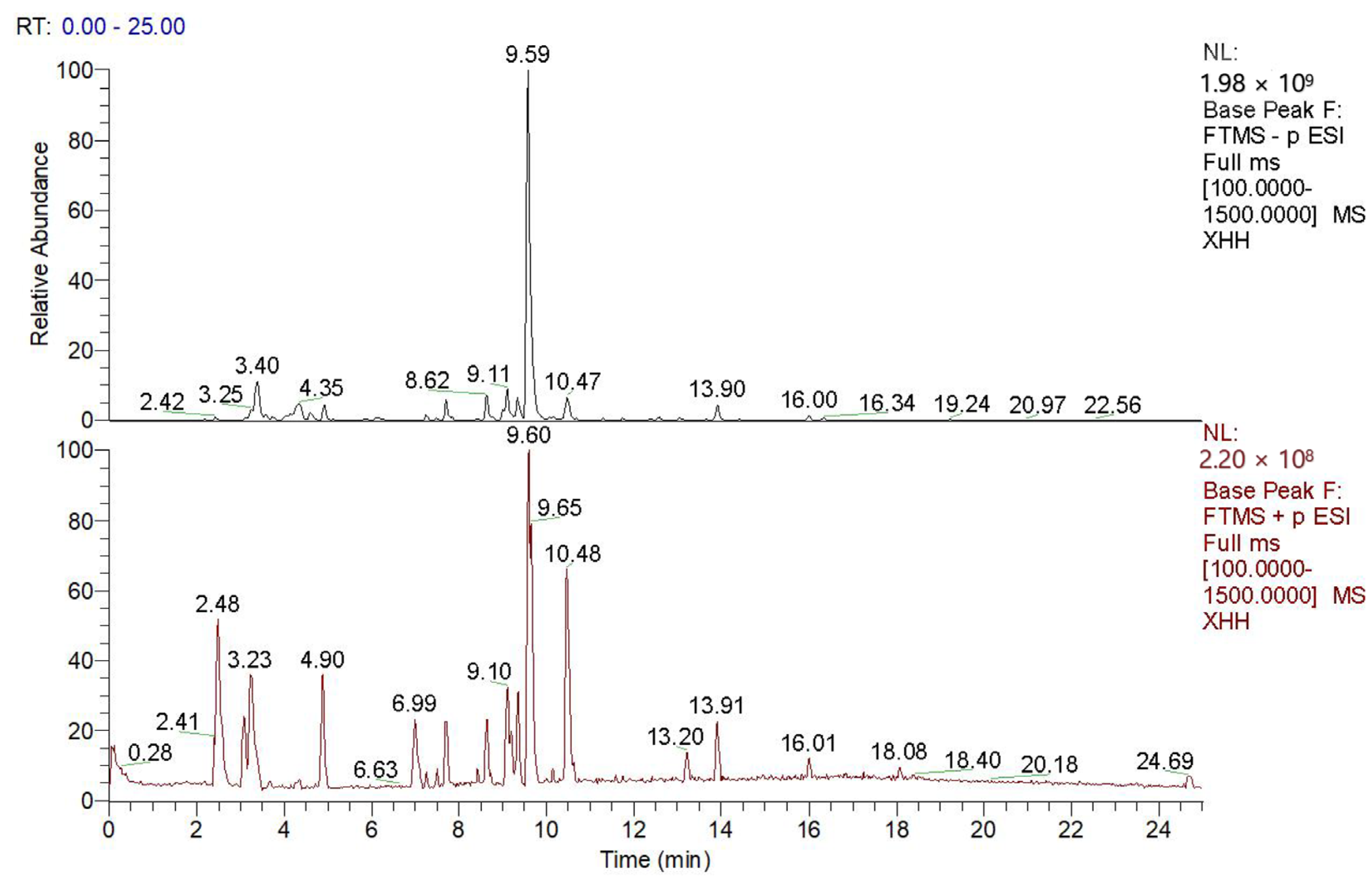
2.1. Chemical Profiling of Saffron Extract by LC-MS
2.2. Network Pharmacological Analysis of Saffron-Alleviated Liver Fibrosis
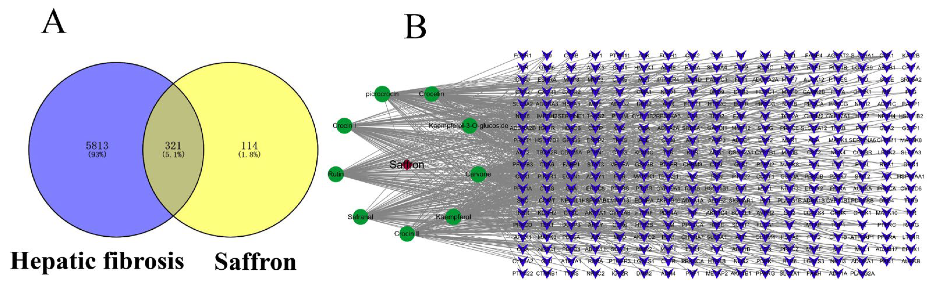
2.2.1. Screening the Main Active Components in Saffron
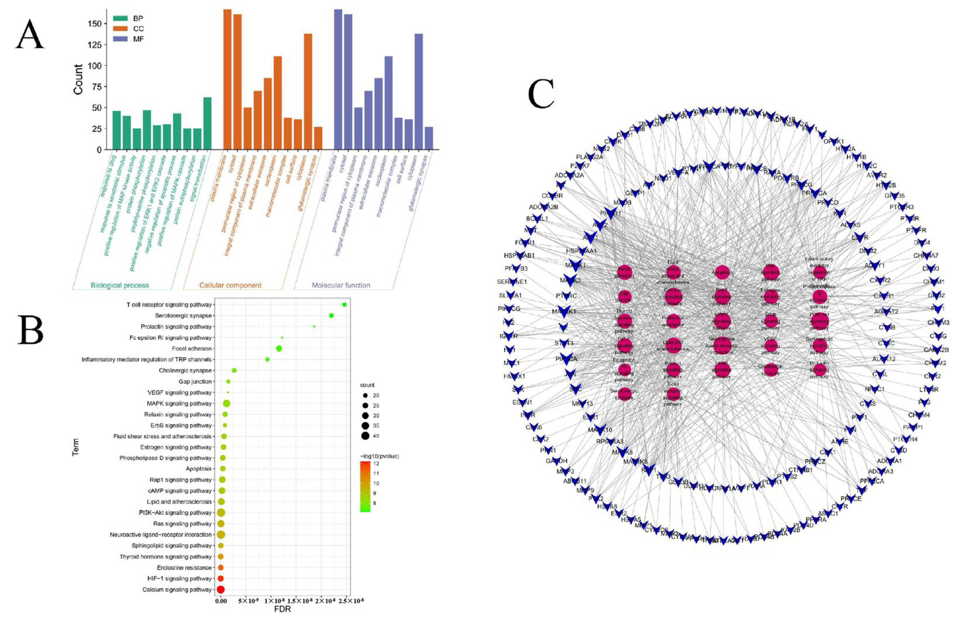
2.2.2. Putative Targets of Saffron for the Treatment of Liver Fibrosis
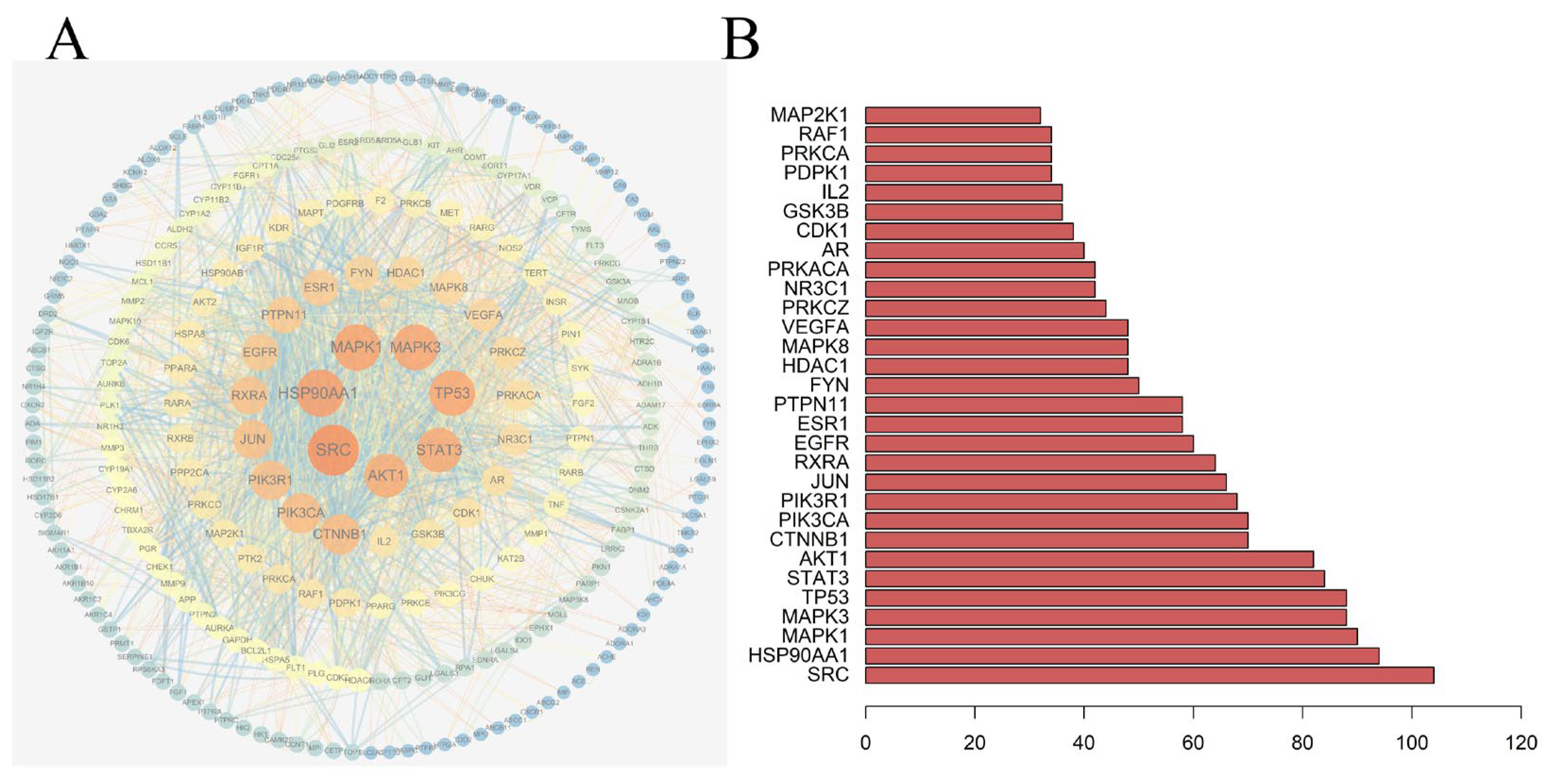
2.2.3. Construction of the PPIs Network and Network Analysis
2.3. Experimental Validations of the Pharmacological Effects and Molecular Mechanisms of Saffron-Alleviated Liver Fibrosis
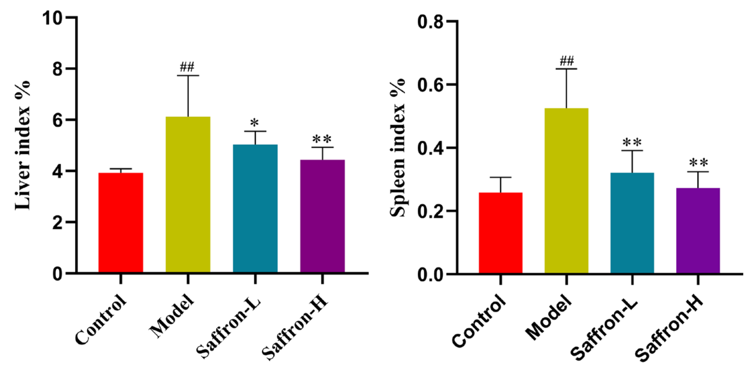
2.3.1. Effects of Saffron Extract on Liver Index and Spleen Index of Liver Fibrosis Mice
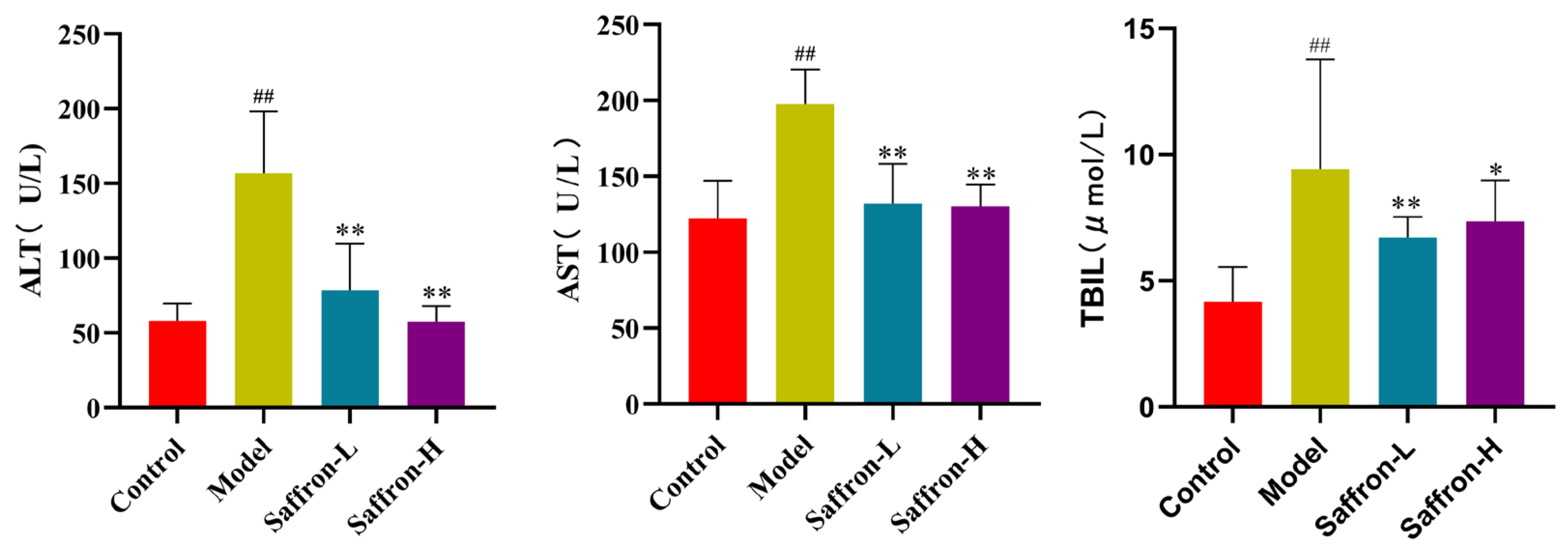
2.3.2. Saffron Extract Ameliorated the Hepatic Injury Markers in Liver Fibrosis Mice
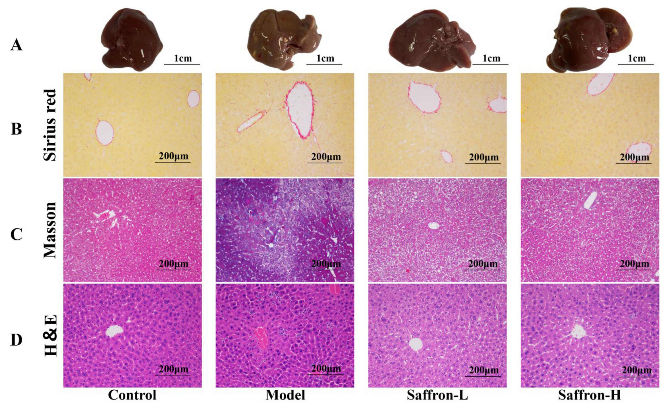
2.3.3. Saffron Extract Alleviated Liver Histopathological Changes in Liver Fibrosis Mice
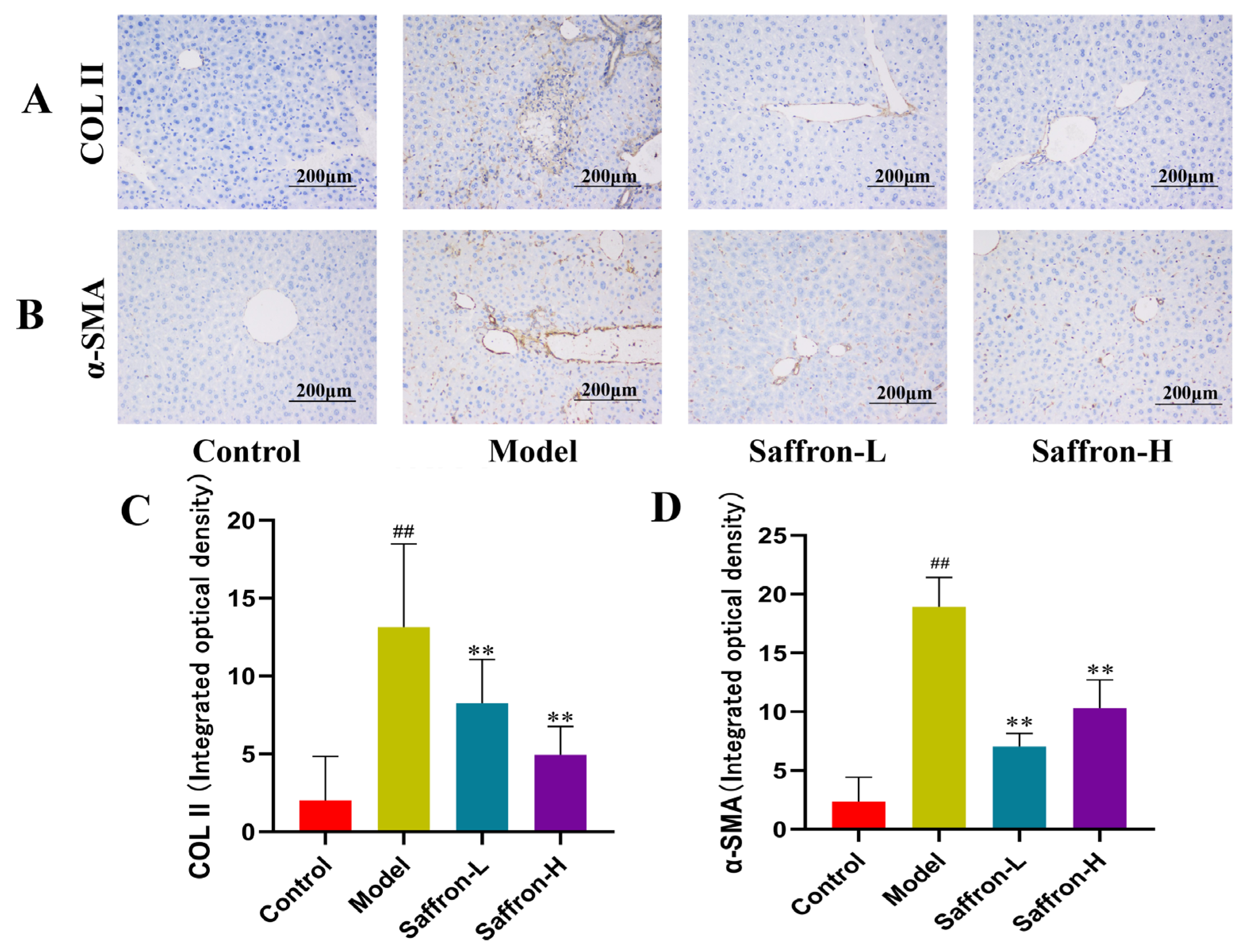
2.3.4. Saffron Extract Attenuated Liver Damage and Collagen Deposition in Liver Fibrosis Mice
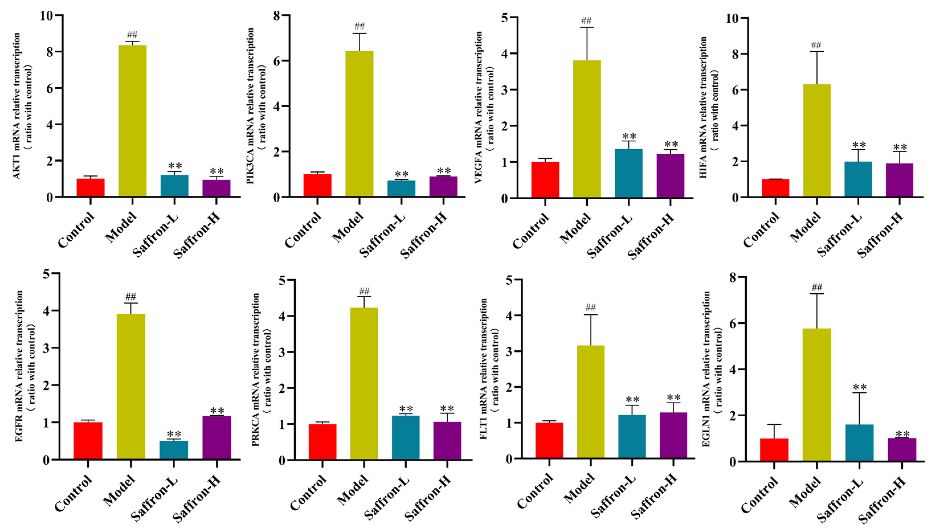
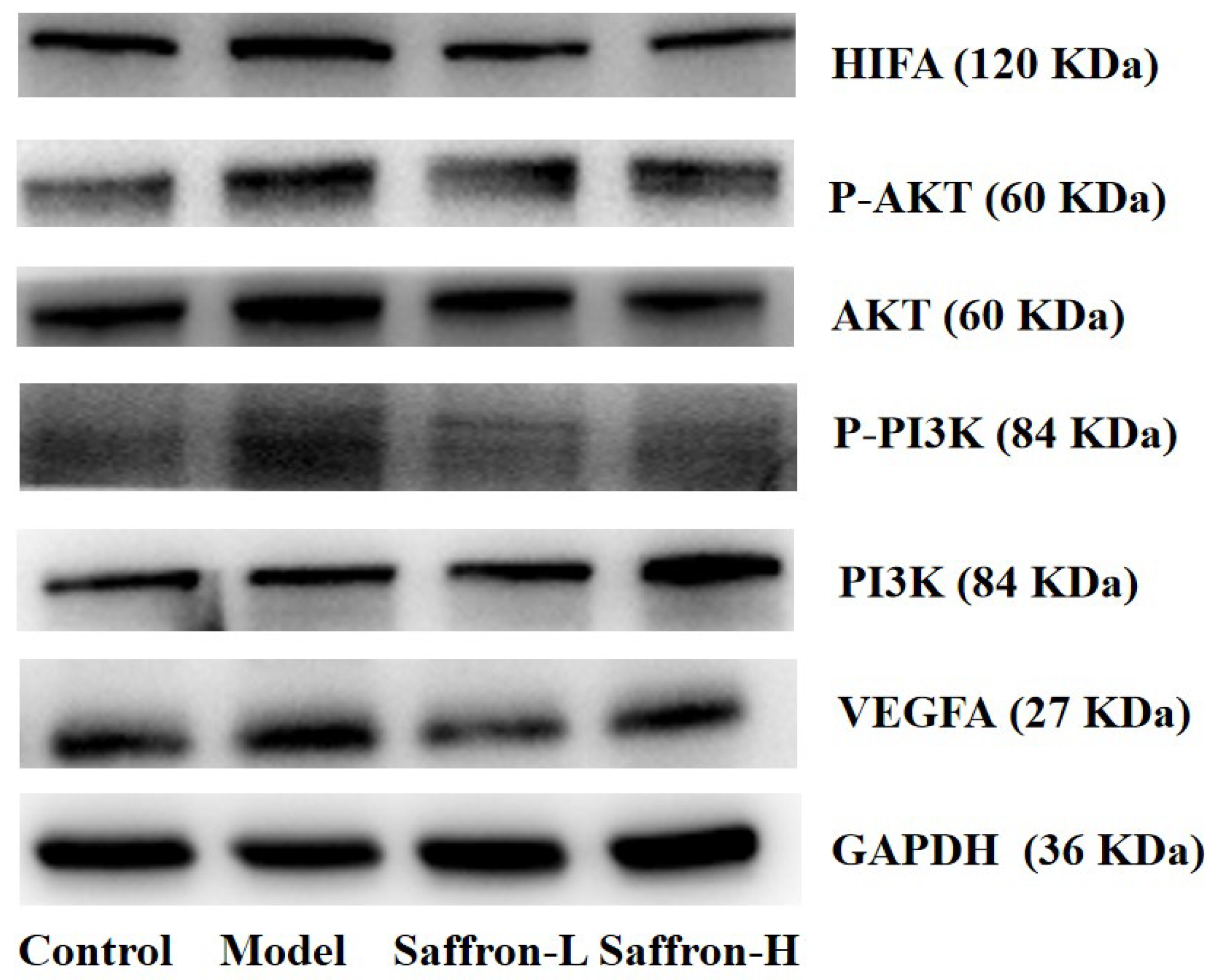
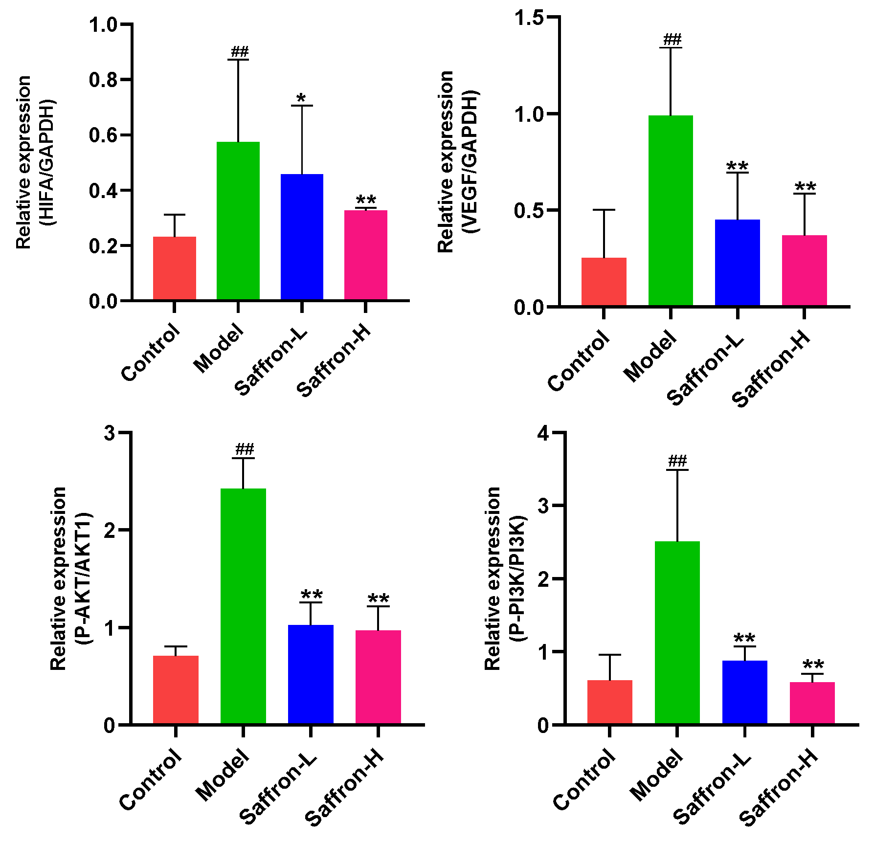
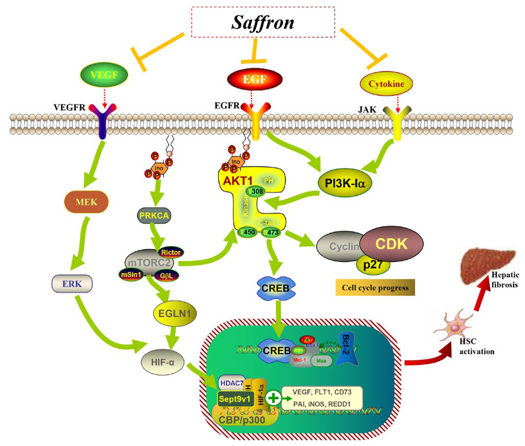
2.3.5. Saffron Extract Inhibited the AKT/HIF-1α/VEGF Signaling Pathways of Liver Fibrosis Mice
3. Discussion
4. Materials and Methods
4.1. Chemicals and Reagents
4.2. Preparation of Saffron Extract
4.3. LC-MS Analysis Conditions
4.4. Network Construction and Analysis
4.5. Animal Study Design
4.6. Organ Coefficient
4.7. Biochemical Analysis
4.8. HE Staining, Sirius Red Staining, and Masson Staining
4.9. Immunohistochemistry
4.10. Western Blotting Analysis
4.11. Real-Time Quantitative PCR Assay
4.12. Statistical Analysis
5. Conclusions
Author Contributions
Funding
Institutional Review Board Statement
Informed Consent Statement
Data Availability Statement
Conflicts of Interest
Sample Availability
References
- Costaguta, G.; Patey, N.; Alvarez, F. Liver disease in pediatric cystic fibrosis. A review of current knowledge. Arch. Argent. Pediatr. 2023, 121, e202202905. [Google Scholar] [CrossRef]
- Dhar, D.; Baglieri, J.; Kisseleva, T.; Brenner, D.A. Mechanisms of liver fibrosis and its role in liver cancer. Exp. Biol. Med. 2020, 245, 96–108. [Google Scholar] [CrossRef] [PubMed]
- Odagiri, N.; Matsubara, T.; Sato-Matsubara, M.; Fujii, H.; Enomoto, M.; Kawada, N. Anti-fibrotic treatments for chronic liver diseases: The present and the future. Clin. Mol. Hepatol. 2021, 27, 413–424. [Google Scholar] [CrossRef] [PubMed]
- Mehal, W.Z.; Schuppan, D. Antifibrotic Therapies in the Liver. Semin. Liver Dis. 2015, 35, 184–198. [Google Scholar] [PubMed]
- Tan, Z.; Sun, H.B.; Xue, T.X.; Gan, C.L.; Liu, H.Y.; Xie, Y.T.; Yao, Y.Q.; Ye, T.H. Liver Fibrosis: Therapeutic Targets and Advances in Drug Therapy. Front. Cell Dev. Biol. 2021, 9, 730176. [Google Scholar] [CrossRef] [PubMed]
- Vijayan, N.; Perumal, M.K. A critical review on anti-fibrotic phytochemicals targeting activated hepatic stellate cells. J. Food Biochem. 2022, 46, e14438. [Google Scholar] [CrossRef] [PubMed]
- Nan, Y.; Su, H.C.; Lian, X.M.; Wu, J.; Liu, S.J.; Chen, P.P.; Liu, S.M. Pathogenesis of Liver Fibrosis and Its TCM Therapeutic Perspectives. Evid.-Based Compl. Alt. 2022, 2022, 5325431. [Google Scholar] [CrossRef]
- Chen, Y.; Liao, W.; Zhu, Z.P.; Chen, J.; Yang, Q.S.; Zheng, Y.F.; Zhang, X.J.; Limsila, B.; Lu, M.G.; Fu, S.; et al. Essential oil from the raw and vinegar-processed Rhizoma Curcumae ameliorate CCl4-induced liver fibrosis: Integrating network pharmacology and molecular mechanism evaluation. Food Funct. 2021, 12, 4199–4220. [Google Scholar] [CrossRef]
- Zhang, Y.Q.; Hua, L.P.; Lin, C.F.; Yuan, M.Z.; Xu, W.; Raj, A.D.; Venkidasamy, B.; Cespedes-Acuna, C.L.; Nile, S.H.; Yan, G.H.; et al. Pien-Tze-Huang alleviates CCl4-induced liver fibrosis through the inhibition of HSC autophagy and the TGF-beta 1/Smad2 pathway. Front. Pharmacol. 2022, 13, 937484. [Google Scholar] [CrossRef]
- Husaini, A.M.; Ul Haq, S.A.; Shabir, A.; Wani, A.B.; Dedmari, M.A. The menace of saffron adulteration: Low-cost rapid identification of fake look-alike saffron using Foldscope and machine learning technology. Front. Plant Sci. 2022, 13, 945291. [Google Scholar] [CrossRef]
- Alavizadeh, S.H.; Hosseinzadeh, H. Bioactivity assessment and toxicity of crocin: A comprehensive review. Food Chem. Toxicol. 2014, 64, 65–80. [Google Scholar] [CrossRef]
- Alizadeh-Sani, M.; Tavassoli, M.; McClements, D.J.; Hamishehkar, H. Multifunctional halochromic packaging materials: Saffron petal anthocyanin loaded-chitosan nanofiber/methyl cellulose matrices. Food Hydrocoll. 2021, 111, 106237. [Google Scholar] [CrossRef]
- Butnariu, M.; Quispe, C.; Herrera-Bravo, J.; Sharifi-Rad, J.; Singh, L.; Aborehab, N.M.; Bouyahya, A.; Venditti, A.; Sen, S.; Acharya, K.; et al. The Pharmacological Activities of Crocus sativus L.: A Review Based on the Mechanisms and Therapeutic Opportunities of its Phytoconstituents. Oxid. Med. Cell. Longev. 2022, 2022, 8214821. [Google Scholar] [CrossRef] [PubMed]
- Gupta, M.; Ghufran, S.M.; Kausar, T.; Ali, R.; Biswas, S.; Nayeem, S.M.; Ishrat, R.; Ali, S.; Ahmad, A.; Rather, I.A.; et al. Z-Guggulsterone Is a Potential Lead Molecule of Dawa-ul-Kurkum against Hepatocellular Carcinoma. Molecules 2022, 27, 5104. [Google Scholar] [CrossRef] [PubMed]
- Li, M.; Ding, L.; Hu, Y.L.; Qin, L.L.; Wu, Y.; Liu, W.; Wu, L.L.; Liu, T.H. Herbal formula LLKL ameliorates hyperglycaemia, modulates the gut microbiota and regulates the gut-liver axis in Zucker diabetic fatty rats. J. Cell. Mol. Med. 2021, 25, 367–382. [Google Scholar] [CrossRef] [PubMed]
- Mashmoul, M.; Azlan, A.; Mohtarrudin, N.; Yusof, B.N.M.; Khaza’ai, H.; Khoo, H.E.; Farzadnia, M.; Boroushaki, M.T. Protective effects of saffron extract and crocin supplementation on fatty liver tissue of high-fat diet-induced obese rats. BMC Complement. Altern. Med. 2016, 16, 401. [Google Scholar] [CrossRef] [PubMed]
- Amin, A.; Hamza, A.A.; Daoud, S.; Khazanehdari, K.; Al Hrout, A.; Baig, B.; Chaiboonchoe, A.; Adrian, T.E.; Zaki, N.; Salehi-Ashtiani, K. Saffron’s Bioactive Molecule Prevents Chemical Induced-Liver Cancer: A Pre-clinical Study. Mol. Biol. Cell 2016, 27, 8120. [Google Scholar]
- Mousavi, S.M.; Mokhtari, P.; Asbaghi, O.; Rigi, S.; Persad, E.; Jayedi, A.; Rezvani, H.; Mahamat-Saleh, Y.; Sadeghi, O. Does saffron supplementation have favorable effects on liver function indicators? A systematic review and meta-analysis of randomized controlled trials. Crit. Rev. Food Sci. 2022, 62, 6315–6327. [Google Scholar] [CrossRef]
- Karimi, E.; Farrokhzad, A.; Darand, M.; Arab, A. The Effect of Saffron Consumption on Liver Function: A Systematic Review and Meta-Analysis of Randomized Controlled Clinical Trials. Complement. Med. Res. 2021, 28, 453–462. [Google Scholar] [CrossRef]
- Xu, Z.J.; Lin, S.S.; Gong, J.J.; Feng, P.S.; Cao, Y.F.; Li, Q.Q.; Jiang, Y.L.; You, Y.; Tong, Y.P.; Wang, P. Exploring the Protective Effects and Mechanism of Crocetin from Saffron Against NAFLD by Network Pharmacology and Experimental Validation. Front. Med. 2021, 8, 681391. [Google Scholar] [CrossRef]
- Wang, X.; Wang, Z.Y.; Zheng, J.H.; Li, S. TCM network pharmacology: A new trend towards combining computational, experimental and clinical approaches. Chin. J. Nat. Med. 2021, 19, 1–11. [Google Scholar] [CrossRef] [PubMed]
- Yuan, Z.Z.; Pan, Y.Y.; Leng, T.; Chu, Y.; Zhang, H.J.; Ma, J.R.; Ma, X.J. Progress and Prospects of Research Ideas and Methods in the Network Pharmacology of Traditional Chinese Medicine. J. Pharm. Pharm. Sci. 2022, 25, 218–226. [Google Scholar] [CrossRef] [PubMed]
- Hao, D.C.; Xiao, P.G. Network Pharmacology: A Rosetta Stone for Traditional Chinese Medicine. Drug Develop. Res. 2014, 75, 299–312. [Google Scholar] [CrossRef] [PubMed]
- Abdalla, Y.; Abdalla, A.; Hamza, A.A.; Amin, A. Safranal Prevents Liver Cancer Through Inhibiting Oxidative Stress and Alleviating Inflammation. Front. Pharmacol. 2022, 12, 777500. [Google Scholar] [CrossRef] [PubMed]
- Zhu, K.; Guo, Y.S.; Zhao, C.H.; Kang, S.X.; Li, J.L.; Wang, J.X.; Tang, Z.H.; Lin, B.; Li, W.H. Etiology Exploration of Non-alcoholic Fatty Liver Disease from Traditional Chinese Medicine Constitution Perspective: A Cross-Sectional Study. Front. Public Health 2021, 9, 635818. [Google Scholar] [CrossRef] [PubMed]
- Chen, Z.M.; Li, W.B.; Quan, L.; Zhou, H.T.; Zhao, Y.F.; Zhang, X.; Hu, L.; Hu, C.J. The Effects of Curcumae Longae Radix, Curcuma phaeocaulis Radix and Their Processed Products on Epo/EpoR Pathway and CD62p. Front. Pharmacol. 2018, 9, 736. [Google Scholar] [CrossRef]
- DeLeve, L.D. Liver Sinusoidal Endothelial Cells in Hepatic Fibrosis. Hepatology 2015, 61, 1740–1746. [Google Scholar] [CrossRef] [PubMed]
- Zhang, H.F.; Gao, X.; Wang, X.; Chen, X.; Huang, Y.; Wang, L.; Xu, Z.W. The mechanisms of renin-angiotensin system in hepatocellular carcinoma: From the perspective of liver fibrosis, HCC cell proliferation, metastasis and angiogenesis, and corresponding protection measures. Biomed. Pharmacother. 2021, 141, 111868. [Google Scholar] [CrossRef]
- Park, S.; Kim, J.W.; Kim, J.H.; Lim, C.W.; Kim, B. Differential Roles of Angiogenesis in the Induction of Fibrogenesis and the Resolution of Fibrosis in Liver. Biol. Pharm. Bull. 2015, 38, 980–985. [Google Scholar] [CrossRef]
- Kajdaniuk, D.; Marek, B.; Borgiel-Marek, H.; Kos-Kudla, B. Vascular endothelial growth factor (VEGF)—Part 1: In physiology and pathophysiology. Endokrynol. Pol. 2011, 62, 444–455. [Google Scholar]
- Zhan, L.; Huang, C.; Meng, X.M.; Song, Y.; Wu, X.Q.; Yang, Y.; Li, J. Hypoxia-inducible factor-1alpha in hepatic fibrosis: A promising therapeutic target. Biochimie 2015, 108, 1–7. [Google Scholar] [CrossRef] [PubMed]
- Ju, C.; Colgan, S.P.; Eltzschig, H.K. Hypoxia-inducible factors as molecular targets for liver diseases. J. Mol. Med. 2016, 94, 613–627. [Google Scholar] [CrossRef] [PubMed]
- Bakshi, H.A.; Quinn, G.A.; Nasef, M.M.; Mishra, V.; Aljabali, A.A.A.; El-Tanani, M.; Serrano-Aroca, A.; Da Silva, M.W.; McCarron, P.A.; Tambuwala, M.M. Crocin Inhibits Angiogenesis and Metastasis in Colon Cancer via TNF-alpha/NF-kB/VEGF Pathways. Cells 2022, 11, 1502. [Google Scholar] [CrossRef] [PubMed]
- Zhao, C.; Kam, H.T.; Chen, Y.; Gong, G.Y.; Hoi, M.P.M.; Skalicka-Wozniak, K.; Dias, A.C.P.; Lee, S.M.Y. Crocetin and Its Glycoside Crocin, Two Bioactive Constituents from Crocus sativus L. (Saffron), Differentially Inhibit Angiogenesis by Inhibiting Endothelial Cytoskeleton Organization and Cell Migration Through VEGFR2/SRC/FAK and VEGFR2/MEK/ERK Signaling Pathways. Front. Pharmacol. 2021, 12, 675359. [Google Scholar] [PubMed]
- Makaritsis, K.P.; Kotidis, C.; Papacharalampous, K.; Kouvaras, E.; Poulakida, E.; Tarantilis, P.; Asprodini, E.; Ntaios, G.; Koukoulis, G.K.; Dalekos, G.N.; et al. Mechanistic insights on the effect of crocin, an active ingredient of saffron, on atherosclerosis in apolipoprotein E knockout mice. Coron. Artery Dis. 2022, 33, 394–402. [Google Scholar] [CrossRef] [PubMed]
- Zhou, Y.; Xu, Q.H.; Shang, J.J.; Lu, L.H.; Chen, G.Y. Crocin inhibits the migration, invasion, and epithelial-mesenchymal transition of gastric cancer cells via miR-320/KLF5/HIF-1 alpha signaling. J. Cell Physiol. 2019, 234, 17876–17885. [Google Scholar] [CrossRef]


| No. | RT (min) | Ionization Mode | Experimental m/z | Molecular | ppm | Tentative Compound | Molecular Formula | MS/MS Fragments |
|---|---|---|---|---|---|---|---|---|
| 1 | 2.384 | [M + H]+ | 175.119 | 175.119 | 0.57 | DL-Arginine | C6H14N4O2 | 175, 151, 116, 70 |
| 2 | 2.490 | [M + H]+ | 104.107 | 104.107 | 3.84 | Choline | C5H13NO | 104, 86, 60 |
| 3 | 3.099 | [M − H]− | 146.045 | 146.044 | 4.72 | L-Glutamic acid | C5H9NO4 | 146, 128, 102 |
| 4 | 3.167 | [M + H]+ | 184.073 | 184.073 | 0.00 | Phosphocholine | C5H14NO4P | 184, 124, 86 |
| 5 | 3.196 | [M + H]+ | 236.110 | 236.108 | 8.05 | 3-methyl-5-oxo-5-(4-toluidino) pentanoic acid | C13H17NO3 | 258, 184, 124, 104 |
| 6 | 3.242 | [M + H]+ | 138.055 | 138.055 | 0.00 | Trigonelline | C7H7NO2 | 138, 120, 110, 92, 78 |
| 7 | 3.252 | [M + H]+ | 360.150 | 360.152 | −7.25 | α-Lactose | C12H22O11 | 316, 163, 145, 127, 97, 85 |
| 8 | 3.270 | [M + H]+ | 116.071 | 116.072 | −6.46 | Proline | C5H9NO2 | 116, 93, 70 |
| 9 | 3.277 | [M − H]− | 179.055 | 179.056 | −2.07 | Glucose | C6H12O6 | 129, 89, 71, 59 |
| 10 | 3.281 | [M + FA − H]− | 549.167 | 549.169 | −3.17 | D-Raffinose | C18H32O16 | 503, 346, 113, 89, 71 |
| 11 | 3.320 | [M + H]+ | 365.105 | 365.106 | −2.16 | D-(+)-Maltose | C12H22O11 | 365, 203, 185, 98, 69 |
| 12 | 3.386 | [M+FA-H]− | 342.116 | 342.116 | −0.50 | Sucrose | C12H22O11 | 341, 179, 119, 101, 89, 71 |
| 13 | 3.395 | [M − H]− | 195.050 | 195.051 | −1.85 | Gluconic acid | C6H12O7 | 195, 177, 129, 75 |
| 14 | 3.436 | [M + H]+ | 118.086 | 118.086 | 2.03 | Valine | C5H11NO2 | 118, 95, 72 |
| 15 | 3.731 | [M − H]− | 135.029 | 135.029 | −2.07 | L-threonic acid | C4H8O5 | 135, 89, 75 |
| 16 | 4.144 | [M + H]+ | 123.056 | 123.056 | −5.85 | Nicotinamide | C6H6N2O | 123, 96, 80 |
| 17 | 4.209 | [M – H]− | 175.024 | 175.024 | −2.00 | Ascorbic acid | C6H8O6 | 175, 115, 87, 71 |
| 18 | 4.225 | [M + H]+ | 348.070 | 348.071 | −2.13 | Adenosine 5′-monophosphate | C10H1N5O7P | 348, 326, 136 |
| 19 | 4.333 | [M − H + HAc]− | 179.055 | 179.056 | −2.18 | D-Fructose | C6H12O6 | 179, 119, 113, 89, 71, 59 |
| 20 | 4.398 | [M − H]− | 193.035 | 193.035 | −1.45 | Galacturonic acid | C6H10O7 | 193, 103, 71, 59 |
| 21 | 4.845 | [M − H]− | 133.013 | 133.014 | −2.33 | DL-Malic acid | C4H6O5 | 133, 115, 71 |
| 22 | 4.876 | [M − H]− | 268.104 | 268.105 | −2.87 | Adenosine | C10H13N5O4 | 268, 136 |
| 23 | 4.878 | [M + H]+ | 136.062 | 136.063 | −6.39 | Adenine | C5H5N5 | 136, 91, 72, 55 |
| 24 | 4.905 | [M + H]+ | 132.102 | 132.103 | −5.45 | Isoleucine | C6H13NO2 | 132, 113, 108, 90, 86, 72 |
| 25 | 5.106 | [M + H]+ | 113.035 | 113.036 | −6.81 | Uracil | C4H4N2O2 | 113, 96, 70 |
| 26 | 5.107 | [M − H]− | 243.062 | 243.062 | −1.56 | Uridine | C9H12N2O6 | 243, 200, 153, 110, 82 |
| 27 | 5.544 | [M − H]− | 259.022 | 259.023 | −1.31 | Glucose 1-phosphate | C6H13O9P | 259, 215, 96, 78 |
| 28 | 5.675 | [M − H]− | 171.006 | 171.006 | −1.99 | Glycerol 3-phosphate | C3H9O6P | 171, 124, 96, 78 |
| 29 | 5.715 | [M + H]+ | 152.057 | 152.057 | −5.20 | Guanine | C5H5N5O | 152, 143, 134, 109, 96 |
| 30 | 5.718 | [M + H]+ | 284.099 | 284.100 | −3.48 | Guanosine | C10H13N5O5 | 152, 135, 110 |
| 31 | 5.869 | [M − H]− | 191.019 | 191.019 | −1.88 | Citric acid | C6H8O7 | 191, 147, 111, 102, 97, 85 |
| 32 | 6.259 | [M − H]− | 117.018 | 117.019 | −2.56 | Succinic acid | C4H6O4 | 117, 99, 73, 71 |
| 33 | 6.587 | [M − H]− | 323.029 | 323.030 | −3.93 | Uridine monophosphate | C9H13N2O9P | 323, 211, 150, 138, 111, 96 |
| 34 | 6.622 | [M + H]+ | 166.086 | 166.086 | 1.14 | L-Phenylalanine | C9H11NO2 | 166, 138, 120, 103, 91, 74 |
| 35 | 7.250 | [M + H]+ | 449.108 | 449.108 | −1.78 | Kaempferol-7-O-glucoside | C21H20O11 | 449, 287, 231, 183, 160, 137 |
| 36 | 7.712 | [M − H]− | 179.055 | 179.056 | −1.62 | Fructose | C6H12O6 | 134, 119, 113, 101, 89, 71 |
| 37 | 8.085 | [M + H]+ | 346.162 | 346.163 | −1.99 | Jasminoside B | C16H26O8 | 238, 185, 167, 139, 121 |
| 38 | 8.734 | [M + H]+ | 167.107 | 167.107 | −4.49 | 6-Pentyl-2H-pyran-2-one | C10H14O2 | 167, 149, 143, 125, 121, 107 |
| 39 | 9.015 | [M − H]− | 179.055 | 179.056 | −1.62 | 9-Fluorenone | C13H8O | 178, 134, 119, 113, 101, 89 |
| 40 | 9.340 | [M − H]− | 609.146 | 609.143 | 4.81 | Rutin | C27H30O16 | 609, 284, 255, 227 |
| 41 | 9.410 | [M − H]− | 145.050 | 145.050 | −2.28 | 3-Methylglutaric acid | C6H10O4 | 145, 121, 111, 101 |
| 42 | 9.443 | [M + H]+ | 169.122 | 169.123 | −3.07 | 10-HAD | C10H18O3 | 169, 151, 123, 109, 95, 81 |
| 43 | 9.564 | [M + H − H2O]+ | 135.080 | 135.081 | −3.33 | 2,4-Dimethylbenzaldehyde | C9H10O | 135, 119, 107, 93, 91, 79 |
| 44 | 9.566 | [M + H]+ | 365.120 | 365.121 | −2.08 | Coniferin | C16H22O8 | 365, 337, 206, 187 |
| 45 | 9.587 | [M + H]+ | 123.117 | 123.118 | −5.28 | 1,2,3,4-Tetramethyl-1,3-cyclopentadiene | C9H14 | 123, 95, 81, 67 |
| 46 | 9.647 | [M − 2H]2− | 975.371 | 975.371 | −0.62 | Crocin I | C44H64O24 | 651, 327, 283 |
| 47 | 10.149 | [M + H]+ | 369.131 | 369.132 | −2.38 | Gibberellic acid | C19H22O6 | 369, 66, 61 |
| 48 | 10.465 | [M − 2H]2− | 813.319 | 813.319 | −0.98 | Crocin II | C38H54O19 | 651, 489, 327 |
| 49 | 10.581 | [M − H]− | 447.094 | 447.095 | −2.26 | Trifolin | C21H20O11 | 447, 380, 284, 255, 227 |
| 50 | 13.047 | [M + H]+ | 329.175 | 329.176 | −4.31 | Crocetin | C20H24O4 | 329, 311, 293, 265, 197 |
| 51 | 13.204 | [M + H]+ | 151.112 | 151.112 | −4.96 | Safranal | C10H14O | 151, 133, 123, 81, 67 |
| 52 | 15.288 | [M − H]− | 327.218 | 327.219 | −3.97 | Corchorifatty acid F | C18H32O5 | 327, 291, 229, 211, 171 |
| 53 | 18.076 | [M + H]+ | 139.112 | 139.112 | −5.61 | Isophorone | C9H14O | 139, 121, 110, 97, 81, 69 |
| 54 | 18.641 | [M + H]+ | 165.091 | 165.092 | −4.66 | 1-(4-methoxyphenyl) propane-1,2-diol | C10H14O3 | 165, 121, 119, 105, 91 |
| 55 | 19.891 | [M + H]+ | 287.055 | 287.055 | −2.61 | Kaempferol | C15H10O6 | 287, 259, 231, 185 |
| 56 | 24.689 | [M + H]+ | 151.112 | 151.112 | −4.24 | Carvone | C10H14O | 151, 133, 123, 81 |
| NO. | Chemical Name | CAS NO | Compound CID | 2D Structure | Formula | Molecular Weight | OB% | DL | Caco-2 |
|---|---|---|---|---|---|---|---|---|---|
| 1 | Crocetin | 27876-94-4 | 5281232 |  | C20H24O4 | 328.40 | 35.30 | 0.26 | 0.54 |
| 2 | Carvone | 99-49-0 | 7439 |  | C10H14O | 150.10 | 49.47 | 0.03 | 1.35 |
| 3 | Kaempferol | 520-18-3 | 5280863 |  | C15H10O6 | 286.05 | 41.88 | 0.26 | 0.24 |
| 4 | Rutin | 153-18-4 | 5280805 |  | C27H30O16 | 610.15 | 3.20 | 0.68 | −1.93 |
| 5 | Crocin Ι | 42553-65-1 | 5281233 |  | C44H64O24 | 976.96 | 2.54 | 0.12 | −4.23 |
| 6 | Crocin ΙΙ | 55750-84-0 | 132399078 | 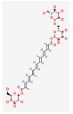 | C38H54O19 | 814.82 | 1.65 | 0.21 | −3.48 |
| 7 | Kaempferol-7-glucoside | 480-10-4 | 5282102 | 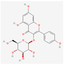 | C21H20O11 | 448.10 | 14.03 | 0.74 | −1.34 |
| 8 | Picrocrocin | 138-55-6 | 130796 | 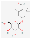 | C16H26O7 | 330.37 | 33.71 | 0.04 | 0.69 |
| 9 | Safranal | 116-26-7 | 61041 |  | C10H14O | 150.22 | 39.56 | 0.04 | 1.39 |
| Gene | Forward Sequence (5′-3′) | Reverse Sequence (5′-3′) |
|---|---|---|
| AKT1 | ATGAACGACGTAGCCATTGTG | TTGTAGCCAATAAAGGTGCCAT |
| VEGFA | GCACATAGAGAGAATGAGCTTCC | CTCCGCTCTGAACAAGGCT |
| HIF1A | ACCTTCATCGGAAACTCCAAAG | ACTGTTAGGCTCAGGTGAACT |
| PIK3CA | CCACGACCATCTTCGGGTG | ACGGAGGCATTCTAAAGTCACTA |
| EGFR | GCCATCTGGGCCAAAGATACC | GTCTTCGCATGAATAGGCCAAT |
| PRKCA | TTGTCCAAGGAAGCCGTCTC | CCTTTGCCACACACTTTGGG |
| EGLN1 | CTGGAGTACATCGTGCCG | GCCGTTTATCCTGTAGTTGC |
| FLT1 | GACTGGTGAGGATAGCTCTACT | ATCCAATCCCTGGCCAGTC |
| GAPDH | GGCCTTCCGTGTTCCTACC | TGCCTGCTTCACCACCTTC |
Disclaimer/Publisher’s Note: The statements, opinions and data contained in all publications are solely those of the individual author(s) and contributor(s) and not of MDPI and/or the editor(s). MDPI and/or the editor(s) disclaim responsibility for any injury to people or property resulting from any ideas, methods, instructions or products referred to in the content. |
© 2023 by the authors. Licensee MDPI, Basel, Switzerland. This article is an open access article distributed under the terms and conditions of the Creative Commons Attribution (CC BY) license (https://creativecommons.org/licenses/by/4.0/).
Share and Cite
Jiang, H.; Huang, X.; Wang, J.; Zhou, Y.; Ren, C.; Zhou, T.; Pei, J. Hepatoprotective Effect of Medicine Food Homology Flower Saffron against CCl4-Induced Liver Fibrosis in Mice via the Akt/HIF-1α/VEGF Signaling Pathway. Molecules 2023, 28, 7238. https://doi.org/10.3390/molecules28217238
Jiang H, Huang X, Wang J, Zhou Y, Ren C, Zhou T, Pei J. Hepatoprotective Effect of Medicine Food Homology Flower Saffron against CCl4-Induced Liver Fibrosis in Mice via the Akt/HIF-1α/VEGF Signaling Pathway. Molecules. 2023; 28(21):7238. https://doi.org/10.3390/molecules28217238
Chicago/Turabian StyleJiang, Huajuan, Xulong Huang, Jiaxin Wang, Yongfeng Zhou, Chaoxiang Ren, Tao Zhou, and Jin Pei. 2023. "Hepatoprotective Effect of Medicine Food Homology Flower Saffron against CCl4-Induced Liver Fibrosis in Mice via the Akt/HIF-1α/VEGF Signaling Pathway" Molecules 28, no. 21: 7238. https://doi.org/10.3390/molecules28217238
APA StyleJiang, H., Huang, X., Wang, J., Zhou, Y., Ren, C., Zhou, T., & Pei, J. (2023). Hepatoprotective Effect of Medicine Food Homology Flower Saffron against CCl4-Induced Liver Fibrosis in Mice via the Akt/HIF-1α/VEGF Signaling Pathway. Molecules, 28(21), 7238. https://doi.org/10.3390/molecules28217238