

In Vitro and In Silico Activities of E. radiata and E. cinerea as an Enhancer of Antibacterial, Antioxidant, and Anti-Inflammatory Agents
 , ,
, ,
Abstract
:1. Introduction
2. Results
2.1. Extraction Yield
Antibacterial Activity
2.2. Antioxidant Activity
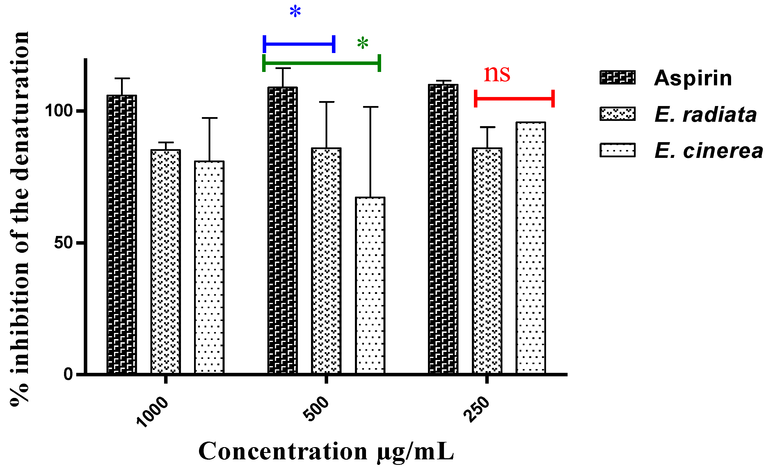
2.3. Anti-Inflammatory Test
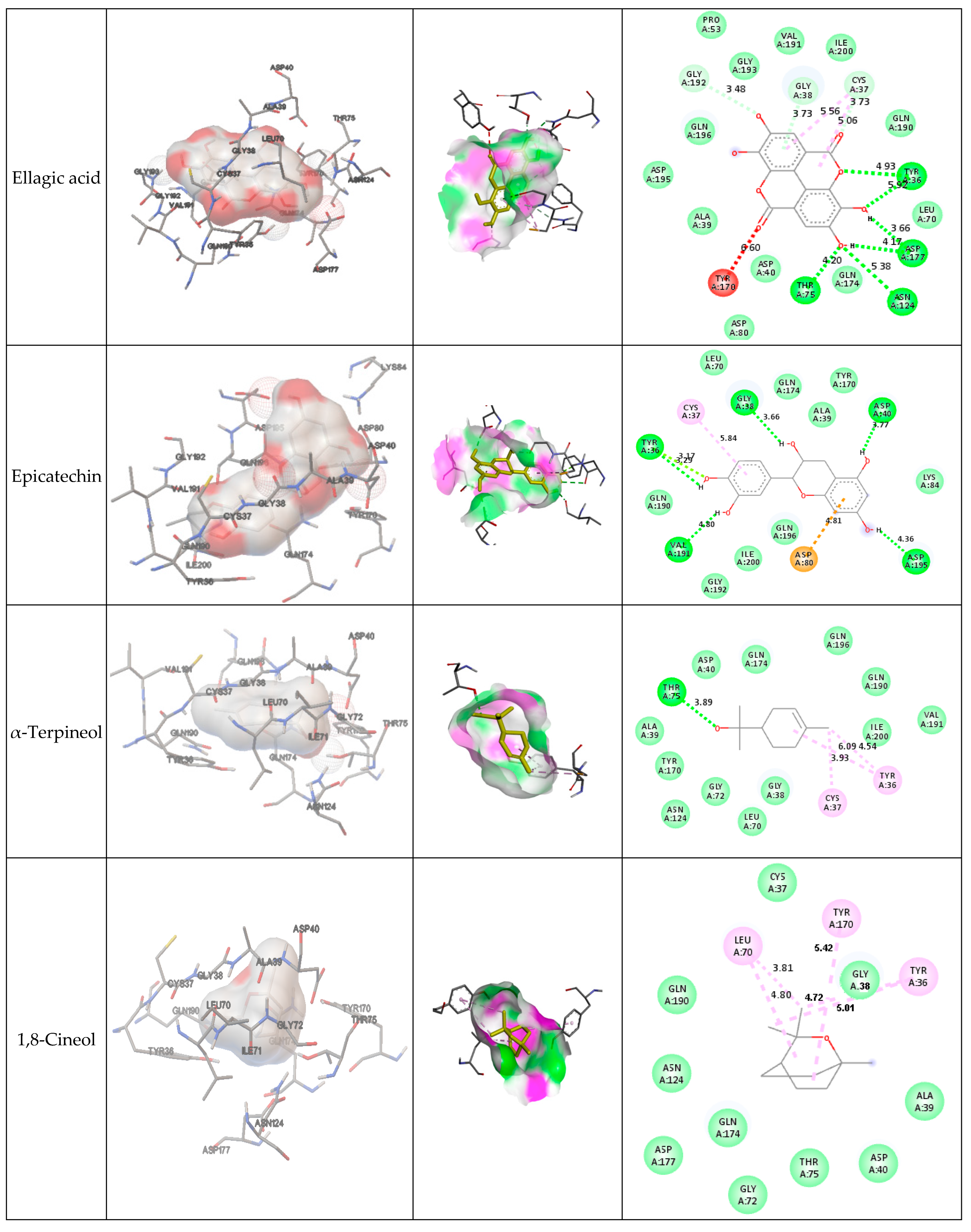
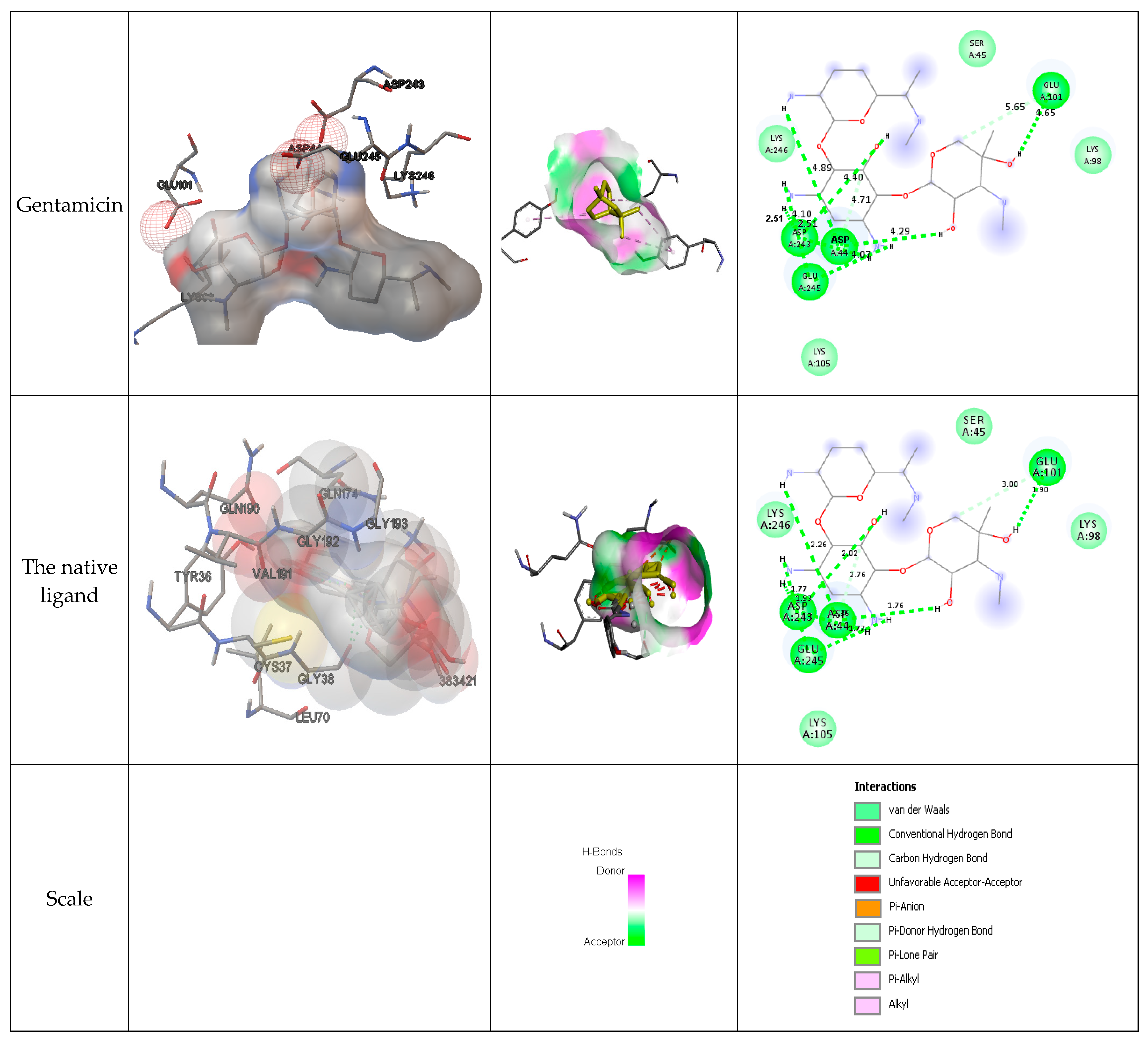
2.4. Docking Results
2.5. Drug Likeness and ADME Prediction
3. Materials and Methods
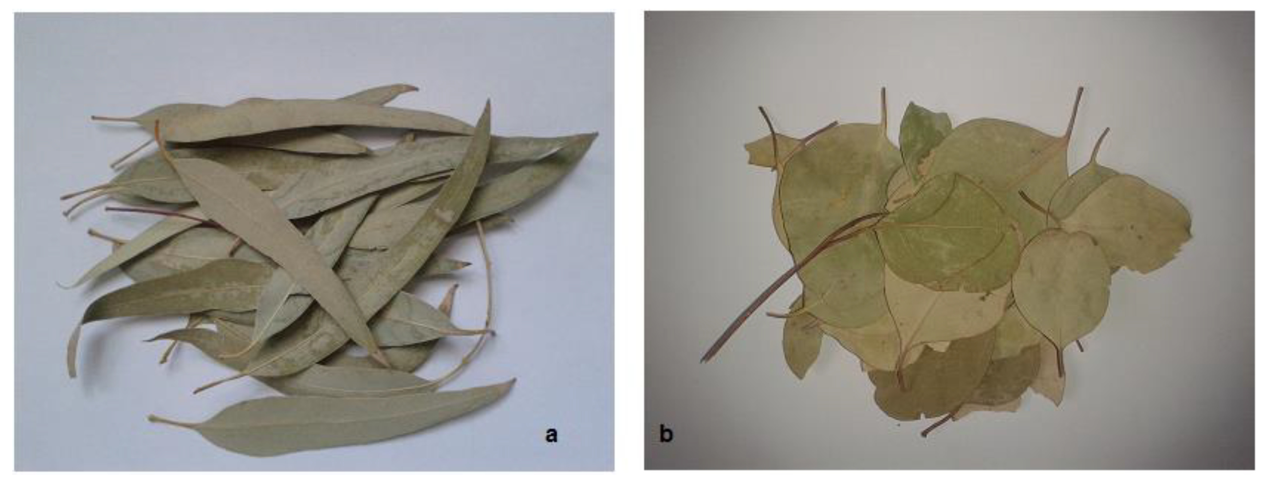
3.1. The Choice of Plants
3.2. Antibacterial Activity
3.3. Antioxidant Activity
3.4. In-Vitro Anti-Inflammatory Test
3.5. Molecular Docking
Phyto-Compounds
3.6. Docking Analysis and Protein Preparation
3.7. Drug Likeness, ADME/Toxicity Prediction
4. Discussion
4.1. Efficacy against Bacteria
4.2. Results in Reducing Inflammation
4.3. Investigation of Docking
4.4. Prediction of ADME/Toxicity and Similarity to Existing Drugs
5. Conclusions
Author Contributions
Funding
Institutional Review Board Statement
Informed Consent Statement
Data Availability Statement
Acknowledgments
Conflicts of Interest
Sample Availability
References
- Kahla, Y.; Zouari-Bouassida, K.; Rezgui, F.; Trigui, M.; Tounsi, S. Efficacy of Eucalyptus cinerea as a Source of Bioactive Compounds for Curative Biocontrol of Crown Gall Caused by Agrobacterium Tumefaciens Strain B6. BioMed Res. Int. 2017, 2017, 9308063. [Google Scholar] [CrossRef] [PubMed]
- Qin, Z.; Li, J.; Zhang, Y.; Xiao, Y.; Zhang, X.; Zhong, L.; Liu, H.; Chen, B. Genome-Wide Identification of MicroRNAs Involved in the Somatic Embryogenesis of Eucalyptus. G3 Genes Genomes Genetics 2021, 11, jkab070. [Google Scholar] [CrossRef] [PubMed]
- Soliman, F.M.; Fathy, M.M.; Salama, M.M.; Saber, F.R. Chemical Composition and Bioactivity of the Volatile Oil from Leaves and Stems of Eucalyptus cinerea. Pharm. Biol. 2014, 52, 1272–1277. [Google Scholar] [CrossRef] [PubMed]
- Abouzkhar, F.A.; Yangui, I.; Messaoud, C.; Ben Jamâa, M.L. Eucalyptus Leaf Diseases Associated with Neofusicoccum spp. in North Africa. J. Arid Environ. 2022, 197, 104662. [Google Scholar] [CrossRef]
- Boeno, D.; Silva, R.F.; Almeida, H.S.; Rodrigues, A.C.; Vanzan, M.; Andreazza, R. Influence of Eucalyptus Development under Soil Fauna. Braz. J. Biol. 2020, 80, 345–353. [Google Scholar] [CrossRef]
- Penín, L.; López, M.; Santos, V.; Alonso, J.L.; Parajó, J.C. Technologies for Eucalyptus Wood Processing in the Scope of Biorefineries: A Comprehensive Review. Bioresour. Technol. 2020, 311, 123528. [Google Scholar] [CrossRef]
- Bhuyan, D.J.; Vuong, Q.V.; Chalmers, A.C.; van Altena, I.A.; Bowyer, M.C.; Scarlett, C.J. Phytochemical, Antibacterial and Antifungal Properties of an Aqueous Extract of Eucalyptus microcorys Leaves. S. Afr. J. Bot. 2017, 112, 180–185. [Google Scholar] [CrossRef]
- Nwabor, O.F.; Singh, S.; Syukri, D.M.; Voravuthikunchai, S.P. Bioactive Fractions of Eucalyptus Camaldulensis Inhibit Important Foodborne Pathogens, Reduce Listeriolysin O-Induced Haemolysis, and Ameliorate Hydrogen Peroxide-Induced Oxidative Stress on Human Embryonic Colon Cells. Food Chem. 2021, 344, 128571. [Google Scholar] [CrossRef]
- Teixeira, A.; DaCunha, D.C.; Barros, L.; Caires, H.R.; Xavier, C.P.R.; Ferreira, I.C.F.R.; Vasconcelos, M.H. Eucalyptus globulus Labill. Decoction Extract Inhibits the Growth of NCI-H460 Cells by Increasing the P53 Levels and Altering the Cell Cycle Profile. Food Funct. 2019, 10, 3188–3197. [Google Scholar] [CrossRef]
- Fetni, S.; Bertella, N. In vitro Study of Anti-Inflammatory Properties of Methanolic Extract Fruits from Rosa Canina L. (Rosaceae). Nutr. Santé 2020, 9, 117–125. [Google Scholar] [CrossRef]
- Olugbodi, J.O.; Tincho, M.B.; Oguntibeju, O.O.; Olaleye, M.T.; Akinmoladun, A.C. Glyphaea brevis—In vitro Antioxidant and In Silico Biological Activity of Major Constituents and Molecular Docking Analyses. Toxicol. In Vitro 2019, 59, 187–196. [Google Scholar] [CrossRef] [PubMed]
- Salaria, D.; Rolta, R.; Patel, C.N.; Dev, K.; Sourirajan, A.; Kumar, V. In vitro and in Silico Analysis of Thymus serpyllum Essential Oil as Bioactivity Enhancer of Antibacterial and Antifungal Agents. J. Biomol. Struct. Dyn. 2021, 40, 10383–10402. [Google Scholar] [CrossRef]
- CLSI M02-A11; Performance Standards for Antimicrobial Disk Susceptibility Tests. Approved Standard—Eleventh Edition. Clinical and Laboratory Standards Institute: Wayne, PA, USA, 2012; p. 76.
- Singh, G.; Marimuthu, P.; de Heluani, C.S.; Catalan, C.A.N. Antioxidant and Biocidal Activities of Carum nigrum (Seed) Essential Oil, Oleoresin, and Their Selected Components. J. Agric. Food Chem. 2006, 54, 174–181. [Google Scholar] [CrossRef]
- Reshma, A.K.; Arun, K.P. In vitro Anti-Inflammatory, Antioxidant and Nephroprotective Studies on Leaves of Aegle marmelos and Ocimum sanctum. Asian J. Pharm. Clin. Res. 2014, 7. [Google Scholar]
- Haddad, M.; Herent, M.-F.; Tilquin, B.; Quetin-Leclercq, J. Effect of Gamma and E-Beam Radiation on the Essential Oils of Thymus vulgaris Thymoliferum, Eucalyptus radiata, and Lavandula angustifolia. J. Agric. Food Chem. 2007, 55, 6082–6086. [Google Scholar] [CrossRef] [PubMed]
- Capetti, F.; Cagliero, C.; Marengo, A.; Bicchi, C.; Rubiolo, P.; Sgorbini, B. Bio-Guided Fractionation Driven by In vitro α-Amylase Inhibition Assays of Essential Oils Bearing Specialized Metabolites with Potential Hypoglycemic Activity. Plants 2020, 9, 1242. [Google Scholar] [CrossRef]
- Coppen, J.J.W. Eucalyptus: The Genus Eucalyptus; Taylor and Francis: London, UK, 2002; ISBN 0-415-27879-1. [Google Scholar]
- Ali, K.; Ahmed, B.; Ansari, S.M.; Saquib, Q.; Al-Khedhairy, A.A.; Dwivedi, S.; Alshaeri, M.; Khan, M.S.; Musarrat, J. Comparative in Situ ROS Mediated Killing of Bacteria with Bulk Analogue, Eucalyptus Leaf Extract (ELE)-Capped and Bare Surface Copper Oxide Nanoparticles. Mater. Sci. Eng. C 2019, 100, 747–758. [Google Scholar] [CrossRef]
- Almeida, I.F.; Fernandes, E.; Lima, J.L.F.C.; Valentão, P.; Andrade, P.B.; Seabra, R.M.; Costa, P.C.; Bahia, M.F. Oxygen and Nitrogen Reactive Species Are Effectively Scavenged by Eucalyptus globulus Leaf Water Extract. J. Med. Food 2009, 12, 175–183. [Google Scholar] [CrossRef]
- Jeevanandam, J.; Chan, Y.S.; Ku, Y.H. Aqueous Eucalyptus globulus Leaf Extract-Mediated Biosynthesis of MgO Nanorods. Appl. Biol. Chem. 2018, 61, 197–208. [Google Scholar] [CrossRef]
- Moreira, P.; Sousa, F.J.; Matos, P.; Brites, G.S.; Gonçalves, M.J.; Cavaleiro, C.; Figueirinha, A.; Salgueiro, L.; Batista, M.T.; Branco, P.C.; et al. Chemical Composition and Effect against Skin Alterations of Bioactive Extracts Obtained by the Hydrodistillation of Eucalyptus globulus Leaves. Pharmaceutics 2022, 14, 561. [Google Scholar] [CrossRef]
- Puig, C.G.; Reigosa, M.J.; Valentão, P.; Andrade, P.B.; Pedrol, N. Unravelling the Bioherbicide Potential of Eucalyptus globulus Labill: Biochemistry and Effects of Its Aqueous Extract. PLoS ONE 2018, 13, e0192872. [Google Scholar] [CrossRef] [PubMed]
- Vuong, Q.V.; Hirun, S.; Chuen, T.L.K.; Goldsmith, C.D.; Munro, B.; Bowyer, M.C.; Chalmers, A.C.; Sakoff, J.A.; Phillips, P.A.; Scarlett, C.J. Physicochemical, Antioxidant and Anti-Cancer Activity of a Eucalyptus robusta (Sm.) Leaf Aqueous Extract. Ind. Crops Prod. 2015, 64, 167–174. [Google Scholar] [CrossRef]
- Bhuyan, D.J.; Vuong, Q.V.; Bond, D.R.; Chalmers, A.C.; Bowyer, M.C.; Scarlett, C.J. Eucalyptus microcorys Leaf Extract Derived HPLC-Fraction Reduces the Viability of MIA PaCa-2 Cells by Inducing Apoptosis and Arresting Cell Cycle. Biomed. Pharmacother. 2018, 105, 449–460. [Google Scholar] [CrossRef] [PubMed]
- Zhang, J.; An, M.; Wu, H.; Liu, D.L.; Stanton, R. Phytotoxic Activity and Chemical Composition of Aqueous Volatile Fractions from Eucalyptus Species. PLoS ONE 2014, 9, e93189. [Google Scholar] [CrossRef] [PubMed]
- Lipinski, C.A. Lead- and Drug-like Compounds: The Rule-of-Five Revolution. Drug Discov. Today Technol. 2004, 1, 337–341. [Google Scholar] [CrossRef]
- Adjissi, L.; Chafai, N.; Benbouguerra, K.; Kirouani, I.; Hellal, A.; Layaida, H.; Elkolli, M.; Bensouici, C.; Chafaa, S. Synthesis, Characterization, DFT, Antioxidant, Antibacterial, Pharmacokinetics and Inhibition of SARS-CoV-2 Main Protease of Some Heterocyclic Hydrazones. J. Mol. Struct. 2022, 1270, 134005. [Google Scholar] [CrossRef]
- Benarba, B. Medicinal Plants Used by Traditional Healers from South-West Algeria: An Ethnobotanical Study. J. Intercult. Ethnopharmacol. 2016, 5, 320. [Google Scholar] [CrossRef]
- Belhouala, K.; Benarba, B. Medicinal Plants Used by Traditional Healers in Algeria: A Multiregional Ethnobotanical Study. Front. Pharmacol. 2021, 12, 760492. [Google Scholar] [CrossRef]
- Bouafia, M.; Amamou, F.; Gherib, M.; Benaissa, M.; Azzi, R.; Nemmiche, S. Ethnobotanical and Ethnomedicinal Analysis of Wild Medicinal Plants Traditionally Used in Naâma, Southwest Algeria. Vegetos 2021, 34, 654–662. [Google Scholar] [CrossRef]
- Bhuyan, D.J.; Van Vuong, Q.; Chalmers, A.C.; van Altena, I.A.; Bowyer, M.C.; Scarlett, C.J. Microwave-Assisted Extraction of Eucalyptus robusta Leaf for the Optimal Yield of Total Phenolic Compounds. Ind. Crops Prod. 2015, 69, 290–299. [Google Scholar] [CrossRef]
- Kaneria, M.; Kanani, B.; Chanda, S. Assessment of Effect of Hydroalcoholic and Decoction Methods on Extraction of Antioxidants from Selected Indian Medicinal Plants. Asian Pac. J. Trop. Biomed. 2012, 2, 195–202. [Google Scholar] [CrossRef] [PubMed]
- Taïbi, K.; Aït Abderrahim, L.; Boussaid, M.; Taibi, F.; Achir, M.; Souana, K.; Benaissa, T.; Farhi, K.H.; Naamani, F.Z.; Nait Said, K. Unraveling the Ethnopharmacological Potential of Medicinal Plants Used in Algerian Traditional Medicine for Urinary Diseases. Eur. J. Integr. Med. 2021, 44, 101339. [Google Scholar] [CrossRef]
- Periche, A.; Castelló, M.L.; Heredia, A.; Escriche, I. Influence of Extraction Methods on the Yield of Steviol Glycosides and Antioxidants in Stevia rebaudiana Extracts. Plant Foods Hum. Nutr. 2015, 70, 119–127. [Google Scholar] [CrossRef] [PubMed]
- Alghoraibi, I.; Soukkarieh, C.; Zein, R.; Alahmad, A.; Walter, J.-G.; Daghestani, M. Aqueous Extract of Eucalyptus camaldulensis Leaves as Reducing and Capping Agent in Biosynthesis of Silver Nanoparticles. Inorg. Nano-Met. Chem. 2020, 50, 895–902. [Google Scholar] [CrossRef]
- Anigbo, A.A.; Avwioroko, O.J.; Cholu, C.O. Phytochemical Constituents, Antimalarial Efficacy, and Protective Effect of Eucalyptus camaldulensis Aqueous Leaf Extract in Plasmodium Berghei-Infected Mice. Prev. Nutr. Food Sci. 2020, 25, 58–64. [Google Scholar] [CrossRef] [PubMed]
- Higgins, C.; Palmer, A.; Nixon, R. Eucalyptus Oil: Contact Allergy and Safety: Eucalyptus Oil Contact Allergy And Safety. Contact Dermat. 2015, 72, 344–346. [Google Scholar] [CrossRef] [PubMed]
- Dhakad, A.K.; Pandey, V.V.; Beg, S.; Rawat, J.M.; Singh, A. Biological, Medicinal and Toxicological Significance of Eucalyptus Leaf Essential Oil: A Review: Biological, Medicinal and Toxicological Significance of Eucalyptus Leaf Essential Oil. J. Sci. Food Agric. 2018, 98, 833–848. [Google Scholar] [CrossRef]
- Tian, Y.; Dong, F.; Zhou, X.; Yang, X. Repellent, Insecticidal and Antimicrobial Activities of Leaf Essential Oils from Three Eucalyptus Species. Chem. Biodivers. 2020, 17, e1900580. [Google Scholar] [CrossRef]
- Juergens, L.J.; Worth, H.; Juergens, U.R. New Perspectives for Mucolytic, Anti-Inflammatory and Adjunctive Therapy with 1,8-Cineol in COPD and Asthma: Review on the New Therapeutic Approach. Adv. Ther. 2020, 37, 1737–1753. [Google Scholar] [CrossRef]
- Vivekanandhan, P.; Usha-Raja-Nanthini, A.; Valli, G.; Subramanian Shivakumar, M. Comparative Efficacy of Eucalyptus globulus (Labill) Hydrodistilled Essential Oil and Temephos as Mosquito Larvicide. Nat. Prod. Res. 2020, 34, 2626–2629. [Google Scholar] [CrossRef]
- Danna, C.; Cornara, L.; Smeriglio, A.; Trombetta, D.; Amato, G.; Aicardi, P.; De Martino, L.; De Feo, V.; Caputo, L. Eucalyptus gunnii and Eucalyptus oulverulenta’ Baby Blue’ Essential Oils as Potential Natural Herbicides. Molecules 2021, 26, 6749. [Google Scholar] [CrossRef] [PubMed]
- Obeizi, Z.; Benbouzid, H.; Ouchenane, S.; Yılmaz, D.; Culha, M.; Bououdina, M. Biosynthesis of Zinc Oxide Nanoparticles from Essential Oil of Eucalyptus globulus with Antimicrobial and Anti-Biofilm Activities. Mater. Today Commun. 2020, 25, 101553. [Google Scholar] [CrossRef]
- Najda, A.; Bains, A.; Chawla, P.; Kumar, A.; Balant, S.; Walasek-Janusz, M.; Wach, D.; Kaushik, R. Assessment of Anti-Inflammatory and Antimicrobial Potential of Ethanolic Extract of Woodfordia Fruticosa Flowers: GC-MS Analysis. Molecules 2021, 26, 7193. [Google Scholar] [CrossRef] [PubMed]
- Antoine, T.A.; Chretien, L.D.; Olive Vivien, N.E.; Djaouda, M.; Aoudou, Y.; Roger, T.; Moïse, N. Use of the Aqueous Extract of Eucalyptus Microcorys for the Treatment in Microcosm, of Water Containing Enterococcus faecalis: Hierarchisation of Cells’ Inhibition Factors. H2Open J. 2018, 1, 47–56. [Google Scholar] [CrossRef]
- Parham, S.; Kharazi, A.Z.; Bakhsheshi-Rad, H.R.; Nur, H.; Ismail, A.F.; Sharif, S.; Ramakrishna, S.; Berto, F. Antioxidant, Antimicrobial and Antiviral Properties of Herbal Materials. Antioxidants 2020, 9, 1309. [Google Scholar] [CrossRef] [PubMed]
- Breidenstein, E.B.M.; de la Fuente-Núñez, C.; Hancock, R.E.W. Pseudomonas Aeruginosa: All Roads Lead to Resistance. Trends Microbiol. 2011, 19, 419–426. [Google Scholar] [CrossRef]
- Chevalier, S.; Bouffartigues, E.; Bodilis, J.; Maillot, O.; Lesouhaitier, O.; Feuilloley, M.G.J.; Orange, N.; Dufour, A.; Cornelis, P. Structure, Function and Regulation of Pseudomonas aeruginosa Porins. FEMS Microbiol. Rev. 2017, 41, 698–722. [Google Scholar] [CrossRef]
- Lupo, A.; Haenni, M.; Madec, J.-Y. Antimicrobial Resistance in Acinetobacter spp. and Pseudomonas spp. Microbiol. Spectr. 2018, 6, 3. [Google Scholar] [CrossRef]
- Bush, N.G.; Evans-Roberts, K.; Maxwell, A. DNA Topoisomerases. EcoSal Plus 2015, 6, 1–34. [Google Scholar] [CrossRef]
- Culp, E.; Wright, G.D. Bacterial Proteases, Untapped Antimicrobial Drug Targets. J. Antibiot. 2017, 70, 366–377. [Google Scholar] [CrossRef]
- Traversier, M.; Gaslonde, T.; Lecso, M.; Michel, S.; Delannay, E. Comparison of Extraction Methods for Chemical Composition, Antibacterial, Depigmenting and Antioxidant Activities of Eryngium maritimum. Int. J. Cosmet. Sci. 2020, 42, 127–135. [Google Scholar] [CrossRef] [PubMed]
- Trabolsi, C.; Takash Chamoun, W.; Hijazi, A.; Nicoletti, C.; Maresca, M.; Nasser, M. Study of Neuroprotection by a Combination of the Biological Antioxidant (Eucalyptus Extract) and the Antihypertensive Drug Candesartan against Chronic Cerebral Ischemia in Rats. Molecules 2021, 26, 839. [Google Scholar] [CrossRef] [PubMed]
- Sousa, A.P.; Oliveira, M.S.; Fernandes, D.A.; Ferreira, M.D.L.; Cordeiro, L.V.; Souza, M.F.V.; Fernandes, L.M.D.; Souza, H.D.S.; Oliveira Filho, A.A.; Pessoa, H.L.F.; et al. In Silico, in vitro, and Ex vivo Studies of the Toxicological and Pharmacological Properties of the Flavonoid 5,7-Dihydroxy-3,8,4’-Trimethoxy. Braz. J. Med. Biol. Res. 2021, 54, e11203. [Google Scholar] [CrossRef]
- Fratianni, F.; d’Acierno, A.; Ombra, M.N.; Amato, G.; De Feo, V.; Ayala-Zavala, J.F.; Coppola, R.; Nazzaro, F. Fatty Acid Composition, Antioxidant, and in vitro Anti-Inflammatory Activity of Five Cold-Pressed Prunus Seed Oils, and Their Anti-Biofilm Effect Against Pathogenic Bacteria. Front. Nutr. 2021, 8, 775751. [Google Scholar] [CrossRef] [PubMed]
- Qamar, M.; Akhtar, S.; Ismail, T.; Yuan, Y.; Ahmad, N.; Tawab, A.; Ismail, A.; Barnard, R.T.; Cooper, M.A.; Blaskovich, M.A.T.; et al. Syzygium cumini(L.), Skeels Fruit Extracts: In vitro and in Vivo Anti-Inflammatory Properties. J. Ethnopharmacol. 2021, 271, 113805. [Google Scholar] [CrossRef] [PubMed]
- Modak, D.; Paul, S.; Sarkar, S.; Thakur, S.; Bhattacharjee, S. Validating Potent Anti-Inflammatory and Anti-Rheumatoid Properties of Drynaria quercifolia Rhizome Methanolic Extract through in vitro, in Vivo, in Silico and GC-MS-Based Profiling. BMC Complement. Med. Ther. 2021, 21, 89. [Google Scholar] [CrossRef]
- Chaiya, P.; Senarat, S.; Phaechamud, T.; Narakornwit, W. In vitro Anti-Inflammatory Activity Using Thermally Inhibiting Protein Denaturation of Egg Albumin and Antimicrobial Activities of Some Organic Solvents. Mater. Today Proc. 2022, 65, 2290–2295. [Google Scholar] [CrossRef]
- Sava, A.; Buron, F.; Routier, S.; Panainte, A.; Bibire, N.; Constantin, S.M.; Lupașcu, F.G.; Focșa, A.V.; Profire, L. Design, Synthesis, In Silico and In vitro Studies for New Nitric Oxide-Releasing Indomethacin Derivatives with 1,3,4-Oxadiazole-2-Thiol Scaffold. Int. J. Mol. Sci. 2021, 22, 7079. [Google Scholar] [CrossRef]
- Babar, Z.M.; Jaswir, I.; Tareq, A.M.; Ali Reza, A.S.M.; Azizi, W.M.; Hafidz, M.; Ahfter, F.; Hasan, M.; Farhad, S.; Uddin, M.M.R.; et al. In Vivo Anxiolytic and in vitro Anti-Inflammatory Activities of Water-Soluble Extract (WSE) of Nigella sativa (L.) Seeds. Nat. Prod. Res. 2021, 35, 2793–2798. [Google Scholar] [CrossRef]
- Xu, T.; Zhao, H.; Wang, M.; Chow, A.; Fang, M. Metabolomics and In Silico Docking-Directed Discovery of Small-Molecule Enzyme Targets. Anal. Chem. 2021, 93, 3072–3081. [Google Scholar] [CrossRef]
- Khan, M.A.; Khan, A.; Azam, M.; Allemailem, K.S.; Alrumaihi, F.; Almatroudi, A.; Alhumaydhi, F.A.; Azam, F.; Khan, S.H.; Zofair, S.F.F.; et al. Liposomal Ellagic Acid Alleviates Cyclophosphamide-Induced Toxicity and Eliminates the Systemic Cryptococcus neoformans Infection in Leukopenic Mice. Pharmaceutics 2021, 13, 882. [Google Scholar] [CrossRef] [PubMed]
- Aishwarya, V.; Solaipriya, S.; Sivaramakrishnan, V. Role of Ellagic Acid for the Prevention and Treatment of Liver Diseases. Phytother. Res. 2021, 35, 2925–2944. [Google Scholar] [CrossRef] [PubMed]
- Cesar, P.H.S.; Trento, M.V.C.; Konig, I.F.M.; Marcussi, S. Catechin, and Epicatechin as an Adjuvant in the Therapy of Hemostasis Disorders Induced by Snake Venoms. J. Biochem. Mol. Toxicol. 2020, 34, e22604. [Google Scholar] [CrossRef] [PubMed]
- Park, S.-N.; Lim, Y.K.; Freire, M.O.; Cho, E.; Jin, D.; Kook, J.-K. Antimicrobial Effect of Linalool and α-Terpineol against Periodontopathic and Cariogenic Bacteria. Anaerobe 2012, 18, 369–372. [Google Scholar] [CrossRef] [PubMed]
- Kong, Q.; Zhang, L.; An, P.; Qi, J.; Yu, X.; Lu, J.; Ren, X. Antifungal Mechanisms of α-Terpineol and Terpene-4-Alcohol as the Critical Components of Melaleuca alternifolia Oil in the Inhibition of Rot Disease Caused by Aspergillus Ochraceus in Postharvest Grapes. J. Appl. Microbiol. 2019, 126, 1161–1174. [Google Scholar] [CrossRef] [PubMed]
- Hussein, K.N.; Csehi, B.; József, S.; Ferenc, H.; Kiskó, G.; Dalmadi, I.; Friedrich, L. Effect of α-Terpineol on Chicken Meat Quality during Refrigerated Conditions. Foods 2021, 10, 1855. [Google Scholar] [CrossRef]
- Moo, C.-L.; Osman, M.A.; Yang, S.-K.; Yap, W.-S.; Ismail, S.; Lim, S.-H.-E.; Chong, C.-M.; Lai, K.-S. Antimicrobial Activity and Mode of Action of 1,8-Cineol against Carbapenemase-Producing Klebsiella pneumoniae. Sci. Rep. 2021, 11, 20824. [Google Scholar] [CrossRef]
- Mączka, W.; Duda-Madej, A.; Górny, A.; Grabarczyk, M.; Wińska, K. Can Eucalyptol Replace Antibiotics? Molecules 2021, 26, 4933. [Google Scholar] [CrossRef]
- Juergens, U. Anti-Inflammatory Properties of the Monoterpene 1.8-Cineol: Current Evidence for Co-Medication in Inflammatory Airway Diseases. Drug Res. 2014, 64, 638–646. [Google Scholar] [CrossRef]
- Moghimi, R.; Aliahmadi, A.; Rafati, H. Ultrasonic Nanoemulsification of Food Grade Trans-Cinnamaldehyde: 1,8-Cineol and investigation of the Mechanism of Antibacterial Activity. Ultrason. Sonochem. 2017, 35, 415–421. [Google Scholar] [CrossRef]
- Ulloa-Benítez, Á.; Medina-Romero, Y.M.; Sánchez-Fernández, R.E.; Lappe-Oliveras, P.; Roque-Flores, G.; Duarte Lisci, G.; Herrera Suárez, T.; Macías-Rubalcava, M.L. Phytotoxic and Antimicrobial Activity of Volatile and Semi-Volatile Organic Compounds from the Endophyte Hypoxylon anthochroum Strain Blaci Isolated from Bursera Lancifolia (Burseraceae). J. Appl. Microbiol. 2016, 121, 380–400. [Google Scholar] [CrossRef] [PubMed]
- Ottaviani, G.; Gosling, D.J.; Patissier, C.; Rodde, S.; Zhou, L.; Faller, B. What Is Modulating Solubility in Simulated Intestinal Fluids? Eur. J. Pharm. Sci. 2010, 41, 452–457. [Google Scholar] [CrossRef] [PubMed]
- Morrison, I.J.; Zhang, J.; Lin, J.; Murray, J.E.; Porter, R.; Langat, M.K.; Sadgrove, N.J.; Barker, J.; Zhang, G.; Delgoda, R. Potential Chemopreventive, Anticancer and Anti-Inflammatory Properties of a Refined Artocarpin-Rich Wood Extract of Artocarpus heterophyllus Lam. Sci. Rep. 2021, 11, 6854. [Google Scholar] [CrossRef] [PubMed]
- Shimada, T.; Tanaka, K.; Takenaka, S.; Murayama, N.; Martin, M.V.; Foroozesh, M.K.; Yamazaki, H.; Guengerich, F.P.; Komori, M. Structure-Function Relationships of Inhibition of Human Cytochromes P450 1A1, 1A2, 1B1, 2C9, and 3A4 by 33 Flavonoid Derivatives. Chem. Res. Toxicol. 2010, 23, 1921–1935. [Google Scholar] [CrossRef]
- Hodek, P.; Trefil, P.; Stiborová, M. Flavonoids-Potent and Versatile Biologically Active Compounds Interacting with Cytochromes P450. Chem. Biol. Interact. 2002, 139, 1–21. [Google Scholar] [CrossRef]


| Plant | E. radiata | E. cinerea | GM | ||||||
|---|---|---|---|---|---|---|---|---|---|
| Concentration (mg/mL) | 200 | 150 | 100 | 50 | 200 | 150 | 100 | 50 | 10 µg/disc |
| E. coli | 20 ± 7.07 | 14 ± 1.00 | 13 ± 0 | 9 ± 00 | 19 ± 2.64 | 20 ± 0 | 20 ± 0 | 19 ± 1.73 | 40 |
| S. aureus | 18 ± 2 | 15.5 ± 0.70 | 16 ± 1.41 | - | 23.5 | 22.6 ± 2.51 | 18 ± 0 | 18.6 ± 1.15 | 40 |
| P. aeruginosa | 11.66 ± 1.52 | 12 ± 00 | - | - | 15.33 ± 0.57 | 12 | - | - | 27 |
| Compound | Best Run | Free Energy of Binding (kcal/mol) | Inhibition Constant, Ki (μM) | vdW + H-Bond + Desolv Energy (kcal/mol) |
|---|---|---|---|---|
| Ellagic acid | 8 | −7.53 | 3.03 | −8.48 |
| Epicatechin | 7 | −6.75 | 11.31 | −7.88 |
| α-Terpineol | 3 | −6.43 | 19.31 | −6.92 |
| 1,8-Cineol | 7 | −5.59 | 79.89 | −5.55 |
| Gentamicin | 8 | −10.04 | 44.00 | −5.40 |
| The native ligand | 2 | −10.42 | 23.10 nM | −10.88 |
| H Bonds | VdW | C-H Bond | Pi-Alkyl Bonds | Alkyl Bonds | Pi-Anion | |
|---|---|---|---|---|---|---|
| Ellagic acid-1TYA | Asp 243, Asp 244, Glu101, Glu 245 | Ser45, Lys98, Lys246 | Lys 105 | |||
| Epicatechin | Asp 40, Asp 195, Gly38, Tyr 36, Val 191 | Leu70, Gln174, Ala39, Tyr170, Lys84, Gln196, Ile200, Gly192, Gln190 | Cys 37 | Asp 80 | ||
| α-Terpineol-1TYA | Thr 75 | Asp40, Gln174, Gln196, Gln190, Ile200, Val191, Gly38, Leu70, Gly72, Asn124, Tyr170, Ala39 | Tyr 36, Cys 37 | Tyr 36, Cys 37 | ||
| 1,8-Cineol-1TYA | Cys37, Gly38, Ala39, Asp40, Thr75, Gly72, Gln174, Asp177, Asn124, Gln190. | Tyr 36, Tyr 170, Leu 70 | Tyr 36, Tyr 170, Leu 70 | |||
| Gentamicin -1TYA | Glu 101, Asp 243, Asp 44, Glu 245 | Lys246, Ser45, Lys98 | Lys 105 | |||
| The native ligand | Glu 101, Asp 243, Asp 44, Glu 245 | Ser 45, Lys 98, Lys 246 | Lys 105 |
| Compound | ||||||
|---|---|---|---|---|---|---|
| Ellagic Acid | Epicatechin | α-Terpineol | 1,8-Cineol | GM | ||
| Physicochemical and pharmacokinetic parameters (Molinspiration Cheminformatics) | ||||||
| miLogP < 5 | 0.94 | 1.37 | 2.60 | 2.72 | −4.21 | |
| TPSA (oA) < 500 | 141.33 | 110.37 | 20.23 | 9.23 | 199.74 | |
| MW < 500 (g/mol) | 302.19 | 290.27 | 154.25 | 154.25 | 477.60 | |
| MV | 221.78 | 244.14 | 170.65 | 166.66 | 450.66 | |
| nON < 10 | 8 | 6 | 1 | 1 | 12 (vio) | |
| nOHNH < 5 | 4 | 5 | 1 | 0 | 11 (vio) | |
| Lipinski’s violation | 0 | 0 | 0 | 0 | 2 | |
| Solubility and pharmacokinetics properties (SwissADME) | ||||||
| Water solubility | Soluble | Soluble | Soluble | Soluble | Highly soluble | |
| Lipophilicity | Yes | Yes | Yes | Yes | ||
| Gastrointestinal absorption | High | High | High | High | Low | |
| Log Kp (skin permeation: cm/s) | −7.36 | −7.82 | −4.83 | −5.30 | −12.12 | |
| Cytochromes inhibitors | CYP1A2 | Yes | No | No | No | No |
| CYP2C19 | No | No | No | No | No | |
| CYP2C9 | No | No | No | No | No | |
| CYP2D6 | No | No | No | No | No | |
| CYP3A4 | No | No | No | No | No | |
| Toxicity risks (OSIRIS Property Explorer) | ||||||
| Mutagenic | No | No | No | Yes | No | |
| Tumorigenic | No | No | No | No | No | |
| Irritant | No | No | MR | No | No | |
| Reproductive effective | No | No | No | Yes | No | |
| Species | Compound | Extract | References |
|---|---|---|---|
| E. radiata | 1,8-Cineol | The main compound in EOs of most species | [16,17] |
| E. cinerea | Ellagic acid Sideroxylonal B Macrocarpal A | Aqueous extract | [18] |
| Aqueous extract | [3] | ||
| E. camaldulensis | Ellagic acid Gallic acid | Aqueous soluble fraction | [8] |
| E. globulus | 1,8-Cineol α-Terpineol | Aqueous extract | [19] |
| Ellagic acid Quercetin | Aqueous extract | [20] | |
| 1,8-Cineol Epicatechin | Aqueous extract | [21] | |
| Ellagic acid | Hydrodistillation residual water | [22] | |
| Ellagic acid | Aqueous extract | [23] | |
| E. robusta | Epicatechin Quercetin | Aqueous extract | [24] |
| E. microcorys | Ellagic acid Epicatechin | Aqueous extract | [25] |
| Different species | 1,8-Cineol α-Terpineol | Aqueous Volatile Fractions | [26] |
Disclaimer/Publisher’s Note: The statements, opinions and data contained in all publications are solely those of the individual author(s) and contributor(s) and not of MDPI and/or the editor(s). MDPI and/or the editor(s) disclaim responsibility for any injury to people or property resulting from any ideas, methods, instructions or products referred to in the content. |
© 2023 by the authors. Licensee MDPI, Basel, Switzerland. This article is an open access article distributed under the terms and conditions of the Creative Commons Attribution (CC BY) license (https://creativecommons.org/licenses/by/4.0/).
Share and Cite
Elkolli, H.; Elkolli, M.; Ataya, F.S.; Salem-Bekhit, M.M.; Zahrani, S.A.; Abdelmageed, M.W.M.; Ernst, B.; Benguerba, Y. In Vitro and In Silico Activities of E. radiata and E. cinerea as an Enhancer of Antibacterial, Antioxidant, and Anti-Inflammatory Agents. Molecules 2023, 28, 7153. https://doi.org/10.3390/molecules28207153
Elkolli H, Elkolli M, Ataya FS, Salem-Bekhit MM, Zahrani SA, Abdelmageed MWM, Ernst B, Benguerba Y. In Vitro and In Silico Activities of E. radiata and E. cinerea as an Enhancer of Antibacterial, Antioxidant, and Anti-Inflammatory Agents. Molecules. 2023; 28(20):7153. https://doi.org/10.3390/molecules28207153
Chicago/Turabian StyleElkolli, Hayet, Meriem Elkolli, Farid S. Ataya, Mounir M. Salem-Bekhit, Sami Al Zahrani, Mostafa W. M. Abdelmageed, Barbara Ernst, and Yacine Benguerba. 2023. "In Vitro and In Silico Activities of E. radiata and E. cinerea as an Enhancer of Antibacterial, Antioxidant, and Anti-Inflammatory Agents" Molecules 28, no. 20: 7153. https://doi.org/10.3390/molecules28207153
APA StyleElkolli, H., Elkolli, M., Ataya, F. S., Salem-Bekhit, M. M., Zahrani, S. A., Abdelmageed, M. W. M., Ernst, B., & Benguerba, Y. (2023). In Vitro and In Silico Activities of E. radiata and E. cinerea as an Enhancer of Antibacterial, Antioxidant, and Anti-Inflammatory Agents. Molecules, 28(20), 7153. https://doi.org/10.3390/molecules28207153