

Development of a New Benzofuran–Pyrazole–Pyridine-Based Molecule for the Management of Osteoarthritis

 , , , , and
Abstract
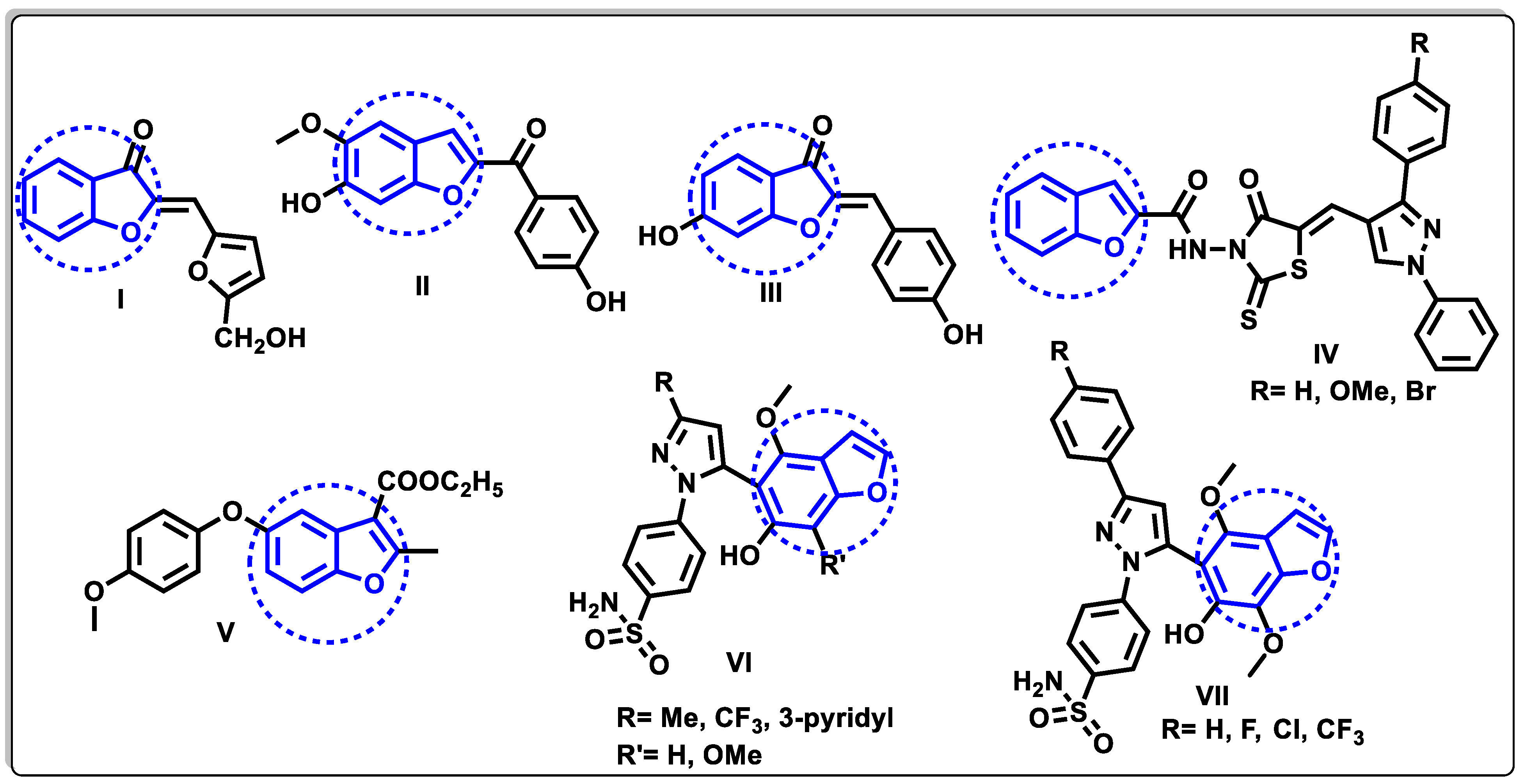
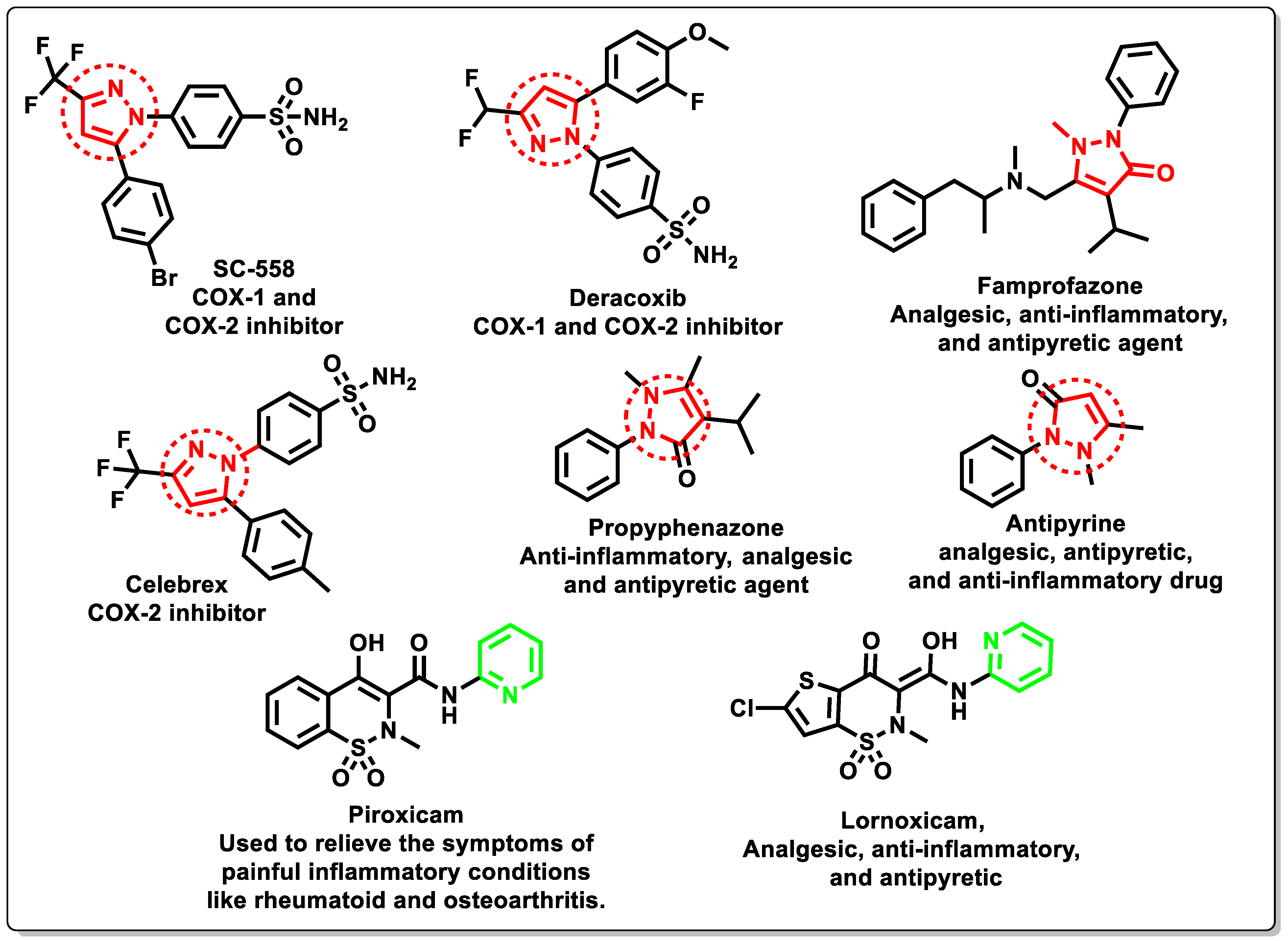
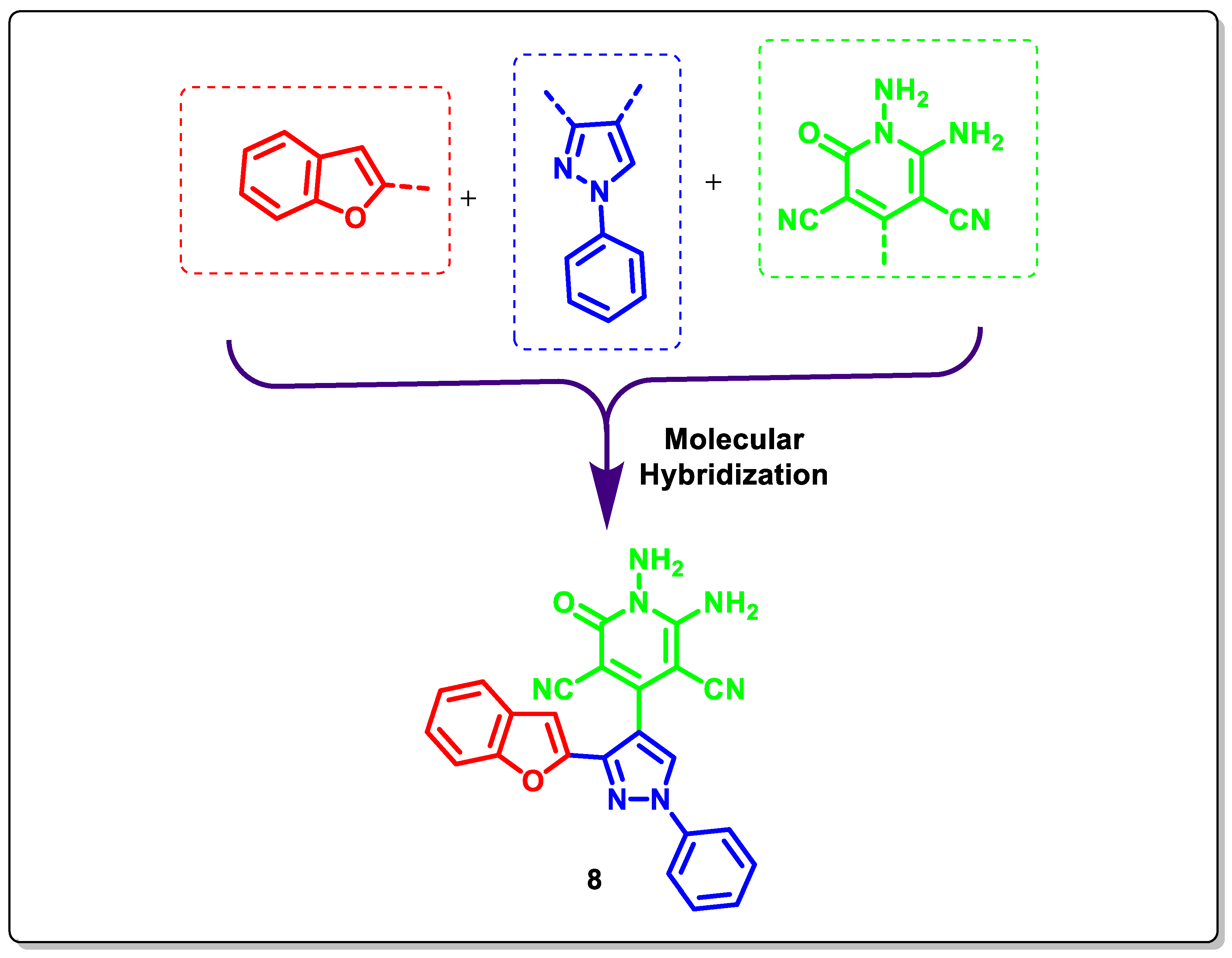
:1. Introduction
2. Results and Discussion
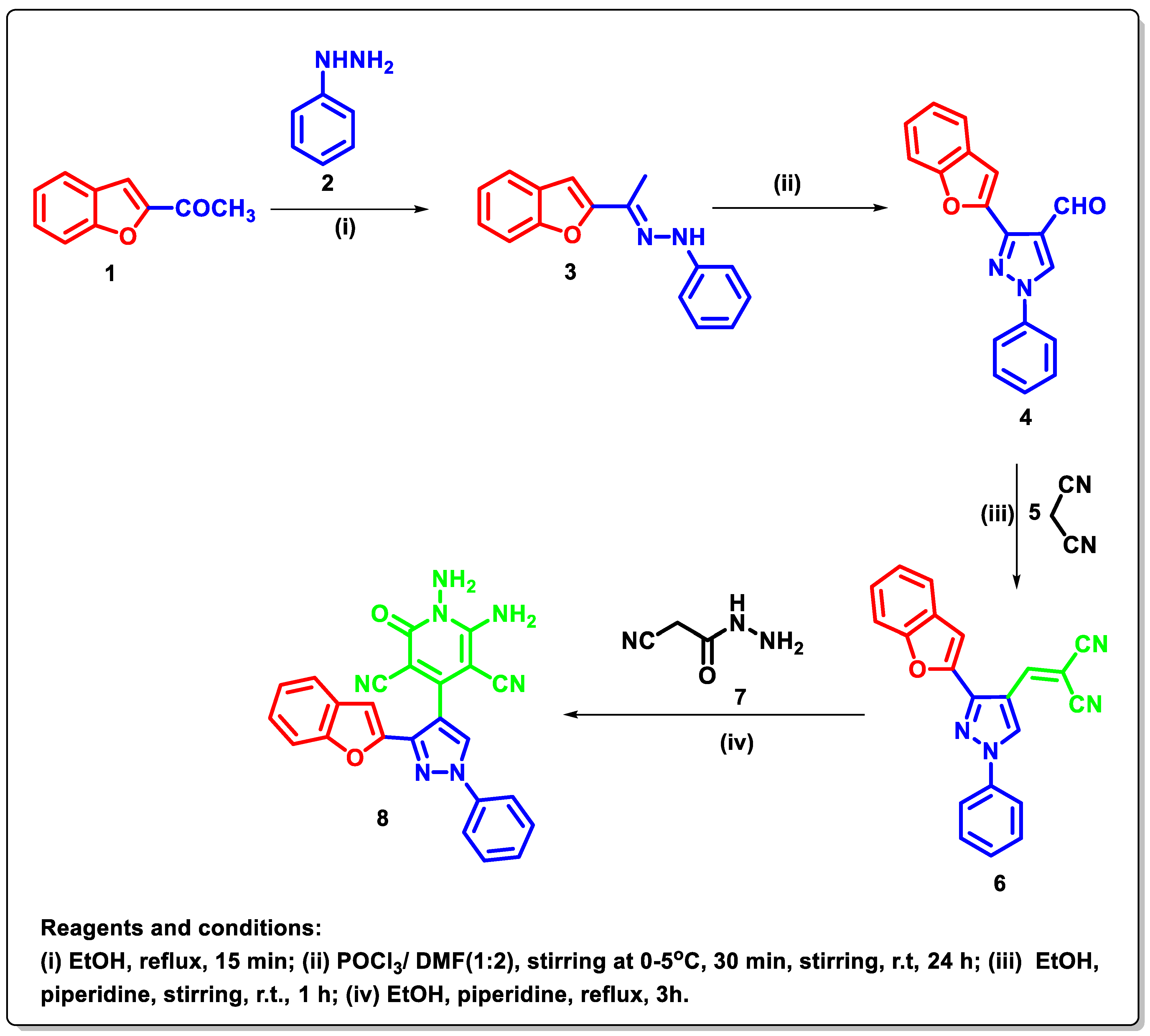
2.1. Chemistry
2.2. Biological Evaluation
2.2.1. DPPH Scavenging Activity
2.2.2. Identification of RANTES, CRP, COMP, LPO, CK, and Protein
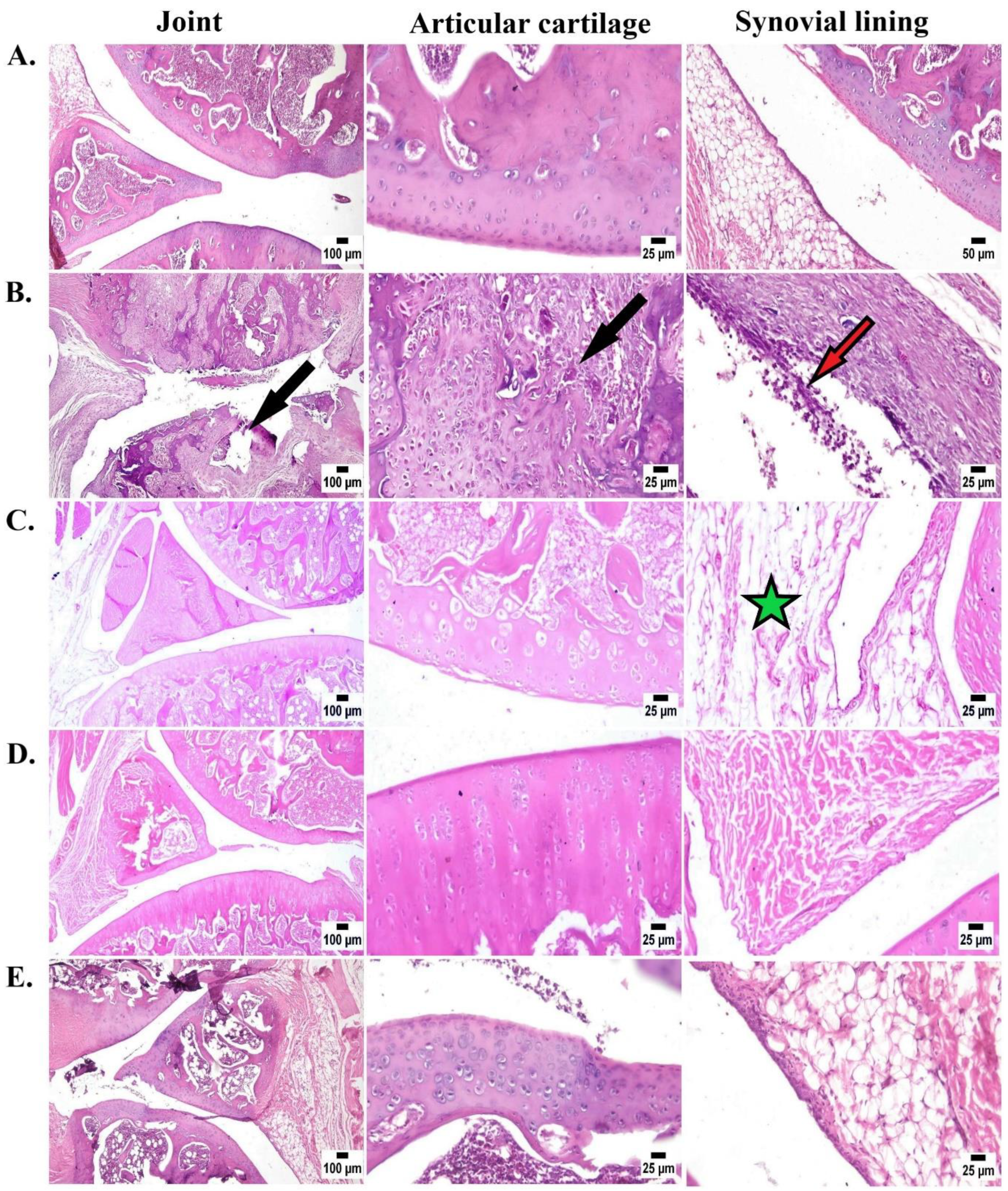
2.2.3. Histopathological Evaluation
3. Computational Studies
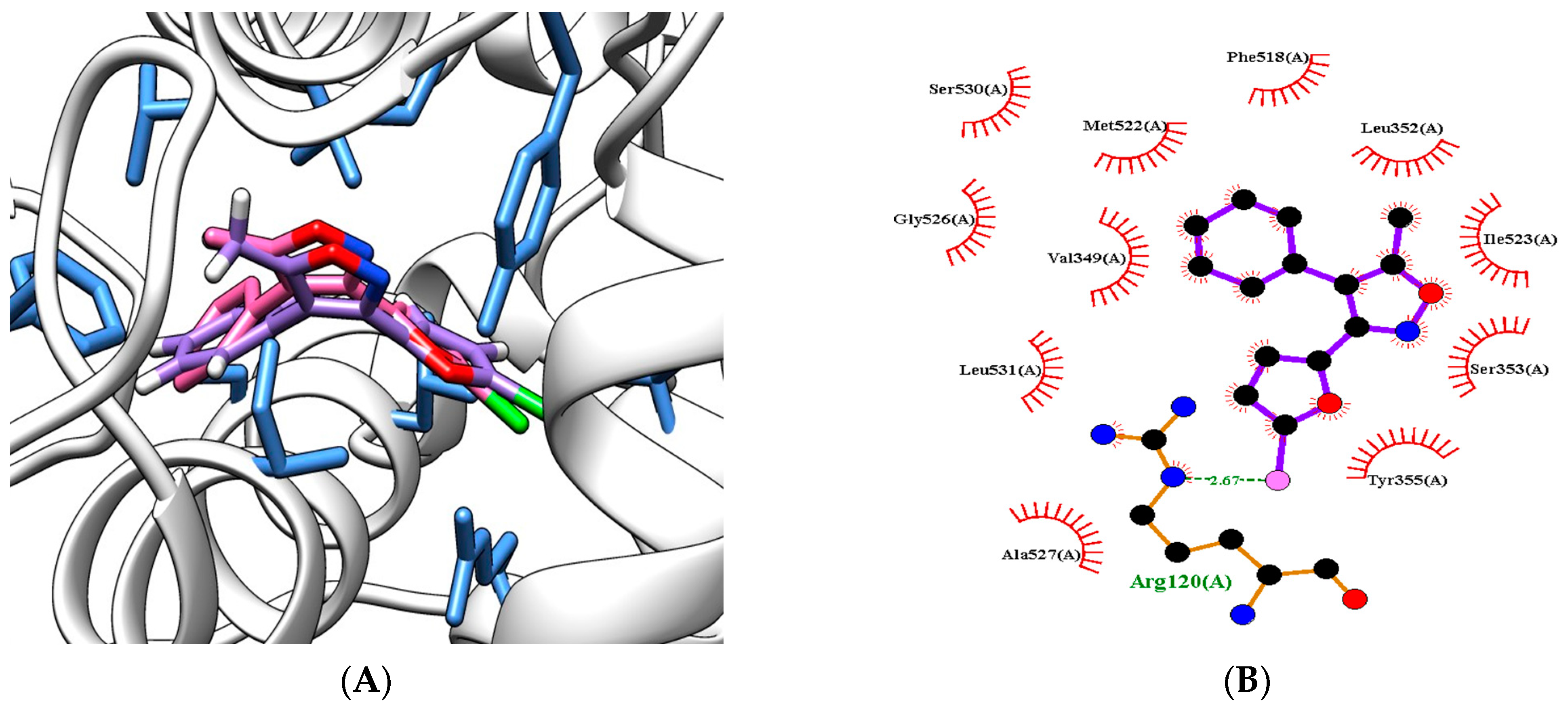
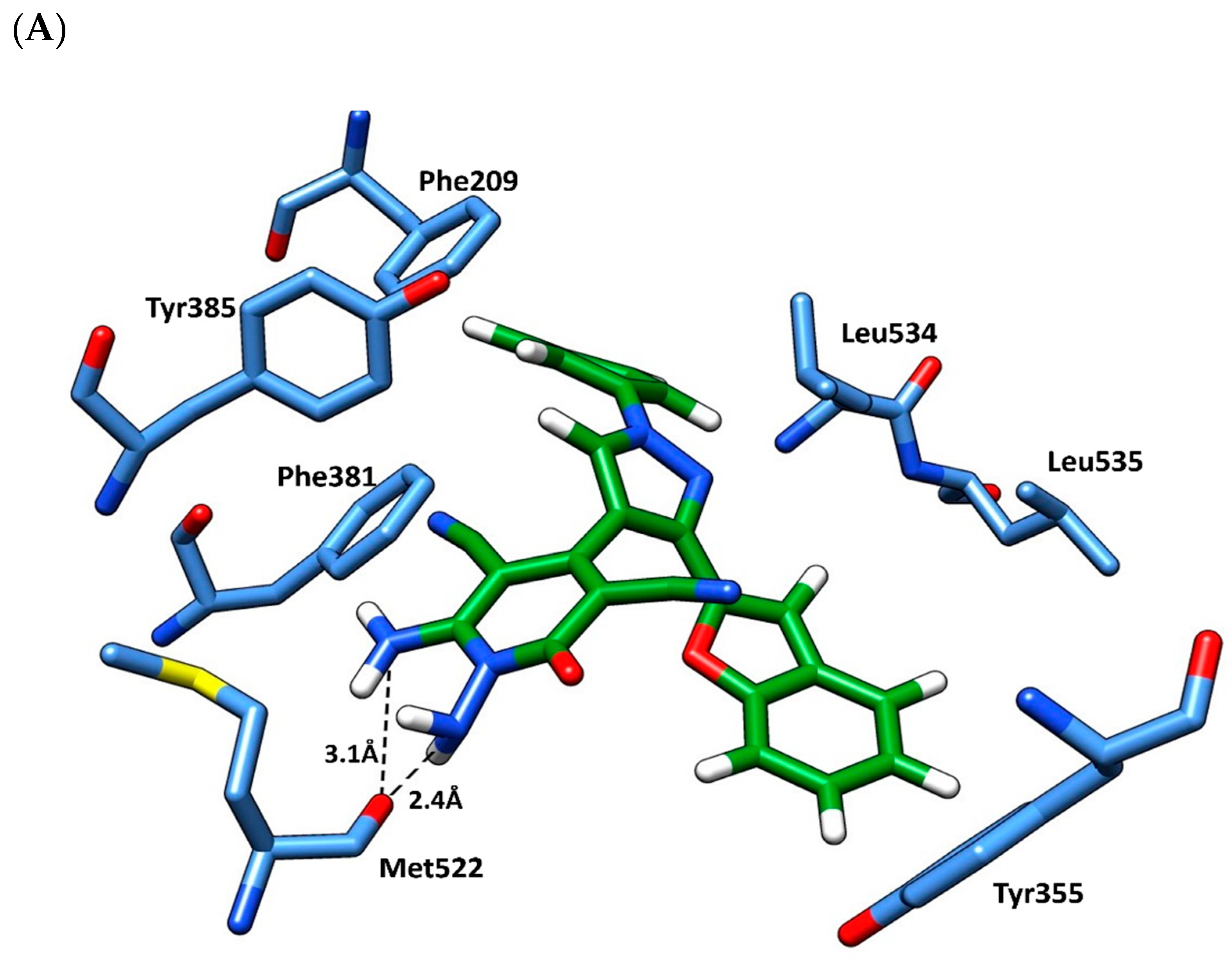
3.1. Molecular Docking Study
3.2. ADMET Analysis
4. Conclusions
5. Experimental Protocols
5.1. Chemistry
5.1.1. Synthesis of 1-(1-(Benzofuran-2-yl)ethylidene)-2-phenylhydrazine (3)
5.1.2. Synthesis of 3-(Benzofuran-2-yl)-1-phenyl-1H-pyrazole-4-carboxyaldehyde (4)
5.1.3. Synthesis of 2-((3-(Benzofuran-2-yl)-1-phenyl-1H-pyrazol-4-yl)methylene)malononilrile (6)
5.1.4. Synthesis of 1,6-Diamino-4-(3-(benzofuran-2-yl)-1-phenyl-1H-pyrazol-4-yl)-2-oxo-1,2-dihydropyridine-3,5-dicarbonitrile (8)
5.2. Biochemical Study
5.2.1. In Vivo Study
Animals
Knee Inflammation Induction in Rats
Experimental Design
5.2.2. Measuring of Biological Parameters
DPPH Radical Scavenging Assay
Evaluation of RANTES, CRP, COMP, LPO, CK, and Protein in KOA Rats with ELISA
5.2.3. Histopathological Study
5.2.4. Molecular Docking
Preparation of the Protein
Preparation of the Ligands
Molecular Modelling
Compliance with Ethical Standards
Supplementary Materials
Author Contributions
Funding
Institutional Review Board Statement
Informed Consent Statement
Data Availability Statement
Acknowledgments
Conflicts of Interest
Sample Availability
References
- Lambova, S.N. Knee Osteoarthritis—How Close Are We to Disease-Modifying Treatment: Emphasis on Metabolic Type Knee Osteoarthritis. Life 2013, 13, 140. [Google Scholar] [CrossRef]
- Berenbaum, F.; Wallace, I.J.; Lieberman, D.E.; Felson, D.T.; Sinusas, K. Osteoarthritis: Diagnosis and treatment. Am. Fam. Phys. 2012, 85, 49–56. [Google Scholar]
- Litwic, A.; Edwards, M.H.; Dennison, E.M.; Cooper, C. Epidemiology and burden of osteoarthritis. Br. Med. Bull. 2013, 105, 185–199. [Google Scholar] [CrossRef]
- Wallace, I.J.; Worthington, S.; Felson, D.T.; Jurmain, R.D.; Wren, K.T.; Maijanen, H.; Woods, R.J.; Lieberman, D.E. Knee osteoarthritis has doubled in prevalence since the mid-20th century. Proc. Natl. Acad. Sci. USA 2017, 114, 9332–9336. [Google Scholar] [CrossRef]
- Zhao, Y.; Xu, J. Synovial fluid-derived exosomal lncRNA PCGEM1 as biomarker for the different stages of osteoarthritis. Int. Orthop. 2018, 24, 2865–2872. [Google Scholar] [CrossRef]
- Mobasheri, A.; Saarakkala, S.; Finnilä, M.; Karsdal, M.A.; Bay-Jensen, A.C.; van Spil, W.E. Recent advances in understanding the phenotypes of osteoarthritis. F1000Research 2019, 8, F1000 Faculty Rev-2091. [Google Scholar] [CrossRef] [PubMed]
- Jette, D.U.; Hunter, S.J.; Burkett, L.; Langham, B.; Logerstedt, D.S.; Piuzzi, N.S.; Poirier, N.M.; Radach, L.J.L.; Ritter, J.E.; Scalzitti, D.A.; et al. Physical therapist management of total knee arthroplasty. Phys Ther. 2020, 100, 1603–1631. [Google Scholar] [CrossRef] [PubMed]
- Bannuru, R.R.; Osani, M.C.; Vaysbrot, E.E.; Arden, N.K.; Bennell, K.; Bierma-Zeinstra, S.M.A.; Kraus, V.B.; Lohmander, L.S.; Abbott, J.H.; Bhandari, M.; et al. OARSI guidelines for the non-surgical management of knee, hip, and polyarticular osteoarthritis. Osteoarthr. Cartil. 2019, 27, 1578–1589. [Google Scholar] [CrossRef]
- Charlesworth, J.; Fitzpatrick, J.; Perera, N.K.P.; Orchard, J. Osteoarthritis a systematic review of long-term safety implications for osteoarthritis of the knee. BMC Musculoskelet Disord. 2019, 20, 151. [Google Scholar] [CrossRef] [PubMed]
- Goldring, M.B.; Otero, M. Inflammation in osteoarthritis. Curr. Opin. Rheumatol. 2011, 23, 471–478. [Google Scholar] [CrossRef]
- Valdes, A.M.; Stocks, J. Osteoarthritis and aging. Eur. Med. J. 2018, 3, 116–123. Available online: https://nottingham-repository.worktribe.com/output/918526 (accessed on 5 September 2023). [CrossRef]
- Kou, L.; Xiao, S.; Sun, R.; Bao, S.; Yao, Q.; Chen, R. Biomaterial-engineered intraarticular drug delivery systems for osteoarthritis therapy. Drug Deliv. 2019, 26, 870–885. [Google Scholar] [CrossRef] [PubMed]
- Khalid, M.; Tufail, S.; Aslam, Z.; Butt, A. Osteoarthritis: From complications to cure. Int. J. Clin. Rheumatol. 2017, 12, 160–167. [Google Scholar] [CrossRef]
- Mulka-Gierek, M.; Krata, N.; Foroncewicz, B.; Pączek, L.; Mucha, K. The Different Patterns of Over-the-Counter Nonsteroidal Anti-Inflammatory Drugs or Analgesics Use in Patients with Chronic Kidney Disease and the General Population. Healthcare 2022, 10, 2035. [Google Scholar] [CrossRef]
- Tayem, Y.I.; Qubaja, M.M.; Shraim, R.K.; Taha, O.B.; Shkheidem, I.A.; Ibrahim, M.A. Non-steroidal anti-inflammatory drugs and antibiotics prescription trends at a central west bank hospital. Sultan Qaboos Univ. Med. J. 2013, 13, 567. [Google Scholar] [CrossRef]
- Shirley, P.Y.; Hunter, D.J. Managing osteoarthritis. Aust. Prescr. 2015, 38, 115. [Google Scholar] [CrossRef]
- Cutolo, M.; Berenbaum, F.; Hochberg, M.; Punzi, L.; Reginster, J.Y. Commentary on recent therapeutic guidelines for osteoarthritis. Semin. Arthritis. Rheum. 2015, 44, 611–617. [Google Scholar] [CrossRef]
- Hena Khanam, S. Bioactive Benzofuran derivatives: A review. Eur. J. Med. Chem. 2015, 97, 483–504. [Google Scholar] [CrossRef] [PubMed]
- Abbas, H.A.; Abd El-Karim, S.S. Design, synthesis and anticervical cancer activity of new benzofuran–pyrazol-hydrazono-thiazolidin-4-one hybrids as potential EGFR inhibitors and apoptosis inducing agents. Bioorg. Chem. 2019, 89, 103035. [Google Scholar] [CrossRef]
- Seo, Y.H.; Damodar, K.; Kim, J.-K.; Jun, J.-G. Synthesis and biological evaluation of 2- aroylbenzofurans, rugchalcones, A.; B and their derivatives as potent antiinflammatory agents. Bioorg. Med. Chem. Lett. 2016, 26, 1521–1524. [Google Scholar] [CrossRef]
- Kadayat, T.M.; Banskota, S.; Gurung, P.P.; Bist, G.; Magar, T.B.; Shrestha, A.; Kim, J.A.; Lee, E.S. Discovery and structure-activity relationship studies of 2-benzylidene-2,3-dihydro-1H-inden-1-one and benzofuran-3(2H)-one derivatives as a novel class of potential therapeutics for inflammatory bowel disease. Eur. J. Med. Chem. 2017, 137, 575–597. [Google Scholar] [CrossRef] [PubMed]
- El-Miligy, M.M.M.; Hazzaa, A.A.; El-Messmary, H.; Nassra, R.A.; El-Hawash, S.A.M. New hybrid molecules combining benzothiophene or benzofuran with rhodanine as dual COX-1/2 and 5- LOX inhibitors: Synthesis, biological evaluation and docking study. Bioorg. Chem. 2017, 72, 102–115. [Google Scholar] [CrossRef] [PubMed]
- Yadav, P.; Singh, P.; Tewari, A.K. Design, synthesis, docking and anti-inflammatory evaluation of novel series of benzofuran based prodrugs. Bioorg. Med. Chem. Lett. 2014, 24, 2251–2255. [Google Scholar] [CrossRef] [PubMed]
- Hassan, G.S.; Abou-Seri, S.M.; Kamel, G.; Ali, M.M. Celecoxib analogs bearing benzofuran moiety as cyclooxygenase-2 inhibitors: Design, synthesis and evaluation as potential anti-inflammatory agents. Eur. J. Med. Chem. 2014, 76, 482–493. [Google Scholar] [CrossRef]
- Bekhit, A.A.; Nasralla, S.N.; El-Agroudy, E.J.; Hamouda, N.; Abd El-Fattah, A.; Bekhit, S.A.; Amagase, K.; Ibrahim, T.M. Investigation of the anti-inflammatory and analgesic activities of promising pyrazole derivative. Eur. J. Pharm. Sci. 2022, 168. [Google Scholar] [CrossRef]
- Nossier, E.S.; Fahmy, H.H.; Khalifa, N.M.; El-Eraky, W.I.; Baset, M.A. Design and synthesis of novel pyrazole-substituted different nitrogenous heterocyclic ring systems as potential anti-inflammatory agents. Molecules 2017, 22, 512. [Google Scholar] [CrossRef]
- Abd El-Karim, S.S.; Mohamed, H.S.; Abdelhameed, M.F.; Amr, A.E.; Almehizia, A.A.; Nossier, E.S. Design, synthesis and molecular docking of new pyrazole-thiazolidinones as potent anti-inflammatory and analgesic agents with TNF-α inhibitory activity. Bioorg. Chem. 2021, 111, 104827. [Google Scholar] [CrossRef]
- Penning, T.D.; Talley, J.J.; Bertenshaw, S.R.; Carter, J.S.; Collins, P.W.; Docter, S.; Graneto, M.J.; Lee, L.F.; Malecha, J.W.; Miyashiro, J.M.; et al. Synthesis and Biological Evaluation of the 1,5-Diarylpyrazole Class of Cyclooxygenase-2 Inhibitors: Identification of 4-[5-(4-Methylphenyl)-3- (trifluoromethyl)-1H-pyrazol-1-yl]benzenesulfonamide (SC-58635, Celecoxib). J. Med. Chem. 1997, 40, 1347–1365. [Google Scholar] [CrossRef]
- Fioravanti, R.; Bolasco, A.; Manna, F.; Rossi, F.; Orallo, F.; Ortuso, F.; Alcaro, S.; Cirilli, R. Synthesis and biological evaluation of N-substituted-3,5-diphenyl-2-pyrazoline derivatives as cyclooxygenase (COX-2) inhibitors. Eur. J. Med. Chem. 2010, 45, 6135–6138. [Google Scholar] [CrossRef]
- Ochiai, K.; Ando, N.; Iwase, K.; Kishi, T.; Fukuchi, K.; Ohinata, A.; Zushi, H.; Yasue, T.; Adams, D.R.; Kohno, Y. Phosphodiesterase inhibitors. Part 2: Design, synthesis, and structure–activity relationships of dual PDE3/4-inhibitory pyrazolo [1, 5- a] pyridines with anti-inflammatory and bronchodilatory activity. Bioorg. Med. Chem. Lett. 2011, 21, 5451–5456. [Google Scholar] [CrossRef]
- Kamat, V.; Santosh, R.; Poojary, B.; Nayak, S.P.; Kumar, B.K.; Sankaranarayanan, M.; Faheem; Khanapure, S.; Barretto, D.A.; Vootla, S.K. Pyridine-and thiazole-based hydrazides with promising anti-inflammatory and antimicrobial activities along with their in silico studies. ACS Omega 2020, 5, 25228–25239. [Google Scholar] [CrossRef] [PubMed]
- Fischer, J.; Ganellin, C.R. Analogue-based drug discovery. Chem. Int. Newsmag. IUPAC 2010, 32, 12–15. [Google Scholar] [CrossRef]
- Othman, E.M.; Fayed, E.A.; Husseiny, E.M.; Abulkhair, H.S. Apoptosis induction, PARP-1 inhibition, and cell cycle analysis of leukemia cancer cells treated with novel synthetic 1, 2, 3-triazole-chalcone conjugates. Bioorg. Chem. 2022, 123, 105762. [Google Scholar] [CrossRef] [PubMed]
- Elliott, E.D. The preparation and properties of 2-vinylbenzofuran. J. Am. Chem. Soc. 1951, 73, 754. [Google Scholar] [CrossRef]
- Aruna Kumar, D.B.; Prakash, G.K.; Kumaraswamy, M.N.; Nandeshwarappa, B.P.; Sherigara, B.S.; Mahadevan, K.M. Synthesis and antimicrobial investigation of some novel phenyl pyrazole, azetidinone and diazenyl ethanone derivatives of benzofurans. Ind. J. Chem. 2007, 46B, 336–343. [Google Scholar]
- El-Zahar, M.I.; Abd El-Karim, S.S.; Anwar, M.M. Synthesis and cytotoxicity screening of some novel benzofuranoyl-pyrazole derivatives against liver and cervix carcinoma cell line. S. Afr. J. Chem. 2009, 62, 189–199. [Google Scholar]
- Abd El-Karim, S.S.; Anwar, M.M.; Mohamed, N.A.; Nasr, T.; Elseginy, S.A. Design, synthesis, biological evaluation and molecular docking studies of novel benzofuran–pyrazole derivatives as anticancer agents. Bioorg. Chem. 2015, 63, 114790. [Google Scholar] [CrossRef]
- Anwar, M.M.; Abd El-Karim, S.S.; Mahmoud, A.H.; Amr, A.G.-E.; Al-Omar, M.A. A Comparative study of the anticancer activity and PARP-1 inhibiting Effect of benzofuran–pyrazole scaffold and its nano-sized particles in human breast cancer cells. Molecules 2019, 24, 2413. [Google Scholar] [CrossRef]
- Nejadhosseinian, M.; Djalalinia, S.; Haerian, H.; Alikhani, M.; Mansour, A.; Mousavian, A.H.; Mardani-Fard, H.A.; Kasaeian, A.; Faezi, S.T. The effects of antioxidants on knee osteoarthritis: A systematic review and meta-analysis. Front. Nutr. 2022, 9, 1026450. [Google Scholar] [CrossRef]
- Ajuebor, M.N.; Hogaboam, C.M.; Kunkel, S.L.; Proudfoot, A.E.; Wallace, J.L. The chemokine RANTES is a crucial mediator of the progression from acute to chronic colitis in the rat. J. Immun. 2001, 166, 552–558. [Google Scholar] [CrossRef]
- Stanczyk, J.; Kowalski, M.L.; Grzegorczyk, J.; Szkudlinska, B.; Jarzebska, M.; Marciniak, M.; Synder, M. RANTES and chemotactic activity in synovial fluids from patients with rheumatoid arthritis and osteoarthritis. Mediat. Inflamm. 2005, 14, 343–348. [Google Scholar] [CrossRef]
- Katschke, K.J., Jr.; Rottman, J.B.; Ruth, J.H.; Qin, S.; Wu, L.; LaRosa, G.; Ponath, P.; Park, C.C.; Pope, R.M.; Koch, A.E. Differential expression of chemokine receptors on peripheral blood, synovial fluid, and synovial tissue monocytes/macrophages in rheumatoid arthritis. Arthritis Rheum. 2001, 44, 1022–1032. [Google Scholar] [CrossRef]
- Zhang, J. Meta-analysis of serum C-reactive protein and cartilage oligomeric matrix protein levels as biomarkers for clinical knee osteoarthritis. BMC Musculoskelet. Disord. 2018, 19, 22. [Google Scholar] [CrossRef]
- Sanchez-Ramirez, D.C.; van der Leeden, M.; van der Esch, M.; Roorda, L.D.; Verschueren, S.; van Dieen, J.; Lems, W.F.; Dekker, J. Increased knee muscle strength is associated with decreased activity limitations in established knee osteoarthritis: Two-year follow-up study in the Amsterdam osteoarthritis cohort. J. Rehabil. Med. 2015, 47, 647–654. [Google Scholar] [CrossRef]
- Mourad, B.H. Prevalence of work-related musculoskeletal disorders among Egyptian printing workers evidenced by using serum biomarkers of inflammation, oxidative stress, muscle injury, and collagen type I turnover. Toxicol. Ind. Health. 2021, 37, 9–22. [Google Scholar] [CrossRef]
- Hong, Z.; Gao, H.; Su, Y.; Xu, B.; Wu, Z. Effect and mechanism of total alkaloids of strychnine on papain induced rabbit knee osteoarthritis. Biomed. Res. 2018, 29, 1590–1597. [Google Scholar] [CrossRef]
- Jones, M.L.; Bancroft, J.D.; Gamble, M. Connective tissues and stains. Theory Pract. Histol. Tech. 2008, 6, 135–160. [Google Scholar]
- Pires, D.E.; Blundell, T.L.; Ascher, D.B. pkCSM: Predicting small-molecule pharmacokinetic and toxicity properties using graph-based signatures. J. Med. Chem. 2015, 58, 4066–4072. [Google Scholar] [CrossRef]
- Elseginy, S.A.; Oliveira, A.S.F.; Shoemark, D.K.; Sessions, R.B. Identification and validation of novel microtubule suppressors with an imidazopyridine scaffold through structure-based virtual screening and docking. RSC Med. Chem. 2022, 13, 929–943. [Google Scholar] [CrossRef]
- Daina, A.; Michielin, O.; Zoete, V. SwissADME: A free web tool to evaluate pharmacokinetics, drug-likeness and medicinal chemistry friendliness of molecules. Sci. Rep. 2017, 7, 42717. [Google Scholar] [CrossRef]
- Udo, M.; Muneta, T.; Tsuji, K.; Ozeki, N.; Nakagawa, Y.; Ohara, T.; Saito, R.; Yanagisawa, K.; Koga, H.; Sekiya, I. Monoiodoacetic acid induces arthritis and synovitis in rats in a dose-and time-dependent manner: Proposed model-specific scoring systems. Osteoarthr. Cartil. 2016, 24, 1284–1291. [Google Scholar] [CrossRef] [PubMed]
- Desmarchelier, C.; Novoa Bermudez, M.J.; Coussio, J.; Ciccia, G.; Boveris, A. Antioxidant and Prooxidant Activities in Aqueous Extracts of Argentine Plants. Int. J. Pharmacogn. 1997, 35, 116–120. [Google Scholar] [CrossRef]
- Elseginy, S.A. Virtual screening and structure-based 3D pharmacophore approach to identify small-molecule inhibitors of SARS-CoV-2 Mpro. J. Biomol. Struct. Dyn. 2022, 40, 13658–13674. [Google Scholar] [CrossRef]
- Dolinsky, T.J.; Nielsen, J.E.; McCammon, J.A.; Baker, N.A. PDB2PQR: An automated pipeline for the setup of Poisson–Boltzmann electrostatics calculations. Nucleic Acids Res. 2004, 32 (Suppl. 2), W665–W667. [Google Scholar] [CrossRef]
- Webb, B.; Sali, A. Comparative protein structure modeling using MODELLER. Curr. Protoc. Bioinform. 2016, 54, 5–6. [Google Scholar] [CrossRef] [PubMed]
- Elseginy, S.A.; Anwar, M.M. In silico analysis of SARS-CoV-2 papain-like protease potential inhibitors. RSC Adv. 2021, 11, 38616–38631. [Google Scholar] [CrossRef] [PubMed]

| Concentrations | DPPH Scavenging Activity (%) of Ascorbic Acid | DPPH Scavenging Activity (%) of Compound 8 |
|---|---|---|
| 10 | 54.88 ± 3.1 | 17.93 ± 2.1 |
| 50 | 97.4 ± 2.1 | 14.02 ± 1.8 |
| 100 | 97 ± 2.3 | 11.59 ± 1.8 |
| 200 | 97 ± 1.7 | 23 ± 2.5 |
| 500 | 97.2 ± 1.9 | 50 ± 2.3 |
| 1000 | 97 ± 2.3 | 84.4 ± 2.7 |
| Groups | RANTES (Pg/mL) | CRP (ng/mL) | COMP (ng/mL) | CK (ng/mL) | LPO (µM) | Protein (g/mL) |
|---|---|---|---|---|---|---|
| 1-Negative control | 60.17 ± 0.85 (2 ***, 3 ***, 4 *, 5 **) | 2.10 ± 0.01 (2 ***) | 1.46 ± 0.012 (2, 4, 5) *** | 262 ± 5.2 (2 ***, 4 **, 5 ***) | 0.76 ± 0.009 (2, 4, 5) *** | 53.4 ± 0.64 (4 ***) |
| 2-Positive control | 88.76 ± 1.8 (1, 3, 4, 5) *** | 2.51 ± 0.03 (1 ***, 3 **, 4 **, 5 **) | 1.75 ± 0.07 (1 ***, 3 ***, 4 *) | 519 ± 6 (1, 3, 4, 5) *** | 0.87 ± 0.03 (1, 3, 4, 5) *** | 63.43 ± 1.82 (4 ***, 5 **) |
| 3-Low dose | 42.35 ± 1.43 (2, 4, 5) *** | 2.23 ± 0.05 (2 **) | 1.41 ± 0.02 (2, 4, 5) *** | 270 ± 4.16 (2 ***, 3 **, 5 ***) | 0.76 ± 0.01 (2, 4, 5) *** | 61 ± 2.27 (4 ***, 5 *) |
| 4-High dose | 55.86 ± 0.94 (1 *, 2 ***, 3 ***, 5 ***) | 2.22 ± 0.11 (2 **) | 1.90 ± 0.01 (1 ***, 2 *, 3 ***) | 308 ± 2.6 (1 **, 2 ***, 3 **) | 0.63 ± 0.017 (1, 2, 3, 5) *** | 120.45 ± 9.16 (1, 2, 3, 5) *** |
| 5-Ref. drug | 66.02 ± 0.96 (1 **, 2 ***, 3 ***, 4 ***) | 1.85 ± 0.05 (1, 3) *** | 3.25 ± 15.76 (1, 2, 3) *** | 216 ± 0.03 (2 **) | 1.06 ± 0.03 (1, 2, 3, 4) *** | 41.8 ± 2.02 (2 **, 3 *, 4 ***) |
| Docking Scores (Kcal/mol) | Residues of Proteins | Moieties of Compounds | Type of Interactions | |
|---|---|---|---|---|
| Native ligand | −6.8 | NH-Arg120 | F | H-bond (2.6 Å) |
| Met522, Ser353 | Furan ring | Pi–pi interaction | ||
| Tyr355 | ||||
| Leu352, Val349, Ile523, Ala527, Leu531 | Phenyl ring | Hydrophobic | ||
| Isoxazole ring | ||||
| Compound 8 | −9.5 | OH-Met522 | NH | H-bond (2.4 Å) |
| NH | H-bond (3.1 Å) | |||
| Phe209 | Phenyl group | Pi–pi interaction | ||
| Phe381 | Phenyl group | |||
| Tyr385 | Pyrazole group | |||
| Tyr355 | Pyrazole group | |||
| Trp387, Leu352 | Benzofuran moiety | Hydrophobic | ||
| Leu534, Leu535 |
| Drug-Likeness Parameters | Compound 8 |
|---|---|
| Smile | [O+]=1c2c(CC=1c1nn(cc1C1=C(C#N)C(=O)N(N)C(N)=C1C#N)-c1ccccc1)cccc2 |
| MW | 434.438 |
| Log Po/w(XLOGP3) | 1.59 |
| Log Po/w(MLOGP) | 1.82 |
| HBD | 2 |
| HBA | 5 |
| NRB | 3 |
| MR | 121.36 |
| Lipinski | Yes (0 violation) |
| SA | 3.79 |
| TPSA | 137.75 |
| Bioavailability score | 0.55 |
| PAINS filtrate | Yes (0 violation) |
| Absorption | ||
|---|---|---|
| Model Name | Predicted Value | Unit |
| Water solubility | −3.37 | log mol/L |
| Human intestinal absorption (HIA) | 89.13 | % |
| Caco2 permeability | 0.34 | nm s−1 |
| Skin permeability | −2.73 | log Kp, cm h−1 |
| Distribution | ||
| BBB permeability | −0.54 | log BB |
| CNS permeability | −2.37 | log PS |
| Metabolism | ||
| CYPC1A2 inhibition | No | |
| CYP2D6 inhibition | No | |
| CYP3A4 substrate | Yes | |
| Excretion | ||
| Total clearance | 1.08 | log ml/min/kg |
| Renal OCT2 substrate | No | |
| Test | Carcinogenicity (Mouse) |
|---|---|
| Mutagen | none |
| Tumorigenic | none |
| Irritant | none |
Disclaimer/Publisher’s Note: The statements, opinions and data contained in all publications are solely those of the individual author(s) and contributor(s) and not of MDPI and/or the editor(s). MDPI and/or the editor(s) disclaim responsibility for any injury to people or property resulting from any ideas, methods, instructions or products referred to in the content. |
© 2023 by the authors. Licensee MDPI, Basel, Switzerland. This article is an open access article distributed under the terms and conditions of the Creative Commons Attribution (CC BY) license (https://creativecommons.org/licenses/by/4.0/).
Share and Cite
Abd El-Karim, S.S.; Mahmoud, A.H.; Al-Mokaddem, A.K.; Ibrahim, N.E.; Alkahtani, H.M.; Zen, A.A.; Anwar, M.M. Development of a New Benzofuran–Pyrazole–Pyridine-Based Molecule for the Management of Osteoarthritis. Molecules 2023, 28, 6814. https://doi.org/10.3390/molecules28196814
Abd El-Karim SS, Mahmoud AH, Al-Mokaddem AK, Ibrahim NE, Alkahtani HM, Zen AA, Anwar MM. Development of a New Benzofuran–Pyrazole–Pyridine-Based Molecule for the Management of Osteoarthritis. Molecules. 2023; 28(19):6814. https://doi.org/10.3390/molecules28196814
Chicago/Turabian StyleAbd El-Karim, Somaia S., Ahlam H. Mahmoud, Asmaa K. Al-Mokaddem, Noha E. Ibrahim, Hamad M. Alkahtani, Amer Alhaj Zen, and Manal M. Anwar. 2023. "Development of a New Benzofuran–Pyrazole–Pyridine-Based Molecule for the Management of Osteoarthritis" Molecules 28, no. 19: 6814. https://doi.org/10.3390/molecules28196814
APA StyleAbd El-Karim, S. S., Mahmoud, A. H., Al-Mokaddem, A. K., Ibrahim, N. E., Alkahtani, H. M., Zen, A. A., & Anwar, M. M. (2023). Development of a New Benzofuran–Pyrazole–Pyridine-Based Molecule for the Management of Osteoarthritis. Molecules, 28(19), 6814. https://doi.org/10.3390/molecules28196814