
Nanoparticles of a Pyrazolo-Pyridazine Derivative as Potential EGFR and CDK-2 Inhibitors: Design, Structure Determination, Anticancer Evaluation and In Silico Studies

 , , , , , and
Abstract
:
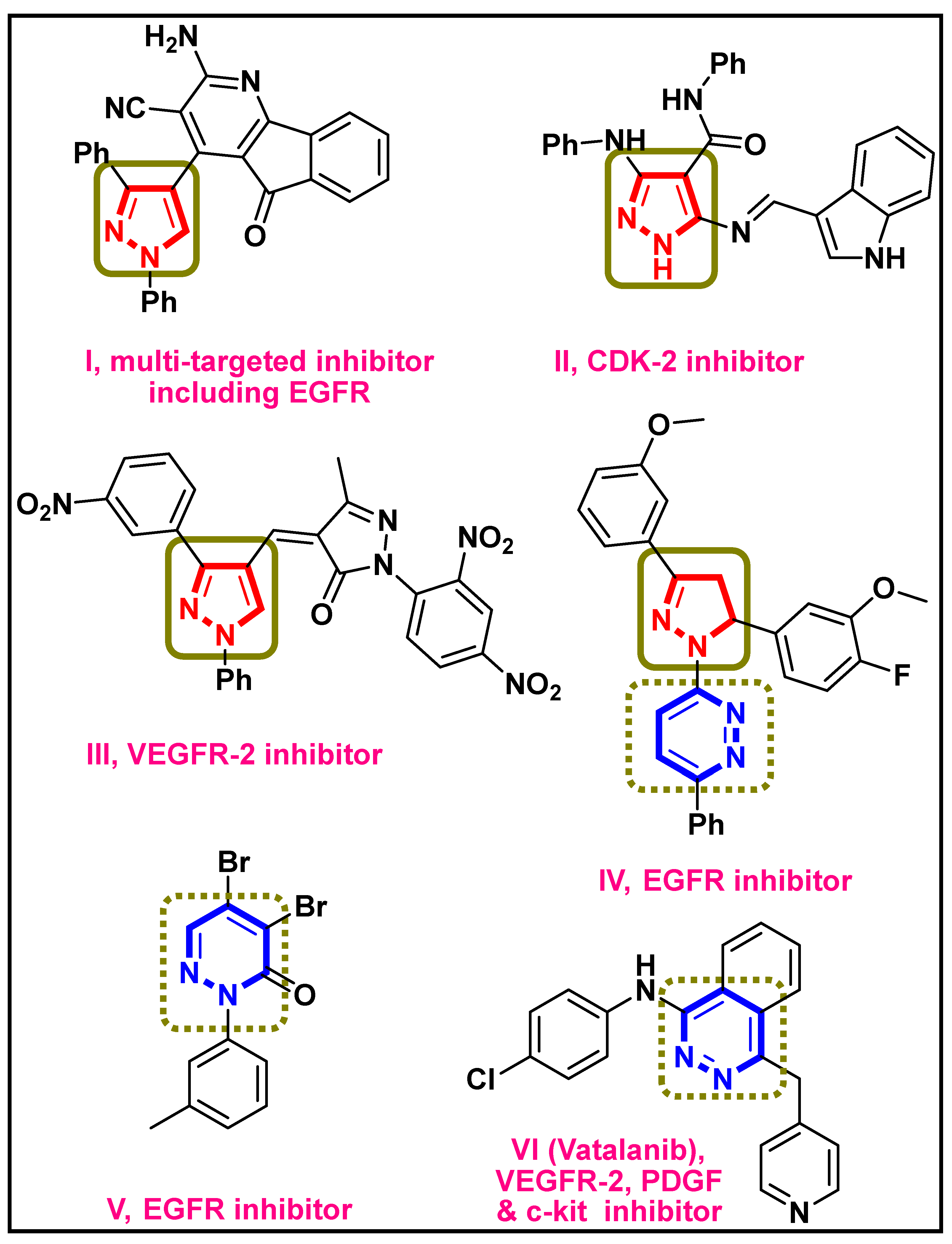
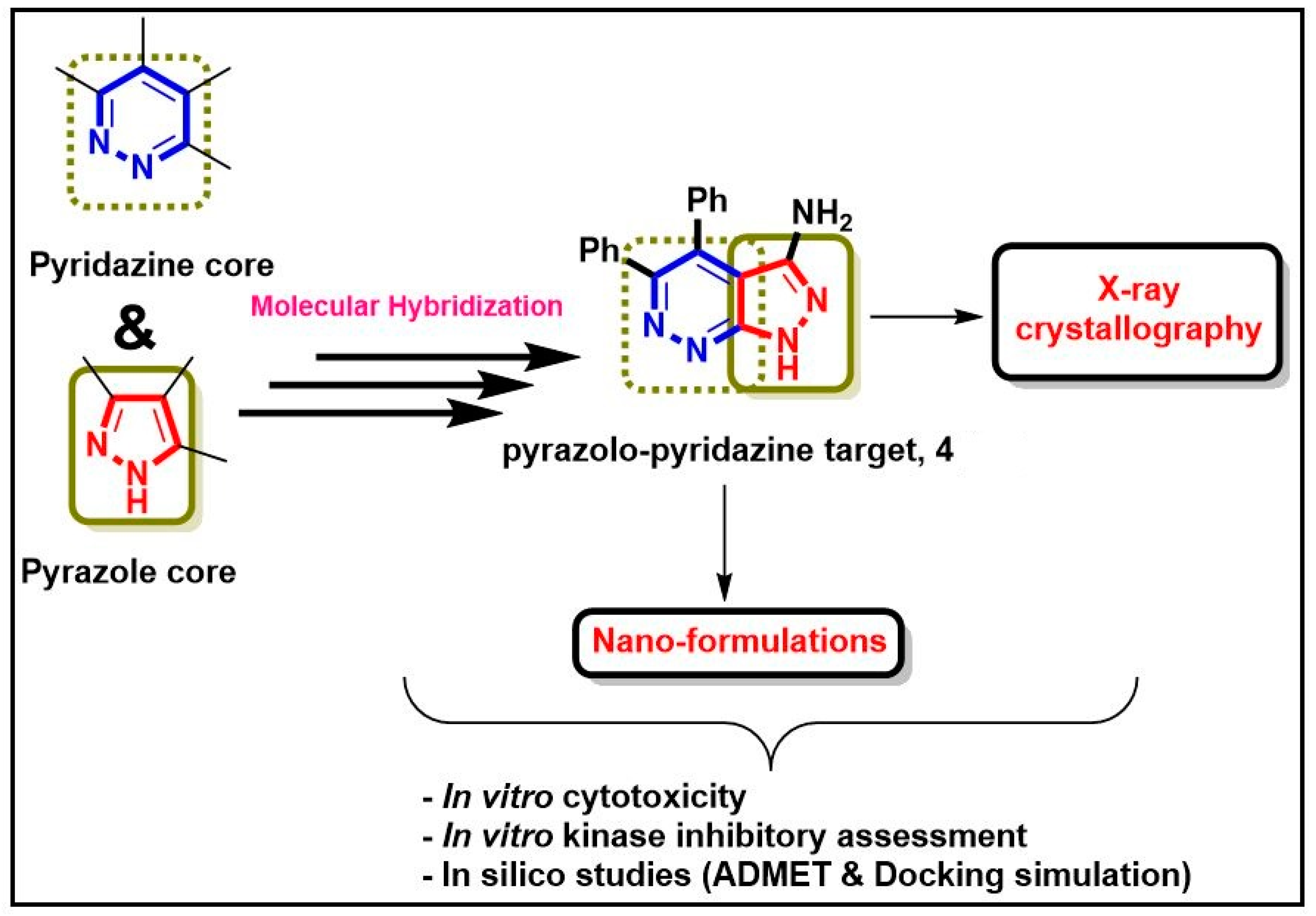
1. Introduction
2. Results and Discussion
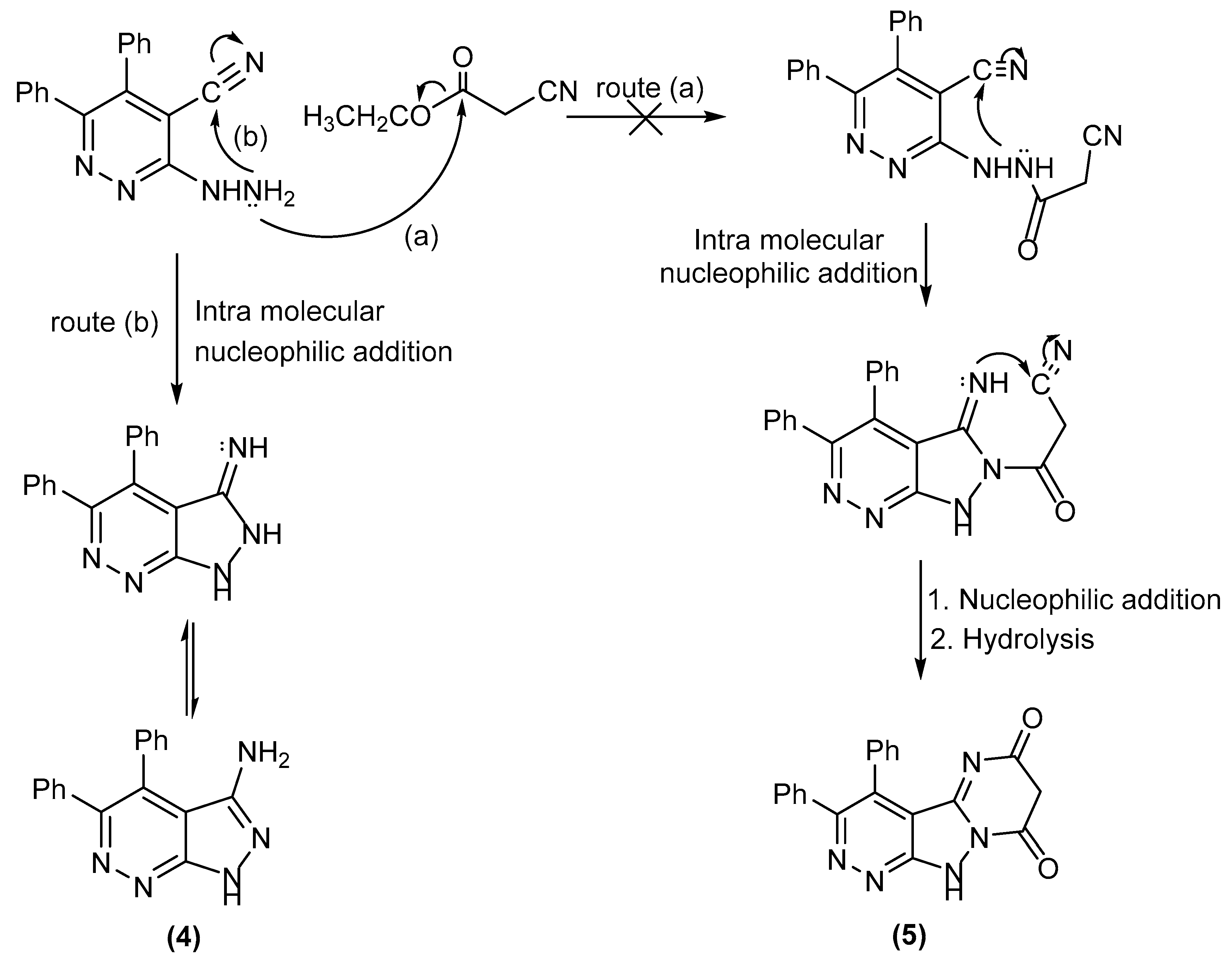
2.1. Synthesis
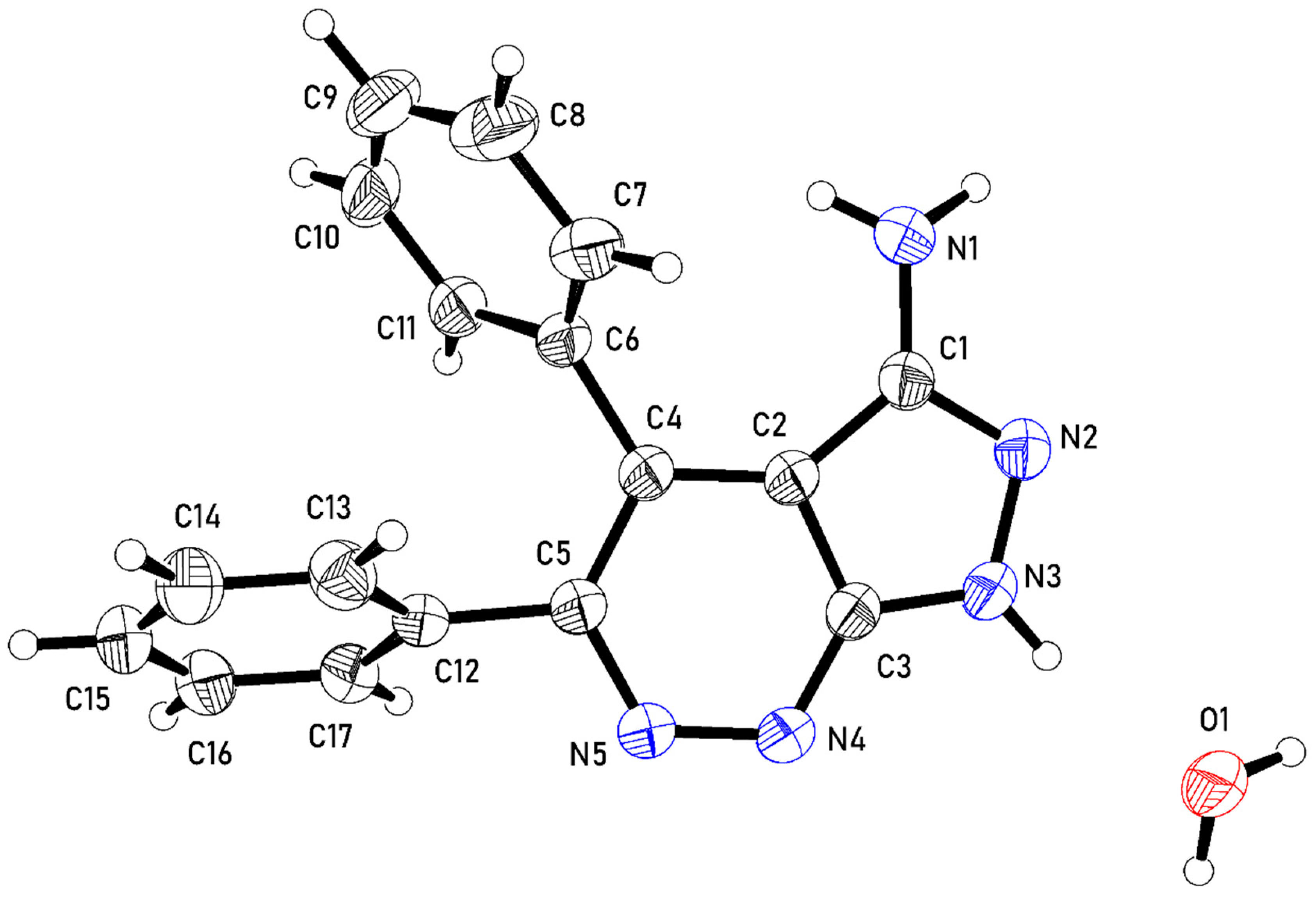
2.2. Crystal Structure of Compound 4
2.3. Development of the Target 4 into Nano-Formulations (4-SLNs and 4-LPHNPs)
Characterization of Developed Nano-Formulations (Particle Size and Zeta Potential)
2.4. Biological Evaluation
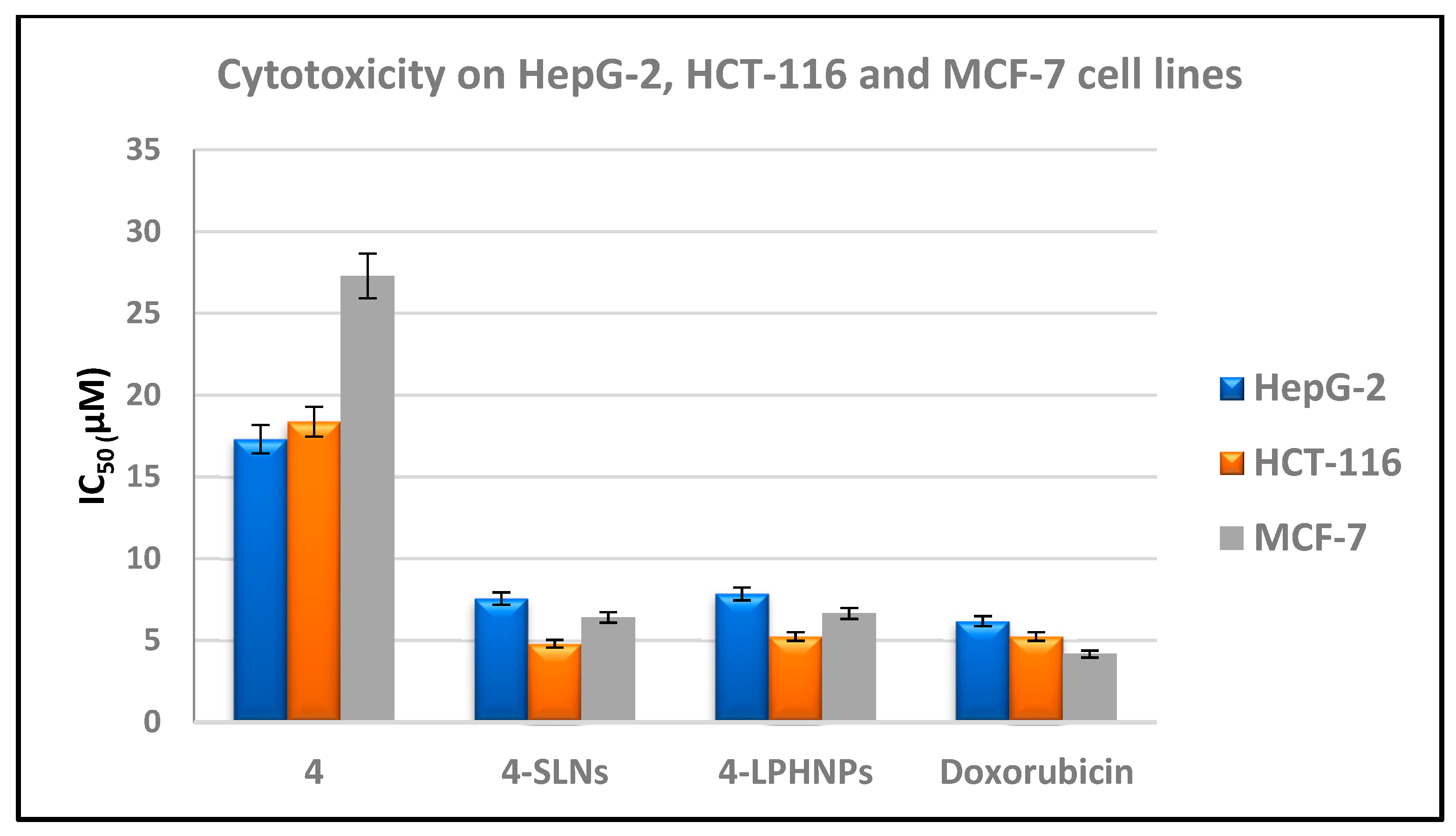
2.4.1. In Vitro Cytotoxicity
2.4.2. In Vitro Kinase Inhibitory Assessment against EGFR and CDK-2
2.4.3. Effect of Target 4 and Its Nanoparticles 4-SLNs and 4-LPHNPs upon Levels of Bax, Bcl-2, Caspase-3 and p53
2.5. In Silico Studies
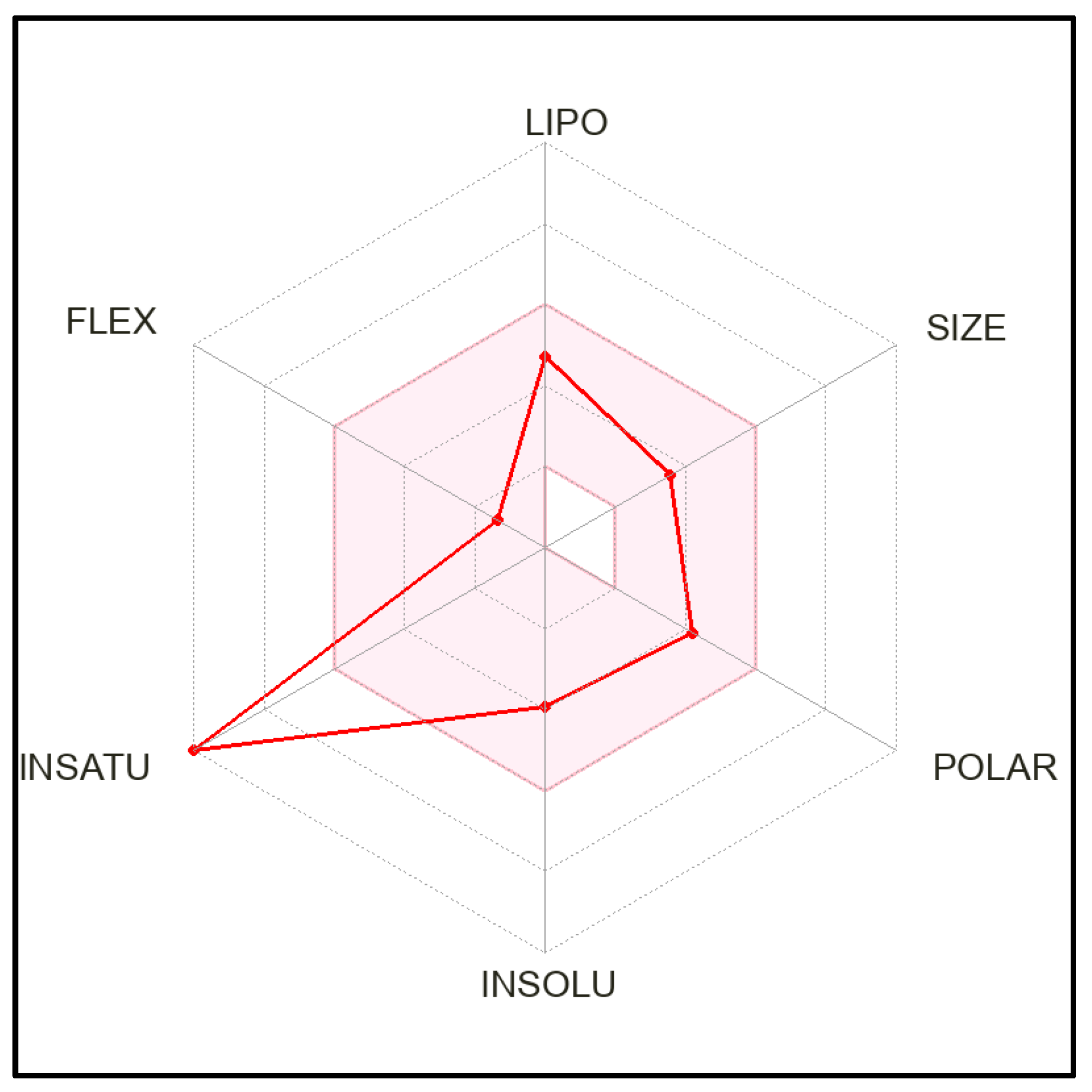
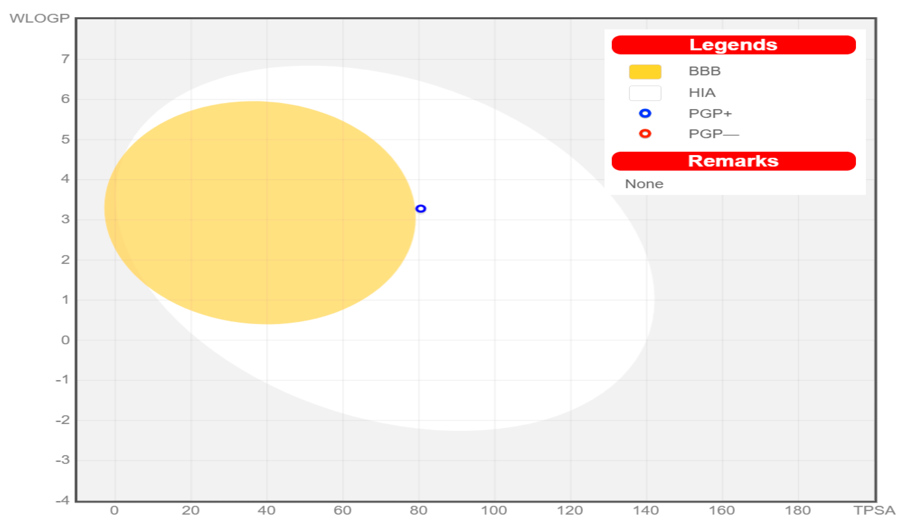
2.5.1. ADME Prediction
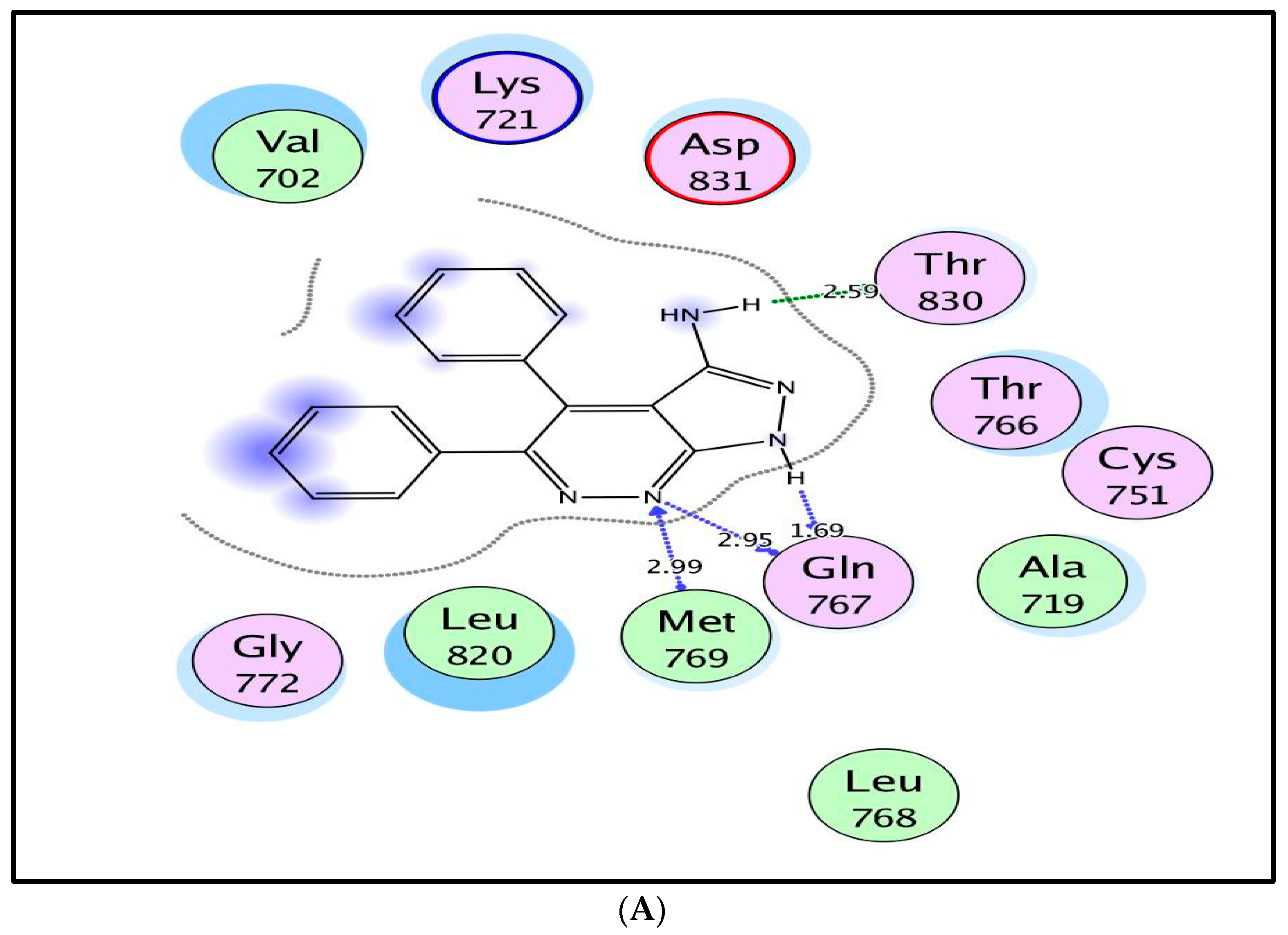
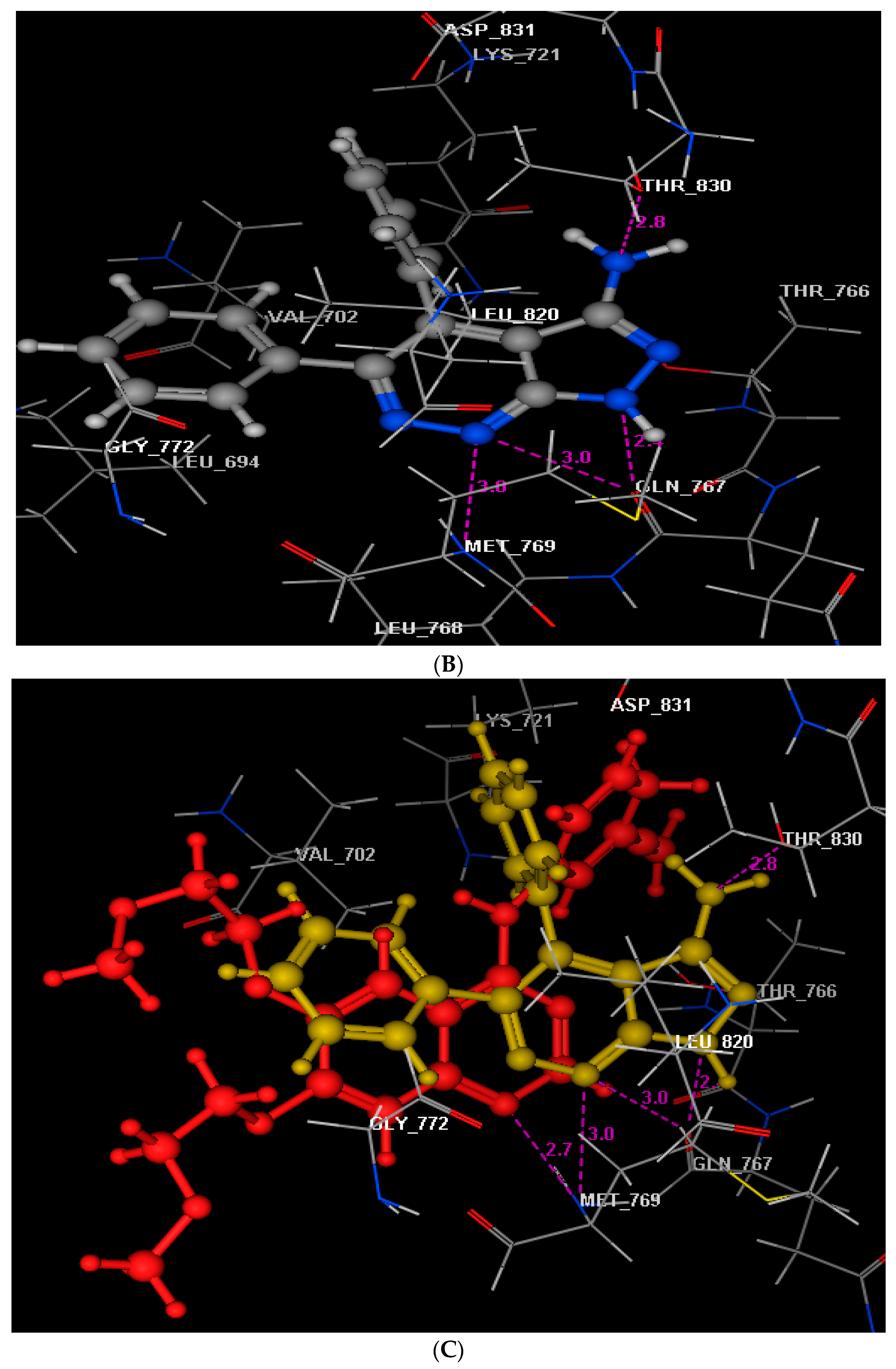
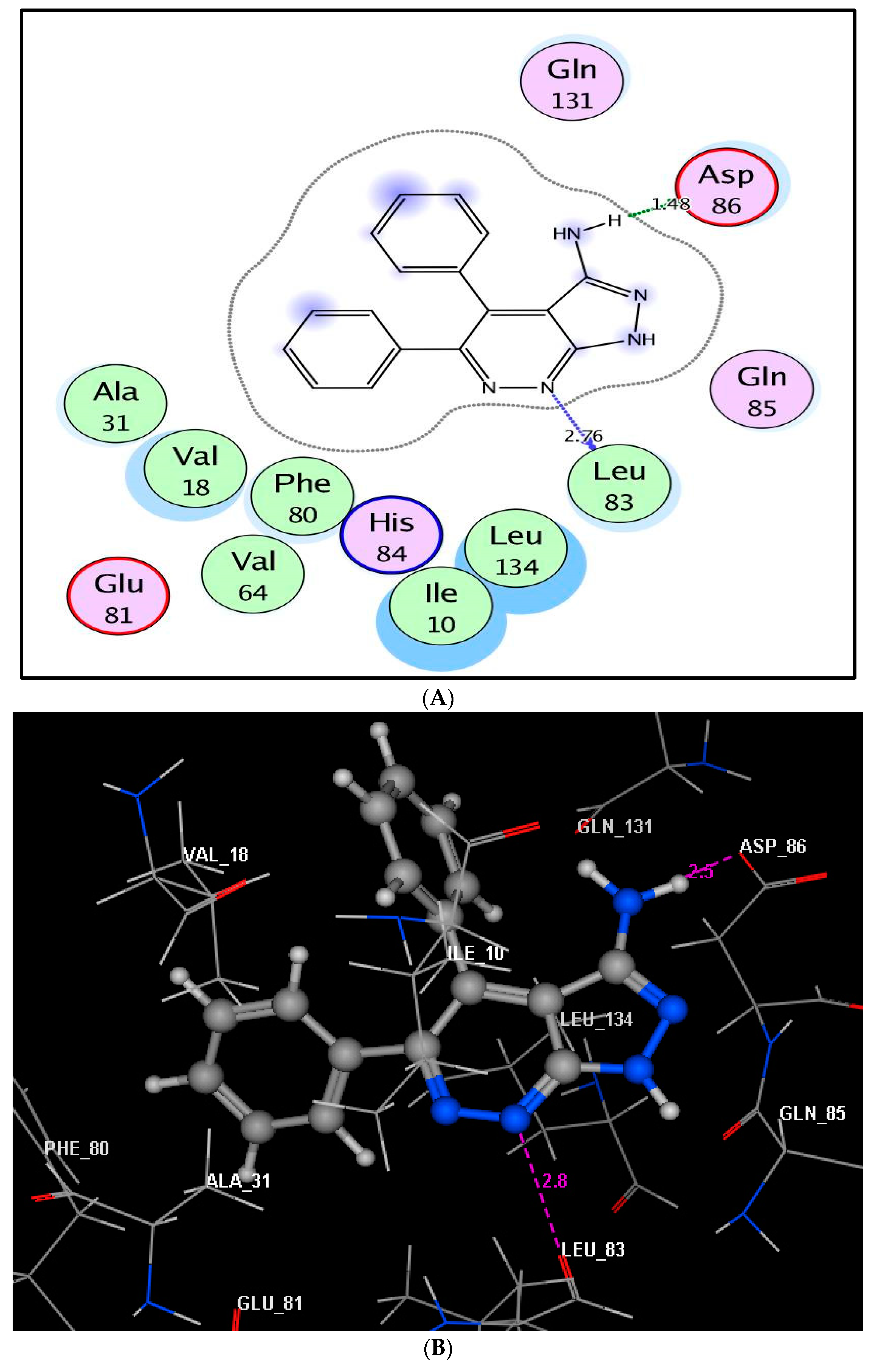
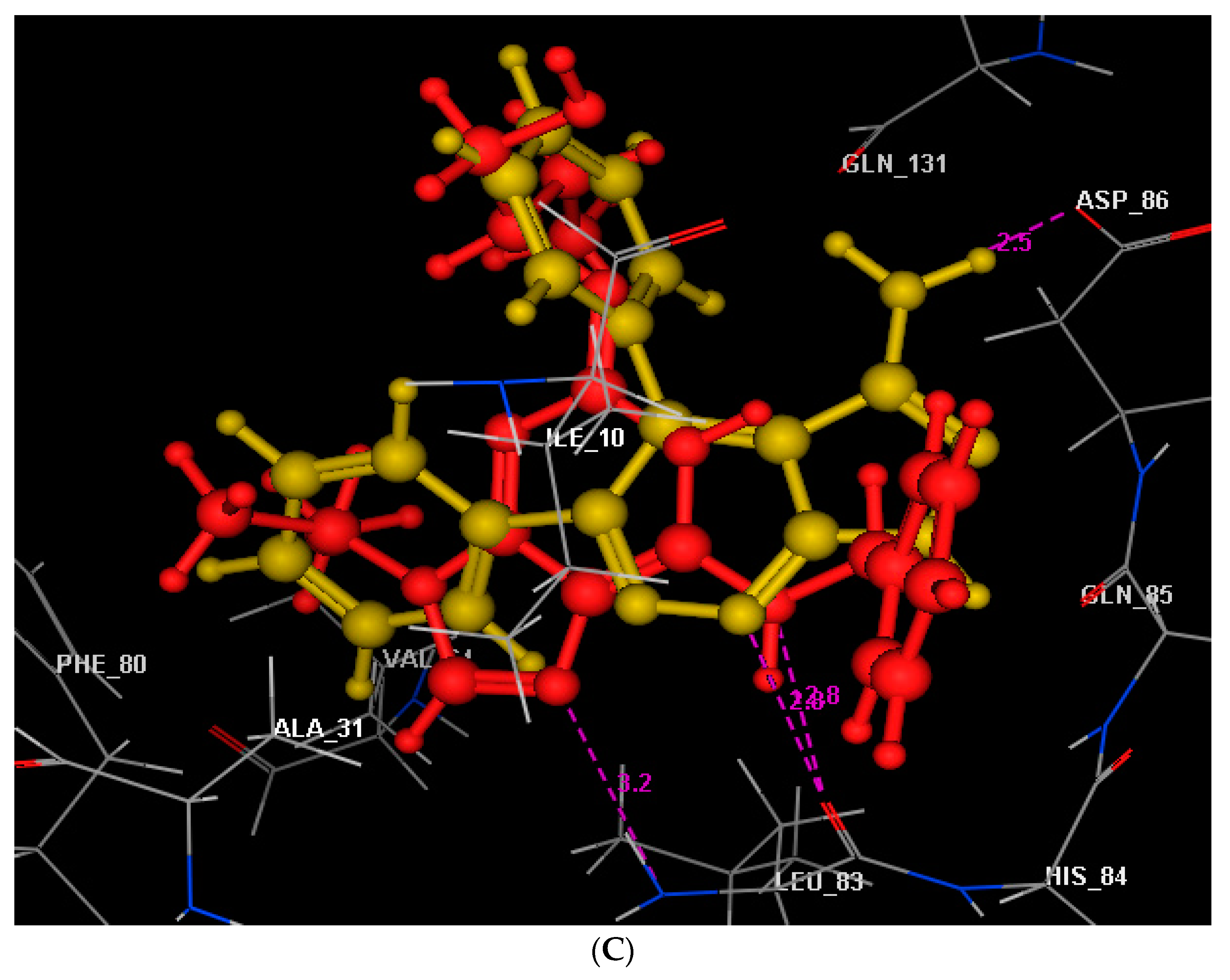
2.5.2. Molecular Docking
3. Materials and Methods
3.1. Synthesis
4,5-Diphenyl-1H-pyrazolo[3,4-c]pyridazin-3-amine (4)
3.2. Crystal Structure Determination
3.3. Development of Target 4 into Nano-Formulations
3.3.1. Formulation of Compound 4 into Solid-Lipid Nanoparticles (4-SLNs) and Lipid–Polymer Hybrid Nanoparticles (4-LPHNPs)
3.3.2. Characterization of Developed Nano-Formulations (Particle Size and Zeta Potential)
3.4. Biological Evaluation
3.4.1. In Vitro Cytotoxic Screening
3.4.2. In Vitro EGFR and CDK-2 Inhibitory Assessment
3.4.3. Effect of Target 4 and Its Nanoparticles 4-SLNs and 4-LPHNPs upon Levels of Bax, Bcl-2, Caspase-3 and p53
3.5. Molecular Docking Study
4. Conclusions
Supplementary Materials
Author Contributions
Funding
Institutional Review Board Statement
Informed Consent Statement
Data Availability Statement
Acknowledgments
Conflicts of Interest
References
- Hull, L.C.; Farrell, D.; Grodzinski, P. Highlights of recent developments and trends in cancer nanotechnology research—View from NCI Alliance for Nanotechnology in Cancer. Biotechnol. Adv. 2014, 32, 666–678. [Google Scholar]
- Zamboni, W.C.; Torchilin, V.; Patri, A.K.; Hrkach, J.; Stern, S.; Lee, R.; Nel, A.; Panaro, N.J.; Grodzinski, P. Best practices in cancer nanotechnology: Perspective from NCI nanotechnology alliance. Clin. Cancer Res. 2012, 18, 3229–3241. [Google Scholar] [CrossRef]
- Hare, J.I.; Lammers, T.; Ashford, M.B.; Puri, S.; Storm, G.; Barry, S.T. Challenges and strategies in anti-cancer nanomedicine development: An industry perspective. Adv. Drug Deliv. Rev. 2017, 108, 25–38. [Google Scholar]
- Salata, O.V. Applications of nanoparticles in biology and medicine. J. Nano Biotechnol. 2004, 2, 1–6. [Google Scholar]
- Dyawanapelly, S.; Mehrotra, P.; Ghosh, G.; Jagtap, D.D.; Dandekar, P.; Jain, R. How the surface functionalized nanoparticles affect conformation and activity of proteins: Exploring through protein-nanoparticle interactions. Bioorg. Chem. 2019, 82, 17–25. [Google Scholar] [CrossRef]
- Saraiva, C.; Praça, C.; Ferreira, R.; Santos, T.; Ferreira, L.; Bernardino, L. Nanoparticle-mediated brain drugdelivery: Overcoming blood–brain barrier to treat neurodegenerative diseases. J. Control. Release 2016, 235, 34–47. [Google Scholar] [CrossRef]
- Popovic, Z.; Liu, W.; Chauhan, V.P.; Lee, J.; Wong, C.; Greytak, A.B.; Insin, N.; Nocera, D.G.; Fukumura, D.; Jain, R.K.; et al. A nanoparticle size series for in vivo fluorescence imaging. Angew. Chem. Int. Ed. Eng. 2010, 122, 8831–8834. [Google Scholar] [CrossRef]
- Cabral, H.; Matsumoto, Y.; Mizuno, K.; Chen, Q.; Murakami, M.; Kimura, M.; Terada, Y.; Kano, M.R.; Miyazono, K.; Uesaka, M.; et al. Accumulation of sub-100 nm polymeric micelles in poorly permeable tumors depends on size. Nat. Nanotechnol. 2011, 6, 815–823. [Google Scholar] [CrossRef]
- Wang, J.; Mao, W.; Lock, L.L.; Tang, J.; Sui, M.; Sun, W.; Cui, H.; Xu, D.; Shen, Y. The role of micelle size in tumor accumulation, penetration, and treatment. ACS Nano 2015, 9, 7195–7206. [Google Scholar] [CrossRef]
- Thakur, S.; Pramod, K.S.; Malviya, R. Utilization of Polymeric Nanoparticle in Cancer Treatment: A Review. J. Pharm. Care Health Sys. 2017, 4, 2. [Google Scholar]
- Nagaraju, B.; Kovvuri, J.; Kumar, C.G.; Routhu, S.R.; Shareef, M.A.; Kadagathur, M.; Adiyala, P.R.; Alavala, S.; Nagesh, N.; Kamal, A. Synthesis and biological evaluation of pyrazole linked benzothiazole-β-naphthol derivatives as topoisomerase I inhibitors with DNA binding ability. Bioorg. Med. Chem. 2019, 27, 708–720. [Google Scholar] [CrossRef]
- Asif, M. The biological potentials of substituted 1,2-diazines: A review on versatile pyridazine derivatives. J. Chin. Pharm. Sci. 2016, 25, 707–725. [Google Scholar]
- Nossier, E.S.; Abd El-Karim, S.S.; Khalifa, N.M.; El-Sayed, A.S.; Hassan, E.S.; El-Hallouty, S.M. Kinase inhibitory activities and molecular docking of a novel series of anticancer pyrazole derivatives. Molecules 2018, 23, 3074. [Google Scholar] [CrossRef]
- Hassan, A.S.; Moustafa, G.O.; Awad, H.M.; Nossier, E.S.; Mady, M.F. Design, synthesis, anticancer evaluation, enzymatic assays, and a molecular modeling study of novel pyrazole–indole hybrids. ACS Omega 2021, 6, 12361–12374. [Google Scholar] [CrossRef]
- Dawood, D.H.; Nossier, E.S.; Ali, M.M.; Mahmoud, A.E. Synthesis and molecular docking study of new pyrazole derivatives as potent anti-breast cancer agents targeting VEGFR-2 kinase. Bioorg. Chem. 2020, 101, 103916. [Google Scholar] [CrossRef]
- Ahmed, M.F.; Santali, E.Y.; El-Deen, E.M.M.; Naguib, I.A.; El-Haggar, R. Development of pyridazine derivatives as potential EGFR inhibitors and apoptosis inducers: Design, synthesis, anticancer evaluation, and molecular modeling studies. Bioorg. Chem. 2021, 106, 104473. [Google Scholar] [CrossRef]
- Djaballah, H.; Varmus, H.E.; Shum, D.; Somwar, R.; Chucholowski, A.; Thiruvazhi, M.S. Substituted Pyridazines as EGFR and/or KRAS Inhibitors. U.S. Patent US9562019B2, 7 February 2017. [Google Scholar]
- Dragovich, T.; Laheru, D.; Dayyani, F.; Bolejack, V.; Smith, L.; Seng, J.; Burris, H.; Rosen, P.; Hidalgo, M.; Ritch, P.; et al. Phase II trial of vatalanib in patients with advanced or metastatic pancreatic adenocarcinoma after first-line gemcitabine therapy (PCRT O4–001). Cancer Chemother. Pharmacol. 2014, 74, 379–387. [Google Scholar]
- Joensuu, H.; De Braud, F.; Grignagni, G.; De Pas, T.; Spitalieri, G.; Coco, P.; Spreafico, C.; Boselli, S.; Toffalorio, F.; Bono, P.; et al. Vatalanib for metastatic gastrointestinal stromal tumour (GIST) resistantto imatinib: Final results of a phase II study. Br. J. Cancer 2011, 104, 1686–1690. [Google Scholar] [CrossRef]
- Brana, M.F.; Cacho, M.; García, M.L.; Mayoral, E.P.; López, B.; de Pascual-Teresa, B.; Ramos, A.; Acero, N.; Llinares, F.; Munoz-Mingarro, D.; et al. Pyrazolo [3, 4-c] pyridazines as novel and selective inhibitors of cyclin-dependent kinases. J. Med. Chem. 2005, 48, 6843–6854. [Google Scholar] [CrossRef]
- Khalifa, F.A. Synthesis and reactions of some pyridazine derivatives. Arch. Pharm. Res. 1990, 13, 198–200. [Google Scholar] [CrossRef]
- Witherington, J.; Bordas, V.; Garland, S.L.; Hickey, D.M.B.; Ife, R.J.; Liddle, J.; Saunders, M.; Smith, D.G.; Ward, R.W. 5-Aryl-pyrazolo [3,4-b]pyridines: Potent Inhibitors of Glycogen Synthase Kinase-3 (GSK-3). Bioorg. Med. Chem. Lett. 2003, 13, 1577–1580. [Google Scholar] [CrossRef]
- Venishetty, V.K.; Chede, R.; Komuravelli, R.; Adepu, L.; Sistla, R.; Diwan, P.V. Design and evaluation of polymer coated carvedilol loaded solid lipid nanoparticles to improve the oral bioavailability: A novel strategy to avoid intraduodenal administration. Colloids Surf. B 2012, 95, 1–9. [Google Scholar] [CrossRef]
- Priya, M.R.K.; Iyer, P.R. Antiproliferative effects on tumor cells of the synthesized gold nanoparticles against Hep2 liver cancer cell line. Egypt. Liver J. 2020, 10, 1–12. [Google Scholar] [CrossRef]
- Shawky, S.M.; Khalifa, M.K.; Eassa, H.A. Lornoxicam-loaded nanosponges for controlled anti-inflammatory effect: In vitro/in vivo assessment. Int. J. Appl. Pharm. 2020, 12, 217–223. [Google Scholar] [CrossRef]
- Zhang, W.; Li, X.; Ye, T.; Chen, F.; Yu, S.; Chen, J.; Yang, X.; Yang, N.; Zhang, J.; Liu, J.; et al. Nanostructured lipid carrier surface modified with Eudragit RS 100 and its potential ophthalmic functions. Int. J. Nanomed. 2014, 9, 4305. [Google Scholar]
- Mosmann, T. Rapid colorimetric assays for cellular growth and survival: Application to proliferation and cytotoxicity assays. J. Immunol. Methods 1983, 65, 55–63. [Google Scholar] [CrossRef]
- El-Sawy, E.R.; Mandour, A.H.; El-Hallouty, S.M.; Shaker, K.H.; Abo-Salem, H.M. Synthesis, antimicrobial and anticancer activities of some new N-methylsulphonyl and N-benzenesulphonyl-3-indolyl hetero-cycles: 1st Cancer Update. Arab. J. Chem. 2013, 6, 67–78. [Google Scholar] [CrossRef]
- Srour, A.M.; Ahmed, N.S.; Abd El-Karim, S.S.; Anwar, M.M.; El-Hallouty, S.M. Design, synthesis, biological evaluation, QSAR analysis and molecular modelling of new thiazol-benzimidazoles as EGFR inhibitors. Bioorg. Med. Chem. 2020, 28, 115657. [Google Scholar]
- Abd El-Meguid, E.A.; El-Deen, E.M.M.; Nael, M.A.; Anwar, M.M. Novel benzimidazole derivatives as anti-cervical cancer agents of potential multi-targeting kinase inhibitory activity. Arab. J. Chem. 2020, 13, 9179–9195. [Google Scholar] [CrossRef]
- Fathalla, O.A.E.F.M.; Ismail, M.A.; Anwar, M.M.; Abouzid, K.A.; Ramadan, A.A. Novel 2-thiopyrimidine derivatives as CDK2 inhibitors: Molecular modeling, synthesis, and anti-tumor activity evaluation. Med. Chem. Res. 2013, 22, 659–673. [Google Scholar] [CrossRef]
- El-Sayed, W.A.; Alminderej, F.M.; Mounier, M.M.; Nossier, E.S.; Saleh, S.M.; Kassem, A.F. Novel 1,2,3-triazole-coumarin hybrid glycosides and their tetrazolyl analogues: Design, anticancer evaluation and molecular docking targeting EGFR, VEGFR-2 and CDK-2. Molecules 2022, 27, 2047. [Google Scholar] [CrossRef]
- El-Deen, E.M.M.; Anwar, M.M.; Abd El-Gwaad, A.A.; Karam, E.A.; El-Ashrey, M.K.; Kassab, R.R. Design and synthesis of some novel pyridothienopyrimidine derivatives and their biological evaluation as antimicrobial and anticancer agents targeting EGFR enzyme. Arab. J. Chem. 2022, 15, 103751. [Google Scholar] [CrossRef]
- Moustafa, G.O.; Shalaby, A.; Naglah, A.M.; Mounier, M.M.; El-Sayed, H.; Anwar, M.M.; Nossier, E.S. Synthesis, characterization, in vitro anticancer potentiality, and antimicrobial activities of novel peptide–glycyrrhetinic-acid-based derivatives. Molecules 2021, 26, 4573. [Google Scholar] [CrossRef]
- Hamdy, N.A.; El Sayed, M.T.; Hussein, H.A.; Mounier, M.M.; Anwar, M.M. Synthesis of novel heterocyclic compounds bearing tetralin moiety of potential anticancer activity targeting the intrinsic apoptotic pathway. Synth. Commun. 2023, 53, 298–315. [Google Scholar] [CrossRef]
- Othman, I.M.; Gad-Elkareem, M.A.; Snoussi, M.; Aouadi, K.; Kadri, A. Novel fused pyridine derivatives containing pyrimidine moiety as prospective tyrosyl-tRNA synthetase inhibitors: Design, synthesis, pharmacokinetics and molecular docking studies. J. Mol. Struct. 2020, 1219, 128651. [Google Scholar] [CrossRef]
- Mohi El-Deen, E.M.; Nossier, E.S.; Karam, E.A. New Quinazolin-4(3H)-one Derivatives Incorporating Hydrazone and Pyrazole Scaffolds as Antimicrobial Agents Targeting DNA Gyrase Enzyme. Sci. Pharm. 2022, 90, 52. [Google Scholar] [CrossRef]
- Valasani, K.R.; Vangavaragu, J.R.; Day, V.W.; Yan, S.S. Structure based design, synthesis, pharmacophore modeling, virtual screening, and molecular docking studies for identification of novel cyclophilin D inhibitors. J. Chem. Inf. Model. 2014, 54, 902–912. [Google Scholar] [CrossRef]
- Al-Resayes, S.I.; Laria, F.Y.; Miloud, M.M.; El-ajaily, M.M.; El-Barasi, N.M.; Sarangi, A.K.; Verma, S.; Azam, M.; Seidel, V.; Mohapatra, R.K. Synthesis, characterization, biological applications, and molecular docking studies of amino-phenol-derived mixed-ligand complexes with Fe (III), Cr (III), and La (III) ions. J. Saudi Chem. Soc. 2023, 27, 101622. [Google Scholar] [CrossRef]
- Ahmed, A.; Saeed, A.; Ejaz, S.A.; Aziz, M.; Hashmi, M.Z.; Channar, P.A.; Abbas, Q.; Raza, H.; Shafiq, Z.; El-Seedi, H.R. Novel adamantyl clubbed iminothiazolidinones as promising elastase inhibitors: Design, synthesis, molecular docking, ADMET and DFT studies. RSC Adv. 2022, 12, 11974–11991. [Google Scholar] [CrossRef]
- Abd El-Meguid, E.A.; Naglah, A.M.; Moustafa, G.O.; Awad, H.M.; El Kerdawy, A.M. Novel benzothiazole-based dual VEGFR-2/EGFR inhibitors targeting breast and liver cancers: Synthesis, cytotoxic activity, QSAR and molecular docking studies. Bioorg. Med. Chem. Lett. 2022, 58, 128529. [Google Scholar] [CrossRef]
- Sheldrick, G.M. Crystal structure refinement with SHELXL. Acta Crystallogr. C Struct. Chem. 2015, 71, 3–8. [Google Scholar] [CrossRef]
- Sheldrick, G.M. SHELXT—Integrated space-group and crystal-structure determination. Acta Crystallogr. A Found. Adv. 2015, 71, 3–8. [Google Scholar] [CrossRef]
- Dave, V.; Yadav, R.B.; Kushwaha, K.; Yadav, S.; Sharma, S.; Agrawal, U. Lipid-polymer hybrid nanoparticles: Development & statistical optimization of norfloxacin for topical drug delivery system. Bioact. Mater. 2017, 2, 269–280. [Google Scholar]
- Saleh, A.; Khalifa, M.; Shawky, S.; Bani-Ali, A.; Eassa, H. Zolmitriptan intranasal spanlastics for enhanced migraine treatment; formulation parameters optimized via quality by design approach. Sci. Pharm. 2021, 89, 24. [Google Scholar] [CrossRef]
- Khalifa, M.K.; Salem, H.A.; Shawky, S.M.; Eassa, H.A.; Elaidy, A.M. Enhancement of zaleplon oral bioavailability using optimized self-nano emulsifying drug delivery systems and its effect on sleep quality among a sample of psychiatric patients. Drug Deliv. 2019, 26, 1243–1253. [Google Scholar] [CrossRef]


| Compound No. | IC50 (Mean ± SEM) (μM) | ||
|---|---|---|---|
| HepG-2 | HCT-116 | MCF-7 | |
| 4 | 17.30 ± 1.42 | 18.38 ± 1.4 | 27.29 ± 2.1 |
| 4-SLNs | 7.56 ± 0.58 | 4.80 ± 0.3 | 6.41 ± 0.4 |
| 4-LPHNPs | 7.85 ± 0.5 | 5.24 ± 0.4 | 6.65 ± 0.5 |
| Doxorubicin | 6.18 ± 0.3 | 5.23 ± 0.3 | 4.17 ± 0.2 |
| Compound No. | IC50 (Mean ± SEM) (µM) | |
|---|---|---|
| EGFR | CDK-2/Cyclin A2 | |
| Erlotinib | 0.126 ± 0.10 | - |
| Roscovitine | - | 0.32 ± 0.05 |
| 4 | 0.391 ± 0.17 | 0.55 ± 0.20 |
| 4-SLNs | 0.088 ± 0.22 | 0.18± 0.15 |
| 4-LPHNPs | 0.096 ± 0.50 | 0.22± 0.04 |
| Compd. | Bax | Bcl-2 | P53 | Casp-3 |
|---|---|---|---|---|
| Conc. Pg/mL | Conc. ng/mL | Conc. Pg/mL | Conc. Pg/mL | |
| 4/MCF-7 | 166.20 ± 0.04 | 2.40 ± 0.30 | 590.22 ± 0.60 | 14.25 ± 0.20 |
| 4-SLNs/MCF-7 | 225.40 ± 0.01 | 1.55 ± 0.20 | 748.10 ± 0.14 | 20.73 ± 0.11 |
| 4-LPHNPs/MCF-7 | 213.50 ± 0.12 | 1.68 ± 0.15 | 735.50 ± 0.22 | 19.62 ± 0.35 |
| Cont./MCF-7 | 40.60 ± 0.05 | 5.30 ± 0.10 | 163.41 ± 0.22 | 1.85 ± 0.35 |
| Compd. | MW a | TPSA (Ų) b | nRB c | nHBA d | nHBD e | MLogP f | Violations g |
|---|---|---|---|---|---|---|---|
| 4 | 287.32 | 80.48 | 2 | 3 | 2 | 3.07 | 0 |
Disclaimer/Publisher’s Note: The statements, opinions and data contained in all publications are solely those of the individual author(s) and contributor(s) and not of MDPI and/or the editor(s). MDPI and/or the editor(s) disclaim responsibility for any injury to people or property resulting from any ideas, methods, instructions or products referred to in the content. |
© 2023 by the authors. Licensee MDPI, Basel, Switzerland. This article is an open access article distributed under the terms and conditions of the Creative Commons Attribution (CC BY) license (https://creativecommons.org/licenses/by/4.0/).
Share and Cite
Hashem, H.E.; Amr, A.E.-G.E.; Almehizia, A.A.; Naglah, A.M.; Kariuki, B.M.; Eassa, H.A.; Nossier, E.S. Nanoparticles of a Pyrazolo-Pyridazine Derivative as Potential EGFR and CDK-2 Inhibitors: Design, Structure Determination, Anticancer Evaluation and In Silico Studies. Molecules 2023, 28, 7252. https://doi.org/10.3390/molecules28217252
Hashem HE, Amr AE-GE, Almehizia AA, Naglah AM, Kariuki BM, Eassa HA, Nossier ES. Nanoparticles of a Pyrazolo-Pyridazine Derivative as Potential EGFR and CDK-2 Inhibitors: Design, Structure Determination, Anticancer Evaluation and In Silico Studies. Molecules. 2023; 28(21):7252. https://doi.org/10.3390/molecules28217252
Chicago/Turabian StyleHashem, Heba E., Abd El-Galil E. Amr, Abdulrahman A. Almehizia, Ahmed M. Naglah, Benson M. Kariuki, Heba A. Eassa, and Eman S. Nossier. 2023. "Nanoparticles of a Pyrazolo-Pyridazine Derivative as Potential EGFR and CDK-2 Inhibitors: Design, Structure Determination, Anticancer Evaluation and In Silico Studies" Molecules 28, no. 21: 7252. https://doi.org/10.3390/molecules28217252
APA StyleHashem, H. E., Amr, A. E.-G. E., Almehizia, A. A., Naglah, A. M., Kariuki, B. M., Eassa, H. A., & Nossier, E. S. (2023). Nanoparticles of a Pyrazolo-Pyridazine Derivative as Potential EGFR and CDK-2 Inhibitors: Design, Structure Determination, Anticancer Evaluation and In Silico Studies. Molecules, 28(21), 7252. https://doi.org/10.3390/molecules28217252