Article

A Novel and Efficient Phthalate Hydrolase from Acinetobacter sp. LUNF3: Molecular Cloning, Characterization and Catalytic Mechanism

1
College of Life Science, Langfang Normal University, Langfang 065000, China
2
Institute of Agricultural Resources and Regional Planning, Chinese Academy of Agricultural Sciences, Beijing 100081, China
3
Technical Innovation Center for Utilization of Edible and Medicinal Fungi in Hebei Province, Langfang 065000, China
4
School of Chemistry and Materials Science, Langfang Normal University, Langfang 065000, China
5
Biotechnology Research Institute, Chinese Academy of Agricultural Sciences, Beijing 100081, China
6
National Nanfan Research Institute (Sanya), Chinese Academy of Agricultural Sciences, Sanya 572024, China
*
Authors to whom correspondence should be addressed.
Molecules 2023, 28(18), 6738; https://doi.org/10.3390/molecules28186738
Submission received: 8 August 2023 / Revised: 14 September 2023 / Accepted: 17 September 2023 / Published: 21 September 2023
(This article belongs to the Special Issue Biomolecules Interactions with Small Molecules)

Abstract

:
Phthalic acid esters (PAEs), which are widespread environmental contaminants, can be efficiently biodegraded, mediated by enzymes such as hydrolases. Despite great advances in the characterization of PAE hydrolases, which are the most important enzymes in the process of PAE degradation, their molecular catalytic mechanism has rarely been systematically investigated. Acinetobacter sp. LUNF3, which was isolated from contaminated soil in this study, demonstrated excellent PAE degradation at 30 °C and pH 5.0–11.0. After sequencing and annotating the complete genome, the gene dphAN1, encoding a novel putative PAE hydrolase, was identified with the conserved motifs catalytic triad (Ser201-Asp295-His325) and oxyanion hole (H127GGG130). DphAN1 can hydrolyze DEP (diethyl phthalate), DBP (dibutyl phthalate) and BBP (benzyl butyl phthalate). The high activity of DphAN1 was observed under a wide range of temperature (10–40 °C) and pH (6.0–9.0). Moreover, the metal ions (Fe2+, Mn2+, Cr2+ and Fe3+) and surfactant TritonX-100 significantly activated DphAN1, indicating a high adaptability and tolerance of DphAN1 to these chemicals. Molecular docking revealed the catalytic triad, oxyanion hole and other residues involved in binding DBP. The mutation of these residues reduced the activity of DphAN1, confirming their interaction with DBP. These results shed light on the catalytic mechanism of DphAN1 and may contribute to protein structural modification to improve catalytic efficiency in environment remediation.

Graphical Abstract

1. Introduction

Phthalic acid esters (PAEs) have been used as plasticizers since the 1920s, improving the flexibility and durability of plastic products [1,2]. However, PAEs are likely to be released into the environment from plastics due to their non-covalent interactions with plastic matrices [3]. Currently, the serious environmental contamination caused by PAEs is leading to increasing concern among researchers worldwide. Moreover, PAEs have similar properties to endocrine disruptors, carcinogens and mutagens [4], posing a potential threat to humans and ecological environments [5,6]. Given the environmental risk, six kinds of PAEs have been listed as priority environmental pollutants: dimethyl phthalate (DMP), diethyl phthalate (DEP), dibutyl phthalate (DBP), benzyl butyl phthalate (BBP), dioctyl phthalate (DOP) and di(2-ethylhexyl) phthalate (DEHP). In view of the serious pollution and toxicological effects of PAEs, an efficient strategy for remediating PAE-contaminated environments must be urgently developed.
Due to their high hydrophobicity, it is difficult for PAEs to be naturally degraded via hydrolysis and photolysis [7,8]. Biodegradation via microorganisms has become the main approach to eliminate PAEs from environments with the advantages of high efficiency and environmental safety. Numerous PAE-degrading bacteria have been isolated from environmental matrices and mainly belong to the genera Sphingomonas [9], Gordonia [10], Rhodococcus [11], Arthrobacter [12], Bacillus [13] and Pseudomonas [14]. These bacteria degrade PAEs mainly via aerobic metabolism, and the degradation characteristics of multiple strains have been revealed. Rhodococcus pyridinivorans XB isolated from activated sludge has a high degradation efficiency towards PAEs, removing 98% of DMP, DEP and DBP within 5 days [11]. In some cases, some functional strains work together to completely degrade PAEs [15]. Some strains or consortiums have also been applied to PAE remediation in various environments. Gordonia sp. QH-11 and Gordonia sp. Lff can efficiently remove PAEs from soil and alter the composition of the bacterial community in soil [10,16].
Traditionally, the initial hydrolysis of PAEs is considered the most important step during the whole degradation process, and causal hydrolases are considered the key enzymes during the metabolism of PAEs via bacteria. The dialkyl PAE hydrolase is responsible for the hydrolysis of the first ester bond of PAEs [17] (Figure 1). The resulting monoalkyl PAEs, with only one ester bond, are transformed into phthalic acid (PA) by monoalkyl PAE hydrolase [18]. Occasionally, the same hydrolase is able to function in the two steps of hydrolysis, such as EstG, GTW28_17760 and EstM2 from Sphingobium sp. SM42, Bacillus subtilis BJQ0005 and a soil metagenomic library, respectively [19,20,21]. Some dialkyl PAE hydrolases have been reported, but their gene sequences remain unidentified. The hydrolase purified from the culture broth of Nocardia erythropolis not only hydrolyzes several kinds of PAEs, including DBP, DEHP, DEP, DOP, etc., but also hydrolyzes dimethyl isophthalate (DMIP), dimethyl terephthalate (DMTP) and diethyl terephthalate (DETP) [22]. The DMP hydrolases from the cell-free extract of Bacillus sp. are characterized, and their genes may be located in the plasmid [23]. Fortunately, metagenomic library, bacterial genomic library and genome sequencing provide effective approaches to identifying gene sequences of dialkyl PAE hydrolases such as Hyd [24], XtjR8 [25], HylD1 [26] and EstYZ5 [17]. These enzymes mainly belong to hydrolase families II, IV, V, VI, VII and VIII [20,21,26] (Table 1). Among them, the hydrolases from family IV, also known as the hormone-sensitive lipase (HSL) family, predominate. These hydrolases from the HSL family possess conserved motifs, such as oxygen hole (HGGG), catalytic triad (Ser-Asp/Glu-His) and GDSAG containing the catalytic Ser [26]. However, the relevant studies of PAE hydrolases mainly focus on gene cloning, function verification, catalytic characteristics and structure analysis. Although the catalytic mechanisms of dialkyl PAE hydrolase PS06828 and Hyd, from family VI and a new family, respectively, have been inferred from spectroscopic and docking analyses [24,27], only the catalytic triad of PS06828 (Ser113, Asp166 and His197) and key residues of Hyd (Thr190 and Ser191) are revealed. According to the results of molecular docking and enzyme assay, the interaction between MIBP and His399 affects the activity of GTW28_17760, a hydrolase from family VII capable of hydrolyzing DIBP and MIBP [20]. The catalytic mechanisms of some monoalkyl PAE hydrolases from family V are also elucidated [18,28]. Altogether, the catalytic mechanisms of PAE hydrolases from other families require to be further resolved.
This study aimed to explore PAE-degrading bacteria, characterize the degradation performance of the isolate, identify efficient PAE hydrolases and elucidate their molecular catalytic mechanisms. The complete genome of Acinetobacter sp. LUNF3, an effective PAE degrader, was sequenced and annotated. The novel dialkyl PAE hydrolase DphAN1 was identified via genomic analysis. After the heterologous expression and affinity purification of DphAN1, its catalytic characteristics were determined. DphAN1 is endowed with a high activity towards PAEs, particularly recalcitrant BBP, and has a good adaptability to some chemicals. The interaction between DphAN1 and DBP via molecular docking revealed active residues related to binding and hydrolyzing DBP. These active residues were validated by site-directed mutagenesis and an enzyme assay. The results provide insights into the molecular catalytic mechanism of DphAN1 and will support its structural modification, aiming for a higher catalytic efficiency.

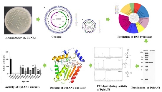
2. Results and Discussion

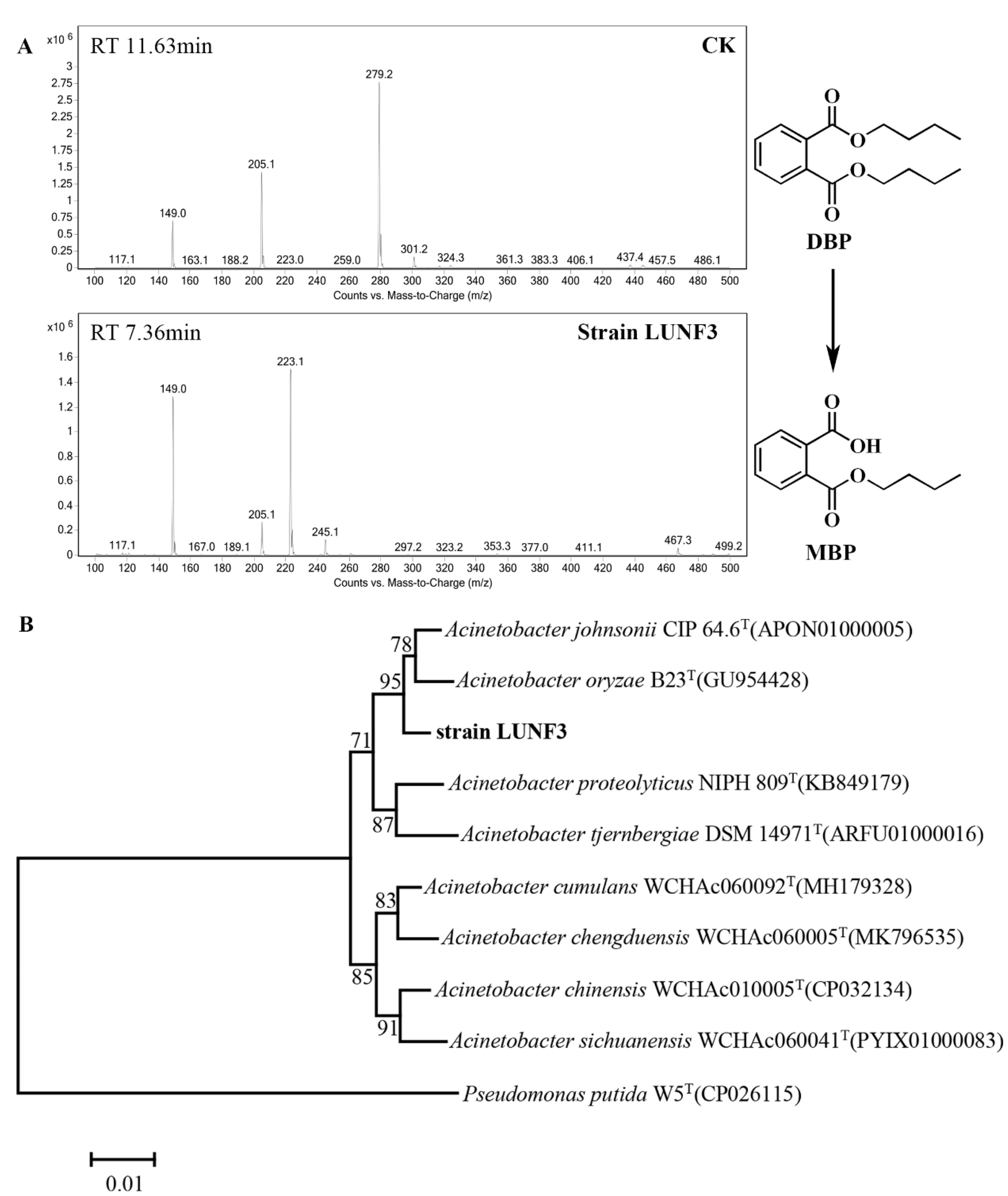
2.1. Isolation and Identification of Acinetobacter sp. LUNF3

After enrichment using DBP as a substrate, the DBP-degrading ability of some candidate strains was assessed by HPLC-MS. One strain, LUNF3, significantly removed DBP and produced MBP (Figure S1 and Figure 2A), indicating its efficiently hydrolyzing DBP. The colony of strain LUNF3 is light yellow with a circular and smooth surface. The 16S rRNA gene of strain LUNF3 shares the maximum sequence identity to that of Acinetobacter sp. based on BLAST against NCBI database (Table S1). According to the phylogenetic tree, strain LUNF3 is taxonomically close to strains Acinetobacter johnsonii CIP 64.6 and Acinetobacter oryzae B23 from EzBioCloud database (Figure 2B). Thus, the DBP-degrading strain here is identified as Acinetobacter sp. LUNF3. It was reported that some strains of the genus Acinetobacter were able to degrade PAEs [29,30,31]. Acinetobacter sp. LMB-5 isolated from soil can quickly remove DMP, DEP and DBP [30]. Acinetobacter sp. SN13 is capable of degrading DEHP [31]. The isolation of Acinetobacter sp. LUNF3 enriches the resources of PAE-degrading bacteria. The genus Acinetobacter can also eliminate other pollutants, such as sulfamethoxazole [32], perfluorooctane sulfonamide [33], crude oil [34] and phenol [35]. Therefore, they have a potential application in the remediation of contaminated environments.

2.2. Characterization of PAE Degradation by Strain LUNF3

Environmental parameters, such as temperature and pH, affect PAE degradation due to their influence on the enzymatic activity of microorganisms. Strain LUNF3 exhibits the highest degradation efficiency at 30 °C, with the complete elimination of DBP (Figure 3A). Approximately 30% of DBP is degraded even at 10 °C, indicating the relatively high activity of LUNF3 towards DBP at low temperature. In addition, strain LUNF3 can effectively remove DBP at a wide range of pH values. The degradation percentage of DBP greatly increases with an increase in pH, rising from 4.0 to 9.0, and is maintained at 100% until pH reached 11.0 (Figure 3B). Strain LUNF3 eliminates around 70% of DBP at pH 5.0 and 6.0. Generally, microorganisms cannot effectively degrade PAEs at a pH too low or too high. Enterobacter sp. DNB-S2 completely degrades DBP at pH 7.0–10.0 but only removes 4.4% and 10.4% of DBP at pH 5.0 and 6.0, respectively [36]. Rhodococcus pyridinivorans DNHP-S2 degrades more than 40% of DEHP at pH 5.0 [37]. The natural environmental conditions are complex and diverse. The high degradation efficiency of strain LUNF3 indicates its promising practical application in environmental remediation across a broad pH range, especially in neutral or alkaline environments (Figure 3A,B).
In a substrate profile test, strain LUNF3 mediates degradation of more than 70% of DEP, as well as 100% of DBP and BBP (Figure S2A). The degradative activity of strain LUNF3 against PAEs varies with their different side chains. Particularly, strain LUNF3 effectively degrades BBP despite the potential steric hindrance by its bulky side chain [38,39]. Based on the HPLC-MS profiles of DEP and BBP, which were degraded by strain LUNF3 (Figure S2B and Figure 3C), the products of MEP, MBP and MBeP were identified based on retention time and molecular ions. The results indicate that strain LUNF3 degrades these kinds of PAEs via hydrolysis. Generally, hydrolysis is a key step for PAE degradation and detoxification [40]. The bacterial degradation of PAEs mainly begins with hydrolysis, such as Burkholderia pyrrocinia B1213 [41], Achromobacter sp. RX [42], Rhodococcus pyridinovorans DNHP-S2 [37] and Pseudarthrobacter defluvii E5 [43]. Only a few bacteria adopt β-oxidation to initiate PAE degradation [15,44].

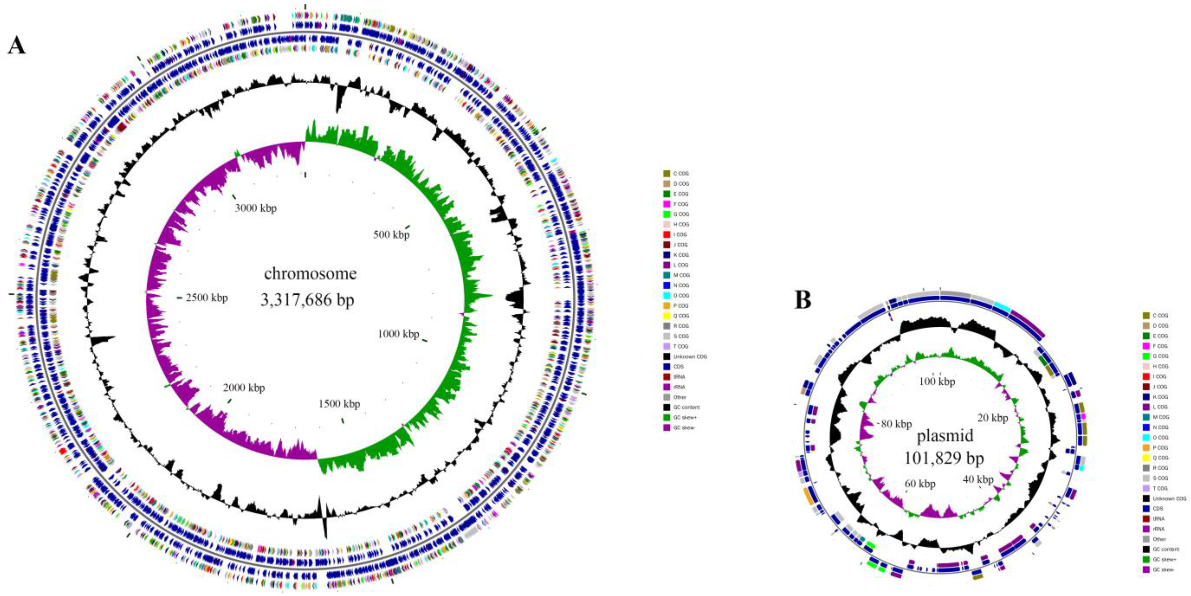
2.3. Cloning of PAE Hydrolase Gene through Complete Genome Sequencing

Strain LUNF3 underwent complete genome sequencing in order to explore the candidate genes involved in PAE degradation. PacBio sequencing produced 281,192 reads with 2,095,890,875 bp (Table S2), which were applied to genome assembly. Illumina sequencing generated high-quality data of 4,840,844 reads with 1,188,882,811 bp, which were used to correct the assembled genome. The obtained complete genome comprises a chromosome with 3,317,686 bp (a GC content of 41.43%) and a circular plasmid with 101,829 bp (a GC content of 38.14%) (Table S2, Figure 4). The genome size and GC content of strain LUNF3 are similar to those of Acinetobacter johnsonii LXL_C1 and Acinetobacter sp. TTH0–4 [45,46]. Acinetobacter sp. TTH0-4 can degrade crude oil and has a chromosome of 2,962,453 bp and 38.74% GC. The cyprodinil degrader Acinetobacter johnsonii LXL_C1 harbors a chromosome of 3,398,706 bp with 41.2% GC content. Based on comparative genomic analysis, the average nucleotide identity (ANI) values of strain LUNF3 against Acinetobacter johnsonii LXL_C1 (GenBank No. CP031011), Acinetobacter johnsonii M19 (GenBank No. CP037424) and Acinetobacter johnsonii FDAARGOS_1093 (GenBank No. CP068195) were 97.75%, 96.17% and 95.85%, respectively. Therefore, strain LUNF3 is genetically close to Acinetobacter johnsonii and can be a member of this bacterial species.
Based on the genome annotation, the chromosome and plasmid of strain LUNF3 harbor 3177 and 98 protein-coding genes (CDSs), respectively (Table 1). Moreover, all seven copies of rRNA genes (5S, 16S, 23S), eighty-nine tRNA genes and one CRISPR structure are located in the chromosome. Among these CDSs, 3221, 2162 and 2270 genes are annotated in NR, Swiss-Prot and GO database (Table S2, Figure S3), respectively. There are 1607 genes distributing across 46 biological pathways in the KEGG database, with 65 genes related to biodegradation and metabolism of xenobiotics (Figure S3C). A total of 2759 genes are divided into 20 function categories according to annotations in the COG database (Figure S4), and 109 genes are involved in lipid transport and metabolism. PAE degradation by bacteria is mediated by metabolism enzymes such as hydrolases, which are involved in cleaving ester bonds of PAEs, the critical step during PAE degradation [1,47,48]. Among these 109 genes mentioned above, one gene encodes a hydrolase DphAN1, which shares 67.0% identity with reported PAE hydrolase from Acinetobacter sp. M673 [29]. The result implies putative PAE-hydrolyzing capability of DphAN1.
According to the phylogenetic relationship between DphAN1 and the hydrolases from families I-VIII, DphAN1 was classified into family IV, which included the reported PAE hydrolases EstS1(AEW03609.1) [49], DphB (AGY55960.1) [50], EstG (AJO67804.1) [19] and the hydrolase (AFK31309.1) from Acinetobacter sp. M673 [29] (Figure 5A). Thus far, the molecular cloning and enzymatic characterization of some bacterial PAE hydrolases, which belong to families II, IV, V, VI, VII and VIII, have been conducted [20,21,26]. Furthermore, after mining the transcriptome of Cylindrotheca closterium using known bacterial PAE hydrolases as probes, two putative DBP hydrolases, DBPH1 and DBPH2, were predicted from the diatom for the first time [51]. The interaction of DBPH1 and DBPH2 with DBP and their expression levels under DBP exposure highlight their potential PAE-hydrolyzing ability. The conserved motifs of DphAN1 were analyzed using multiple sequence alignment. The typical catalytic triad (Ser201-Asp295-His325) was detected in DphAN1 (Figure S5). EstJ6 and EstYZ5, the PAE hydrolases from family IV, possess Glu instead of Asp in the catalytic triad [17,48]. The conserved G199DSAGG204 is also present in DphAN1 and contains the Ser201 of the catalytic triad (Figure 5B). In DphB, a cold-active PAE hydrolase identified from the metagenomic library of biofilms, Glu replaces Asp of the GDSAGG motif [50]. The motif H127GGG130 is also conserved (Figure 5C) and may form an oxyanion hole to stabilize reaction intermediates during the catalyzation of PAEs via DphAN1. Other conserved motifs were also observed in DphAN1, including D295LLHDEG301 containing the catalytic Asp295, H325GF327 harboring the catalytic His325, and V154LSIDYPLAPE164 (Figure S6). The functions of these conserved residues are further confirmed in this study.

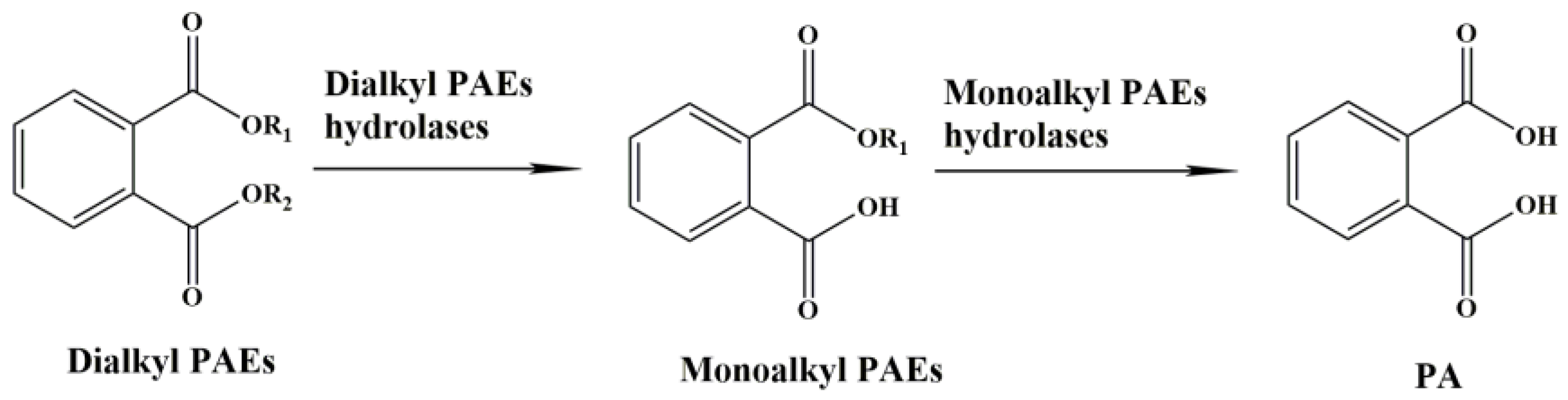
2.4. Functional Identification and Characterization of DphAN1

To identify the enzymatic activity of DphAN1 toward PAEs, the gene dphAN1 was expressed in ArcticExpress (DE3) cells. After purification via affinity chromatography, the recombinant DphAN1 with 6 × His tag was analyzed using SDS-PAGE. One single band was observed with a molecular weight of approximately 40 kDa (Figure 6A), in accordance with the theoretically calculated value of recombinant DphAN1 (41.46 kDa). The high PAE-hydrolyzing activity of recombinant DphAN1 was detected. After treatment with DphAN1, 51.34%, 77.02% and 58.60% of DEP, BBP and DBP were eliminated with corresponding enzymatic specific activity of 1.81, 2.72 and 2.07 U/mg protein (Figure 6B), respectively. DphAN1 catalyzed the hydrolysis of PAEs and generated the corresponding monoalkyl PAEs according to HPLC-MS (Figure 6C and Figure S7). Importantly, the hydrolysis of BBP generates MBP and MBeP, suggesting that DphAN1 is capable of cleaving either of the two esters of BBP (Figure S7B).
Environmental factors have a critical impact on enzymatic activity [52]. So, it is necessary to explore the optimal conditions for enzymatic catalysis. The enzyme assay of DphAN1 was investigated under various environmental conditions. The highest activity was observed at pH 8.0, and more than 57% of activity was maintained at pH 6.0–9.0 (Figure 7A). EstYZ5 displays less than 10% of the highest activity at pH 5.0 [17]. The activity of EstM2 is not reported at pH < 6.0 [21]. Therefore, DphAN1 has the advantage of high activity in slightly acid to alkaline environments. The high activity of DphAN1 remains between 20 °C and 40 °C, with the optimum activity at 30 °C (Figure 7B). Notably, DphAN1 displays 47.08% of maximal activity even at 10 °C. Some PAE hydrolases show relatively low activity at 10 °C in contrast to those at optimal temperatures, such as Est3563 and EstS1 [49,53]. The better environmental adaptability of DphAN1 will make possible its practical application in environments with a wide range of pH and temperature (Figure 7A,B).
The effects of chemicals on the activity of DphAN1 were also investigated. The metal ions at a concentration of 1 mM positively or negatively affect the activity of DphAN1 to different extents (Figure 7C). The addition of Mn2+, Cr2+, Fe2+ and Fe3+ causes an increase in the activity of DphAN1 by 18–89%. In a previous report, Fe3+ also enhances oxytetracycline degradation by Pseudomonas sp. T4 [54]. DphAN1 is not very sensitive to Ca2+, Mg2+ and Cd2+, indicating the relative stability of DphAN1 under these ions. However, Co2+, Ni2+, Zn2+ and Cu2+ reduce its activity by 20–40%, while Hg2+ inactivates DphAN1 completely. A significant decrease in activity was observed under 1% of Tween20 and Tween80, while the activity was lost when the concentration of Tween20 and Tween80 reached 5% (Figure 7D). These results demonstrate that the inhibitory effects were gradually enhanced with the increased concentration of Tween20 and Tween80. The strong denaturant SDS brings about a complete loss of activity, which may be caused by the destruction of DphAN1 by SDS. The surfactant Triton X-100 serves as an activator of DphAN1 and leads to an increase in activity by 49%. The Ser-specific inhibitor PMSF and His modifier DEPC cause complete inactivation of DphAN1. These findings suggest that the residues Ser and His are involved in catalysis, which is consistent with the components of the catalytic triad (Ser201-Asp295-His325). Phenyl methane sulfonyl fluoride (PMSF) and diethylpyrocarbonate (DEPC) possibly modify Ser201 and His325, respectively, and ultimately influence the hydrolytic activity. More than 60% of activity is maintained after the addition of β-mercaptoethanol (β-ME). Three Cys residues of DphAN1 may form disulfide bonds, which are disrupted under the action of β-ME, thus reducing the activity.

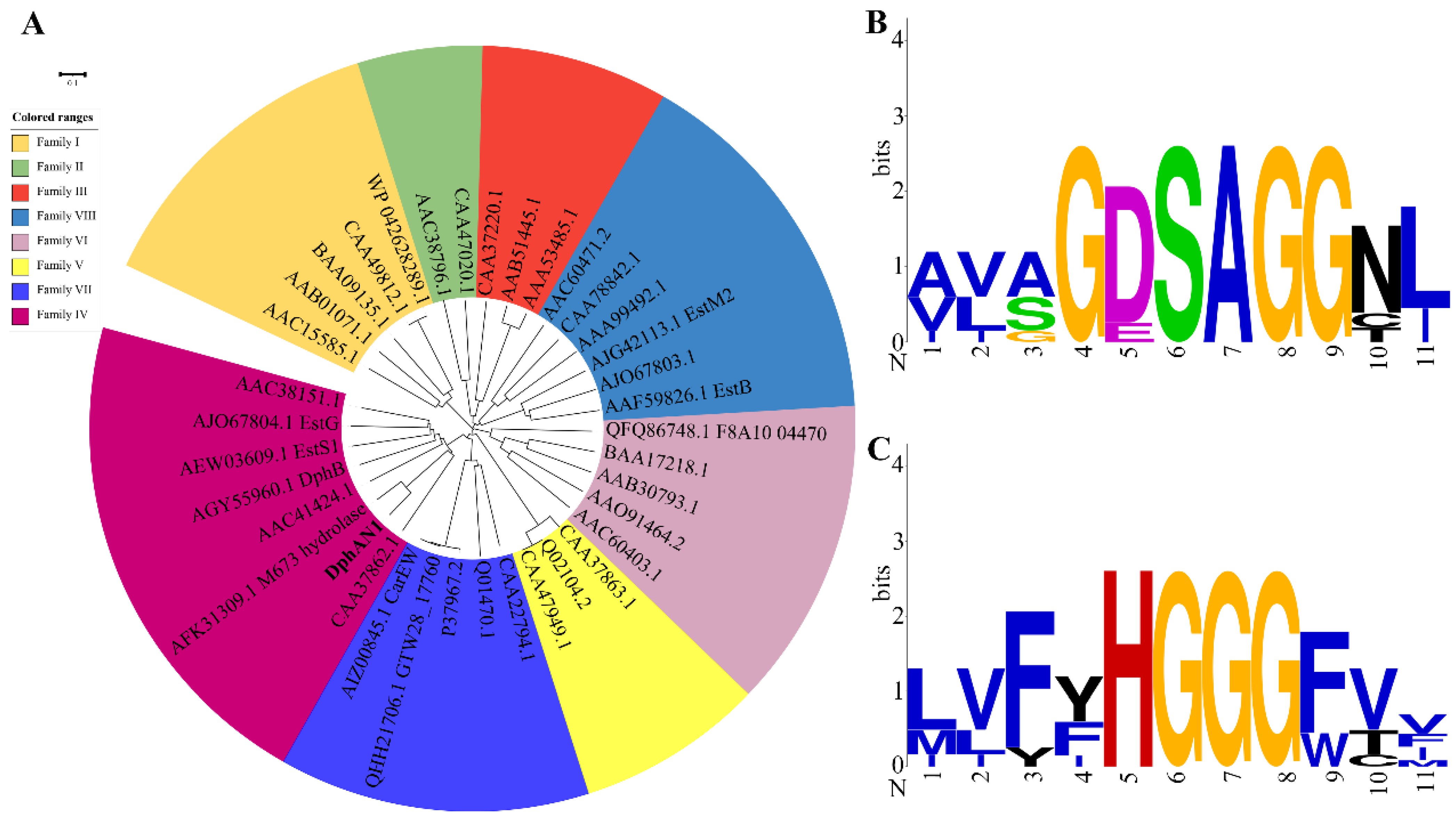
2.5. Structural Analysis of DphAN1

The three-dimensional structure of DphAN1 was modeled with the template of Est8 (PDB:4ypv) [55] (Figure 8A). According to validation by Molprobity [56], 94.8% of residues are in favored (98%) regions and 98.7% in allowed (>99.8%) regions (Figure S8), demonstrating the good quality of the modeled DphAN1 and its suitability for future research. The superposition of the structures of DphAN1 and Est8 resulted in an RMSD value of 0.149 (Figure 8B), indicating that DphAN1 belongs to the a/β hydrolase superfamily and harbors catalytic domain and cap domain (Figure 8A,B). In the catalytic domain, the central mixed β-sheet is sandwiched by several α-helices. The putative catalytic triad (Ser201-Asp295-His325) and oxyanion hole components H127GGG130 are situated in flexible loops or coils of the catalytic domain (Figure 8A), implying the location of the substrate-binding pocket. The residue Ser201 was in proximity with the motif H127GGG130 in accordance with their related functions. The orientation of the catalytic triad and oxyanion hole of DphAN1 may facilitate its interacting and hydrolyzing PAEs. The cap domain is composed of five α-helices and might determine the entrance of PAEs to the substrate-binding pocket [55].
Generally, molecular docking serves as an effective method for evaluating the interaction between the protein and ligand [25]. Molecular docking analysis of DphAN1 and DBP was performed to explore the molecular basis of DphAN1 catalyzing DBP hydrolysis (Figure 8C). DBP is located in the area between the catalytic domain and cap domain (Figure 8C), which is a long and hydrophobic pocket (Figure 8D and Figure S9). The catalytic Ser201 was situated around the substrate-binding pocket. The hydroxyl oxygen of Ser201 is 3.5 Å away from the carboxyl carbon of DBP, which could facilitate nucleophilic attack of Ser201 against the carboxyl carbon of DBP like other hydrolases [18]. In addition, the interactions among Ser201, His325 and Asp295 in the putative catalytic triad are mediated by hydrogen bonds, which might participate in electron transport during the process of DphAN1 catalyzing DBP (Figure 8D). To validate the deduction, Ser201, His325 and Asp295 were replaced by Ala via site-directed mutagenesis, respectively (Figure S10). After the enzyme assay, all variants displayed no hydrolytic activity towards DBP (Figure 8E), confirming that these amino acids are components of the catalytic triad. The catalytic triads are also identified in PAE hydrolases Xtj8 (Ser152-Glu246-His276) and EstJ6 (Ser146-Glu240-His270) from the HSL family [25,48], and the residue Glu substitutes Asp of the catalytic triad. The backbone amide (NH) of Gly129 and Gly130 forms an oxyanion hole (Figure 8D) and may stabilize the reaction tetrahedral intermediate of DphAN1-DBP during a hydrolysis process similar to that observed in other hydrolases [57,58,59]. Although the oxyanion hole is a common feature of hydrolases [57,59], the constituent residues are of diversity among hydrolases, such as Phe71 and Met176 of Est22 [60], Cys40 and Ser120 of AlAXEA [61], and Tyr87 and Met160 of IsPETase [57]. The variants G129A and G130A exhibited approximately 40% hydrolytic activity compared to the wild type of DphAN1 (Figure 8D), implying their key roles in the oxyanion hole. Moreover, some other residues of DphAN1 are involved in interaction with DBP to enhance the substrate-binding force (Figure 8D). The phenyl group of DBP forms π-π stack with Phe78 and π-anion interaction with Asp253, respectively. The residues Val133, Val254, Val230, Val257 and Phe330 provide a relatively hydrophobic pocket to stably bind to DBP (Figure 8D and Figure S9). When these residues were mutated to Ala, the activity of variants was significantly reduced. The possible reason is the mutation causes weak interaction between the enzyme and DBP. Based on the reduced activity of DphAN1 variants, the active residues responsible for binding and hydrolyzing DBP are identified in DphAN1 and can facilitate structural modification by rational design to improve catalytic performance [58].

3. Materials and Methods

3.1. Reagents and Media

PAEs and their metabolites were obtained from Aladdin Chemistry Co., Ltd. (Shanghai, China) or Sinopharm Chemical Reagent Co., Ltd. (Shanghai, China) and included the following: DEP, DBP, BBP, monoethyl phthalate (MEP), monobutyl phthalate (MBP), monobenzyl phthalate (MBeP) and phthalic acid (PA). Methanol was of HPLC grade, and other chemicals were of analytical grade. PAEs were dissolved in methanol to prepare the stock solution. The restriction enzymes, ligase, TA/Blunt-Zero Cloning vector, Mut Express II and Ni-NTA Resin were purchased from Vazyme Biotech Co., Ltd. (Nanjing, China) or TransGen Biotech Co., Ltd. (Beijing, China). The binding buffer and elution buffer were prepared using our existing methods [59]. The media for bacteria culture included Luria-Bertani (LB) medium and trace element medium (TEM) [62].

3.2. Enrichment, Isolation and Identification of PAE-Degrading Bacteria

Soil was collected from a wasteyard in Xingtai, China. The sample (10 g) was added to 100 mL of liquid LB medium containing 0.5 mM DBP and incubated at 180 rpm and 30 °C for 5 days. Then, 1% of the culture was transferred to fresh TEM (pH 8.0) with 0.5 mM DBP for further cultivation under the same conditions. The enrichment step was repeated five times in TEM (pH 8.0). The culture was streaked on a TEM agar plate with 0.5 mM DBP and incubated at 30 °C for 5 days. Some colonies, potentially capable of degrading DBP, were incubated in 100 mL of LB liquid medium to OD600 = 1.0, respectively. The cells were centrifuged, washed and resuspended in 200 mL of TEM (pH 8.0). The suspension was divided into 10 mL, supplemented with 0.5 mM DBP and cultivated for 5 days at 180 rpm and 30 °C. The control contained 10 mL of TEM (pH 8.0) with 0.5 mM DBP. All experiments were conducted in triplicate in the present study. The degradative activity of strain LUNF3 was estimated by HPLC-MS assay of DBP hydrolysis.
The genomic DNA of DBP-degrading strain LUNF3 was prepared using EasyPure® Bacteria Genomic DNA Kit (TransGen, Beijing, China). The DNA was used as template for amplification of 16S rRNA gene with primers 27F and 1492R [37]. The amplified products were ligated with TA/Blunt-Zero Cloning vector (Vazyme, Nanjing, China), which were transferred into DH5α. After sequencing and assembly of the 16S rRNA gene, the gene sequence was submitted in NCBI (https://blast.ncbi.nlm.nih.gov/Blast.cgi?PROGRAM=blastn&PAGE_TYPE=BlastSearch&LINK_LOC=blasthome (accessed on 1 April 2022)) and EzBioCloud Database (https://www.ezbiocloud.net/ (accessed on 1 April 2022)) to search for the relatives. The phylogenetic tree was constructed via MEGA 6.0 using the neighbor-joining method.

3.3. Degradation of PAEs by Strain LUNF3

The suspension of strain LUNF3 was prepared in liquid TEM (pH 8.0) with 0.5 mM DBP, as mentioned above. To investigate DBP degradation performance of the isolated strain at a range of temperatures (20, 30, 40 and 50 °C), the cultures were incubated at 180 rpm and a certain temperature for 5 days. To examine the effect of pH on DBP degradation, the strain LUNF3 was suspended in TEM (0.5 mM DBP) at a range of pH 4.0–11.0, and other culture conditions were set as above. The TEM (pH 8.0) with 0.5 mM DBP was adopted as the control. The concentration of residual DBP was determined by HPLC. To investigate the substrate spectra of the isolated strain, 0.5 mM of PAEs (DEP, DBP and BBP individually) were added into the strain suspension in TEM (pH 8.0). The mixtures were cultivated at 180 rpm, pH 8.0 and 30 °C for 5 days. The concentration of residual PAEs was determined using HPLC, and the degradation metabolites were detected by HPLC-MS [63].

3.4. Complete Genome Sequencing and Annotation

The genomic DNA of strain LUNF3 was prepared using EasyPure®Bacteria Genomic DNA Kit (TransGen Biotech, Beijing, China) according to the manufacturer’s instructions. The integrity and purity were detected via agarose gel electrophoresis and Qubit Fluorometer, respectively. After the construction of genome sequencing libraries, sequencing was carried out on the Illumina NovaSeq platform and PacBio Sequel platform. The genome sequence was assembled using PacBio sequencing reads via HGAP and CANU [64,65] and rectified using sequencing results from the Illumina platform. Based on the complete genome sequence, protein-coding genes, tRNA genes and rRNA genes were predicted using GeneMarkS, Trnascan-se and RNAmmer [66,67,68], respectively. The genes were annotated in public databases such as NR, eggNOG, KEGG, Swiss-Prot and GO [10]. The average nucleotide identity (ANI) analysis of the genomes was performed using FastANI [11].

3.5. Sequence Analysis and Expression of PAE Hydrolase DphAN1

The genome of strain LUNF3 was mined and searched for similar sequences of PAE hydrolases reported, such as XtjR8, HylD1, HylD2 and EstM2 [21,25,26]. The phylogenetic analysis of predicted PAE hydrolase DphAN1 and lipolytic enzymes from families I-VIII was performed using MEGA 6.0 using the neighbor-joining method. Multiple sequence alignment was conducted using CLUSTALW (https://www.ebi.ac.uk/Tools/msa/clustalo/ (accessed on 1 April 2022), and the conserved sequences were analyzed. After the codon optimization of gene dphAN1, the gene was synthesized and cloned into pCold II at the restriction sites of NdeI and XbaI. The recombinant expression vector pC-dphAN1 was introduced into strain ArcticExpress (DE3), and the strain was incubated in LB medium with 50 μg/mL ampicillin at 37 °C and 180 rpm. Then, 0.1 mM IPTG was added into the culture when OD600 of the culture reached approximately 0.8, inducing the expression of dphAN1. After induction of dphAN1 expression at 16 °C for 20h, the cells were collected by centrifugation and cell disruption was conducted by ultrasonication. The lysate underwent centrifugation to collect the supernatant, which was used to purify recombinant DphAN1 via the column of Ni-NTA Resin (TransGen, Beijing, China) [62]. The concentration of purified DphAN1 was measured using the BCA Protein Assay Kit (TIANGEN, Beijing, China).

3.6. Biochemical Characterization of DphAN1

To verify the PAE-hydrolyzing capability of DphAN1, DEP, DBP and BBP were adopted as substrates for tests. For the assays, 0.5 mM substrate and 6.38 μg DphAN1 were added into 0.9 mL of Tris–HCl (50 mM, pH 8.0). The reaction was carried out at 30 °C and 180 rpm for 20 min, and the reaction was terminated by adding 10 µL of 1M HCl. PAEs and hydrolysis products were detected by HPLC or HPLC-MS. The control contained no hydrolase DphAN1. One unit of specific activity of DphAN1 was defined as the amount of enzyme required for hydrolysis of 1 µmol of PAEs per minute. To examine the optimal pH for DphAN1 catalysis, the following buffer Na2HPO4–citric acid (pH 4.0–7.0), Tris–HCl (pH 8.0–8.9) and Glycine–NaOH (pH 9.0–10.0) was used as the reaction buffer. To examine the optimal temperature, the reaction was performed in Tris–HCl (pH 8.0) at temperatures ranging from 10 °C to 50 °C. Other reaction conditions were set as above. The highest activity under a certain pH or temperature was defined as 100%, and the relative activity under other conditions was calculated.
To investigate the effects of metal ions or chemicals on the activity of DphAN1,these additives, which included metal ions (1 mM Ca2+, Mg2+, Fe3+, Mn2+, Zn2+, Co2+, Ni2+, Cu2+, Cr2+, Cd2+, Fe2+ and Hg2+), surfactants (1 mM and 5 mM SDS, 1% and 5% Tween 20, 1% and 5% Tween 80 and 1% Triton X-100) and esterase inhibitors (1% DEPC, PMSF and β-mercaptoethanol (β-ME)), were individually added into Na2HPO4–citric acid (pH 7.0). Other reaction conditions were set as above. The activity of DphAN1 was expressed as relative activity to the control without any additives.

3.7. Homology Modeling and Molecular Docking

To construct the homology model of DphAN1, the PDB database was searched for homolog proteins using NCBI BLAST. The protein Est8 (PDB:4ypv), an alkaline esterase from a diesel oil-degrading consortium [55], was adopted as a model template due to 34.09% amino acid identity with DphAN1 (over 86% coverage). After homology modeling of DphAN1 using the Modeller software, the modeled structure was checked by Molprobity [56]. The molecular docking of DphAN1 and DBP was performed using AutoDock4.2 [18]. The three-dimensional structure of DBP was obtained from PubChem (http://pubchem.ncbi.nlm.nih.gov/ (accessed on 22 February 2022), and the rotations and torsions of DBP were automatically set in AutoDock tools.

3.8. Site-Directed Mutagenesis of DphAN1

The variants of DphAN1 were prepared by site-directed mutagenesis according to the instruction of the kit Mut Express II (Vazyme, Nanjing, China). The residues of DphAN1 (S201, D295, H325, G129, G130, F78, V133, V230, D253, V254, V257 and F330) were replaced by alanine. The mutant primers in Table S3 were applied to amplify the expression vector pC-dphAN1. The products were cyclized and transformed into ArcticExpress(DE3) cells. The mutated DphAN1 was overexpressed in these cells under induction by 0.1 mM IPTG. The purification and enzyme assay of variants of DphAN1 were conducted following the procedure described above.

3.9. Analytical Methods

PAEs or intermediates in the cultures of strain LUNF3 or reaction mixture of hydrolase DphAN1 were detected by HPLC or HPLC-MS. To extract PAEs or intermediates in the mixture above, an equal volume of ethyl acetate was added with strong agitation. Ethyl acetate in the organic phase was evaporated, and the extracts were dissolved in an equal volume of methanol. The solution was filtrated through a 0.22 µm membrane and then analyzed using HPLC (Agilent 1200, Santa Clara, CA, USA) or HPLC-MS equipped with a triple quadrupole mass spectrometer (Agilent 6420, Santa Clara, CA, USA). The chromatographic conditions were as follows: reverse phase column ZORBAX Eclipse Plus C18 (4.6 mm × 250 mm, 5 µm), the mobile phase of 90% methanol and 10% water containing 0.1% acetic acid, and the flow rate of 0.8 mL/min or 0.5 mL/min.

4. Conclusions

In summary, an efficient PAE degrader Acinetobacter sp. LUNF3 was isolated from soil under a wasteyard. According to the complete genome sequencing and genomic analysis of strain LUNF3, a novel PAE hydrolase DphAN1 from the HSL family was identified. DphAN1 possesses canonical α/β hydrolase structure and conserved motifs such as catalytic triad and oxyanion hole. DphAN1 is able to hydrolyze numerous kinds of PAEs, especially BBP with bulky side chains. Moreover, DphAN1 displays high activity under a broad range of temperatures and pH values and can tolerate several metal ions and chemicals. The identification and characterization of DphAN1 enriches the resources of PAE hydrolases and significantly highlights its potential application in the remediation of PAE-contaminated environments. The interaction between DphAN1 and DBP was investigated by molecular docking to ascertain key active sites (catalytic triad and other binding residues) and was validated by mutation of these residues. These results provide insights into the catalytic mechanism of hydrolase DphAN1 and may be a basis for protein structural modification to improve catalytic efficiency in environment remediation.

Supplementary Materials

The following supporting information can be downloaded at: https://www.mdpi.com/article/10.3390/molecules28186738/s1, Figure S1: The HPLC profile of DBP degraded by strain LUNF3 (B) compared with control (A). CK: TEM (pH 8.0) with 0.5 mM DBP; Figure S2: The substrates detection of strain LUNF3 (A) and metabolites detected by HPLC-MS (B) (* p < 0.05, ** p < 0.01 and *** p < 0.0001). CK (A): TEM (pH 8.0) with 0.5 Mm DEP, DBP or BBP. CK (B): TEM (pH 8.0) with 0.5 mM DEP; Figure S3: The genes from chromosome (A) and plasmid (B) of strain LUNF3 annotated in GO database and the distribution of these genes in metabolic pathways based on KEGG database (C); Figure S4: The functional classification of genes from chromosome (A) and plasmid (B) based on COG database; Figure S5: The alignment of DphAN1 and other hydrolases from family IV. The conserved amino acid residues are shown in rectangle. The catalytic triad (Ser201-Asp295-His325) is marked by pentastar; Figure S6: The highly conserved motifs D295LLHDEG301 (A), H325GF327 (B) and V154LSIDYPLAPE164 (C) detected from DphAN1; Figure S7: The HPLC-MS spectrum of DEP (A) and BBP (B) catalyzed by DphAN1. CK (A): Tris-HCl (50 mM, pH 8.0) with 0.5 mM DEP. CK (B): Tris-HCl (50 mM, pH 8.0) with 0.5 mM BBP; Figure S8: The evaluation of the model structure of DphAN1 by MolProbity Ramachandran analysis; Figure S9: The hydrophobic pocket of hydrolase DphAN1 where DBP was docked; Figure S10: The SDS-PAGE analysis of hydrolase DphAN1 and its mutants. M: protein marker, 1: DphAN1, 2: S201A, 3: D295A, 4: H325A, 5: G129A, 6: G130A, 7: F78A, 8: V133A, 9: V230A, 10: D253A, 11: V254A, 12: V257A, 13: F330A.; Table S1: The top 100 sequences similar to the 16S sequence of strain LUNF3; Table S2: The genomic characteristics of Acinetobacter sp. LUNF3; Table S3: The sequence of primers used for site-directed mutagenesis of DphAN1.

Author Contributions

Conceptualization, X.H. and W.W.; software, S.F. and J.G.; investigation, S.F.; data curation, S.F.; writing—original draft, S.F.; writing—review and editing, S.H., H.D., Z.W., Y.F. and H.H.; supervision, X.H. and W.W. All authors have read and agreed to the published version of the manuscript.

Funding

This research was funded by the Science Research Project of Hebei Education Department (QN2021123), the Nanfan special project of the Chinese Academy of Agricultural Sciences (YYLH2305) and The Agricultural Science and Technology Innovation Program (ASTIP).

Institutional Review Board Statement

Not applicable.

Informed Consent Statement

Not applicable.

Data Availability Statement

The data in this study can be obtained from the corresponding authors or the NCBI database. The sequences of 16S rRNA gen and dphAN1 were deposited in GenBank under accession numbers OM900052 and OM908374, respectively. The sequences of chromosome and plasmid were deposited in the GenBank database under accession numbers CP093968 and CP093969, respectively.

Acknowledgments

We thank Fei Pan from the Institute of Apicultural Research, Chinese Academy of Agricultural Sciences, for technical assistance of homology modeling and molecular docking.

Conflicts of Interest

The authors declare no conflict of interest.

Sample Availability

The sample of Acinetobacter sp. LUNF3 is available from the authors.

References

  1. Hu, R.; Zhao, H.; Xu, X.; Wang, Z.; Yu, K.; Shu, L.; Yan, Q.; Wu, B.; Mo, C.; He, Z.; et al. Bacteria-driven phthalic acid ester biodegradation: Current status and emerging opportunities. Environ. Int. 2021, 154, 106560. [Google Scholar] [CrossRef]
  2. Peijnenburg, W.J.; Struijs, J. Occurrence of phthalate esters in the environment of The Netherlands. Ecotox Environ. Safe 2006, 63, 204–215. [Google Scholar] [CrossRef] [PubMed]
  3. Clausen, P.A.; Liu, Z.; Kofoed-Sørensen, V.; Little, J.; Wolkoff, P. Influence of temperature on the emission of di-(2-ethylhexyl) phthalate (DEHP) from PVC flooring in the emission cell FLEC. Environ. Sci. Technol. 2012, 46, 909–915. [Google Scholar] [CrossRef]
  4. Ambe, K.; Sakakibara, Y.; Sakabe, A.; Makino, H.; Ochibe, T.; Tohkin, M. Comparison of the developmental/reproductive toxicity and hepatotoxicity of phthalate esters in rats using an open toxicity data source. J. Toxicol. Sci. 2019, 44, 245–255. [Google Scholar] [CrossRef] [PubMed]
  5. Weaver, J.A.; Beverly, B.E.J.; Keshava, N.; Mudipalli, A.; Arzuaga, X.; Cai, C.; Hotchkiss, A.K.; Makris, S.L.; Yost, E.E. Hazards of diethyl phthalate (DEP) exposure: A systematic review of animal toxicology studies. Environ. Int. 2020, 145, 105848. [Google Scholar] [CrossRef]
  6. Radke, E.G.; Braun, J.M.; Nachman, R.M.; Cooper, G.S. Phthalate exposure and neurodevelopment: A systematic review and meta-analysis of human epidemiological evidence. Environ. Int. 2020, 137, 105408. [Google Scholar] [CrossRef]
  7. Jonsson, S.; Vavilin, V.; Svensson, B. Phthalate hydrolysis under landfill conditions. Water Sci. Technol. A J. Int. Assoc. Water Pollut. Res. 2006, 53, 119–127. [Google Scholar] [CrossRef]
  8. Lau, T.K.; Chu, W.; Graham, N. The degradation of endocrine disruptor Di-n-butyl Phthalate by UV irradiation: A photolysis and product study. Chemosphere 2005, 60, 1045–1053. [Google Scholar] [CrossRef]
  9. Mahajan, R.; Verma, S.; Kushwaha, M.; Singh, D.; Akhter, Y.; Chatterjee, S. Biodegradation of dinbutyl phthalate by psychrotolerant Sphingobium yanoikuyae strain P4 and protein structural analysis of carboxylesterase involved in the pathway. Int. J. Biol. Macromol. 2019, 122, 806–816. [Google Scholar] [CrossRef] [PubMed]
  10. Wang, Y.; Zhan, W.; Ren, Q.; Cheng, S.; Wang, J.; Ma, X.; Zhang, C.; Wang, Y. Biodegradation of di-(2-ethylhexyl) phthalate by a newly isolated Gordonia sp. and its application in the remediation of contaminated soils. Sci. Total Environ. 2019, 689, 645–651. [Google Scholar] [CrossRef]
  11. Zhao, H.-M.; Hu, R.-W.; Chen, X.-B.; Lü, H.; Li, Y.-W.; Li, H.; Mo, C.-H.; Cai, Q.-Y.; Wong, M.-H. Biodegradation pathway of di-(2-ethylhexyl) phthalate by a novel Rhodococcus pyridinivorans XB and its bioaugmentation for remediation of DEHP contaminated soil. Sci. Total Environ. 2018, 640–641, 1121–1131. [Google Scholar] [CrossRef] [PubMed]
  12. Liu, T.; Li, J.; Qiu, L.; Zhang, F.; Linhardt, R.J.; Zhong, W. Combined genomic and transcriptomic analysis of Dibutyl phthalate metabolic pathway in Arthrobacter sp. ZJUTW. Biotechnol. Bioeng. 2020, 117, 3712–3726. [Google Scholar] [CrossRef] [PubMed]
  13. Feng, N.-X.; Yu, J.; Mo, C.-H.; Zhao, H.-M.; Li, Y.-W.; Wu, B.-X.; Cai, Q.-Y.; Li, H.; Zhou, D.-M.; Wong, M.-H. Biodegradation of di-n-butyl phthalate (DBP) by a novel endophytic Bacillus megaterium strain YJB3. Sci. Total Environ. 2018, 616–617, 117–127. [Google Scholar] [CrossRef] [PubMed]
  14. Tao, Y.; Li, H.; Gu, J.; Shi, H.; Han, S.; Jiao, Y.; Zhong, G.; Zhang, Q.; Akindolie, M.S.; Lin, Y.; et al. Metabolism of diethyl phthalate (DEP) and identification of degradation intermediates by Pseudomonas sp. DNE-S1. Ecotox Environ. Safe 2019, 173, 411–418. [Google Scholar] [CrossRef]
  15. Wu, X.; Liang, R.; Dai, Q.; Jin, D.; Wang, Y.; Chao, W. Complete degradation of di-n-octyl phthalate by biochemical cooperation between Gordonia sp. strain JDC-2 and Arthrobacter sp. strain JDC-32 isolated from activated sludge. J. Hazard. Mater. 2010, 176, 262–268. [Google Scholar] [CrossRef]
  16. Kong, X.; Jin, D.; Tai, X.; Yu, H.; Duan, G.; Yan, X.; Pan, J.; Song, J.; Deng, Y. Bioremediation of dibutyl phthalate in a simulated agricultural ecosystem by Gordonia sp. strain QH-11 and the microbial ecological effects in soil. Sci. Total Environ. 2019, 667, 691–700. [Google Scholar] [CrossRef]
  17. Yan, Z.; Ding, L.; Zou, D.; Qiu, J.; Shao, Y.; Sun, S.; Li, L.; Xin, Z. Characterization of a novel carboxylesterase with catalytic activity toward di(2-ethylhexyl) phthalate from a soil metagenomic library. Sci. Total Environ. 2021, 785, 147260. [Google Scholar] [CrossRef]
  18. Fan, S.; Wang, J.; Yan, Y.; Wang, J.; Jia, Y. Excellent Degradation Performance of a Versatile Phthalic Acid Esters-Degrading Bacterium and Catalytic Mechanism of Monoalkyl Phthalate Hydrolase. Int. J. Mol. Sci. 2018, 19, 2803. [Google Scholar] [CrossRef]
  19. Whangsuk, W.; Sungkeeree, P.; Nakasiri, M.; Thiengmag, S.; Mongkolsuk, S.; Loprasert, S. Two endocrine disrupting dibutyl phthalate degrading esterases and their compensatory gene expression in Sphingobium sp. SM42. Int. Biodeter Biodegr 2015, 99, 45–54. [Google Scholar] [CrossRef]
  20. Xu, Y.; Liu, X.; Zhao, J.; Huang, H.; Wu, M.; Li, X.; Li, W.; Sun, X.; Sun, B. An efficient phthalate ester-degrading Bacillus subtilis: Degradation kinetics, metabolic pathway, and catalytic mechanism of the key enzyme. Environ. Pollut. 2021, 273, 116461. [Google Scholar] [CrossRef]
  21. Sarkar, J.; Dutta, A.; Chowdhury, P.P.; Chakraborty, J.; Dutta, T.K. Characterization of a novel family VIII esterase EstM2 from soil metagenome capable of hydrolyzing estrogenic phthalates. Microb. Cell Fact. 2020, 19, 77. [Google Scholar] [CrossRef]
  22. Kurane, R.; Suzuki, T.; Fukuoka, S. Purification and Some Properties of a Phthalate Ester-Hydrolyzing Enzyme From Nocardia erythropolis. Appl. Microbiol. Biot. 1984, 20, 378–383. [Google Scholar] [CrossRef]
  23. Niazi, J.H.; Prasad, D.T.; Karegoudar, T.B. Initial degradation of dimethylphthalate by esterases from Bacillus species. Fems Microbiol. Lett. 2001, 196, 201–205. [Google Scholar] [CrossRef] [PubMed]
  24. Du, H.; Hu, R.-W.; Zhao, H.-M.; Huang, H.-B.; Xiang, L.; Liu, B.-L.; Feng, N.-X.; Li, H.; Li, Y.-W.; Cai, Q.-Y.; et al. Mechanistic insight into esterase-catalyzed hydrolysis of phthalate esters (PAEs) based on integrated multi-spectroscopic analyses and docking simulation. J. Hazard. Mater. 2021, 408, 124901. [Google Scholar] [CrossRef]
  25. Qiu, J.; Yang, H.; Yan, Z.; Shi, Y.; Zou, D.; Ding, L.; Shao, Y.; Li, L.; Khan, U.; Sun, S.; et al. Characterization of XtjR8: A novel esterase with phthalate-hydrolyzing activity from a metagenomic library of lotus pond sludge. Int. J. Biol. Macromol. 2020, 164, 1510–1518. [Google Scholar] [CrossRef] [PubMed]
  26. Xu, Y.; Minhazul, K.A.; Wang, X.; Liu, X.; Li, X.; Meng, Q.; Li, H.; Zhang, C.; Sun, X.; Sun, B. Biodegradation of phthalate esters by Paracoccus kondratievae BJQ0001 isolated from Jiuqu (Baijiu fermentation starter) and identification of the ester bond hydrolysis enzyme. Environ. Pollut. 2020, 263, 114506. [Google Scholar] [CrossRef] [PubMed]
  27. Cheng, J.; Du, H.; Zhou, M.-S.; Ji, Y.; Xie, Y.-Q.; Huang, H.-B.; Zhang, S.-H.; Li, F.; Xiang, L.; Cai, Q.-Y.; et al. Substrate-enzyme interactions and catalytic mechanism in a novel family VI esterase with dibutyl phthalate-hydrolyzing activity. Environ. Int. 2023, 178, 108054. [Google Scholar] [CrossRef] [PubMed]
  28. Chen, Y.; Wang, Y.; Xu, Y.; Sun, J.; Yang, L.; Feng, C.; Wang, J.; Zhou, Y.; Zhang, Z.-M.; Wang, Y. Molecular insights into the catalytic mechanism of plasticizer degradation by a monoalkyl phthalate hydrolase. Commun. Chem. 2023, 6, 45. [Google Scholar] [CrossRef]
  29. Wu, J.; Liao, X.; Yu, F.; Wei, Z.; Yang, L. Cloning of a dibutyl phthalate hydrolase gene from Acinetobacter sp. strain M673 and functional analysis of its expression product in Escherichia coli. Appl. Microbiol. Biot. 2013, 97, 2483–2491. [Google Scholar] [CrossRef]
  30. Fang, Y.; Zhang, L.; Wang, J.; Zhou, Y.; Ye, B. Biodegradation of Phthalate Esters by a Newly Isolated Acinetobacter sp. Strain LMB-5 and Characteristics of Its Esterase. Pedosphere 2017, 27, 606–615. [Google Scholar] [CrossRef]
  31. Xu, J.; Lu, Q.; de Toledo, R.A.; Shim, H. Degradation of di-2-ethylhexyl phthalate (DEHP) by an indigenous isolate Acinetobacter sp. SN13. Int. Biodeter Biodegr 2017, 117, 205–214. [Google Scholar] [CrossRef]
  32. Wang, S.; Wang, J. Biodegradation and metabolic pathway of sulfamethoxazole by a novel strain Acinetobacter sp. Appl. Microbiol. Biot. 2018, 102, 425–432. [Google Scholar] [CrossRef]
  33. Hao, J.; Wang, P.; Kang, Y.; He, H.; Luo, H.; Kim, S.; Niu, L.; Jiang, H.; Ma, K. Degradation of Perfluorooctane Sulfonamide by Acinetobacter Sp. M and Its Extracellular Enzymes. Chem. Asian J. 2019, 14, 2780–2784. [Google Scholar] [CrossRef]
  34. Zhang, X.; Kong, D.; Liu, X.; Xie, H.; Lou, X.; Zeng, C. Combined microbial degradation of crude oil under alkaline conditions by Acinetobacter baumannii and Talaromyces sp. Chemosphere 2021, 273, 129666. [Google Scholar] [CrossRef] [PubMed]
  35. Sun, J.-Q.; Xu, L.; Tang, Y.-Q.; Chen, F.-M.; Wu, X.-L. Simultaneous degradation of phenol and n-hexadecane by Acinetobacter strains. Bioresour. Technol. 2012, 123, 664–668. [Google Scholar] [CrossRef] [PubMed]
  36. Sun, R.; Wang, L.; Jiao, Y.; Zhang, Y.; Zhang, X.; Wu, P.; Chen, Z.; Feng, C.; Li, Y.; Li, X.; et al. Metabolic process of di-n-butyl phthalate (DBP) by Enterobacter sp. DNB-S2, isolated from Mollisol region in China. Environ. Pollut. 2019, 255, 113344. [Google Scholar] [CrossRef]
  37. Wang, L.; Gan, D.; Gong, L.; Zhang, Y.; Wang, J.; Guan, R.; Zeng, L.; Qu, J.; Dong, M. Analysis of the performance of the efficient di-(2-ethylhexyl) phthalate-degrading bacterium Rhodococcus pyridinovorans DNHP-S2 and associated catabolic pathways. Chemosphere 2022, 306, 135610. [Google Scholar] [CrossRef] [PubMed]
  38. Xu, Y.; Zhao, J.; Huang, H.; Guo, X.; Li, X.; Zou, W.; Li, W.; Zhang, C.; Huang, M. Biodegradation of phthalate esters by Pantoea dispersa BJQ0007 isolated from Baijiu. J. Food Compos. Anal. 2022, 105, 104201. [Google Scholar] [CrossRef]
  39. Feng, N.-X.; Feng, Y.-X.; Liang, Q.-F.; Chen, X.; Xiang, L.; Zhao, H.-M.; Liu, B.-L.; Cao, G.; Li, Y.-W.; Li, H.; et al. Complete biodegradation of di-n-butyl phthalate (DBP) by a novel Pseudomonas sp. YJB6. Sci. Total Environ. 2020, 761, 143208. [Google Scholar] [CrossRef]
  40. Tian, M.; Zhang, X.; Liu, L.; Martin, F.L.; Wang, H.; Zhang, J.; Huang, Q.; Wang, X.; Shen, H. Phthalate side-chain structures and hydrolysis metabolism associated with steroidogenic effects in MLTC-1 Leydig cells. Toxicol. Lett. 2019, 308, 56–64. [Google Scholar] [CrossRef]
  41. Li, J.; Zhang, J.; Yadav, M.P.; Li, X. Biodegradability and biodegradation pathway of di-(2-ethylhexyl) phthalate by Burkholderia pyrrocinia B1213. Chemosphere 2019, 225, 443–450. [Google Scholar] [CrossRef]
  42. Wang, P.; Gao, J.; Zhao, Y.; Zhang, M.; Zhou, S. Biodegradability of di-(2-ethylhexyl) phthalate by a newly isolated bacterium Achromobacter sp. RX. Sci. Total Environ. 2021, 755, 142476. [Google Scholar] [CrossRef] [PubMed]
  43. Chen, F.; Chen, Y.; Chen, C.; Feng, L.; Dong, Y.; Chen, J.; Lan, J.; Hou, H. High-efficiency degradation of phthalic acid esters (PAEs) by Pseudarthrobacter defluvii E5: Performance, degradative pathway, and key genes. Sci. Total Environ. 2021, 794, 148719. [Google Scholar] [CrossRef]
  44. Wang, Y.; Liu, H.; Peng, Y.; Tong, L.; Feng, L.; Ma, K. New pathways for the biodegradation of diethyl phthalate by Sphingobium yanoikuyae SHJ. Process Biochem. 2018, 71, 152–158. [Google Scholar] [CrossRef]
  45. Zhang, G.; Chen, T.; Chang, S.; Zhang, W.; Wu, X.; Wu, M.; Wang, Y.; Long, H.; Chen, X.; Wang, Y.; et al. Complete genome sequence of Acinetobacter sp. TTH0-4, a cold-active crude oil degrading strain isolated from Qinghai-Tibet Plateau. J. Biotechnol. 2016, 226, 54–55. [Google Scholar] [CrossRef]
  46. Wang, W.; Chen, X.; Yan, H.; Hu, J.; Liu, X. Complete genome sequence of the cyprodinil-degrading bacterium Acinetobacter johnsonii LXL_C1. Microb. Pathog. 2019, 127, 246–249. [Google Scholar] [CrossRef]
  47. Wright, R.J.; Bosch, R.; Gibson, M.I.; Christie-Oleza, J.A. Plasticizer Degradation by Marine Bacterial Isolates: A Proteogenomic and Metabolomic Characterization. Environ. Sci. Technol. 2020, 54, 2244–2256. [Google Scholar] [CrossRef] [PubMed]
  48. Qiu, J.; Zhang, Y.; Shi, Y.; Jiang, J.; Wu, S.; Li, L.; Shao, Y.; Xin, Z. Identification and characterization of a novel phthalate-degrading hydrolase from a soil metagenomic library. Ecotox Environ. Safe 2020, 190, 110148. [Google Scholar] [CrossRef]
  49. Zhang, X.-Y.; Fan, X.; Qiu, Y.-J.; Li, C.-Y.; Xing, S.; Zheng, Y.-T.; Xu, J.-H. Newly Identified Thermostable Esterase from Sulfobacillus acidophilus: Properties and Performance in Phthalate Ester Degradation. Appl. Environ. Microb. 2014, 80, 6870–6878. [Google Scholar] [CrossRef] [PubMed]
  50. Jiao, Y.; Chen, X.; Wang, X.; Liao, X.; Xiao, L.; Miao, A.; Wu, J.; Yang, L. Identification and Characterization of a Cold-Active Phthalate Esters Hydrolase by Screening a Metagenomic Library Derived from Biofilms of a Wastewater Treatment Plant. PLoS ONE 2013, 8, e75977. [Google Scholar] [CrossRef]
  51. Vingiani, G.M.; Leone, S.; De Luca, D.; Borra, M.; Dobson, A.D.; Ianora, A.; De Luca, P.; Lauritano, C. First identification and characterization of detoxifying plastic-degrading DBP hydrolases in the marine diatom Cylindrotheca closterium. Sci. Total Environ. 2022, 812, 152535. [Google Scholar] [PubMed]
  52. Duan, X.; Xiang, M.; Wang, L.; Yan, Q.; Yang, S.; Jiang, Z. Biochemical characterization of a novel lipase from Malbranchea cinnamomea suitable for production of lipolyzed milkfat flavor and biodegradation of phthalate esters. Food Chem. 2019, 297, 124925. [Google Scholar] [CrossRef] [PubMed]
  53. Ren, L.-Q.; Chang, T.-T.; Ren, D.-P.; Zhou, Y.; Ye, B.-C. Rational design to improve activity of the Est3563 esterase from Acinetobacter sp. LMB-5. Enzym. Microb. Tech. 2019, 131, 109331. [Google Scholar] [CrossRef] [PubMed]
  54. Qi, W.; Long, J.; Feng, C.; Feng, Y.; Cheng, D.; Liu, Y.; Xue, J.; Li, Z. Fe3+ enhanced degradation of oxytetracycline in water by pseudomonas. Water Res. 2019, 160, 361–370. [Google Scholar] [CrossRef]
  55. Pereira, M.R.; Maester, T.C.; Mercaldi, G.F.; de Macedo Lemos, E.G.; Hyvönen, M.; Balan, A. From a metagenomic source to a high-resolution structure of a novel alkaline esterase. Appl. Microbiol. Biot. 2017, 101, 4935–4949. [Google Scholar] [CrossRef]
  56. Davis, I.W.; Leaver-Fay, A.; Chen, V.B.; Block, J.N.; Kapral, G.J.; Wang, X.; Murray, L.W.; Arendall, W.B.; Snoeyink, J.; Richardson, J.S.; et al. MolProbity: All-atom contacts and structure validation for proteins and nucleic acids. Nucleic Acids Res. 2007, 35, W375–W383. [Google Scholar] [CrossRef]
  57. Joo, S.; Cho, I.J.; Seo, H.; Son, H.F.; Sagong, H.-Y.; Shin, T.J.; Choi, S.Y.; Lee, S.Y.; Kim, K.-J. Structural insight into molecular mechanism of poly(ethylene terephthalate) degradation. Nat. Commun. 2018, 9, 382. [Google Scholar] [CrossRef]
  58. Austin, H.P.; Allen, M.D.; Donohoe, B.S.; Rorrer, N.A.; Kearns, F.L.; Silveira, R.L.; Pollard, B.C.; Dominick, G.; Duman, R.; El Omari, K.; et al. Characterization and engineering of a plastic-degrading aromatic polyesterase. Proc. Natl. Acad. Sci. USA 2018, 115, E4350–E4357. [Google Scholar] [CrossRef]
  59. Han, X.; Liu, W.; Huang, J.-W.; Ma, J.; Zheng, Y.; Ko, T.-P.; Xu, L.; Cheng, Y.-S.; Chen, C.-C.; Guo, R.-T. Structural insight into catalytic mechanism of PET hydrolase. Nat. Commun. 2017, 8, 2106. [Google Scholar] [CrossRef]
  60. Li, P.-Y.; Yao, Q.-Q.; Wang, P.; Zhang, Y.; Li, Y.; Zhang, Y.-Q.; Hao, J.; Zhou, B.-C.; Chen, X.-L.; Shi, M.; et al. A Novel Subfamily Esterase with a Homoserine Transacetylase-like Fold but No Transferase Activity. Appl. Environ. Microb. 2017, 83, e117–e131. [Google Scholar] [CrossRef]
  61. Komiya, D.; Hori, A.; Ishida, T.; Igarashi, K.; Samejima, M.; Koseki, T.; Fushinobu, S. Crystal Structure and Substrate Specificity Modification of Acetyl Xylan Esterase from Aspergillus luchuensis. Appl. Environ. Microb. 2017, 83, e1217–e1251. [Google Scholar] [CrossRef] [PubMed]
  62. Fan, S.; Wang, J.; Li, K.; Yang, T.; Jia, Y.; Zhao, B.; Yan, Y. Complete genome sequence of Gordonia sp. YC-JH1, a bacterium efficiently degrading a wide range of phthalic acid esters. J. Biotechnol. 2018, 279, 55–60. [Google Scholar] [CrossRef]
  63. Kanaujiya, D.K.; Sivashanmugam, S.; Pakshirajan, K. Biodegradation and toxicity removal of phthalate mixture by Gordonia sp. in a continuous stirred tank bioreactor system. Environ. Technol. Inno 2022, 26, 102324. [Google Scholar] [CrossRef]
  64. Chin, C.-S.; Peluso, P.; Sedlazeck, F.J.; Nattestad, M.; Concepcion, G.T.; Clum, A.; Dunn, C.; O’Malley, R.; Figueroa-Balderas, R.; Morales-Cruz, A.; et al. Phased diploid genome assembly with single-molecule real-time sequencing. Nat. Methods 2016, 13, 1050–1054. [Google Scholar] [CrossRef]
  65. Koren, S.; Walenz, B.P.; Berlin, K.; Miller, J.R.; Bergman, N.H.; Phillippy, A.M. Canu: Scalable and accurate long-read assembly via adaptive κ-mer weighting and repeat separation. Genome Res. 2017, 27, 215087–215116. [Google Scholar] [CrossRef] [PubMed]
  66. Lowe, T.M.; Eddy, S.R. tRNAscan-SE: A program for improved detection of transfer RNA genes in genomic sequence. Nucleic Acids Res. 1997, 25, 955–964. [Google Scholar] [CrossRef] [PubMed]
  67. Besemer, J.; Lomsadze, A.; Borodovsky, M. GeneMarkS: A self-training method for prediction of gene starts in microbial genomes. Nucleic Acids Res. Nar. 2001, 29, 2607–2618. [Google Scholar] [CrossRef]
  68. Lagesen, K.; Hallin, P.; Rødland, E.A.; Staerfeldt, H.-H.; Rognes, T.; Ussery, D.W. RNAmmer: Consistent and rapid annotation of ribosomal RNA genes. Nucleic Acids Res. 2007, 35, 3100–3108. [Google Scholar] [CrossRef]
Figure 1. The pathways of PAE hydrolysis by microorganisms.
Molecules 28 06738 g001
Figure 2. Functional and molecular identification of strain LUNF3. (A) HPLC-MS profile of DBP degraded by strain LUNF3 with the metabolite of MBP. Strain LUNF3 was cultivated in TEM (pH 8.0) with 0.5 mM DBP for 5 days at 180 rpm and 30 °C. CK: TEM (pH 8.0) with 0.5 mM DBP. (B) The phylogenetic tree constructed using 16S rRNA gene sequences of strain LUNF3 and its relatives. Bar, 0.01 nucleotide substitutions per nucleotide position.
Molecules 28 06738 g002
Figure 3. The characterization of strain LUNF3 degrading PAEs. The degradation performance of strain LUNF3 against DBP under a range of temperature (A) and pH values (B). Strain LUNF3 was cultivated in TEM (pH 8.0) with 0.5 mM DBP for 5 days at 10–50 °C (A). Strain LUNF3 was cultivated for 5 days at 30 °C in TEM (pH 4.0–11.0) supplemented with 0.5 mM DBP (B). (C) The metabolites of BBP detected using HPLC-MS. Strain LUNF3 was cultivated in TEM (pH 8.0) with 0.5 mM BBP for 5 days at 180rpm and 30 °C. CK: TEM (pH 8.0) with 0.5 mM BBP.
Molecules 28 06738 g003
Figure 4. The circle genome map of strain LUNF3 composed of a chromosome (A) and a plasmid (B). From inside to outside: scale (circle1), GC skew (circle2), GC content (circle3), COG, in which each CDS belonged (circle4 and circle7), and locations of genes (CDS, tRNA and rRNA) in the genome (circle5 and circle6).
Molecules 28 06738 g004
Figure 5. Phylogenetic analysis and conserved sequence analysis of hydrolase DphAN1. (A) The phylogenetic tree based on the amino acid sequence of DphAN1 and lipolytic enzymes from families I–VIII. The conserved motifs GDSAGG (B) and HGGG (C) identified from DphAN1.
Molecules 28 06738 g005
Figure 6. The purification and hydrolysis activity of DphAN1 toward PAEs. (A) SDS-PAGE analysis of purified recombinant DphAN1. (B) The PAE substrates of DphAN1 (** p < 0.01 and *** p < 0.0001). In Tris–HCl (50 mM, pH 8.0) supplemented with DEP, DBP or BBP, the hydrolysis reaction by DphAN1 was conducted at 30 °C and 180 rpm for 20 min. CK: Tris–HCl (50 mM, pH 8.0) with 0.5 mM DEP, DBP or BBP. (C) HPLC-MS profile of DBP hydrolysis catalyzed by DphAN1. CK: Tris–HCl (50 mM, pH 8.0) with 0.5 mM DBP.
Molecules 28 06738 g006
Figure 7. The activity of DphAN1 under various pH values (A), temperatures (B), metal ions (C), surfactants and inhibitors (D). In buffer Na2HPO4–citric acid (pH 4.0–7.0), Tris–HCl (pH 8.0–8.9) and Glycine–NaOH (pH 9.0–10.0) supplemented with DBP, the hydrolysis reaction by DphAN1 was conducted at 30 °C and 180 rpm for 20 min (A). DphAN1 and DBP were incubated in Tris–HCl (pH 8.0) at 10–50 °C and 180 rpm for 20 min (B). In Na2HPO4–citric acid (pH 7.0) supplemented with metal ions, DBP hydrolysis reaction by DphAN1 was conducted at 30 °C and 180 rpm for 20 min (C) (* p < 0.05, ** p < 0.01 and *** p < 0.0001). In Na2HPO4–citric acid (pH 7.0) supplemented with surfactants or inhibitors, DBP hydrolysis reaction by DphAN1 was conducted at 30 °C and 180 rpm for 20 min (D) (* p < 0.05, ** p < 0.01 and *** p < 0.0001).
Molecules 28 06738 g007
Figure 8. The interaction of DphAN1 and DBP analyzed via homology modeling, molecular docking and site-directed mutagenesis. (A) The modeled structure of DphAN1 showing the cap domain and catalytic domain. The catalytic triad (Ser201-Asp295-His325) and oxyanion hole (H127GGG130) are represented as sticks. (B) The structural superposition of DphAN1 (cyan) and Est8 (gray). (C) The molecular docking of DphAN1 and DBP displaying the substrate-binding pocket between the cap domain and catalytic domain. (D) The interaction between amino acid residues of DphAN1 and DBP. (E) The relative activity of DphAN1 mutants (*** p < 0.0001). In Tris–HCl (pH 8.0) supplemented with DBP, the hydrolysis reaction by DphAN1 or variants was conducted at 30 °C and 180 rpm for 20 min.
Molecules 28 06738 g008
Table 1. The PAE hydrolases from bacteria.
HydrolaseFunctionAccession No.SubstrateHydrolase FamilyOriginReference
PAE ydrolasesDialkyl PAE hydrolase/DBP, DEHP, DEP, DOP, DMIP, DMTP, DETPGene not identifiedNocardia erythropolis[22]
DMP hydrolasesDialkyl PAE hydrolase/DMPGene not identifiedBacillus sp.[23]
GTW28_09400Dialkyl PAE hydrolaseQHH20153.1DBP, DIBP, DEHPIIBacillus subtilis BJQ0005[20]
GTW28_13725Dialkyl PAE hydrolaseQHH20954.1DEHPVBacillus subtilis BJQ0005[20]
HylD1Dialkyl PAE hydrolaseQFQ86055.1DMP, DEPIVParacoccus kondratievae BJQ0001[26]
HylD2Dialkyl PAE hydrolaseQFQ86748.1DEHPVIParacoccus kondratievae BJQ0001[26]
HydDialkyl PAE hydrolaseAYW76486DMP, DEP, DBP, DOP, DEHP, BBP, DINPNew familyRhodococcus sp. 2G[24]
GTW28_17760 Dialkyl/monoalkyl PAE hydrolaseQHH21706.1DMP, DEP, DBP, DIBP, DEHP, MBP, MEHPVIIBacillus subtilis BJQ0005[20]
EstM2Dialkyl/monoalkyl PAE hydrolaseAJG42113.1DMP, DEP, BBP, DBP, DPP, MBzP, MMP, MEP, MBP, MPPVIIIsoil metagenomic library[21]
EstGDialkyl/monoalkyl PAE hydrolase AJO67804.1DBPVIIISphingobium sp. SM42[19]
MphG1Monoalkyl PAE hydrolaseAUH70054.1MEP, MBP, MHP, MEHPVGordonia sp. YC-JH1[18]
DIBP, Diisobutyl phthalate; DINP, Diisononyl phthalate; DPP, Diphenyl phthalate; MEHP, Mono-(2-ethylhexyl) phthalate; MBzP, Monobenzyl phthalate; MMP, Monomethyl phthalate; MEP, Monoethyl phthalate; MBP, Monobutyl phthalate; MPP, Monophenyl phthalate; MHP, Monohexyl phthalate.
Disclaimer/Publisher’s Note: The statements, opinions and data contained in all publications are solely those of the individual author(s) and contributor(s) and not of MDPI and/or the editor(s). MDPI and/or the editor(s) disclaim responsibility for any injury to people or property resulting from any ideas, methods, instructions or products referred to in the content.

