Review

Calcium Phosphate-Based Nanomaterials: Preparation, Multifunction, and Application for Bone Tissue Engineering

1
Department of Orthopedics, Jiading District Central Hospital Affiliated Shanghai University of Medicine & Health Sciences, Shanghai 201800, China
2
Institute of Translational Medicine, Shanghai University, Shanghai 200444, China
*
Authors to whom correspondence should be addressed.
Molecules 2023, 28(12), 4790; https://doi.org/10.3390/molecules28124790
Submission received: 1 April 2023 / Revised: 1 June 2023 / Accepted: 9 June 2023 / Published: 15 June 2023

Abstract

:
Calcium phosphate is the main inorganic component of bone. Calcium phosphate-based biomaterials have demonstrated great potential in bone tissue engineering due to their superior biocompatibility, pH-responsive degradability, excellent osteoinductivity, and similar components to bone. Calcium phosphate nanomaterials have gained more and more attention for their enhanced bioactivity and better integration with host tissues. Additionally, they can also be easily functionalized with metal ions, bioactive molecules/proteins, as well as therapeutic drugs; thus, calcium phosphate-based biomaterials have been widely used in many other fields, such as drug delivery, cancer therapy, and as nanoprobes in bioimaging. Thus, the preparation methods of calcium phosphate nanomaterials were systematically reviewed, and the multifunction strategies of calcium phosphate-based biomaterials have also been comprehensively summarized. Finally, the applications and perspectives of functionalized calcium phosphate biomaterials in bone tissue engineering, including bone defect repair, bone regeneration, and drug delivery, were illustrated and discussed by presenting typical examples.

1. Introduction

Nowadays, over two million bone grafts are needed for bone defects [1,2], which are caused by trauma, infection, and tumors, since the size of the defect is far larger than the self-healing capability of bone tissue. Autologous bone grafts are still considered the gold standard to achieve bone defect repair [3,4]. Allografts also show excellent bioactivity, but the drawbacks are still obvious, including possible disease transmission, immune rejection, second injury, and donor site morbidity [5]. Thus, various bone substitutes have been made to solve the limitation of bone repair caused by the autologous bone, such as inorganic implants or organic implants. Usually, inorganic implants used for bone defects tend to be peri-implantitis [6], be non-degradable [7], and lack osteoinductivity [8], which bring about the continuous inhibition of bone regeneration and cause the absence of strong and effective mechanical support from newborn bone. Thus, patients may suffer a lot from default treatment.
Bone tissue engineering is a promising approach for the regeneration and repair of damaged bone tissues. Biomaterial-based approaches have emerged as an alternative to these traditional methods. Biomaterials can provide a scaffold for cell adhesion, proliferation, and differentiation, and they can also release growth factors and other bioactive molecules to promote bone regeneration. Biomaterials used for bone tissue engineering should have properties such as biocompatibility, biodegradability, osteoconductivity, and osteoinductivity. Biomaterials can be classified into the following four categories based on their composition: calcium phosphate-based biomaterials, metallic biomaterials, polymeric biomaterials, and composite biomaterials. Among these, calcium phosphate-based biomaterials have received the most attention due to their excellent biocompatibility, bioactivity, and similarity to the mineral component of bone [9,10].
Compared to conventional calcium phosphate-based graft materials, calcium phosphate nanomaterials offer several distinct advantages and show unique properties for bone tissue engineering, including enhanced bioactivity, tailored physical and chemical properties, controlled drug delivery, better integration with host tissue, and ease of fabrication and scalability. Specifically, these biomaterials have a high surface area to volume ratio, which provides more space for cell adhesion and proliferation and also promotes cell differentiation [11,12], as well as loading more therapeutic ingredients [13,14]. Additionally, the nano-sized particles can enhance the mechanical properties and show a profile of controlled drug release.
Thus, calcium phosphate nanomaterials have attracted more and more attention, and various preparation strategies have been developed to satisfy clinical requirements. The preparation methods include wet chemical precipitation, solvothermal synthesis, the sol-gel method, microwave-assisted method, sonochemical synthesis, the enzyme-assisted method, as well as spray drying and electrospinning. Among these, the precipitation method is the most commonly used method for the preparation of nano-calcium phosphate-based biomaterials, while the other methods also have their advantages, which are further discussed in the following section.
Furthermore, calcium phosphate-based nanomaterials can be tailored by adjusting their size, shape, and surface chemistry. Functionalized calcium phosphates are endowed with osteogenic properties, angiogenic properties, antimicrobial properties, bioimaging capabilities, and so on. Herein, calcium phosphate-based nanomaterials showed great potential in bone regeneration, antitumor therapy, and drug delivery. Thus, the review is intended to give a comprehensive summary of the preparation, multifunctionality, and application of nano calcium phosphate-based biomaterials for bone tissue engineering, and it also addresses the attention of readers and provides inspiration for the design of bioactive materials for bone tissue engineering.

2. Synthesis Strategies for Calcium Phosphate-Based Nanomaterials

Recently, lots of synthesis methods have been developed to prepare calcium phosphate nanomaterials, such as wet chemical precipitation, solvothermal synthesis, the sol-gel method, the microwave-assisted method, sonochemical synthesis, the enzyme-assisted method, as well as spray drying and electrospinning. This section gives a thorough introduction of the above-mentioned synthesis methods due to their unique advantages for the fabrication of calcium phosphate-based nanomaterials.

2.1. Wet Chemical Precipitation

Wet chemical precipitation is thought to be the simplest method to prepare calcium phosphate nanomaterials, including amorphous calcium phosphate, which is the first solid phases of calcium phosphate, as well as hydroxyapatite, by providing mixed aqueous solutions of calcium ions and phosphate ions. As shown in Figure 1, the preparation of calcium phosphate-based nanomaterials usually involves the dispersion of the starting materials, typically calcium and phosphate precursors, in an aqueous solution, followed by a reaction between them to form a precipitate. The reaction is typically carried out under controlled conditions, such as fixed pH values and reaction times, to obtain the desired properties of the final biomaterials. After the reaction, the precipitate is washed with water or other solvents to remove impurities and obtain the final compound. The specific method used for the preparation of calcium phosphate-based nanomaterials may vary depending on the desired properties and application of the biomaterials.
The wet chemical precipitation method is a relatively simple and widely used method for the preparation of calcium phosphate-based nanomaterials [1]. However, there are some limitations and drawbacks to this method. For instance, the reaction time required for the precipitation process can be quite long, which may result in an incomplete reaction or the formation of impurities [15]. In addition, the resulting compound may not have optimal properties or functionality for certain applications. To overcome these limitations, researchers have explored various modification strategies. For example, the addition of dopants such as magnesium, cerium, and silicon can improve the functionality of the resulting biomaterials [16]. Surfactants, polymers, proteins, and bioactive molecules have been used to regulate the formation of CaP nanomaterials to regulate their components and phase. For example, in the presence of special medium, mimicking the situation of biomineralization in vivo, proteins and calcium ions can generate a kind of nanoparticle, which leads to a sustained release profile and increased bioactivity [17]. Additionally, microwaves and ultrasounds have also been introduced to improve reaction efficiency.
Moreover, precursor concentration, pH value, reaction temperature, mixing and stirring, and solvent influence the structure, morphology, and properties of nano calcium phosphate obtained through wet chemical precipitation [18,19]. Higher precursor concentrations can lead to larger particle sizes and increased crystallinity, while lower concentrations may result in smaller and more amorphous particles [20]. The optimization of precursor concentrations helps to achieve desired particle properties. The pH value of the reaction solution influences the solubility of calcium and phosphate ions and also affects the precipitation kinetics. Varying the pH can lead to different crystalline phases and morphologies of calcium phosphate nanoparticles, and it can also affect the subsequent implications for bioactivity and biocompatibility [21]. Higher temperatures during the precipitation process generally promote faster crystal growth, leading to larger particles with enhanced crystallinity. Conversely, lower temperatures tend to favor the formation of amorphous or poorly crystalline particles [22]. The method and intensity of mixing or stirring have an impact on particle size distribution, agglomeration, and homogeneity. Proper mixing promotes the uniform dispersion of precursors, resulting in more homogeneous nanoparticles. The solvent being in wet chemical precipitation can also affect the reaction kinetics [23], solubility of precursors, and crystal growth.
Overall, though the wet chemical precipitation method has its limitations, it remains an important and widely used method for the preparation of calcium phosphate-based nanomaterials. Researchers continue to explore and optimize this synthesis method to enrich the functionality of CaP nanomaterials for bone tissue engineering.
Figure 1. Schematic representation of wet chemical precipitation for the preparation of calcium phosphate-based biomaterial. Reprinted with permission from Ref. [24], Copyright 2019, American Chemical Society (Washington, DC, USA).
Molecules 28 04790 g001

2.2. Solvothermal Synthesis

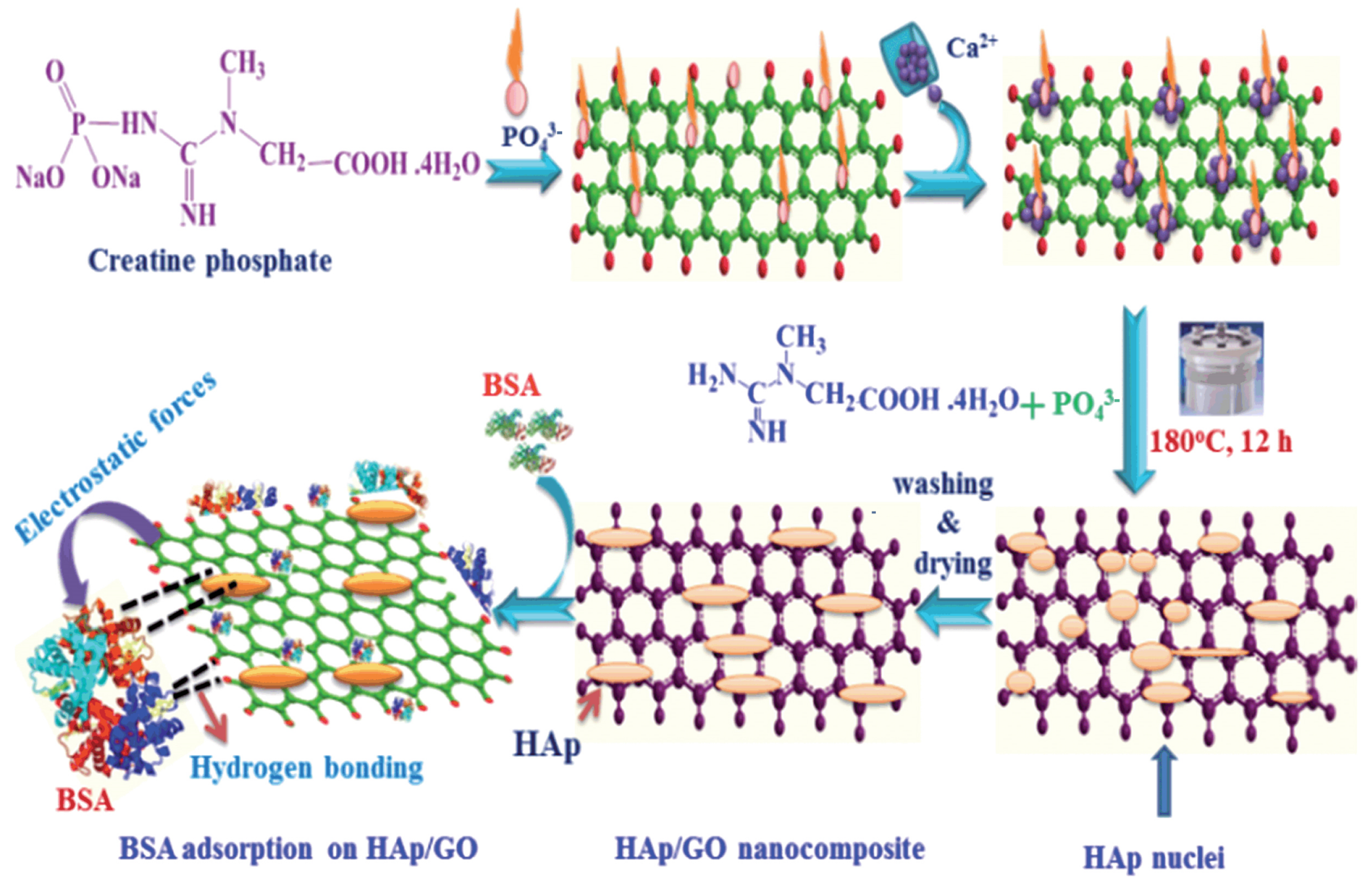
The solvothermal synthesis method is a variation of the hydrothermal method, where organic solvents are used instead of water. This method allows for the use of higher temperatures and pressures, which can increase the solubility of the reactants and promote more rapid reaction rates (Figure 2) [25,26].
In a typical solvothermal synthesis procedure, the starting materials are mixed in a solvent using magnetic stirring. The mixture is then loaded into a sealed container, such as an autoclave, and heated to a high temperature for a fixed period of time. The pressure inside the container is also increased, which can further enhance the solubility of the reactants and accelerate the reaction. After the reaction is complete, the resulting product is typically washed with water or alcohol to remove any impurities or residual reactants. Finally, the product is vacuum-dried to remove any remaining solvent, and the final product can be obtained. Temperature and pressure, reaction time, precursor concentration, solvent, additives, and catalysts influence the structure, morphology, and properties of nano calcium phosphate obtained through solvothermal synthesis. Solvothermal synthesis involves heating the reaction mixture at elevated temperatures and pressures within a sealed autoclave. Temperature and pressure conditions have a significant effect on crystal growth, phase formation, and particle size distribution. Higher temperatures and pressures generally promote faster crystal growth, resulting in larger particles with enhanced crystallinity [27]. Longer reaction times allow for more extensive crystal growth, leading to larger particles, while prolonged reaction times may also result in increased agglomeration or phase transformation [28]. Higher precursor concentrations can lead to the formation of larger particles with increased crystallinity, but at excessively high concentrations, particle agglomeration may occur [29]. Different solvents have varying solubility properties, which affect the precursor dissolution and subsequent precipitation processes. Additional additives or catalysts can also be introduced in solvothermal synthesis to modify the particle characteristics [30]. For example, surfactants or templates can be employed to control particle size or morphology. Catalytic agents may be used to enhance the reaction rate or promote specific crystal phases.
The solvothermal synthesis method has been used to prepare a variety of calcium phosphate-based nanoparticles with controlled size, morphology, and composition. This method allows for greater control over the reaction conditions and can result in the production of biomaterials with improved properties and functionality compared to those obtained by other methods [31]. One of the main benefits of the solvothermal synthesis method is its ability to dissolve almost any material in the solvent, which can be heated and pressurized to its melting point. This allows for the synthesis of highly ordered nanostructures with controlled size, morphology, and composition. For example, the solvothermal method has been used to synthesize highly ordered arrays of microtubes made from a calcium chloride (CaCl2) powder precursor, which can form a block of three-dimensional structure. In addition, the solvothermal method has also been used to transform unstable di-tert-butyl phosphate into a stable precursor for calcium phosphate ceramic biomaterials using different calcium sources [32].
However, the solvothermal method does have some challenges; one of the major weaknesses is the difficulty in controlling the morphology and structure of the resulting nanostructures. This is due to multiple variables, such as the type, volume ratio, and concentration of the starting materials, as well as the reaction conditions, such as temperature and pressure [33]. Furthermore, the use of organic solvents can also pose some challenges, such as the need for appropriate safety precautions and the potential for toxicity [34]. Despite these challenges, the solvothermal method remains an important technique for the synthesis of CaP nanoparticles with tailored properties and functionality for bone tissue engineering and other applications.
Figure 2. Schematic representation of solvothermal synthesis for the preparation of calcium phosphate-based biomaterial. Reprinted with permission from Ref. [31], Copyright 2016, Royal Society of Chemistry (London, UK).
Molecules 28 04790 g002

2.3. Sol-Gel Method

As shown in Figure 3, the sol-gel method is another wet-chemical method for the synthesis of calcium phosphate-based nanoparticles. It is a relatively new procedure that does not require high temperature to help the compound reaction [35]. The sol-gel method typically involves several stages, including hydrolysis and polycondensation, gelation, drying, and crystallization. In the first step, inorganic materials, such as chlorides, nitrates, and sulfides, or metal alkoxides are dissolved in an acidic or alkaline solution and are stirred for a long time to obtain precursors. Metal alkoxides are then added to the precursors for polymerization, which leads to the development of a gel. Finally, the gel is converted into solid materials by freeze-drying or drying with high temperature [36,37] and can be used for coating scaffolds without any other drying.
Precursor composition, pH and solvent, hydrolysis and condensation ratios, drying methods, and annealing and heat treatment contribute to the regulation of the structure, morphology, and properties of nano calcium phosphate obtained through the sol-gel method [38]. Different precursors, such as inorganic salts or alkoxides, can lead to variations in the stoichiometry, crystallinity, and phase composition of the nanoparticles [39]. Varying the pH can affect the hydrolysis and condensation reactions, which, in turn, influence the particle size, surface charge, and morphology. The hydrolysis of the precursor and polycondensation of the resolved chemical products are the most significant steps in this synthesis [40]. Through this process, a wide range of diameters of nanoparticles can be synthesized by varying the ratio of materials [41]. These steps also affect the features of the final composites, such as specific surface area and microporosity, which can be influenced by using different reagents, various ratios of raw materials, or different temperature ranges [36,42,43]. Different drying techniques, such as freeze-drying or high-temperature drying, can lead to variations in the particle size distribution, agglomeration, and phase composition. Annealing can induce phase transformations, crystal growth, and improved crystallinity. The temperature and duration of heat treatment can be optimized to achieve desired properties, such as enhanced crystallinity, improved stability, and controlled release behavior.
Though there are still big challenges, the sol-gel method is still widely used due to its ability to produce CaP nanomaterials with high purity, good crystallinity, and controllable morphologies, making it suitable for various biomedical applications, including bone tissue engineering.

2.4. Microwave-Assisted Method

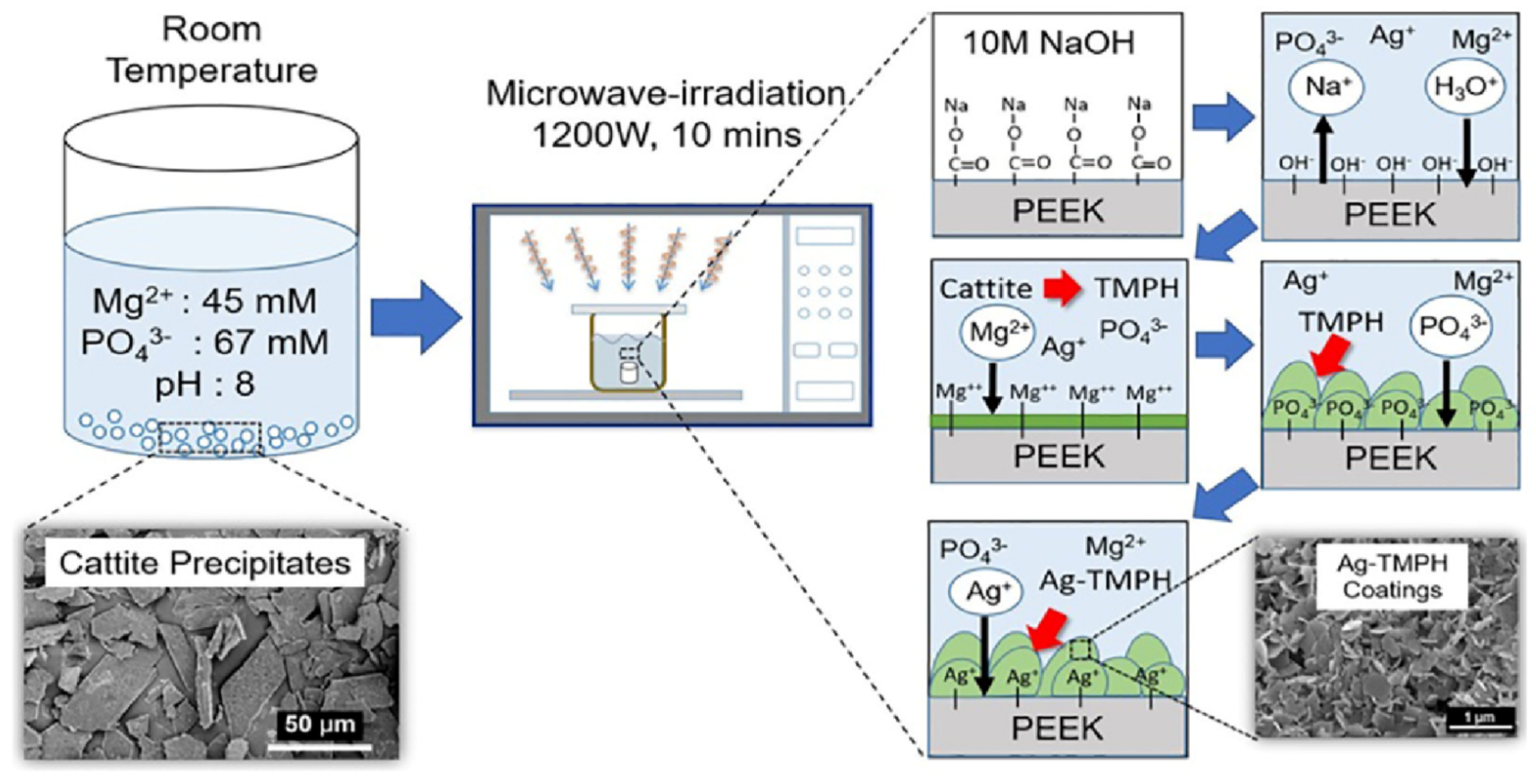
Microwave-assisted synthesis is a method of heating and accelerating the chemical reaction between materials to obtain CaP particles [44,45]. As shown in Figure 4 [45], the process involves dissolving soluble materials in a solvent, adjusting the pH value of the mixture, subjecting the mixture to microwave irradiation at a fixed temperature and time, and finally isolating the product through centrifugation, cleaning, and drying [46,47].
Power and duration, solvent and pH, precursor concentration and ratio, additives and surfactants, and cooling rate influence the formation of CaP nanoparticles [48]. Higher power and longer durations can lead to increased heating and higher reaction temperatures, affecting the nucleation, growth, and crystallinity of the nano calcium phosphate particles [49]. Different solvents and pH conditions can affect the particle size, morphology, and phase composition of the nano calcium phosphate [17]. Varying the precursor concentrations and ratios can affect the nucleation and growth kinetics, leading to variations in particle size, crystallinity, and surface properties [50]. These additives can act as nucleation promoters, growth modifiers, or surface modifiers, resulting in controlled particle formation, improved dispersibility, and enhanced stability. Rapid cooling can lead to the formation of amorphous or partially crystalline phases, while slower cooling rates may promote the growth of larger crystals [51]. The cooling rate can influence the phase composition, crystallinity, and surface properties of the synthesized nanoparticles. Thus, the structure, morphology, phase, as well as particle size can be regulated via the parameters.
Microwave-assisted synthesis has some advantages, such as high energy efficiency, shorter synthesis time, as well as consuming few samples [39], which make it a promising method for the laboratory. However, the high heat generated can also lead to some disadvantages, such as the deterioration of the sample constituents and the high cost of industry-level equipment maintenance. It is important to consider both the advantages and disadvantages of this method before using it in research or industrial applications.

2.5. Sonochemical Synthesis

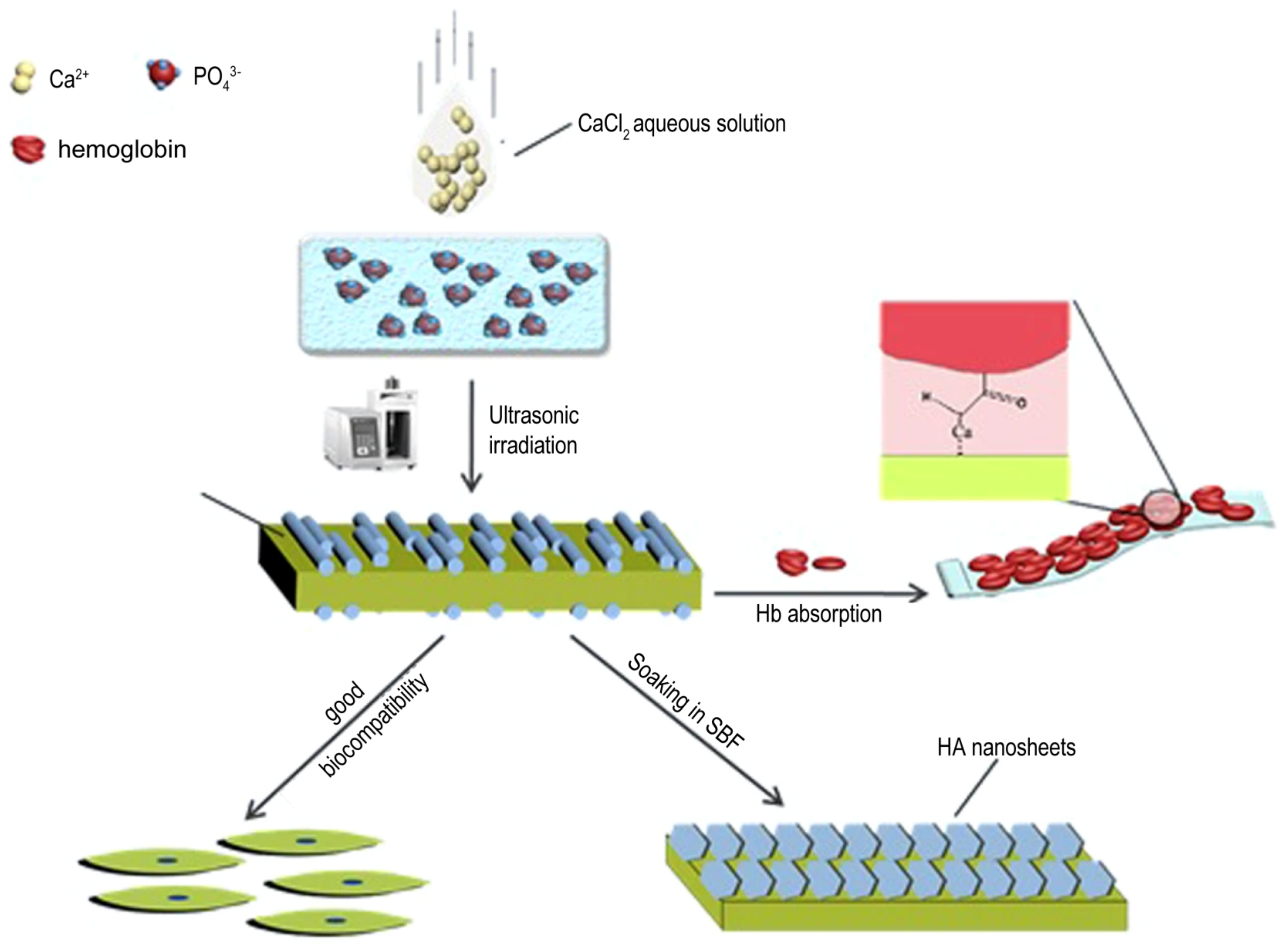
As shown in Figure 5, sonochemical synthesis has the potential to offer several advantages over other synthesis methods, such as being safer and more environmentally friendly, as it does not require the use of toxic solvents [52]. This method also has the advantage of the reduction in reaction time, the ability to control particle size, and the production of highly homogeneous materials. The method can also be carried out at relatively low temperatures and does not require high-pressure conditions, which reduces the energy consumption and cost of the process. Additionally, sonochemistry can be used to modify the surface properties of materials, such as increasing their porosity, improving their biocompatibility, and enhancing their drug delivery capabilities. Additionally, the cavitation effect can lead to smaller and more uniform particle size, which can increase the protein absorption of the nanoparticles [53]. Herein, ultrasonic power and frequency, [54] reactant concentration, [55] pH and temperature, the addition of surfactants and additives, and reaction time [56] all influence the formation of calcium phosphate nanoparticles via sonochemical synthesis.
Further research is needed to fully understand the effects of sonochemical synthesis on the properties of calcium phosphate-based nanoparticles, including their crystallinity and morphology [56]. With continued exploration and the refinement of this method, it may prove to be a valuable approach for the synthesis of high-quality calcium phosphate-based materials with a wide range of potential applications. The main disadvantage of sonochemistry is that it is difficult to scale up to industrial-level production due to the limitations of the equipment and the variability in the cavitation effect.

2.6. Enzyme-Assisted Method

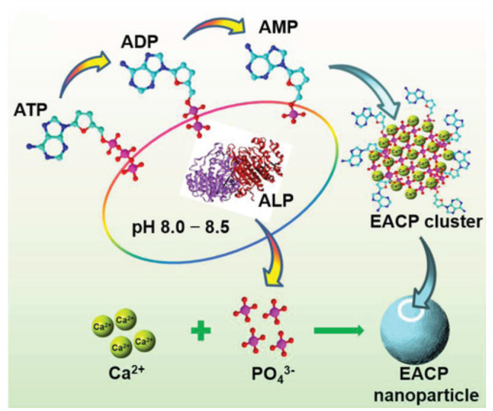
There is also research that shows that calcium phosphate-based nanoparticles, obtained in the enzymatic approach, have strong bioactivity in vivo/vitro experiments due to functional enzymes in promoting biomineralization [57]. As shown in Figure 6, Jiang et al. developed bioactive minerals via the enzymatic reaction based on the use of alkaline phosphatase (ALP) to catalyze the hydrolysis of ATP molecules and the release of phosphate ions combined with the unconstrained calcium ions in the precursor solutions. Thereafter, the organic-inorganic composites containing amorphous calcium phosphate, ATP, ADP, and AMP were obtained, and the composites showed superior biocompatibility, enhanced osteogenic differentiation, and reduced repair time for bone regeneration [58]. Additionally, the control of rate of deposition and final coating morphology can be reached by adjusting the value of the pH, the concentration of the enzyme, the ratio of calcium and phosphate ions, and the time of reaction [59].
Enzymatic methods offer several advantages in the synthesis of calcium phosphate-based biomaterials. Firstly, they are mild and environmentally friendly because the reaction is performed under mild conditions without the use of harsh chemicals or high temperatures [60]. Secondly, the obtained materials are usually of highly purity, which is essential for their biological activity. Thirdly, enzymatic methods have the potential to mimic the biomineralization processes that occur in living organisms, leading to the synthesis of biomaterials with unique properties that are not possible to achieve via other methods [57]. The enzymatic synthesis method also has some drawbacks. The enzymatic reaction is often slow and requires longer reaction times than other methods. Additionally, the enzyme used in the process can be expensive and can affect the final product yield.

2.7. Spray Drying and Electrospinning

Spray drying and electrospinning are two other methods used for the preparation of calcium phosphate nanomaterials. In the spray drying method, a solution containing the calcium and phosphate precursors is sprayed into a hot air stream, resulting in the formation of nanoparticles. In addition, the spray process can also be applied to the preparation of calcium phosphate coating [61]. In the electrospinning method, a polymer solution containing calcium and phosphate precursors is used to form nanofibers, which are then calcined to obtain the desired calcium phosphate nanomaterials.
Additionally, plasma spraying can also be used for the preparation of calcium phosphate coating, which can effectively prevent the corrosion and loosening of metal implants in clinic. Plasma spraying requires a high cost; therefore, the application is limited. Liu et al. developed a double-layer calcium phosphate-sandwiched siloxane composite coating that can decrease the cost by using the chemical conversion method and the sequential mineralization method to promote the loading of Ca and P elements with the coating so that this magnesium alloy with the composite coating can achieve good mineralization ability and biocompatibility in vivo [62]. Moreover, calcium phosphate coatings can also be obtained via sequential mineralization in SBF [63] or electrochemical-assisted deposition [64].
For spray drying and electrospinning, the concentration of the calcium and phosphate precursors in the spray drying and electrospinning solutions affects the viscosity and surface tension of the solution. Higher concentrations typically result in increased particle size and improved crystallinity. Different solvents have varying evaporation rates and compatibility with the precursors, [65] leading to differences in particle size, porosity, and surface characteristics. Higher airflow rates and temperatures generally result in smaller particle sizes and improved crystallinity. Optimal feed rates ensure uniform particle distribution [66]. Different polymers have varying viscosities, conductivity, and compatibility with the precursors, leading to differences in fiber morphology, diameter, and mechanical properties [67]. Post-treatment techniques, such as heat treatment or cross-linking, can influence the crystallinity, porosity, and mechanical strength of the nanoparticles.
As described above, each synthesis method has its own unique advantages and limitations for the preparation of calcium phosphate nanomaterials, and a summary is given in Table 1. Proper synthesis methods can be employed for preparing calcium phosphate nanomaterials with desired properties.

3. Multifunctional Properties of Calcium Phosphate-Based Nanomaterials

The various synthesis methods contribute to the diversity of calcium phosphate-based nanoparticles. Herein, calcium phosphate-based nanomaterials have favorable properties, involving biocompatibility, biodegradability, and the fact that calcium is a critical mineral composition of human bones and teeth [85], and calcium phosphate-based nanomaterials can also decompose in an aqueous solution with the pH value in human internal environment [86]. Thus, it can be used as a great carrier to load drugs and can protect them in blood [87], leading to controllable release in the human internal situation.
Moreover, large specific surface areas of calcium phosphate-based nanomaterials lead to loading more therapeutic drugs, increasing the concentration of the drug around the targeted sites to achieve efficient bone healing [88,89]. Based on this reason, Kans et al. developed CaP-based coatings with interconnected pores via ultrasound-assisted microarc oxidation method. The porous structure of the coatings contributes to the improvement of the vancomycin loading capacity to achieve more effective antibacterial properties. Additionally, by taking advantage of the electrostatic interaction between Ca2+ ions, on the surface of the CaP, and COO- in bovine serum albumin (BSA), Kanupriya et al. found that anhydrous dicalcium phosphate synthesized via rapid microwave-assisted synthesis had higher porosity (77.8 m2/g), providing more binding sites for BSA. The porous anhydrous dicalcium phosphate showed about a 59 wt% loading capacity of BSA, which is much higher than that of hydroxyapatite with a lower specific surface area (16.1 m2/g) [90].
As shown above, calcium phosphates have the ability to incorporate biomolecules, inside or on the surface, to reach the target sites and deliver therapeutic compounds. In addition, some biomolecules can combine with inorganic structures to repair bone defects by suppressing the acute inflammatory response [91,92] or various active ingredients, promoting the formation of new blood vessels, inhibiting the fusion and migration of osteoclasts [93] or directly encouraging osteogenic differentiation [94,95,96]. As shown in Figure 7a, Kai-Chi et al. developed uniform carbonate-based apatite nanorods, which upregulate cellular attachment and mineralization, by combining ferulic acid through the regulation of heparin [97]. Additionally, many growth factors show various functions to control the evolution, differentiation, and metabolism of stem cells in the process of bone regeneration, including platelet-derived growth factor (PDGF), transforming growth factor beta1 (TGFbeta1), vascular endothelial growth factor (VEGF), basic fibroblast growth factor (bFGF), hepatocyte growth factor (HGF), epidermal growth factor (EGF), insulin-like growth factor-1 (IGF-1), and so on. Bone-morphogenetic proteins (BMP) show especially promising results when used in bone regeneration. As shown in Figure 7b, Seong et al. combined BMP-2 with the crosslinked porous biphasic calcium phosphate, invented by applying a water-in-oil emulsion technique with camphene as a pore generator, to achieve high loading efficiency and constant release [95]. In another study shown in Figure 7c, there is a phenomenon that the calcium pyrophosphate can keep the shape of nanofiber microspheres in the presence of a low pH value (pH~4), consisting of a stomach environment, and can reorganize into fiber and even into mineral plates in the intestinal environment (pH~7), which means that the large specific surface area of 65 m2/g turns into only 1.5 m2/g; thus, the loaded protein can keep its high activity when exposed to the stomach environment. Subsequently, the loaded proteins would be released in the intestinal environment, which leads to targeted enteric delivery [98].
Stem cells, such as hBMSCs [10], hESC-MSCs [99], and hiPSC-MSCAs [100], play a critical role in the process of bone regeneration. Several studies have investigated that calcium-based materials can load these cells [101], and the interaction between nanostructures and cells contributes to bone healing [102,103]. Calcium phosphate bone cement (CPC) was compounded with bone marrow mesenchymal stem cells (BMSCs) and was condensed in alginate chitosan alginate (ACA) microcapsules. This can protect cells from the negative influence of the CPC setting reaction and effectively form macropores in the composite to help CPC display better bone regeneration functions in the performance of ectopic osteogenesis and high effective degradability in vivo compared to the CPC with the addition of pure microcapsules or cell microcapsules [103]. Calcium phosphates, which are osteoconductive, injectable, and moldable, can provide strong sites for the proliferation and differentiation of cells and can promote bone healing.
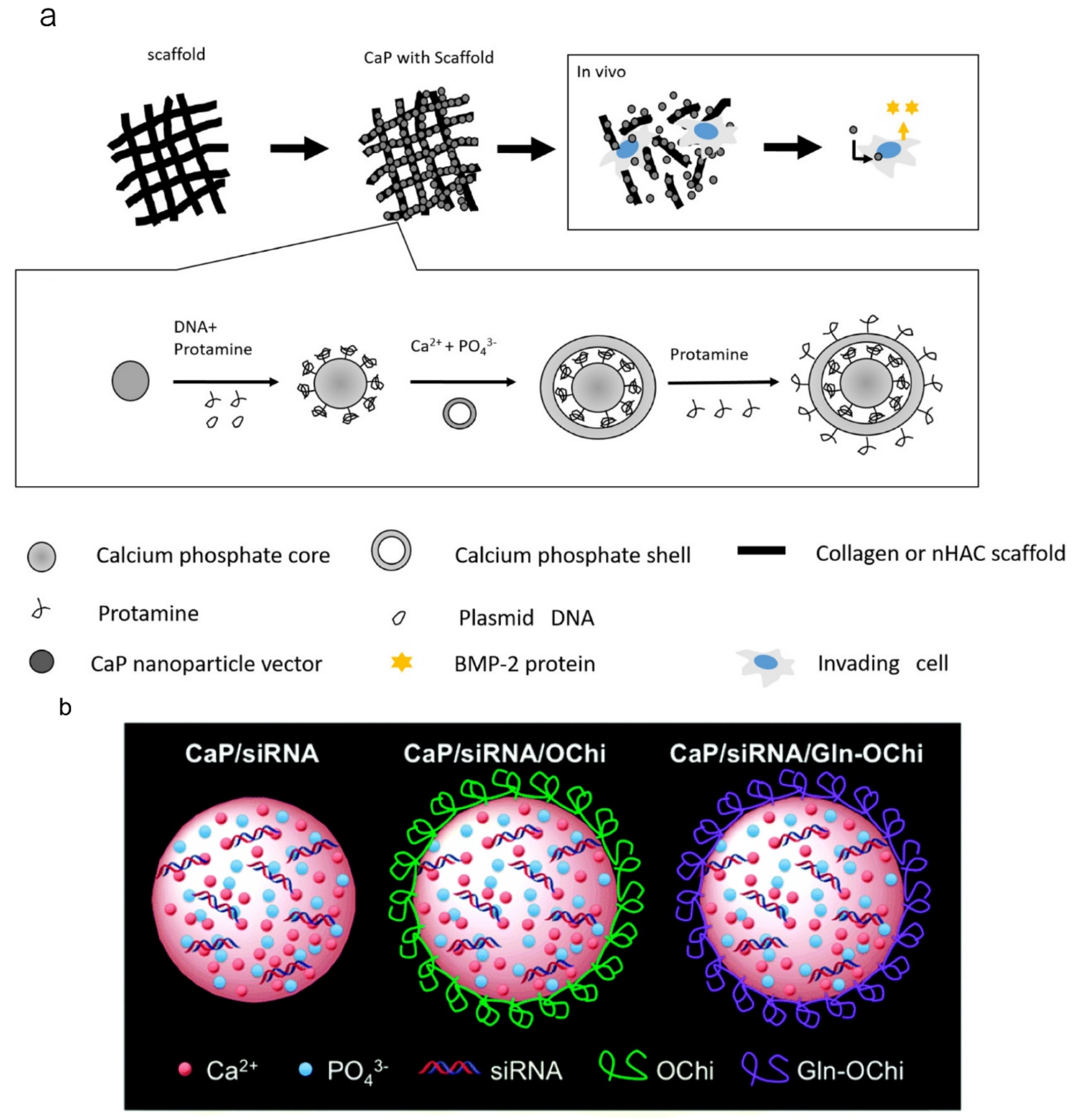
Gene therapy is a potential and new approach to achieve highly efficient bone regeneration [104]. Calcium phosphate nanomaterials can be used as carriers for RNA [85,105] or DNA [106,107]. BMP-2 promotes bone regeneration effectively, and a high dosage of it can be loaded on the calcium phosphate nanocarriers to achieve the satisfied release of the growth factor during the stage of bone regeneration. However, there is difficulty in achieving the controlled release of growth factors during bone regeneration. A fast degradation rate leads to the release of BMP-2 at a high level, as well as bone or tooth resorption. To solve this problem, achieving larger, longer, and a more gradual release of BMP-2, as shown in Figure 8a, Tenkumo et al. designed a multi-shell calcium phosphate nanoparticle as a carrier with BMP-2-encoding-plasmid DNA. Thereafter, the drug delivery system was incorporated with a nanohydroxyapatite-collagen scaffold to promote gene transfection successfully [96]. The efficient cellular uptake of nanoparticle-loaded nucleic acid molecules has a huge influence on the efficiency of bone regeneration. In Figure 8b, it is shown that Bogyu Choi et al. found that nucleic acid-loaded CaP nanoparticles with a Gln-Ochi chitosan coating can protect these particles from forming large aggregates due to electrostatic repulsion and can achieve higher cellular uptake to promote bone tissue regeneration.
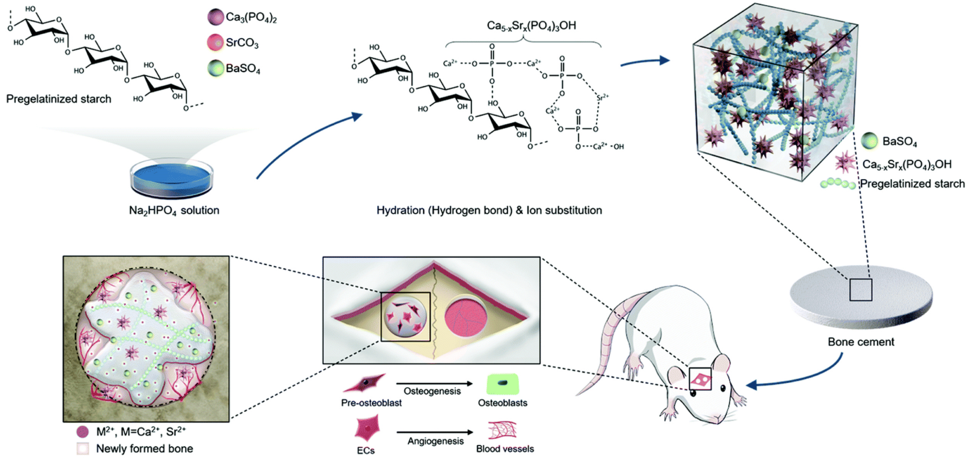
Metal ions can also be integrated within calcium phosphate-based nanomaterials, regulating gene expression and promoting bone repair [109,110,111,112,113]. As shown in Figure 9, Wu et al. introduced strontium ions, which came from SrCO3, into the modified calcium phosphate cement, composed of 20 wt% pregelatinized starch and 10 wt% BaSO4, to improve the flexibility, uniformity, and radiopacity performance of the cement. A total of 0.25 M of the Na2HPO4 solution acted as the setting liquid. This mixture performed well in a calvarial defect model of a rat and showed suitable mechanical strength [114].

4. Applications of Calcium Phosphate-Based Nanomaterials in Bone Tissue Engineering

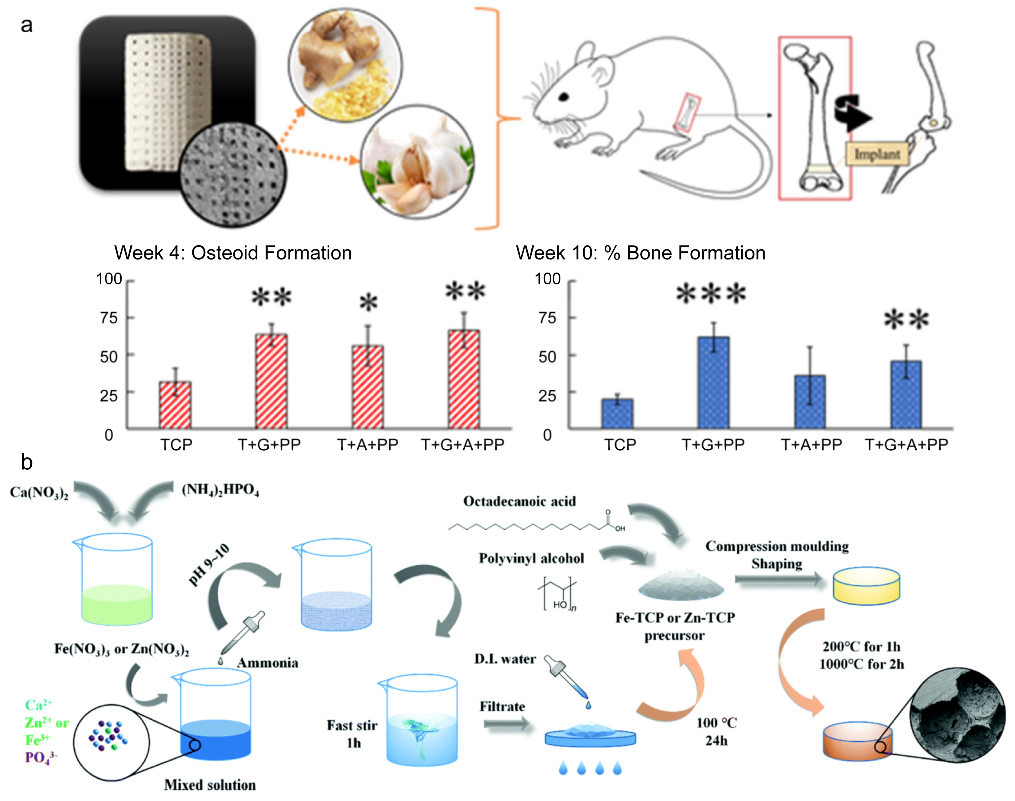
Calcium phosphate-based nanomaterials show superior biocompatibility, show osteoinductivity, and have high surface area and porosity. Thus, they have been widely used for bone regeneration [115]. Additionally, calcium phosphate-based nanomaterials can be doped with special compounds to promote bone repair directly. Susmita et al. designed 3D calcium phosphate scaffolds, resembling the structure of cortical and cancellous bones, to provide a stronger, denser structure to develop compressive strengths of the scaffold and maintain the central interconnected porosity for cell proliferation and vascularization. The scaffold was incorporated with natural compounds from ginger root and garlic powder, which can enhance bone healing when used in vivo, and improved the bioavailability of these medicinal compounds [116] (Figure 10a). Furthermore, many metal elements also have various influences on the process of bone regeneration, such as silicon, magnesium, cerium, and so on. Based on this theory, Fe and Zn were combined with tricalcium phosphate (TCP) via co-deposition because these two elements can improve new blood vessel formation, which can significantly promote bone regeneration; then, the Fe/Zn-modified TCP powders were turned into a solid, and micropores were formed inside at room temperature. The composites achieved the sustained release of metal (Ca2+, Zn2+ and Fe3+) ions, which can promote the process of bone repair [117] (Figure 10b). Additionally, because of the low mechanical strength, brittleness, and quick resorption rate, CaP-based scaffolds need to be reinforced with mechanical strength and be improved regarding bioactivity. It is reported that bioceramic powders (hydroxyapatite and β-tricalcium phosphate) have been embedded in mixed solutions of chitosan and silk fibroin to improve both the mechanical properties and the differentiation of mesenchymal stem cells since chitosan and silk fibroin have shown better bioactivity in supporting the growth and differentiation of BMSCs. Additionally, the polymer solution can be used as the bioink of robocasting equipment to print 3D scaffolds with fixed system parameters at room temperature, and this scaffold achieved great performance regarding mechanical properties and biocompatibility [118]. Calcium phosphate-based materials can also be used as carriers to load cells or growth factors, which can improve effective therapeutic ingredients in bone defect areas, can keep the sustained release of these factors, and can provide enough room for osteoblasts and new blood vessels [119]. Li et al. reported that bone marrow mesenchymal stem cells and platelet-rich plasma combined with a calcium phosphate cement scaffold achieved more newly formed bone areas than normal calcium phosphate cement scaffolds in an in vivo experiment [120].
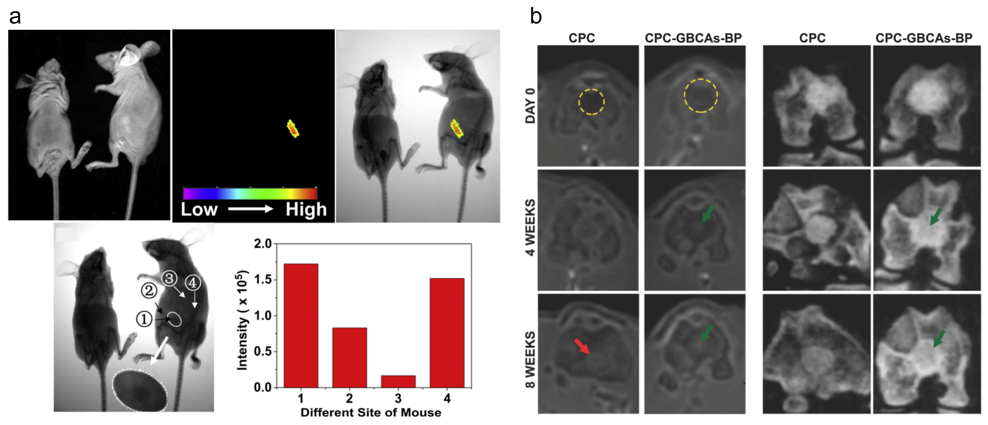
Recently, near-infrared-emitting persistent luminescence nanoparticles have been used as an optical probe for bioimaging and biosensing [121,122]. Though they show low absorptivity in deep-tissue imaging, achieving stable in vivo luminescence is still a great challenge [123]. Chen et al. developed Eu3+/Gd3+ ion-dual-doped CaP nanoparticles via co-precipitation using block copolymer polylactide-block-monomethoxy as a template. The nanoparticles showed prolonged stability (more than 80 days), improved the T1-weighted MRI signal intensity, and decreased T2-weighted MRI signal intensity, which resulted in darker images for the better observation of changes in local areas in vivo [124] (Figure 11a). Additionally, to monitor the in vivo performance of calcium phosphate cements, which show a high structural similarity to bone [125], calcium phosphate cements linked with gadolinium (III) by bisphosphonates were developed, and they showed shortened T1 relaxation times of the water in tissues and achieved signal enhancement in a T1-weighted MRI. In this way, the highly accurate evaluation of the implant shape both in MRI and CT for a prolonged time period (about 8 weeks) was achieved [126] (Figure 11b).
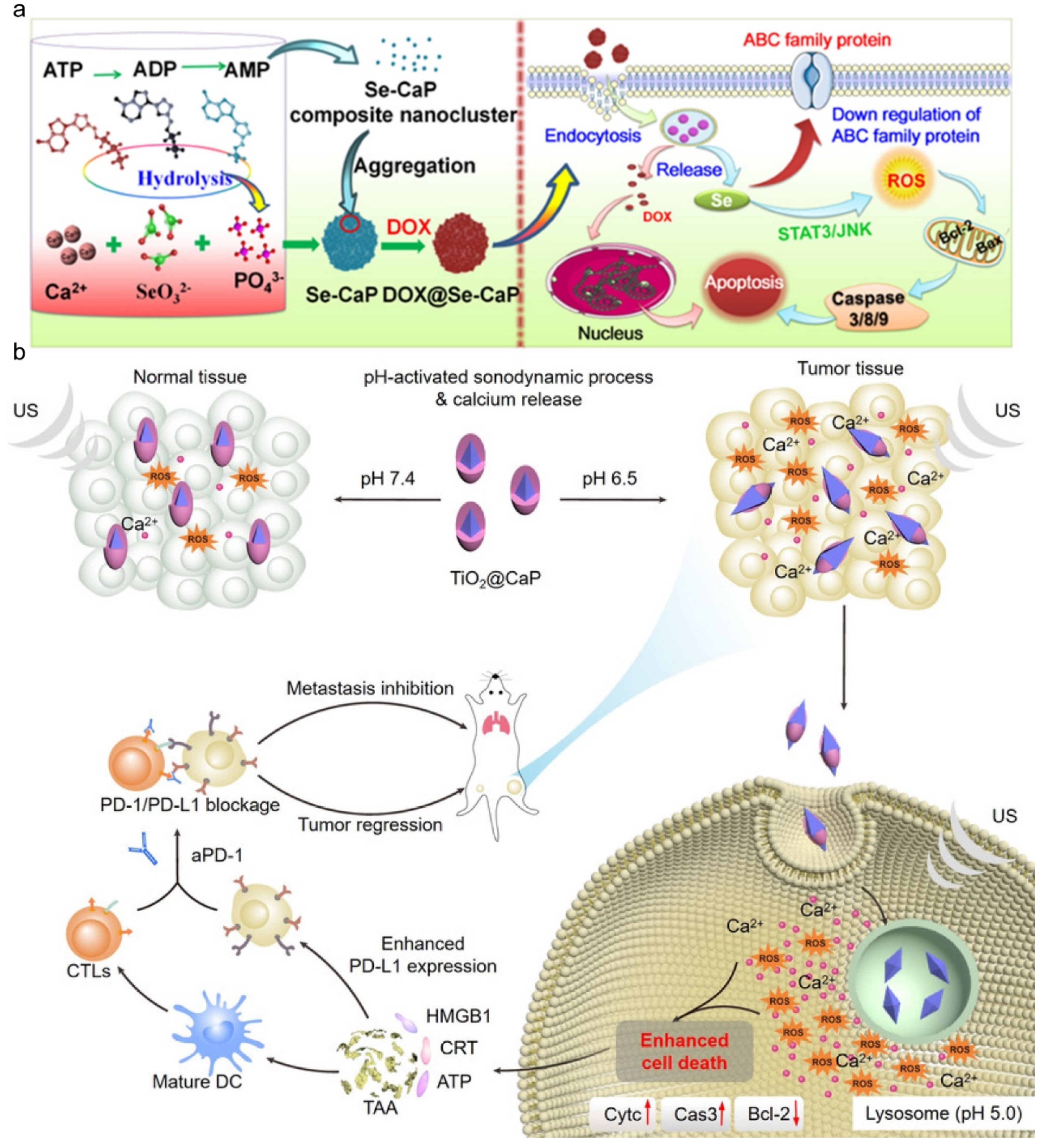
Calcium phosphate-based nanoparticles are also desirable materials for drug delivery [127]. Doxorubicin could activate the immunogenic cell death of tumor cells through an apoptosis pathway, including reactive oxygen species, which could lead to DNA cleavage and mitochondrial dysfunction [128], while the multidrug resistance of cancers may result in poor prognosis [129]. To address this issue, a selenium-doped CaP, serving as the doxorubicin carrier, was designed for reversing the multidrug resistance of tumors. Se-doped CaP is pH-sensitive and dox can be released specifically in the tumor sites due to the acid microenvironment. Thus, the uptake of anticancer drugs of tumor cells was raised, and Se ions released could decrease reactive oxygen species of tumor cells and the expression of ATP-binding cassette transporters, which reverse multidrug resistance [130] (Figure 12a). The surgery of bone tumors causes large bone defects, and calcium phosphate bone pastes have been widely used as bone substitutes in clinics to replace allogenous bone, providing strong and stable support for the defect sites without internal fixation [131,132], for many years, and the result of anticancer is also well described in the long run [133,134]. Furthermore, there is the theory that calcium ions play a vital role in tumor necrosis by regulating the stability of the intracellular concentration of calcium ions [135,136,137], and a high concentration of calcium in the microenvironment of the tumor can encourage tumor calcification [138,139]. Based on this phenomenon, calcium phosphate can combine with some special materials, such as ferritin, which is a promising load for targeted delivery, to protect ferritin from interception to achieve the function of targeted delivery to the tumor effectively, and the shell of calcium phosphate not only counterbalances the acidic microenvironment around tumors but also accelerates the immunomodulation and calcification of cancers [140,141]. CaP nanoparticles can also be coated to camouflage, which may result in intracellular calcium overload and induce apoptosis. For example, TiO2-coated CaP can elevate the generation of reactive oxygen species, due to the demonstrative property of inorganic sonosensitizers, and can provide calcium ions due to the acidic microenvironment of the tumor (Figure 12b) [142,143,144].

5. Conclusions and Perspectives

Nano-calcium phosphate-based biomaterials have shown great potential in bone tissue engineering due to their unique properties, including their small size, high surface area to volume ratio, and multifunctionality. Various preparation methods, including sol-gel, hydrothermal synthesis, spray drying, and electrospinning, have been used to obtain these biomaterials with controlled size, morphology, and composition.
The multifunctionality of nano-calcium phosphate-based biomaterials is attributed to their abilities to enhance the osteogenic differentiation and mineralization of mesenchymal stem cells, to promote angiogenesis, and to exhibit antimicrobial properties. These properties make them attractive for various applications in bone tissue engineering, including bone defect repair, bioimaging, and anticancer applications.
The preparation, multifunctionality, and application of nano-calcium phosphate-based biomaterials for bone tissue engineering have been extensively studied and reviewed in the literature. These biomaterials have shown great potential in the regeneration and repair of bone tissues and can provide a scaffold for the adhesion, proliferation, and differentiation of stem cells. Further research is needed to optimize the preparation methods, functionalization, and characterization of these biomaterials to improve their performance and clinical outcomes. Nonetheless, these biomaterials represent a promising avenue for the development of effective and safe bone tissue engineering therapies.
The future exploration of calcium phosphate-based nanomaterials could be based on the following points: more effective binding of active substances; more effective targeting methods to achieve more effective treatment goals in local areas; and the interaction mechanism between calcium phosphate and substances. Calcium phosphate binds to the active ingredient and can protect the bioactivity of the component from being affected to maximize the function of the ingredient, such as promoting bone regeneration and anti-cancer properties. In addition, effective targeting strategies can deliver calcium phosphate-loaded components to the relevant areas so that the local area has a high concentration of therapeutic components, which minimizes the harm caused by therapeutic components. In order to achieve the above objectives, it is necessary to further explore the interaction mechanism between calcium phosphate and various substances to achieve the stable synthesis of calcium phosphate materials with related functions and even commercial production into clinical applications. Thus, it is conducive to the stable output of bioactive calcium phosphate material for large-scale production and application in practice.
Thus far, based on the consideration of biosafety, only a few calcium phosphate-based nanoparticles have been applied in clinics. Therefore, we hope that this review inspires readers to obtain more ideas for the design of bone repair materials.

Author Contributions

X.C. and H.L. gave the conceptualization, conducted the investigation, and wrote the original draft; Y.M. and Y.J. revised the manuscript; Y.J. contributed to the funding acquisition. All authors have read and agreed to the published version of the manuscript.

Funding

The authors acknowledge the grants by support from National Natural Science Foundation of China (No. 82102217), Science and Technology Commission of Shanghai Municipality (No. 21YF1413100).

Institutional Review Board Statement

Not applicable.

Informed Consent Statement

Not applicable.

Data Availability Statement

Not applicable.

Conflicts of Interest

The authors declare no conflict of interest.

Sample Availability

Not applicable.

References

  1. Kahar, N.N.F.; Ahmad, N.; Jaafar, M.; Yahaya, B.H.; Sulaiman, A.R.; Hamid, Z.A.A. A review of bioceramics scaffolds for bone defects in different types of animal models: HA andbeta-TCP. Biomed. Phys. Eng. Express 2022, 8, 21. [Google Scholar] [CrossRef]
  2. Greenwald, A.S.; Boden, S.D.; Goldberg, V.M.; Khan, Y.; Laurencin, C.T.; Rosier, R.N. Bone-graft substitutes: Facts, fictions, and applications. J. Bone Joint Surg. Am. 2001, 83 (Suppl. 2), S98–S103. [Google Scholar] [CrossRef] [PubMed]
  3. Wang, J.; Li, W.; He, X.; Li, S.; Pan, H.; Yin, L. Injectable platelet-rich fibrin positively regulates osteogenic differentiation of stem cells from implant hole via the ERK1/2 pathway. Platelets 2023, 34, 2159020. [Google Scholar] [CrossRef] [PubMed]
  4. Ng, J.; Spiller, K.; Bernhard, J.; Vunjak-Novakovic, G. Biomimetic Approaches for Bone Tissue Engineering. Tissue Eng. Part B Rev. 2017, 23, 480–493. [Google Scholar] [CrossRef] [PubMed]
  5. Fu, Q.; Saiz, E.; Rahaman, M.N.; Tomsia, A.P. Toward Strong and Tough Glass and Ceramic Scaffolds for Bone Repair. Adv. Funct. Mater. 2013, 23, 5461–5476. [Google Scholar] [CrossRef] [PubMed]
  6. Tomasi, C.; Regidor, E.; Ortiz-Vigón, A.; Derks, J. Efficacy of reconstructive surgical therapy at peri-implantitis-related bone defects. A systematic review and meta-analysis. J. Clin. Periodontol. 2019, 46 (Suppl. 21), 340–356. [Google Scholar] [CrossRef] [Green Version]
  7. Amini, Z.; Lari, R. A systematic review of decellularized allograft and xenograft-derived scaffolds in bone tissue regeneration. Tissue Cell 2021, 69, 101494. [Google Scholar] [CrossRef]
  8. Fillingham, Y.; Jacobs, J. Bone grafts and their substitutes. Bone Joint J. 2016, 98-B, 6–9. [Google Scholar] [CrossRef]
  9. Zhao, R.; Yang, R.; Cooper, P.R.; Khurshid, Z.; Shavandi, A.; Ratnayake, J. Bone Grafts and Substitutes in Dentistry: A Review of Current Trends and Developments. Molecules 2021, 26, 3007. [Google Scholar] [CrossRef]
  10. Xu, H.H.; Wang, P.; Wang, L.; Bao, C.; Chen, Q.; Weir, M.D.; Chow, L.C.; Zhao, L.; Zhou, X.; Reynolds, M.A. Calcium phosphate cements for bone engineering and their biological properties. Bone Res. 2017, 5, 17056. [Google Scholar] [CrossRef] [Green Version]
  11. Yang, Y.; Su, S.; Liu, S.; Liu, W.; Yang, Q.; Tian, L.; Tan, Z.; Fan, L.; Yu, B.; Wang, J.; et al. Triple-functional bone adhesive with enhanced internal fixation, bacteriostasis and osteoinductive properties for open fracture repair. Bioact. Mater. 2023, 25, 273–290. [Google Scholar] [CrossRef] [PubMed]
  12. Idaszek, J.; Jaroszewicz, J.; Choinska, E.; Gorecka, Z.; Hyc, A.; Osiecka-Iwan, A.; Wielunska-Kus, B.; Swieszkowski, W.; Moskalewski, S. Toward osteomimetic formation of calcium phosphate coatings with carbonated hydroxyapatite. Biomater. Adv. 2023, 149, 213403. [Google Scholar] [CrossRef] [PubMed]
  13. Tabaković, A.; Kester, M.; Adair, J.H. Calcium phosphate-based composite nanoparticles in bioimaging and therapeutic delivery applications. Wiley Interdiscip. Rev.—Nanomed. Nanobiotechnol. 2012, 4, 96–112. [Google Scholar] [CrossRef] [PubMed]
  14. Zhu, Q.; Chen, Z.; Paul, P.K.; Lu, Y.; Wu, W.; Qi, J. Oral delivery of proteins and peptides: Challenges, status quo and future perspectives. Acta Pharm. Sin. B 2021, 11, 2416–2448. [Google Scholar] [CrossRef] [PubMed]
  15. Szurkowska, K.; Szeleszczuk, Ł.; Kolmas, J. Effects of Synthesis Conditions on the Formation of Si-Substituted Alpha Tricalcium Phosphates. Int. J. Mol. Sci. 2020, 21, 9164. [Google Scholar] [CrossRef]
  16. Kim, H.-W.; Kim, Y.-J. Effect of silicon or cerium doping on the anti-inflammatory activity of biphasic calcium phosphate scaffolds for bone regeneration. Prog. Biomater. 2022, 11, 421–430. [Google Scholar] [CrossRef]
  17. Sanchez-Campos, D.; Valderrama, M.I.R.; Lopez-Ortiz, S.; Salado-Leza, D.; Fernandez-Garcia, M.E.; Mendoza-Anaya, D.; Salinas-Rodriguez, E.; Rodriguez-Lugo, V. Modulated Monoclinic Hydroxyapatite: The Effect of pH in the Microwave Assisted Method. Minerals 2021, 11, 13. [Google Scholar] [CrossRef]
  18. Latocha, J.; Wojasinski, M.; Sobieszuk, P.; Gierlotka, S.; Ciach, T. Impact of morphology-influencing factors in lecithin-based hydroxyapatite precipitation. Ceram. Int. 2019, 45, 21220–21227. [Google Scholar] [CrossRef]
  19. Esposti, L.D.; Dotti, A.; Adamiano, A.; Fabbi, C.; Quarta, E.; Colombo, P.; Catalucci, D.; De Luca, C.E.; Iafisco, M. Calcium Phosphate Nanoparticle Precipitation by a Continuous Flow Process: A Design of an Experiment Approach. Crystals 2020, 10, 17. [Google Scholar] [CrossRef]
  20. Khayrutdinova, D.R.; Goldberg, M.A.; Antonova, O.S.; Krokhicheva, P.A.; Fomin, A.S.; Obolkina, T.O.; Konovalov, A.A.; Akhmedova, S.A.; Sviridova, I.K.; Kirsanova, V.A.; et al. Effects of Heat Treatment on Phase Formation in Cytocompatible Sulphate-Containing Tricalcium Phosphate Materials. Minerals 2023, 13, 147. [Google Scholar] [CrossRef]
  21. Goh, K.W.; Wong, Y.H.; Ramesh, S.; Chandran, H.; Krishnasamy, S.; Ramesh, S.; Sidhu, A.; Teng, W.D. Effect of pH on the properties of eggshell-derived hydroxyapatite bioceramic synthesized by wet chemical method assisted by microwave irradiation. Ceram. Int. 2021, 47, 8879–8887. [Google Scholar] [CrossRef]
  22. Latocha, J.; Wojasinski, M.; Janowska, O.; Chojnacka, U.; Gierlotka, S.; Ciach, T.; Sobieszuk, P. Morphology-controlled precipitation/remodeling of plate and rod-shaped hydroxyapatite nanoparticles. Aiche J. 2022, 68, 12. [Google Scholar] [CrossRef]
  23. Lv, L.; Liao, H.; Zhang, T.; Tang, S.W.; Liu, W.Z. Separation of calcium chloride from waste acidic raffinate in HCl wet process for phosphoric acid manufacture: Simulated and experimental study. J. Environ. Chem. Eng. 2022, 10, 108076. [Google Scholar] [CrossRef]
  24. Cai, F.; Yang, L.; Shan, S.; Mott, D.; Chen, B.H.; Luo, J.; Zhong, C.-J. Preparation of PdCu Alloy Nanocatalysts for Nitrate Hydrogenation and Carbon Monoxide Oxidation. Catalysts 2016, 6, 96. [Google Scholar] [CrossRef] [Green Version]
  25. Zhong, H.; Mirkovic, T.; Scholes, G.D. 5.06—Nanocrystal Synthesis. In Comprehensive Nanoscience and Technology; Andrews, D.L., Scholes, G.D., Wiederrecht, G.P., Eds.; Academic Press: Amsterdam, The Netherlands, 2011; pp. 153–201. [Google Scholar] [CrossRef]
  26. Mutalib, A.A.A.; Jaafar, N.F. Potential of deep eutectic solvent in photocatalyst fabrication methods for water pollutant degradation: A review. J. Environ. Chem. Eng. 2022, 10, 107422. [Google Scholar] [CrossRef]
  27. Verma, S.; Murugavel, R. Alkali Metal Di-tert-butyl Phosphates: Single-Source Precursors for Homo- and Heterometallic Inorganic Phosphate Materials. Inorg. Chem. 2022, 61, 6807–6818. [Google Scholar] [CrossRef]
  28. Qi, C.; Zhu, Y.-J.; Ding, G.-J.; Wu, J.; Chen, F. Solvothermal synthesis of hydroxyapatite nanostructures with various morphologies using adenosine 5′-monophosphate sodium salt as an organic phosphorus source. RSC Adv. 2015, 5, 3792–3798. [Google Scholar] [CrossRef]
  29. Hao, H.; Wang, Y.; Shi, B. NaLa(CO3)(2) hybridized with Fe3O4 for efficient phosphate removal: Synthesis and adsorption mechanistic study. Water Res. 2019, 155, 1–11. [Google Scholar] [CrossRef]
  30. Nur, A.; Jumari, A.; Budiman, A.W.; Ruzicka, O.; Fajri, M.A.; Nazriati, N.; Fajaroh, F. Electrosynthesis of Cobalt—Hydroxyapatite Nanoparticles. In Proceedings of the 4th International Conference on Industrial, Mechanical, Electrical, and Chemical Engineering (ICIMECE), Surakarta, Indonesia, 9–11 October 2018. [Google Scholar]
  31. Bharath, G.; Latha, S.B.; Alsharaeh, E.H.; Prakash, P.; Ponpandian, N. Enhanced hydroxyapatite nanorods formation on graphene oxide nanocomposite as a potential candidate for protein adsorption, pH controlled release and an effective drug delivery platform for cancer therapy. Anal. Methods 2016, 9, 240–252. [Google Scholar] [CrossRef]
  32. Verma, S.; Murugavel, R. Di-tert-butylphosphate Derived Thermolabile Calcium Organophosphates: Precursors for Ca(H2PO4)2, Ca(HPO4), α-/β-Ca(PO3)2, and Nanocrystalline Ca10(PO4)6(OH)2. Inorg. Chem. 2020, 59, 13233–13244. [Google Scholar] [CrossRef]
  33. Zhang, Y.-G.; Zhu, Y.-J.; Chen, F.; Sun, T.-W.; Jiang, Y.-Y. Ultralong hydroxyapatite microtubes: Solvothermal synthesis and application in drug loading and sustained drug release. Crystengcomm 2017, 19, 1965–1973. [Google Scholar] [CrossRef]
  34. Yang, M.S.; Xu, P.F.; Zhao, X.Y.; Tian, C.; Han, C.R.; Song, J. Systematic studies of hydroxyapatite/terpene hybrid microspheres: Natural rosin-based phosphorus sources and chemical structures of coordination compounds. Mater. Res. Express 2019, 6, 8. [Google Scholar] [CrossRef]
  35. Bueno, O.M.V.M.; Herrera, C.L.; Bertran, C.A.; San-Miguel, M.A.; Lopes, J.H. An experimental and theoretical approach on stability towards hydrolysis of triethyl phosphate and its effects on the microstructure of sol-gel-derived bioactive silicate glass. Mater. Sci. Eng. C Mater. Biol. Appl. 2021, 120, 111759. [Google Scholar] [CrossRef]
  36. Coelho, S.A.R.; Almeida, J.C.; Unalan, I.; Detsch, R.; Miranda Salvado, I.M.; Boccaccini, A.R.; Fernandes, M.H.V. Cellular Response to Sol-Gel Hybrid Materials Releasing Boron and Calcium Ions. ACS Biomater. Sci. Eng. 2021, 7, 491–506. [Google Scholar] [CrossRef] [PubMed]
  37. Snyder, K.L.; Holmes, H.R.; McCarthy, C.; Rajachar, R.M. Bioactive vapor deposited calcium-phosphate silica sol-gel particles for directing osteoblast behavior. J. Biomed. Mater. Res. A 2016, 104, 2135–2148. [Google Scholar] [CrossRef] [PubMed]
  38. Akbarzade, T.; Farmany, A.; Farhadian, M.; Khamverdi, Z.; Dastgir, R. Synthesis and characterization of nano bioactive glass for improving enamel remineralization ability of casein phosphopeptide-amorphous calcium phosphate (CPP-ACP). BMC Oral. Health 2022, 22, 9. [Google Scholar] [CrossRef]
  39. Christ, B.; Glaubitt, W.; Berberich, K.; Weigel, T.; Probst, J.; Sextl, G.; Dembski, S. Sol-Gel-Derived Fibers Based on Amorphous alpha-Hydroxy-Carboxylate-Modified Titanium(IV) Oxide as a 3-Dimensional Scaffold. Materials 2022, 15, 12. [Google Scholar] [CrossRef] [PubMed]
  40. Yilmaz, E.; Soylak, M. 15—Functionalized Nanomaterials for Sample Preparation Methods. In Handbook of Nanomaterials in Analytical Chemistry; Mustansar Hussain, C., Ed.; Elsevier: Amsterdam, The Netherlands, 2020; pp. 375–413. [Google Scholar] [CrossRef]
  41. Singh, L.P.; Bhattacharyya, S.K.; Kumar, R.; Mishra, G.; Sharma, U.; Singh, G.; Ahalawat, S. Sol-Gel processing of silica nanoparticles and their applications. Adv. Colloid. Interface Sci. 2014, 214, 17–37. [Google Scholar] [CrossRef] [PubMed]
  42. Joughehdoust, S.; Behnamghader, A.; Jahandideh, R.; Manafi, S. Effect of aging temperature on formation of sol-gel derived fluor-hydroxyapatite nanoparticles. J. Nanosci. Nanotechnol. 2010, 10, 2892–2896. [Google Scholar] [CrossRef]
  43. Jmal, N.; Bouaziz, J. Synthesis, characterization and bioactivity of a calcium-phosphate glass-ceramics obtained by the sol-gel processing method. Mater. Sci. Eng. C Mater. Biol. Appl. 2017, 71, 279–288. [Google Scholar] [CrossRef]
  44. Morsali, A.; Hashemi, L. Chapter Two—Nanoscale coordination polymers: Preparation, function and application. In Advances in Inorganic Chemistry; Ruiz-Molina, D., van Eldik, R., Eds.; Academic Press: Cambridge, MA, USA, 2020; Volume 76, pp. 33–72. [Google Scholar]
  45. Sikder, P.; Ren, Y.; Bhaduri, S.B. Microwave processing of calcium phosphate and magnesium phosphate based orthopedic bioceramics: A state-of-the-art review. Acta Biomater. 2020, 111, 29–53. [Google Scholar] [CrossRef] [PubMed]
  46. Zhao, J.; Zhu, Y.-J.; Cheng, G.-F.; Ruan, Y.-J.; Sun, T.-W.; Chen, F.; Wu, J.; Zhao, X.-Y.; Ding, G.-J. Microwave-assisted hydrothermal rapid synthesis of amorphous calcium phosphate nanoparticles and hydroxyapatite microspheres using cytidine 5′-triphosphate disodium salt as a phosphate source. Mater. Lett. 2014, 124, 208–211. [Google Scholar] [CrossRef]
  47. Dharmalingam, K.; Padmavathi, G.; Kunnumakkara, A.B.; Anandalakshmi, R. Microwave-assisted synthesis of cellulose/zinc-sulfate-calcium-phosphate (ZSCAP) nanocomposites for biomedical applications. Mater. Sci. Eng. C Mater. Biol. Appl. 2019, 100, 535–543. [Google Scholar] [CrossRef]
  48. Jodati, H.; Tezcaner, A.; Alshemary, A.Z.; Sahin, V.; Evis, Z. Effects of the doping concentration of boron on physicochemical, mechanical, and biological properties of hydroxyapatite. Ceram. Int. 2022, 48, 22743–22758. [Google Scholar] [CrossRef]
  49. Srinivasan, B.; Kolanthai, E.; Nivethaa, E.A.K.; Pandian, M.S.; Ramasamy, P.; Catalani, L.H.; Kalkura, S.N. Enhanced in vitro inhibition of MCF-7 and magnetic properties of cobalt incorporated calcium phosphate (HAp and ss-TCP) nanoparticles. Ceram. Int. 2023, 49, 855–861. [Google Scholar] [CrossRef]
  50. Matamoros-Veloza, Z.; Rendon-Angeles, J.C.; Yanagisawa, K.; Ueda, T.; Zhu, K.J.; Moreno-Perez, B. Preparation of Silicon Hydroxyapatite Nanopowders under Microwave-Assisted Hydrothermal Method. Nanomaterials 2021, 11, 16. [Google Scholar] [CrossRef]
  51. Sozugecer, S.; Bayramgil, N.P. Preparation and characterization of polyacrylic acid-hydroxyapatite nanocomposite by microwave-assisted synthesis method. Heliyon 2021, 7, 8. [Google Scholar] [CrossRef] [PubMed]
  52. Fu, L.-H.; Qi, C.; Liu, Y.-J.; Cao, W.-T.; Ma, M.-G. Sonochemical synthesis of cellulose/hydroxyapatite nanocomposites and their application in protein adsorption. Sci. Rep. 2018, 8, 8292. [Google Scholar] [CrossRef]
  53. Gausterer, J.C.; Schüßler, C.; Gabor, F. The impact of calcium phosphate on FITC-BSA loading of sonochemically prepared PLGA nanoparticles for inner ear drug delivery elucidated by two different fluorimetric quantification methods. Ultrason. Sonochemistry 2021, 79, 105783. [Google Scholar] [CrossRef]
  54. Muthumariyappan, A.; Rajaji, U.; Chen, S.-M.; Baskaran, N.; Chen, T.-W.; Ramalingam, R.J. Sonochemical synthesis of perovskite-type barium titanate nanoparticles decorated on reduced graphene oxide nanosheets as an effective electrode material for the rapid determination of ractopamine in meat samples. Ultrason. Sonochemistry 2019, 56, 318–326. [Google Scholar] [CrossRef] [PubMed]
  55. Liu, X.; Wu, Z.; Cavalli, R.; Cravotto, G. Sonochemical Preparation of Inorganic Nanoparticles and Nanocomposites for Drug Release-A Review. Ind. Eng. Chem. Res. 2021, 60, 10011–10032. [Google Scholar] [CrossRef]
  56. Qi, C.; Zhou, D.; Zhu, Y.J.; Sun, T.W.; Chen, F.; Zhang, C.Q. Sonochemical synthesis of fructose 1,6-bisphosphate dicalcium porous microspheres and their application in promotion of osteogenic differentiation. Mater. Sci. Eng. C-Mater. Biol. Appl. 2017, 77, 846–856. [Google Scholar] [CrossRef]
  57. Guibert, C.; Landoulsi, J. Enzymatic Approach in Calcium Phosphate Biomineralization: A Contribution to Reconcile the Physicochemical with the Physiological View. Int. J. Mol. Sci. 2021, 22, 24. [Google Scholar] [CrossRef]
  58. Jiang, Y.-Y.; Zhou, Z.-F.; Zhu, Y.-J.; Chen, F.-F.; Lu, B.-Q.; Cao, W.-T.; Zhang, Y.-G.; Cai, Z.-D.; Chen, F. Enzymatic Reaction Generates Biomimic Nanominerals with Superior Bioactivity. Small 2018, 14, e1804321. [Google Scholar] [CrossRef]
  59. Nijhuis, A.W.G.; Nejadnik, M.R.; Nudelman, F.; Walboomers, X.F.; te Riet, J.; Habibovic, P.; Tahmasebi Birgani, Z.; Li, Y.; Bomans, P.H.H.; Jansen, J.A.; et al. Enzymatic pH control for biomimetic deposition of calcium phosphate coatings. Acta Biomater. 2014, 10, 931–939. [Google Scholar] [CrossRef] [PubMed]
  60. Medvecky, L.; Stulajterova, R.; Giretova, M.; Luptakova, L.; Sopcak, T. Injectable Enzymatically Hardened Calcium Phosphate Biocement. J. Funct. Biomater. 2020, 11, 74. [Google Scholar] [CrossRef] [PubMed]
  61. Chahal, H.K.; Matthews, S.; Jones, M.I. Effect of process conditions on spray dried calcium carbonate powders for thermal spraying. Ceram. Int. 2021, 47, 351–360. [Google Scholar] [CrossRef]
  62. Liu, Z.; Wang, T.; Xu, Y.; Liang, C.; Li, G.; Guo, Y.; Zhang, Z.; Lian, J.; Ren, L. Double-layer calcium phosphate sandwiched siloxane composite coating to enhance corrosion resistance and biocompatibility of magnesium alloys for bone tissue engineering. Prog. Org. Coat. 2023, 177, 107417. [Google Scholar] [CrossRef]
  63. Chen, S.J.; Wang, Q.; Eltit, F.; Guo, Y.B.; Cox, M.; Wang, R.Z. An Ammonia-Induced Calcium Phosphate Nanostructure: A Potential Assay for Studying Osteoporosis and Bone Metastasis. Acs Appl. Mater. Interfaces 2021, 13, 17207–17219. [Google Scholar] [CrossRef] [PubMed]
  64. Zai, W.; Sun, S.; Man, H.; Lian, J.S.; Zhang, Y.S. Preparation and anticorrosion properties of electrodeposited calcium phosphate (CaP) coatings on Mg-Zn-Ca metallic glass. Mater. Chem. Phys. 2022, 290, 15. [Google Scholar] [CrossRef]
  65. Le Grill, S.; Soulie, J.; Coppel, Y.; Roblin, P.; Lecante, P.; Marsan, O.; Charvillat, C.; Bertrand, G.; Rey, C.; Brouillet, F. Spray-drying-derived amorphous calcium phosphate: A multi-scale characterization. J. Mater. Sci. 2021, 56, 1189–1202. [Google Scholar] [CrossRef]
  66. Ye, H.; Zhu, J.; Deng, D.; Jin, S.; Li, J.; Man, Y. Enhanced osteogenesis and angiogenesis by PCL/chitosan/Sr-doped calcium phosphate electrospun nanocomposite membrane for guided bone regeneration. J. Biomater. Sci. -Polym. Ed. 2019, 30, 1505–1522. [Google Scholar] [CrossRef] [PubMed]
  67. Singh, Y.P.; Mishra, B.; Gupta, M.K.; Bhaskar, R.; Han, S.S.; Mishra, N.C.; Dasgupta, S. Gelatin/monetite electrospun scaffolds to regenerate bone tissue: Fabrication, characterization, and in-vitro evaluation. J. Mech. Behav. Biomed. Mater. 2023, 137, 11. [Google Scholar] [CrossRef]
  68. Sadiq, T.O.; Sudin, I.; Idris, J.; Fadil, N.A. Synthesis Techniques of Bioceramic Hydroxyapatite for Biomedical. J. Biomim. Biomater. Biomed. Eng. 2023, 59, 59–80. [Google Scholar] [CrossRef]
  69. Chou, Y.J.; Ningsih, H.S.; Shih, S.J. Preparation, characterization and investigation of antibacterial silver-zinc co-doped beta-tricalcium phosphate by spray pyrolysis. Ceram. Int. 2020, 46, 16708–16715. [Google Scholar] [CrossRef]
  70. Nosrati, H.; Sarraf-Mamoory, R.; Kazemi, M.H.; Perez, M.C.; Shokrollahi, M.; Emameh, R.Z.; Falak, R. Characterization of hydroxyapatite-reduced graphene oxide nanocomposites consolidated via high frequency induction heat sintering method. J. Asian Ceram. Soc. 2020, 8, 1296–1309. [Google Scholar] [CrossRef]
  71. Karalkeviciene, R.; Raudonyte-Svirbutaviciene, E.; Zarkov, A.; Yang, J.C.; Popov, A.I.; Kareiva, A. Solvothermal Synthesis of Calcium Hydroxyapatite via Hydrolysis of Alpha-Tricalcium Phosphate in the Presence of Different Organic Additives. Crystals 2023, 13, 265. [Google Scholar] [CrossRef]
  72. Farahani, S.D.; Zolgharnein, J. Removal of Alizarin red S by calcium-terephthalate MOF synthesized from recycled PET-waste using Box-Behnken and Taguchi designs optimization approaches. J. Solid State Chem. 2022, 316, 123560. [Google Scholar] [CrossRef]
  73. Ray, S.; Yadav, J.; Mishra, A.; Dasgupta, S. In-Vitro antimicrobial & biocompatibility study of Spherical 52S4.6 Submicron-Bioactive glass synthesized by Stober Method: Effect of Ag Doping. J. Sol-Gel Sci. Technol. 2023, 106, 67–84. [Google Scholar] [CrossRef]
  74. Novak, S.; Orives, J.R.; Nalin, M.; Unalan, I.; Boccaccini, A.R.; de Camargo, E.R. Quaternary bioactive glass-derived powders presenting submicrometric particles and antimicrobial activity. Ceram. Int. 2022, 48, 29982–29990. [Google Scholar] [CrossRef]
  75. Kazeli, K.; Tsamesidis, I.; Theocharidou, A.; Malletzidou, L.; Rhoades, J.; Pouroutzidou, G.K.; Likotrafiti, E.; Chrissafis, K.; Lialiaris, T.; Papadopoulou, L.; et al. Synthesis and Characterization of Novel Calcium-Silicate Nanobioceramics with Magnesium: Effect of Heat Treatment on Biological, Physical and Chemical Properties. Ceramics 2021, 4, 628–651. [Google Scholar] [CrossRef]
  76. Guo, X.; Hu, Y.P.; Yuan, K.Z.; Qiao, Y. Review of the Effect of Surface Coating Modification on Magnesium Alloy Biocompatibility. Materials 2022, 15, 3291. [Google Scholar] [CrossRef] [PubMed]
  77. Citradewi, P.W.; Hidayat, H.; Purwiandono, G.; Fatimah, I.; Sagadevan, S. Clitorea ternatea-mediated silver nanoparticle-doped hydroxyapatite derived from cockle shell as antibacterial material. Chem. Phys. Lett. 2021, 769, 8. [Google Scholar] [CrossRef]
  78. Akhtar, K.; Pervez, C. Evaluation of the experimental parameters for the morphological tunning of monodispersed calcium hydroxyapatite. J. Dispers. Sci. Technol. 2021, 42, 984–997. [Google Scholar] [CrossRef]
  79. Colaco, E.; Guibert, C.; Lee, J.; Maisonhaute, E.; Brouri, D.; Dupont-Gillain, C.; El Kirat, K.; Demoustier-Champagne, S.; Landoulsi, J. Embedding Collagen in Multilayers for Enzyme-Assisted Mineralization: A Promising Way to Direct Crystallization in Confinement. Biomacromolecules 2021, 22, 3460–3473. [Google Scholar] [CrossRef]
  80. Kordesedehi, R.; Asadollahi, M.A.; Shahpiri, A.; Biria, D.; Nikel, P.I. Optimized enantioselective (S)-2-hydroxypropiophenone synthesis by free- and encapsulated-resting cells of Pseudomonas putida. Microb. Cell Factories 2023, 22, 11. [Google Scholar] [CrossRef] [PubMed]
  81. Colaco, E.; Lefevre, D.; Maisonhaute, E.; Brouri, D.; Guibert, C.; Dupont-Gillain, C.; El Kirat, K.; Demoustier-Champagne, S.; Landoulsi, J. Enzyme-assisted mineralization of calcium phosphate: Exploring confinement for the design of highly crystalline nano-objects. Nanoscale 2020, 12, 10051–10064. [Google Scholar] [CrossRef]
  82. Nakhaei, M.; Jirofti, N.; Ebrahimzadeh, M.H.; Moradi, A. Different methods of hydroxyapatite-based coatings on external fixator pin with high adhesion approach. Plasma Process. Polym. 2023, e2200219. [Google Scholar] [CrossRef]
  83. Akhtar, M.; Uzair, S.A.; Rizwan, M.; Rehman, M.A.U. The Improvement in Surface Properties of Metallic Implant via Magnetron Sputtering: Recent Progress and Remaining Challenges. Front. Mater. 2022, 8, 18. [Google Scholar] [CrossRef]
  84. Miller, F.; Wenderoth, S.; Wintzheimer, S.; Mandel, K. Rising Like a Phoenix from the Ashes: Fire-Proof Magnetically Retrievable Supraparticles with an Optical Fingerprint for Postmortem Identification of Products. Adv. Opt. Mater. 2022, 10, 11. [Google Scholar] [CrossRef]
  85. Levingstone, T.J.; Herbaj, S.; Redmond, J.; McCarthy, H.O.; Dunne, N.J. Calcium Phosphate Nanoparticles-Based Systems for RNAi Delivery: Applications in Bone Tissue Regeneration. Nanomaterials 2020, 10, 146. [Google Scholar] [CrossRef] [PubMed] [Green Version]
  86. Qi, C.; Zhu, Y.-J.; Zhang, Y.-G.; Jiang, Y.-Y.; Wu, J.; Chen, F. Vesicle-like nanospheres of amorphous calcium phosphate: Sonochemical synthesis using the adenosine 5′-triphosphate disodium salt and their application in pH-responsive drug delivery. J. Mater. Chem. B 2015, 3, 7347–7354. [Google Scholar] [CrossRef] [PubMed]
  87. Yang, J.; Wang, X.; Wang, B.; Park, K.; Wooley, K.; Zhang, S. Challenging the fundamental conjectures in nanoparticle drug delivery for chemotherapy treatment of solid cancers. Adv. Drug. Deliv. Rev. 2022, 190, 114525. [Google Scholar] [CrossRef] [PubMed]
  88. Ding, G.-J.; Zhu, Y.-J.; Qi, C.; Lu, B.-Q.; Chen, F.; Wu, J. Porous hollow microspheres of amorphous calcium phosphate: Soybean lecithin templated microwave-assisted hydrothermal synthesis and application in drug delivery. J. Mater. Chem. B 2015, 3, 1823–1830. [Google Scholar] [CrossRef] [PubMed]
  89. Sun, R.; Ahlen, M.; Tai, C.W.; Bajnoczi, E.G.; de Kleijne, F.; Ferraz, N.; Persson, I.; Stromme, M.; Cheung, O. Highly Porous Amorphous Calcium Phosphate for Drug Delivery and Bio-Medical Applications. Nanomaterials 2020, 10, 18. [Google Scholar] [CrossRef] [Green Version]
  90. Ching Lau, C.; Reardon, P.J.T.; Campbell Knowles, J.; Tang, J. Phase-Tunable Calcium Phosphate Biomaterials Synthesis and Application in Protein Delivery. ACS Biomater. Sci. Eng. 2015, 1, 947–954. [Google Scholar] [CrossRef]
  91. Ge, Y.-W.; Chu, M.; Zhu, Z.-Y.; Ke, Q.-F.; Guo, Y.-P.; Zhang, C.-Q.; Jia, W.-T. Nacre-inspired magnetically oriented micro-cellulose fibres/nano-hydroxyapatite/chitosan layered scaffold enhances pro-osteogenesis and angiogenesis. Mater. Today Bio 2022, 16, 100439. [Google Scholar] [CrossRef]
  92. Ge, Y.-W.; Fan, Z.-H.; Ke, Q.-F.; Guo, Y.-P.; Zhang, C.-Q.; Jia, W.-T. SrFe12O19-doped nano-layered double hydroxide/chitosan layered scaffolds with a nacre-mimetic architecture guide in situ bone ingrowth and regulate bone homeostasis. Mater. Today Bio 2022, 16, 100362. [Google Scholar] [CrossRef]
  93. Zhou, F.; Shen, Y.; Liu, B.; Chen, X.; Wan, L.; Peng, D. Gastrodin inhibits osteoclastogenesis via down-regulating the NFATc1 signaling pathway and stimulates osseointegration in vitro. Biochem. Biophys. Res. Commun. 2017, 484, 820–826. [Google Scholar] [CrossRef]
  94. Kuro-O, M. Klotho and calciprotein particles as therapeutic targets against accelerated ageing. Clin. Sci. 2021, 135, 1915–1927. [Google Scholar] [CrossRef]
  95. Seong, Y.-J.; Song, E.-H.; Park, C.; Lee, H.; Kang, I.-G.; Kim, H.-E.; Jeong, S.-H. Porous calcium phosphate-collagen composite microspheres for effective growth factor delivery and bone tissue regeneration. Mater. Sci. Engineerin. C Mater. Biol. Appl. 2020, 109, 110480. [Google Scholar] [CrossRef]
  96. Tenkumo, T.; Vanegas Sáenz, J.R.; Nakamura, K.; Shimizu, Y.; Sokolova, V.; Epple, M.; Kamano, Y.; Egusa, H.; Sugaya, T.; Sasaki, K. Prolonged release of bone morphogenetic protein-2 in vivo by gene transfection with DNA-functionalized calcium phosphate nanoparticle-loaded collagen scaffolds. Mater. Sci. Eng. C Mater. Biol. Appl. 2018, 92, 172–183. [Google Scholar] [CrossRef]
  97. Chang, K.C.; Chen, J.C.; Cheng, I.T.; Haung, S.M.; Liu, S.M.; Ko, C.L.; Sun, Y.S.; Shih, C.J.; Chen, W.C. Strength and Biocompatibility of Heparin-Based Calcium Phosphate Cement Grafted with Ferulic Acid. Polymers 2021, 13, 19. [Google Scholar] [CrossRef] [PubMed]
  98. Bassett, D.C.; Robinson, T.E.; Hill, R.J.; Grover, L.M.; Barralet, J.E. Self-assembled calcium pyrophosphate nanostructures for targeted molecular delivery. Biomater. Adv. 2022, 140, 213086. [Google Scholar] [CrossRef] [PubMed]
  99. Zhao, L.; Weir, M.D.; Xu, H.H.K. Human umbilical cord stem cell encapsulation in calcium phosphate scaffolds for bone engineering. Biomaterials 2010, 31, 3848–3857. [Google Scholar] [CrossRef] [PubMed] [Green Version]
  100. Wang, P.; Zhao, L.; Liu, J.; Weir, M.D.; Zhou, X.; Xu, H.H.K. Bone tissue engineering via nanostructured calcium phosphate biomaterials and stem cells. Bone Res. 2014, 2, 14017. [Google Scholar] [CrossRef] [PubMed] [Green Version]
  101. Kim, H.D.; Amirthalingam, S.; Kim, S.L.; Lee, S.S.; Rangasamy, J.; Hwang, N.S. Biomimetic Materials and Fabrication Approaches for Bone Tissue Engineering. Adv. Healthc. Mater. 2017, 6, 18. [Google Scholar] [CrossRef]
  102. Hou, X.D.; Zhang, L.; Zhou, Z.F.; Luo, X.; Wang, T.L.; Zhao, X.Y.; Lu, B.Q.; Chen, F.; Zheng, L.P. Calcium Phosphate-Based Biomaterials for Bone Repair. J. Funct. Biomater. 2022, 13, 39. [Google Scholar] [CrossRef]
  103. Yuan, Y.; Shen, L.; Liu, T.; He, L.; Meng, D.; Jiang, Q. Physicochemical properties of bone marrow mesenchymal stem cells encapsulated in microcapsules combined with calcium phosphate cement and their ectopic bone formation. Front. Bioeng. Biotechnol. 2022, 10, 1005954. [Google Scholar] [CrossRef]
  104. Evans, C.H.; Huard, J. Gene therapy approaches to regenerating the musculoskeletal system. Nat. Rev. Rheumatol. 2015, 11, 234–242. [Google Scholar] [CrossRef] [Green Version]
  105. Dorozhkin, S.V. Bioceramics of calcium orthophosphates. Biomaterials 2010, 31, 1465–1485. [Google Scholar] [CrossRef]
  106. Zhu, C.; Chang, Q.; Zou, D.; Zhang, W.; Wang, S.; Zhao, J.; Yu, W.; Zhang, X.; Zhang, Z.; Jiang, X. LvBMP-2 gene-modified BMSCs combined with calcium phosphate cement scaffolds for the repair of calvarial defects in rats. J. Mater. Sci. Mater. Med. 2011, 22, 1965–1973. [Google Scholar] [CrossRef] [PubMed]
  107. Tenkumo, T.; Vanegas Sáenz, J.R.; Takada, Y.; Takahashi, M.; Rotan, O.; Sokolova, V.; Epple, M.; Sasaki, K. Gene transfection of human mesenchymal stem cells with a nano-hydroxyapatite-collagen scaffold containing DNA-functionalized calcium phosphate nanoparticles. Genes Cells 2016, 21, 682–695. [Google Scholar] [CrossRef] [PubMed] [Green Version]
  108. Choi, B.; Cui, Z.-K.; Kim, S.; Fan, J.; Wu, B.M.; Lee, M. Glutamine-chitosan modified calcium phosphate nanoparticles for efficient siRNA delivery and osteogenic differentiation. J. Mater. Chem. B 2015, 3, 6448–6455. [Google Scholar] [CrossRef] [Green Version]
  109. Chowdhury, E.H.; Kunou, M.; Nagaoka, M.; Kundu, A.K.; Hoshiba, T.; Akaike, T. High-efficiency gene delivery for expression in mammalian cells by nanoprecipitates of Ca-Mg phosphate. Gene 2004, 341, 77–82. [Google Scholar] [CrossRef] [PubMed]
  110. Khalifehzadeh, R.; Arami, H. DNA-Templated Strontium-Doped Calcium Phosphate Nanoparticles for Gene Delivery in Bone Cells. ACS Biomater. Sci. Eng. 2019, 5, 3201–3211. [Google Scholar] [CrossRef] [PubMed]
  111. Mao, L.; Xia, L.; Chang, J.; Liu, J.; Jiang, L.; Wu, C.; Fang, B. The synergistic effects of Sr and Si bioactive ions on osteogenesis, osteoclastogenesis and angiogenesis for osteoporotic bone regeneration. Acta Biomater. 2017, 61, 217–232. [Google Scholar] [CrossRef]
  112. Zhang, M.; Wu, C.; Lin, K.; Fan, W.; Chen, L.; Xiao, Y.; Chang, J. Biological responses of human bone marrow mesenchymal stem cells to Sr-M-Si (M = Zn, Mg) silicate bioceramics. J. Biomed. Mater. Res. A 2012, 100, 2979–2990. [Google Scholar] [CrossRef]
  113. Rentsch, B.; Bernhardt, A.; Henß, A.; Ray, S.; Rentsch, C.; Schamel, M.; Gbureck, U.; Gelinsky, M.; Rammelt, S.; Lode, A. Trivalent chromium incorporated in a crystalline calcium phosphate matrix accelerates materials degradation and bone formation in vivo. Acta Biomater. 2018, 69, 332–341. [Google Scholar] [CrossRef]
  114. Wu, X.; Tang, Z.; Wu, K.; Bai, Y.; Lin, X.; Yang, H.; Yang, Q.; Wang, Z.; Ni, X.; Liu, H.; et al. Strontium-calcium phosphate hybrid cement with enhanced osteogenic and angiogenic properties for vascularised bone regeneration. J. Mater. Chem. B 2021, 9, 5982–5997. [Google Scholar] [CrossRef]
  115. Duraisamy, K.; Gangadharan, A.; Martirosyan, K.S.; Sahu, N.K.; Manogaran, P.; Easwaradas Kreedapathy, G. Fabrication of Multifunctional Drug Loaded Magnetic Phase Supported Calcium Phosphate Nanoparticle for Local Hyperthermia Combined Drug Delivery and Antibacterial Activity. ACS Appl. Bio Mater. 2023, 6, 104–116. [Google Scholar] [CrossRef] [PubMed]
  116. Bose, S.; Banerjee, D.; Vu, A.A. Ginger and Garlic Extracts Enhance Osteogenesis in 3D Printed Calcium Phosphate Bone Scaffolds with Bimodal Pore Distribution. ACS Appl. Mater. Interfaces 2022, 14, 12964–12975. [Google Scholar] [CrossRef] [PubMed]
  117. Xie, L.; Yang, Y.; Fu, Z.; Li, Y.; Shi, J.; Ma, D.; Liu, S.; Luo, D. Fe/Zn-modified tricalcium phosphate (TCP) biomaterials: Preparation and biological properties. RSC Adv. 2019, 9, 781–789. [Google Scholar] [CrossRef] [PubMed] [Green Version]
  118. Torres, P.M.C.; Ribeiro, N.; Nunes, C.M.M.; Rodrigues, A.F.M.; Sousa, A.; Olhero, S.M. Toughening robocast chitosan/biphasic calcium phosphate composite scaffolds with silk fibroin: Tuning printable inks and scaffold structure for bone regeneration. Biomater. Adv. 2022, 134, 112690. [Google Scholar] [CrossRef]
  119. Shakoor, S.; Kibble, E.; El-Jawhari, J.J. Bioengineering Approaches for Delivering Growth Factors: A Focus on Bone and Cartilage Regeneration. Bioengineering 2022, 9, 15. [Google Scholar] [CrossRef]
  120. Li, G.; Shen, W.; Tang, X.; Mo, G.; Yao, L.; Wang, J. Combined use of calcium phosphate cement, mesenchymal stem cells and platelet-rich plasma for bone regeneration in critical-size defect of the femoral condyle in mini-pigs. Regen. Med. 2021, 16, 451–464. [Google Scholar] [CrossRef]
  121. Xie, W.; Jiang, W.; Zhou, R.; Li, J.; Ding, J.; Ni, H.; Zhang, Q.; Tang, Q.; Meng, J.-X.; Lin, L. Disorder-Induced Broadband Near-Infrared Persistent and Photostimulated Luminescence in Mg2SnO4:Cr3. Inorg. Chem. 2021, 60, 2219–2227. [Google Scholar] [CrossRef]
  122. Xue, Z.; Li, X.; Li, Y.; Jiang, M.; Liu, H.; Zeng, S.; Hao, J. X-ray-Activated Near-Infrared Persistent Luminescent Probe for Deep-Tissue and Renewable In Vivo Bioimaging. ACS Appl. Mater. Interfaces 2017, 9, 22132–22142. [Google Scholar] [CrossRef]
  123. Shiu, W.-T.; Chang, L.-Y.; Jiang, Y.; Shakouri, M.; Wu, Y.-H.; Lin, B.-H.; Liu, L. Synthesis and characterization of a near-infrared persistent luminescent Cr-doped zinc gallate-calcium phosphate composite. Phys. Chem. Chem. Phys. 2022, 24, 21131–21140. [Google Scholar] [CrossRef]
  124. Chen, F.; Huang, P.; Zhu, Y.-J.; Wu, J.; Cui, D.-X. Multifunctional Eu3+/Gd3+ dual-doped calcium phosphate vesicle-like nanospheres for sustained drug release and imaging. Biomaterials 2012, 33, 6447–6455. [Google Scholar] [CrossRef]
  125. Zhang, J.; Liu, W.; Schnitzler, V.; Tancret, F.; Bouler, J.-M. Calcium phosphate cements for bone substitution: Chemistry, handling and mechanical properties. Acta Biomater. 2014, 10, 1035–1049. [Google Scholar] [CrossRef] [PubMed]
  126. Mastrogiacomo, S.; Kownacka, A.E.; Dou, W.; Burke, B.P.; de Rosales, R.T.M.; Heerschap, A.; Jansen, J.A.; Archibald, S.J.; Walboomers, X.F. Bisphosphonate Functionalized Gadolinium Oxide Nanoparticles Allow Long-Term MRI/CT Multimodal Imaging of Calcium Phosphate Bone Cement. Adv. Healthc. Mater. 2018, 7, e1800202. [Google Scholar] [CrossRef] [PubMed] [Green Version]
  127. Rong, Z.-J.; Yang, L.-J.; Cai, B.-T.; Zhu, L.-X.; Cao, Y.-L.; Wu, G.-F.; Zhang, Z.-J. Porous nano-hydroxyapatite/collagen scaffold containing drug-loaded ADM-PLGA microspheres for bone cancer treatment. J. Mater. Sci. Mater. Med. 2016, 27, 89. [Google Scholar] [CrossRef]
  128. Xie, L.; Wang, G.; Sang, W.; Li, J.; Zhang, Z.; Li, W.; Yan, J.; Zhao, Q.; Dai, Y. Phenolic immunogenic cell death nanoinducer for sensitizing tumor to PD-1 checkpoint blockade immunotherapy. Biomaterials 2021, 269, 120638. [Google Scholar] [CrossRef] [PubMed]
  129. Schwartz, C.L.; Gorlick, R.; Teot, L.; Krailo, M.; Chen, Z.; Goorin, A.; Grier, H.E.; Bernstein, M.L.; Meyers, P. Multiple drug resistance in osteogenic sarcoma: INT0133 from the Children’s Oncology Group. J. Clin. Oncol. 2007, 25, 2057–2062. [Google Scholar] [CrossRef]
  130. Hu, J.; Jiang, Y.; Tan, S.; Zhu, K.; Cai, T.; Zhan, T.; He, S.; Chen, F.; Zhang, C. Selenium-doped calcium phosphate biomineral reverses multidrug resistance to enhance bone tumor chemotherapy. Nanomedicine 2021, 32, 102322. [Google Scholar] [CrossRef] [PubMed]
  131. Takeuchi, A.; Suwanpramote, P.; Yamamoto, N.; Shirai, T.; Hayashi, K.; Kimura, H.; Miwa, S.; Higuchi, T.; Abe, K.; Tsuchiya, H. Mid- to long-term clinical outcome of giant cell tumor of bone treated with calcium phosphate cement following thorough curettage and phenolization. J. Surg. Oncol. 2018, 117, 1232–1238. [Google Scholar] [CrossRef]
  132. Wang, Y.; Li, X.; Luo, Y.; Zhang, L.; Chen, H.; Min, L.; Chang, Q.; Zhou, Y.; Tu, C.; Zhu, X.; et al. Application of osteoinductive calcium phosphate ceramics in giant cell tumor of the sacrum: Report of six cases. Regen. Biomater. 2022, 9, rbac017. [Google Scholar] [CrossRef]
  133. Higuchi, T.; Yamamoto, N.; Hayashi, K.; Takeuchi, A.; Kimura, H.; Miwa, S.; Igarashi, K.; Abe, K.; Taniguchi, Y.; Aiba, H.; et al. Calcium Phosphate Cement in the Surgical Management of Benign Bone Tumors. Anticancer. Res. 2018, 38, 3031–3035. [Google Scholar]
  134. Verlaan, J.J.; van Helden, W.H.; Oner, F.C.; Verbout, A.J.; Dhert, W.J.A. Balloon vertebroplasty with calcium phosphate cement augmentation for direct restoration of traumatic thoracolumbar vertebral fractures. Spine 2002, 27, 543–548. [Google Scholar] [CrossRef] [Green Version]
  135. Blumenthal, D.T.; Aisenstein, O.; Ben-Horin, I.; Ben Bashat, D.; Artzi, M.; Corn, B.W.; Kanner, A.A.; Ram, Z.; Bokstein, F. Calcification in high grade gliomas treated with bevacizumab. J. Neurooncol. 2015, 123, 283–288. [Google Scholar] [CrossRef] [PubMed]
  136. Kozai, D.; Ogawa, N.; Mori, Y. Redox regulation of transient receptor potential channels. Antioxid. Redox Signal. 2014, 21, 971–986. [Google Scholar] [CrossRef] [PubMed] [Green Version]
  137. Sun, Q.; Liu, B.; Zhao, R.; Feng, L.; Wang, Z.; Dong, S.; Dong, Y.; Gai, S.; Ding, H.; Yang, P. Calcium Peroxide-Based Nanosystem with Cancer Microenvironment-Activated Capabilities for Imaging Guided Combination Therapy via Mitochondrial Ca2+ Overload and Chemotherapy. ACS Appl. Mater. Interfaces 2021, 13, 44096–44107. [Google Scholar] [CrossRef] [PubMed]
  138. Yao, S.S.; Jin, B.A.; Liu, Z.M.; Shao, C.Y.; Zhao, R.B.; Wang, X.Y.; Tang, R.K. Biomineralization: From Material Tactics to Biological Strategy. Adv. Mater. 2017, 29, 19. [Google Scholar] [CrossRef] [PubMed]
  139. Zhou, Y.; Zhang, J.; Dan, P.; Bi, F.; Chen, Y.; Liu, J.; Li, Q.; Gou, H.; Wu, B.; Qiu, M. Tumor calcification as a prognostic factor in cetuximab plus chemotherapy-treated patients with metastatic colorectal cancer. Anticancer. Drugs 2019, 30, 195–200. [Google Scholar] [CrossRef] [PubMed]
  140. Wang, C.; Wang, X.; Zhang, W.; Ma, D.; Li, F.; Jia, R.; Shi, M.; Wang, Y.; Ma, G.; Wei, W. Shielding Ferritin with a Biomineralized Shell Enables Efficient Modulation of Tumor Microenvironment and Targeted Delivery of Diverse Therapeutic Agents. Adv. Mater. 2022, 34, e2107150. [Google Scholar] [CrossRef]
  141. Zhao, R.; Wang, B.; Yang, X.; Xiao, Y.; Wang, X.; Shao, C.; Tang, R. A Drug-Free Tumor Therapy Strategy: Cancer-Cell-Targeting Calcification. Angew. Chem. Int. Ed. Engl. 2016, 55, 5225–5229. [Google Scholar] [CrossRef] [PubMed]
  142. Tan, X.; Huang, J.; Wang, Y.; He, S.; Jia, L.; Zhu, Y.; Pu, K.; Zhang, Y.; Yang, X. Transformable Nanosensitizer with Tumor Microenvironment-Activated Sonodynamic Process and Calcium Release for Enhanced Cancer Immunotherapy. Angew. Chem. Int. Ed. Engl. 2021, 60, 14051–14059. [Google Scholar] [CrossRef] [PubMed]
  143. Hu, H.; Xu, Q.; Mo, Z.; Hu, X.; He, Q.; Zhang, Z.; Xu, Z. New anti-cancer explorations based on metal ions. J. Nanobiotechnology 2022, 20, 457. [Google Scholar] [CrossRef] [PubMed]
  144. Zhao, H.; Wang, L.; Zeng, K.; Li, J.; Chen, W.; Liu, Y.-N. Nanomessenger-Mediated Signaling Cascade for Antitumor Immunotherapy. ACS Nano 2021, 15, 13188–13199. [Google Scholar] [CrossRef]
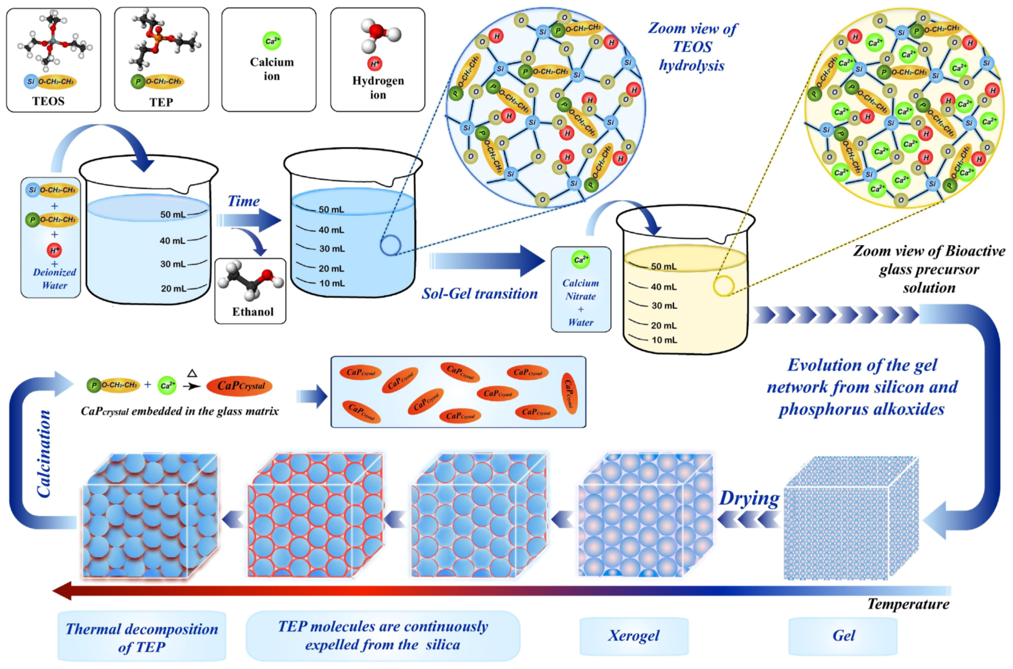
Figure 3. Schematic representation of the classical sol-gel procedure for the preparation of calcium phosphate-based bio-material. Reprinted with permission from Ref. [35], Copyright 2020, Elsevier (Amsterdam, The Netherlands).
Molecules 28 04790 g003
Figure 4. Schematic representation of the microwave-assisted procedure for the preparation of calcium phosphate-based biomaterials. Reprinted with permission from Ref. [45], Copyright 2020, Elsevier.
Molecules 28 04790 g004
Figure 5. Schematic representation of the sonochemical synthesis for the preparation of calcium phosphate-based biomaterials. Reprinted with permission from Ref. [52], Copyright 2018, Nature Publishing Group (Berlin, Germany).
Molecules 28 04790 g005
Figure 6. Schematic representation of the enzyme-assisted method for the preparation of organic-inorganic nanocomposites. Reprinted with permission from Ref. [58], Copyright 2018, Wiley (Hoboken, NJ, USA).
Molecules 28 04790 g006
Figure 7. Strategies for the component engineering of nano-calcium phosphate-based biomaterials. (a) Calcium phosphate-based biomaterial coated with heparin to crosslink with ferulic acid; reprinted with permission from Ref. [97], Copyright 2021, MDPI (Basel, Switzerland). (b) The crosslinked porous biphasic calcium phosphate loading more BMP-2; reprinted with permission from Ref. [95]. (c) The structure of the calcium phosphate-based biomaterial influenced by the pH value, leading to the drug release; reprinted with permission from Ref. [98], Copyright 2019, Elsevier.
Molecules 28 04790 g007
Figure 8. Strategies for calcium phosphate-based material loading genes. (a) Calcium phosphate-loading DNA fixed in a scaffold to promote BMP-2 protein; reprinted with permission from Ref. [96], Copyright 2018, Elsevier. (b) The nucleic acid-loaded nanoparticles, combining nucleic acid molecule-loaded CaP nanoparticles with a Gln-Ochi chitosan coating to avoid aggregating; reprinted with permission from Ref. [108], Copyright 2015, Royal Society of Chemistry.
Molecules 28 04790 g008
Figure 9. Strategies for mixing metal ions with calcium phosphate. Calcium phosphate cement mixed with Ba/Sr ions to achieve bone regeneration. Reprinted with permission from Ref. [114], Copyright 2021, Royal Society of Chemistry.
Molecules 28 04790 g009
Figure 10. Calcium phosphate composite materials with different properties for bone repair. (a) Calcium phosphate scaffolds loaded with ginger and garlic extracts can improve bone regeneration and osteogenic differentiation (* p < 0.05; ** 0.05 < p < 0.01; and *** p < 0.01); reprinted with permission from Ref. [116], Copyright 2022, American Chemical Society. (b) Calcium phosphate materials were combined with Zn/Fe ions to turn micropores into a dispersed solid; reprinted with permission from Ref. [117], Copyright 2019, Royal Society of Chemistry.
Molecules 28 04790 g010
Figure 11. Multi-application in the imaging area. (a) Calcium phosphate-based materials used in bioimaging; reprinted with permission from Ref. [124], Copyright 2012, Elsevier. (b) Simple flow chart of calcium phosphate composite gadolinium and for CT and MRI bone defect repair evaluation. Green arrows indicate the bright area which appears in the middle of the implant on the MRI acquisitions after 4 weeks from the surgery. Red arrow displays the calcium phosphate cements that become indistinguishable from the surrounding bone after 8 weeks from implantation in vivo; reprinted with permission from Ref. [126], Copyright 2018, Wiley.
Molecules 28 04790 g011
Figure 12. Strategies for calcium phosphate-based materials to kill cancer cells. (a) Reversal of the multidrug resistance of tumors by combining calcium phosphate with Se element; reprinted with permission from Ref. [130], Copyright 2020, Elsevier. (b) Construction of calcium phosphate and TiO2 to promote cancer therapy; reprinted with permission from Ref. [142], Copyright 2021, Wiley.
Molecules 28 04790 g012
Table 1. A summary of synthesis methods for the preparation of calcium phosphate-based nanomaterials.
Synthesis MethodsAdvantagesDisadvantages
Wet chemical precipitation
  • Simple, efficient [68]
  • Easy control of the reaction system environment [19]
  • Needs a long reaction time [19]
  • Difficult to purify composites [69]
Solvothermal synthesis
  • Short reaction time [28]
  • Simple operation process [29]
  • Security risks in the reaction process [70]
  • Hard to control materials’ properties [71]
  • More energy consumption [72]
Sol-gel method
  • Simple synthesis process [73]
  • Excellent calcium phosphate crystallization [74]
  • The physicochemical properties of the product are uncontrollable [75]
  • Mixed with impurities [76]
Microwave-assisted method
  • Short reaction time [51]
  • Less raw material consumption [48]
  • Raw materials require full dissolution [45]
  • Limited application range [17]
  • High equipment maintenance costs [77]
Sonochemical synthesis
  • Less cost of the process [55]
  • The cavitation effect [52]
  • Few studies on the preparation of calcium phosphate materials [78]
  • Hard to industrialize [53]
Enzyme-assisted method
  • Environmentally friendly [79]
  • The reaction conditions are controllable [58]
  • Longer reaction time [80]
  • Whole process monitoring [81]
Spray drying and electrospinning
  • Machine parameters control product structure [82]
  • High equipment costs [83]
  • Difficult preparation of precursor solution [84]
Disclaimer/Publisher’s Note: The statements, opinions and data contained in all publications are solely those of the individual author(s) and contributor(s) and not of MDPI and/or the editor(s). MDPI and/or the editor(s) disclaim responsibility for any injury to people or property resulting from any ideas, methods, instructions or products referred to in the content.