Abstract
Sweat, a biofluid secreted naturally from the eccrine glands of the human body, is rich in several electrolytes, metabolites, biomolecules, and even xenobiotics that enter the body through other means. Recent studies indicate a high correlation between the analytes’ concentrations in the sweat and the blood, opening up sweat as a medium for disease diagnosis and other general health monitoring applications. However, low concentration of analytes in sweat is a significant limitation, requiring high-performing sensors for this application. Electrochemical sensors, due to their high sensitivity, low cost, and miniaturization, play a crucial role in realizing the potential of sweat as a key sensing medium. MXenes, recently developed anisotropic two-dimensional atomic-layered nanomaterials composed of early transition metal carbides or nitrides, are currently being explored as a material of choice for electrochemical sensors. Their large surface area, tunable electrical properties, excellent mechanical strength, good dispersibility, and biocompatibility make them attractive for bio-electrochemical sensing platforms. This review presents the recent progress made in MXene-based bio-electrochemical sensors such as wearable, implantable, and microfluidic sensors and their applications in disease diagnosis and developing point-of-care sensing platforms. Finally, the paper discusses the challenges and limitations of MXenes as a material of choice in bio-electrochemical sensors and future perspectives on this exciting material for sweat-sensing applications.
1. Introduction
The current healthcare system is taking a transition from a conventional approach, where patients visit a healthcare professional for any medical aid after developing the disease or its symptoms, towards more sophisticated continuous monitoring of vital biomarkers and then informing patients (sometimes by healthcare professionals remotely) of any deviation from the normal level. This enables early detection of any deviation from the level of the normal biomarker and brings in new possibilities for the prevention of disease that are not possible through the conventional system. This requires highly specific and sensitive biosensing platforms for precisely and continuously monitoring the biomarkers. The electrochemical-based wearable sensors are currently being explored on this line and are found to be an excellent sensing platform to address the above challenges [1,2,3]. Thus, wearable electrochemical biosensors can provide continuous, instantaneous data on patients’ physiologies via dynamic, non-invasive measures of biomarkers in biofluids without causing discomfort to the wearing individuals [4,5,6,7,8]. In recent years, the growth of wearable electronic technologies that can precisely quantify vital responses such as body temperature, blood pressure, blood sugar level, and heart rate, aiding in depicting and monitoring the individual’s health conditions, has accelerated [3,9]. However, these biological parameters cannot provide complete information on the human body’s robust biochemical and metabolic functions [10]. So, biofluids such as tears, saliva, and sweat are of massive interest to biosensors researchers for their ease of sampling and their demonstrated capabilities to provide continuous and instantaneous monitoring of vital biomarkers and parameters, which could give insights into the subject’s physiology in a non-invasive manner [11,12,13,14,15,16]. Compared to other biofluids, sweat is high in several essential analytes such as glucose [17], lactate [18], cortisol [18], uric acid [19], interleukin [20], ammonium [21], sodium [22], potassium [23], calcium [24], iron [25], and zinc [26]. They provide critical physiological information about the human body and are closely intertwined with capillaries and nerve fibers, offering an immense advantage for biosensing applications. Moreover, wearable analytes monitoring technologies at sweat production sites open a huge possibility of autonomous, continuous, and instantaneous sensing of vital analytes present in sweat.
The first work on wearable electrochemical sensors for real-time monitoring of sweat lactate was developed by Jia et al. [27]. Further, many researchers have executed the monitoring of metabolites, electrolytes, drugs, and trace elements present in sweat. Electrochemical sensing is a familiar and deep-rooted method for detecting sweat analytes that is extensively used in wearable sensing platforms and often used in clinical diagnostics due to its simplicity, portability, high performance, and low cost [28,29,30]. Some recent technological developments are steering the sophistication of wearable sweat sensors based on electrochemical methods. A complete unified multiplexed sensing device for simultaneous monitoring of multiple analytes makes the system more practical. It offers a versatile wearable electrochemical biosensing platform for large-scale clinical diagnostics and physiological understandings [31,32,33]. Thus, the performance and effectiveness of the wearable biosensor rely heavily on the properties of materials used to make the device.
Some of the most common materials used in biosensors include and are not limited to the following: graphene and its derivatives [34,35], transition metal nanoparticles [36,37,38,39], transitional metal dichalcogenides [40,41], gold nanoparticles [42,43], silver nanoparticles [44,45], and very recently, transition metal carbides/nitrides, called as MXenes [46]. Because of their excellent multifaceted characteristics including high surface area, size control, tunable electronic and mechanical properties, high biocompatibility, exceptional sensitivity, specificity, and low detection limit (LOD), MXenes are currently being explored heavily for fabricating biosensors compared to other nanomaterials. MXenes are a class of 2D inorganic nanomaterials with few-atom thicknesses made of layered transition carbides, nitrides, and carbonitrides [47,48,49]. They are typically exfoliated by etching of the A layer from the MAX phase, specifically [50]. The general formula of an MXene is Mn+1XnTx, where M represents the early transition metals, X is for carbon or nitrogen, T is for surface functional groups (–OH, –F, =O), and n represents an integer.
This review systematically summarizes recent breakthroughs in MXene-based electrochemical biosensors for sweat analytes. We review and discuss various synthesis methods for MXenes, their surface chemistry, and functionalization strategies in sensor applications. The advantages and importance of sweat analytes in continuously monitoring human physiology and disease conditions are also highlighted. Finally, we also discuss various MXene-based wearable biosensors for vital analytes in sweat. Most importantly, this review sheds light on future scopes and recommendations for researchers working on MXenes for electrochemical biosensor applications.
2. Advantages of Sweat as an Analyte in Biosensing
In the current healthcare systems, blood serum is considered as the gold standard to determine the concentration of analytes in most cases [51,52]. However, the invasive methods of sampling blood from patients produce more impediments, particularly for hemophobic patients, new-borns, and elderly persons. Urine is also a biofluid widely used as clinical samples but has some limitations for autonomous and continuous monitoring [53]. Saliva is another biofluid that contains many biomarkers, including enzymes, hormones, antibodies, and antimicrobial agents, which can precisely provide details on the molecular state of human physiology [54]. However, saliva monitoring also has specific drawbacks, such as saliva in the mouth containing many impurities and food particles, which could affect the precision of results. Tears, another biofluid, contain enzymes, proteins, lipids, and certain salts, which can reflect the conditions and disease in the eyes and others [55]. Unfortunately, the present tear sample collection protocols produce reflex tears and eye irritation, which can also affect the results of sensor analysis.
In contrast to other biofluids such as blood, urine, saliva, and tears, sweat has a massive advantage in biosensing applications. Sweat regulates heat balance in the body and plays crucial physiological roles including immune defense, thermoregulation, moisturization, and pH balance [56,57]. Thus, sweat contains several biomarkers that can provide the complete physiological status of the body at the molecular level [58,59]. The sweat glands in the body secrete the sweat. Thus, sweat analytes can be collected in a non-invasive manner from different body parts and are optimal for continuous monitoring. The average density of sweat glands in the body and the approximate range of analytes’ concentrations in the sweat fluid are shown in Figure 1a,b [60]. Key analytes in human sweat and associated health conditions are shown in Table 1.
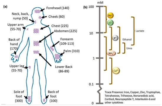
Figure 1.
(a) Schematic representation of the average density of sweat glands in the different parts of the human body(glands/cm2). (b) Approximate range of analytes’ concentrations present in the sweat. Reproduced with permission from Ref. [60]. Copyright 2019 Elsevier.

Table 1.
Key analytes present in human sweat and associated with health issues.
3. MXenes–A Material of Choice for Biosensors
In contrast to metal nanoparticles- and graphene derivative-based biosensors, MXenes have gained much attention in developing biosensors due to their excellent biocompatibility and high electrical conductivity properties. A suitable sensor possesses high specificity, sensitivity, LOD, quick response, and a wide operating range. In addition, it also needs to be less expensive not only in lab-scale use, but also while scaling up. MXenes have all/most of these very important properties in the development of biosensors [81,82,83,84].
3.1. Synthesis Strategies of MXenes
MXenes, a new class of 2D anisotropic nanomaterial, have significantly improved from their first discovery in 2011 (Ti3C2Tx) by the selective etching of the MAX phase precursor Ti3AlC2 [85]. Typically, MXenes are developed by removing the A layer from their MAX phase via specific etching. The synthetic methods of MXenes can be classified into two types: the top-down approach and the bottom-up approach. The top-down approach involves the direct exfoliation of the A layer, while the bottom-up approach is based on 2D ordered growth from atoms/molecules. Both the methods are discussed in detail in the below sections.
3.1.1. Top–Down Approach
The top–down approach involves the direct exfoliation of the bulk parent MAX phase material with its original integrity retained. These exfoliation processes are typically carried out using chemical and mechanical methods. The top-down approach can be further classified based on the type of precursors used, the delamination process, and the etchants involved in the reaction. The following section describes these methods in more detail.
Based on Precursors
Based on the precursors selected, the preparation strategy of MXenes is classified into MAX-phase and non-MAX-phase methods. In the MAX phase-based preparation method, the A layer is eliminated by using specific selective etchants with optimized concentrations and time intervals, followed by filtration and sonication to obtain few-layered 2D nanosheets. A typical example of this method is the elimination of the A layer from the MAX phase (Ti3AlC2) to form Ti3C2Tx MXene by Naguib et al. (Figure 2a), with hydrofluoric acid (HF) as an etchant in room temperature conditions [85].
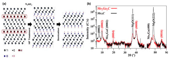
Figure 2.
(a) Schematic illustration of the synthesis process of Ti3AlC2. Reproduced with permission from Ref. [85] Copyright 2011 John Wiley and Sons. (b) X-ray diffraction pattern of Mo2Ga2C film before and after HF etching. Reproduced with permission from Ref. [86]. Copyright 2015 Elsevier.
Non-MAX phase-based synthesis was obtained from the selective etching of the gallium (Ga) layer from the Mo2Ga2C with 50% concentrated HF acid to form Mo2CTx MXene [86]. In contrast to the known MAX phase formula, the non-MAX phase method of prepared Mo2CTx MXene consists of two A layers of Ga stacked between the Mo2C layers. To confirm perfectly etched treatment, the X-ray diffraction patterns of Mo2CTx and Mo2Ga2C before and after etching were compared (Figure 2b), in which there was a significant reduction in the peak intensity of Mo2C, which confirms the Ga phase was dissolved during the etching process.
Based on Delamination
In general, synthesizing single-layered MXenes requires further processing steps on the multi-layered MXenes, called delamination. The delamination of multi-layered MXenes can be by the intercalation method or through mechanical exfoliation. It was reported that mechanical exfoliation is ineffective because the interlayer interactions in MXenes produced by this method are highly tacky (the interlayer interaction in the MXene is two- to six-folds stronger than that present in graphite and bulk MoS2 [87]). Moreover, the sonication time also affects the lamellar structure, decreases MXene sheet size, and produces defects in the structure [88,89,90].
In the case of the intercalation method, intercalants are introduced to reduce the interlayer spacing, weaken the interaction between the MXene layers, and facilitate the formation of individual nanosheets. This dramatically improves the surface area and abundant surface terminations directly associated with the MXene’s electrically conductive properties. The intercalants widely used for intercalations of the MXene are classified into two types: ionic aqueous solutions that include metal hydroxides or halide salts as aqueous solutions [91,92] and organic intercalants such as dimethyl sulfoxide (DMSO) [93], tetrabutylammonium hydroxide (TBAOH) [94], tetra propylammonium hydroxide (TPAOH) [95], isopropyl amine [96], n-butyllithium [97], and bovine serum albumin (BSA) [98].
Based on Etchants
- HF etching
In the HF etching-based MXene synthesis method, the layered MAX phase is stirred with HF aqueous solution at room temperature with specified concentration and time. Here, the multi-layered MXene is produced by an etched A layer from the MAX phase, and M-A bonds are replaced by the weak intercalations of Tx termination such as (–OH, –F, =O) on the surface of the multi-layered MXene. In agreement with numerous studies in recent times, several etching parameters such as temperature, time, and concentration of etchant play a deterministic role in the standard of the prepared MXene nanosheets. Kumar et al. demonstrated the effectiveness of Ti3C2Tx MXene etched at elevated temperatures for a binder-free supercapacitor application (Figure 3a–d) [99].
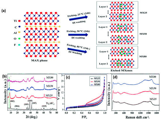
Figure 3.
(a) Schematic representation of the etching mechanism at elevated temperature and their high exfoliation. (b) XRD pattern of exfoliated material at different temperatures and reduced intensity at high–temperature etching of MX80 indicates deterioration of crystallinity. (c) BET isotherm studies confirm the increase in surface area with high–temperature etching. (d) Raman spectra of exfoliated MXene at different temperatures were high–intensity peaks observed for MX80, indicating the defects introduced at high–temperature etching. Reproduced from Ref. [99] with permission from the Royal Society of Chemistry.
- Non–HF etching
Even though HF is extensively and effectively utilized to prepare MXenes from the MAX phase, it is highly corrosive and hazardous to humans and the environment [100,101,102]. A trace amount of HF remaining in the MXene sample will harm the composite preparation and biological studies carried out further. Thus, the etching of MXenes by non-HF-based solvents is also extensively studied. The alternative etchants used for etching MXenes are weak acids and environmentally friendly bifluorides such as sodium bifluoride (NaHF2), ammonium bifluoride (NH4HF2), and potassium bifluoride (KHF2). Feng et al. demonstrated the synthesis of Ti3C2Tx MXene from the Ti3AlC2 MAX phase with bi-fluoride in a single-stage process. They found that the NH4+, Na+, or K+ ions enter the interlayer spacing of the MXene and enlarge the interplanar spacing and delamination efficiency [103].
In comparison to carbide-based MXenes, the Al layer is strongly bonded in the nitride-based MXene. Thus, more energy is required to remove the A layer from the nitride-based MXene. Moreover, the nitride-based MXene is less stable and might be able to dissolve in HF [104,105]. To overcome this issue, molten fluoride is used with the support of high-temperature heating to remove the A layer from the nitride-based MXene. Urbankowski et al. heated the Ti4AlN3 powder at 550 degrees for 30 min under an argon atmosphere with molten fluoride, where the free F− ions were active enough to etch the Al layer from the nitride MXene MAX phases [106]. Studies revealed that the etchants are an essential determinant in the surface termination of the synthesized MXene. The fluorine-based etchants increase the percentage of F content in the surface terminations. Therefore, further modifications are needed for particular applications such as biomedical and sensors [107,108].
3.1.2. Bottom–Up Approach
MXenes can also be synthesized by crystal growth methods using a small organic or inorganic molecule as the precursor. The bottom-up approaches enable precise manipulation of the size of the MXene sheet, geometry, and the surface termination groups in the MXene, which are impossible using top-down approaches [109,110,111,112]. However, compared to the top-down approach of MXene preparation, only a few studies have been performed with the bottom-up approach, perhaps due to the difficulty in the bottom-up approach to build atom-by-atom the MXene’s complex structure and multi-component atomic-thick layer. Moreover, the mechanisms of interaction between the layers are still not clear.
Hong et al. demonstrated the preparation of Mo2N MXene sheets by the chemical vapor deposition (CVD) method with NH3 as the source in the temperature range of 1080 °C [112]. Similarly, Ren et al., 2015 developed ultrathin α-Mo2CTx crystals with excellent stability and defect free by CVD [113]. Buke et al. reported a detailed study on the effect of temperature, time, and copper layer thickness in the preparation of an MXene by the CVD method. In addition, they also found that Mo2C MXene crystals formed on the graphene sheets are thinner than those formed on the copper sheets [114]. In continuation to the CVD method, pulse laser deposition (PLD) and salt template methods were also investigated for the preparation of MXenes through a bottom-up approach [115,116].
3.2. Properties of Mxenes
MXenes have already proven their steadfast importance in multiple applications such as catalysis [117]; sensors (optical [118,119]; electrochemical [120,121,122,123,124,125,126]; surface-enhanced Raman scattering [127,128]; biomedical applications [129,130]; electromagnetic shielding [131,132,133]; and energy storage [134,135]. This success is significantly due to its unique properties, such as high young’s modulus [136,137], adjustable band gap [138], and thermal and electrical conductivity [139]. Comparison of fundamental properties of nanomaterials are shown in Table 2.
3.2.1. Electrical Properties
Similar to the MAX phase, a pristine MXene is all-metallic. Thus, the research has been focused on enhancing the conductivity nature of MXenes by engineering their surface chemistry [140,141]. The prepared MXene using etchants leaves a surface termination group (–OH, –F, =O) which will bond to the metal atom in the MXene. Based on the experimental and theoretical studies, a few reports showed that some termination groups may affect the conductive property of the MXene [142,143,144] and electron mobility [145,146,147]. Yan et al. demonstrated an alkaline-treated MXene for humidity sensors, where they showed similar effects of –OH and –O termination groups in enhancing the electrochemical capacitance and sensor properties [107,148,149]. Pandey et al. investigated the electronic properties of MnCn−1O2 MXenes (M = Ti, W, Ta, Hf, Sc Mo, Nb, Cr, Zr, Y, Mn, V) [150], in which they found that oxygen-terminated MXenes are favorable for many applications.

Table 2.
Comparison of fundamental properties of nanomaterials.
Table 2.
Comparison of fundamental properties of nanomaterials.
| Nanomaterials | Conductivity (S/cm) | Surface Area (m2 g−1) | Biocompatibility | References |
|---|---|---|---|---|
| Graphene | 2700 | 450 | Biocompatible | [151,152] |
| Single-walled carbon nanotubes | 102 to 106 | 600 | Under debate | [153,154,155] |
| Multiwalled carbon nanotubes | 103 to 105 | 122 | Under debate | [153,154,155] |
| Hexagonal boron nitride | Insulator | 150–550 | Depends on the shape and size | [156,157] |
| MnO2 | 10−5 to 10−6 | 257.5 | Biocompatible | [158,159,160] |
| MoS2 | 10−4 | 8.6 | Biocompatible | [161,162,163] |
| MXene-Ti3C2 | 15100 | 93.6 | Biocompatible | [164,165,166,167,168] |
3.2.2. Biocompatibility of MXenes
The principal mechanisms of nanomaterials’ toxicity could be one of the following: (1) damage to cells, (2) genotoxicity, (3) slow clearance in the renal pathway and accumulation in the organs, and (4) specific toxicity to the neural and reproductive system. Thus, an extensive biological safety evaluation is required to understand all the possible interactions between the nanomaterials and the physiological systems. MXenes, due to their high surface area and hydrophilicity, provide a suitable matrix for fabricating wearable biosensors [169]. Some preliminary results on MXene cytocompatibility and biocompatibility are already explored in the literature. Ti3C2 MXene is the most commonly investigated system in biomedical applications [166,170,171,172]. Liu et al. developed Ti3C2 MXene nanosheets for theranostics applications. They revealed that the MXene nanosheets passed in the bloodstream are excreted through human urine via the renal clearance pathway or accumulated in the tumor tissue due to the enhanced permeability and retention effect (EPR). In this study, the Ti3C2 nanosheets’ biosafety and biocompatibility are confirmed by the absence of significant weight loss and the absence of necrotic process. Han et al. investigated the in vivo biocompatibility of Ti3C2 MXene by administering the MXene to mice at elevated dosages (6.25, 12.5, 25, and 50 mg/kg), which resulted in excellent in vivo biocompatibility at 25 mg/kg [173]. In a similar study, Zong et al. developed a Ti3C2-GdW10 nanocomposite for cancer theranostics application. The in vivo biocompatibility studies concluded that up to 50 mg/kg of Ti3C2-GdW10 nanocomposite was biocompatible using female Kunming mice [174]. Similar results have been reported for systematic cytocompatibility and biocompatibility of tantalum carbide-based MXene Ta4C3 [171,175] and niobium carbide MXene Nb2C [176,177].
4. MXene-Based Electrochemical Sweat Sensors
Due to the fascinating physiochemical properties of MXenes and their composites, they have gained strong attention in biosensing with their excellent sensitivity, specificity, LOD, mechanical properties, and biocompatibility [136,144,178,179].
4.1. Glucose Sensing
Peng et al. developed a Pt/Ti3C2 MXene-based wearable, flexible non-enzymatic electrochemical biosensor for continuous monitoring of glucose in sweat [180]. Figure 4a–c illustrates the fabrication process of the sensor, microfluidic patch, and integration of the flexible sensor to the prepared patch. Figure 4d–f shows the conceptual scheme of the sensor for glucose detection, the cross-sectional view of the sensor on the skin, and the oxidation mechanism of the material and analyte. Figure 4g,h shows the electrochemical reaction mechanism of Pt/Ti3C2 coated on a glassy carbon electrode (GCE) and fabricated flexible sensor. This system offers a LOD of 29.15 μmol L–1 with a correlation coefficient of 0.9793.
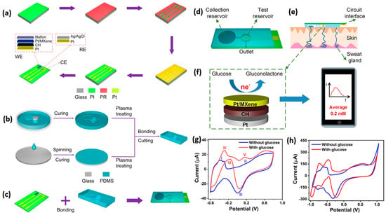
Figure 4.
(a) Schematics show the flexible sensor’s fabrication process. (b) The preparation process of the microfluidic patch. (c) Integration of MXene–based sensor to microfluidic patch. (d) Schematic overview of the proposed flexible wearable sensor. (e) Cross–sectional view of the sensor on the skin. (f) The electrochemical oxidation reaction of glucose on the MXene. (g) CV response of Pt/Ti3C2 MXene coated on GCE. (h) CV response of Pt/Ti3C2–fabricated flexible wearable sensor. The figure is reproduced with permission from [180]. The copyright year is 2023 American Chemical Society.
In another work by Park et al., 2022, a butterfly-inspired hybrid epidermal biosensing (bi-HEB) patch, made with carbon Ti3C2Tx MXene-based nanocomposite, was developed for simultaneous monitoring of glucose and electrocardiograms (ECGs) in human subjects while performing indoor physical activities [73]. This system demonstrated excellent sensitivity of 100.85 µAmm−1 cm−2 within physiological levels (0.003−1.5 mm). Moreover, variations in pH and temperature from on-body sweat monitoring are calibrated. Figure 5a–c is a detailed schematic overview of bi-HEB patch fabrication. Feng et al. demonstrated a 3D porous Ti3C2Tx/graphene/AuNP composite electrochemical biosensor for quick, responsive glucose detection. This system has a LOD of 2 μM in a concise time of fewer than 3 s with a sensitivity of 169.49 μA/(mM·cm2) in the range of 2 μM–0.4 mM concentration of glucose [181]. Hosseini et al. fabricated a Ti3C2/nickel-samarium-layered double hydroxide for enzyme-free real-time glucose detection. The glucose sensing was investigated with the DPV method, resulting in a LOD value of 0.24 μM and a linear range from 0.001 to 0.1 mM and 0.25–7.5 Mm [182].
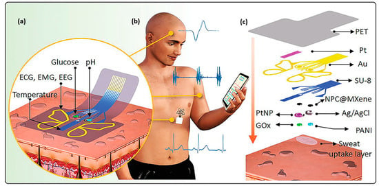
Figure 5.
(a) Schematic illustration of layer-by-layer assembled multiplexed electrochemical sensor mounted on the skin containing various analyte-templated patterns designed on thin polyethylene terephthalate (PET) substrate. (b) bi-HEB patch mounted at the chest to monitor sweat glucose, temperature, and pH simultaneously. (c) Schematic representation of layer-by-layer fabrication process of the bi-HEB patch from top to bottom. Reproduced with permission from Ref. [73]. Copyright 2022 John Wiley and Sons.
Li et al. demonstrated a highly integrated wearable sensing paper (HIWSP) for electrochemical analysis of glucose and lactate from sweat [183]. This paper composition contains hydrophobic-layered protecting wax, a hydrophilic layer for sweat diffusion, and a Ti3C2Tx MXene/methylene blue composite. This sensor had a sensitivity of 2.4 nA μM−1 and 0.49 μA mM−1 for the simultaneous detection of glucose and lactate, respectively. In another work, Lei et al. fabricated a high-performing wearable biosensor for in vitro perspiration analysis [184]. This multifunctional biosensor is designed by incorporating Ti3C2Tx MXene and Persian blue composite for durable and sensitive monitoring of glucose and lactate in sweat. Figure 6a–i illustrates the real-time monitoring of wearable patches indicating pH, glucose, and lactate. Magesh et al. fabricated a palladium hydroxide integrated Ti3C2Tx MXene composite electrode to detect nicotine analyte in human sweat. This analysis was performed with the help of CV and amperometric studies and exhibited a LOD value of 27 nM with a sensitivity of 0.286 µA µM−1 cm−2 [185].
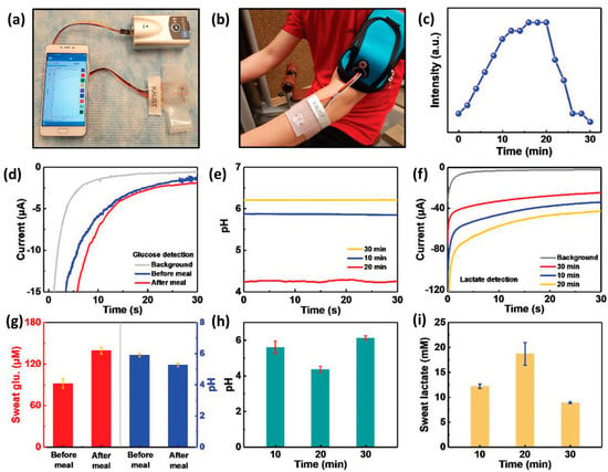
Figure 6.
(a) Photograph of wearable sweat monitoring patch connected to a portable electrochemical analyzer that supplies power and can wirelessly communicate the body’s status with mobile phones via Bluetooth. (b) Skin–attached wearable sweat sensor. (c) Graphical data of on–body test cycling resistance profile. (d) Chronoamperometric response of pH changes and glucose sensor before and after a meal. (e) pH response at different times during exercise. (f) The electrochemical response of lactate sensor at different times during exercise. (g) Comparison of pH level and glucose after a meal with three different sensors. (h) Comparison of pH levels at different times during exercise. (i) Comparison of lactate sensor responses at different exercise times with different lactate sensors. Reproduced with permission from Ref. [184]. Copyright 2019 John Wiley and Sons.
Ti3C2Tx MXene nanoflakes decorated ZnO tetrapods (ZnO TPs), a graphene oxide composite, and a skin-attachable enzymatic electrothermal glucose sensor was fabricated by Myndrul et al. [186]. This system showed a LOD of 17 μM with a broad linear detection range of 0.05–0.7 mM for detecting glucose from human sweat. Figure 7a–d shows stretchable electrodes at various strains, glucose sensing performance at applied strain (artificial sweat), skin-attachable sensor performance, and current density changes in the fabricated sensors on volunteer sweet consumption for glucose monitoring.
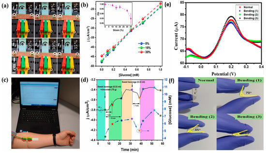
Figure 7.
(a) Picture of flexible MXene/ZnO TPs/Gox composite electrode at strain range of 0–35%. (b) Working performance of fabricated sensor at applied strains (electrode current density vs applied strains) graph. (c) Photograph of skin–attachable sensor functioning for monitoring of glucose in sweat. (d) Graph of current density changes in composite material under sweet consumption for glucose detection. Reprinted with permission from Ref. [186]. (e) SWASV response of composite material–based fabricated electrode for 300 ppb Cu (II) ion on normal and bending mode. (f) Photographs of bending processes of the fabricated electrode. Reproduced with permission from Ref. [187]. Copyright 2020, American Chemical Society.
4.2. Cortisol Sensing
Park et al. developed a microfluidic integrated wearable impedimetric immune sensor based on Ti3C2Tx MXene incorporating laser-mediated porous graphene. This immune-sensing patch analyzes cortisol from the sweat by collecting sweat samples at one touch using a microfluidic channel network. Thus, wearable immune-sensing patches exhibit a dynamic range of 0.01–100 nM with a LOD of 88pM [188]. In another work, Laochai et al. fabricated Ti3C2 MXene/AuNPs/L-cysteine composited electrodes for real-time detection of cortisol from sweat [71]. Under optimal conditions, this fabricated sensor shows sensitivity with wide linearity of 5–180 ng mL−1 and a LOD value of 0.54 ng mL−1. The real-time analysis of cortisol from artificial sweat results in a 94.47–102% recovery value. The fabrication of cortisol immune sensors and immobilization steps are discussed with the help of Figure 8a,b.
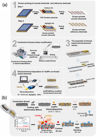
Figure 8.
(a) Schematic illustration of the fabrication of counter and reference electrodes, surface modification, and electrochemical deposition of gold nanoparticles on the surface of the electrode. (b) Immobilization process. Reproduced with permission from Ref. [71]. Copyright 2022 Elsevier.
4.3. Other Analytes
Zhi et al. developed a bioinspired directional moisture-wicking electronic skin for static health monitoring with the support of triboelectric energy harvesting. The device is made of heterogeneous fibrous polyvinylidene fluoride (PVDF) as a hydrophobic layer and polyacrylonitrile (PAN) as the hydrophilic layer with Ti3C2Tx MXene/CNT conductive ink [189]. Zhang et al. fabricated a wireless, battery-free, wearable, skin-interfaced electrochemical sensing patch for K+ monitoring from sweat. They used valinomycin as a K+ selective carrier specific to K+, enabling the selective detection of K+ by the membrane. Figure 9a–e demonstrates the conceptual operation mechanism of the fabricated device, electrode fabrication, sensor fabrication, and CV analysis of the material, followed by EIS analysis. This fabricated sensor measures the K+ ion concentration from human sweat with an excellent sensitivity of 63 mV/dec and a high linear detection range from 1 to 32 mM [77]. In another work, Hui et al. designed a flexible electrochemical heavy metal sensor for the non-invasive detection of Cu and Zn ions in human sweat with a material composition of Ti3C2Tx/multi-walled carbon nanotubes (MWCNTs) in a layer-by-layer self-assembly [187]. The square wave anodic stripping voltammetric method (SWASV) was used for electrochemical analysis. The resulting LOD of Cu and Zn ions is 0.1 and 1.5 ppb. The response of SWASV for Cu (II) ion and the bending process of the fabricated electrode are shown in Figure 7e,f.
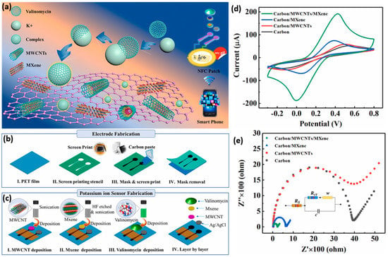
Figure 9.
(a) Schematic illustration of the operating mechanism of wearable, wireless, battery–free integrated electrochemical sensing patch (valinomycin is a selective K+ carrier and naturally has specific permeability to K+). (b) Steps involved in electrode fabrication. (c) Sensor fabrication process. (d) CV study bare carbon, carbon/MWCNTs, carbon/MXene, and carbon/MWCNTs/MXene. (e) EIS Nyquist analysis. Reproduced with the permission form. Ref. [77]. Copyright 2020 Elsevier.
Cui et al. demonstrated a heterostructure Ti3C2 MXene/MoS2 composite for detecting ascorbic acid in human sweat. The study resulted in high sensitivity of 54.6 nA μM−1 and a LOD of 4.2 μM [190]. In another work, Saleh et al. fabricated inkjet-printed Ti3C2Tx MXene-based electrodes for cutaneous biosensing [74]. They demonstrated an exciting finding on Inkjet-printed MXene electrodes for Na+ ion detection and cytokine protein with 3.9 mV per decade by simple changes in the functionalization of the target analyte. These findings simplify the fabrication of wearable electronic platforms that enable multimodal biosensing. Qiao et al. developed a Ti3C2 MXene@TiO2 (anatase/rutile) ternary heterostructured electrode to detect phosphoprotein in sweat. They showed a LOD of 1.52 μM with a broad linear range from 0.01 to 1 mg/mL [191]. Chen et al. developed a fluoroalkyl functionalized F–Ti3C2Tx/polyaniline (PANI) superhydrophobic skin-attachable and wearable electrochemical pH biosensor for real-time perspiration analysis [192]. Thus, the schematic view of the sensing device setup, pH analysis of sweat in males/females, and comparison of measured values from the fabricated patch with other equipment are discussed in detail in Figure 10a–d.
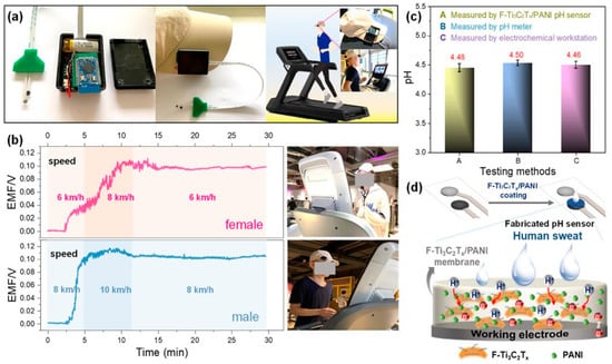
Figure 10.
(a) Digital photograph of wearable pH sensor with a lithium-ion battery, mini–type potentiometer, and F–Ti3C2Tx/PANI material. (b) Continuously monitored real–time pH in human male and female volunteers. (c) Comparison of F–Ti3C2Tx/PANI sensor with ex situ electrochemical workstation and pH meter results. (d) Schematic illustration of F–Ti3C2Tx/PANI electrochemical behavior. Reproduced with permission from Ref. [192]. Copyright 2022 American Chemical Society.
5. Summary and Outlook
In recent years, more attention has been paid to MXenes due to their outstanding characteristic features such as high conductivity, surface area, oxidation and redox capacity, biocompatibility, and significant electrocatalytic and electrochemical properties. Moreover, MXenes also hold great potential as conductive substrates for various electrode-based devices, especially for specific-patterned electrodes. This paves the way for the fabrication of highly integrated electrochemical sensing gadgets such as wearable sensors for non-invasive monitoring of biomarkers in the body fluid (sweat) and miniaturized electronic devices for practical standards. However, for the faster translation of MXene-based electrochemical sensors from lab to market, we propose the following research directions in the near future:
(i) Device fabrication with precise nano-/micro-patterned structures are essential to enhance the performance of wearable sensors, and their shortcomings cannot be ignored [193].
(ii) It is challenging to synthesize MXenes on a large scale due to their atomic-thick layer design. To overcome this, future research should address the industrial-scale nanostructured design of MXenes and cost-effective technologies [194,195].
(iii) The malleable structure is crucial in changing the conductivity and internal resistance of the MXene. Hence future research should focus on the enlargement of interlayer distance in MXenes.
(iv) The problems with the flexibility of MXenes can be solved by performing focused research on MXene-polymer nanocomposites using biocompatible polymers [196].
(v) MXene-based biosensors strongly rely on nanohybrid biocompatibility. Thus, there should be focused research on the surface chemistry of MXenes to solve the problems based on the affinity and stability of biomolecules on MXene surfaces [197].
(vi) In the case of wearable sensors, MXene nanomaterial is oxidized when continuously in contact with air. This reduces the conductivity and affects the sensing ability. However, the external polymer coating to prevent oxidation in the MXene affects the breathability and comfort of the wearable biosensors. Thus, an in-depth understanding is needed to design sensors that could maintain the conductivity of the MXene without losing the convenience of the user [169].
Author Contributions
S.G. and A.P. conceptualized the paper; S.G. and K.R. wrote the paper; T.K. and A.P. reviewed and edited the manuscript. All authors have read and agreed to the published version of the manuscript.
Funding
The author (T.K) gratefully acknowledge the financial support of this project by the Ministry of Science and Technology, Taiwan (MOST 111-2221-E-027-085). A.P. would like to acknowledge the financial support from the Science and Engineering Research Board (SERB), Department of Science and Technology, Government of India for its start-up research grant (File No. SRG/2020/001115) scheme and expresses his appreciation to “VIT SEED GRANT”. K.R. thank DST-INSPIRE JRF and SRF (Inspire Fellow No. IF210172) schemes for her Ph.D. fellowship.
Institutional Review Board Statement
Not applicable.
Informed Consent Statement
Not applicable.
Data Availability Statement
All the data and materials that support the results or analyses presented in their paper are freely available upon request.
Acknowledgments
The authors gratefully acknowledge the financial support of this project by the Ministry of Science and Technology, Taiwan (MOST 111-2221-E-027-085). S.G, A.P, and K.R. would also like to acknowledge the support received from the Center for Biomaterials, Cellular, and Molecular Theranostics (CBCMT); the School of Advanced Sciences (SAS); and the Periyar Central Library of Vellore Institute of Technology, Vellore.
Conflicts of Interest
The authors declare no conflict of interest.
Sample Availability
Not applicable.
Abbreviations
| cm | Centimeter |
| CNT | Carbon nanotube |
| CV | Cyclic voltammetry |
| CVD | Chemical vapor deposition |
| dec | Decade |
| DMSO | Dimethyl sulfoxide |
| DPV | Differential pulse voltammetry |
| ECG | Electrocardiogram |
| EIS | Electrochemical impendence spectroscopy |
| GCE | Glassy carbon electrode |
| HF | Hydrofluoric acid |
| KHF2 | Potassium bifluoride |
| LOD | Limit of detection |
| mL | Milliliter |
| mM | Millimolar |
| μM | Micromolar |
| MoS2 | Molybdenum disulfide |
| mV | Millivolts |
| MWCNT | Multi-walled carbon nanotube |
| NaHF2 | Sodium bifluoride |
| ng | Nanogram |
| NH4+ | Ammonium |
| NH4HF2 | Ammonium bifluoride |
| nM | Nanomolar |
| PAN | Polyacrylonitrile |
| PANI | Polyaniline |
| PET | Polyethylene terephthalate |
| PLD | Pulse laser deposition |
| pM | Picomolar |
| ppb | Parts per billion |
| PVDF | Polyvinylidene difluoride |
| SWASV | Square wave anodic stripping voltammetric method |
| TBAOH | Tetrabutylammonium hydroxide |
| TPAOH | Tetrapropylammonium hydroxide |
References
- Kim, J.; Campbell, A.S.; de Ávila, B.E.-F.; Wang, J. Wearable biosensors for healthcare monitoring. Nat. Biotechnol. 2019, 37, 389–406. [Google Scholar] [CrossRef] [PubMed]
- Smith, A.A.; Li, R.; Tse, Z.T.H. Reshaping healthcare with wearable biosensors. Sci. Rep. 2023, 13, 4998. [Google Scholar] [CrossRef]
- Grattieri, M.; Minteer, S.D. Self-Powered Biosensors. ACS Sens. 2018, 3, 44–53. [Google Scholar] [CrossRef]
- Xiang, L.; Wang, Y.; Xia, F.; Liu, F.; He, D.; Long, G.; Zeng, X.; Liang, X.; Jin, C.; Wang, Y.; et al. An epidermal electronic system for physiological information acquisition, processing, and storage with an integrated flash memory array. Sci. Adv. 2023, 8, eabp8075. [Google Scholar] [CrossRef] [PubMed]
- Kim, D.-H.; Lu, N.; Ma, R.; Kim, Y.-S.; Kim, R.-H.; Wang, S.; Wu, J.; Won, S.M.; Tao, H.; Islam, A.; et al. Epidermal Electronics. Science 2011, 333, 838–843. [Google Scholar] [CrossRef] [PubMed]
- Ghaffari, R.; Choi, J.; Raj, M.S.; Chen, S.; Lee, S.P.; Reeder, J.T.; Aranyosi, A.J.; Leech, A.; Li, W.; Schon, S.; et al. Soft Wearable Systems for Colorimetric and Electrochemical Analysis of Biofluids. Adv. Funct. Mater. 2020, 30, 1907269. [Google Scholar] [CrossRef]
- Singh, S.U.; Chatterjee, S.; Lone, S.A.; Ho, H.-H.; Kaswan, K.; Peringeth, K.; Khan, A.; Chiang, Y.-W.; Lee, S.; Lin, Z.-H. Advanced wearable biosensors for the detection of body fluids and exhaled breath by graphene. Microchim. Acta 2022, 189, 236. [Google Scholar] [CrossRef]
- Wang, L.; Wang, J.; Fan, C.; Xu, T.; Zhang, X. Skin-like hydrogel-elastomer based electrochemical device for comfortable wearable biofluid monitoring. Chem. Eng. J. 2023, 455, 140609. [Google Scholar] [CrossRef]
- Hong, Y.J.; Lee, H.; Kim, J.; Lee, M.; Choi, H.J.; Hyeon, T.; Kim, D.-H. Multifunctional Wearable System that Integrates Sweat-Based Sensing and Vital-Sign Monitoring to Estimate Pre-/Post-Exercise Glucose Levels. Adv. Funct. Mater. 2018, 28, 1805754. [Google Scholar] [CrossRef]
- Wang, M.; Yang, Y.; Min, J.; Song, Y.; Tu, J.; Mukasa, D.; Ye, C.; Xu, C.; Heflin, N.; McCune, J.S.; et al. A wearable electrochemical biosensor for the monitoring of metabolites and nutrients. Nat. Biomed. Eng. 2022, 6, 1225–1235. [Google Scholar] [CrossRef]
- Gómez, J.; Oviedo, B.; Zhuma, E. Patient Monitoring System Based on Internet of Things. Procedia Comput. Sci. 2016, 83, 90–97. [Google Scholar] [CrossRef]
- Mohammadzadeh, N.; Safdari, R. Patient monitoring in mobile health: Opportunities and challenges. Med. Arh. 2014, 68, 57–60. [Google Scholar] [CrossRef]
- Cameron, J.M.; Butler, H.J.; Palmer, D.S.; Baker, M.J. Biofluid spectroscopic disease diagnostics: A review on the processes and spectral impact of drying. J. Biophotonics 2018, 11, e201700299. [Google Scholar] [CrossRef] [PubMed]
- Leal, L.B.; Nogueira, M.S.; Canevari, R.A.; Carvalho, L.F.C.S. Vibration spectroscopy and body biofluids: Literature review for clinical applications. Photodiagn. Photodyn. Ther. 2018, 24, 237–244. [Google Scholar] [CrossRef]
- Shende, P.; Trivedi, R. Biofluidic material-based carriers: Potential systems for crossing cellular barriers. J. Control. Release 2021, 329, 858–870. [Google Scholar] [CrossRef] [PubMed]
- Pucchio, A.; Krance, S.H.; Pur, D.R.; Miranda, R.N.; Felfeli, T. Artificial Intelligence Analysis of Biofluid Markers in Age-Related Macular Degeneration: A Systematic Review. Clin. Ophthalmol. 2022, 16, 2463–2476. [Google Scholar] [CrossRef]
- Zafar, H.; Channa, A.; Jeoti, V.; Stojanović, G.M. Comprehensive Review on Wearable Sweat-Glucose Sensors for Continuous Glucose Monitoring. Sensors 2022, 22, 638. [Google Scholar] [CrossRef]
- Derbyshire, P.J.; Barr, H.; Davis, F.; Higson, S.P.J. Lactate in human sweat: A critical review of research to the present day. J. Physiol. Sci. 2012, 62, 429–440. [Google Scholar] [CrossRef] [PubMed]
- Huang, C.T.; Chen, M.L.; Huang, L.L.; Mao, I.F. Uric acid and urea in human sweat. Chin. J. Physiol. 2002, 45, 109–115. [Google Scholar]
- Jones, A.P.; Webb, L.M.C.; Anderson, A.O.; Leonardo, E.J.; Rot, A. Normal human sweat contains interleukin-8. J. Leukoc. Biol. 1995, 57, 434–437. [Google Scholar] [CrossRef]
- Itoh, S.; Nakayama, T. Ammonia in human sweat and its origin. Jpn. J. Physiol. 1952, 3, 133–137. [Google Scholar] [CrossRef]
- Bates, G.P.; Miller, V.S. Sweat rate and sodium loss during work in the heat. J. Occup. Med. Toxicol. 2008, 3, 4. [Google Scholar] [CrossRef]
- Vairo, D.; Bruzzese, L.; Marlinge, M.; Fuster, L.; Adjriou, N.; Kipson, N.; Brunet, P.; Cautela, J.; Jammes, Y.; Mottola, G.; et al. Towards Addressing the Body Electrolyte Environment via Sweat Analysis:Pilocarpine Iontophoresis Supports Assessment of Plasma Potassium Concentration. Sci. Rep. 2017, 7, 11801. [Google Scholar] [CrossRef] [PubMed]
- Consolazio, C.F.; Matoush, L.O.; Nelson, R.A.; Hackler, L.R.; Preston, E.E. Relationship Between Calcium in Sweat, Calcium Balance, and Calcium Requirements. J. Nutr. 1962, 78, 78–88. [Google Scholar] [CrossRef]
- Aruoma, O.I.; Reilly, T.; MacLaren, D.; Halliwell, B. Iron, copper and zinc concentrations in human sweat and plasma; the effect of exercise. Clin. Chim. Acta 1988, 177, 81–87. [Google Scholar] [CrossRef]
- Stauber, J.L.; Florence, T.M. A comparative study of copper, lead, cadmium and zinc in human sweat and blood. Sci. Total Environ. 1988, 74, 235–247. [Google Scholar] [CrossRef]
- Jia, W.; Bandodkar, A.J.; Valdés-Ramírez, G.; Windmiller, J.R.; Yang, Z.; Ramírez, J.; Chan, G.; Wang, J. Electrochemical tattoo biosensors for real-time noninvasive lactate monitoring in human perspiration. Anal. Chem. 2013, 85, 6553–6560. [Google Scholar] [CrossRef] [PubMed]
- Bandodkar, A.J.; Wang, J. Non-invasive wearable electrochemical sensors: A review. Trends Biotechnol. 2014, 32, 363–371. [Google Scholar] [CrossRef] [PubMed]
- Tabasum, H.; Gill, N.; Mishra, R.; Lone, S. Wearable microfluidic-based e-skin sweat sensors. RSC Adv. 2022, 12, 8691–8707. [Google Scholar] [CrossRef]
- Mohan, A.M.V.; Rajendran, V.; Mishra, R.K.; Jayaraman, M. Recent advances and perspectives in sweat based wearable electrochemical sensors. Trends Anal. Chem. 2020, 131, 116024. [Google Scholar] [CrossRef]
- Gao, F.; Liu, C.; Zhang, L.; Liu, T.; Wang, Z.; Song, Z.; Cai, H.; Fang, Z.; Chen, J.; Wang, J.; et al. Wearable and flexible electrochemical sensors for sweat analysis: A review. Microsyst. Nanoeng. 2023, 9, 1. [Google Scholar] [CrossRef] [PubMed]
- Yang, P.; Wei, G.; Liu, A.; Huo, F.; Zhang, Z. A review of sampling, energy supply and intelligent monitoring for long-term sweat sensors. NPJ Flex. Electron. 2022, 6, 33. [Google Scholar] [CrossRef]
- Kwon, K.; Kim, J.U.; Deng, Y.; Krishnan, S.R.; Choi, J.; Jang, H.; Lee, K.; Su, C.-J.; Yoo, I.; Wu, Y.; et al. An on-skin platform for wireless monitoring of flow rate, cumulative loss and temperature of sweat in real time. Nat. Electron. 2021, 4, 302–312. [Google Scholar] [CrossRef]
- Krishnan, S.K.; Singh, E.; Singh, P.; Meyyappan, M.; Nalwa, H.S. A review on graphene-based nanocomposites for electrochemical and fluorescent biosensors. RSC Adv. 2019, 9, 8778–8881. [Google Scholar] [CrossRef] [PubMed]
- Shao, Y.; Wang, J.; Wu, H.; Liu, J.; Aksay, I.A.; Lin, Y. Graphene Based Electrochemical Sensors and Biosensors: A Review. Electroanalysis 2010, 22, 1027–1036. [Google Scholar] [CrossRef]
- Kokulnathan, T.; Wang, T.-J.; Kumar, E.A.; Duraisamy, N. An-Ting Lee An electrochemical platform based on yttrium oxide/boron nitride nanocomposite for the detection of dopamine. Sens. Actuators B Chem. 2021, 349, 130787. [Google Scholar] [CrossRef]
- Kokulnathan, T.; Ahmed, F.; Chen, S.-M.; Chen, T.-W.; Hasan, P.M.Z.; Bilgrami, A.L.; Darwesh, R. Rational Confinement of Yttrium Vanadate within Three-Dimensional Graphene Aerogel: Electrochemical Analysis of Monoamine Neurotransmitter (Dopamine). ACS Appl. Mater. Interfaces 2021, 13, 10987–10995. [Google Scholar] [CrossRef]
- Agnihotri, A.S.; Varghese, A.; Nidhin, M. Transition metal oxides in electrochemical and bio sensing: A state-of-art review. Appl. Surf. Sci. Adv. 2021, 4, 100072. [Google Scholar] [CrossRef]
- Xu, M.; Song, Y.; Wang, J.; Li, N. Anisotropic transition metal–based nanomaterials for biomedical applications. VIEW 2021, 2, 20200154. [Google Scholar] [CrossRef]
- Wang, Y.-H.; Huang, K.-J.; Wu, X. Recent advances in transition-metal dichalcogenides based electrochemical biosensors: A review. Biosens. Bioelectron. 2017, 97, 305–316. [Google Scholar] [CrossRef]
- Mia, A.K.; Meyyappan, M.; Giri, P.K. Two-Dimensional Transition Metal Dichalcogenide Based Biosensors: From Fundamentals to Healthcare Applications. Biosensors 2023, 13, 169. [Google Scholar] [CrossRef]
- Hegde, M.; Pai, P.; Shetty, M.G.; Babitha, K.S. Gold nanoparticle based biosensors for rapid pathogen detection: A review. Environ. Nanotechnol. Monit. Manag. 2022, 18, 100756. [Google Scholar] [CrossRef]
- Nooranian, S.; Mohammadinejad, A.; Mohajeri, T.; Aleyaghoob, G.; Kazemi Oskuee, R. Biosensors based on aptamer-conjugated gold nanoparticles: A review. Biotechnol. Appl. Biochem. 2022, 69, 1517–1534. [Google Scholar] [CrossRef]
- Beck, F.; Loessl, M.; Baeumner, A.J. Signaling strategies of silver nanoparticles in optical and electrochemical biosensors: Considering their potential for the point-of-care. Microchim. Acta 2023, 190, 91. [Google Scholar] [CrossRef] [PubMed]
- Tan, P.; Li, H.; Wang, J.; Gopinath, S.C.B. Silver nanoparticle in biosensor and bioimaging: Clinical perspectives. Biotechnol. Appl. Biochem. 2021, 68, 1236–1242. [Google Scholar] [CrossRef]
- Amara, U.; Hussain, I.; Ahmad, M.; Mahmood, K.; Zhang, K. 2D MXene-Based Biosensing: A Review. Small 2023, 19, 2205249. [Google Scholar] [CrossRef]
- Gogotsi, Y.; Anasori, B. The Rise of MXenes. ACS Nano 2019, 13, 8491–8494. [Google Scholar] [CrossRef] [PubMed]
- Ankitha, M.; Arjun, A.M.; Shabana, N.; Rasheed, P.A. A Mini Review on Recent Advances in MXene Based Electrochemical Wearable Sensing Devices. Biomed. Mater. Devices 2022. [Google Scholar] [CrossRef]
- Wang, Q.; Han, N.; Shen, Z.; Li, X.; Chen, Z.; Cao, Y.; Si, W.; Wang, F.; Ni, B.-J.; Thakur, V.K. MXene-based electrochemical (bio) sensors for sustainable applications: Roadmap for future advanced materials. Nano Mater. Sci. 2023, 5, 39–52. [Google Scholar] [CrossRef]
- Alnoor, H.; Elsukova, A.; Palisaitis, J.; Persson, I.; Tseng, E.N.; Lu, J.; Hultman, L.; Persson, P.O.Å. Exploring MXenes and their MAX phase precursors by electron microscopy. Mater. Today Adv. 2021, 9, 100123. [Google Scholar] [CrossRef]
- Buono, M.J. Sweat Ethanol Concentrations are Highly Correlated with Co-Existing Blood Values in Humans. Exp. Physiol. 1999, 84, 401–404. [Google Scholar] [CrossRef]
- Fogh-Andersen, N.; Altura, B.M.; Altura, B.T.; Siggaard-Andersen, O. Composition of interstitial fluid. Clin. Chem. 1995, 41, 1522–1525. [Google Scholar] [CrossRef] [PubMed]
- Bariya, M.; Nyein, H.Y.Y.; Javey, A. Wearable sweat sensors. Nat. Electron. 2018, 1, 160–171. [Google Scholar] [CrossRef]
- Mannoor, M.S.; Tao, H.; Clayton, J.D.; Sengupta, A.; Kaplan, D.L.; Naik, R.R.; Verma, N.; Omenetto, F.G.; McAlpine, M.C. Graphene-based wireless bacteria detection on tooth enamel. Nat. Commun. 2012, 3, 763. [Google Scholar] [CrossRef] [PubMed]
- Ravishankar, P.; Daily, A. Tears as the Next Diagnostic Biofluid: A Comparative Study between Ocular Fluid and Blood. Appl. Sci. 2022, 12, 2884. [Google Scholar] [CrossRef]
- Fishberg, E.H.; Bierman, W. Acid-Base Balance in Sweat. J. Biol. Chem. 1932, 97, 433–441. [Google Scholar] [CrossRef]
- Shiohara, T.; Mizukawa, Y.; Shimoda-Komatsu, Y.; Aoyama, Y. Sweat is a most efficient natural moisturizer providing protective immunity at points of allergen entry. Allergol. Int. 2018, 67, 442–447. [Google Scholar] [CrossRef]
- Bandodkar, A.J.; Jeerapan, I.; Wang, J. Wearable Chemical Sensors: Present Challenges and Future Prospects. ACS Sens. 2016, 1, 464–482. [Google Scholar] [CrossRef]
- Choi, J.; Ghaffari, R.; Baker, L.B.; Rogers, J.A. Skin-interfaced systems for sweat collection and analytics. Sci. Adv. 2023, 4, eaar3921. [Google Scholar] [CrossRef]
- Legner, C.; Kalwa, U.; Patel, V.; Chesmore, A.; Pandey, S. Sweat sensing in the smart wearables era: Towards integrative, multifunctional and body-compliant perspiration analysis. Sens. Actuators A Phys. 2019, 296, 200–221. [Google Scholar] [CrossRef]
- Emaminejad, S.; Gao, W.; Wu, E.; Davies, Z.A.; Yin Yin Nyein, H.; Challa, S.; Ryan, S.P.; Fahad, H.M.; Chen, K.; Shahpar, Z.; et al. Autonomous sweat extraction and analysis applied to cystic fibrosis and glucose monitoring using a fully integrated wearable platform. Proc. Natl. Acad. Sci. USA 2017, 114, 4625–4630. [Google Scholar] [CrossRef] [PubMed]
- Zhao, Y.; Zhai, Q.; Dong, D.; An, T.; Gong, S.; Shi, Q.; Cheng, W. Highly Stretchable and Strain-Insensitive Fiber-Based Wearable Electrochemical Biosensor to Monitor Glucose in the Sweat. Anal. Chem. 2019, 91, 6569–6576. [Google Scholar] [CrossRef] [PubMed]
- Komkova, M.A.; Eliseev, A.A.; Poyarkov, A.A.; Daboss, E.V.; Evdokimov, P.V.; Eliseev, A.A.; Karyakin, A.A. Simultaneous monitoring of sweat lactate content and sweat secretion rate by wearable remote biosensors. Biosens. Bioelectron. 2022, 202, 113970. [Google Scholar] [CrossRef] [PubMed]
- Gao, W.; Emaminejad, S.; Nyein, H.Y.Y.; Challa, S.; Chen, K.; Peck, A.; Fahad, H.M.; Ota, H.; Shiraki, H.; Kiriya, D.; et al. Fully integrated wearable sensor arrays for multiplexed in situ perspiration analysis. Nature 2016, 529, 509–514. [Google Scholar] [CrossRef]
- Al-Tamer, Y.Y.; Hadi, E.A.; Al-Badrani, I. eldin I. Sweat urea, uric acid and creatinine concentrations in uraemic patients. Urol. Res. 1997, 25, 337–340. [Google Scholar] [CrossRef]
- Gao, W.; Nyein, H.Y.Y.; Shahpar, Z.; Fahad, H.M.; Chen, K.; Emaminejad, S.; Gao, Y.; Tai, L.-C.; Ota, H.; Wu, E.; et al. Wearable Microsensor Array for Multiplexed Heavy Metal Monitoring of Body Fluids. ACS Sens. 2016, 1, 866–874. [Google Scholar] [CrossRef]
- Kim, J.; de Araujo, W.R.; Samek, I.A.; Bandodkar, A.J.; Jia, W.; Brunetti, B.; Paixão, T.R.L.C.; Wang, J. Wearable temporary tattoo sensor for real-time trace metal monitoring in human sweat. Electrochem. Commun. 2015, 51, 41–45. [Google Scholar] [CrossRef]
- Yang, Q.; Rosati, G.; Abarintos, V.; Aroca, M.A.; Osma, J.F.; Merkoçi, A. Wearable and fully printed microfluidic nanosensor for sweat rate, conductivity, and copper detection with healthcare applications. Biosens. Bioelectron. 2022, 202, 114005. [Google Scholar] [CrossRef]
- Munje, R.D.; Muthukumar, S.; Jagannath, B.; Prasad, S. A new paradigm in sweat based wearable diagnostics biosensors using Room Temperature Ionic Liquids (RTILs). Sci. Rep. 2017, 7, 1950. [Google Scholar] [CrossRef]
- Gaines Das, R.E.; Poole, S. The international standard for interleukin-6: Evaluation in an international collaborative study. J. Immunol. Methods 1993, 160, 147–153. [Google Scholar] [CrossRef]
- Laochai, T.; Yukird, J.; Promphet, N.; Qin, J.; Chailapakul, O.; Rodthongkum, N. Non-invasive electrochemical immunosensor for sweat cortisol based on L-cys/AuNPs/ MXene modified thread electrode. Biosens. Bioelectron. 2022, 203, 114039. [Google Scholar] [CrossRef] [PubMed]
- Cizza, G.; Marques, A.H.; Eskandari, F.; Christie, I.C.; Torvik, S.; Silverman, M.N.; Phillips, T.M.; Sternberg, E.M. Elevated Neuroimmune Biomarkers in Sweat Patches and Plasma of Premenopausal Women with Major Depressive Disorder in Remission: The POWER Study. Biol. Psychiatry 2008, 64, 907–911. [Google Scholar] [CrossRef] [PubMed]
- Zahed, M.A.; Sharifuzzaman, M.; Yoon, H.; Asaduzzaman, M.; Kim, D.K.; Jeong, S.; Pradhan, G.B.; Shin, Y.D.; Yoon, S.H.; Sharma, S.; et al. A Nanoporous Carbon-MXene Heterostructured Nanocomposite-Based Epidermal Patch for Real-Time Biopotentials and Sweat Glucose Monitoring. Adv. Funct. Mater. 2022, 32, 2208344. [Google Scholar] [CrossRef]
- Saleh, A.; Wustoni, S.; Bihar, E.; El-demellawi, J.K.; Zhang, Y.; Hama, A. Inkjet-printed Ti3C2Tx MXene electrodes for multimodal cutaneous biosensing. J. Phys. 2020, 3, 044004. [Google Scholar] [CrossRef]
- Dang, W.; Manjakkal, L.; Navaraj, W.T.; Lorenzelli, L.; Vinciguerra, V.; Dahiya, R. Stretchable wireless system for sweat pH monitoring. Biosens. Bioelectron. 2018, 107, 192–202. [Google Scholar] [CrossRef] [PubMed]
- Rosenstein, B.J.; Cutting, G.R. The diagnosis of cystic fibrosis: A consensus statement. Cystic Fibrosis Foundation Consensus Panel. J. Pediatr. 1998, 132, 589–595. [Google Scholar] [CrossRef]
- Zhang, S.; Zahed, M.A.; Sharifuzzaman, M.; Yoon, S.; Hui, X.; Chandra Barman, S.; Sharma, S.; Yoon, H.S.; Park, C.; Park, J.Y. A wearable battery-free wireless and skin-interfaced microfluidics integrated electrochemical sensing patch for on-site biomarkers monitoring in human perspiration. Biosens. Bioelectron. 2021, 175, 112844. [Google Scholar] [CrossRef]
- Nyein, H.Y.Y.; Gao, W.; Shahpar, Z.; Emaminejad, S.; Challa, S.; Chen, K.; Fahad, H.M.; Tai, L.-C.; Ota, H.; Davis, R.W.; et al. A Wearable Electrochemical Platform for Noninvasive Simultaneous Monitoring of Ca2+ and pH. ACS Nano 2016, 10, 7216–7224. [Google Scholar] [CrossRef]
- Terse-Thakoor, T.; Punjiya, M.; Matharu, Z.; Lyu, B.; Ahmad, M.; Giles, G.E.; Owyeung, R.; Alaimo, F.; Shojaei Baghini, M.; Brunyé, T.T.; et al. Thread-based multiplexed sensor patch for real-time sweat monitoring. NPJ Flex. Electron. 2020, 4, 18. [Google Scholar] [CrossRef]
- Guinovart, T.; Bandodkar, A.J.; Windmiller, J.R.; Andrade, F.J.; Wang, J. A potentiometric tattoo sensor for monitoring ammonium in sweat. Analyst 2013, 138, 7031–7038. [Google Scholar] [CrossRef]
- Radecki, J.; Radecka, H. Voltammetric Biosensors in Bioanalysis BT. In Handbook of Bioanalytics; Buszewski, B., Baranowska, I., Eds.; Springer International Publishing: Cham, Switzerland, 2022; pp. 747–760. ISBN 978-3-030-95660-8. [Google Scholar]
- Yunus, S.; Jonas, A.M.; Lakard, B. Potentiometric Biosensors BT. In Encyclopedia of Biophysics; Roberts, G.C.K., Ed.; Springer: Berlin/Heidelberg, Germany, 2013; pp. 1941–1946. [Google Scholar]
- Sadeghi, S.J. Amperometric Biosensors BT. In Encyclopedia of Biophysics; Roberts, G.C.K., Ed.; Springer: Berlin/Heidelberg, Germany, 2013; pp. 61–67. [Google Scholar]
- Radhakrishnan, R.; Suni, I.I.; Bever, C.S.; Hammock, B.D. Impedance Biosensors: Applications to Sustainability and Remaining Technical Challenges. ACS Sustain. Chem. Eng. 2014, 2, 1649–1655. [Google Scholar] [CrossRef] [PubMed]
- Naguib, M.; Kurtoglu, M.; Presser, V.; Lu, J.; Niu, J.; Heon, M.; Hultman, L.; Gogotsi, Y.; Barsoum, M.W. Two-Dimensional Nanocrystals Produced by Exfoliation of Ti3AlC2. Adv. Mater. 2011, 23, 4248–4253. [Google Scholar] [CrossRef] [PubMed]
- Meshkian, R.; Näslund, L.-Å.; Halim, J.; Lu, J.; Barsoum, M.W.; Rosen, J. Synthesis of two-dimensional molybdenum carbide, Mo2C, from the gallium based atomic laminate Mo2Ga2C. Scr. Mater. 2015, 108, 147–150. [Google Scholar] [CrossRef]
- Hu, T.; Hu, M.; Li, Z.; Zhang, H.; Zhang, C.; Wang, J.; Wang, X. Interlayer coupling in two-dimensional titanium carbide MXenes. Phys. Chem. Chem. Phys. 2016, 18, 20256–20260. [Google Scholar] [CrossRef]
- Malaki, M.; Maleki, A.; Varma, R.S. MXenes and ultrasonication. J. Mater. Chem. A 2019, 7, 10843–10857. [Google Scholar] [CrossRef]
- Zhang, W.; Zhang, X. The effect of ultrasound on synthesis and energy storage mechanism of Ti3C2Tx MXene. Ultrason. Sonochem. 2022, 89, 106122. [Google Scholar] [CrossRef]
- Lipatov, A.; Alhabeb, M.; Lukatskaya, M.R.; Boson, A.; Gogotsi, Y.; Sinitskii, A. Effect of Synthesis on Quality, Electronic Properties and Environmental Stability of Individual Monolayer Ti3C2 MXene Flakes. Adv. Electron. Mater. 2016, 2, 1600255. [Google Scholar] [CrossRef]
- Zhang, T.; Chang, L.; Zhang, X.; Wan, H.; Liu, N.; Zhou, L.; Xiao, X. Simultaneously tuning interlayer spacing and termination of MXenes by Lewis-basic halides. Nat. Commun. 2022, 13, 6731. [Google Scholar] [CrossRef]
- Li, C.; Xue, Z.; Qin, J.; Sawangphruk, M.; Yu, P.; Zhang, X.; Liu, R. Synthesis of nickel hydroxide/delaminated-Ti3C2 MXene nanosheets as promising anode material for high performance lithium ion battery. J. Alloys Compd. 2020, 842, 155812. [Google Scholar] [CrossRef]
- Lv, G.; Wang, J.; Shi, Z.; Fan, L. Intercalation and delamination of two-dimensional MXene (Ti3C2Tx) and application in sodium-ion batteries. Mater. Lett. 2018, 219, 45–50. [Google Scholar] [CrossRef]
- Liu, L.; Orbay, M.; Luo, S.; Duluard, S.; Shao, H.; Harmel, J.; Rozier, P.; Taberna, P.-L.; Simon, P. Exfoliation and Delamination of Ti3C2Tx MXene Prepared via Molten Salt Etching Route. ACS Nano 2022, 16, 111–118. [Google Scholar] [CrossRef]
- Lin, H.; Gao, S.; Dai, C.; Chen, Y.; Shi, J. A Two-Dimensional Biodegradable Niobium Carbide (MXene) for Photothermal Tumor Eradication in NIR-I and NIR-II Biowindows. J. Am. Chem. Soc. 2017, 139, 16235–16247. [Google Scholar] [CrossRef]
- Li, G.; Tan, L.; Zhang, Y.; Wu, B.; Li, L. Highly Efficiently Delaminated Single-Layered MXene Nanosheets with Large Lateral Size. Langmuir 2017, 33, 9000–9006. [Google Scholar] [CrossRef]
- Chen, X.; Zhu, Y.; Zhang, M.; Sui, J.; Peng, W.; Li, Y.; Zhang, G.; Zhang, F.; Fan, X. N-Butyllithium-Treated Ti3C2Tx MXene with Excellent Pseudocapacitor Performance. ACS Nano 2019, 13, 9449–9456. [Google Scholar] [CrossRef] [PubMed]
- Seredych, M.; Maleski, K.; Mathis, T.S.; Gogotsi, Y. Delamination of MXenes using bovine serum albumin. Colloids Surfaces A Physicochem. Eng. Asp. 2022, 641, 128580. [Google Scholar] [CrossRef]
- Kumar, S.; Kang, D.; Hong, H.; Rehman, M.A.; Lee, Y.; Lee, N.; Seo, Y. Effect of Ti3C2Tx MXenes etched at elevated temperatures using concentrated acid on binder-free supercapacitors. RSC Adv. 2020, 10, 41837–41845. [Google Scholar] [CrossRef]
- Omulepu, O.; Bryan, D.J. Chapter 100—Chemical Injuries. In Plastic Surgery Secrets Plus, 2nd ed.; Weinzweig, J., Ed.; Mosby: Philadelphia, MS, USA, 2010; pp. 652–656. ISBN 978-0-323-03470-8. [Google Scholar]
- Gad, S.E.; Sullivan, D.W. Hydrofluoric Acid. In Encyclopedia of Toxicology, 3rd ed.; Wexler, P., Ed.; Academic Press: Oxford, UK, 2014; pp. 964–966. ISBN 978-0-12-386455-0. [Google Scholar]
- Dai, H.; Shi, S.; Yang, L.; Guo, C.; Chen, X. Recent progress on the corrosion behavior of metallic materials in HF solution. Corros. Rev. 2021, 39, 313–337. [Google Scholar] [CrossRef]
- Feng, A.; Yu, Y.; Wang, Y.; Jiang, F.; Yu, Y.; Mi, L.; Song, L. Two-dimensional MXene Ti3C2 produced by exfoliation of Ti3AlC2. Mater. Des. 2017, 114, 161–166. [Google Scholar] [CrossRef]
- Venkateshalu, S.; Grace, A.N. MXenes—A new class of 2D layered materials: Synthesis, properties, applications as supercapacitor electrode and beyond. Appl. Mater. Today 2020, 18, 100509. [Google Scholar] [CrossRef]
- Li, H.; Fan, R.; Zou, B.; Yan, J.; Shi, Q.; Guo, G. Roles of MXenes in biomedical applications: Recent developments and prospects. J. Nanobiotechnol. 2023, 21, 73. [Google Scholar] [CrossRef] [PubMed]
- Urbankowski, P.; Anasori, B.; Makaryan, T.; Er, D.; Kota, S.; Walsh, P.L.; Zhao, M.; Shenoy, V.B.; Barsoum, M.W.; Gogotsi, Y. Synthesis of two-dimensional titanium nitride Ti4N3 (MXene). Nanoscale 2016, 8, 11385–11391. [Google Scholar] [CrossRef] [PubMed]
- Björk, J.; Rosen, J. Functionalizing MXenes by Tailoring Surface Terminations in Different Chemical Environments. Chem. Mater. 2021, 33, 9108–9118. [Google Scholar] [CrossRef]
- Huang, K.; Li, Z.; Lin, J.; Han, G.; Huang, P. Two-dimensional transition metal carbides and nitrides (MXenes) for biomedical applications. Chem. Soc. Rev. 2018, 47, 5109–5124. [Google Scholar] [CrossRef]
- Verger, L.; Xu, C.; Natu, V.; Cheng, H.-M.; Ren, W.; Barsoum, M.W. Overview of the synthesis of MXenes and other ultrathin 2D transition metal carbides and nitrides. Curr. Opin. Solid State Mater. Sci. 2019, 23, 149–163. [Google Scholar] [CrossRef]
- Shen, B.; Huang, H.; Liu, H.; Jiang, Q.; He, H. Bottom-up construction of three-dimensional porous MXene/nitrogen-doped graphene architectures as efficient hydrogen evolution electrocatalysts. Int. J. Hydrogen Energy 2021, 46, 29984–29993. [Google Scholar] [CrossRef]
- Jiang, L.; Zhou, D.; Yang, J.; Zhou, S.; Wang, H.; Yuan, X.; Liang, J.; Li, X.; Chen, Y.; Li, H. 2D single- and few-layered MXenes: Synthesis, applications and perspectives. J. Mater. Chem. A 2022, 10, 13651–13672. [Google Scholar] [CrossRef]
- Hong, Y.-L.; Liu, Z.; Wang, L.; Zhou, T.; Ma, W.; Xu, C.; Feng, S.; Chen, L.; Chen, M.-L.; Sun, D.-M.; et al. Chemical vapor deposition of layered two-dimensional MoSi2N4 materials. Science 2020, 369, 670–674. [Google Scholar] [CrossRef]
- Xu, C.; Wang, L.; Liu, Z.; Chen, L.; Guo, J.; Kang, N.; Ma, X.-L.; Cheng, H.-M.; Ren, W. Large-area high-quality 2D ultrathin Mo2C superconducting crystals. Nat. Mater. 2015, 14, 1135–1141. [Google Scholar] [CrossRef]
- Turker, F.; Caylan, O.R.; Mehmood, N.; Kasirga, T.S.; Sevik, C.; Cambaz Buke, G. CVD synthesis and characterization of thin Mo2C crystals. J. Am. Ceram. Soc. 2020, 103, 5586–5593. [Google Scholar] [CrossRef]
- Zhang, Z.; Zhang, F.; Wang, H.; Chan, C.H.; Lu, W.; Dai, J. Substrate orientation-induced epitaxial growth of face centered cubic Mo2C superconductive thin film. J. Mater. Chem. C 2017, 5, 10822–10827. [Google Scholar] [CrossRef]
- Xiao, X.; Yu, H.; Jin, H.; Wu, M.; Fang, Y.; Sun, J.; Hu, Z.; Li, T.; Wu, J.; Huang, L.; et al. Salt-Templated Synthesis of 2D Metallic MoN and Other Nitrides. ACS Nano 2017, 11, 2180–2186. [Google Scholar] [CrossRef]
- Bai, S.; Yang, M.; Jiang, J.; He, X.; Zou, J.; Xiong, Z.; Liao, G.; Liu, S. Recent advances of MXenes as electrocatalysts for hydrogen evolution reaction. NPJ 2D Mater. Appl. 2021, 5, 78. [Google Scholar] [CrossRef]
- Wu, L.; Yuan, X.; Tang, Y.; Wageh, S.; Al-Hartomy, O.A.; Al-Sehemi, A.G.; Yang, J.; Xiang, Y.; Zhang, H.; Qin, Y. MXene sensors based on optical and electrical sensing signals: From biological, chemical, and physical sensing to emerging intelligent and bionic devices. PhotoniX 2023, 4, 15. [Google Scholar] [CrossRef]
- Bhardwaj, S.K.; Singh, H.; Khatri, M.; Kim, K.-H.; Bhardwaj, N. Advances in MXenes-based optical biosensors: A review. Biosens. Bioelectron. 2022, 202, 113995. [Google Scholar] [CrossRef]
- Wu, X.; Ma, P.; Sun, Y.; Du, F.; Song, D.; Xu, G. Application of MXene in Electrochemical Sensors: A Review. Electroanalysis 2021, 33, 1827–1851. [Google Scholar] [CrossRef]
- Joseph, X.B.; Baby, J.N.; Wang, S.-F.; Sriram, B.; George, M. Interfacial Superassembly of Mo2C@NiMn-LDH Frameworks for Electrochemical Monitoring of Carbendazim Fungicide. ACS Sustain. Chem. Eng. 2021, 9, 14900–14910. [Google Scholar] [CrossRef]
- Kokulnathan, T.; Wang, T.-J. Vanadium Carbide-Entrapped Graphitic Carbon Nitride Nanocomposites: Synthesis and Electrochemical Platforms for Accurate Detection of Furazolidone. ACS Appl. Nano Mater. 2020, 3, 2554–2561. [Google Scholar] [CrossRef]
- Kokulnathan, T.; Wang, T.-J.; Ahmed, F.; Kumar, S. Deep eutectic solvents-assisted synthesis of NiFe-LDH/Mo2C nanocomposite for electrochemical determination of nitrite. J. Mol. Liq. 2023, 369, 120785. [Google Scholar] [CrossRef]
- Kokulnathan, T.; Wang, T.-J.; Ahmed, F.; Alshahrani, T. Hydrothermal synthesis of ZnCr-LDH/Tungsten carbide composite: A disposable electrochemical strip for mesalazine analysis. Chem. Eng. J. 2023, 451, 138884. [Google Scholar] [CrossRef]
- Kokulnathan, T.; Ashok Kumar, E.; Wang, T.-J. Design and In Situ Synthesis of Titanium Carbide/Boron Nitride Nanocomposite: Investigation of Electrocatalytic Activity for the Sulfadiazine Sensor. ACS Sustain. Chem. Eng. 2020, 8, 12471–12481. [Google Scholar] [CrossRef]
- Kumar, E.A.; Kokulnathan, T.; Wang, T.-J.; Anthuvan, A.J.; Chang, Y.-H. Two-dimensional titanium carbide (MXene) nanosheets as an efficient electrocatalyst for 4-nitroquinoline N-oxide detection. J. Mol. Liq. 2020, 312, 113354. [Google Scholar] [CrossRef]
- Yoo, S.S.; Ho, J.-W.; Shin, D.-I.; Kim, M.; Hong, S.; Lee, J.H.; Jeong, H.J.; Jeong, M.S.; Yi, G.-R.; Kwon, S.J.; et al. Simultaneously intensified plasmonic and charge transfer effects in surface enhanced Raman scattering sensors using an MXene-blanketed Au nanoparticle assembly. J. Mater. Chem. A 2022, 10, 2945–2956. [Google Scholar] [CrossRef]
- Satheeshkumar, E.; Makaryan, T.; Melikyan, A.; Minassian, H.; Gogotsi, Y.; Yoshimura, M. One-step Solution Processing of Ag, Au and Pd@MXene Hybrids for SERS. Sci. Rep. 2016, 6, 32049. [Google Scholar] [CrossRef] [PubMed]
- Huang, J.; Li, Z.; Mao, Y.; Li, Z. Progress and biomedical applications of MXenes. Nano Sel. 2021, 2, 1480–1508. [Google Scholar] [CrossRef]
- Ganesan, S.; Ethiraj, K.R.; Kesarla, M.K.; Palaniappan, A. Biomedical Applications of MXenes BT. In Fundamental Aspects and Perspectives of MXenes; Khalid, M., Grace, A.N., Arulraj, A., Numan, A., Eds.; Springer International Publishing: Cham, Switzerland, 2022; pp. 271–300. ISBN 978-3-031-05006-0. [Google Scholar]
- Song, P.; Liu, B.; Qiu, H.; Shi, X.; Cao, D.; Gu, J. MXenes for polymer matrix electromagnetic interference shielding composites: A review. Compos. Commun. 2021, 24, 100653. [Google Scholar] [CrossRef]
- Liang, L.; Yao, C.; Yan, X.; Feng, Y.; Hao, X.; Zhou, B.; Wang, Y.; Ma, J.; Liu, C.; Shen, C. High-efficiency electromagnetic interference shielding capability of magnetic Ti3C2Tx MXene/CNT composite film. J. Mater. Chem. A 2021, 9, 24560–24570. [Google Scholar] [CrossRef]
- Han, M.; Shuck, C.E.; Rakhmanov, R.; Parchment, D.; Anasori, B.; Koo, C.M.; Friedman, G.; Gogotsi, Y. Beyond Ti3C2Tx: MXenes for Electromagnetic Interference Shielding. ACS Nano 2020, 14, 5008–5016. [Google Scholar] [CrossRef]
- Liu, S.; Song, Z.; Jin, X.; Mao, R.; Zhang, T.; Hu, F. MXenes for metal-ion and metal-sulfur batteries: Synthesis, properties, and electrochemistry. Mater. Rep. Energy 2022, 2, 100077. [Google Scholar] [CrossRef]
- Anasori, B.; Lukatskaya, M.R.; Gogotsi, Y. 2D metal carbides and nitrides (MXenes) for energy storage. Nat. Rev. Mater. 2017, 2, 16098. [Google Scholar] [CrossRef]
- Firestein, K.L.; von Treifeldt, J.E.; Kvashnin, D.G.; Fernando, J.F.S.; Zhang, C.; Kvashnin, A.G.; Podryabinkin, E.V.; Shapeev, A.V.; Siriwardena, D.P.; Sorokin, P.B.; et al. Young’s Modulus and Tensile Strength of Ti3C2 MXene Nanosheets as Revealed by In Situ TEM Probing, AFM Nanomechanical Mapping, and Theoretical Calculations. Nano Lett. 2020, 20, 5900–5908. [Google Scholar] [CrossRef]
- Lipatov, A.; Lu, H.; Alhabeb, M.; Anasori, B.; Gruverman, A.; Gogotsi, Y.; Sinitskii, A. Elastic properties of 2D Ti3C2Tx MXene monolayers and bilayers. Sci. Adv. 2018, 4, eaat0491. [Google Scholar] [CrossRef] [PubMed]
- Zhang, Y.; Xia, W.; Wu, Y.; Zhang, P. Prediction of MXene based 2D tunable band gap semiconductors: GW quasiparticle calculations. Nanoscale 2019, 11, 3993–4000. [Google Scholar] [CrossRef] [PubMed]
- Yu, L.; Huang, D.; Wang, X.; Yu, W.; Yue, Y. Tuning thermal and electrical properties of MXenes via dehydration. Phys. Chem. Chem. Phys. 2022, 24, 25969–25978. [Google Scholar] [CrossRef] [PubMed]
- Peng, Y.; Cai, P.; Yang, L.; Liu, Y.; Zhu, L.; Zhang, Q.; Liu, J.; Huang, Z.; Yang, Y. Theoretical and Experimental Studies of Ti3C2 MXene for Surface-Enhanced Raman Spectroscopy-Based Sensing. ACS Omega 2020, 5, 26486–26496. [Google Scholar] [CrossRef]
- Khazaei, M.; Ranjbar, A.; Arai, M.; Sasaki, T.; Yunoki, S. Electronic properties and applications of MXenes: A theoretical review. J. Mater. Chem. C 2017, 5, 2488–2503. [Google Scholar] [CrossRef]
- Li, T.; Yan, X.; Huang, L.; Li, J.; Yao, L.; Zhu, Q.; Wang, W.; Abbas, W.; Naz, R.; Gu, J.; et al. Fluorine-free Ti3C2Tx (T = O, OH) nanosheets (∼50–100 nm) for nitrogen fixation under ambient conditions. J. Mater. Chem. A 2019, 7, 14462–14465. [Google Scholar] [CrossRef]
- Enyashin, A.N.; Ivanovskii, A.L. Atomic structure, comparative stability and electronic properties of hydroxylated Ti2C and Ti3C2 nanotubes. Comput. Theor. Chem. 2012, 989, 27–32. [Google Scholar] [CrossRef]
- Hart, J.L.; Hantanasirisakul, K.; Lang, A.C.; Anasori, B.; Pinto, D.; Pivak, Y.; van Omme, J.T.; May, S.J.; Gogotsi, Y.; Taheri, M.L. Control of MXenes’ electronic properties through termination and intercalation. Nat. Commun. 2019, 10, 522. [Google Scholar] [CrossRef]
- Yang, L.; Dall’Agnese, C.; Dall’Agnese, Y.; Chen, G.; Gao, Y.; Sanehira, Y.; Jena, A.K.; Wang, X.-F.; Gogotsi, Y.; Miyasaka, T. Surface-Modified Metallic Ti3C2Tx MXene as Electron Transport Layer for Planar Heterojunction Perovskite Solar Cells. Adv. Funct. Mater. 2019, 29, 1905694. [Google Scholar] [CrossRef]
- Schultz, T.; Frey, N.C.; Hantanasirisakul, K.; Park, S.; May, S.J.; Shenoy, V.B.; Gogotsi, Y.; Koch, N. Surface Termination Dependent Work Function and Electronic Properties of Ti3C2Tx MXene. Chem. Mater. 2019, 31, 6590–6597. [Google Scholar] [CrossRef]
- Yun, T.; Kim, H.; Iqbal, A.; Cho, Y.S.; Lee, G.S.; Kim, M.-K.; Kim, S.J.; Kim, D.; Gogotsi, Y.; Kim, S.O.; et al. Electromagnetic Shielding of Monolayer MXene Assemblies. Adv. Mater. 2020, 32, 1906769. [Google Scholar] [CrossRef] [PubMed]
- Yang, S.; Zhang, P.; Wang, F.; Ricciardulli, A.G.; Lohe, M.R.; Blom, P.W.M.; Feng, X. Fluoride-Free Synthesis of Two-Dimensional Titanium Carbide (MXene) Using a Binary Aqueous System. Angew. Chem. Int. Ed. 2018, 57, 15491–15495. [Google Scholar] [CrossRef] [PubMed]
- Yang, Z.; Liu, A.; Wang, C.; Liu, F.; He, J.; Li, S.; Wang, J.; You, R.; Yan, X.; Sun, P.; et al. Improvement of Gas and Humidity Sensing Properties of Organ-like MXene by Alkaline Treatment. ACS Sens. 2019, 4, 1261–1269. [Google Scholar] [CrossRef]
- Pandey, M.; Thygesen, K.S. Two-Dimensional MXenes as Catalysts for Electrochemical Hydrogen Evolution: A Computational Screening Study. J. Phys. Chem. C 2017, 121, 13593–13598. [Google Scholar] [CrossRef]
- Chang, W.-C.; Cheng, S.-C.; Chiang, W.-H.; Liao, J.-L.; Ho, R.-M.; Hsiao, T.-C.; Tsai, D.-H. Quantifying Surface Area of Nanosheet Graphene Oxide Colloid Using a Gas-Phase Electrostatic Approach. Anal. Chem. 2017, 89, 12217–12222. [Google Scholar] [CrossRef] [PubMed]
- Soliman, A.B.; Hassan, M.H.; Abugable, A.A.; Karakalos, S.G.; Alkordi, M.H. Post-Synthetic Immobilization of Ni Ions in a Porous-Organic Polymer-Graphene Composite for Non-Noble Metal Electrocatalytic Water Oxidation. ChemCatChem 2017, 9, 2946–2951. [Google Scholar] [CrossRef]
- Meskher, H.; Mustansar, H.C.; Thakur, A.K.; Sathyamurthy, R.; Lynch, I.; Singh, P.; Han, T.K.; Saidur, R. Recent trends in carbon nanotube (CNT)-based biosensors for the fast and sensitive detection of human viruses: A critical review. Nanoscale Adv. 2023, 5, 992–1010. [Google Scholar] [CrossRef]
- Ma, P.-C.; Siddiqui, N.A.; Marom, G.; Kim, J.-K. Dispersion and functionalization of carbon nanotubes for polymer-based nanocomposites: A review. Compos. Part A Appl. Sci. Manuf. 2010, 41, 1345–1367. [Google Scholar] [CrossRef]
- Zhu, S.; Luo, F.; Li, J.; Zhu, B.; Wang, G.-X. Biocompatibility assessment of single-walled carbon nanotubes using Saccharomyces cerevisiae as a model organism. J. Nanobiotechnol. 2018, 16, 44. [Google Scholar] [CrossRef]
- Molaei, M.J.; Younas, M.; Rezakazemi, M. A Comprehensive Review on Recent Advances in Two-Dimensional (2D) Hexagonal Boron Nitride. ACS Appl. Electron. Mater. 2021, 3, 5165–5187. [Google Scholar] [CrossRef]
- Kim, J.; Han, J.; Seo, M.; Kang, S.; Kim, D.; Ihm, J. High-surface area ceramic-derived boron-nitride and its hydrogen uptake properties. J. Mater. Chem. A 2013, 1, 1014–1017. [Google Scholar] [CrossRef]
- Chen, J.; Meng, H.; Tian, Y.; Yang, R.; Du, D.; Li, Z.; Qu, L.; Lin, Y. Recent advances in functionalized MnO2 nanosheets for biosensing and biomedicine applications. Nanoscale Horiz. 2019, 4, 321–338. [Google Scholar] [CrossRef]
- Mahmood, N.; De Castro, I.A.; Pramoda, K.; Khoshmanesh, K.; Bhargava, S.K.; Kalantar-Zadeh, K. Atomically thin two-dimensional metal oxide nanosheets and their heterostructures for energy storage. Energy Storage Mater. 2019, 16, 455–480. [Google Scholar] [CrossRef]
- Jia, Z.; Wang, J.; Wang, Y.; Li, B.; Wang, B.; Qi, T.; Wang, X. Interfacial Synthesis of δ-MnO2 Nano-sheets with a Large Surface Area and Their Application in Electrochemical Capacitors. J. Mater. Sci. Technol. 2016, 32, 147–152. [Google Scholar] [CrossRef]
- Ikram, M.; Liu, L.; Liu, Y.; Ma, L.; Lv, H.; Ullah, M.; He, L.; Wu, H.; Wang, R.; Shi, K. Fabrication and characterization of a high-surface area MoS2@WS2 heterojunction for the ultra-sensitive NO2 detection at room temperature. J. Mater. Chem. A 2019, 7, 14602–14612. [Google Scholar] [CrossRef]
- El Beqqali, O.; Zorkani, I.; Rogemond, F.; Chermette, H.; Chaabane, R.B.; Gamoudi, M.; Guillaud, G. Electrical properties of molybdenum disulfide MoS2. Experimental study and density functional calculation results. Synth. Met. 1997, 90, 165–172. [Google Scholar] [CrossRef]
- Barua, S.; Dutta, H.S.; Gogoi, S.; Devi, R.; Khan, R. Nanostructured MoS2-Based Advanced Biosensors: A Review. ACS Appl. Nano Mater. 2018, 1, 2–25. [Google Scholar] [CrossRef]
- Zhang, J.; Kong, N.; Uzun, S.; Levitt, A.; Seyedin, S.; Lynch, P.A.; Qin, S.; Han, M.; Yang, W.; Liu, J.; et al. Scalable Manufacturing of Free-Standing, Strong Ti3C2Tx MXene Films with Outstanding Conductivity. Adv. Mater. 2020, 32, 2001093. [Google Scholar] [CrossRef]
- Ren, C.E.; Zhao, M.-Q.; Makaryan, T.; Halim, J.; Boota, M.; Kota, S.; Anasori, B.; Barsoum, M.W.; Gogotsi, Y. Porous Two-Dimensional Transition Metal Carbide (MXene) Flakes for High-Performance Li-Ion Storage. ChemElectroChem 2016, 3, 689–693. [Google Scholar] [CrossRef]
- Chen, L.; Dai, X.; Feng, W.; Chen, Y. Biomedical Applications of MXenes: From Nanomedicine to Biomaterials. Accounts Mater. Res. 2022, 3, 785–798. [Google Scholar] [CrossRef]
- Hang, G.; Wang, X.; Zhang, J.; Wei, Y.; He, S.; Wang, H.; Liu, Z. Review of MXene Nanosheet Composites for Flexible Pressure Sensors. ACS Appl. Nano Mater. 2022, 5, 14191–14208. [Google Scholar] [CrossRef]
- Shahzad, F.; Iqbal, A.; Kim, H.; Koo, C.M. 2D Transition Metal Carbides (MXenes): Applications as an Electrically Conducting Material. Adv. Mater. 2020, 32, 2002159. [Google Scholar] [CrossRef] [PubMed]
- Soomro, R.A.; Zhang, P.; Fan, B.; Wei, Y.; Xu, B. Progression in the Oxidation Stability of MXenes. Nano-Micro Lett. 2023, 15, 108. [Google Scholar] [CrossRef] [PubMed]
- Liu, G.; Zou, J.; Tang, Q.; Yang, X.; Zhang, Y.; Zhang, Q.; Huang, W.; Chen, P.; Shao, J.; Dong, X. Surface Modified Ti3C2 MXene Nanosheets for Tumor Targeting Photothermal/Photodynamic/Chemo Synergistic Therapy. ACS Appl. Mater. Interfaces 2017, 9, 40077–40086. [Google Scholar] [CrossRef]
- Lin, H.; Wang, Y.; Gao, S.; Chen, Y.; Shi, J. Theranostic 2D Tantalum Carbide (MXene). Adv. Mater. 2018, 30, 1703284. [Google Scholar] [CrossRef]
- Basara, G.; Saeidi-Javash, M.; Ren, X.; Bahcecioglu, G.; Wyatt, B.C.; Anasori, B.; Zhang, Y.; Zorlutuna, P. Electrically conductive 3D printed Ti3C2Tx MXene-PEG composite constructs for cardiac tissue engineering. Acta Biomater. 2022, 139, 179–189. [Google Scholar] [CrossRef] [PubMed]
- Han, X.; Huang, J.; Lin, H.; Wang, Z.; Li, P.; Chen, Y. 2D Ultrathin MXene-Based Drug-Delivery Nanoplatform for Synergistic Photothermal Ablation and Chemotherapy of Cancer. Adv. Healthc. Mater. 2018, 7, 1701394. [Google Scholar] [CrossRef] [PubMed]
- Zong, L.; Wu, H.; Lin, H.; Chen, Y. A polyoxometalate-functionalized two-dimensional titanium carbide composite MXene for effective cancer theranostics. Nano Res. 2018, 11, 4149–4168. [Google Scholar] [CrossRef]
- Dai, C.; Chen, Y.; Jing, X.; Xiang, L.; Yang, D.; Lin, H.; Liu, Z.; Han, X.; Wu, R. Two-Dimensional Tantalum Carbide (MXenes) Composite Nanosheets for Multiple Imaging-Guided Photothermal Tumor Ablation. ACS Nano 2017, 11, 12696–12712. [Google Scholar] [CrossRef]
- Liu, J.; Jiang, X.; Zhang, R.; Zhang, Y.; Wu, L.; Lu, W.; Li, J.; Li, Y.; Zhang, H. MXene-Enabled Electrochemical Microfluidic Biosensor: Applications toward Multicomponent Continuous Monitoring in Whole Blood. Adv. Funct. Mater. 2019, 29, 1807326. [Google Scholar] [CrossRef]
- Ren, X.; Huo, M.; Wang, M.; Lin, H.; Zhang, X.; Yin, J.; Chen, Y.; Chen, H. Highly Catalytic Niobium Carbide (MXene) Promotes Hematopoietic Recovery after Radiation by Free Radical Scavenging. ACS Nano 2019, 13, 6438–6454. [Google Scholar] [CrossRef] [PubMed]
- Lim, G.P.; Soon, C.F.; Ma, N.L.; Morsin, M.; Nayan, N.; Ahmad, M.K.; Tee, K.S. Cytotoxicity of MXene-based nanomaterials for biomedical applications: A mini review. Environ. Res. 2021, 201, 111592. [Google Scholar] [CrossRef] [PubMed]
- Imani Yengejeh, S.; Kazemi, S.A.; Wen, W.; Wang, Y. Oxygen-terminated M4X3 MXenes with superior mechanical strength. Mech. Mater. 2021, 160, 103957. [Google Scholar] [CrossRef]
- Li, Q.-F.; Chen, X.; Wang, H.; Liu, M.; Peng, H.-L. Pt/MXene-Based Flexible Wearable Non-Enzymatic Electrochemical Sensor for Continuous Glucose Detection in Sweat. ACS Appl. Mater. Interfaces 2023, 15, 13290–13298. [Google Scholar] [CrossRef]
- Feng, L.; Qin, W.; Wang, Y.; Gu, C.; Li, X.; Chen, J.; Chen, J.; Qiao, H.; Yang, M.; Tian, Z.; et al. Ti3C2Tx MXene/Graphene/AuNPs 3D porous composites for high sensitivity and fast response glucose biosensing. Microchem. J. 2023, 184, 108142. [Google Scholar] [CrossRef]
- Gilnezhad, J.; Firoozbakhtian, A.; Hosseini, M.; Adel, S.; Xu, G.; Ganjali, M.R. An enzyme-free Ti3C2/Ni/Sm-LDH-based screen-printed-electrode for real-time sweat detection of glucose. Anal. Chim. Acta 2023, 1250, 340981. [Google Scholar] [CrossRef]
- Li, M.; Wang, L.; Liu, R.; Li, J.; Zhang, Q.; Shi, G.; Li, Y.; Hou, C.; Wang, H. A highly integrated sensing paper for wearable electrochemical sweat analysis. Biosens. Bioelectron. 2021, 174, 112828. [Google Scholar] [CrossRef]
- Lei, Y.; Zhao, W.; Zhang, Y.; Jiang, Q.; He, J.-H.; Baeumner, A.J.; Wolfbeis, O.S.; Wang, Z.L.; Salama, K.N.; Alshareef, H.N. A MXene-Based Wearable Biosensor System for High-Performance In Vitro Perspiration Analysis. Small 2019, 15, 1901190. [Google Scholar] [CrossRef]
- Magesh, V.; Sundramoorthy, A.K.; Ganapathy, D.; Atchudan, R.; Arya, S.; Alshgari, R.A.; Aljuwayid, A.M. Palladium Hydroxide (Pearlman’s Catalyst) Doped MXene (Ti3C2Tx) Composite Modified Electrode for Selective Detection of Nicotine in Human Sweat. Biosensors 2023, 13, 54. [Google Scholar] [CrossRef]
- Myndrul, V.; Coy, E.; Babayevska, N.; Zahorodna, V.; Balitskyi, V.; Baginskiy, I.; Gogotsi, O.; Bechelany, M.; Giardi, M.T.; Iatsunskyi, I. MXene nanoflakes decorating ZnO tetrapods for enhanced performance of skin-attachable stretchable enzymatic electrochemical glucose sensor. Biosens. Bioelectron. 2022, 207, 114141. [Google Scholar] [CrossRef]
- Hui, X.; Sharifuzzaman, M.; Sharma, S.; Xuan, X.; Zhang, S.; Ko, S.G.; Yoon, S.H.; Park, J.Y. High-Performance Flexible Electrochemical Heavy Metal Sensor Based on Layer-by-Layer Assembly of Ti3C2Tx/MWNTs Nanocomposites for Noninvasive Detection of Copper and Zinc Ions in Human Biofluids. ACS Appl. Mater. Interfaces 2020, 12, 48928–48937. [Google Scholar] [CrossRef]
- Nah, J.S.; Barman, S.C.; Zahed, M.A.; Sharifuzzaman, M.; Yoon, H.; Park, C.; Yoon, S.; Zhang, S.; Park, J.Y. A wearable microfluidics-integrated impedimetric immunosensor based on Ti3C2Tx MXene incorporated laser-burned graphene for noninvasive sweat cortisol detection. Sens. Actuators B Chem. 2021, 329, 129206. [Google Scholar] [CrossRef]
- Zhi, C.; Shi, S.; Zhang, S.; Si, Y.; Yang, J.; Meng, S.; Fei, B.; Hu, J. Bioinspired All-Fibrous Directional Moisture-Wicking Electronic Skins for Biomechanical Energy Harvesting and All-Range Health Sensing. Nano-Micro Lett. 2023, 15, 60. [Google Scholar] [CrossRef]
- Zhang, Y.; Wang, Z.; Liu, X.; Liu, Y.; Cheng, Y.; Cui, D.; Chen, F.; Cao, W. A MXene/MoS2 heterostructure based biosensor for accurate sweat ascorbic acid detection. FlatChem 2023, 39, 100503. [Google Scholar] [CrossRef]
- Qiao, Y.; Liu, X.; Jia, Z.; Zhang, P.; Gao, L.; Liu, B.; Qiao, L.; Zhang, L. In Situ Growth Intercalation Structure MXene@Anatase/Rutile TiO2 Ternary Heterojunction with Excellent Phosphoprotein Detection in Sweat. Biosensors 2022, 12, 865. [Google Scholar] [CrossRef]
- Chen, L.; Chen, F.; Liu, G.; Lin, H.; Bao, Y.; Han, D.; Wang, W.; Ma, Y.; Zhang, B.; Niu, L. Superhydrophobic Functionalized Ti3C2Tx MXene-Based Skin-Attachable and Wearable Electrochemical pH Sensor for Real-Time Sweat Detection. Anal. Chem. 2022, 94, 7319–7328. [Google Scholar] [CrossRef] [PubMed]
- Ates, H.C.; Nguyen, P.Q.; Gonzalez-Macia, L.; Morales-Narváez, E.; Güder, F.; Collins, J.J.; Dincer, C. End-to-end design of wearable sensors. Nat. Rev. Mater. 2022, 7, 887–907. [Google Scholar] [CrossRef] [PubMed]
- Shuck, C.E.; Sarycheva, A.; Anayee, M.; Levitt, A.; Zhu, Y.; Uzun, S.; Balitskiy, V.; Zahorodna, V.; Gogotsi, O.; Gogotsi, Y. Scalable Synthesis of Ti3C2Tx MXene. Adv. Eng. Mater. 2020, 22, 1901241. [Google Scholar] [CrossRef]
- Chen, N.; Duan, Z.; Cai, W.; Wang, Y.; Pu, B.; Huang, H.; Xie, Y.; Tang, Q.; Zhang, H.; Yang, W. Supercritical etching method for the large-scale manufacturing of MXenes. Nano Energy 2023, 107, 108147. [Google Scholar] [CrossRef]
- Luo, Y.; Que, W.; Bin, X.; Xia, C.; Kong, B.; Gao, B.; Kong, L.B. Flexible MXene-Based Composite Films: Synthesis, Modification, and Applications as Electrodes of Supercapacitors. Small 2022, 18, 2201290. [Google Scholar] [CrossRef]
- Huang, H.; Jiang, R.; Feng, Y.; Ouyang, H.; Zhou, N.; Zhang, X.; Wei, Y. Recent development and prospects of surface modification and biomedical applications of MXenes. Nanoscale 2020, 12, 1325–1338. [Google Scholar] [CrossRef] [PubMed]
Disclaimer/Publisher’s Note: The statements, opinions and data contained in all publications are solely those of the individual author(s) and contributor(s) and not of MDPI and/or the editor(s). MDPI and/or the editor(s) disclaim responsibility for any injury to people or property resulting from any ideas, methods, instructions or products referred to in the content. |
© 2023 by the authors. Licensee MDPI, Basel, Switzerland. This article is an open access article distributed under the terms and conditions of the Creative Commons Attribution (CC BY) license (https://creativecommons.org/licenses/by/4.0/).