Article

Synthesis of Transition-Metal-Doped Nanocatalysts with Antibacterial Capabilities Using a Complementary Green Method

1
Department of Chemistry, Baba Mastnath University, Rohtak 124021, India
2
Department of Chemistry, IPS Academy, Institute of Engineering and Science, Indore 452012, India
3
Department of Biochemistry, Government Medical College, Latur 413512, India
4
Department of Chemistry, Government E. Raghavendra Rao P.G. Science College, Bilaspur 495001, India
5
Joint Doctoral School, Silesian University of Technology, 44-100 Gliwice, Poland
6
Department of Chemistry, Government Engineer Vishwesarraiya Post Graduate College, Korba 495677, India
7
Bar-Ilan Institute for Nanotechnology and Advanced Materials, Ramat Gan 5290002, Israel
8
Department of Chemical Sciences, Siddhachalam Laboratory, Raipur 493221, India
*
Authors to whom correspondence should be addressed.
Molecules 2023, 28(10), 4182; https://doi.org/10.3390/molecules28104182
Submission received: 24 March 2023 / Revised: 14 May 2023 / Accepted: 15 May 2023 / Published: 19 May 2023
(This article belongs to the Special Issue Metal Nanoparticles for a New Generation of Antibacterial Agents)

Abstract

:
A facile single-step wet chemical synthesis of a transition-metal-doped molybdate derivative was achieved via an Ocimum tenuiflorum extract-mediated green approach. The Synthesized nanomaterials of doped molybdate were characterized by optical and other spectroscopic techniques, which confirmed the size of nanocrystalline (~27.3 nm). The thermal stability of the nanomaterials confirmed through thermogravimetric analysis showed similarity with nanomaterials of Mn-ZnMoO4. Moreover, the nanoparticles displayed a non-toxic nature and showed antibactericidal activity. The impact of doping was reflected in band gap measurements; undoped ZnMoO4 showed relatively lower band gap in comparison to Mn-doped ZnMoO4. In the presence of light, ZnMoO4 nanomaterials a exhibited photocatalytic response to solochrome dark blue dye with a concentration of 50 ppm. OH and O2* radicals also destroyed the blue color of the dye within 2 min and showed potential antibactericidal activity towards both Gram-positive and Gram-negative bacteria, representing a unique application of the green-synthesized nanocatalyst.

1. Introduction

Zinc molybdate is one of the most useful ternary oxides in the family of transition metal elements with MoO4 (AMoO4; where A = transition metal element). ZnMoO4 is insoluble in water and has low toxicity; therefore, it is used as white pigment [1], as well as in various other applications such as photocatalysis [2,3,4], as phosphor for light-emitting diodes [5,6], as an electrochemical anode material for lithium batteries [7], in scintillating bolometers for double beta decay of 100 Mo [8,9], humidity sensors [10], antibacterial applications [11], biological imaging of deep tumors [12], supercapacitors [13] and as a catalyst of oxidation reactions [14,15]. MeMoO4 compounds (such as ZnMoO4) can be categorized into two groups: (i) those based on MoO6 octahedral units and (ii) those based on MoO4 tetrahedral units. The O → Mo charge transfer band occurs within UV-visible ranges and depends on Mo6+ ions. When molybdenum ions have tetrahedral sites, band gap occurs in the UV region. Band gap enters the visible region when more molybdenum ions are inserted into octahedral sites. As a consequence of the additional covalent character of Mo-O tetrahedral bonds over Mo-O, optical properties controlled through d-d transitions of inserted 3D metal chromophores and pigment develop via divalent cations. Thus, excited Mn2+ doping ions with a high quantum yield have a large band gap matrix. ZnMoO4 structural networks were built with [MoO4] tetrahedral units and used in d10 and [Ne] electronic configurations of Zn2+ [16]. Mn2+ ions with 3d5 configuration exhibited d-d transitions, which were forbidden by the spin rule [17]. Mn2+ was employed in this study due to its luminescent properties.
Tulsi (Ocimum tenuiflorum), which is also known as “elixir” in Ayurveda grows in southeast Asia, including in India. It is considered a quintessential apoptogenic herb. It is also widely known for its antimicrobial, antibacterial [18] and antioxidant activities [19]. Nanomaterials exhibit intriguing electronic and optical properties that render them useful in a variety of domains, including the biomedical field, photovoltaic devices, energy storage, etc. [20,21,22,23,24,25,26]. Thus, in the present study, we synthesized ZnMoO4 nanoparticles and Mn2+-doped ZnMoO4 from Ocimum tenuiflorum leaves via green synthesis. The synthesized nanoparticles were characterized by different spectroscopic techniques. In comparison, Mn-doped zinc molybdate (Eg = 4.22 eV) had higher band gap energy than zinc molybdate (Eg = 3.07 eV). FTIR spectroscopy showed the clear attachment of Mn to ZnMoO4. The crystalline size of both nanomaterials was 27.3 nm. According to the TGA/TDA spectra, when temperatures were high, Mn-doped zinc molybdate nanoparticles were more stable than ZnMoO4 nanoparticles. Photoluminescence spectra showed that Mn-doped ZnMoO4 was more photoluminescent in nature than ZnMoO4. There is a need to further explore biological and environment applications of doped and undoped zinc molybdate nanomaterials. In the presence of light and air, both Mn-doped and undoped ZnMoO4 nanomaterials generate OH and O2* free radicals, which showed photocatalytic activity that can degrade the blue color of SDB dye and penetrate the cell membrane of E. coli and S. aureus bacterial strains. Both nanomaterials inhibit bacterial strains. The presence of Mn in combination with ZnMoO4 enhances the photocatalytic properties and antimicrobial activity than undoped ZnMoO4 nanomaterial. Therefore, Mn-ZnMoO4 was more prominent than ZnMoO4 nanomaterials.

2. Results and Discussion

2.1. Characterization of Nanocomposites

UV-vis spectroscopy was used to study the optical properties of synthesized zinc molybdates (Figure 1a). According to the UV spectrum, there was a sharp absorption peak at 376 nm and a broad peak at 272 nm. Typically, the optical band gap (Eg) of nanomaterial can be estimated by the classical Tauc approach [27], which presents the relationship between the incident photoenergy (hν) and the absorption coefficient (α) near the absorption edge as follows
αhν = A0(hν − Eg)n
It depends on the mechanism of interband transition (for example, n = 1/2 for direct transitions and n = 2 for indirect transitions). A0 is a constant called the band tailing parameter, and Eg is the intercept of the extrapolated linear when (αhν)1/n is plotted against hν. Figure 1b shows a Tauc plot of ZnMoO4 with a band gap value of 3.07 eV. On the other hand, Mn-doped ZnMoO4 nanomaterial showed an absorption peak at 293 nm and two shoulder peaks at wavelengths of 312 and 334 nm (Figure 1c). Figure 1d shows the Tauc plot of Mn-doped ZnMoO4 with a band gap value of 4.22 eV.
The chemical structures of ZnMoO4 were identified by the FTIR spectrum. Figure 2a, corresponds to a range of 500–4000 cm−1. Several absorption bands were observed, such as infrared bands at 3334 and 1608 cm−1 that correspond to the OH stretching vibration and bending vibration of water molecules (H-O-H) [28]. Bands at 1135, 925, 799 and 751 cm−1 are due to [MoOy]n−, and that at 473 cm−1 is attributed to Zn-O in zinc molybdates [29,30,31]. The bands at 2347 are attributed to organic contamination during sample preparation. Figure 2b shows the FTIR spectrum of Mn-doped ZnMoO4, which resembles ZnMoO4. However, the identical peaks at 473 and 450 cm−1 are attributed to Zn-O and Mn-Zn in Mn-doped ZnMoO4 nanomaterial.
The X-ray diffraction (XRD) method was used to analyze the resultant complexes. According to Figure 2c, the synthesized zinc molybdate nanomaterials were crystalline in nature [32,33]. The identical XRD peaks at 2θ values of 12.9, 17.5, 25.4, 27.3, 29.3, 31.9, 34.3, 40.4, 51.9 and 52.8 belong to planes (001), (101), (112), (004), (114), (211), (200), (312) and (224) and (43), respectively. The crystalline size of zinc molybdate was 24.9 nm at 2θ = 27.3°. According to Figure 2d, the XRD spectra of Mn-doped zinc molybdate resemble those of zinc molybdate. The XRD peaks at 27.3, 29.3 and 31.7 indicate the presence of zinc molybdate nanomaterials in Mn-doped zinc molybdate. However, interlayer spacing (d) was 0.247 nm and 0.372 nm for ZnMoO4 and Mn-doped ZnMoO4, respectively.
Figure 2e shows the XPS spectra curves of ZnMoO4 and Mn-ZnMoO4. The spectra for both ZnMoO4 and Mn-ZnMoO4 display peaks of Zn2p, Mo3d and O1s [34], whereas a peak of Mn was detected in the Mn-ZnMoO4 sample but was absent in the ZnMoO4 spectrum [35].
The thermal stability of green synthesized nanomaterials of zinc molybdate (ZnMoO4) and Mn-doped zinc molybdate (Mn-ZnMoO4) were characterized by TGA and DTA analysis. According to Figure 3a,b, the TGA spectra of ZnMoO4 and Mn-doped ZnMoO4 exhibited weight loss in four steps. The total weight loss was found to be around 10%.
The first weight loss was observed at temperatures greater than 150 °C due to the removal of physically adsorbed hydrated water from the surface. The second weight loss was observed at temperatures above 250 °C due to the removal of lattice water. The third weight loss was observed at temperatures above 350 °C due to hydroxide decomposition and partial removal of residues and evaporation of various gases, such as NO2, CO2, NH3, etc. The fourth weight loss was observed at temperatures of 520 °C due to phase transformation [36]. These results demonstrate that zinc molybdate and Mn-doped zinc molybdate nanoparticles were more thermally stable at higher temperatures.
In the DTA spectra, we observed a shift in transition temperature because of the fast heating rate. The thermal differential endothermic signal was observed to be distributed over a wide temperature range (260 °C). In the slow cooling process, we observed exothermic peaks because of crystallization (768 °C) and phase transition.
Figure 4a,b show the emission spectra of the ZnMoO4 and Mn-doped ZnMoO4, which were excited at λ = 200 nm at room temperature [37]. The recombination of ē + h pairs in complex [MoO4] caused the emission bands of ZnMoO4 [38]. During the excitation process, some electrons located near the valence band (VB) in the 2p orbitals of the O absorbed energy (hv) and were promoted to unoccupied levels near the conduction band (CB) in Mo 4d orbitals. Electrons participated in the emission processes, which involved recombination phenomena in centers located in the band gap, and increases the recombination rate and an intensity of photoluminescence [39].
According to the ZnMoO4 (Figure 4a) emission spectrum, at 200 nm excitation, a sharp peak was emitted at 422 nm, which belongs to Mo (4d) → O(2P) transition. It is also evident that another emission peak was emitted at 535 nm, which belongs to 5D37F6 transition, and at 587 nm, which belongs to 5D47F4. According to the emission spectrum of Mn-doped ZnMoO4, at 200 nm excitation, sharp emission peaks were observed at 422, 440, 486 and 537 nm (Figure 4b).

2.2. Antibacterial Activities of ZnMoO4 and Mn-Doped ZnMoO4

2.2.1. Bacterial Species Collection

Overall, two E. coli and S. aureus strains were analyzed to show the antibacterial activity of both doped and undoped nanomaterials. The strains were previously isolated from patients with urinary tract infections and sewage water.

2.2.2. Antibacterial Effect

The antibacterial activities of ZnMoO4 and Mn-doped ZnMoO4 both prevented the further growth of the two bacterial strains, i.e., E. coli and S. aureus, by inhibiting protein synthesis [40].
As shown in Figure 5 different ZOIs (zones of inhibition) for antibacterial activity were obtained for ZnMoO4 and Mn-doped ZnMoO4 with different concentrations (50, 100 and 150 µg/mL) in methanol (Table 1).
It was clear that ZnMoO4 produced a minimum ZOI for E. coli (Figure 5a). However, in the case of S. aureus, there was a good ZOI and a better response (Figure 5b). The clear area around the sample indicates complete inhibition. Comparatively, Mn-doped ZnMoO4 (Figure 5c,d) showed a good ZOI and a better response for the bacterial response to both (a) E. coli and (b) S. aureus. Therefore, antibacterial activity was higher for Mn-doped ZnMoO4 than ZnMoO4 against both bacterial strains.

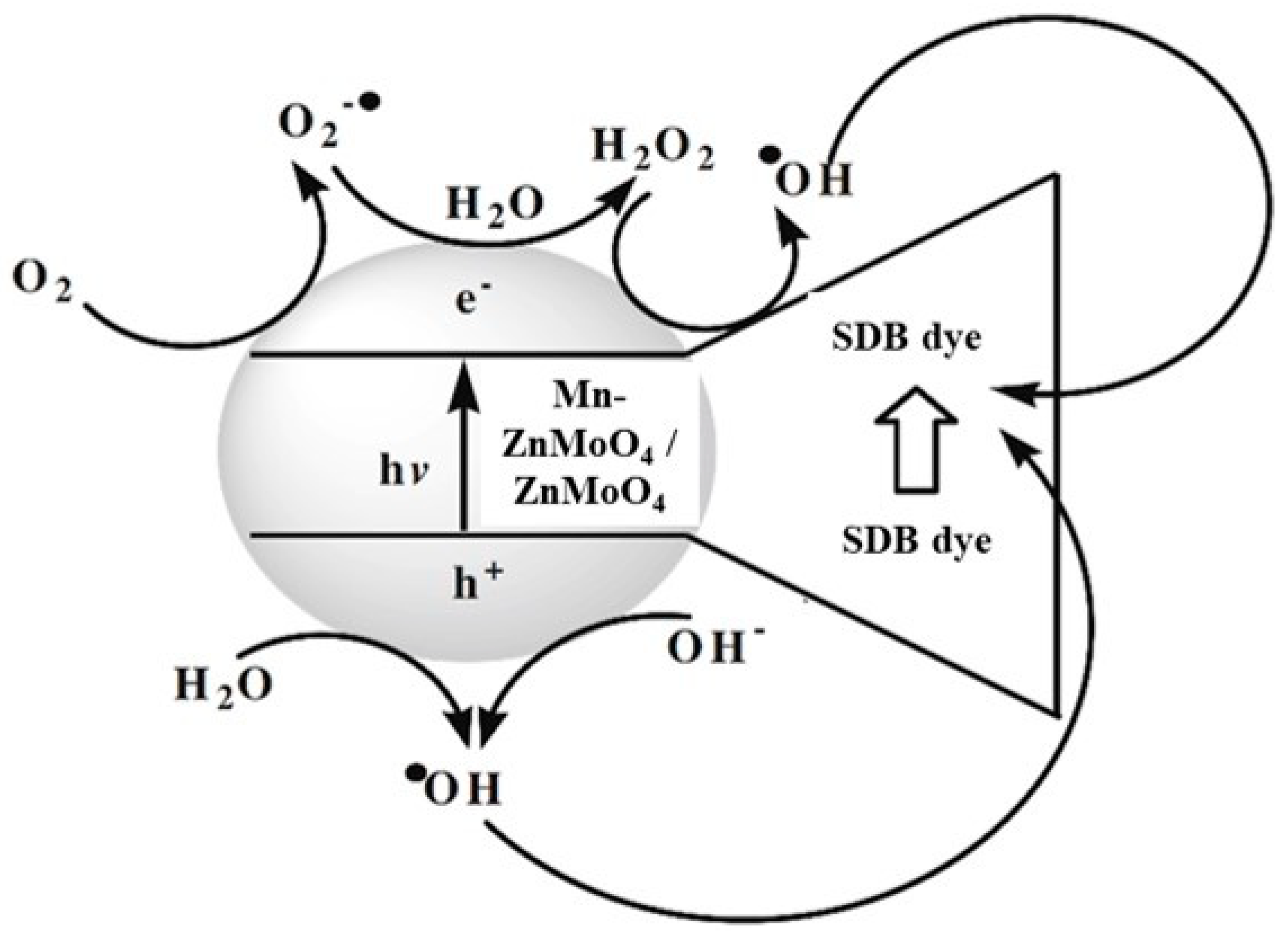
2.3. Photocatalytic Activity

Photocatalytic experiments were conducted on SDB dye using ZnMoO4 and Mn-doped ZnMoO4.
Figure 6 shows that the photocatalytic reaction of 50 ppm SDB dye in the presence of light after 360 min resulted in slight changes. After the addition of 40 mg ZnMoO4 or Mn-doped ZnMoO4, a drastic degradation was observed in the presence of light and air during the process of photocatalysis. The photocatalytic activity of the molybdates was due to transfer of charges from the valence band orbital of O2p to the empty conduction band orbital of Mo 4d (2p 4d) (49). Here, we describe the photocatalytic activity of ZnMoO4 on solochrome dark blue in by process of photodegradation.
In the presence of light and air, 40 mg ZnMoO4 was added to 10.0 mL aq. solution of 50 ppm SDB (pH = 6). With a constant time (10 min) interval, ZnMoO4 degraded the SDB (Figure 7a). After 120 min, the blue solution became colorless (Figure 7b). With increased time, a decrease in the absorption of blue color was observed at a constant time interval and a 420 nm wavelength (Figure 7c).
For comparison, 40.0 mg Mn-doped ZnMoO4 was added to 10.0 mL aq. Solution with a concentration of 50 ppm SDB (pH = 6) in the presence of light and air with a constant time (10 min) interval. Mn-doped ZnMoO4 degraded the dark blue color of the solution at a wavelength of 588 nm (Figure 8a). After 80 min, the blue solution turned colorless (Figure 8b). With increased time, a decrease in the absorption of blue color was observed when the time interval was held constant with a wavelength of 588 nm (Figure 8c).
According to absorption spectra of Mn, ZnMoO4 nanomaterial showed one absorption peak at 293 nm and two shoulder peaks at wavelengths of 312 and 334 nm, with a band gap value of 4.22 eV. In the case of undoped ZnMoO4, there was one sharp peak and one small, broad peak at 376 and 272 cm−1 wavelengths, respectively, with a band gap value of 3.07 eV. FTIR spectra showed identical peaks at 473 and 450 nm, which were attributed to Zn-O and Mn-Zn, respectively, for Mn-doped ZnMoO4 nanomaterial. At high temperatures, Mn-doped zinc molybdate nanoparticles were more stable than undoped ZnMoO4. According to the photoluminescence spectra, Mn-doped ZnMoO4 was more photoluminescent than ZnMoO4. Undoped ZnMoO4 had a characteristic band, and a sharp peak was observed at 422 nm, which belonged to the Mo (4d) → O(2P) transition. It was also evident that there were emission peaks at 535 nm and 587 nm, which belonged to 5D37F6 and 5D47F4 transitions, respectively. However, in the case of Mn-doped ZnMoO4, at an excitation of 200 nm, sharp emission peaks were observed at 422, 440, 486 and 537 nm.
In ZnMoO4, absorbed photons were equal to or greater than its band gap energy, and electrons were excited from the VB band to the conduction band (CB), leaving a “hole” in the VB of ZnMoO4. These pairs of electron holes normally recombine rapidly; therefore, the photocatalytic activity of the material decreases. The photogenerated e and h+ reacted with H2O, O2 and the organic substrate adsorbed on photocatalytic surface for generation of reactive species such as OH and O2. The oxidative action of OH and O2 decomposed organic compounds into degradation products [41]. These radicals may be involved in processes of organic compound mineralization (Figure 9). Since SDB is cationic in nature, it is readily absorbed on the surfaces of catalytic ZnMoO4 and Mn-ZnMoO4 at alkaline pH, which accelerates the process of photodegradation [42,43].
The probable mechanism of photocatalytic degradation of solochrome dark blue can be summarized as follows:
ZnMoO4 + hν → ZnMoO4 + e + h+
h+ + H2O → OH
e + O2 → O2*
OH + O2* + SDB → degradation in solution
In the presence of light and air, ZnMoO4 (40.0 mg) was activated to form OH and O2* radicals. After 120 min, the blue solution of the monochromatic dark blue dye deteriorated, and the solution became colorless. On the other hand, a photocatalytic process was developed for Mn-ZnMoO4 nanomaterials that follow the same photocatalytic process and also generate OH and O2* radicals. Due to the presence of Mn, the blue color of the SDB dye was degraded by the Mn-ZnMoO4 nanomaterials after 80 min, resulting in a colorless dye, which makes Mn-doped ZnMoO4 more important than ZnMoO4 in the photocatalytic process.
Free-radical °OH and O2° produced by non-toxic Mn-doped ZnMoO4 and undoped ZnMoO4 nanomaterials entered the cell membrane of bacterial cells and inhibited cell growth of Escherichia coli and Staphylococcus aureus species. In contrast, the difference between Mn-ZnMoO4 nanomaterials inhibited bacterial cell growth more than ZnMoO4 (undoped) (Table 1) because more free radicals were produced, which easily penetrated the cell membrane and inhibited cell growth.

3. Materials and Methods

3.1. Chemicals and Reagents

The raw materials used in the synthesis procedure were zinc sulphate (ZnSO4·7H2O; Merck 99.8%), sodium molybdate (Na2MoO4·2H2O; Merck 99.8%), manganese sulphate (MnSO4; Merck 99.5%), ethylene glycol (Merck), urea (Hi-media), E. coli and S. aureus pure culture, natural agar media (Hi-Media) and triple-distilled water.

3.2. Instrumentation

In the absorbance mode, UV-visible spectra were acquired using a UV-1900i double-beam spectrophotometer. Samples were dispersed in ethanol to determine absorbance. Photoluminescence measurements of powder were performed using 266 nm radiation from an Nd: YAG laser and detected by a CCD (charge coupled device) detector (model: QE 65000, Ocean Optics, Orlando, FL, USA) attached to the fiber sample, which was analyzed using an Advanced D8 Bruker X-ray diffractometer (XRD) with Ni-filtered Cu-K (1.5405) (2–10–80°; step size, 0.02°). The vibration spectra were recorded using an Avtar 370 Thermo Nicolet Fourier transform infrared (FT-IR) spectrophotometer equipped with a DTGS detector with a set resolution of 4 cm−1, and the samples were prepared as KBr discs for this study.

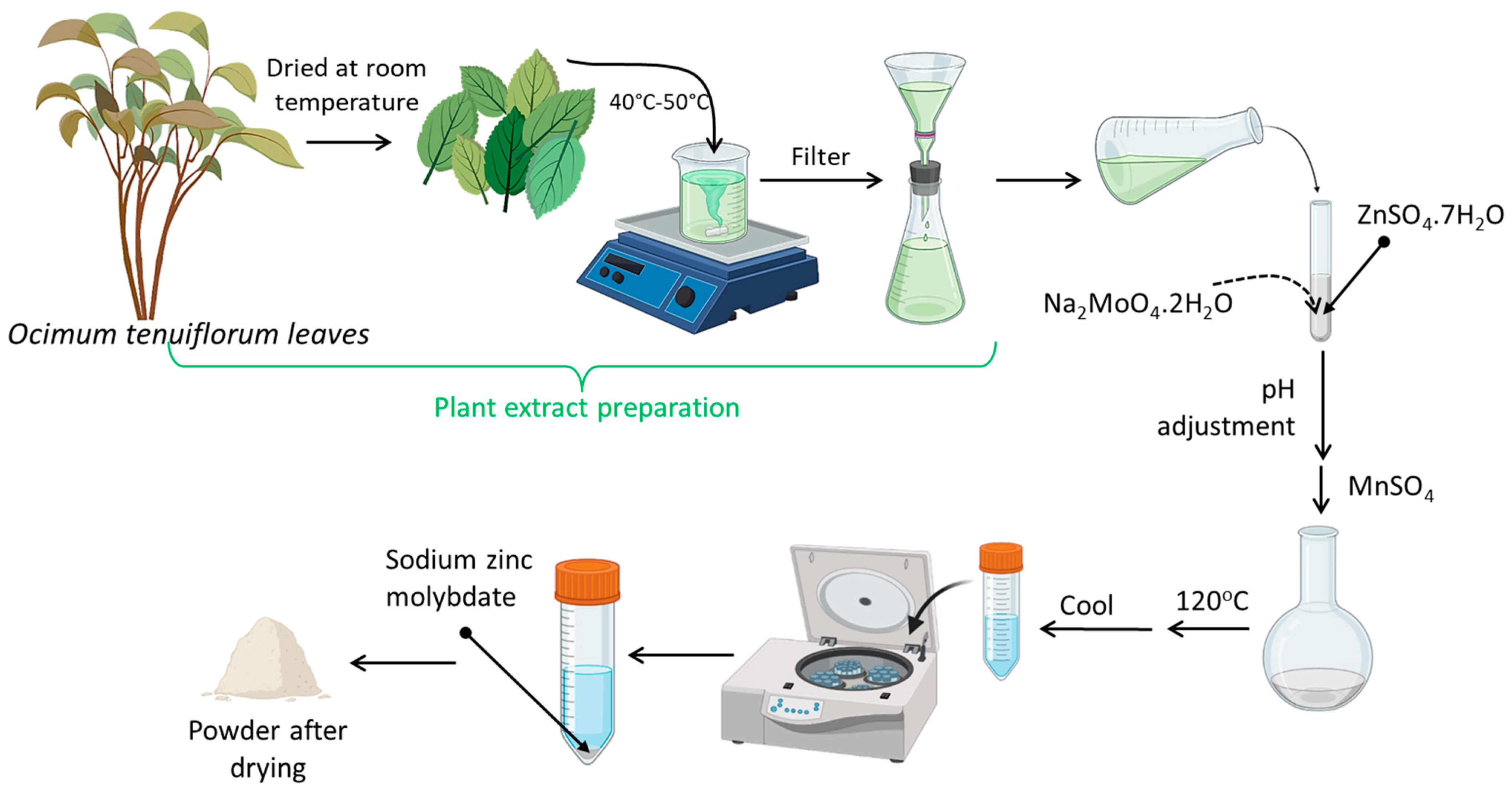
3.3. Extraction of Ocimum Tenuiflorum Leaves

Leaves of Ocimum Tenuiflorum were collected in January 2022 from a natural ecosystem at India. Plant leaves were washed with double-distilled water to remove impurities and dried at room temperature (25 °C). Subsequently, the washed plant leaves (100 g) were boiled with distilled water (110 mL) at 40–50 °C. The whole extract solution was filtered using Whatman filter paper No. 42 and stored at 4–5 °C. Then, the extracted filtrate was used in the synthesis of zinc molybdate nanomaterials.

3.4. Synthesis of Sodium Zinc Molybdate

The total synthesis process is graphically illustrated in Scheme 1. Green synthesis of zinc molybdate and Mn-doped zinc molybdate NPs was carried out using crude plant extract (100 mL) heated (40–60 °C) on a magnetic stirrer with continuous stirring using a magnetic bar; then, the temperature was maintained at 50 °C. A solution of ZnSO4·7H2O and ethylene glycol was added to aforementioned solution. The pH of the solution was adjusted to 9 with urea. The entire solution was heated for an hour at 80 °C with stirring. White precipitate was developed after the addition of 2 M solution of Na2MoO4·2H2O. The solution was then transferred to a round-bottom flask and heated up to 120 °C with continuous stirring. The white precipitate was allowed to settle; then, the mixture was filtered to separate the precipitate from the solution. The precipitate was washed with deionized water, methanol and acetone. The resultant material was dried in an oven at 60 °C for 2 h.

3.5. Synthesis of Mn-Doped Sodium Zinc Molybdate

In a beaker, solutions of 1 M ZnSO4·7H2O and ethylene glycol (10 mL) were mixed together, followed by the addition of 2 M Na2MoO4·2H2O. After the addition of urea, the pH of the solution was adjusted to 9, followed by heating to 80 °C for one hour. The solution was stirred well with 1 gm of MnSO4. This solution was transferred to a round-bottom flask and heated up to 120 °C with continuous stirring. The solution was allowed to stand until white precipitate settled, which was subsequently and filtered from the solution. The precipitate was washed with deionized water, methanol and acetone. The resultant material was dried in an oven at 60 °C for 2 h.

3.6. Antibacterial Activity

Antibacterial activity was determined in a well diffusion assay on nutrient agar medium (NAM). The medium was cast into a Petri dish under sterile conditions and kept up to 1 h for solidification. Then, fresh overnight cultured E. coli and S. aureus (100 µg/mL) were spread onto a solidified nutrient agar Petri dish. The dishes were kept up to 15–20 min for complete absorption of bacterial cultures. Wells were prepared by gel puncture (7–8 mm). Then, ZnMoO4 and Mn-ZnMoO4 nanomaterials with different concentrations (50, 100 and 150 µg/mL) were introduced into these wells. The Petri dishes with medium were kept at room temperature for 30 min to allow for the diffusion of the extracts. Then, incubation was carried out at 37 °C for 24 h to allow for maximum growth of the microorganisms. Both nanomaterials inhibited the growth of microorganisms, displaying a clear zone of inhibition (ZOI) around the well after incubation.

3.7. Dye Remediation

Solochrome dark blue dye (SDB) adsorption experiments were conducted under batch conditions using green-synthesized ZnMoO4 and Mn-ZnMoO4 nanomaterials. A standard solochrome dark blue dye (SDB) stock solution was diluted with deionized water in order to obtain different concentrations. The obtained solochrome dark blue dye (SDB) solutions were kept in a flask with a fixed volume (10 mL of 5 ppm), and ZnMoO4 and Mn-ZnMoO4 nanomaterials were added. The flask was placed in a sonicator for 120 min at a pH of 6 at room temperature. After a certain time, the upper layer of liquid was analyzed by a UV-vis spectrophotometer (UV-Visible 1900i, Shimadzu, Japan) at a wavelength of 600 nm. The ZnMoO4 and Mn-ZnMoO4 were removed with the help of centrifugation after the completion of experiment. The removal (R, %) was calculated according to Equation (2) as follows:
R ( % ) = C o C e C o × 100
where Co and Ce are the initial and equilibrium concentrations of SDB (mg L−1), respectively.

4. Conclusions

In this paper, we described the green synthesis of ZnMoO4 and Mn-ZnMoO4 nanomaterials from Ocimum tenuiflorum (Tulsi), as well as their characterization by UV-vis absorption spectroscopy, FTIR spectroscopy, X-ray diffraction (X-ray diffraction) and luminance spectra (PL). The band gaps of ZnMoO4 and Mn-ZnMoO4 were 3.07 and 4.22 eV, respectively. Both doped and undoped nanomaterials have potential for biological and environmental applications. Both nanomaterials exhibited antibacterial activity and photocatalytic properties.
In contrast, Mn-ZnMoO4 nanomaterials responded better to two different bacterial species: (a) Escherichia coli and (b) Staphylococcus aureus. ZnMoO4 showed a positive response only against Staphylococcus aureus. Due to the formation of reactive oxidative species such as OH and O2 radicals, both nanomaterials were found to have photocatalytic properties, as demonstrated by solochrome dark blue dye (SDB) in the process of photodegradation. In the presence of light and air, after 80 min, Mn-doped ZnMoO4 (Mn-ZnMoO4) resulted in the decolorization of the blue dye, whereas ZnMnO4 took 120 min to induce decolorization.

Author Contributions

Conceptualization, A.S., K.B., M.A., J.R., P.D., S.G. and R.R.; Methodology, A.S., M.A., S.G. and R.R.; Formal analysis, R.C.A. and N.G.; Investigation, A.S., K.B., J.R. and P.D.; Data curation, A.S.; Writing—original draft, A.S., K.B., M.A., J.R., P.D., S.G. and R.R.; Writing—review & editing, A.S., R.C.A., K.B., N.G., M.A., J.R., P.D., S.G. and R.R.; Visualization, S.G. and R.R.; Supervision, P.D. and R.R. All authors have read and agreed to the published version of the manuscript.

Funding

This research received no external funding.

Institutional Review Board Statement

Not applicable.

Informed Consent Statement

Not applicable.

Data Availability Statement

Not applicable.

Conflicts of Interest

The authors declare no conflict of interest.

Sample Availability

Not applicable.

References

  1. Cornu, L.; Jubera, V.; Demourgues, A.; Salek, G.; Gaudon, M. Luminescence properties and pigment properties of A-doped (Zn, Mg) MoO4 triclinic oxides (with A = Co, Ni, Cu or Mn). Ceram. Int. 2017, 43, 13377–13387. [Google Scholar] [CrossRef]
  2. Wang, D.; Huang, M.; Zhuang, Y.; Jia, H.-L.; Sun, J.; Guan, M. Phase-and Morphology-Controlled Synthesis of Zinc Molybdate for Excellent Photocatalytic Properties. Eur. J. Inorg. Chem. 2017, 2017, 4939–4946. [Google Scholar] [CrossRef]
  3. Ramezani, M.; Hosseinpour-Mashkani, S.M.; Sobhani-Nasab, A.; Ghasemi Estarki, H. Synthesis, characterization, and morphological control of ZnMoO4 nanostructures through precipitation method and its photocatalyst application. J. Mater. Sci. Mater. Electron. 2015, 26, 7588–7594. [Google Scholar] [CrossRef]
  4. Lv, L.; Tong, W.; Zhang, Y.; Su, Y.; Wang, X. Metastable monoclinic ZnMoO4: Hydrothermal synthesis, optical properties and photocatalytic performance. J. Nanosci. Nanotechnol. 2011, 11, 9506–9512. [Google Scholar] [CrossRef] [PubMed]
  5. Lovisa, L.X.; Oliveira, M.C.; Andrés, J.; Gracia, L.; Li, M.S.; Longo, E.; Tranquilina, R.L.; Paskocimasa, C.A.; Bomioa, M.R.D.; Motta, F.V. Structure, morphology and photoluminescence emissions of ZnMoO4: RE3+ = Tb3+ − Tm3+ − X Eu3+ (x = 1, 1.5, 2, 2.5 and 3 mol%) particles obtained by the sonochemical method. J. Alloys Compd. 2018, 750, 55–70. [Google Scholar] [CrossRef]
  6. Ryu, J.H.; Koo, S.-M.; Yoon, J.-W.; Lim, C.S.; Shim, K.B. Synthesis of nanocrystalline MMoO4 (M = Ni, Zn) phosphors via a citrate complex route assisted by microwave irradiation and their photoluminescence. Mater. Lett. 2006, 60, 1702–1705. [Google Scholar] [CrossRef]
  7. Zazhigalov, V.; Sachuk, O.; Kopachevska, N.; Starchevskyy, V.; Sawlowicz, Z. Effect of ultrasonic treatment on formation of nanodimensional structures in ZnO–MoO3 system. Theor. Exp. Chem. 2017, 53, 53–59. [Google Scholar] [CrossRef]
  8. Chernyak, D.; Danevich, F.; Degoda, V.Y.; Dmitruk, I.; Ferri, F.; Galashov, E.; Giuliani, A.; Ivanov, I.; Kobychev, V.; Mancuso, M.; et al. Optical, luminescence and thermal properties of radiopure ZnMoO4 crystals used in scintillating bolometers for double beta decay search. Nucl. Instrum. Methods Phys. Res. Sect. A Accel. Spectrometers Detect. Assoc. Equip. 2013, 729, 856–863. [Google Scholar] [CrossRef]
  9. Beeman, J.; Danevich, F.; Degoda, V.; Galashov, E.; Giuliani, A.; Ivanov, I.; Mancuso, M.; Marnieros, S.; Nones, C.; Pessina, G.E.; et al. An improved ZnMoO4 scintillating bolometer for the search for neutrinoless double beta decay of 100 Mo. J. Low Temp. Phys. 2012, 167, 1021–1028. [Google Scholar] [CrossRef]
  10. Jeseentharani, V.; Dayalan, A.; Nagaraja, K. Co-precipitation synthesis, humidity sensing and photoluminescence properties of nanocrystalline Co2+ substituted zinc (II) molybdate (Zn1–xCoxMoO4; x = 0, 0.3, 0.5, 0.7, 1). Solid State Sci. 2017, 67, 46–58. [Google Scholar] [CrossRef]
  11. Mardare, C.C.; Tanasic, D.; Rathner, A.; Müller, N.; Hassel, A.W. Growth inhibition of Escherichia coli by zinc molybdate with different crystalline structures. Phys. Status Solidi 2016, 213, 1471–1478. [Google Scholar] [CrossRef]
  12. Luitel, H.N.; Chand, R.; Hamajima, H.; Gaihre, Y.R.; Shingae, T.; Yanagita, T.; Watari, T. Highly efficient NIR to NIR upconversion of ZnMoO4: Tm3+, Yb3+ phosphors and their application in biological imaging of deep tumors. J. Mater. Chem. B 2016, 4, 6192–6199. [Google Scholar] [CrossRef] [PubMed]
  13. Gao, Y.-P.; Huang, K.-J.; Zhang, C.-X.; Song, S.-S.; Wu, X. High-performance symmetric supercapacitor based on flower-like zinc molybdate. J. Alloys Compd. 2018, 731, 1151–1158. [Google Scholar] [CrossRef]
  14. Yoon, Y.-S.; Fujikawa, N.; Ueda, W.; Moro-Oka, Y.; Lee, K.-W. Propane oxidation over various metal molybdate catalysts. Catal. Today 1995, 24, 327–333. [Google Scholar] [CrossRef]
  15. Maggiore, R.; Galvagno, S.; Bart, J.C.; Giannetto, A.; Crisafulli, C.; Toscano, G. Catalytic oxidation of propene over zinc, cadmium and nickel molybdates. Z. Phys. Chem. 1983, 137, 111–118. [Google Scholar] [CrossRef]
  16. Cavalcante, L.S.; Moraes, E.; Almeida, M.; Dalmaschio, C.; Batista, N.; Varela, J.A.; Longo, E.; Siu Li, M.; Andrés, J.; Beltrán, A. A combined theoretical and experimental study of electronic structure and optical properties of β-ZnMoO4 microcrystals. Polyhedron 2013, 54, 13–25. [Google Scholar] [CrossRef]
  17. Pasiński, D.; Sokolnicki, J. Broadband orange phosphor by energy transfer between Ce3+ and Mn2+ in Ca3Al2Ge3O12 garnet host. J. Alloys Compd. 2019, 786, 808–816. [Google Scholar] [CrossRef]
  18. Mallikarjun, S.; Rao, A.; Rajesh, G.; Shenoy, R.; Pai, M. Antimicrobial efficacy of Tulsi leaf (Ocimum sanctum) extract on periodontal pathogens: An in vitro study. J. Indian Soc. Periodontol. 2016, 20, 145. [Google Scholar] [PubMed]
  19. Hussain, E.H.; Jamil, K.; Rao, M. Hypoglycaemic, hypolipidemic and antioxidant properties of tulsi (Ocimum sanctum Linn) on streptozotocin induced diabetes in rats. Indian J. Clin. Biochem. 2001, 16, 190–194. [Google Scholar] [CrossRef]
  20. Ganguly, S.; Das, P.; Saha, A.; Noked, M.; Gedanken, A.; Margel, S. Mussel-inspired polynorepinephrine/MXene-based magnetic nanohybrid for electromagnetic interference shielding in X-band and strain-sensing performance. Langmuir 2022, 38, 3936–3950. [Google Scholar] [CrossRef] [PubMed]
  21. Ganguly, S.; Kanovsky, N.; Das, P.; Gedanken, A.; Margel, S. Photopolymerized thin coating of polypyrrole/graphene nanofiber/iron oxide onto nonpolar plastic for flexible electromagnetic radiation shielding, strain sensing, and non-contact heating applications. Adv. Mater. Interfaces 2021, 8, 2101255. [Google Scholar] [CrossRef]
  22. Das, P.; Ganguly, S.; Margel, S.; Gedanken, A. Immobilization of heteroatom-doped carbon dots onto nonpolar plastics for antifogging, antioxidant, and food monitoring applications. Langmuir 2021, 37, 3508–3520. [Google Scholar] [CrossRef]
  23. Das, P.; Maruthapandi, M.; Saravanan, A.; Natan, M.; Jacobi, G.; Banin, E.; Gedanken, A. Carbon dots for heavy-metal sensing, pH-sensitive cargo delivery, and antibacterial applications. ACS Appl. Nano Mater. 2020, 3, 11777–11790. [Google Scholar] [CrossRef]
  24. Ganguly, S.; Das, P.; Itzhaki, E.; Hadad, E.; Gedanken, A.; Margel, S. Microwave-synthesized polysaccharide-derived carbon dots as therapeutic cargoes and toughening agents for elastomeric gels. ACS Appl. Mater. Interfaces 2020, 12, 51940–51951. [Google Scholar] [CrossRef]
  25. Sanderson, K. Quantum dots go large: A small industry could be on the verge of a boom. Nature 2009, 459, 760–762. [Google Scholar] [CrossRef]
  26. Das, P.; Ganguly, S.; Perelshtein, I.; Margel, S.; Gedanken, A. Acoustic green synthesis of graphene-gallium nanoparticles and PEDOT: PSS hybrid coating for textile to mitigate electromagnetic radiation pollution. ACS Appl. Nano Mater. 2022, 5, 1644–1655. [Google Scholar] [CrossRef]
  27. Tauc, J. Amorphous and Liquid Semiconductors; Springer Science & Business Media: Berlin/Heidelberg, Germany, 2012. [Google Scholar]
  28. Frost, R.L.; Palmer, S.J. Raman spectrum of decrespignyite [(Y, REE) 4Cu(CO3) 4Cl(OH)5 2H2O] and its relation with those of other halogenated carbonates including bastnasite, hydroxybastnasite, parisite and northupite. J. Raman Spectrosc. 2011, 42, 2042–2048. [Google Scholar] [CrossRef]
  29. Liang, Y.; Liu, P.; Li, H.; Yang, G. ZnMoO4 micro-and nanostructures synthesized by electrochemistry-assisted laser ablation in liquids and their optical properties. Cryst. Growth Des. 2012, 12, 4487–4493. [Google Scholar] [CrossRef]
  30. Hardcastle, F.D.; Wachs, I.E. Determination of vanadium-oxygen bond distances and bond orders by Raman spectroscopy. J. Phys. Chem. 1991, 95, 5031–5041. [Google Scholar] [CrossRef]
  31. Pholnak, C.; Sirisathitkul, C.; Harding, D.J. Characterizations of octahedral zinc oxide synthesized by sonochemical method. J. Phys. Chem. Solids 2011, 72, 817–823. [Google Scholar] [CrossRef]
  32. de Azevedo Marques, A.P.; de Melo, D.M.; Paskocimas, C.A.; Pizani, P.S.; Joya, M.R.; Leite, E.R.; Longo, E. Photoluminescent BaMoO4 nanopowders prepared by complex polymerization method (CPM). J. Solid State Chem. 2006, 179, 671–678. [Google Scholar] [CrossRef]
  33. Ryu, J.H.; Yoon, J.-W.; Lim, C.S.; Oh, W.-C.; Shim, K.B. Microwave-assisted synthesis of CaMoO4 nano-powders by a citrate complex method and its photoluminescence property. J. Alloys Compd. 2005, 390, 245–249. [Google Scholar] [CrossRef]
  34. Li, P.; Wang, J.; Li, L.; Song, S.; Yuan, X.; Jiao, W.; Hao, Z.; Li, X. Design of a ZnMoO4 porous nanosheet with oxygen vacancies as a better performance electrode material for supercapacitors. New J. Chem. 2021, 45, 9026–9039. [Google Scholar] [CrossRef]
  35. Wang, Y.; Cheng, J.; Yu, S.; Alcocer, E.J.; Shahid, M.; Wang, Z.; Pan, W. Synergistic effect of N-decorated and Mn2+ doped ZnO nanofibers with enhanced photocatalytic activity. Sci. Rep. 2016, 6, 32711. [Google Scholar] [CrossRef] [PubMed]
  36. Zbair, M.; Ezahri, M.; Benlhachemi, A.; Arab, M.; Bakiz, B.; Guinneton, F.; Gavarri, J.-R. Rietveld refinements, impedance spectroscopy and phase transition of the polycrystalline ZnMoO4 ceramics. Ceram. Int. 2015, 41, 15193–15201. [Google Scholar]
  37. Xianju, Z.; Xiaodong, Y.; Tengjiao, X.; Kaining, Z.; Tianyu, C.; Hao, Y.; Wang, Z. Luminescence properties and energy transfer of host sensitized CaMoO4: Tb3+ green phosphors. J. Rare Earths 2013, 31, 655–659. [Google Scholar]
  38. Sczancoski, J.; Cavalcante, L.; Marana, N.; da Silva, R.O.; Tranquilin, R.; Joya, M.; Pizani, P.; Varela, J.; Sambrano, J.; Li, M.S.; et al. Electronic structure and optical properties of BaMoO4 powders. Curr. Appl. Phys. 2010, 10, 614–624. [Google Scholar] [CrossRef]
  39. Liao, J.; Lin, S.; Zhang, L.; Pan, N.; Cao, X.; Li, J. Photocatalytic degradation of methyl orange using a TiO2/Ti mesh electrode with 3D nanotube arrays. ACS Appl. Mater. Interfaces 2012, 4, 171–177. [Google Scholar] [CrossRef]
  40. Liang, J.-H.; Han, X. Structure-activity relationships and mechanism of action of macrolides derived from erythromycin as antibacterial agents. Curr. Top. Med. Chem. 2013, 13, 3131–3164. [Google Scholar] [CrossRef]
  41. Jiang, Y.-R.; Lee, W.W.; Chen, K.-T.; Wang, M.-C.; Chang, K.-H.; Chen, C.-C. Hydrothermal synthesis of β-ZnMoO4 crystals and their photocatalytic degradation of Victoria Blue R and phenol. J. Taiwan Inst. Chem. Eng. 2014, 45, 207–218. [Google Scholar] [CrossRef]
  42. Sarwar, N.; Humayoun, U.B.; Kumar, M.; Zaidi, S.F.A.; Yoo, J.H.; Ali, N.; Jeong, D.I.; Lee, J.H.; Yoon, D.H. Citric acid mediated green synthesis of copper nanoparticles using cinnamon bark extract and its multifaceted applications. J. Clean. Prod. 2021, 292, 125974. [Google Scholar] [CrossRef]
  43. Sarwar, N.; Bin Humayoun, U.; Kumar, M.; Nawaz, A.; Zafar, M.S.; Rasool, U.; Kim, Y.H.; Yoon, D.H. A bio based immobilizing matrix for transition metal oxides (TMO) crosslinked cotton: A facile and green processing for photocatalytic self-cleaning and multifunctional textile. Mater. Lett. 2022, 309, 131338. [Google Scholar] [CrossRef]
Figure 1. (a) UV−vis absorption spectrum of ZnMoO4. (b)Tauc Plot of ZnMoO4 derived from UV-vis absorption. (c) UV−vis absorption spectrum of Mn-doped ZnMoO4. (d) Tauc plot of Mn-doped ZnMoO4 derived from UV-vis absorption.
Molecules 28 04182 g001
Figure 2. FTIR spectra of (a) ZnMoO4 and (b) Mn-ZnMoO4 nanomaterial. XRD patterns of (c) ZnMoO4 and (d) Mn-ZnMoO4 nanostructures. (e) Full XPS spectra of ZnMoO4 and Mn-ZnMoO4 nanomaterials.
Molecules 28 04182 g002
Figure 3. TGA and DTA analysis of (a) ZnMoO4 nanomaterials and (b) Mn-doped ZnMoO4 nanomaterials. TGA is represented by the red line, and DTA is represented but the dark blue line.
Molecules 28 04182 g003
Figure 4. Photoluminescence spectra of (a) ZnMoO4 and (b) Mn-doped ZnMoO4.
Molecules 28 04182 g004
Figure 5. Antibacterial activity of (a,b) ZnMoO4 and (c,d) Mn-doped ZnMoO4 nanomaterials against E. coli and S. aureus.
Molecules 28 04182 g005
Figure 6. Photocatalysis reaction of SDB dye in the presence of light and air.
Molecules 28 04182 g006
Figure 7. (a) Photocatalytic SDB degradation of ZnMoO4 under visible light and light at a 420 nm wavelength. (b) With the naked eye: (i) aq. solution of 50 ppm SDB (blank); (ii) after 120 min, the blue color of the solution degraded with 40.0 mg undoped ZnMoO4. (c) At 420 nm wavelength after adding ZnMoO4 in solution with increased time, there was decrease in the absorption in the presence of light and air.
Molecules 28 04182 g007
Figure 8. (a) Photocatalytic SDB degradation of Mn-doped ZnMoO4 under visible light and light at a 588 nm wavelength. (b) With the naked eye: (i) aq. solution of 50 ppm SDB (blank); (ii) after 80 min, the blue color of solution degraded with 40.0 mg undoped ZnMoO4. (c) At 588 nm wavelength, after the addition of Mn-doped ZnMoO4 in solution, there was decrease in absorption with an increase in time in the presence of light and air.
Molecules 28 04182 g008
Figure 9. Mechanism of the photocatalytic degradation process of solochrome dark blue (SDB) dye with ZnMoO4/Mn-ZnMoO4 nanomaterials.
Molecules 28 04182 g009
Scheme 1. Graphical illustration of green synthesis procedure for Mn-doped zinc molybdate.
Molecules 28 04182 sch001
Table 1. Zones of inhibition (ZOIs) of antimicrobial activity for ZnMoO4 and Mn-doped ZnMoO4 at different concentrations in methanol solvent (50, 100 and 150 µg/mL).
Bacterial SpeciesConcentration of Mn-Doped ZnMoO4
50 µL100 µL150 µL
E. coli3.0 mm1.0 mm1.5 mm
S. aureus1.4 mm6.0 mm8.0 mm
Disclaimer/Publisher’s Note: The statements, opinions and data contained in all publications are solely those of the individual author(s) and contributor(s) and not of MDPI and/or the editor(s). MDPI and/or the editor(s) disclaim responsibility for any injury to people or property resulting from any ideas, methods, instructions or products referred to in the content.

