Article

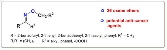
The Oxime Ethers with Heterocyclic, Alicyclic and Aromatic Moiety as Potential Anti-Cancer Agents

1
Department of Organic Chemistry, Faculty of Pharmacy, Collegium Medicum in Bydgoszcz, Nicolaus Copernicus University in Toruń, Jurasza 2, 85-089 Bydgoszcz, Poland
2
Department of Biochemistry, Institute of Biology, Faculty of Biological and Veterinary Sciences, Nicolaus Copernicus University in Toruń, Lwowska 1, 87-100 Torun, Poland
3
Department of Medical Biology and Biochemistry, Faculty of Medicine, Collegium Medicum in Bydgoszcz, Nicolaus Copernicus University in Toruń, Karłowicza 24, 85-092 Bydgoszcz, Poland
*
Authors to whom correspondence should be addressed.
Molecules 2022, 27(4), 1374; https://doi.org/10.3390/molecules27041374
Submission received: 29 November 2021 / Revised: 14 February 2022 / Accepted: 15 February 2022 / Published: 17 February 2022
(This article belongs to the Special Issue Novel Design and Synthesis of Anticancer Agents)

Abstract

:
Chemotherapy is one of the most commonly used methods of cancer disease treatment. Due to the acquisition of drug resistance and the possibility of cancer recurrence, there is an urgent need to search for new molecules that would be more effective in destroying cancer cells. In this study, 1-(benzofuran-2-yl)ethan-1-one oxime and 26 oxime ethers containing heterocyclic, alicyclic or aromatic moiety were screened for their cytotoxicity against HeLa cancer cell line. The most promising derivatives with potential antitumor activity were 2-(cyclohexylideneaminoxy)acetic acid (18) and (E)-acetophenone O-2-morpholinoethyl oxime (22), which reduced the viability of HeLa cells below 20% of control at concentrations of 100–250 μg/mL. Some oxime ethers, namely thiazole and benzothiophene derivatives (2427), also reduced HeLa cell viability at similar concentrations but with lower efficiency. Further cytotoxicity evaluation confirmed the specific toxicity of (E)-acetophenone O-2-morpholinoethyl oxime (22) against A-549, Caco-2, and HeLa cancer cells, with an EC50 around 7 μg/mL (30 μM). The most potent and specific compound was (E)-1-(benzothiophene-2-yl)ethanone O-4-methoxybenzyl oxime (27), which was selective for Caco-2 (with EC50 116 μg/mL) and HeLa (with EC50 28 μg/mL) cells. Considering the bioavailability parameters, the tested derivatives meet the criteria for good absorption and permeation. The presented results allow us to conclude that oxime ethers deserve more scientific attention and further research on their chemotherapeutic activity.

Graphical Abstract

1. Introduction

The cancer diseases are a very serious health problem. Currently, about 18 million cases of various types of cancer are diagnosed each year. According to WHO predictions, this number is expected to reach 29.4 million in 2040 [1]. Chemotherapy is one of the most important methods of cancer treatment. Among the many compounds currently in use, new molecules are being developed that may be even more effective in destroying cancer cells. Success in treating cancer depends on the ability to detect it early, but also on the type of cancer. The problem in the treatment of neoplasms is the acquisition of drug resistance, and, unfortunately, the possibility of relapse of the neoplastic disease. The causes of cancer are difficult to estimate, but are certainly a very complex problem [2]. Therefore, there is a need to search for and investigate new structures that offer the potential to cure cancer.
The oxime ethers represent an interesting class of compounds with diverse biological activities. They exhibit antimicrobial [3], insecticidal [4], and anticonvulsant [5] properties and are often examined for their cytotoxic activity. Many articles related to the activities of oxime ethers concern their antitumor properties. Chakravati et al. reported the synthesis of thioarylnaphthylmethanone oxime ethers to treat breast cancer (MCF-7) cells [6]. The anti-tumor activities of the compounds against the cell lines tested were very high. Díaz et al. tested the cytotoxicity of oxime ethers of flavone and 6-hydroxy flavone oxime derivatives against breast (MDA-MB-231) and prostate (PC-3) adenocarcinomas as well as against human lung cells (A-549 and MRC-5 cell lines) [7]. Naringenin oxime derivative ethers showed very high antiproliferative activity in studies on promyelocytic leukemia (HL-60) and breast cancer cell lines (MCF-7, MDA-MB-231) [8]. Bis(4-hydroxy)benzophenone oxime ethers have been identified as novel estrogen receptor ligands [9]. These compounds also inhibit the growth of cancer cells through an estrogen receptor independent mechanism.
Recent years have indicated a growing research interest in benzofuran moiety, which significantly influences the biological activity of compounds. For example, benzofuran derivatives are being investigated for anticancer activity [10,11], as well as antibacterial, antiprotozoal and other activities [12,13]. Other heterocyclic moieties—thiazole and benzothiophene—may determine the biological activities of these compounds. Many of the thiazole derivatives show antitumor activity [14]. On the other hand, many interesting biological activities are exhibited by benzothiophene derivatives, including antitumor activity [15]. The benzofuran derivatives of the oxime ethers have also shown high antifungal activities against Candida albicans [16]. The thiophene derivative oxime ethers exhibit a number of biological activities, e.g., 2-benzoylthiophene derivatives show anti-aggregating activity against platelets [17] and antibacterial [18], anti-inflammatory, and anticonvulsant effects [19]. The derivatives O-uracil oxime of 2-acetylthiophene show antitumor effects [20], and in the case of many thiophene derivatives, it is also possible to present those structures that have shown antitumor activity [21]. Oxime ethers, for example, acetophenone derivatives, are also biologically active compounds and exhibit antidepressant activities [22].
Considering the above, oxime ethers constitute a potential class of compounds with possible and promising anti-cancer properties. Therefore, it seems justified to conduct research on the cytotoxic activity of various derivatives from this group of compounds.
In our previous studies, we obtained a series of new functionalized ethers of 2-acetylbenzofuran [23] and 2-acetylthiophene oxime [24]. The obtained products were tested microbiologically. In this report, we performed cytotoxicity studies of compounds with oxime ether moiety and benzofuran, benzothiophene, thiophene and thiazole substituents, as well as other cyclic substituents to assess their anti-proliferative potential. Thus, we have tested their influence on human dermal fibroblasts (HDF) used as healthy cell models and human cervical adenocarcinoma (HeLa) cells, lung epithelial carcinoma (A549) cells, and intestinal epithelial carcinoma (Caco-2) cell line representing different human cancer cells.

2. Results and Discussion
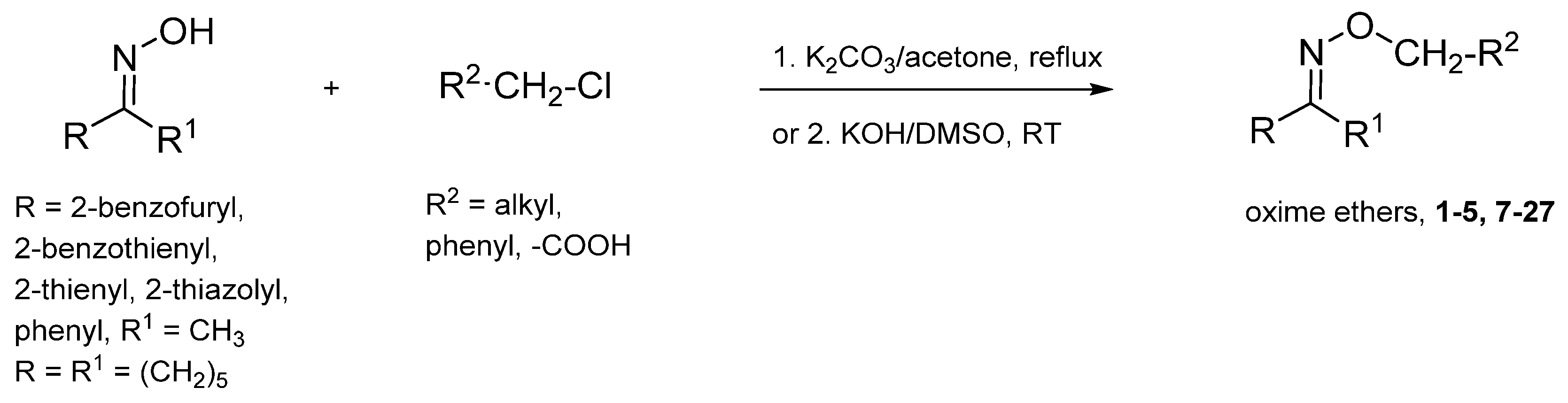
2.1. Chemistry

Most of the presented compounds (15, 727) were obtained by the reaction of oximes with the corresponding halides in the presence of K2CO3 or KOH base and were described in our earlier articles [23,24,25] (Scheme 1). The products were obtained with good yields and high purities. The following derivatives of thiophene (1315), thiazole (2425) and benzothiophene (2627) are newly synthesized compounds. The compounds were obtained in the reaction of 2-acetylthiophene oxime (1315), 2-acetylthiazole oxime (2425) or 2-acetylbenzothiophene oxime (2627) with corresponding chloroalkylamine in DMSO in KOH medium. The structures of all compounds together with their half-maximal effective concentrations (EC50) in HeLa cell culture are presented in Table 1.

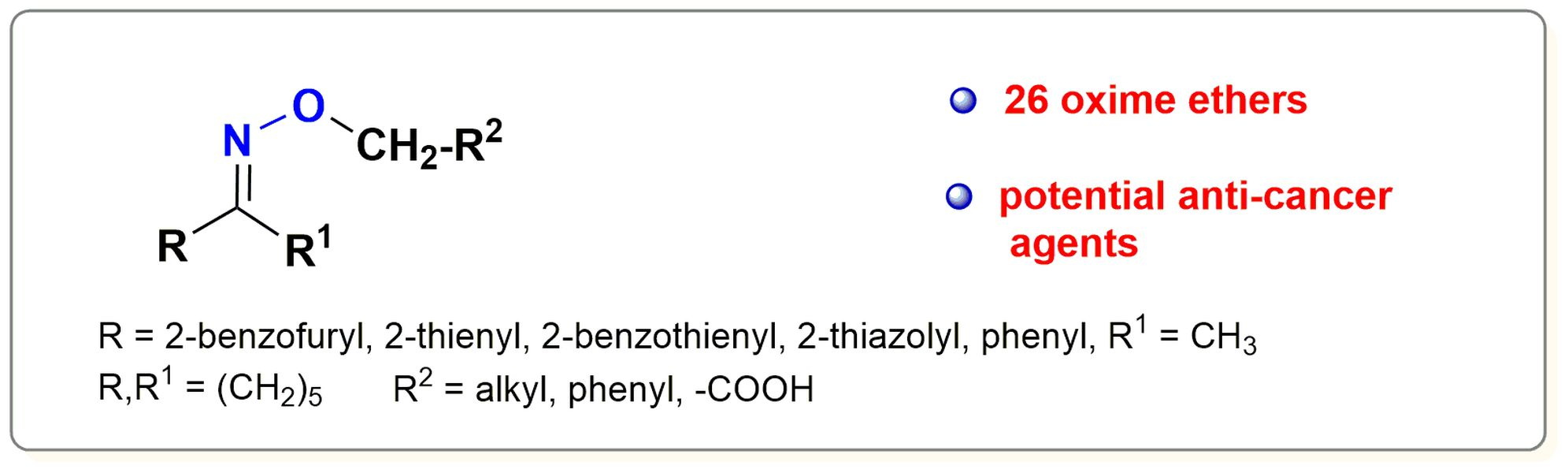
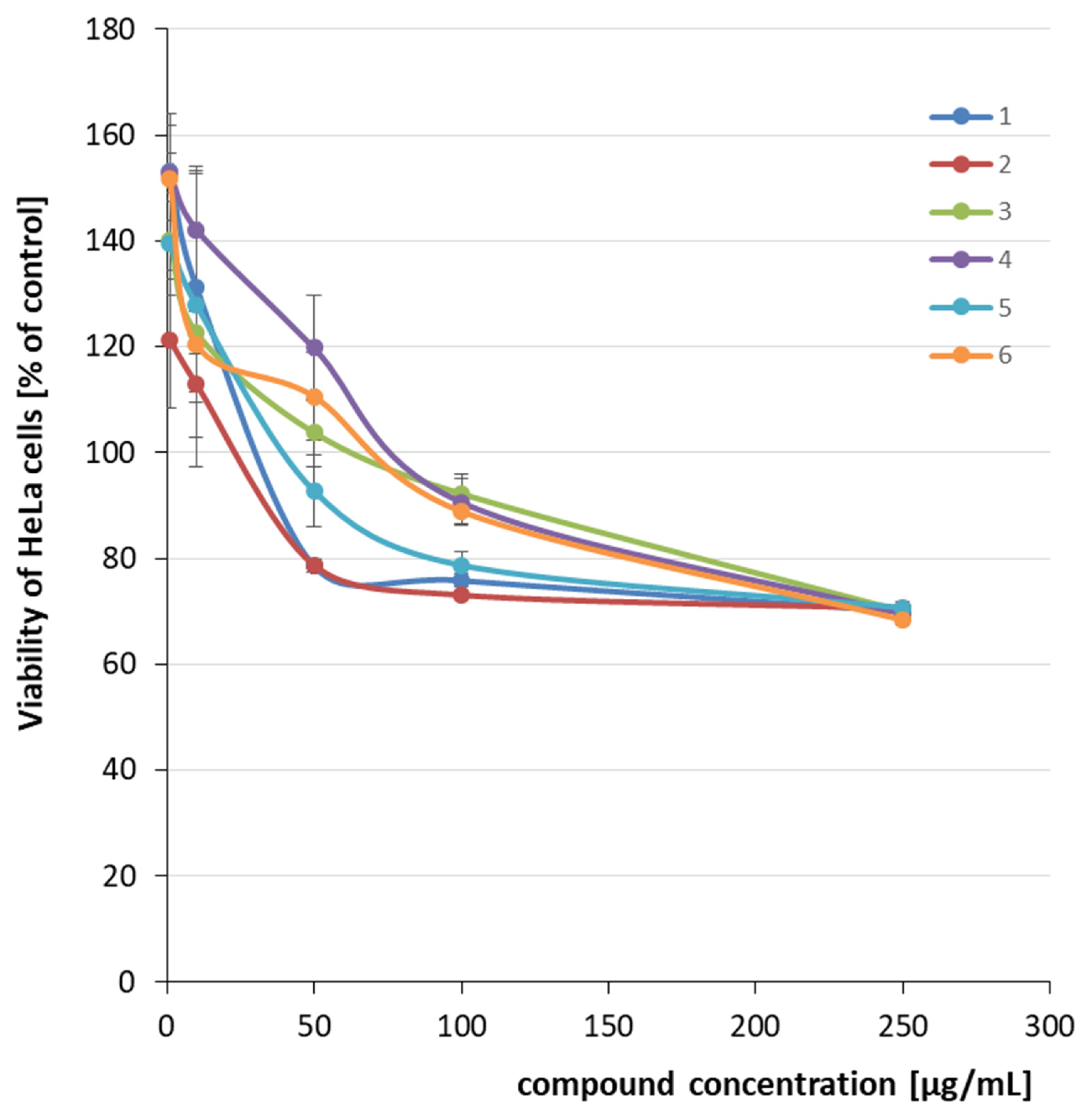
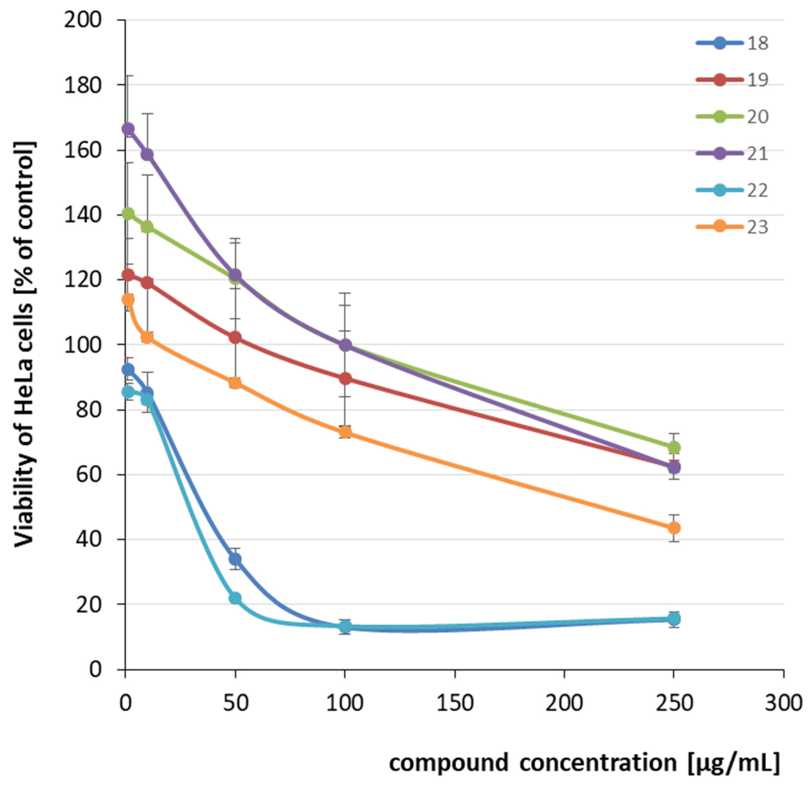
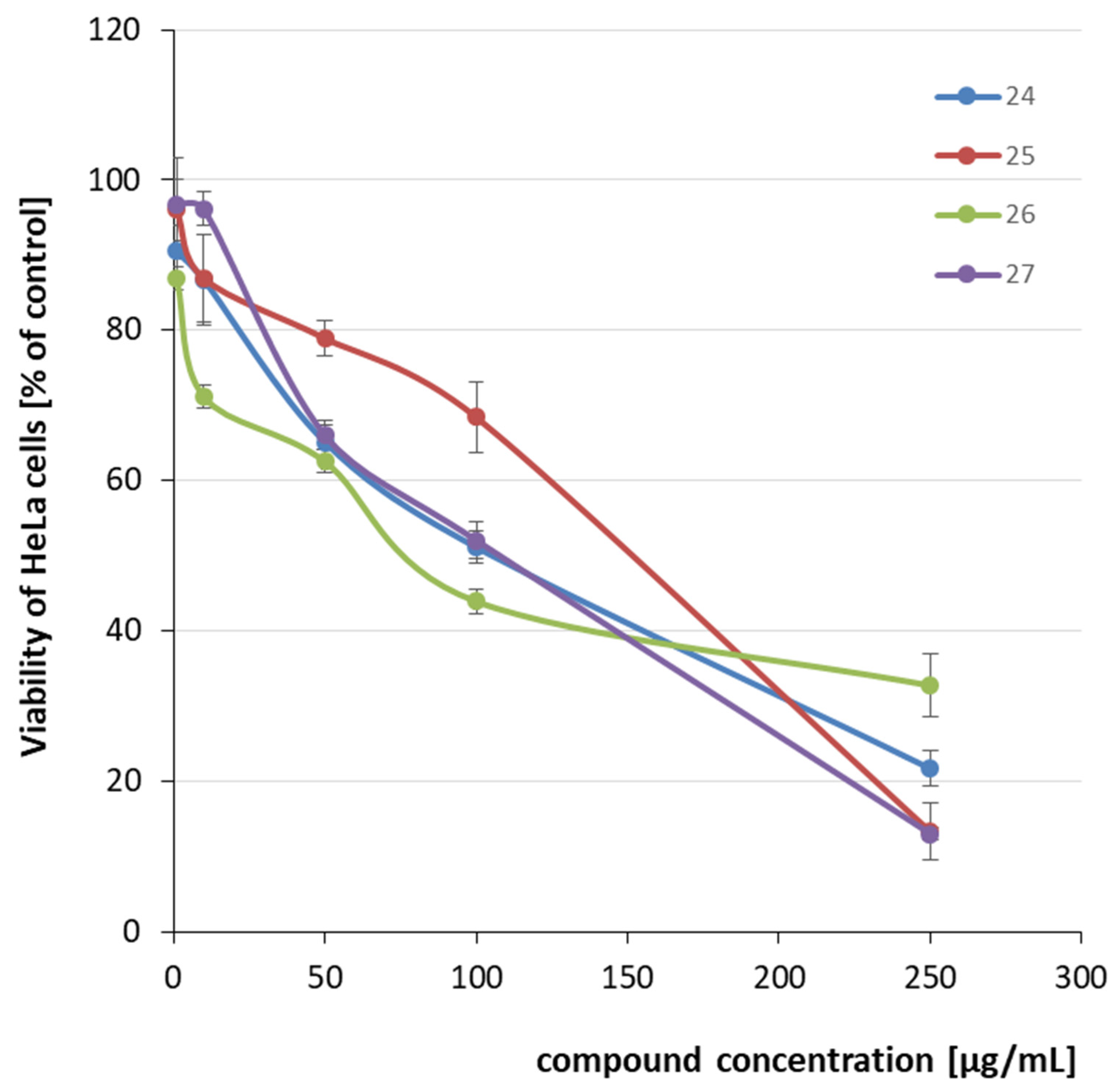
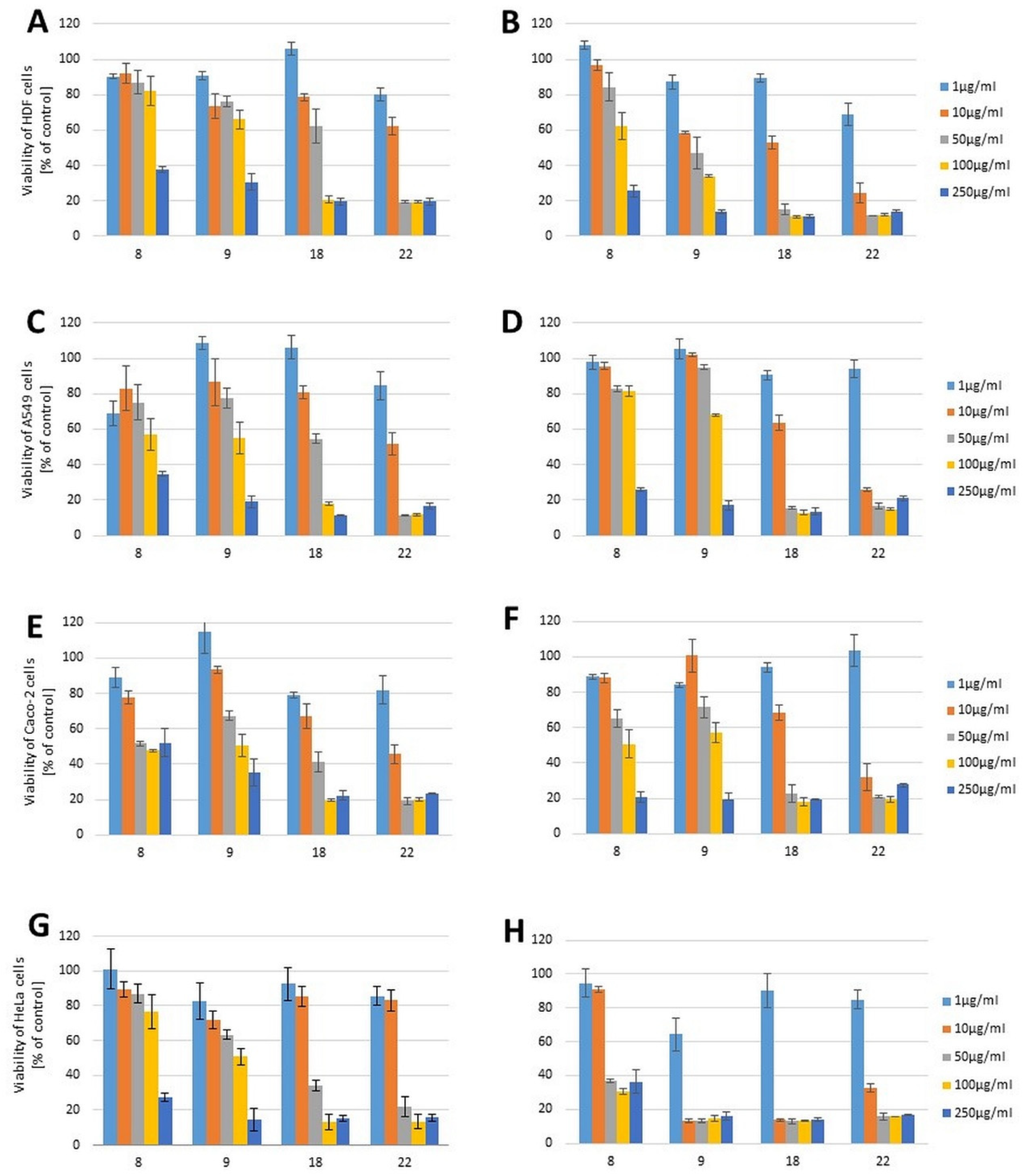
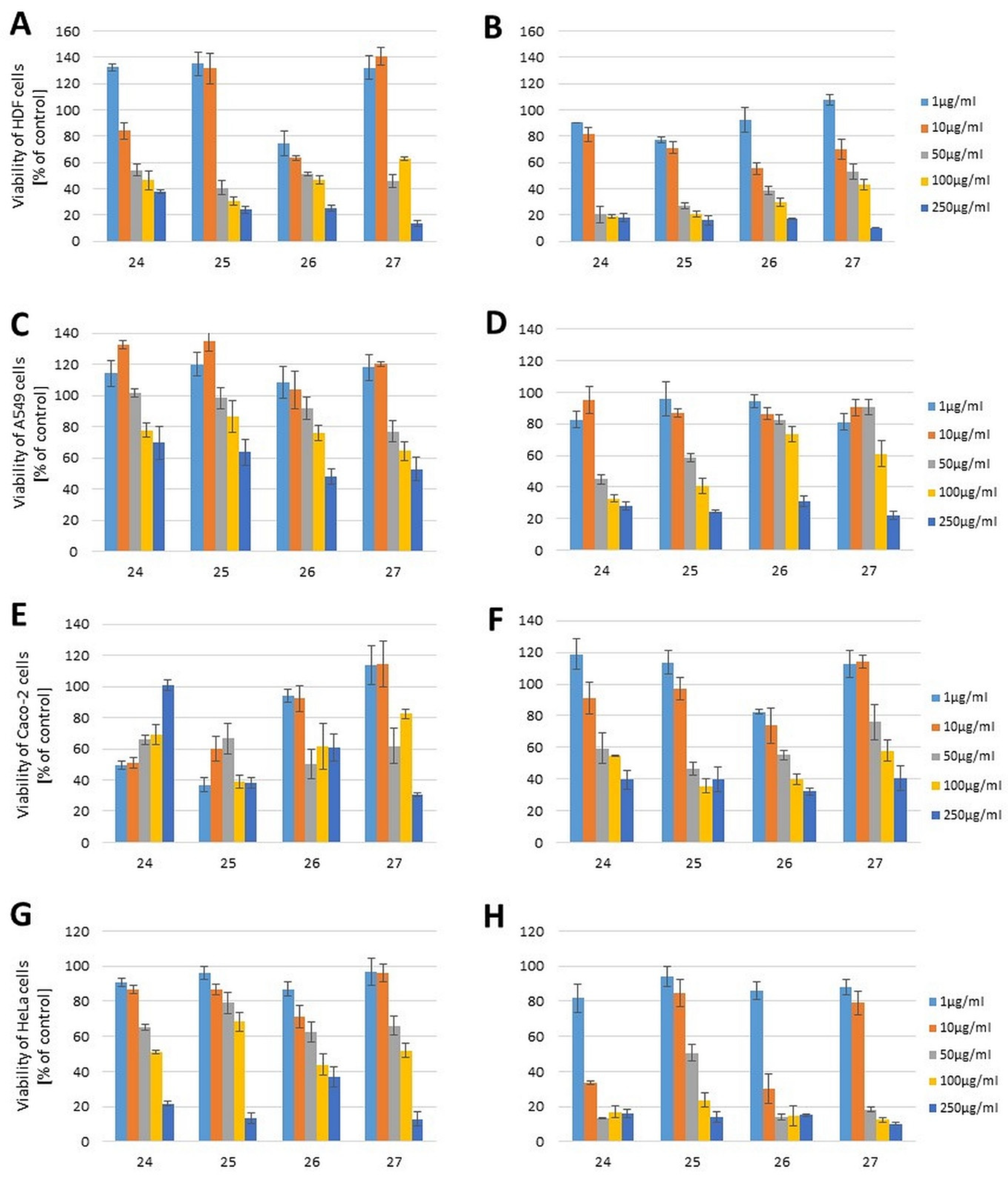
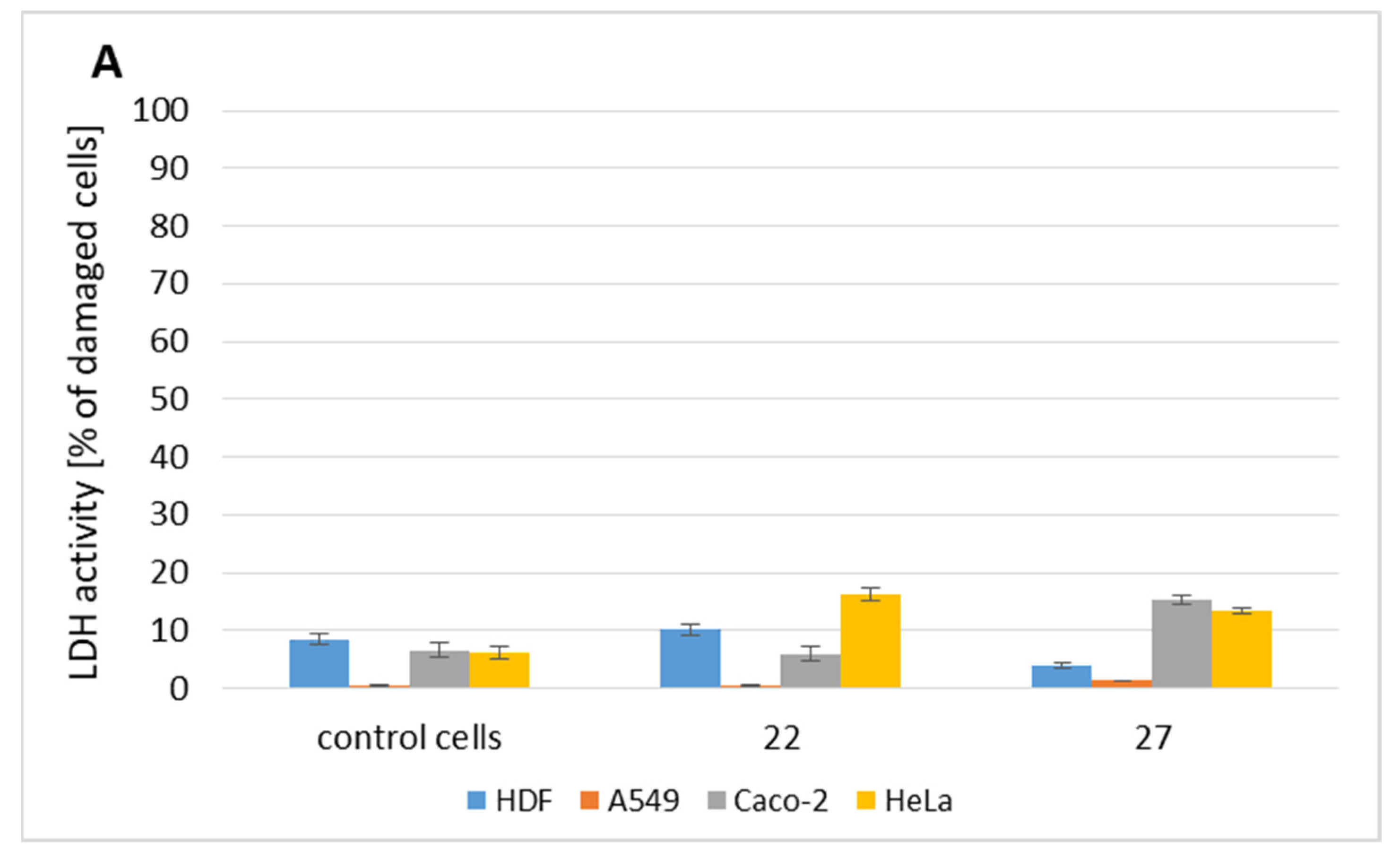
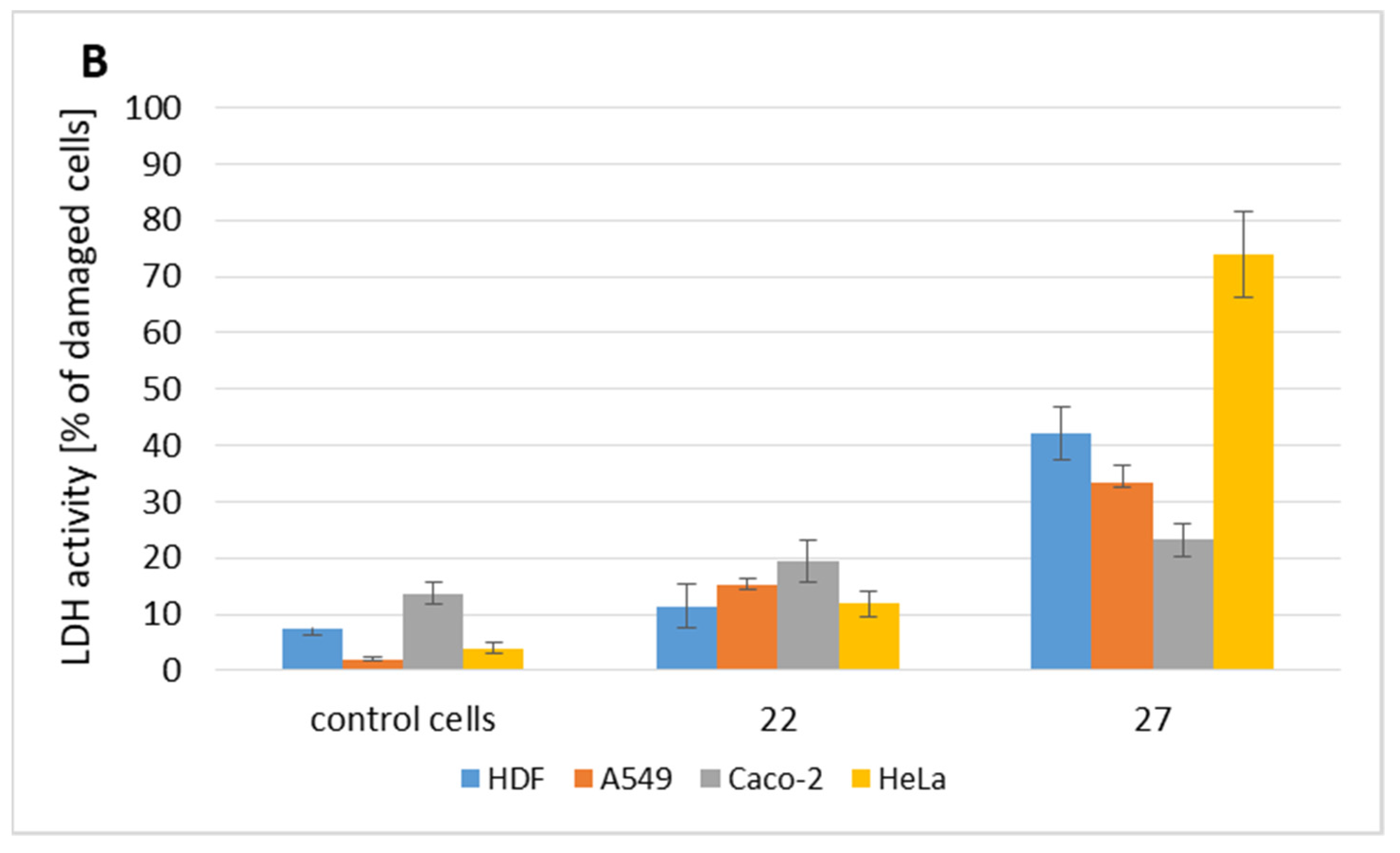
2.2. Cytotoxicity Evaluation

Cytotoxic properties of all compounds were determined by 3-(4,5-dimethylthiazol-2-yl)-2,5-diphenyltetrazolium bromide (MTT) test reflecting cell viability. Cytotoxicity screening of all 27 compounds—2-acetylbenzofuran oxime (6) and its oxime ethers (15), as well as: 2-acetylthiophene (717), cyclohexanone (1820), acetophenone (2123), thiazole (2425), benzothiazole (2627) oxime ethers—was performed in vitro on human cervical adenocarcinoma (HeLa) cells as the representatives of cancerous cells.
A majority of the derivatives are quite biocompatible in the HeLa cell culture, and in the concentration range of 1–50 μg/mL even increased the cell viability under study. This increase does not specifically mean that the number of cells increased; sometimes the stressor initiates higher metabolic activity (as we used the MTT assay reflecting activity of mitochondrial enzymes) that facilitates cell adaptation to stress conditions. All benzofuran derivatives (16) in the concentration range of 100–250 μg/mL reduced the survival of HeLa cells only to about 80–70% (Figure 1). The half-maximal effective concentrations (EC50) in HeLa cell culture were calculated and are summarized in Table 1.
Thiophene derivatives in the concentration range of 1–10 μg/mL were in general characterized by a lack of cytotoxicity against HeLa cells. On the other hand, compounds 7, 12, 16 and 17 in the concentration range of 1–10 μg/mL increased cell viability, and in the concentration range of 50–250 μg/mL slightly reduced the cell viability to about 70–60% (Figure 2). The most promising compounds in this group were 8 and 9 with an EC50 value of approximately 100 μg/mL (Table 1).
The results of studies using cyclohexanone and acetophenone derivatives on HeLa cell culture indicated no cytotoxicity in the concentrations of 1 and 10 μg/mL. The most promising derivatives were 18 and 22, as they reduced the viability of HeLa cells to below 20% at a concentration range of 50–250 μg/mL, and EC50 was achieved after 24 h culture at the concentrations of 32.26 and 23.15 μg/mL for (18) and (22), respectively (Figure 3, Table 1).
The last group of oxime ethers were the thiazole and benzothiophene derivatives (E)-1-(thiazol-2-yl)ethanone O-4-methoxybenzyl oxime (24) and (E)-1-(thiazol-2-yl)ethanone O-4-bromobenzyl oxime (25), and the benzothiophene derivatives (E)-1-(benzothiophene-2-yl)ethanone O-2,4-dichlorobenzyl oxime (26) and (E)-1-(benzothiophene-2-yl)ethanone O-4-methoxybenzyl oxime (27).
Based on the obtained outcomes, we can conclude that all derivatives from this group have cytotoxic activity against HeLa cells only at the highest concentration of 250 μg/mL, but reduce their viability even to below 20% (Figure 4, Table 1).
The screening results indicate that some of the compounds have the potential to act as anti-cancer agents and deserve more detailed assessment. Therefore, compounds showing the highest cytotoxicity (8, 9, 18, 22, 2427) were further tested on normal human dermal fibroblasts (HDF), A549, Caco-2, and HeLa cells to compare their activity after 24 h and 72 h culture (Figure 5 and Figure 6).
The most potent derivatives were compounds 18 and 22, as they reduced the viability of HDF, A549, Caco-2, and HeLa cells to approximately 20% at a concentration of 100 μg/mL for 18 and 50 μg/mL for 22 after 24 h exposure (Figure 5). Unfortunately, there was no clear specificity towards cancer cell lines, mainly for compound 18, as its EC50 value after 72 h culture was similar for all types of cells, excluding HeLa, and calculated as 90–100 μM. The low sensitivity of cancer cells is their specific and undesired feature, one of the hallmarks of cancer, or a result of the presence of highly resistant cancer stem cells [26,27]. A small difference in EC50 for compound 22 observed between normal and cancer cells allowed us to assume that this type of modification could be promising for the design of new anti-cancer agents, summarized in Table 2.
Compounds 2427 showed time- and dose-dependent cytotoxicity in relation to the tested cell lines. As predicted, their influence on normal and cancer cells was comparable (Figure 6). On the other hand, compound 27 exhibited a specific cytotoxic activity against Caco-2 and HeLa cells, while exhibiting relatively low toxicity against HDF cells. A decrease in HDF viability by 50% after 72 h exposure required a 0.45 mM concentration of this agent, whereas concentrations of 0.37 mM and 0.09 mM were sufficient against Caco-2 cells and HeLa cells, respectively (Table 2).
Summing up, only a few of the 27 tested derivatives could be promising as potential anti-cancer agents. Two of them—compound 22 and 27—would be a good starting point for future research. Therefore, we sought to compare their ability to induce necrotic cell death. Since necrotic cell death relies on the disruption of cell membrane, it results in the extracellular release of cytoplasmic enzymes such as lactate dehydrogenase (LDH). Thus, we compared LDH activity in the extracellular environment (culture media). The results are presented in Figure 7.
Surprisingly, compound 22, although toxic to cells, induced only a slight increase in LDH activity outside the cells, not exceeding 20% that of positive control. It can be assumed that its mode of action is underpinned by a decrease in proliferative rate and/or apoptotic cell death. Conversely, compound 27 induced extreme membrane damage and release of LDH, primarily in HeLa cells but not in Caco-2 cells, which also suffer from this compound’s toxicity. The higher LDH activity after 72 h of cell exposure to toxic compounds confirmed that initially the cells decreased their proliferation and subsequently necrosis occurred. It can be concluded that antitumor activity of various tested compounds depends on their chemistry, concentration and, on the other hand, also on the specific features of the cells tested.

2.3. Bioavailability of Tested Compounds

Lipinski’s rule and Veber’s rule are the main principles considered in medicinal chemistry when evaluating the bioavailability of drugs after oral administration. According to Lipinski’s rule, a compound is characterized by good absorption and permeation after oral administration when it does not exceed more than one of the following descriptors: molecular mass ≤ 500 Da, log p ≤ 5, number of hydrogen donors (nOHNH) ≤ 5 and number of hydrogen acceptors ≤ 10 [28]. On the other hand, by Veber’s rule, a compound is characterized by good bioavailability after per os administration when the topological polar surface of the molecule ≤ 140 Å2 and the number of rotatable bonds (Nrotb) < 10 [29]. The above parameters were calculated for compounds 127 using Molinspiration software [30], and the results are presented in Table 3.
Analysis of the calculated parameters showed that all the tested ethers satisfy Veber’s rule, while the rule of five without any violations was satisfied by compounds 425. For the ethers 13 and 2627, there was one violation of the rule of five (log p > 5).
The topological polar surface area (TPSA) of a molecule is an important descriptor considered during drug design, due to the fact that it allows predicting the permeation of a compound across biological membranes, including but not limited to penetration of the blood–brain barrier [31]. Molecules having topological polar surface area values of less than 60–70 Å2 show a high probability of penetrating the blood–brain barrier [32]. Among the studied ethers, derivatives 4 and 10 had TPSA >60–70 Å2, while the remaining compounds did not exceed 70 Å2, which suggests that they would be characterized by high penetration into the central nervous system. Considering the bioavailability parameters, the tested derivatives met the criteria for good absorption and permeation.
Regarding their potential anti-cancer activity, the selected oxime ether derivatives can be described as promising therapeutic compounds for cancer disease. The presented outcomes created a basis for further research on the oxime ethers with cytotoxic activity. We hope that it will pave the way for further structural modifications of these compounds by addition of different substituents in order to increase their activity, and to evaluate their modes of action in detail.

3. Materials and Methods

3.1. General Information

1H- and 13C-NMR spectra were recorded on the Bruker Avance 400 and 700 apparatus (TMS as an internal standard). High-resolution mass spectrometry (HRMS) measurements were made using a Synapt G2 Si mass spectrometer (Waters) equipped with an ESI source and a quadrupole time-of-flight mass analyzer. In order to achieve the highest accuracy of mass measurement, data were collected in center of gravity mode and the mass was corrected during acquisition using enkephalin leucine solution as external reference (Lock-SprayTM), which generated reference ion at m/z 556.2771 Da ([M + H]+) in positive ESI mode. The measurement results were processed with MassLynx 4.1 software (Waters).

3.2. Reagents and Solvents

Solvents: chloroform, dimethylsulfoxide, ethyl acetate, ethyl alcohol, hexane, triethylamine—POCh, Poland (Avantor Performance Materials Poland S.A., Gliwice Poland).
Reagents for synthesis: 2-acetylhiophene, 2,4-dichlorobenzyl chloride, 4-methoxybenzyl chloride, 2-chloroethylamine hydrochloride 99%, 3-chloropropylamine hydrochloride 98%, 2-chloro-N,N-diethylethylamine hydrochloride 99%, Sigma Aldrich Poznań Poland. 2-Acetylbenzothiophene 98% and 2-acetylthiazole 98% AmBeed (Chemat, Poland).
Auxiliary reagents: hydroxylamine hydrochloride, magnesium sulfate, potassium iodide, potassium hydroxide—POCh Poland (Avantor Performance Materials Poland S.A., Gliwice Poland).
TLC: 5 cm × 10 cm TLC plates coated with silica gel with F-254 (Merck, Darmstadt Germany).

3.3. Synthesis of Compounds 1315, 2425 and 2627—General Procedure

In a flask (25 mL) equipped with a plug, to a solution of 2-acetylthiophene oxime (5.0 mmol) for the synthesis oxime ethers 1315, (2-acetylthiazole oxime to the synthesis 2425, 2-acetylbenzothiophene oxime to the synthesis 2627) and DMSO (6 mL), an appropriate chloride (5.0 mmol), KI (0.10 g), and strongly pulverized KOH (20 mmol) were added [25]. The mixture was intensively stirred for 1 h at room temperature (monitored by TLC). Water (30 mL) and chloroform (30 mL) were added. The water phase was extracted with chloroform (30 mL). The combined organic layers were washed with water (4 × 25 mL) and dried (MgSO4). Evaporation yielded the oxime ethers.
For the preparation oxime ethers 2425 and 2627 the same procedure was used, with the exception of the amount of KOH—10 mmol.
NMR and MS spectra of the tested compounds are available in Supplementary Materials. (E)-1-(thiophen-2-yl)ethanone O-2-aminoethyl oxime (13)—Yield: 71%, pale yellow oil. 1H-NMR (500 MHz, CDCl3), δ ppm, J Hz): 7.27 (d, 5.0, 1H, CH), 7.22 (dd, 1.0 3.5, 1H, CH), 7.02 (dd, 1.0 5.0, 1H, CH), 4.20 (t, 5.0, 2H, CH2), 3.02 (t, 5.0, 2H, CH2), 2.27 (s, 3H, CH3), 1.6–1.8 (s, 2H, NH2). 13C-NMR (125 MHz, CDCl3, δ ppm): 150.83 (CC=N), 140.41 (C), 126.97 (CH), 126.77 (CH), 126.14 (CH), 76.63 (OCH2), 41.69 (NCH2), 12.92 (CH3). HR-MS m/z 185.0752 [M + H]+ (calcd for C8H13N2OS: 185.0743). Rf (silica gel, AcOEt:hexane:triethylamine 1:1:0.02): 0.05.
(E)-1-(thiophen-2-yl)ethanone O-2-(diethylamino)ethyl oxime (14)—Yield: 78%, orange oil. 1H-NMR (400 MHz, CDCl3), δ ppm, J Hz): 7.26 (dd, 1.2 4.0, 1H, CH), 7.21 (dd, 1.2 4.0, 1H, CH), 7.02 (dd, 1.2 4.0, 1H, CH), 4.27 (t, 2H, OCH2), 2.85 (t, 2H, NCH2), 2.65 (q, 4H, 2 × CH2), 2.26 (s, 3H, CH3), 1.08 (t, 7.2, 6H, 2 × CH3). 13C-NMR (100 MHz, CDCl3, δ ppm): 150.41 (CC=N), 140.62 (C), 126.89 (CH), 125.62 (CH), 125,91 (CH), 72.70 (OCH2), 51.31 (CH2), 47.86 (NCH2, 2 × CH2), 12.98 (CH3), 11.87 (2 × CH3). HR-MS m/z 241.1373 [M + H]+ (calcd for C12H21N2OS: 241.1369). Rf (silica gel, AcOEt:hexane:triethylamine 1:1:0.02): 0.2.
(E)-1-(thiophen-2-yl)ethanone O-3-aminopropyl oxime (15)—Yield: 76%, pale yellow oil. 1H-NMR (500 MHz, CDCl3), δ ppm, J Hz): 7.27 (d, 5.0 1H, CH), 7.22 (d, 3.5, 1H, CH), 7.02 (dd, 1.0 5.0, 1H, CH), 4.26 (t, 6.0 Hz, 2H, CH2), 2.87 (dd, 6.5 7.0, 2H, CH2), 2.26 (s, 3H, CH3), 1.90–2.10 (s, 2H, NH2), 1.90 (m, 2H, CH2). 13C-NMR (125 MHz, CDCl3, δ ppm): 150.33 (CC=N), 140.63 (C), 126.89 (CH), 126.59 (CH), 125.90 (CH), 72.03 (OCH2), 39.20 (NCH2), 33.15, 12.85 (CH3). HR-MS m/z 199.0909 [M + H]+ (calcd for C9H15N2OS: 199.0900). Rf (silica gel, AcOEt:hexane:triethylamine 1:1:0.02): 0.05.
(E)-1-(thiazol-2-yl)ethanone O-4-methoxybenzyl oxime (24)—Yield: 84%, yellow oil. 1H NMR: (500 MHz, CDCl3): δ (ppm) 7.84 (d, 3.0, 1H, CH), 7.38 (d, 8.5, 2H, 2 × CH), 7.30 (d, 3.0, 1H, CH), 6.93 (d, 8.5, 2H, 2 × CH), 5,21 (s, 2H, OCH2), 3.83 (s, 3H, OCH3), 2.41 (s, 3H, CH3). 13C NMR: (125 MHz, CDCl3): δ (ppm) 165.44 (C=N), 159.58 (C), 151.88 (C), 142.79 (CH), 130.21 (2 × CH), 129.35 (C), 120.07 (CH), 113.84 (2 × CH), 76.79 (CH2), 55.28 (OCH3), 12.14 (CH3). HR-MS m/z 263.0848 [M + H]+ (calcd for C13H15N2O2S: 263.0849). Rf (silica gel, AcOEt:hexane 1:2): 0.85.
(E)-1-(thiazol-2-yl)ethanone O-4-bromobenzyl oxime (25)—Yield: 69%, white solid, mp. 67–70 °C (ethanol). 1H NMR: (500 MHz, CDCl3): δ (ppm) 7.85 (d, 3.5, 1H, CH), 7.52 (d, 8.5, 2H, 2 × CH), 7.29–7.32 (m, 3H, 3×CH), 5.22 (s, 2H, OCH2), 2.41 (s, 3H, CH3). 13C NMR: (125 MHz, CDCl3): δ (ppm) 165.05 (C=N), 152.39 (C), 142.87 (CH), 136.36 (C), 131.59 (2 × CH), 130.03 (2 × CH), 122.10 (C), 120.24 (CH), 76.12 (CH2), 12.18 (CH3). HR-MS m/z 310.9856 [M + H]+ (calcd for C12H12N2OS79Br: 310.9848). Rf (silica gel, AcOEt:hexane 1:1): 0.85.
(E)-1-(benzothiophene-2-yl)ethanone O-2,4-dichlorobenzyl oxime (26)—Yield: 51%, pale yellow solid, mp. 113–117 °C (ethanol). 1H-NMR: (500 MHz, CDCl3): δ (ppm) 7.79 (dd, 2.0 6.5 1H, CH), 7.77 (dd, 2.0 6.5 1H, CH), 7.48 (s, 1H, CH), 7.44 (dd, 2.0 8.0, 1H, CH), 7.35 (ddd, 2.0 5.5 6.0, 2H, 2 × CH), 7.29 (dd, 2.0 8.0, 1H, CH), 5.34 (s, 2H, OCH2), 2.38 (s, 3H, CH3). 13C NMR: (125 MHz, CDCl3): δ (ppm) 151.90 (C=N), 140.39 (C), 140.06 (C), 139.33 (C), 134.18 (CH), 134.11 (C), 130.82 (CH), 129.25 (CH), 127.05 (CH), 125.56 (CH), 124.39 (C), 123.92 (CH), 123.52 (CH), 122.26 (CH), 72.96 (CH2), 12.65 (CH3). HR-MS m/z 350.0172 [M + H]+ (calcd for C17H14NOSCl2: 350.0168). Rf (silica gel, AcOEt:hexane 1:2): 0.8.
(E)-1-(benzothiophene-2-yl)ethanone O-4-methoxybenzyl oxime (27)—Yield: 51%, white solid, mp. 132–134 °C (ethanol). 1H NMR: (500 MHz, CDCl3): δ (ppm) 7.79 (dd, 2.5 6.0, 1H, CH), 7.74 (dd, 2.5 6.0, 1H, CH), 7.44 (s, 1H, CH), 7.41 (d, 8.5, 2H, 2 × CH), 7.35 (dd, 2.0 5.5, 2H, 2 × CH), 6.94 (d, 8.5, 2H, 2 × CH), 5.20 (s, 2H, OCH2), 3.84 (s, 3H, OCH3), 2.32 (s, 3H, CH3). 13C NMR: (125 MHz, CDCl3): δ (ppm) 159.41 (C=N), 150.99 (C), 140.97 (C), 140.01 (C), 139.44 (C), 130.27 (2 × CH), 129.66 (C), 125.38 (CH), 124.32 (CH), 123.83 (CH), 123.06 (CH), 122.23 (CH), 113.80 (2 × CH), 76.40 (CH2), 55.28 (OCH3), 12.61 (CH3). HR-MS m/z 312.1054 [M + H]+ (calcd for C18H18NO2S: 312.1053). Rf (silica gel, AcOEt:hexane 1:2): 0.7.

3.4. Cytotoxicity Evaluation of Tested Compounds

2-Acetylbenzofuran, 2-acetylbenzothiophene, 2-acetylthiophene, 2-acetylthiazole, cyclohexanone and acetophenone derivatives were used for in vitro cytotoxicity assays with cervical adenocarcinoma (HeLa), human dermal fibroblasts (HDF), lung epithelial carcinoma (A549), and intestinal epithelial carcinoma (Caco-2) cell lines. A549, Caco-2, and HeLa cells were obtained from Sigma-Aldrich (Germany), and HDF from Biokom (Poland). All cell lines were cultured according to the manufacturer’s protocol under sterile conditions in the presence of 4.9% CO2 at 37 °C. A549 cells were grown in F-12 medium containing 10% fetal bovine serum (FBS) and 1% penicillin/streptomycin. HDF cells were grown in DMEM (low glucose, with 2 mM glutamine) medium containing 10% fetal bovine serum (FBS) and 1% penicillin/streptomycin. Caco-2 cells were grown in EMEM medium containing 10% fetal bovine serum (FBS), 1% non-essential amino acids (NEAA) and 1% penicillin/streptomycin. HeLa cells were grown in DMEM (low glucose, with 2 mM glutamine) medium containing 10% fetal bovine serum (FBS) and 1% gentamycin. After thawing, cells were cultured until they reached sub-confluent state. The cells were then detached using 0.25% trypsin solution and seeded into 24-well or 96-well plates (3 × 10³ cells per well) for further experiments. After 24 h growth, the tested compounds were added to the cell cultures in concentrations of 1, 10, 50, 100 and 250 μg mL−1, respectively, and incubated for the next 24 h or 72 h. The MTT test, based on the ability to reduce 3-(4,5-dimethylthiazol 2-yl)-2,5-diphenyltetrazolium bromide (MTT) by mitochondrial dehydrogenases, was performed in triplicate to assess cell metabolic activity and viability. The plates were then read spectrophotometrically at a wavelength of 570 nm. The dose-dependence curves were charted directly on x-/y-axes, with the drug dosages on the x-axis and the cell viability (percentage of viable cells) on the y-axis.
Additionally, the lactate dehydrogenase (LDH) activity test reflecting cell-membrane damage was performed for cells exposed to selected tested compounds. LDH activity was determined in the culture medium by measuring the decrease in NADH (nicotinamide adenine dinucleotide, reduced disodium salt). The decrease in the amount of NADH was directly correlated to the increase in the number of damaged cells. To measure LDH activity, the culture medium was collected from the wells with cells exposed to concentrations equal to the EC50 values of tested compounds after short- (24 h), and long-time (72 h) exposure. The LDH activity test was performed as follows: 25 μL of NADH (2.5 mg/mL) and 25 μL of sodium pyruvate (2.5 mg/mL) were added to 150 μL of the collected culture medium. Absorbance at 340 nm was spectrophotometrically measured. The number of damaged cells was compared to the untreated control sample, also considering the positive control treated with 1% Triton X-100 as 100% damaged cells.

4. Conclusions

In conclusion, 1-(benzofuran-2-yl)ethan-1-one oxime and 26 oxime ethers containing benzofuran, benzothiophene, thiophene, thiazole, cyclohexyl or phenyl moiety were screened for cytotoxicity against the human cervical adenocarcinoma (HeLa cell line) representative cancer cell model. The most promising derivatives were 2-(cyclohexylideneaminoxy)acetic acid (18) and (E)-acetophenone O-2-morpholinoethyl oxime (22), which reduced the viability of HeLa cells to below 20% of control at concentrations over 100 μg/mL. However, in further evaluations of cytotoxic influence, compound (18) showed higher toxicity against normal cells than cancerous cells, a common feature of many molecules that results from the lower sensitivity of cancer cells. We confirmed the specific toxicity of acetophenone derivative (22) against the cancer cell lines (EC50 ≈ 30 µM for Caco-2, HeLa, and A549 cells after 72 h treatment). From another group of oxime ether derivatives, compound 27 exhibited specific toxicity against Caco-2 and HeLa cells with EC50 values of 370 µM and 90 µM, respectively. Comparison of 22 and 27 modes of action revealed that they initiate different processes, leading to reduced proliferation and cell death. Considering the bioavailability parameters, the tested derivatives met the criteria for good absorption and permeation, and can be further investigated.

Supplementary Materials

The following supporting information can be downloaded at. Figure S1: The cytotoxicity evaluation on HeLa cell culture after 24 h of exposure to the compounds 127. Figure S2: Spectral data of the new synthesized compounds 1315 and 2427. Figure S3: NMR Spectra of the other compounds.

Author Contributions

Conceptualization, T.K.; Methodology, T.K. and A.H.; Validation, T.K. and A.H.; Investigation, T.K., A.H., R.S. and S.B.; Resources, T.K., A.H. and S.B.; Data Curation, T.K. and A.H.; Writing—Original Draft Preparation, T.K., A.H. and S.B.; Writing—Review and Editing, R.S, D.K. and K.R.; Visualization, T.K. and R.S.; Supervision, T.K. and R.S.; Project Administration, T.K., D.K. and R.S.; Funding Acquisition, R.S. and K.R. All authors have read and agreed to the published version of the manuscript.

Funding

This research received no external funding.

Institutional Review Board Statement

Not applicable.

Informed Consent Statement

Not applicable.

Conflicts of Interest

The authors declare no conflict of interest.

Sample Availability

Samples of the compounds 127 are available from the authors.

References

  1. WHO Report on Cancer: Setting Priorities, Investing Wisely and Providing Care for All. Available online: https://www.who.int/publications/i/item/who-report-on-cancer-setting-priorities-investing-wisely-and-providing-care-for-all (accessed on 1 March 2021).
  2. Meegan, M.J.; O’Boyle, N.M. Special Issue, Anticancer Drugs. Pharmaceuticals 2019, 12, 134. [Google Scholar] [CrossRef] [PubMed] [Green Version]
  3. Parthiban, P.; Rathika, P.; Ramkumar, V.; Son, S.M.; Jeong, Y.T. Stereospecific synthesis of oximes and oxime ethers of 3-azabicycles: A SAR study towards antimicrobial agents. Bioorg. Med. Chem. Lett. 2010, 20, 1642–1647. [Google Scholar] [CrossRef] [PubMed]
  4. Hu, Z.-B.; Luo, H.-A.; Wang, X.-G.; Huang, M.-Z.; Huang, L.; Pang, H.-L.; Mao, C.-H.; Pei, H.; Huang, C.-Q.; Sun, J.; et al. Synthesis and evaluation O-benzyl oxime-ether derivatives containing β-methoxyacrylate moiety for insecticidal and fungicidal activities. Bull. Korean Chem. Soc. 2014, 35, 1073–1076. [Google Scholar] [CrossRef] [Green Version]
  5. Özdemir, Z.; Sari, S.; Karakurt, A.; Dalkara, S. Synthesis, anticonvulsant screening, and molecular modeling studies of new arylalkylimidazole oxime ether derivatives. Drug Dev. Res. 2019, 80, 269–280. [Google Scholar] [CrossRef]
  6. Chakravarti, B.; Akhtar, T.; Rai, B.; Yadav, M.; Siddiqui, J.A.; Dwivedi, S.K.D.; Thakur, R.; Singh, A.K.; Kumar, H.; Khan, K.; et al. Thioaryl naphthylmethanone oxime ether analogs as novel anticancer agents. J. Med. Chem. 2014, 57, 8010–8025. [Google Scholar] [CrossRef]
  7. Díaz, J.E.; Martinez, D.C.; López, L.V.; Mendez, G.M.; Vera, R.; Loaiza, A.E. Synthesis and in vitro antiproliferative activity of flavone and 6-hydroxyflavone oxime ethers derivatives. J. Braz. Chem. Soc. 2018, 29, 177–184. [Google Scholar] [CrossRef]
  8. Latif, A.D.; Gonda, T.; Vágvölgyi, M.; Kúsz, N.; Kulmány, Á.; Ocsovszki, I.; Zomborszki, Z.P.; Zupkó, I.; Hunyadi, A. Synthesis and in vitro antitumor activity of naringenin oxime and oxime ether derivatives. Int. J. Mol. Sci. 2019, 20, 2184. [Google Scholar] [CrossRef] [Green Version]
  9. Kim, T.; Kim, H.-I.; An, J.-Y.; Lee, J.; Lee, N.-R.; Heo, J.; Kim, J.-E.; Yu, J.; Lee, J.S.; Inn, K.-S.; et al. Identification of novel estrogen receptor (ER) agonists that have additional and complementary anti-cancer activities via ER-independent mechanism. Bioorg. Med. Chem. Lett. 2016, 26, 1844–1848. [Google Scholar] [CrossRef]
  10. Hayakawa, I.; Shioya, R.; Agatsuma, T.; Furukawa, H.; Sugano, Y. Thienopyridine and benzofuran derivatives as potent antitumor agents possessing different structure-activity relationship. Bioorg. Med. Chem. Lett. 2004, 14, 3411–3414. [Google Scholar] [CrossRef]
  11. Coşkun, D.; Tekin, S.; Sandal, S.; Coşkun, M.F. Synthesis, Characterization, and Anticancer Activity of New Benzofuran Substituted Chalcones. J. Chem. 2016, 2016, 7678486. [Google Scholar] [CrossRef] [Green Version]
  12. Kamal, M.; Shakya, A.K.; Jawaid, T. Benzofurans: A New profile of biological activities. Int. J. Med. Pharm. Sci. 2011, 1, 1–15. [Google Scholar]
  13. Liu, Y.T.; Sun, J.; Wang, X.-J. Natural source, bioactivity and synthesis of benzofuran derivatives. RSC Adv. 2019, 9, 27510–27540. [Google Scholar]
  14. Petrou, A.; Fesaidou, M.; Geronikaki, A. Thiazole Ring—A Biologically Active Scaffold. Molecules 2021, 26, 3166. [Google Scholar] [CrossRef] [PubMed]
  15. Keri, R.S.; Chand, K.; Budagumpi, S.; Somappa, S.B.; Patil, S.A.; Nagaraja, B.M. An overview of benzo[b]thiophene-based medicinal chemistry. Eur. J. Med. Chem. 2017, 138, 1002–1033. [Google Scholar] [CrossRef]
  16. Demirayak, S.; Uçucu, U.; Benkli, K.; Gündoğdu-Karaburun, N.; Karaburun, A.C.; Akar, D.; Karabacak, M.; Kiraz, N. Synthesis and antifungal activities of some aryl(benzofuran-2-yl)ketoximes. Il Farmaco 2002, 57, 609–612. [Google Scholar] [CrossRef]
  17. Varache-Lembège, M.; Nuhrich, A.; Renard, P.; Duboudin, F.; Vercauteren, J.; Devaux, G. Platelet antiaggregant methoxyphenylthienyl ketoxime ethers. Synthesis and structure-activity relationships. Arch. Pharm. 1995, 328, 417–424. [Google Scholar] [CrossRef]
  18. Letafat, B.; Emami, S.; Mohammadhosseini, N.; Faramarzi, M.A.; Samadi, N.; Shafiee, A.; Foroumadi, A. Synthesis and antibacterial activity of new N-[2-(thiophen-3-yl)ethyl] piperazinyl quinolones. Chem. Pharm. Bull. 2007, 55, 894–898. [Google Scholar] [CrossRef] [Green Version]
  19. Abele, E.; Lukevics, E. Furan and thiophene oximes: Synthesis, reactions, and biological activity review. Chem. Heterocycl. Comp. 2001, 37, 141–167. [Google Scholar] [CrossRef]
  20. Matsumura, K.; Miyashita, O.; Shimadzu, H.; Hashimoto, N. Uracil Derivatives and Production Thereof. U.S. Patent 4190656, 26 February 1980. [Google Scholar]
  21. Pathania, A.S.; Chawla, P.A. Thiophene-based derivatives as anticancer agents: An overview on decade’s work. Bioorg. Chem. 2020, 101, 104026–104043. [Google Scholar]
  22. Bozdag, O.; Gümüse, B.; Demirdamar, R.; Büyükbingöl, E.; Rolland, Y.; Ertan, R. Synthesis of some novel oxime ether derivatives and their activity in the ‘behavioral despair test’. Eur. J. Med. Chem. 1998, 33, 133–141. [Google Scholar] [CrossRef]
  23. Kosmalski, T.; Kutkowska, J.; Gzella, A.K.; Nowakiewicz, A. New heterocyclic oxime ethers of 1-(benzofuran-2-yl)ethan-1-one and their antimicrobial activity. Acta Pol. Pharm. 2015, 72, 289–295. [Google Scholar] [PubMed]
  24. Kosmalski, T.; Kutkowska, J.; Dwojak, I.; Studzińska, R.; Sikora, A.; Modzelewska-Banachiewicz, B.; Gzella, A. Novel O-benzyl oxime ethers of 1-(thiophen-2-yl)ethan-1-one—Synthesis and antimicrobial activity. Heterocycles 2017, 94, 523–530. [Google Scholar] [CrossRef] [Green Version]
  25. Kosmalski, T.; Studzińska, R.; Daniszewska, N.; Ullrich, M.; Sikora, A.; Marszałł, M.; Modzelewska-Banachiewicz, B. Study of the room-temperature synthesis of oxime ethers by using a super base. ChemistryOpen 2018, 7, 551–557. [Google Scholar] [CrossRef] [PubMed] [Green Version]
  26. Rebucci, M.; Michiels, C. Molecular aspects of cancer cell resistance to chemotherapy. Biochem. Pharmacol. 2013, 85, 1219–1226. [Google Scholar] [CrossRef]
  27. Bai, X.; Ni, J.; Beretov, P.; Graham, P.; Li, Y. Cancer stem cell in breast cancer therapeutic resistance. Cancer Treat. Rev. 2018, 69, 152–163. [Google Scholar] [CrossRef]
  28. Lipinski, C.A.; Lombardo, F.; Dominy, B.W.; Feeney, P.J. Experimental and computational approaches to estimate solubility and premeability in drug discovery and development settings. Adv. Drug Deliv. Rev. 1997, 23, 3–25. [Google Scholar] [CrossRef]
  29. Veber, D.F.; Johnson, S.R.; Cheng, H.; Smith, B.R.; Ward, K.W.; Kopple, K.D. Molecular Properties That Influence the Oral Bioavailability of DrugCandidates. J. Med. Chem. 2002, 45, 2615–2623. [Google Scholar] [CrossRef]
  30. Molinspiration. Available online: https://www.molinspiration.com/ (accessed on 15 September 2021).
  31. Ertl, P.; Rohde, B.; Selzer, P. Fast Calculation of Molecular Polar Surface Area as a Sum of Fragment-Based Contributions and Its Application on the Prediction of Drug Transport Properties. J. Med. Chem. 2000, 43, 3714–3717. [Google Scholar] [CrossRef]
  32. Kelder, J.; Grootenhuis, P.D.J.; Bayada, M.D.; Delbressine, L.P.C.; Ploemen, J.P. Polar Molecular Surface as a Dominating Determinant for Oral Absorption and Brain Penetration of Drugs. Pharm. Res. 1999, 16, 1514–1519. [Google Scholar] [CrossRef]
Scheme 1. Synthesis of the tested compounds 15 and 727.
Molecules 27 01374 sch001
Figure 1. The dose-dependent curves for cytotoxicity screening of the benzofuran oxime ethers (15) and 2-acetylbenzofuran oxime (6) on HeLa cells. Each compound was added to growing cells at concentrations of 1, 10, 50, 100, and 250 µg/mL, and cell viability was assayed with the MTT test.
Molecules 27 01374 g001
Figure 2. The dose-dependent curves for cytotoxicity screening of thiophene oxime ethers (717) on HeLa cells. Each compound was added to growing cells at concentrations of 1, 10, 50, 100, and 250 µg/mL, and cell viability was assayed with the MTT test.
Molecules 27 01374 g002
Figure 3. The dose-dependent curves for cytotoxicity of the cyclohexanone and acetophenone oxime ethers (1823) on HeLa cells. Each compound was added to the growing cells at concentrations of 1, 10, 50, 100, and 250 µg/mL, and cell viability was assayed with the MTT test.
Molecules 27 01374 g003
Figure 4. The dose-dependent curves for cytotoxicity of the thiazole and benzothiophene oxime ethers (2427) on HeLa cells. Each compound was added to the growing cells at concentrations of 1, 10, 50, 100, and 250 µg/mL, and cell viability was assayed with the MTT test.
Molecules 27 01374 g004
Figure 5. The cytotoxicity of selected oxime ether derivatives (8, 9, 18, and 22) towards: (A)—HDF after 24 h, (B)—HDF after 72 h, (C)—A549 after 24 h, (D)—A549 after 72 h, (E)—Caco-2 cells after 24 h, (F)—Caco-2 cells after 72 h, (G)—HeLa cells after 24 h, and (H)—HeLa cells after 72 h in culture.
Molecules 27 01374 g005
Figure 6. The cytotoxicity of the oxime ethers 2427 towards: (A)—HDF after 24 h, (B)—HDF after 72 h, (C)—A549 after 24 h, (D)—A549 after 72 h, (E)—Caco-2 cells after 24 h, (F)—Caco-2 cells after 72 h, (G)—HeLa cells after 24 h and (H)—HeLa cells after 72 h in culture.
Molecules 27 01374 g006
Figure 7. Comparison of LDH activity (relative to positive control where appropriate cell lysates were assumed as 100% damaged cells) in cell media after 24 h (A) and 72 h (B) of exposure to concentrations equal to EC50 value of selected compounds. The cells not treated with tested compounds were treated as negative controls.
Molecules 27 01374 g007aMolecules 27 01374 g007b
Table 1. The structures of oxime ethers (15, 727) and benzofuran oxime (6) and their summarized EC50 values in 24 h culture of HeLa cells.
No.RR1R2EC50 for HeLa Cells *
μg/mLmM
1 Molecules 27 01374 i001CH3C6H3-2.4-diCl>250>0.75
2C6H4-4-CF3>250>0.75
3C6H4-4-Br>250>0.73
4C6H4-4-NO2>250>0.81
5C6H3-2.6-diF>250>0.83
6H (oxime)>250>1.43
7 Molecules 27 01374 i002CH3C6H3-2.4-diCl>250>0.83
8C6H4-4-Br188.120.61
9C6H4-4-NO2126.300.46
10C6H3-5-MeO-2-NO2>250>0.82
11CH2-NMe2>250>1.18
12 Molecules 27 01374 i003>250 >0.98
13CH2-NH2>250 >1.36
14CH2-NEt2>250>1.04
15CH2CH2-NH2>250>1.10
16CH2CH2-NMe2>250>1.10
17COOH>250>1.25
18(CH2)5COOH32.26 0.19
19CH2-NMe2>250>1.36
20 Molecules 27 01374 i004>250 >1.10
21 Molecules 27 01374 i005CH3 Molecules 27 01374 i006>250 >0.67
22 Molecules 27 01374 i00723.150.09
23CH2-NMe2233.141.13
24 Molecules 27 01374 i008CH3C6H4-4-OMe110.300.42
25C6H4-4-Br130.100.42
26 Molecules 27 01374 i009CH3C6H3-2.4-diCl121.230.35
27C6H4-4-OMe93.800.30
* The EC50 values were calculated using Quest Graph™ ED50 Calculator retrieved from https://www.aatbio.com/tools/ed50-calculator (accessed on 10 January 2022).
Table 2. Summarized EC50 values of compounds 8, 9, 18, 22 and 2427 in 24 h and 72 h cultures of HDF, A549, Caco-2, and HeLa cells.
EC50 Value * in μg/mL (mM)
CellsHDFA549Caco-2HeLa
Time
No.
24 h72 h24 h72 h24 h72 h24 h72 h
8126.70
(0.41)
118.21
(0.38)
193.59
(0.62)
228.30
(0.74)
229.79
(0.74)
118.43
(0.38)
188.12
(0.61)
40.00
(0.13)
9171.57
(0.62)
44.74
(0.16)
106.70
(0.39)
130.75
(0.47)
77.96
(0.28)
104.88
(0.38)
126.30
(0.46)
6.90
(0.02)
1843.66
(0.26)
14.69
(0.09)
47.79
(0.28)
17.88
(0.10)
44.36
(0.26)
17.46
(0.10)
32.26
(0.19)
4.90
(0.03)
2227.31
(0.11)
8.04
(0.03)
10.19
(0.04)
6.72
(0.03)
9.45
(0.04)
6.79
(0.03)
23.15
(0.09)
7.00
(0.03)
2435.26
(0.13)
30.65
(0.12)
>250
(>0.95)
35.74
(0.14)
>250
(>0.95)
69.10
(0.26)
110.30
(0.42)
8.70
(0.03)
2537.88
(0.12)
25.66
(0.08)
>250
(>0.80)
79.86
(0.26)
70.85
(0.23)
50.47
(0.16)
130.10
(0.42)
47.33
(0.15)
26134.78
(0.38)
25.66
(0.07)
199.12
(0.57)
176.95
(0.51)
>250
(>0.71)
50.08
(0.14)
121.23
(0.35)
8.18
(0.02)
27149.64
(0.48)
139.57
(0.45)
>250
(>0.80)
142.64
(0.46)
122.00
(0.39)
116.47
(0.37)
93.80
(0.30)
27.9
(0.09)
* The EC50 values were calculated using Quest Graph™ ED50 Calculator retrieved from https://www.aatbio.com/tools/ed50-calculator (accessed on 10 January 2022).
Table 3. Physicochemical properties of oxime ethers 15, 727 and oxime 6.
No.miLog P atPSA [Å2] aMW anOH anOHNH aNrotb aLipinski Violations aVeber Violations a
15.6234.74334.2030410
25.2337.74333.3130510
35.1534.74344.2130410
44.3080.56310.3160500
54.5734.74301.2930400
62.4745.73175.1931100
74.9621.60300.2120400
84.4821.60310.2220400
93.6367.42276.3250500
103.6276.66306.3460600
112.1124.84212.3230500
121.9534.07254.3540500
130.8847.62184.2632400
142.8624.84240.3730700
151.1547.62198.2932500
162.3824.84226.3430600
171.3258.90199.2341400
181.3758.90171.2041300
192.1624.84184.2830400
202.0034.07226.3240400
214.9321.60375.3520400
222.0534.07248.3340500
232.2124.84206.2730500
242.4943.72262.3340500
253.2534.49311.2030400
266.2621.60350.2720410
275.0430.83311.4130510
a Molinspiration; abbreviations: MW, molecular mass; tPSA, topological polar surface area; Nrotb, number of rotatable bonds.
Publisher’s Note: MDPI stays neutral with regard to jurisdictional claims in published maps and institutional affiliations.

