Article

MiodesinTM Positively Modulates the Immune Response in Endometrial and Vaginal Cells

by
Carlos Rocha Oliveira
1,2,*,
Hudson Polonini
3,
Maria Cristina Marcucci
4 and
Rodolfo P. Vieira
5,6,7
1
School of Medicine, Anhembi Morumbi University, Avenida Deputado Benedito Matarazzo 6070, Sao Jose dos Campos 12231-280, SP, Brazil
2
Postgraduate Program in Biomedical Engineering, Department of Science and Technology, Federal University of Sao Paulo (UNIFESP), Rua Talim, 330, Vila Nair, Sao Jose dos Campos 12231-280, SP, Brazil
3
Fagron BV, Fascinatio Boulevard 350, 3065 WB Rotterdam, The Netherlands
4
Department of Biosciences and Oral Diagnosis, Institute of Science and Technology, Universidade Estadual Paulista-UNESP, Sao Jose dos Campos 12231-280, SP, Brazil
5
Post-graduation Program in Sciences of Human Movement and Rehabilitation, Federal University of Sao Paulo (UNIFESP), Avenida Ana Costa 95, Santos 11060-001, SP, Brazil
6
Post-Graduation Program in Human Movement and Rehabilitation, Unievangelica, Avenida Universitária KM 3,5, Anapolis 75083-515, GO, Brazil
7
Brazilian Institute of Teaching and Research in Pulmonary and Exercise Immunology (IBEPIPE), Rua Pedro Ernesto 240, Sao Jose dos Campos 12231-280, SP, Brazil
*
Author to whom correspondence should be addressed.
Molecules 2022, 27(3), 782; https://doi.org/10.3390/molecules27030782
Submission received: 22 December 2021 / Revised: 14 January 2022 / Accepted: 24 January 2022 / Published: 25 January 2022
(This article belongs to the Special Issue Characterization, Properties and Applications of Natural Products)

Abstract

:
Endometriosis presents high prevalence and its physiopathology involves hyperactivation of endometrial and vaginal cells, especially by bacteria. The disease has no cure and therapies aiming to inhibit its development are highly desirable. Therefore, this study investigated whether MiodesinTM (10 µg/mL = IC80; 200 µg/mL = IC50), a natural compound constituted by Uncaria tomentosa, Endopleura uchi, and astaxanthin, could exert anti-inflammatory and anti-proliferative effects against Lipopolysaccharides (LPS) stimulation in endometrial and Candida albicans vaginal cell lines. VK2 E6/E7 (vaginal) and KLE (epithelial) cell lines were stimulated with Candida albicans (1 × 10⁷ to 5 × 10⁷/mL) and LPS (1 μg/mL), respectively. MiodesinTM inhibited mRNA expression for Nuclear factor kappa B (NF-κB), ciclo-oxigenase 1 (COX-1), and phospholipase A2 (PLA2), beyond the C–C motif chemokine ligand 2 (CCL2), CCL3, and CCL5 in VK2 E6/E7 cells (p < 0.05). In addition, the inhibitory effects of both doses of MiodesinTM (10 µg/mL and 200 µg/mL) resulted in reduced secretion of interleukin-1β (IL-1β), IL-6, IL-8, tumor necrosis factor α (TNF-α) (24 h, 48 h, and 72 h) and CCL2, CCL3, and CLL5 (p < 0.05) by VK2 E6/E7 cells. In the same way, COX-1 MiodesinTM inhibited LPS-induced hyperactivation of KLE cells, as demonstrated by reduced secretion of IL-1β, IL-6, IL-8, TNF-α (24 h, 48 h, and 72 h) and CCL2, CCL3, and CLL5 (p < 0.05). Furthermore, MiodesinTM also inhibited mRNA expression and secretion of matrix metalloproteinase-2 (MMP-2), MMP-9, and vascular endothelial growth factor (VEGF), which are key regulators of invasion of endometrial cells. Thus, the study concludes that MiodesinTM presents beneficial effects in the context of endometriosis, positively affecting the inflammatory and proliferative response.

1. Introduction

Endometriosis is estimated to affect around 10% of the female population in reproductive age, reaching 20–90% in those with pelvic pain and/or infertility. It currently has no cure, and it is not possible to predict whether it will have relapses. Many patients report changes in quality of life, mainly due to pain and emotional problems related to infertility, among other factors [1].
This condition can be understood as a chronic and estrogen-dependent disease, characterized by the growth of endometrial tissue outside of the uterine cavity. Although its etiology remains elusive, a growing body of evidence indicates a key role of the immunological and inflammatory component involved in its development and progression [2]. The pro-inflammatory cascade in endometriosis triggers changes in cell proliferation, adhesion, and migration of endometrial tissue, which are fundamental for the success of ectopic implantation [3]. These events are also essential for induction of endometrial lesions in the peritoneal cavity, which have a key role of matrix metalloproteinase (MMP), such as MMP-2 [4], MMP-9 [5], and VEGF [6]. Furthermore, such a process occurs due to an orchestration by nuclear factor kappa beta (NF-kβ) [7]. NF-kβ has been proven to be essential in the physiopathology of endometriosis and is directly related to invasion of endometrial cells [7]. Cyclooxygenase enzyme (COX), in its two isoforms COX-1 and COX-2, has been described to centrally regulates the inflammatory process in endometriosis [8]. In addition, phospholipase A2 (PLA2) which possesses a potent pro-inflammatory property has been found to be increased in the peritoneal fluid of patients with endometriosis [9]. More recently, a group of researchers demonstrated increased expression of PLA2 in endometriotic ovarian cysts and positive correlations with the progesterone receptor (PR-B) and estrogen receptor (Erβ) [10].
Additionally, endometriosis and leiomyoma can coexist and trigger a significant number of symptomatic patients, which may explain the high pain scores reported by some patients with leiomyoma [11]. In this group of patients, the development of medical therapies that could ameliorate pelvic pain, abnormal bleeding, and reduce uterine volume would be of utmost relevance [12]. Leiomyomas are benign gynecological tumors, composed of monoclonal cells arising from the myometrium that have specific genetic mutations and are highly sensitive to the effects of steroid hormones, characterized by increased expression of estrogen and progesterone receptors when compared to the normal myometrium [13]. Treatment for both conditions includes surgery and medication, including pain medications, such as non-steroidal anti-inflammatory agents or hormonal therapy, such as gonadotropin-releasing hormone (GnRH) agonists and androgens [14].
Regarding the drug treatment of leiomyoma, gonadotropin releasing hormone analogs (GnRHa) are agents approved by the Food and Drug Administration (FDA) for temporary preoperative use to reduce leiomyoma-related blood loss and correct the resulting iron deficiency anemia. Other agents, such as selective estrogen receptor modulators, antiprogestins, aromatase inhibitors, carbegoline, danazol, and gestrinone, have been evaluated for the treatment of uterine leiomyomas with varying degrees of success [15].
Through searches for new options to treat endometriosis and leiomyoma, a natural alternative called MiodesinTM was developed. It is a natural, phytotherapeutic anti-inflammatory food supplement developed to prevent and treat subclinical inflammation and its harmful effects to the body, reducing the risk of chronic degenerative diseases of inflammatory origin. MiodesinTM is a proprietary blend of three natural ingredients: Uncaria tomentosa, Endopleura uchi and Haematococcus pluvialis. More than a simple mix of extracts, the starting materials are processed together through a special technological process responsible for its distinctive chemical composition: it has a minimum of 2.7% of alkaloids (in terms of bergenin), 0.54% of tanins (in terms of pyrogallol) and 0.24% of astaxanthin. U. tomentosa (Willd.) DC. (Rubiaceae) (popular name: cat’s claw) is a species native to the forests of Central and South America, used for thousands of years by the peoples of the Peruvian and Brazilian Amazon—the first register of its use dates back to the Incas. It possesses indole and oxindole alkaloids, glucosinolates, flavonoids, sterols, carbolines, and polyunsaturated fatty acids, which confer immunostimulant, antioxidant and anti-inflammatory activities [16]. E. uchi (Huber) Cuatrec. (Humiriaceae) (popular name: uxi) is a tree found throughout the entire Brazilian part of the Amazon basin. Its major constituents are phenolic compounds, specially bergenin [17]. Traditional medicinal applications of the stem bark of E. uchi are diverse and include anti-inflammatory, antioxidant, antibacterial, and antifungal activities [17,18,19,20]. H. pluvialis (Chlorophyceae, Volvocales) is a unicellular freshwater microalga found in temperate regions around the world and considered to be the main source of astaxanthin (3,3′-dihydroxy-β-carotene-4,4′-dione) [21]. Astaxanthin is often referred as a “super-antioxidant” molecule, because of its capacity to reduce free radicals and oxidative stress: 65 times greater than vitamin C, 54 times more than β-carotene, and 100 times more than α-tocopherol [22]. Additionally, the molecule also presents immunomodulating activity [23,24,25].
MiodesinTM has previously been shown to exert anti-inflammatory effects in reducing pelvic pain in patients with endometriosis and leiomyoma, supporting the role of naturally occurring anti-inflammatory medications in the treatment of this disease [12,26,27]. However, the underlying mechanisms involved in such beneficial effects remains unexplored.
Therefore, the present study investigated the effects of Miodesin™ on activation, proliferation, and cytotoxicity of key cells (vaginal and endometrial) involved in endometriosis and leiomyoma.

2. Results

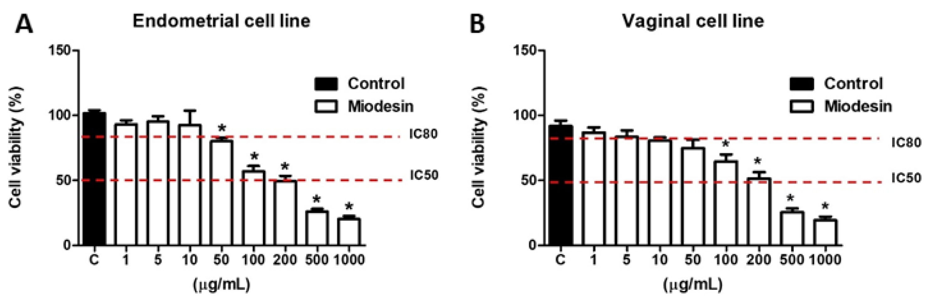
2.1. Effects of MiodesinTM on Cell Line Viability

Different concentrations of MiodesinTM were tested (0–1000 µg/mL) and the IC50 value (200 µg/mL) and IC80 value (10 µg/mL) were defined as the study dose in endometrial (Figure 1A) and vaginal (Figure 1B) cell lines.

2.2. Pro-Inflammatory Response Induced by Candida Albicans in Vaginal Cell Line VK2 E6/E7 Is Inhibited by MiodesinTM

Table 1 shows the secretion of interleukins (IL-6, IL-8, IL-1β, and TNF-α) by vaginal cell line (VK2 E6/E7). Miodesin™ (10 µg/mL and 200 µg/mL) significantly reduced Candida albicans-induced release of pro-inflammatory interleukins (IL-6, p < 0.05; IL-8, p < 0.05; IL-1β, p < 0.05, and TNF-α, p < 0.05). Similarly, Miodesin™ (10µg/mL and 200µg/mL) also reduced Candida albicans-induced release of chemokines (CCL2, p < 0.05; CCL3, p < 0.05), but not for (CCL5/RANTES, p > 0.05), which are shown in Table 2. In summary, such results clearly show the anti-inflammatory effects of Miodesin™ on vaginal cells, not only by inhibiting the release of pro-inflammatory cytokines, but also inhibiting the release of chemokines.

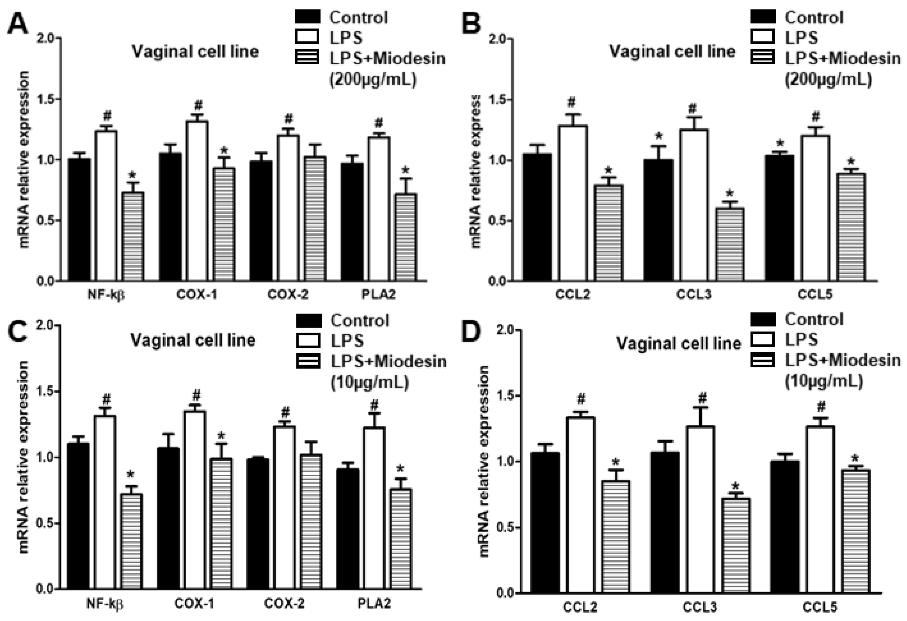
2.3. Expression of Cytokine and Inflammatory Mediators at mRNA Levels in Vaginal Cell Line (VK2 E6/E7)

We also investigated the effects of MiodesinTM (10 µg/mL and 200 µg/mL) on the mRNA expression of transcription factor, inflammatory mediators, and chemokines in VK2 E6/E7 cells. Similar results were found for both doses of MiodesinTM (Figure 2A,B, 200 µg/mL; Figure 2C,D, 10 µg/mL), resulting in reduced expression of total NF-κβ transcription factor, inflammatory mediators, and chemokines at mRNA levels. These results indicate that MiodesinTM significantly suppressed the expression of NF-κβ transcription factor and also that of enzymes that participate in the arachidonic acid cascade (inflammatory mediators), such as COX-1 and PLA2, as well as reducing chemokine expression, CCL2/MCP-1, CCL3/MIP-1α, and CCL5/RANTES.

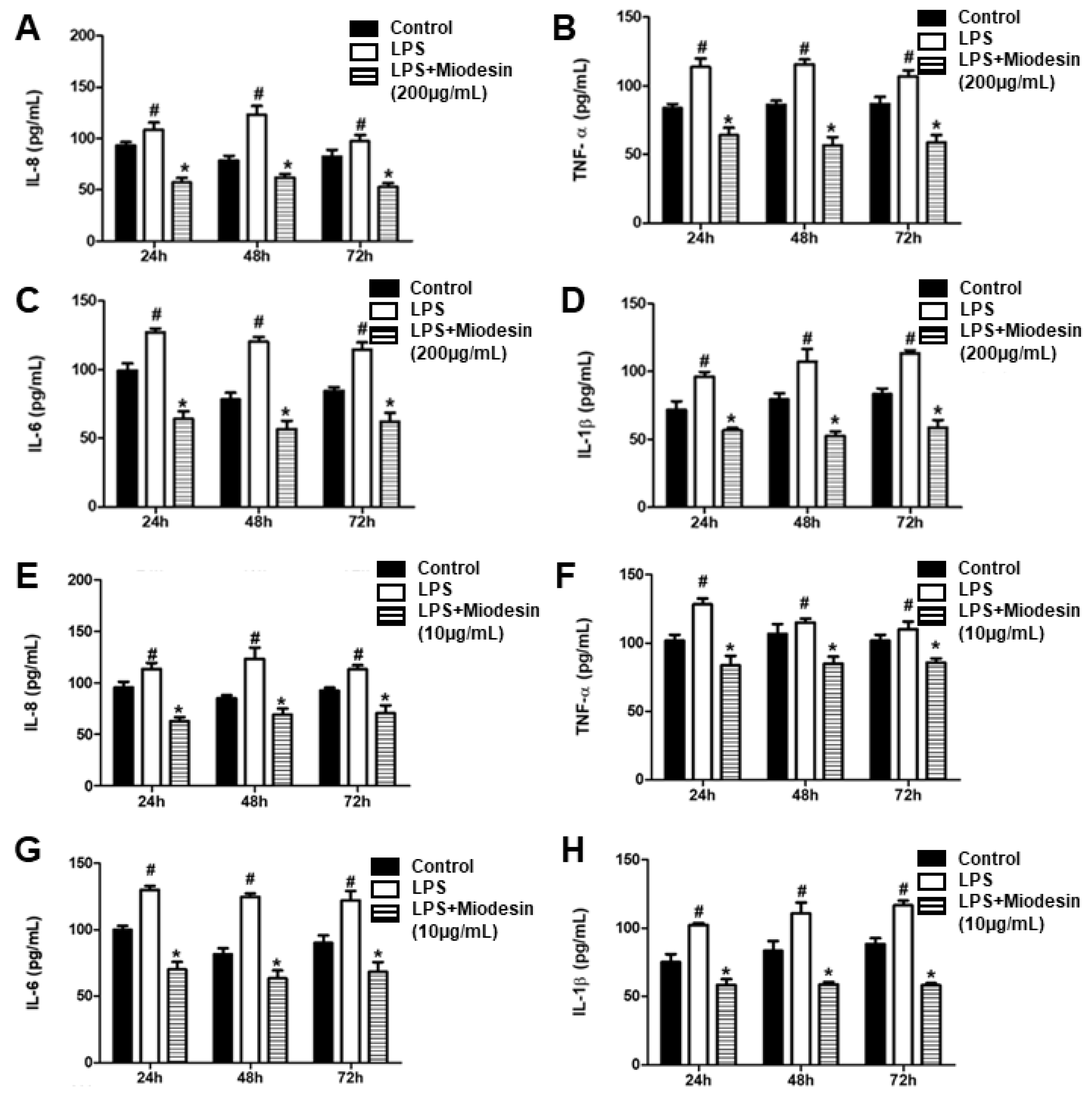
2.4. MiodesinTM Down-Regulates LPS-Induced Inflammatory Cytokine and Chemokine Release by Endometrial Cell Line (KLE)

Secretion of pro-inflammatory cytokines IL-8 (Figure 3A), TNF-α (Figure 3B), IL-6 (Figure 3C), and IL-1β (Figure 3D) was significantly reduced in endometrial cells pre-treated with MiodesinTM (200 µg/mL). In addition, the effects of Miodesin™ over time (24, 48, and 72 h) was maintained, reinforcing the anti-inflammatory effects of Miodesin™ in the time-course studied. Similarly, lower dose of Miodesin™ (10 µg/mL) also resulted in decreased secretion of pro-inflammatory cytokines IL-8 (Figure 3E), TNF-α (Figure 3F), IL-6 (Figure 3G), and IL-1β (Figure 3H).
Table 3 shows the results obtained with endometrial cells (KLE) stimulated with LPS which were pre-treated with 200 µg/mL and 10 µg/mL of MiodesinTM. MiodesinTM significantly reduced the chemokine release (CCL2/MCP-1, p < 0.05; CCL3/MIP-1α, p < 0.05; CCL5/RANTES, p < 0.05). Again, such results clearly show the anti-inflammatory effects of Miodesin™ on endometrial cells, not only by inhibiting the release of pro-inflammatory cytokines, but also by inhibiting the release of chemokines.

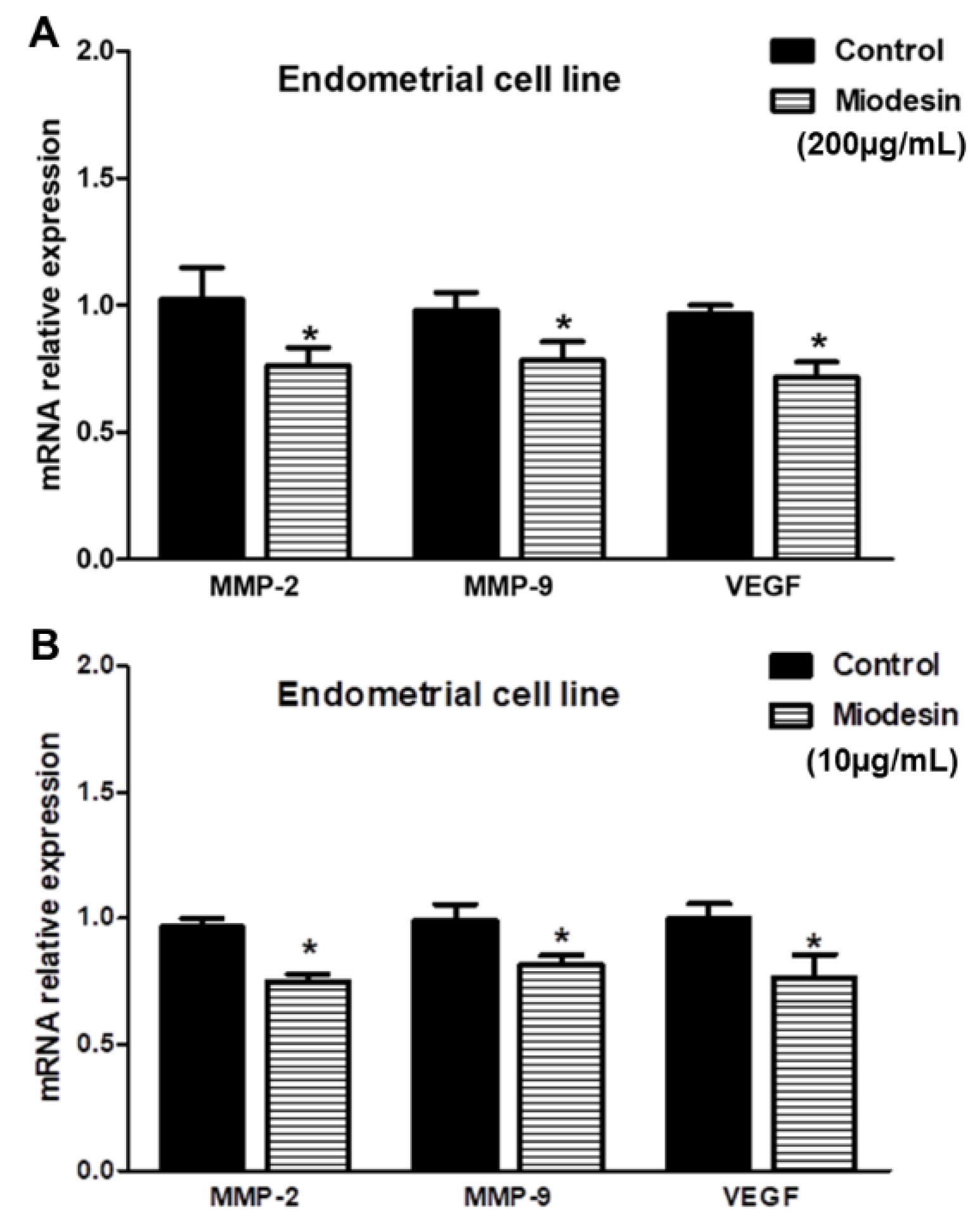
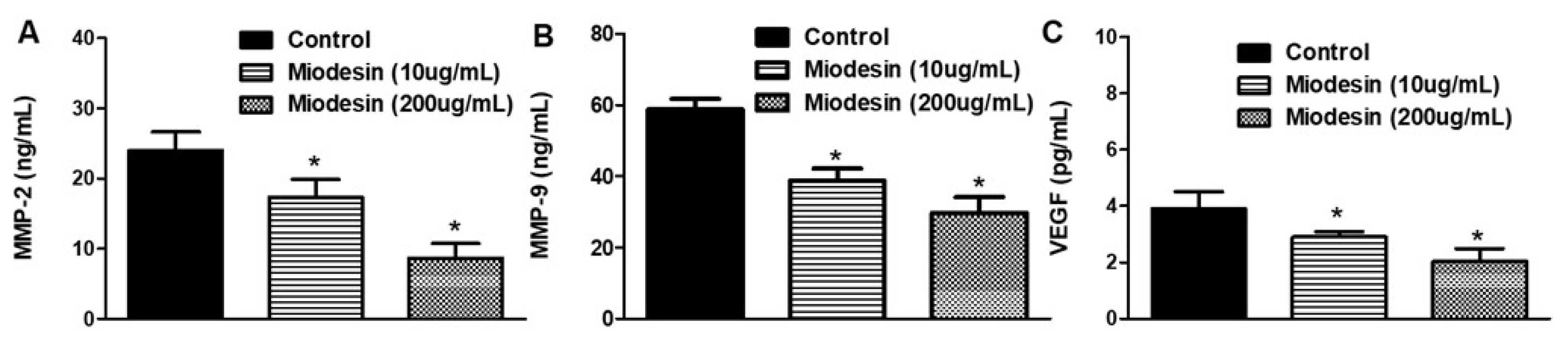
2.5. MiodesinTM Regulates MMP-2, MMP-9, and VEGF in Endometrial Cells

To investigate whether MiodesinTM regulates MMP-2, MMP-9, and VEGF expression at transcriptional level, reverse transcription polymerase chain reaction (RT-PCR) was performed. As shown in Figure 4, MiodesinTM (Figure 4A, 200 µg/mL; Figure 4B, 10 µg/mL) significantly inhibited the expression of MMP-2, MMP-9, and VEGF mRNA in KLE cells. Such results at mRNA levels were confirmed at protein levels, since MiodesinTM (200 µg/mL and 10 µg/mL) treatment led to significant decreases in MMP-2, MMP-9, and VEGF levels in the culture supernatant of KLE cells (Figure 5), as measured by ELISA. These findings are especially important in the context of endometrial cell proliferation and activation, since MMP-2, MMP-9, and VEGF are key regulators of inflammatory and fibrotic processes in the endometrium.

3. Discussion

The present study shows for the first time that MiodesinTM presents anti-inflammatory effects, inhibiting hyperactivation of endometrial epithelial cell KLE and vaginal epithelial cell VK2 E6/E7, which are cells presenting a key role in the pathophysiology of endometriosis and leiomyoma [2,3]. Such effects of MiodesinTM were evidenced by inhibition in the cytokines (IL-6, IL-8, IL-1β, and TNF-α) and chemokine release (CCL2/MCP-1; CCL3/MIP-1α), beyond the inhibition of other inflammatory (COX-1, COX-2, PLA2) and pro-fibrotic (MMP-2, MMP-9, and VEGF) mediators. In addition, MiodesinTM efficiently inhibited the transcription factor NF-kβ.
Endometriosis is still a disease with uncertain pathophysiology. However, the inflammatory character associated with the role of cytokines, hormones, and soluble factors seems to play an important role in its pathophysiology [28]. Indeed, the inflammatory character in endometriosis has been studied in the peritoneal fluid and serum, in addition to the eutopic and ectopic endometrium [29,30,31,32]. In this way, increased levels of TNF-α, IL-6, IL-8, IL-10, VEGF, and MCP-1/CCL2 [29,32,33], as well as different metalloproteinases (MMPs), are associated with the establishment and progression of endometriosis [34]. In fact, the present study revealed that vaginal mucosa and endometriotic cells responded to increased levels of IL-6, IL-8, IL-1β, and TNF-α when challenged by Candida albicans or by LPS, and these cells present a pivotal role in the inflammatory process in endometriosis and leiomyoma.
On the other hand, in 2018 and 2019, Maia et al. published the first manuscripts showing the role of MiodesinTM in reducing pelvic pain in patients with endometriosis and leiomyoma, supporting the role of naturally occurring anti-inflammatory medications in the treatment of this disease [8,26,27]. However, no analysis concerning the anti-inflammatory effects of MiodesinTM was addressed in such studies. So, the present study shows for the first time that MiodesinTM (10 µg/mL and 200 µg/mL) inhibited the hyperactivation of vaginal mucosa and endometriotic cells driving an inflammatory response, as demonstrated by reduced release of cytokines (IL-1β, IL-6, IL-8 and TNF-α; p < 0.01) and chemokines (CCL2, CCL3 and CCL5; p < 0.01)—in addition to the reduction on the expression at mRNA levels of transcription factor (NF-kβ; p < 0.01), inflammatory enzymes (COX-1, COX-2, PLA2, iNOS; p < 0.01), and chemokines (CCL2, CCL3 and CCL5; p < 0.01). Of note, the cytokines inhibited by MiodesinTM, such as IL-8 [32,35], IL-6 [36,37], IL-1β [38,39], and TNF-α [40,41] are thought to have a key role in the inflammatory process in endometriosis, becoming an interesting tool as an adjuvant for therapeutic schemes in the treatment of endometriosis. Thus, the inhibitory effects of MiodesinTM on epithelial cells in the human lower female genital tract clearly showed its anti-inflammatory potential, ensuring further randomized clinical trials.
Although inflammation plays a pivotal role in endometriosis, which is regulated by pro-inflammatory cytokines [42,43], it is important to note the modulatory capacity of MiodesinTM reducing the release of IL-8 by vaginal mucosa and endometriotic cells. IL-8 is also considered a chemokine, which is involved in the pathogenesis of endometriosis [44]. Chemokines are proteins with the ability to control mainly immune cell chemoattraction, but also centrally involved in cell proliferation, migration, and apoptosis [45]. In the present study, stimulation with C. albicans induced a significant increase in the secretion of chemokines CCL2, CCL3, and CCL5 by vaginal mucosa cells. Again, treatment with MiodesinTM resulted in significant reduction in CCL2, CCL3, and CCL5 expression at mRNA level and only for CCL2 and CCL3 at protein levels. In addition, chemokines form a complex network and stimulate other cells, such as macrophages, stromal cells, epidermal cells, and smooth muscle cells [46]. Recent studies have shown several types of chemokines produced by the endometrial implant foci, thus affecting the progression of endometriosis [46]. In this sense, both IL-8 and CCL2 are elevated in endometriotic tissues and in peritoneal implants, reinforcing the role of chemokines in the progression of ovarian and peritoneal endometriosis [47]. CCL5 is another example of a chemokine found high in the peritoneal fluid of women with endometriosis and is proportional to the stage of the disease that seems to mediate the chemotactic activity of monocytes in the peritoneal cavity, suggesting that it may contribute to the progression of this disease [48]. Thus, in this study we also evaluated the behavior in the secretion of chemokines CCL2, CCL3, and CCL5 in the KLE endometrial cell line. The results showed, once again, the ability of MiodesinTM to reverse the increase in chemokine secretion, when endometrial cells were stimulated by LPS, reinforcing the therapeutic potential of MiodesinTM in therapeutic endometriosis treatment schemes.
Furthermore, in endometrial cells, we found that the secretion and expression of the mRNA levels of the metalloprotease enzymes MMP-2 and MMP-9 and of growth factor VEGF were reduced. The results showed that treatment with MiodesinTM led to a significant reduction in the secretion of MMP-2, MMP-9, and VEGF by KLE cells, measured at protein levels. Similarly, such inhibitory effects of MiodesinTM were also seen at mRNA levels. Studies suggest that MMPs are associated with the establishment and progression of endometriosis and their levels appear to be increased in ectopic endometriotic tissues [5]. MMP-2 and MMP-9 have been extensively studied in the scope of endometriosis, mainly because they are present at high levels in the peritoneal fluid of patients with endometriosis [49]. In addition, MMP-2 and MMP-9 are found to have high levels and activity in human endometriotic tissues [50]. Downregulation of MMP-2 and MMP-9 by MiodesinTM may involve the inhibition of migration and invasion in human endometriotic cells, considering that MMPs have been implicated in this phenomenon. VEGF is one of the main growth factors in angiogenesis and since endometriosis is characterized by marked vascularization in and around ectopic tissue, VEGF-induced angiogenesis may be a critical aspect of the pathophysiology of endometriosis [46].
Finally, in addition to VEGF, chemokines and cytokines can play important roles in angiogenesis associated with endometriosis. IL-8, for example, a chemoattractant for neutrophils, is an angiogenic agent, inducing proliferation of endothelial cells [51]. TNF-α, which induces the growth of new blood vessels and secreted by macrophages, also stimulates the proliferation of endometriotic stromal cells [52].

4. Materials and Methods

4.1. Reagents

Dulbecco’s modified Eagle’s medium (DMEM), keratinocyte serum-free medium, fetal bovine serum (FBS), penicillin, streptomycin, and phosphate-buffered saline (PBS) were obtained from Gibco BRL (Grand Island, CA, USA). 3-(4,5-Dimethylthiazol-2-yl)-2,5-diphenyltetrazolium bromide (MTT) was purchased from Sigma-Aldrich (St. Louis, MO, USA). The Miodesin™ was supplied by Fagron™ (São Paulo, Brazil). The purity and quality of the raw materials used, as well as the formulation of Miodesin™ (Uncaria tomentosa, Endopleura uchi and Astaxanthin) were monitored by the Fagron™ Brazil quality control department.

4.2. Culture and Cytotoxicity Evaluation of Miodesin by MTT Assay

Endometrial cell line KLE (epithelial) and vaginal cell line VK2 E6/E7 (epithelial) were cultured in Dulbecco’s modified Eagles’ medium (DMEM high glucose) supplemented with 10% v/v fetal bovine serum (FBS), 1% L-glutamine, 100 U/mL penicillin and 100 mg/mL streptomycin, and in keratinocyte serum-free medium (Gibco) supplemented with 50 µg of bovine pituitary extract, 0.1 ng of epidermal growth factor, 100 U of penicillin, and 100 µg of streptomycin, respectively and maintained at 37 °C in a humidified atmosphere of 5% CO2. The cells were trypsinized every 72 h using 0.01% trypsin and 1 mmol ethylenediamine tetra-acetic acid (EDTA). For all the experiments, the Miodesin™ was dissolved in the culture medium in appropriate concentrations. The cell viability of control and MiodesinTM (1–1.000 μg/mL)-treated cells were measured using a standard MTT assay. Briefly, 5 × 10⁴ viable cells were seeded into clear 96-well flat-bottom plates (Corning) in RPMI medium supplemented with 10% fetal bovine serum and incubated with different concentrations of the extract for 24 h. Then, 10 μL/well of MTT (5 mg/mL) was added and the cells were incubated for 4 h. Following incubation, 100 μL of 10% sodium dodecyl sulfate solution in deionized water was added to each well and left overnight. The absorbance was measured at 595 nm in a benchtop multimode reader (Molecular Device). The established cell lines KLE and VK2 E6/E7 were kindly provided by Dr. Andre Luis Lacerda Bachi, from University Santo Amaro, São Paulo, SP, Brazil.

4.3. Candida Albicans (Stimulator Cells)

The isolate of Candida albicans was grown on Sabouraud dextrose agar (Becton Dickinson, Cockeysville, MD, USA) at 30 °C, and one colony was used to inoculate 10 mL of Phytone-peptone broth (Becton Dickinson) supplemented with 0.1% glucose. Broth cultures were grown to stationary phase for 18 h at 25 °C in a shaking water bath. The blastoconidia were collected, washed with phosphate-buffered saline, and enumerated on a hemacytometer by using trypan blue dye exclusion.

4.4. Coculture Supernatant Collection for Examination of Cytokines and Chemokines

For examination of cytokines and chemokines, epithelial cells (1 × 10⁵ to 5 × 10⁵ cells/mL) were cocultured with Candida albicans (1 × 10⁷ to 5 × 10⁷/mL) at a ratio of 1:100 in separate wells for 0, 24, 48, and 72 h for VK2 E6/E7 cells in a total volume of 2 mL of tissue culture medium in 24-well tissue culture plates (Costar, Corning, NY, USA). Controls included VK2 E6/E7 cells (constitutive production) cultured alone in separate wells, Candida albicans cultured alone (negative control), and cells cultured with tumor necrosis factor alpha (TNF-alpha; 10 to 20 ng/mL [quality control]; Pharmingen, San Diego, CA, USA) independently in tissue culture medium alone over the same 96-h period. At each time point, the VK2 E7/E6 cell-Candida coculture and the respective control cultures were aspirated from a new set of individual wells and centrifuged for 5 min at 800× g. Thereafter, the supernatants were collected and stored at −70 °C until assayed.

4.5. Cytokine and Chemokine Analysis of Coculture and Endometrial Cells Supernatants

The concentrations of TNF-α, IL-6, IL-8, IL-1β, MCP-1 (CCL2), MIP-1-α (CCL3), and RANTES (CCL5), in the supernatants obtained from different groups were analyzed using enzyme-linked immunosorbent assay (ELISA) kits (R&D Systems, Minneapolis, MN, USA) following the manufacturer’s instructions. For endometrial cell line cells, they were treated with LPS (1 μg/mL) with or without Miodesin (10 µg/mL and 200 μg/mL) for 6 h. The culture supernatant (100 μL) was removed to determine the level of cytokines and chemokines, according to the manufacturer’s instructions.

4.6. Reverse Transcription-Quantitative PCR (RT-qPCR)

Total RNA extracted from cells samples was converted to cDNA using a SuperScript® III RT kit (Invitrogen, Carlsbad, CA, USA), according to the manufacturer’s protocol. The concentration of RNA was detected using a NanoDrop 2000 (Thermo Fisher Scientific, Inc., Waltham, MA, USA). GAPDH and 18 S rRNA were used as the internal control. The thermocycling conditions were as follows: 95 °C for 10 min followed by 35 cycles of 95 °C for 15 s and 55 °C for 40 s. The 2−ΔΔCq method was used to quantify the relative gene expression levels of the target genes. Relative standard curves were generated by serial dilutions and all samples were run in triplicates. The table below indicates the sequences of primers used in qRT-PCR analysis.
GenePrimer sequences
NF-κβForward 5′-ATGGCTTCTATGAGGCTGAG- 3′
Reverse 5′-GTTGTTGTTGGTCTGGATGC- 3′
COX-1Forward 5′-AGGAGATGGCTGCTGAGTTGG-3′
Reverse 5′-AATCTGACTTTCTGAGTTGCC-3′
COX-2Forward 5′-ACACCTTCAACATTGAAGACC-3′
Reverse 5′-ATCCCTTCACTAAATGCCCTC-3′
PLA2Forward 5′-AAAGAACACTATAGGGAGAG-3′
Reverse 5´-AAAGAGGTAAAGGGCATTGT-3′
CCL2Forward 5′-GATCCCAATGAGTAGGCTGG-3′
Reverse 5′-CGGGTCAACTTCACATTCAAAG-3′
CCL3Forward 5′-ACACCAGAAGGATACAAGCAG-3′
Reverse 5′-CGATGAATTGGCGTGGAATC-3′
CCL5Forward 5′-CCCACGTCAAGGAGTATTTCTAC-3′
Reverse 5′-CTAGGACTAGAGCAAGCGATG-3′
MMP-2Forward 5′-ACCGCGACAAGAAGTATGGC-3′
Reverse 5′-CCACTTGCGGTCA TCATCGT-3´
MMP-9Forward 5′-CGATGACGAGTTGTGGTCCC-3′
Reverse 5′-TCGTAGTTG GCCGTGGTACT-3′
VEGFForward 5′-TGCAGATTATGCGGATCAAACC-3′
Reverse 5′-TGCATTCACATTTGTTGTGCTGTAG-3′
GAPDHForward 5′-CGGTGTGAACGGATTTGGC-3′
Reverse 5′-GTGAGTGGAGTCATACTGGAAC-3′

4.7. Enzyme-Linked Immunosorbent Assay for the Determination of MMP2 and MMP9

Cell supernatant was centrifuged at 12,000× g for 15 min at 4 °C. MMP2 ELISA kits (RAB0365; Sigma, San Louis, MO, USA) and MMP9 ELISA kits (RAB0372; Sigma, Nanjing, China) were used to measure the levels of protein secretion of MMP2 and MMP9 by glioma cells, respectively, according to the manufacturer’s instructions. To detect VEGF concentration in supernatants, ELISA was performed with a commercially available ELISA kit (Abcam, Cambridge, UK). The VEGF concentrations were detected according to the manufacturer’s instructions.

4.8. Statistical Analysis

The unpaired Student’s t test was used to analyze constitutive and Candida-specific cytokine production between primary oral and vaginal epithelial cells. Significant differences were defined as having a p value ≤ 0.05. One-way analysis of variance (ANOVA) was used for multiple comparisons. The obtained results were expressed as the mean ± standard error of mean from at least three independent experiments, unless stated otherwise.

5. Conclusions

MiodesinTM exerts anti-inflammatory effects against C. albicans and LPS stimulation, inhibiting vaginal mucosa and endometriotic cells hyperactivation. The results show that MiodesinTM is a promising phytotherapeutic candidate for treatment of endometriosis and leiomyoma.

Author Contributions

Conceptualization, methodology, investigation, writing—original draft preparation, C.R.O., M.C.M. and R.P.V.; writing—review and editing, H.P. All authors have read and agreed to the published version of the manuscript.

Funding

This study was supported by a research grant from Fagron do Brasil Pharmaceutical LTDA.

Institutional Review Board Statement

Not applicable.

Informed Consent Statement

Not applicable.

Data Availability Statement

All raw data will be available upon a reasonable justification request.

Acknowledgments

In this section, you can acknowledge any support given which is not covered by the author contribution or funding sections. This may include administrative and technical support, or donations in kind (e.g., materials used for experiments).

Conflicts of Interest

All authors disclose any influence of companies or manufacturers in the present study. In addition, all authors declare that the results of the study are presented clearly, honestly, and without fabrication, falsification, or inappropriate data manipulation.

Sample Availability

Samples of the compounds will be available upon a reasonable justification request.

References

  1. Murji, A.; Bedaiwy, M.; Singh, S.S.; Bougie, O.; Committee, C.R.S. Influence of Ethnicity on Clinical Presentation and Quality of Life in Women with Uterine Fibroids: Results from a Prospective Observational Registry. J. Obstet. Gynaecol. Can. 2020, 42, 726–733. [Google Scholar] [CrossRef] [PubMed]
  2. Symons, L.K.; Miller, J.E.; Kay, V.R.; Marks, R.M.; Liblik, K.; Koti, M.; Tayade, C. The Immunopathophysiology of Endometriosis. Trends Mol. Med. 2018, 24, 748–762. [Google Scholar] [CrossRef] [PubMed]
  3. Giudice, L.C. Endometriosis. N. Engl. J. Med. 2010, 362, 2389–2398. [Google Scholar] [CrossRef] [PubMed]
  4. Yu, H.; Zhong, Q.; Xia, Y.; Li, E.; Wang, S.; Ren, R. MicroRNA-2861 Targets STAT3 and MMP2 to Regulate the Proliferation and Apoptosis of Ectopic Endometrial Cells in Endometriosis. Die Pharm.-Int. J. Pharm. Sci. 2019, 74, 243–249. [Google Scholar]
  5. Bostanci Durmus, A.; Dincer Cengiz, S.; Yılmaz, H.; Candar, T.; Gursoy, A.Y.; Sinem Caglar, G. The Levels of Matrix Metalloproteinase-9 and Neutrophil Gelatinase-Associated Lipocalin in Different Stages of Endometriosis. J. Obstet. Gynaecol. 2019, 39, 991–995. [Google Scholar] [CrossRef] [PubMed]
  6. Zhang, F.; Liu, X.; Wang, W.; Dong, H.; Xia, Y.; Ruan, L.; Liu, L. Expression of MMIF, HIF-1α and VEGF in Serum and Endometrial Tissues of Patients with Endometriosis. Curr. Med. Sci. 2018, 38, 499–504. [Google Scholar] [CrossRef] [PubMed]
  7. Nasiri, N.; Babaei, S.; Moini, A.; Eftekhari-Yazdi, P. Controlling Semi-Invasive Activity of Human Endometrial Stromal Cells by Inhibiting NF-kB Signaling Pathway Using Aloe-emodin and Aspirin. J. Reprod. Infertil. 2021, 22, 227–240. [Google Scholar] [CrossRef]
  8. Huang, F.; Cao, J.; Liu, Q.; Zou, Y.; Li, H.; Yin, T. MAPK/ERK signal pathway involved expression of COX-2 and VEGF by IL-1β induced in human endometriosis stromal cells in vitro. Int. J. Clin. Exp. Pathol. 2013, 6, 2129–2136. [Google Scholar]
  9. Sano, M.; Morishita, T.; Nozaki, M.; Yokoyama, M.; Watanabe, Y.; Nakano, H. Elevation of the phospholipase A2 activity in peritoneal fluid cells from women with endometriosis. Fertil. Steril. 1994, 61, 657–662. [Google Scholar] [CrossRef]
  10. Kowalczyk-Zieba, I.; Woclawek-Potocka, I.; Wasniewski, T.; Boruszewska, D.; Grycmacher, K.; Sinderewicz, E.; Staszkiewicz, J.; Wolczynski, S. LPAR2 and LPAR4 are the Main Receptors Responsible for LPA Actions in Ovarian Endometriotic Cysts. Reprod. Sci. 2019, 26, 139–150. [Google Scholar] [CrossRef]
  11. Nicolaus, K.; Bräuer, D.; Sczesny, R.; Lehmann, T.; Diebolder, H.; Runnebaum, I.B. Unexpected Coexistent Endometriosis in Women with Symptomatic Uterine Leiomyomas Is Independently Associated with Infertility, Nulliparity and Minor Myoma Size. Arch. Gynecol. Obstet. 2019, 300, 103–108. [Google Scholar] [CrossRef] [PubMed]
  12. Maia, H.; Saback, W.; Haddad, C.; Sitya, P.R. Treatment of Endometriosis and Leiomyoma with the Association of Miodesin and Gestrinone in Pentravan Through the Vaginal Route. J. Clin. Rev. Case Rep. 2018, 3, 1–5. [Google Scholar] [CrossRef]
  13. Florence, A.M.; Fatehi, M. Leiomyoma. Available online: https://www.ncbi.nlm.nih.gov/books/NBK430685/ (accessed on 20 December 2021).
  14. Brown, J.; Crawford, T.J.; Allen, C.; Hopewell, S.; Prentice, A. Nonsteroidal Anti-inflammatory Drugs for Pain in Women with Endometriosis. Cochrane Database Syst. Rev. 2017, 1, CD004753. [Google Scholar] [CrossRef] [PubMed]
  15. Sabry, M.; Al-Hendy, A. Medical Treatment of Uterine Leiomyoma. Reprod. Sci. 2012, 19, 339–353. [Google Scholar] [CrossRef] [Green Version]
  16. Valerio, L.G.; Gonzales, G.F. Toxicological Aspects of the South American Herbs Cat’s Claw (Uncaria Tomentosa) and Maca (Lepidium Meyenii). Toxicol. Rev. 2005, 24, 11–35. [Google Scholar] [CrossRef]
  17. Silva, L.R.; Teixeira, R. Phenolic Profile and Biological Potential of Endopleura Uchi Extracts. Asian Pac. J. Trop. Med. 2015, 8, 889–897. [Google Scholar] [CrossRef] [Green Version]
  18. Muniz, M.P. Estudo Fitoquímico e Da Atividade Biológica de Endopleura Uchi Huber Cuatrecasas. Ph.D. Thesis, Federal University of Amazonas, Manaus, AM, Brazil, 2013. [Google Scholar]
  19. da Silva, S.L.; de Oliveira, V.G.; Yano, T.; Nunomura, R.d.C.S. Antimicrobial Activity of Bergenin from Endopleura Uchi (Huber) Cuatrec. Acta Amaz. 2009, 39, 187–191. [Google Scholar] [CrossRef] [Green Version]
  20. Correa, M.; Penna, L. Dicionário Das Plantas Úteis Do Brasil e Das Exóticas Cultivadas; Instituto Brasileiro de Desenvolvimento Florestal: Rio de Janeiro, Brazil, 1974; Volume 5. [Google Scholar]
  21. Rao, A.R.; Reddy, A.H.; Aradhya, S.M. Antibacterial Properties of Spirulina Platensis, Haematococcus Pluvialis, Botryococcus Braunii Micro Algal Extracts. Curr. Trends Biotechnol. Pharm. 2010, 4, 809–819. [Google Scholar]
  22. Shah, M.; Mahfuzur, R.; Liang, Y.; Cheng, J.J.; Daroch, M. Astaxanthin-Producing Green Microalga Haematococcus Pluvialis: From Single Cell to High Value Commercial Products. Front. Plant Sci. 2016, 7, 531. [Google Scholar] [CrossRef] [Green Version]
  23. Park, J.S.; Mathison, B.D.; Hayek, M.G.; Massimino, S.; Reinhart, G.A.; Chew, B.P. Astaxanthin Stimulates Cell-Mediated and Humoral Immune Responses in Cats. Vet. Immunol. Immunopathol. 2011, 144, 455–461. [Google Scholar] [CrossRef]
  24. Park, J.S.; Chyun, J.H.; Kim, Y.K.; Line, L.L.; Chew, B.P. Astaxanthin Decreased Oxidative Stress and Inflammation and Enhanced Immune Response in Humans. Nutr. Metab. 2010, 7, 18. [Google Scholar] [CrossRef] [PubMed] [Green Version]
  25. Chew, B.P.; Mathison, B.D.; Hayek, M.G.; Massimino, S.; Reinhart, G.A.; Park, J.S. Dietary Astaxanthin Enhances Immune Response in Dogs. Vet. Immunol. Immunopathol. 2011, 140, 199–206. [Google Scholar] [CrossRef] [PubMed]
  26. Maia, H.; Saback, W.; Haddad, C.; Sitya, P.R. Effect of Vaginal MiodesinTM in PentravanTM on the Response to Progestin Therapy in Patients with Deep Endometriosis and Adenomyosis. J. Clin. Rev. Case Rep. 2019, 4, 1–5. [Google Scholar]
  27. Maia, H.; Saback, W.; Coutinho, E. Short Term Effects of the Vaginal Administration of Gestrinone and MiodesinTM on Endometriosis Pain. J. Clin. Rev. Case Rep. 2019, 4, 1–6. [Google Scholar] [CrossRef]
  28. Luckow Invitti, A.; Schor, E.; Martins Parreira, R.; Kopelman, A.; Kamergorodsky, G.; Gonçalves, G.A.; Batista Castello Girão, M.J. Inflammatory Cytokine Profile of Co-cultivated Primary Cells from the Endometrium of Women with and without Endometriosis. Mol. Med. Rep. 2018, 18, 1287–1296. [Google Scholar] [CrossRef] [Green Version]
  29. Ilie, I.; Ilie, R. Cytokines and Endometriosis-the Role of Immunological Alterations. Biotechnol. Mol. Biol. Nanomed. 2013, 1, 8–19. [Google Scholar]
  30. Gazvani, R.; Templeton, A. Peritoneal Environment, Cytokines and Angiogenesis in the Pathophysiology of Endometriosis. Reproduction 2002, 123, 217–226. [Google Scholar] [CrossRef]
  31. Fan, Y.-Y.; Chen, H.-Y.; Chen, W.; Liu, Y.-N.; Fu, Y.; Wang, L.-N. Expression of Inflammatory Cytokines in Serum and Peritoneal Fluid from Patients with Different Stages of Endometriosis. Gynecol. Endocrinol. 2018, 34, 507–512. [Google Scholar] [CrossRef]
  32. Kalu, E.; Sumar, N.; Giannopoulos, T.; Patel, P.; Croucher, C.; Sherriff, E.; Bansal, A. Cytokine Profiles in Serum and Peritoneal Fluid from Infertile Women with and without Endometriosis. J. Obstet. Gynaecol. Res. 2007, 33, 490–495. [Google Scholar] [CrossRef]
  33. Jørgensen, H.; Hill, A.S.; Beste, M.T.; Kumar, M.P.; Chiswick, E.; Fedorcsak, P.; Isaacson, K.B.; Lauffenburger, D.A.; Griffith, L.G.; Qvigstad, E. Peritoneal Fluid Cytokines Related to Endometriosis in Patients Evaluated for Infertility. Fertil. Steril. 2017, 107, 1191–1199. [Google Scholar] [CrossRef] [Green Version]
  34. Zhou, H.-E.; Nothnick, W.B. The Relevancy of the Matrix Metalloproteinase System to the Pathophysiology of Endometriosis. Front. Biosci. 2005, 10, 569–575. [Google Scholar] [CrossRef] [PubMed]
  35. Garcia-Velasco, J.A.; Arici, A. Interleukin-8 Stimulates the Adhesion of Endometrial Stromal Cells to Fibronectin. Fertil. Steril. 1999, 72, 336–340. [Google Scholar] [CrossRef]
  36. Herington, J.L.; Bruner-Tran, K.L.; Lucas, J.A.; Osteen, K.G. Immune Interactions in Endometriosis. Expert Rev. Clin. Immunol. 2011, 7, 611–626. [Google Scholar] [CrossRef] [PubMed] [Green Version]
  37. Young, V.J.; Brown, J.K.; Saunders, P.T.K.; Horne, A.W. The Role of the Peritoneum in the Pathogenesis of Endometriosis. Hum. Reprod. Update 2013, 19, 558–569. [Google Scholar] [CrossRef]
  38. Lebovic, D.I.; Baldocchi, R.A.; Mueller, M.D.; Taylor, R.N. Altered Expression of a Cell-Cycle Suppressor Gene, Tob-1, in Endometriotic Cells by CDNA Array Analyses. Fertil. Steril. 2002, 78, 849–854. [Google Scholar] [CrossRef]
  39. Sikora, J.; Mielczarek-Palacz, A.; Kondera-Anasz, Z. Association of the Precursor of Interleukin-1β and Peritoneal Inflammation—Role in Pathogenesis of Endometriosis. J. Clin. Lab. Anal. 2016, 30, 831–837. [Google Scholar] [CrossRef] [Green Version]
  40. Sakamoto, Y.; Harada, T.; Horie, S.; Iba, Y.; Taniguchi, F.; Yoshida, S.; Iwabe, T.; Terakawa, N. Tumor Necrosis Factor-α-Induced Interleukin-8 (IL-8) Expression in Endometriotic Stromal Cells, Probably through Nuclear Factor-ΚB Activation: Gonadotropin-Releasing Hormone Agonist Treatment Reduced IL-8 Expression. J. Clin. Endocrinol. Metab. 2003, 88, 730–735. [Google Scholar] [CrossRef]
  41. McKinnon, B.D.; Kocbek, V.; Nirgianakis, K.; Bersinger, N.A.; Mueller, M.D. Kinase Signalling Pathways in Endometriosis: Potential Targets for Non-Hormonal Therapeutics. Hum. Reprod. Update 2016, 22, 382–403. [Google Scholar] [CrossRef] [Green Version]
  42. Steele, C.; Fidel Jr, P.L. Cytokine and Chemokine Production by Human Oral and Vaginal Epithelial Cells in Response to Candida Albicans. Infect. Immun. 2002, 70, 577–583. [Google Scholar] [CrossRef] [Green Version]
  43. Fichorova, R.N.; Anderson, D.J. Differential Expression of Immunobiological Mediators by Immortalized Human Cervical and Vaginal Epithelial Cells. Biol. Reprod. 1999, 60, 508–514. [Google Scholar] [CrossRef]
  44. Borrelli, G.; Carvalho, K.; Kallas, E.; Mechsner, S.; Baracat, E.; Abrao, M. Chemokines in the Pathogenesis of Endometriosis and Infertility. J. Reprod. Immunol. 2013, 98, 1–9. [Google Scholar] [CrossRef] [PubMed]
  45. Złotkowska, A.; Andronowska, A. Chemokines as the Modulators of Endometrial Epithelial Cells Remodelling. Sci. Rep. 2019, 9, 12968. [Google Scholar] [CrossRef] [PubMed]
  46. Nishida, M.; Nasu, K.; Narahara, H. Role of Chemokines in the Pathogenesis of Endometriosis. Front. Biosci. Sch. Ed. 2011, 3, 1196–1204. [Google Scholar] [CrossRef] [PubMed]
  47. Chegini, N.; Roberts, M.; Ripps, B. Differential Expression of Interleukins (IL)-13 and IL-15 in Ectopic and Eutopic Endometrium of Women with Endometriosis and Normal Fertile Women. Am. J. Reprod. Immunol. 2003, 49, 75–83. [Google Scholar] [CrossRef] [PubMed]
  48. Guo, P.; Bi, K.; Lu, Z.; Wang, K.; Xu, Y.; Wu, H.; Cao, Y.; Jiang, H. CCR5/CCR5 Ligand-Induced Myeloid-Derived Suppressor Cells Are Related to the Progression of Endometriosis. Reprod. BioMedicine Online 2019, 39, 704–711. [Google Scholar] [CrossRef] [PubMed]
  49. Huang, H.-F.; Hong, L.-H.; Tan, Y.; Sheng, J.-Z. Matrix Metalloproteinase 2 Is Associated with Changes in Steroid Hormones in the Sera and Peritoneal Fluid of Patients with Endometriosis. Fertil. Steril. 2004, 81, 1235–1239. [Google Scholar] [CrossRef] [PubMed]
  50. Chung, H.-W.; Wen, Y.; Chun, S.-H.; Nezhat, C.; Woo, B.-H.; Lake Polan, M. Matrix Metalloproteinase-9 and Tissue Inhibitor of Metalloproteinase-3 MRNA Expression in Ectopic and Eutopic Endometrium in Women with Endometriosis: A Rationale for Endometriotic Invasiveness. Fertil. Steril. 2001, 75, 152–159. [Google Scholar] [CrossRef]
  51. Schadendorf, D.; Möller, A.; Algermissen, B.; Worm, M.; Sticherling, M.; Czarnetzki, B.M. IL-8 Produced by Human Malignant Melanoma Cells in vitro Is an Essential Autocrine Growth Factor. J. Immunol. 1993, 151, 2667–2675. [Google Scholar]
  52. Iwabe, T.; Harada, T.; Tsudo, T.; Nagano, Y.; Yoshida, S.; Tanikawa, M.; Terakawa, N. Tumor Necrosis Factor-Alpha Promote Proliferation of Endometriotic Stromal Cells by Inducing Gene and Protein Expression. J. Clin. Endocrinol. Metab. 2000, 85, 824–829. [Google Scholar] [CrossRef] [Green Version]
Figure 1. Effects of MiodesinMT on endometrial (KLE) (A) and vaginal cell (VK2 E6/E7) (B) lines. Cells were treated with different concentrations of MiodesinMT for 24 h. The red dotted line represents the IC80 and IC50 point. The IC80 was defined as the study test concentration (10 µg/mL) while IC50 was defined as the study test concentration (200 µg/mL). Date shown are representative of three replicates in at least three independent experiments. * p < 0.05 indicates significant differences (ANOVA). Values are expressed as means ± SEM.
Molecules 27 00782 g001
Figure 2. Effects of MiodesinTM (200 µg/mL and 10 µg/mL) on the expression of mRNA of NF-κβ, inflammatory enzymes and chemokines from endometrial (VK2 E6/E7) cells in presence or absence of LPS. (A) mRNA levels were significantly reduced (* p < 0.05) for NF-kβ, COX-1 and PLA2, except for COX-2 mRNA levels. (B) The mRNA levels of the chemokines CCL2/MCP-1, CCL3/MIP-1α, and CCL5/RANTES were significantly reduced. (C) mRNA levels were significantly reduced (* p < 0.05) for NF-kβ, COX-1, COX-2, and PLA2. (D) mRNA levels were significantly reduced (* p < 0.05) for CCL2, CCL3, and CCL5. mRNA levels were determined using real-time RT-PCR. Values are expressed as means ± SEM. (#) p < 0.05 LPS vs. control cells. (*) p < 0.05 MiodesinTM vs. LPS.
Molecules 27 00782 g002
Figure 3. MiodesinMT regulates LPS-induced release of inflammatory cytokines. Endometrial cells (KLE cells) were pretreated with MiodesinTM (200 µg/mL and 10 µg/mL) for 2 h and treated with LPS (1 µg/mL) for 24 h. In subfigures (AD), the dose of MiodesinTM was 200ug/mL, and in subfigures (EH) the dose of MiodesinTM was 10ug/mL. The effect of MiodesinTM on decreasing release of interleukines evaluated was determined by ELISA. Values are expressed as means ± SEM. Values are expressed as means ± SEM. (#) p < 0.05 LPS vs. control (non-treated cells). (*) p < 0.05 MiodesinTM vs. LPS.
Molecules 27 00782 g003
Figure 4. Effects of MiodesinTM (A), 200 µg/mL; (B), 10 µg/mL on the expression of mRNA of MMP-2, MMP-9, and VEGF in KLE cells. mRNA levels were significantly reduced for MMP-2, MMP-9, and VEGF. KLE cells were treated with MiodesinTM (200 µg/mL and 10 µg/mL) for 24 h. Real-time RT-PCR was performed to measure the mRNA levels of MMP-2, MMP-9, and VEGF in KLE cells. Glyceraldehyde-3-phosphate dehydrogenase (GADPH) was used as an internal control. Data are presented as the means ± SEM of three independent experiments. (*) p < 0.05.
Molecules 27 00782 g004
Figure 5. Effects of MiodesinTM (200 µg/mL and 10 µg/mL) on the production of MMP2, MMP9, and VEGF. Following treatment with MiodesinTM (200 µg/mL) for 24 h. ELISA analysis was performed to determine the levels of MMP2 (A), MMP9 (B), and VEGF (C) in the cell culture supernatant. Data are presented as the means ± SEM of three independent experiments. (*) p < 0.05.
Molecules 27 00782 g005
Table 1. Effects of MiodesinTM on the cytokine secretion by vaginal cell line (VK2 E6/E7). Cells were stimulated with Candida albicans in the presence of MiodesinTM (10 µg/mL and 200 µg/mL) and cultured for 24 h. Cytokines were determined in the culture supernatants by ELISA. Representative data obtained in one of three different experimental series are shown. Mean values ± SEM (of triplicate cultures) are given. (#) p < 0.05. (Candida albicans vs. medium). (*) p < 0.05. (Candida albicans-stimulated cells + MiodesinTM vs. Candida albicans-stimulated cells).
CytokineStimulusCytokine Concentration (Mean pg/mL ± SEM)
24 h48 h72 h
IL-6Medium4.9 ± 0.87.8 ± 2.48.2 ± 2.6
C. albicans17.1 ± 1.0 #31.0 ± 2.9 #64.3 ± 3.7 #
C. albicans + MiodesinTM (10 µg/mL)6.2 ± 2.3 *12.9 ± 3.8 *28.2 ± 3.9 *
Medium3.4 ± 0.512.6 ± 2.826.0 ± 3.7
C. albicans19.0 ± 0.9 #39.0 ± 3.7 #68.0± 4.5 #
C. albicans + MiodesinTM (200 µg/mL)11.0 ± 2.9 *15.0 ± 3.5 *29.0 ± 3.2 *
IL-8Medium31.4 ± 2.435.2 ± 2.932.1 ± 3.2
C. albicans63.0 ± 4.0 #76.6 ± 3.0 #102.4 ± 3.7 #
C. albicans +MiodesinTM (10 µg/mL)33.9 ± 2.7 *26.8 ± 1.9 *29.7 ± 3.1 *
Medium30.0 ± 3.731.3 ± 1.629.6 ± 3.8
C. albicans69.0 ± 5.1 #87 ± 2.5 #107.6 ± 3.4 #
C. albicans + MiodesinTM (200 µg/mL)36.0 ± 3.5 *21 ± 2.9 *20.3 ± 1.7 *
IL-1βMedium4.7 ± 2.65.8 ± 2.15.7 ± 1.4
C. albicans15.3 ± 2.3 #23.2 ± 2.8 #31.5 ± 3.3 #
C. albicans + MiodesinTM (10 µg/mL)9.0 ± 1.6 *11.5 ± 2.5 *17.3 ± 1.1 *
Medium3.3 ± 2.13.9 ± 1.86.0 ± 2.3
C. albicans18.0 ± 1.9 #22.0 ± 3.9 #32.0 ± 3.1 #
C. albicans + MiodesinTM (200 µg/mL)12.0 ± 1.1 *17.0 ± 3.5 *18.0 ± 2.1 *
TNF-αMedium7.1 + 0.99.2 + 3.28.6 + 1.7
C. albicans25.2 + 2.1 #29.9 + 4.1 #37.0 + 3.2 #
C. albicans + MiodesinTM (10 µg/mL)11.1 + 1.9 *13.2 + 2.1 *14.9 + 4,2 *
Medium5.7 ± 0.212.6 ± 2.818.6 ± 3.7
C. albicans21.5 ± 0.5 #31.9 ± 3.7 #67.2 ± 2.5 #
C. albicans + MiodesinTM (200 µg/mL)11.0 ± 2.7 *16.0 ± 3.5 *31.0 ± 3.2 *
Table 2. Effects of MiodesinTM on the chemokine secretion by vaginal cell line (VK2 E6/E7). Cells were stimulated with C. albicans in the presence of MiodesinTM (10 µg/mL and 200 µg/mL) and cultured for 24 h. Chemokines were determined in the culture supernatants by ELISA. Representative data obtained in one of three different experimental sets are shown. Mean values ± SEM (of triplicate cultures) are given. (#) p < 0.05. (Candida albicans vs. medium). (*) p < 0.05. (Candida albicans-stimulated cells + MiodesinTM vs. Candida albicans-stimulated cells).
CytokineStimulusCytokine Concentration (Mean pg/mL ± SEM)
24 h48 h72 h
CCL2
(MCP-1)
Medium3.3 ± 2.94.7 ± 1.26.5 ± 2.8
C. albicans10.1 ± 2.2 #14.2 ± 2.9 #21.5 ± 1.5 #
C. albicans + MiodesinTM (10 µg/mL)6.7 ± 3.2 *8.9 ± 3.5 *10.1 ± 1.1 *
Medium2.1 ± 3.24.9 ± 2.28.10 ± 1.7
C. albicans9.8 ± 1.2 #16.0 ± 3.2 #31.0 ± 2.5 #
C. albicans + MiodesinTM (200 µg/mL)5.2 ± 1.1 *12.6 ± 1.5 *19.8 ± 1.7 *
CCL3
(MIP-1α)
Medium33.0 ± 2.835.2 ± 3.931.4 ± 2.3
C. albicans58.6 ± 3.3 #69.8 ± 4.1 #75.2 ± 2.8 #
C. albicans + MiodesinTM (10 µg/mL)27.1 ± 2.5 *31.6 ± 1.9 *25.2 ± 3.2 *
Medium30.0 ± 3.732.3 ± 1.629.6 ± 3.8
C. albicans59.0 ± 3.1 #67 ± 2.5 #81.7 ± 3.4 #
C. albicans + MiodesinTM (200 µg/mL)26.0 ± 3.5 *29 ± 2.9 *24.0 ± 1.7 *
CCL5
(RANTES)
Medium3.7 ± 1.93.1 ± 2.83.6 ± 1.7
C. albicans19.1 ± 3.7 #24.2 ± 2.7 #29.9 ± 2.9 #
C. albicans + MiodesinTM (10 µg/mL)9.2 ± 4.511.9 ± 2.512.8 ± 5.8
Medium3.3 ± 2.13.9 ± 1.84.0 ± 2.3
C. albicans16.0 ± 2.9 #22.0 ± 3.9 #32.0 ± 3.1 #
C. albicans + MiodesinTM (200 µg/mL)9.3 ± 4.119.9 ± 3.928.0 ± 5.5
Table 3. Effects of MiodesinTM on the chemokine secretion by endometrial cell line (KLE). Cells were stimulated with LPS in the presence of MiodesinTM (10 µg/mL and 200 µg/mL) and cultured for 24 h. Chemokines were determined in the culture supernatants by ELISA. Mean values ± SEM (of triplicate cultures) are given. (#) p < 0.05. (LPS-stimulated cells vs. medium). (*) p < 0.05. (MiodesinTM vs. LPS-stimulated cells).
CytokineStimulusCytokine Concentration (Mean pg/mL ± SEM)
24 h48 h72 h
CCL2
(MCP-1)
Medium7.1 ± 2.16.2 ± 2.27.9 ± 4.1
LPS15.2 ± 3.1 #17.2 ± 3.6 #21.3 ± 3.7 #
LPS + MiodesinTM (10 µg/mL)6.1 ± 1.4 *8.9 ± 3.3 *9.2 ± 1.1 *
Medium4.2 ± 2.96.4 ± 3.911.3 ± 3.2
LPS13.3 ± 3.9 #21.4 ± 5.1 #42.2 ± 3.5 #
LPS + MiodesinTM (200 µg/mL)6.9 ± 2.4 *12.3 ± 2.3 *19.1 ± 2.1 *
CCL3
(MIP-1α)
Medium33.2 ± 1.739.4 ± 2.236.3 ± 3.7
LPS67.2 ± 3.8 #75.2 ± 2.9 #81.1 ± 6.2 #
LPS + MiodesinTM (10 µg/mL)31.2 ± 4.2 *33.8 ± 5.2 *29.8 ± 11.1 *
Medium31.1 ± 2.242.1 ± 3.731.2 ± 5.8
LPS69.1 ± 2.9 #77.8 ± 3.8 #93.2 ± 8.9 #
LPS + MiodesinTM (200 µg/mL)21.1 ± 4.5 *31.2 ± 6.9 *20.4 ± 10.7 *
CCL5
(RANTES)
Medium5.2 ± 4.18.4 ± 2.57.4 ± 2.8
LPS30.3 ± 3.1 #36.2 ± 4.1 #39.6 ± 3.7 #
LPS + MiodesinTM (10 µg/mL)9.4 ± 6.2 *11.3 ± 4.1 *12.9 ± 4.7 *
Medium4.4 ± 7.93.9 ± 1.86.7 ± 2.3
LPS29.9 ± 5.1 #32.0 ± 3.9 #41.0 ± 3.1 #
LPS + MiodesinTM (200 µg/mL)13.5 ± 8.1 *18.1 ± 5.2 *21.8 ± 4.9 *
Publisher’s Note: MDPI stays neutral with regard to jurisdictional claims in published maps and institutional affiliations.

