Article

The Synergistic Hepatoprotective Activity of Rosemary Essential Oil and Curcumin: The Role of the MEK/ERK Pathway

by
Maged E. Mohamed
1,2,*,
Nancy S. Younis
1,3,
Hossam S. El-Beltagi
4,5,* and
Omar M. Mohafez
6
1
Department of Pharmaceutical Sciences, College of Clinical Pharmacy, King Faisal University, Al-Ahsa 31982, Saudi Arabia
2
Department of Pharmacognosy, Faculty of Pharmacy, University of Zagazig, Zagazig 44519, Egypt
3
Zagazig University Hospitals, Zagazig 44511, Egypt
4
Agricultural Biotechnology Department, College of Agriculture and Food Sciences, King Faisal University, Al-Ahsa 31982, Saudi Arabia
5
Biochemistry Department, Faculty of Agriculture, Cairo University, Giza 12613, Egypt
6
Department of Biochemistry, Faculty of Pharmacy, University of Al-Azhar, Assiut Branch 71524, Egypt
*
Authors to whom correspondence should be addressed.
Molecules 2022, 27(24), 8910; https://doi.org/10.3390/molecules27248910
Submission received: 28 November 2022 / Revised: 8 December 2022 / Accepted: 13 December 2022 / Published: 15 December 2022

Abstract

:
Background: Curcumin is a natural product obtained from the rhizome of Curcuma longa. Rosemary (Rosmarinus officinalis) is a medicinal and aromatic plant that is widely spread in the Mediterranean region. Both Curcumin and rosemary essential oil are natural products of high medicinal and pharmacological significance. The hepatoprotective effect of both natural products is well-established; however, the mechanism of such action is not fully understood. Thus, this study is an attempt to explore the hepatoprotective mechanism of action of these remedies through their effect on MEK and ERK proteins. Furthermore, the effect of rosemary essential oil on the plasma concentration of curcumin has been scrutinized. Materials and methods: The major constituents of REO were qualitatively and quantitatively determined by GC/MS and GC/FID, respectively. Curcumin and rosemary essential oil were given to mice in a pre-treatment model, followed by induction of liver injury through a high dose of paracetamol. Serum liver enzymes, lipid peroxidation, antioxidant activities, the inflammatory and apoptotic biomarkers, as well as the MEK and ERK portions, were verified. The plasma levels of curcumin were determined in the presence and absence of rosemary essential oil. Results: The major constituents of REO were 1,8-cineole (51.52%), camphor (10.52%), and α-pinene (8.41%). The results revealed a superior hepatoprotective activity of the combination when compared to each natural product alone, as demonstrated by the lowered liver enzymes, lipid peroxidation, mitigated inflammatory and apoptotic biomarkers, and enhanced antioxidant activities. Furthermore, the combination induced the overexpression of MEK and ERK proteins, providing evidence for the involvement of this cascade in the hepatoprotective activity of such natural products. The administration of rosemary essential oil with curcumin enhanced the curcuminoid plasma level. Conclusion: The co-administration of both curcumin and rosemary essential oil together enhanced both their hepatoprotective activity and the level of curcumin in plasma, indicating a synergistic activity between both natural products.

1. Introduction

Curcumin (Cur) is a hydrophobic polyphenol derived from the rhizome of turmeric (Curcuma longa L., Zingiberaceae). Cur retains a comprehensive range of biological, medicinal, and pharmacological activities and has been used for many diseases and syndromes [1]. Cur has demonstrated to have antioxidant [2], antimicrobial [3], anti-malarial [4], and anticancer [5] activities. Furthermore, the anti-thrombotic [6], anti-hyperlipidemic [7], hypoglycemic [8], anti-inflammatory agent [1], anti-rheumatic [9], and myocardial infarction protective [10] effects of Cur were also well documented. Cur is well-recognized for its hepatoprotective activity, which was emphasized using different in vivo hepatic injury and hepatic carcinoma models such as alcohol-induced hepatic fibrosis [11], aflatoxin-induced liver injury [12], heavy metals-induced liver damage [13], and paracetamol (Para) induced liver toxicity [14]. Although the hepatoprotective activity of Cur is well investigated, the mechanism of action of this protective activity is still unclear and needs further inspections. However, with all these pharmacological actions, the main difficulty of translating these beneficial effects of Cur to human medicinal purposes is its low bioavailability [15]. Diverse formulations were applied to escalate Cur solubility, thereby improving its bioavailability as well as its pharmacokinetic profile [16]. Microemulsions [17,18], nanotechnological procedures such as nano-emulsions [19,20], and liposomes [21] are a few such examples of ways to increase Cur bioavailability. Natural Cur is commercially available as a mixture of three curcuminoids: Cur (nearly 75%), demethoxycurcumin (nearly 15%), and bisdemethoxycurcumin (nearly 5%) [22]. The antioxidant potency of those curcuminoids is arranged from Cur as the best, to bisdemethoxycurcumin as the lowest, as the number of methoxy groups decreases [23]. Similarly, the antiulcer potency and anti-inflammatory activity of the curcuminoids follow the same pattern of the antioxidant activity [24,25]. However, the curcuminoids’ activity pattern is totally reversed if the anticancer activity is the purpose, with bisdemethoxycurcumin as the most potent anticancer agent [26].
Rosemary (Rosmarinus officinalis L., Lamiaceae) is a medicinal and aromatic plant widely spread in the Mediterranean region, but is now cultivated all over the world. Traditionally, it has been used as a stimulant and mild analgesic and has been prescribed for headaches, poor blood circulation, inflammatory diseases, and physical and mental fatigue [27]. The pharmacological effects of R. officinalis are mainly due to its distinguished antioxidant activity. These potent antioxidant properties can be attributed to its major diterpenes, carnosol and carnosic acid, as well as to the essential oil components [28]. Rosemary essential oil (REO) is a colorless or pale yellow liquid with the characteristic odour of the plant. The essential oil of R. officinalis retains numerous health benefits and therapeutic effects. The oil relieved dyspepsia and mild spasms of the gastrointestinal tract (GIT), as well as relieving minor peripheral circulatory disorders [29]. Furthermore, several pharmacological activities can be attributed to the oil components such as antidepressant [30], anti-inflammatory [31], and anticancer effects [32]. The hepatoprotective effects of R. officinalis are well documented and have been observed in different experimental models of liver injury. The rosemary methanol extract has been effective against carbon tetrachloride-induced acute liver damage [33], whereas the rosemary water extract prevented azathioprine-induced acute liver injury in rats [34]. Furthermore, the hepatoprotective of REO is mentioned in a few reports. For example, Raskovic et al. [35] demonstrated the ability of REO to prevent carbon tetrachloride-induced hepatotoxicity by limiting the rate of lipid peroxidation [35]. Additionally, Pinho et al. [36] investigated the oil effect on acetaminophen-induced liver injury [36]; however, for all these studies, the mechanism by which the oil exerts these effects was not clear and needs more investigation.
The Ras/Raf/Mitogen-activated protein kinase/ERK kinase (MEK)/extracellular-signal-regulated kinase (ERK) cascade is one of the most important pathways. This pathway links signals from cell surface mitogen to transcription factors, which regulate gene expression and cell cycle, division, and survival. MEK can originate from two gene isoforms: MEK1 and MEK2 [37]. MEK1-deficient mice exhibit a lupus-like syndrome [38], and its deletion leads to embryonic lethality, whereas the interruption of MEK2 has no consequences [37]. The activation of the MEK1 isoform plays a critical role in the ERK1/2 activation [39], stimulates epidermal proliferation, and regulates the fibroblast migration [40]. The triggering of MEK1 increases the proliferative potential of thyroid epithelial cells [41]. The extracellular signal-regulated protein kinases 1 and 2 (ERK1/2) are members of the mitogen-activated protein kinase family, which can encourage cell survival through post-translational modification and inactivation of the cell death machinery component [42].
To the knowledge of the authors, mechanisms of the hepatoprotective effect of Cur and REO have not been thoroughly studied. In the present study, the effect of the REO and Cur combination on Para-induced liver-injured mice was scrutinized. The effect of these natural products on the expression of the MEK/ERK cascade was explored to interpret the liver protective effects. Finally, the effect of REO on the plasma level of Cur was investigated.

2. Results

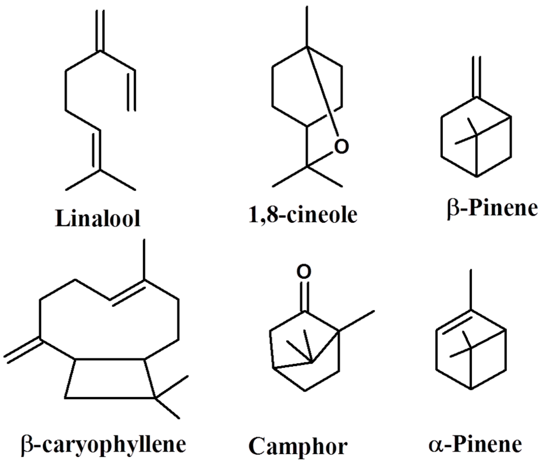
2.1. Analysis of Volatile Components in REO

Any essential oil is composed of a mixture of different oxygenated and non-oxygenated hydrocarbons, from which some components dominate. Volatile components from R. officinalis leaves were separated and identified using GC and GC/MS, and their relative abundance is listed according to their retention indices in Table 1, Figure 1. The extraction yield of REO was 1.8% v/fresh weight, in which thirteen components (with average composition of >1%) were identified, representing 94.06% of total oil components, with monoterpene hydrocarbons as the major constituents of the oil (90.04%). Importantly, 1,8-cineole (51.52%) constitutes the major component of REO and together with camphor (10.52%) and α-pinene (8.41%), they represent the most abundant monoterpenes (Table 1). Oxygenated monoterpenes have relatively higher concentrations than non-oxygenated monoterpenes hydrocarbons, representing 69.75% of the total oil components. Sesquiterpenes are shown in relatively low levels in the oil by the presence of β-caryophyllene (4.57%). Most non-identified components are present as traces with less than 0.01% relative abundances. These results are in accordance with other reports published on the composition of rosemary oil elsewhere [43], especially with supercritical fluid extracted rosemary oil from Egypt [44].

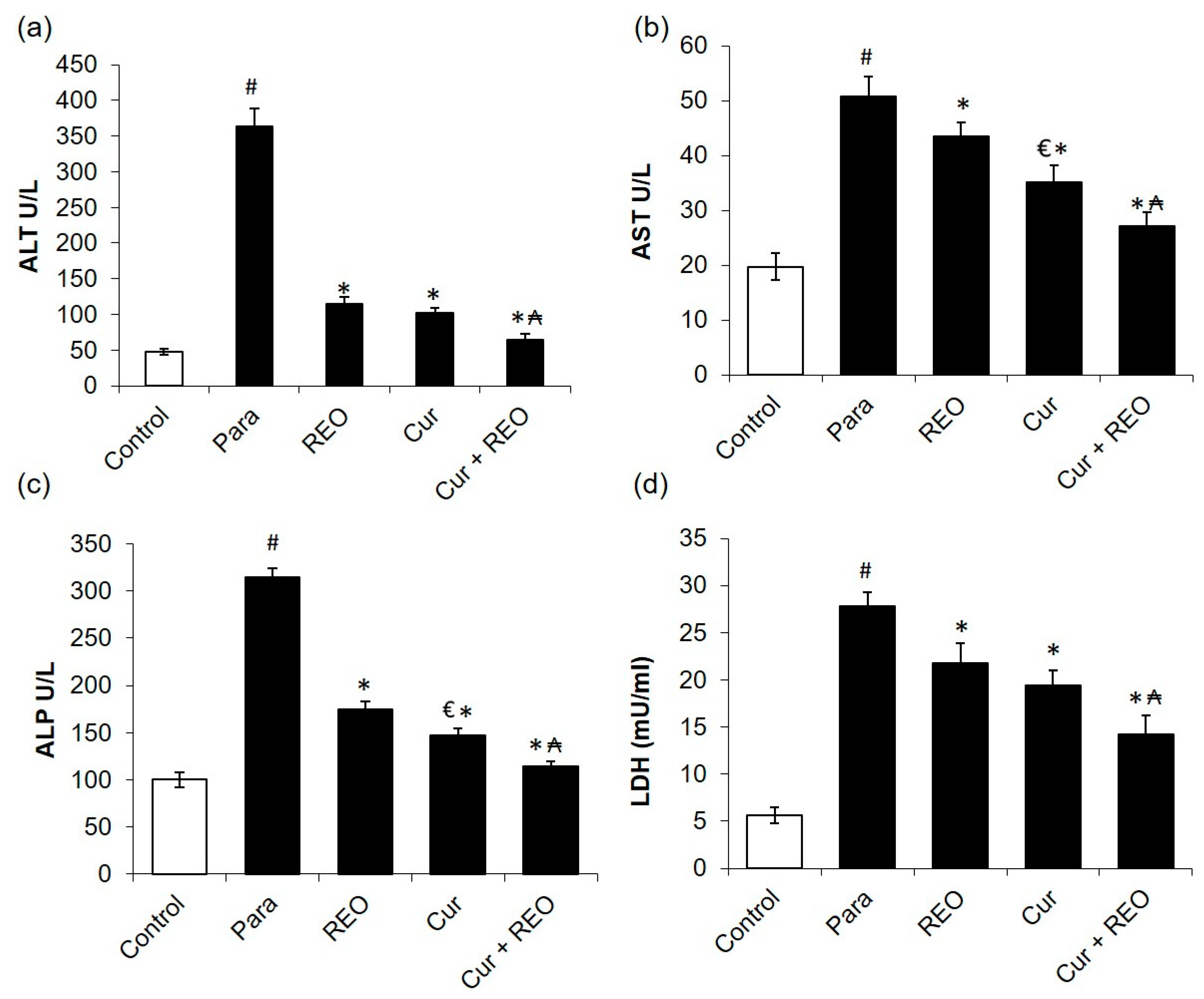
2.2. The Protective Effects of Cur and REO on Para-Induced Liver Injury

In order to confirm the induction of liver injury-induced Para, serum ALT, AST, ALP, and LDH activities were assessed. Para significantly elevated the levels of serum ALT, AST, ALP, and LDH by 7.6-, 3.1-, 2.5-, and 4.9-folds (p < 0.05), respectively, in comparison to the normal group (Figure 2), signifying the damage of hepatocytes and the successful induction of liver injury. The pre-treatment of animals with Cur or REO significantly attenuated ALT, AST, ALP, and LDH levels (p < 0.05) compared to the Para untreated group (Figure 2). There is no significant difference between the effect of both Cur and REO on the serum of ALT and LDH, whereas Cur alone causes a significant reduction in ALP and AST compared to REO alone (Figure 2). However, the combination of both natural drugs causes a further reduction of serum levels of ALT, AST, ALP, and LDH when related to Cur, REO, and Para groups (Figure 2).

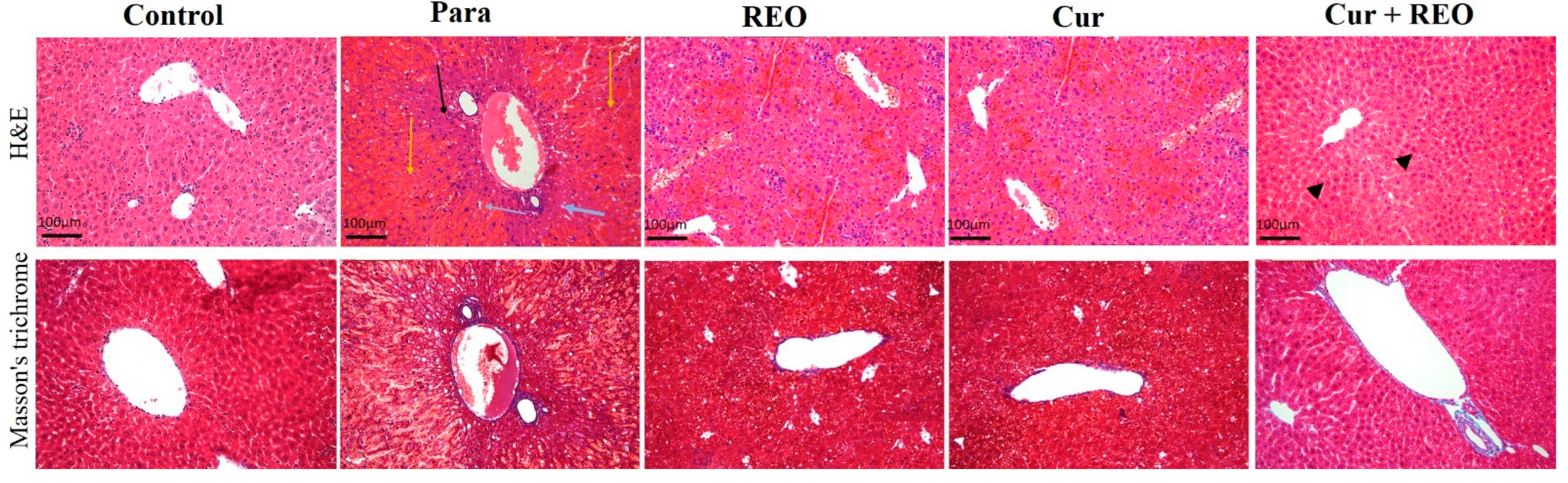
2.3. Protective Effect of Cur and REO on Para-Induced Histopathological Changes

Figure 3 demonstrates that mice hepatic tissue stained with H&E showed regular architectures of hepatic parenchyma in the control group. The Para group showed massive necrosis (yellow arrows) with a few zones of surviving hepatocytes around the central vein (black arrow), mild hydropic degeneration (blue arrows), central vein, and hepatic sinusoid congestion and hemorrhage. The Cur and REO groups exhibited less necrosis with better-surviving hepatocytes around the central vein, fewer hydropic degeneration around the central vein, and hepatic sinusoid congestion and hemorrhage. The combination group restored hepatic damage characterized by binucleated hepatocytes (head arrows) with mild congestion.

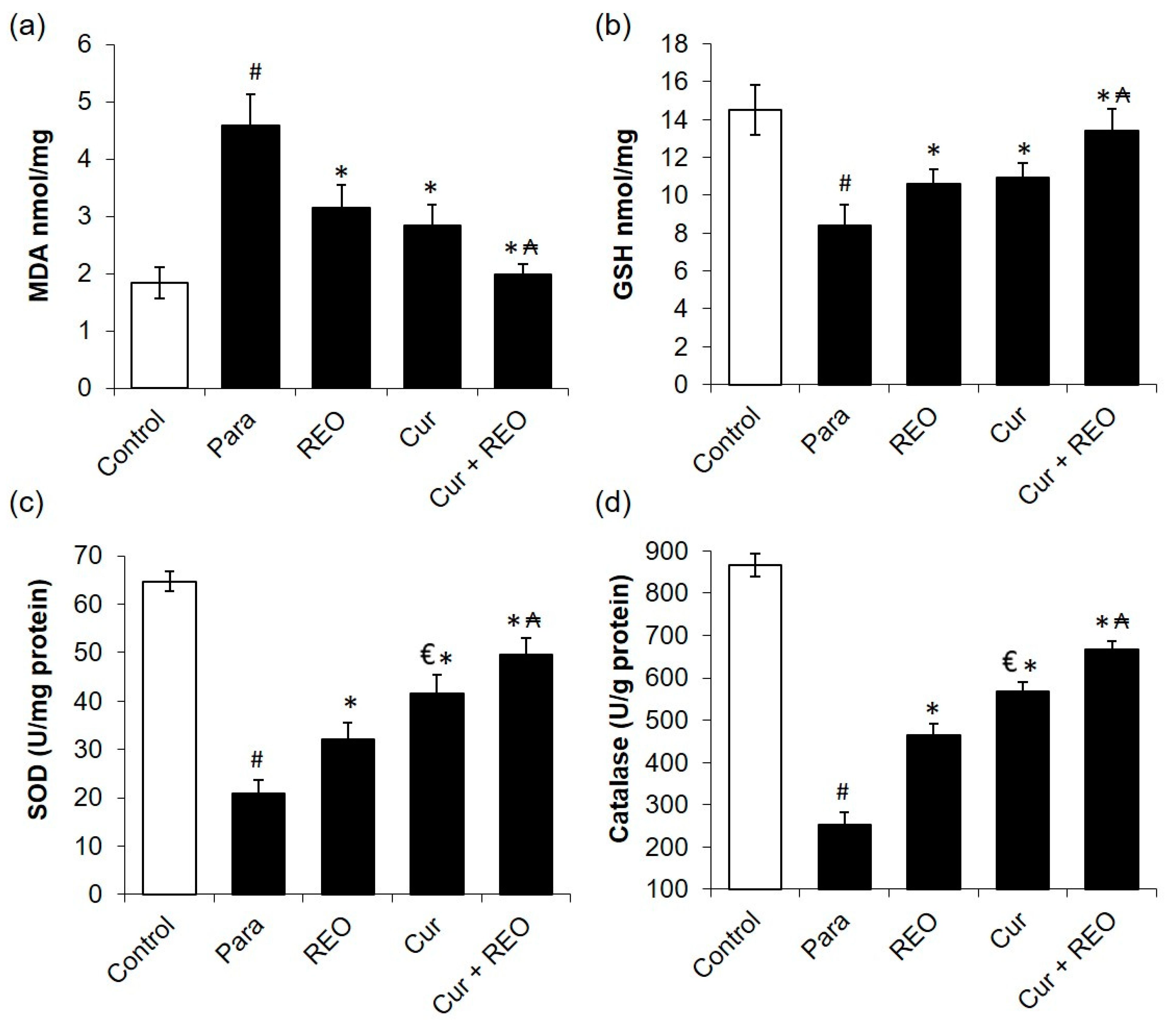
2.4. Protective Effect of Cur and REO on Para-Induced Oxidative Stress and Antioxidant Enzymes Activity

Cur and REO’s antioxidant potential were confirmed by determining MDA, GSH, SOD, and CAT levels in liver homogenates as biomarkers of oxidative stress and antioxidant enzymes. Consumption of Para-induced a 2.48-fold increase in MDA level compared to the Para alone group, suggesting the oxidative damage of cell membranes. This was accompanied by a significant reduction of GSH concentration, reaching 8.40 ± 1.1 nmol GSH/mg versus 14.52 ± 1.33 nmol GSH/mg, SOD reaching 20.82 ± 2.76 versus 64.74 ± 2.12 U/mg protein, and CAT reaching 252.78 ± 30.19 versus 866.00 ± 27.44 U/g protein compared to Para-induced hepatic injury group. Pre-treatment with Cur alone significantly reduced MDA (p < 0.05) levels as well as elevated GSH SOD and CAT levels (Figure 4). Interestingly, pretreatment with combined Cur and REO significantly (p < 0.001) reversed the oxidative stress-related biochemical parameters in liver homogenates and normalized MDA and GSH, as well as increased SOD and CAT significantly compared to each Cur or REO alone.

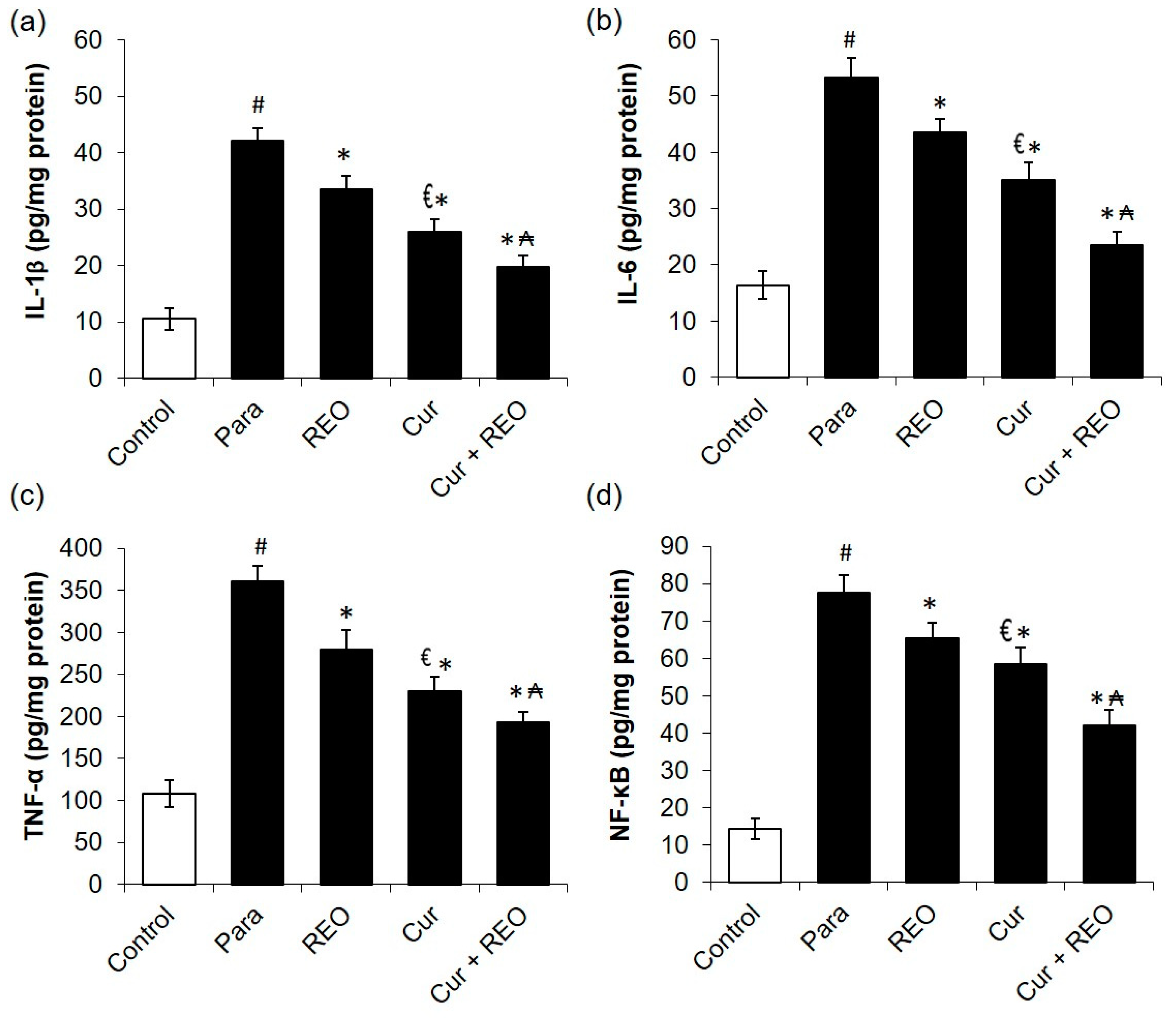
2.5. Protective Effect of Cur and REO on Para-Induced Inflammation

The anti-inflammatory potential of Cur and REO was confirmed through the measurement of numerous inflammatory cytokines biomarkers, including IL-1β, IL-6, TNF-α and NF-κB levels in liver homogenates. Para consumption intensified IL-1β, IL-6, TNF-α and NF-κB levels by 3.9-, 3-, 3.3-, and 5.4-fold increase compared to the normal group, suggesting severe hepatic inflammation status. Pre-treatment with Cur or REO significantly reduced IL-1β, IL-6,TNF-α, and levels of NF-κB hepatic levels (Figure 5). Remarkably, pretreatment with the combination of Cur and Reo treatment significantly reduced IL-1β, IL-6, TNF-α and NF-κB hepatic levels more than each one alone, signifying a reversal of the inflammation associated with Para liver toxicity.

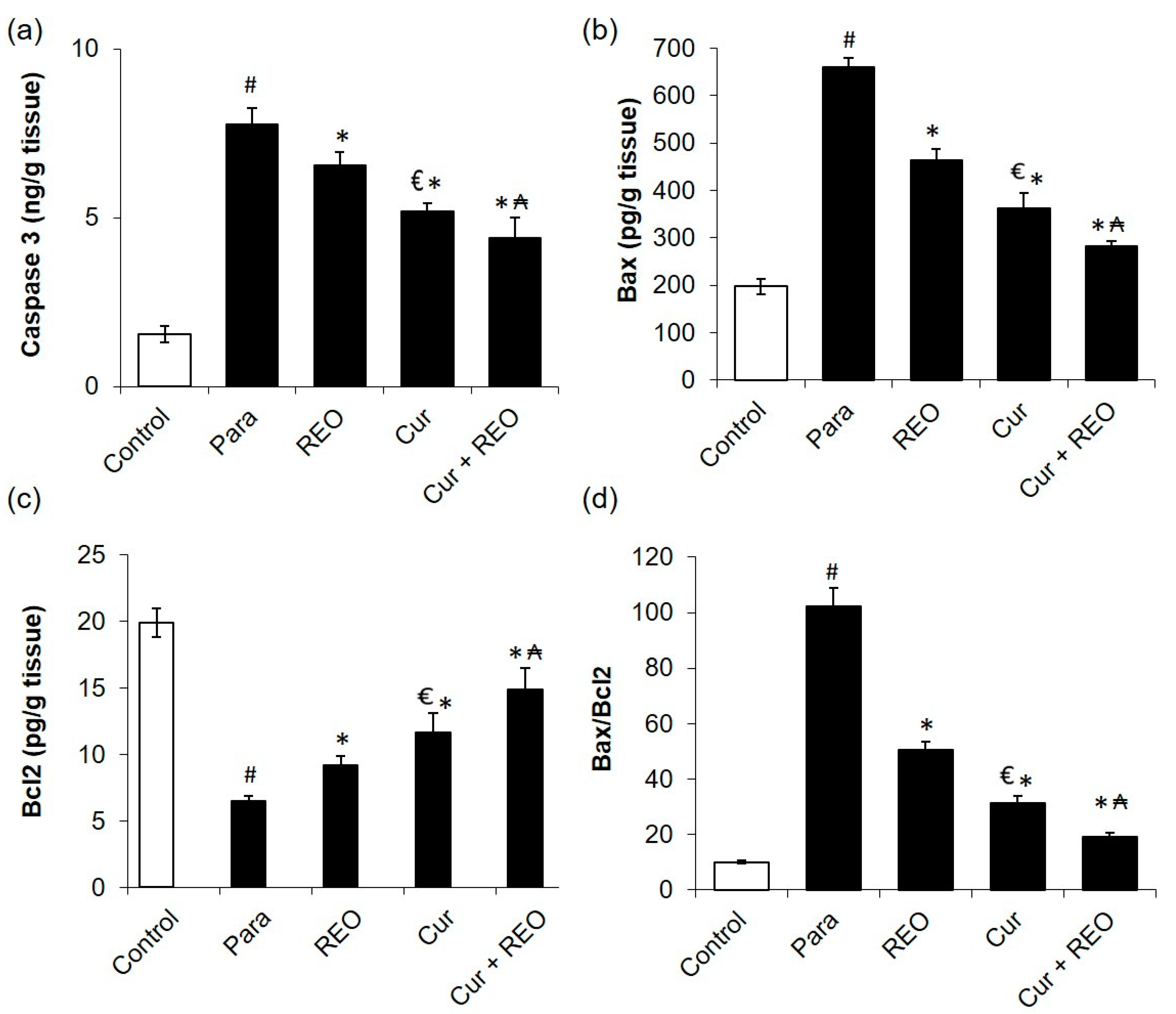
2.6. Protective Effect of Cur and REO on Para-Induced Apoptosis

The anti-apoptotic prospective of Cur and REO and their combination were studied through the measurement of apoptotic markers including caspase-3, Bcl2, Bax and Bax/Bcl2 ratio in liver homogenates. Para-induced hepatic injury was associated with apoptosis intensification as demonstrated by the intensified caspase-3, Bax as well as the ration of Bax/Bcl2 ratio compared to the normal group, suggesting severe hepatic apoptosis state. Pre-treatment with Cur or REO significantly reduced caspase-3, Bcl2, Bax and levels of Bax/Bcl2 ratio hepatic levels (Figure 6). Pretreatment with the combination of Cur and REO treatment significantly reduced caspase-3, Bcl2, Bax and levels of Bax/Bcl2 ratio hepatic levels more than each one alone, signifying deterrence of the apoptosis intensification accompanying the Para hepatic toxicity.

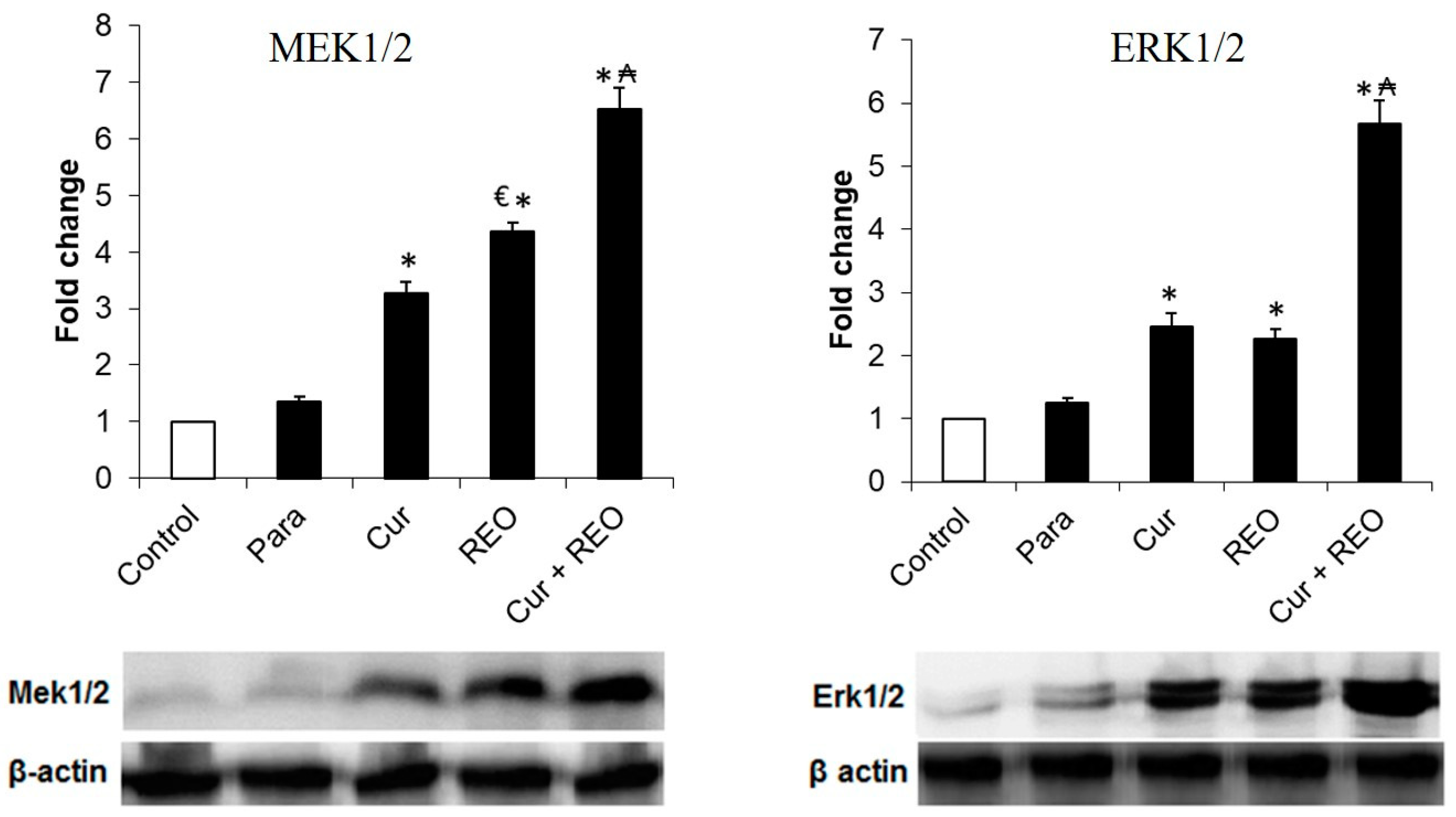
2.7. Effect of Cur and REO on MEK1 Expression

To investigate whether the MEK pathway is involved in the mechanism of hepatoprotective effect for both Cur and REO, the level of total MEK1 expression in liver homogenates was inspected. Pretreatment with Cur or REO induced the over-expression of MEK1 by 3.27- and 4.37-folds, respectively, in comparison to the control. On the other side, pretreatment with the combination of Cur and REO resulted in a 6.53-fold increase in expression of MEK1 in comparison to the control group (Figure 7).

2.8. Effect of Cur and REO on ERK1 Expression

To reveal the effect of the above observed over-expression of MEK1 by Cur and REO on ERK1, the total ERK1 protein expression in the liver homogenate was determined. The western blot analysis revealed that the administration of Cur and REO resulted in 2.06- and 1.94-folds increase in the total ERK1 protein level compared to the control group, respectively (Figure 7). Interestingly, pretreatment with the combined Cur and REO resulted in 5.85-folds increase in ERK1 expression in comparison to the control group (Figure 7).

2.9. High-Performance Liquid Chromatography (HPLC) Analysis of Cur in the Presence and Absence of REO

Blood samples were taken from the different animal groups under investigation to determine the effect of REO on the plasma concentrations of Cur. The results indicated low concentrations of Cur in the plasma of animals administrated Cur alone, although the drug has been given to the animals for 10 successive days. When both Cur and REO were given to the animals, the Cur plasma concentration increased by 14.6-fold more than Cur alone, Figure 8.

3. Discussion

Many studies were carried out to emphasize the hepatoprotective activity of many natural products; however, only a few studies were conducted for the rationale of disclosing the mechanism of action. REO and Cur are both distinguished for their hepatoprotective activity on many models of liver injury, yet, the precise mechanisms of action of these natural products are still unclear. The study hereby aims to reveal part of the mechanism of action of Cur, REO, and their synergistic combination through investigating their effect on the MEK/ERK signaling pathway.
Para consumption resulted in hepatocellular damage represented by high levels of serum liver enzymes; ALT, AST, ALP, and LDH. The liver protection effect of Cur and REO was clearly disclosed by the significant decline in serum liver enzymes ALT, AST, ALP, and LDH levels. Cur hepatoprotection activity was documented against numerous liver disorders, for instance, nonalcoholic fatty liver disease [45], aflatoxin B1-induced liver injury [46], and CCl4-induced liver fibrosis [47], among other models. Furthermore, the hepatoprotective potential of REO is verified against carbon tetrachloride [35] and hexavalent chromium-induced [48] liver injury. Remarkably, the combination of Cur and REO exhibited superior outcomes than each one alone, indicating a synergistic hepatoprotective effect.
Meanwhile, Para induced a dramatic increase in lipid peroxidation, reflected by the elevated level of MDA, diminished GSH level, and SOD and CAT activities in the hepatic homogenate. On the other hand, pretreatment with Cur and REO caused a significant drop in the level of MDA concomitant with an increased level of GSH and SOD, and CAT activities. These results are in accordance with the published data on the antioxidant effect of Cur [13,14] and REO [36,44,49] elsewhere. The combination of Cur and REO displayed better results than each one alone, indicating synergistic antioxidant activities and lipid peroxidation mitigation against hepatocellular injury induced by Para.
In the current study, Para consumption intensified inflammatory cytokines biomarkers, including IL-1β, IL-6, TNF-α and NF-κB levels, and apoptosis indicators, including caspase -3, Bax and Bax/Bcl2 suggesting severe hepatic inflammation and apoptosis, as similarly mentioned in previous studies [50,51]. On the other hand, the anti-inflammatory and anti-apoptotic potential of each of Cur and REO alone was confirmed through the reduction in IL-1β, IL-6, TNF-α, NF-κB, caspase -3, Bax and Bax/Bcl2 ratio levels. The anti-inflammatory and anti-apoptotic activity of Cur was documented in several extrahepatic [52,53,54] and hepatic diseases [1,12,55]. Likewise, REO presented anti-inflammatory and anti-apoptotic characteristics in various animal models [31,56]. Remarkably, pretreatment with combined Cur and REO treatment significantly reduced IL-1β, IL-6, TNF-α, NF-κB, caspase -3, Bax, and Bax/Bcl2 ratio levels more than each one alone, signifying the synergistic anti-inflammatory and anti-apoptotic effects.
Several mechanisms have been reported for Cur as an anti-inflammatory, cytoprotective, and antitumor agent. The reported mechanisms depended on the suppression of pro-inflammatory mediators [57], induction of HSP-70 that has cytoprotective effects, inhibition of HSP-90 that has anti-proliferative and pro-apoptotic activities [58,59], or induction of HSP-30 that has protection from oxidative stress [60]. The hepatoprotective activity of REO against carbon tetrachloride [35], cyclophosphamide [61], and hexavalent chromium-induced [48] liver injury was established; still, the mechanism of action was not identified. Thus, in the current study, we tried to propose a mechanism of action through which both Cur and REO may act to achieve this hepatoprotective effect.
ERK is attached to MEK in the cytoplasm [62]. Upon stimulation, ERK1/2 dissociates from MEK1/2 [63]. It was reported that ERK activation may exert an anti-apoptotic [60] and has a protective effect of glutamate treatment via restoration of the glutathione levels [64]. It also has a shielding effect against hydrogen peroxide-induced apoptosis in the cardio-myoblast [65]. Other cellular action of ERK1/2 pathway activation includes its cytoprotective effect against oxidative stress [66]. ERK1/2 has a signaling role in the regulation of proliferation during the liver regeneration process [67] and protects renal epithelial cells against oxidative stress-induced damage [68].
The results of the current study illustrate the ameliorative effect of the combination of Cur and REO pretreatment on the expression of both MEK1 and ERK1 proteins. Para Induced a slight but non-significant increase in the level of these proteins, especially in ERK expression, when compared to the untreated group, and this elevation can be explained by the necessity of cells to counter the Para toxic effect. The combination of both natural components has potentiated the liver for the production of a massive amount of these proteins that can play important role in apoptosis regulation and the protective effect from Para-induced liver cell damage. Accordingly, the hepatoprotective effect of Cur and REO was proven to involve the Ras/Raf/MEK/ERK cascade in the current study. This cascade has a critical role in coordinating cell survival, cell cycle progression, and differentiation in response to various signals from different receptor tyrosine kinases and other cell surface receptors [69]. The decontrolled Ras/Raf/MEK/ERK signaling pathway is an essential therapeutic target in many epithelial cancers [40]. Additionally, other signal transduction pathways interact with the Raf/MEK/ERK pathway to positively or negatively control its activity or to adjust the phosphorylation status of downstream targets [70].
Despite its efficacy and safety, Cur has not been widely used as a therapeutic agent, mainly for problems related to its bioavailability [1] and solubility [71]. The pharmacokinetic studies of Cur have exhibited poor absorption, fast metabolism, and elimination of Cur as major reasons for its poor bioavailability [15]. This problem has been the target of many research projects, which may be overcome through several approaches [72]. The use of compounds that can hinder the metabolic pathways of Cur is one of the major tactics implemented to improve Cur bioavailability. An example of these metabolic blockers is piperine, which produced an increase in the bioavailability of Cur in humans by 2000% when both drugs were co-administrated [73]. Other synergistically acting drugs with Cur included quercetin [74], genistein [75], and epigallocatechin gallate (a green tea component) [76]. Pharmaceutical formulation into nanoparticles, liposomes, or micelles, solved, in part, the problem of Cur bioavailability [15]. A microemulsion system of Cur, which consists of an oil (Capryol 90) and a surfactant (Cremophor RH40), increased the relative absorption of Cur in rats by 22.6-fold [77]. The inclusion of Cur in a lipophilic matrix has increased the relative human absorption of Cur by 19.2-fold [78]. The approach of using co-administered essential oils was also implemented to affect Cur’s bioavailability. Eugenol and terpineol, both essential oil components, were used as enhancers for Cur penetration to the skin [79]. Comparing the production of essential oils in underground parts to the aerial parts of plants reveals that they are rarely produced under the soil level. However, turmeric, the plant which produces Cur in its rhizomes, is known for the presence of essential oil in its underground parts [80,81]. Turmeric rhizome essential oil is highly oxygenated; the major principles in this oil belong to the sesquiterpene turmerone derivatives (40%) [81]. The combination of curcuminoids and volatile oils of turmeric rhizome resulted in a 6.9-fold increase in human absorption of Cur [82]. The patent number US8153172B2 for the United States of America patent office (USAPO) discussed “A composition having a curcuminoid and an essential oil of turmeric, wherein the essential oil is present in an amount sufficient to cause an enhancement of bioavailability of curcumin”. However, the approach of using essential oil as enhancers for Cur bioavailability is still uncommon and needs development. In the current study, the administration of REO with Cur resulted in a significant increase in Cur plasma concentration. This effect could be a result of enhanced absorption (i.e., the essential oil solubilized Cur), or due to hindering in metabolism or elimination of the compound. The presence of the oxygenated hydrocarbons in the REO (Table 1) can act as hydrophilic carriers for Cur, thus enhancing its absorption and bioavailability. The percentage of oxygenated compounds in REO is similar to that of turmeric essential oil and can act in a similar way in increasing the absorption rate of Cur. This study is considered an initial investigation of the REO effect on Cur and should be followed by a whole pharmacokinetic study to investigate, in-depth, the effect of the oil on Cur bioavailability.

4. Materials and Methods

4.1. Plant Material

The fresh healthy leaves of rosemary (R. officinalis L.) were collected from the Medicinal Plants Experimental Garden of the Faculty of Pharmacy, Zagazig University, Egypt, in March 2015. The identity of the plants was confirmed by Dr. A. H. Abdel-Baset, plant taxonomist, Egyptian Agricultural museum, Cairo, Egypt. Voucher specimens were deposited in the herbarium of the Department of Pharmacognosy, Faculty of Pharmacy, Zagazig University, Egypt, under number L236.

4.2. Isolation of REO

The fresh R. officinalis leaves (100 g) were cut into small pieces (approximately 1 cm in length) and immediately subjected to hydrodistillation using a Clevenger-type apparatus for 3 h [83]. The volatile fraction (yield: 1.8% v/fresh weight) was recovered by decantation and dried over anhydrous sodium sulphate. The essential oil samples were kept in brown vials in the refrigerator at 4 °C until the commencement of analyses.

4.3. Gas Chromatography Analysis

Gas chromatography/flame ionization (GC/FID) analysis was done using a GC-2010 Plus gas chromatograph (Shimadzu Corporation, Kyoto, Japan) equipped with an FID-2010 Plus detector. The following conditions were applied: column, RTX-5MS®fused silica capillary (30 m × 0.25 mm i.d and 0.25 μm film thickness); carrier gas He (2 mL/min); temperature 300 °C, injection temperature 250 °C; oven temperature program: initial temperature 45 °C, 2 min isothermal, 300 °C, 4 °C/1 min, then 20 min isothermal; the split ratio was 1:15, the injection volume is 5 μL. Gas chromatography/mass spectrometry (GC/MS) data were recorded on a GCMS-QP2010 Plus, Shimadzu Corporation, Kyoto, Japan. The ionization energy for the mass spectrometer was 70 eV. The split ratio was 1:30; other conditions were identical to those mentioned for GC/FID.
Kovat’s retention indices (RI) were calculated with respect to a set of co-injected standard hydrocarbons (C10-C28). Compounds were identified by comparing their spectral data and retention indices with the Wiley Registry of Mass Spectral Data, 10th edition (April 2013), the NIST 11 Mass Spectral Library (NIST11/2011/EPA/NIH), and data from the literature [84].

4.4. Animals and Ethical Approval

Thirty albino mice weighing (20 ± 5 gm) were obtained from the College of Veterinary Medicine, King Faisal University, Al-Hasa, KSA. The experimental protocol was permitted by the Institutional Animal Care and Use Committee of King Faisal University (approval number KFU-REC-2022-OCT-ETHICS212). All the experiments were executed in harmony with the relevant procedures and regulations of the Ethical Conduct for the Use of Animals in Research at King Faisal University. The animals were housed in well-ventilated large spacious polypropylene cages and had 12 h light/dark cycles throughout the experimental period. All animals received water and rodent standard pellets during the experiment.

4.5. Experimental Protocol

Mice were divided into five groups (n = 6). Group 1 is the control group, and group 2 represents the Para-induced hepatotoxicity group (Para). Animals in the control and the Para groups received normal saline for ten days. In the third group (Cur), mice were administered Cur (20 mg/kg) once daily orally by gavage for ten days. In the fourth group (REO), mice were given REO (20 mg/kg) [27] once a day orally by gavage for ten days. The fifth group represented the combination group (Cur + REO) in which mice were administered both REO (10 mg/kg) and Cur (10 mg/kg) orally once daily for ten days. On the last day of the experiment, liver toxicity was induced in fasted animals in groups 2, 3, 4, and 5 by giving Para (450 mg/kg) IP [85]. The mice were anesthetized and sacrificed 3 h after Para injection.

4.6. Blood and Tissue Collection and Processing

Blood samples were collected by retro-orbital sinus puncture using capillary tubes under diethyl ether anesthesia from all animals. Collected blood samples were left to clot for 10 min at room temperature and centrifuged at 2000 rpm for 15 min at 4 °C to obtain the serum. Liver homogenates (10%) were prepared on ice with ice-cold Tris-Hcl (10 mM, pH 7.4) in the presence of protease inhibitors. The resulting suspension was centrifuged at 10,000 rpm for 10 min, and the supernatants were collected for further analysis.

4.7. Histopathological Investigation

For hepatic histopathological assessment, a portion of mouse hepatic tissues was fixed with 10% formalin and processed; sections (4 μm) were obtained and stained with hematoxylin and eosin (H&E). Digital images were collected under a light microscope at 100× magnification. The histological changes were quantified as normal, moderate, and severe based on the hepatic cytoplasm inflammation, centrilobular necrosis, cellular hypertrophy, vacuolization, and steatosis [86]. The histopathological changes were quantified by two independent histopathologists and scored double blindly.

4.8. Hepatic Function Tests Determination

Colorimetric kits were used to determine the serum level of the hepatic function enzymes, including alanine aminotransferase (ALT), aspartate aminotransferase (AST), alkaline phosphatase (ALP) and lactate dehydrogenase (LDH) following the manufacturer’s instructions.

4.9. Hepatic Oxidative Stress Status Determination

A Malondialdehyde (MDA; ab238537) ELISA kit and GSH Assay Kit (ab239727) Colorimetric kit were acquired from Abcam Inc. (Cambridge, UK). Superoxide dismutase (SOD; MBS036924) and catalase (MBS726781) ELISA kits were obtained from My BioSource (San Diego, CA, USA). All the procedures were executed in agreement with the manufacturer’s directions.

4.10. Determination of Inflammation and Apoptotic Signaling Markers

Inflammation markers, including TNF-α (ab46070), IL-1β (ab100768), IL-6 (ab100772), and NF-κB (ab176648) ELISA kits were obtained from Abcam Co. (Eugene, OR, USA). As for the apoptotic signaling markers, cleaved caspase-3 (KHO1091) was purchased from Thermo Fisher Scientific Inc. (Waltham, MA, USA), whereas Bcl2 (MBS2881713) and Bax (MBS935667) ELISA kits were acquired from My BioSource (San Diego, CA, USA). These markers were measured according to the manufacturer’s instructions using the microplate reader SpectraMax i3X (Molecular devices San Jose, CA, USA).

4.11. Western Blot Analysis

Western blot analysis was conducted as described elsewhere [87]. Briefly, the liver protein homogenate was separated by sodium dodecyl sulfate gel electrophoresis and transferred to a PVDF membrane. The membrane was blocked by incubation in Tris-buffered saline (TBS) containing 3% bovine serum albumin for 1 h at room temperature. After washing with TBS containing 0.1% Tween-20, the membranes were incubated with primary antibodies for 2 h at room temperature and then incubated with the secondary antibody (HRP-conjugated antibodies). The chemiluminescence produced from the luminol reagent was detected with LI-COR C-DiGit Chemiluminescence Scanner.

4.12. HPLC Determination of Cur in Serum

The sample preparation and HPLC separation were conducted according to Jäger et al. [88]. Blood samples, collected after Para administration for 3 h, were centrifuged (2000× g) for 10 min to separate plasma. A measured 0.2 mL of plasma was mixed with 100 μL of β-Glucuronidase/Arylsulfatase (1000 U, Merck, 10127698001 St. Louis, MO, USA) in phosphate buffer (0.1 M, pH 6.80) and incubated (37 °C, 1 h). After incubation, the solution was mixed with 1 mL of ethyl acetate, vortexed (1 min), sonicated (15 min), and then centrifuged (15,000× g, 6 min). The ethyl acetate layer was then evaporated at 30 °C under reduced pressure. The ethyl acetate extraction was repeated three times for each sample. The residue was dissolved in 100 μL of methanol.
Natural Cur (extracted from Curcuma longa, family Zingiberaceae) was purchased from Merck (product number C1386, St. Louis, MO, USA). The stock solution of Cur in methanol (100 μg/mL) was prepared, from which a serial dilution (50 pg/mL to 10 μg/mL) was spiked into the plasma of normal control animals. A prepared 50 ng/mL solution of salbutamol was used as an internal standard. A five-point calibration curve was established by plotting the peak area ratio of Cur serial dilutions to internal standard versus the Cur concentration, from which regression equations were determined to calculate the concentration of Cur.
Prepared samples and standard Cur were injected into a Shimadzu Prominence HPLC system provided with a CBM-20A controller, LC-20A solvent unit, SIL-20A auto-sampler, CTO-20A column oven, and SPD-20A UV-VIS detector. Separation was conducted on Luna-C18 (L1, Phenomenex, 150 mm × 4.6 mm × 5 μm). The mobile phase was a combination of acetonitrile-methanol-water (40:20:40, v/v) in isocratic mode. The flow rate was adjusted at 1 mL/min, and the UV detector was adjusted to 360 nm.

4.13. Statistical Analysis

Data were expressed as mean ± standard deviation (SD) for five animals in each group. Statistically significant differences between the groups were determined using one-way ANOVA followed by Tukey’s multiple comparison test. In all cases, probability values of p < 0.05 were taken as statistically significant.

5. Conclusions

REO and Cur are both natural products of high value due to their medicinally and pharmacologically proven activities, including their hepatoprotective actions. However, the mechanism through which these remedies can act still needs to be determined. The hereby study sheds light on these mechanisms by investigating these natural products’ effect on the MEK/ERK signaling cascade. The components of REO were analyzed using GC, accompanied by FID and MS detectors. The analysis indicated the abundance of monoterpenes in the essential oil, particularly 1,8-cineole, camphor, and α-pinene. Both REO and Cur modulated the levels of serum liver enzymes: ALT and ALP; lipid peroxidation proteins; and MDA and GSH, in liver homogenate. Pretreatment with REO and Cur demonstrated an over-expression of both MEK1 and ERK1 proteins, indicating the contribution of these natural products in the Raf/Ras/MEK/ERK flow. The two natural remedies exhibited all the above solitary actions, and their activity was amplified after combination, maybe due to the synergistic effect of REO, a hepatoprotective agent itself, or through enhancing Cur bioavailability. The enhanced plasma concentration of Cur after the co-administration of REO was proven in this study; however, further studies are required to study the essential oil’s whole effect on Cur’s pharmacokinetics. Future exploration of the impact of the major contents of the REO (1,8-cineole, camphor, and α-pinene) along with Cur is also recommended as a result of this study.

Author Contributions

Conceptualization, M.E.M. and O.M.M.; methodology, M.E.M., N.S.Y. and O.M.M.; software, M.E.M., N.S.Y. and O.M.M.; formal analysis, M.E.M., N.S.Y. and O.M.M.; investigation, M.E.M., and O.M.M.; writing—original draft preparation, M.E.M., N.S.Y. and O.M.M.; writing—review and editing, M.E.M., N.S.Y. and O.M.M.; supervision, M.E.M., N.S.Y., H.S.E.-B. and O.M.M.; funding acquisition, M.E.M. and H.S.E.-B. All authors have read and agreed to the published version of the manuscript.

Funding

This research was funded by the Deputyship for Research and Innovation, the Ministry of Education in Saudi Arabia, project number [INST011].

Institutional Review Board Statement

The experimental protocol was permitted by the Institutional Animal Care and Use Committee of King Faisal University (approval number KFU-REC-2022-OCT-ETHICS212). All the experiments were executed in harmony with the relevant procedures and regulations of the Ethical Conduct for the Use of Animals in Research at King Faisal University.

Data Availability Statement

Not applicable.

Acknowledgments

The authors acknowledge all the effort exerted by Tamim Alyahian in preparations, assistance, and support in the experimental part of this study.

Conflicts of Interest

The authors declare no conflict of interest.

References

  1. Jurenka, J.S. Anti-inflammatory properties of curcumin, a major constituent of Curcuma longa: A review of preclinical and clinical research. Altern. Med. Rev. J. Clin. Ther. 2009, 14, 141–153. [Google Scholar]
  2. Sharma, O.P. Antioxidant activity of curcumin and related compounds. Biochem. Pharmacol. 1976, 25, 1811–1812. [Google Scholar] [CrossRef] [PubMed]
  3. Kim, M.K.; Choi, G.J.; Lee, H.S. Fungicidal property of Curcuma longa L. rhizome-derived curcumin against phytopathogenic fungi in a greenhouse. J. Agric. Food Chem. 2003, 51, 1578–1581. [Google Scholar] [CrossRef] [PubMed]
  4. Reddy, R.C.; Vatsala, P.G.; Keshamouni, V.G.; Padmanaban, G.; Rangarajan, P.N. Curcumin for malaria therapy. Biochem. Biophys. Res. Commun. 2005, 326, 472–474. [Google Scholar] [CrossRef] [PubMed]
  5. Aggarwal, B.B.; Kumar, A.; Bharti, A.C. Anticancer potential of curcumin: Preclinical and clinical studies. Anticancer. Res. 2003, 23, 363–398. [Google Scholar]
  6. Srivastava, R.; Dikshit, M.; Srimal, R.; Dhawan, B. Anti-thrombotic effect of curcumin. Thromb. Res. 1985, 40, 413–417. [Google Scholar] [CrossRef]
  7. Babu, P.S.; Srinivasan, K. Hypolipidemic action of curcumin, the active principle of turmeric (Curcuma longa) in streptozotocin induced diabetic rats. Mol. Cell. Biochem. 1997, 166, 169–175. [Google Scholar] [CrossRef]
  8. Srinivasan, M. Effect of curcumin on blood sugar as seen in a diabetic subject. Indian J. Med. Sci. 1972, 26, 269–270. [Google Scholar]
  9. Deodhar, S.D.; Sethi, R.; Srimal, R.C. Preliminary study on antirheumatic activity of curcumin (diferuloyl methane). Indian J. Med. Res. 1980, 71, 632–634. [Google Scholar]
  10. Nirmala, C.; Puvanakrishnan, R. Protective role of curcumin against isoproterenol induced myocardial infarction in rats. Mol. Cell. Biochem. 1996, 159, 85–93. [Google Scholar] [CrossRef]
  11. Chen, N.; Geng, Q.; Zheng, J.; He, S.; Huo, X.; Sun, X. Suppression of the TGF-β/Smad signaling pathway and inhibition of hepatic stellate cell proliferation play a role in the hepatoprotective effects of curcumin against alcohol-induced hepatic fibrosis. Int. J. Mol. Med. 2014, 34, 1110–1116. [Google Scholar] [CrossRef] [PubMed] [Green Version]
  12. El-Agamy, D.S. Comparative effects of curcumin and resveratrol on aflatoxin B(1)-induced liver injury in rats. Arch. Toxicol. 2010, 84, 389–396. [Google Scholar] [CrossRef] [PubMed]
  13. García-Niño, W.R.; Pedraza-Chaverrí, J. Protective effect of curcumin against heavy metals-induced liver damage. Food Chem. Toxicol. 2014, 69, 182–201. [Google Scholar] [CrossRef] [PubMed]
  14. Girish, C.; Koner, B.C.; Jayanthi, S.; Rao, K.R.; Rajesh, B.; Pradhan, S.C. Hepatoprotective activity of picroliv, curcumin and ellagic acid compared to silymarin on paracetamol induced liver toxicity in mice. Fundam. Clin. Pharmacol. 2009, 23, 735–745. [Google Scholar] [CrossRef]
  15. Anand, P.; Kunnumakkara, A.B.; Newman, R.A.; Aggarwal, B.B. Bioavailability of curcumin: Problems and promises. Mol. Pharm. 2007, 4, 807–818. [Google Scholar] [CrossRef]
  16. Anjana, D.; Nair, K.; Somashekara, N.; Venkata, M.; Ravichandran, S.; Yelucheri, R.; Parmar, H.; Upadhyay, R.; Verma, R.; Ramchand, C.; et al. Development of Curcumin Based Ophthalmic Formulation. Am. J. Infect. Dis. 2014, 8, 41–49. [Google Scholar]
  17. Scomoroscenco, C.; Teodorescu, M.; Burlacu, S.G.; Gîfu, I.C.; Mihaescu, C.I.; Petcu, C.; Raducan, A.; Oancea, P.; Cinteza, L.O. Synergistic Antioxidant Activity and Enhanced Stability of Curcumin Encapsulated in Vegetal Oil-Based Microemulsion and Gel Microemulsions. Antioxidants 2022, 11, 854. [Google Scholar] [CrossRef]
  18. Rahdar, A.; Hajinezhad, M.; Sargazi, S.; Zaboli, M.; Barani, M.; Baino, F.; Bilal, M.; Sanchooli, E. Biochemical, Ameliorative and Cytotoxic Effects of Newly Synthesized Curcumin Microemulsions: Evidence from In Vitro and In Vivo Studies. Nanomaterials 2021, 11, 817. [Google Scholar] [CrossRef]
  19. Vaz, G.; Clementino, A.; Bidone, J.; Villetti, M.; Falkembach, M.; Batista, M.; Barros, P.; Sonvico, F.; Dora, C. Curcumin and Quercetin-Loaded Nanoemulsions: Physicochemical Compatibility Study and Validation of a Simultaneous Quantification Method. Nanomaterials 2020, 10, 1650. [Google Scholar] [CrossRef]
  20. Adena, S.; Herneisey, M.; Pierce, E.; Hartmeier, P.; Adlakha, S.; Hosfeld, M.; Drennen, J.; Janjic, J. Quality by Design Methodology Applied to Process Optimization and Scale up of Curcumin Nanoemulsions Produced by Catastrophic Phase Inversion. Pharmaceutics 2021, 13, 880. [Google Scholar] [CrossRef]
  21. Chen, C.; Johnston, T.D.; Jeon, H.; Gedaly, R.; McHugh, P.P.; Burke, T.G.; Ranjan, D. An in vitro study of liposomal curcumin: Stability, toxicity and biological activity in human lymphocytes and Epstein-Barr virus-transformed human B-cells. Int. J. Pharm. 2009, 366, 133–139. [Google Scholar] [CrossRef] [PubMed]
  22. Li, S. Chemical Composition and Product Quality Control of Turmeric (Curcuma longa L.). Pharm. Crops 2011, 2, 28–54. [Google Scholar] [CrossRef]
  23. Jayaprakasha, G.K.; Jagan Mohan Rao, L.; Sakariah, K.K. Antioxidant activities of curcumin, demethoxycurcumin and bisdemethoxycurcumin. Food Chem. 2006, 98, 720–724. [Google Scholar] [CrossRef]
  24. Mahattanadul, S.; Panichayupakaranant, P.; Tungsinmonkong, K. Comparison of the inhibitory potency of curcumin, demethoxycurcumin and bisdemethoxycurcumin on iNOS-derived NO in activated macrophages and on gastric ulcer in rats. Planta Med. 2009, 75, PJ22. [Google Scholar] [CrossRef]
  25. Sandur, S.K.; Pandey, M.K.; Sung, B.; Ahn, K.S.; Murakami, A.; Sethi, G.; Limtrakul, P.; Badmaev, V.; Aggarwal, B.B. Curcumin, demethoxycurcumin, bisdemethoxycurcumin, tetrahydrocurcumin and turmerones differentially regulate anti-inflammatory and anti-proliferative responses through a ROS-independent mechanism. Carcinogenesis 2007, 28, 1765–1773. [Google Scholar] [CrossRef] [Green Version]
  26. Ali, I.; Haque, A.; Saleem, K. Separation and identification of curcuminoids in turmeric powder by HPLC using phenyl column. Anal. Methods 2014, 6, 2526–2536. [Google Scholar] [CrossRef]
  27. Al-Sereiti, M.R.; Abu-Amer, K.M.; Sena, P. Pharmacology of rosemary (Rosmarinus officinalis Linn.) and its therapeutic potentials. Indian J. Exp. Biol. 1999, 37, 124–130. [Google Scholar]
  28. Ngo, S.N.T.; Williams, D.; Head, R. Rosemary and Cancer Prevention: Preclinical Perspectives. Crit. Rev. Food Sci. Nutr. 2011, 51, 946–954. [Google Scholar] [CrossRef]
  29. European Medicines Agency. Community Herbal Monograph on Rosmarinus officinalis L., Folium; European Medicines Agency: London, UK, 2010.
  30. Machado, D.; Cunha, M.; Neis, V.; Balen, G.; Colla, A.; Bettio, L.; Oliveira, A.; Pazini, F.; Dalmarco, J.; Simionatto, E.; et al. Antidepressant-like effects of fractions, essential oil, carnosol and betulinic acid isolated from Rosmarinus officinalis L. Food Chem. 2013, 136, 999–1005. [Google Scholar] [CrossRef] [Green Version]
  31. Takaki, I.; Bersani-Amado, L.; Vendruscolo, A.; Sartoretto, S.; Diniz, S.; Bersani-Amado, C.; Cuman, R. Anti-inflammatory and antinociceptive effects of Rosmarinus officinalis L. essential oil in experimental animal models. J. Med. Food 2008, 11, 741–746. [Google Scholar] [CrossRef]
  32. Wang, W.; Li, N.; Luo, M.; Zu, Y.; Efferth, T. Antibacterial Activity and Anticancer Activity of Rosmarinus officinalis L. Essential Oil Compared to That of Its Main Components. Molecules 2012, 17, 2704–2713. [Google Scholar] [CrossRef] [PubMed] [Green Version]
  33. Sotelo-Félix, J.; Martinez-Fong, D.; Muriel, P.; Santillán, R.; Castillo, D.; Yahuaca, P. Evaluation of the effectiveness of Rosmarinus officinalis (Lamiaceae) in the alleviation of carbon tetrachloride-induced acute hepatotoxicity in the rat. J. Ethnopharmacol. 2002, 81, 145–154. [Google Scholar] [CrossRef]
  34. Amin, A.; Hamza, A. Hepatoprotective effects of Hibiscus, Rosmarinus and Salvia on azathioprine-induced toxicity in rats. Life Sci. 2005, 77, 266–278. [Google Scholar] [CrossRef]
  35. Rašković, A.; Milanović, I.; Pavlović, N.; Ćebović, T.; Vukmirović, S.; Mikov, M. Antioxidant activity of rosemary (Rosmarinus officinalis L.) essential oil and its hepatoprotective potential. BMC Complement. Altern. Med. 2014, 14, 225. [Google Scholar] [CrossRef] [PubMed] [Green Version]
  36. Pinho, R.J.D.; Aguiar, R.P.; Spironello, R.A.; Silva-Comar, F.M.D.S.; Silva-Filho, S.E.; Nogami, E.M.; Bersani-Amado, C.A.; Cuman, R.K.N. Hepatoprotective Effect of Pretreatment with Rosemary and Ginger Essential Oil in Experimental Model of Acetaminophen-induced Injury. Br. J. Pharm. Res. 2014, 4, 2126–2135. [Google Scholar] [CrossRef]
  37. Bélanger, L.-F.; Roy, S.; Tremblay, M.; Brott, B.; Steff, A.-M.; Mourad, W.; Hugo, P.; Erikson, R.; Charron, J. Mek2 Is Dispensable for Mouse Growth and Development. Mol. Cell. Biol. 2003, 23, 4778–4787. [Google Scholar] [CrossRef] [PubMed]
  38. Scholl, F.A.; Dumesic, P.A.; Khavari, P.A. Mek1 Alters Epidermal Growth and Differentiation. Cancer Res. 2004, 64, 6035–6040. [Google Scholar] [CrossRef] [Green Version]
  39. Bouhamdan, M.; Bauerfeld, C.; Talreja, J.; Beuret, L.; Charron, J.; Samavati, L. MEK1 dependent and independent ERK activation regulates IL-10 and IL-12 production in bone marrow derived macrophages. Cell. Signal. 2015, 27, 2068–2076. [Google Scholar] [CrossRef] [Green Version]
  40. Zmajkovicova, K.; Jesenberger, V.; Catalanotti, F.; Baumgartner, C.; Reyes, G.; Baccarini, M. MEK1 Is Required for PTEN Membrane Recruitment, AKT Regulation, and the Maintenance of Peripheral Tolerance. Mol. Cell 2013, 50, 43–55. [Google Scholar] [CrossRef] [Green Version]
  41. Cobellis, G.; Missero, C.; Di Lauro, R. Concomitant activation of MEK-1 and Rac-1 increases the proliferative potential of thyroid epithelial cells, without affecting their differentiation. Oncogene 1998, 17, 2047–2057. [Google Scholar] [CrossRef] [Green Version]
  42. Roux, P.P.; Blenis, J. ERK and p38 MAPK-Activated Protein Kinases: A Family of Protein Kinases with Diverse Biological Functions. Microbiol. Mol. Biol. Rev. 2004, 68, 320–344. [Google Scholar] [CrossRef] [PubMed] [Green Version]
  43. Jalali-Heravi, M.; Moazeni, R.S.; Sereshti, H. Analysis of Iranian rosemary essential oil: Application of gas chromatography–mass spectrometry combined with chemometrics. J. Chromatogr. A 2011, 1218, 2569–2576. [Google Scholar] [CrossRef] [PubMed]
  44. El-Ghorab, A.H. Supercritical Fluid Extraction of the Egyptian Rosemary (Rosmarinus officinalis) Leaves and NigellasativaL. Seeds Volatile Oils and Their Antioxidant Activities. J. Essent. Oil Bear. Plants 2003, 6, 67–77. [Google Scholar] [CrossRef]
  45. Lee, D.E.; Lee, S.J.; Kim, S.J.; Lee, H.-S.; Kwon, O.-S. Curcumin Ameliorates Nonalcoholic Fatty Liver Disease through Inhibition of O-GlcNAcylation. Nutrients 2019, 11, 2702. [Google Scholar] [CrossRef] [PubMed] [Green Version]
  46. Wang, Y.; Liu, F.; Liu, M.; Zhou, X.; Wang, M.; Cao, K.; Jin, S.; Shan, A.; Feng, X. Curcumin mitigates aflatoxin B1-induced liver injury via regulating the NLRP3 inflammasome and Nrf2 signaling pathway. Food Chem. Toxicol. 2022, 161, 112823. [Google Scholar] [CrossRef]
  47. Zhao, Y.; Ma, X.; Wang, J.; He, X.; Hu, Y.; Zhang, P.; Wang, R.; Li, R.; Gong, M.; Luo, S.; et al. Curcumin Protects against CCl4-Induced Liver Fibrosis in Rats by Inhibiting HIF-1α Through an ERK-Dependent Pathway. Molecules 2014, 19, 18767–18780. [Google Scholar] [CrossRef]
  48. El-Demerdash, F.M.; El-Sayed, R.A.; Abdel-Daim, M.M. Hepatoprotective potential of Rosmarinus officinalis essential oil against hexavalent chromium-induced hematotoxicity, biochemical, histological, and immunohistochemical changes in male rats. Environ. Sci. Pollut. Res. 2021, 28, 17445–17456. [Google Scholar] [CrossRef]
  49. Christopoulou, S.D.; Androutsopoulou, C.; Hahalis, P.; Kotsalou, C.; Vantarakis, A.; Lamari, F.N. Rosemary Extract and Essential Oil as Drink Ingredients: An Evaluation of Their Chemical Composition, Genotoxicity, Antimicrobial, Antiviral, and Antioxidant Properties. Foods 2021, 10, 3143. [Google Scholar] [CrossRef]
  50. Jiang, W.-P.; Deng, J.-S.; Huang, S.-S.; Wu, S.-H.; Chen, C.-C.; Liao, J.-C.; Chen, H.-Y.; Lin, H.-Y.; Huang, G.-J. Sanghuangporus sanghuang Mycelium Prevents Paracetamol-Induced Hepatotoxicity through Regulating the MAPK/NF-κB, Keap1/Nrf2/HO-1, TLR4/PI3K/Akt, and CaMKKβ/LKB1/AMPK Pathways and Suppressing Oxidative Stress and Inflammation. Antioxidants 2021, 10, 897. [Google Scholar] [CrossRef]
  51. Wang, Z.; Hao, W.; Hu, J.; Mi, X.; Han, Y.; Ren, S.; Jiang, S.; Wang, Y.; Li, X.; Li, W. Maltol Improves APAP-Induced Hepatotoxicity by Inhibiting Oxidative Stress and Inflammation Response via NF-κB and PI3K/Akt Signal Pathways. Antioxidants 2019, 8, 395. [Google Scholar] [CrossRef] [Green Version]
  52. Loganes, C.; Lega, S.; Bramuzzo, M.; Brumatti, L.V.; Piscianz, E.; Valencic, E.; Tommasini, A.; Marcuzzi, A. Curcumin Anti-Apoptotic Action in a Model of Intestinal Epithelial Inflammatory Damage. Nutrients 2017, 9, 578. [Google Scholar] [CrossRef] [PubMed] [Green Version]
  53. Hussain, Y.; Khan, H.; Alotaibi, G.; Khan, F.; Alam, W.; Aschner, M.; Jeandet, P.; Saso, L. How Curcumin Targets Inflammatory Mediators in Diabetes: Therapeutic Insights and Possible Solutions. Molecules 2022, 27, 4058. [Google Scholar] [CrossRef]
  54. Ułamek-Kozioł, M.; Czuczwar, S.J.; Januszewski, S.; Pluta, R. Substantiation for the Use of Curcumin during the Development of Neurodegeneration after Brain Ischemia. Int. J. Mol. Sci. 2020, 21, 517. [Google Scholar] [CrossRef] [PubMed] [Green Version]
  55. Li, G.; Chen, J.B.; Wang, C.; Xu, Z.; Nie, H.; Qin, X.Y.; Chen, X.M.; Gong, Q. Curcumin protects against acetaminophen-induced apoptosis in hepatic injury. World J. Gastroenterol. 2013, 19, 7440–7446. [Google Scholar] [CrossRef] [PubMed]
  56. Minaiyan, M.; Ghannadi, A.R.; Afsharipour, M.; Mahzouni, P. Effects of extract and essential oil of Rosmarinus officinalis L. on TNBS-induced colitis in rats. Res. Pharm. Sci. 2011, 6, 13–21. [Google Scholar] [PubMed]
  57. Menon, V.P.; Sudheer, A.R. Antioxidant and anti-inflammatory properties of curcumin. Adv. Exp. Med. Biol. 2007, 595, 105–125. [Google Scholar] [CrossRef] [PubMed]
  58. Dunsmore, K.E.; Chen, P.; Wong, H.R. Curcumin, a medicinal herbal compound capable of inducing the heat shock response. Crit. Care Med. 2001, 29, 2199–2204. [Google Scholar] [CrossRef]
  59. Giommarelli, C.; Zuco, V.; Favini, E.; Pisano, C.; Piaz, F.D.; De Tommasi, N.; Zunino, F. The enhancement of antiproliferative and proapoptotic activity of HDAC inhibitors by curcumin is mediated by Hsp90 inhibition. Cell. Mol. Life Sci. 2009, 67, 995–1004. [Google Scholar] [CrossRef]
  60. Motterlini, R.; Foresti, R.; Bassi, R.; Green, C.J. Curcumin, an antioxidant and anti-inflammatory agent, induces heme oxygenase-1 and protects endothelial cells against oxidative stress. Free. Radic. Biol. Med. 2000, 28, 1303–1312. [Google Scholar] [CrossRef]
  61. Fahim, F.; Esmat, A.; Fadel, H.M.; Hassan, K.; Fahim, K.F. Allied studies on the effect of Rosmarinus officinalis L. on experimental hepatotoxicity and mutagenesis. Int. J. Food Sci. Nutr. 1999, 50, 413–427. [Google Scholar] [CrossRef]
  62. Fukuda, M.; Gotoh, Y.; Nishida, E. Interaction of MAP kinase with MAP kinase kinase: Its possible role in the control of nucleocytoplasmic transport of MAP kinase. EMBO J. 1997, 16, 1901–1908. [Google Scholar] [CrossRef] [PubMed] [Green Version]
  63. Ebisuya, M.; Kondoh, K.; Nishida, E. The duration, magnitude and compartmentalization of ERK MAP kinase activity: Mechanisms for providing signaling specificity. J. Cell Sci. 2005, 118, 2997–3002. [Google Scholar] [CrossRef] [PubMed] [Green Version]
  64. Winter, J.; Klumpe, I.; Heger, J.; Rauch, U.; Schultheiss, H.-P.; Landmesser, U.; Dörner, A. Adenine nucleotide translocase 1 overexpression protects cardiomyocytes against hypoxia via increased ERK1/2 and AKT activation. Cell. Signal. 2016, 28, 152–159. [Google Scholar] [CrossRef] [PubMed]
  65. Kim, D.-E.; Kim, B.; Shin, H.-S.; Kwon, H.J.; Park, E.-S. The protective effect of hispidin against hydrogen peroxide-induced apoptosis in H9c2 cardiomyoblast cells through Akt/GSK-3β and ERK1/2 signaling pathway. Exp. Cell Res. 2014, 327, 264–275. [Google Scholar] [CrossRef]
  66. Kim, H.-R.; Kim, Y.S.; Yoon, J.A.; Lyu, S.W.; Shin, H.; Lim, H.J.; Hong, S.-H.; Lee, D.R.; Song, H. Egr1 is rapidly and transiently induced by estrogen and bisphenol A via activation of nuclear estrogen receptor-dependent ERK1/2 pathway in the uterus. Reprod. Toxicol. 2014, 50, 60–67. [Google Scholar] [CrossRef]
  67. Chen, X.; Xu, C.; Liu, Y. Involvement of ERK1/2 signaling in proliferation of eight liver cell types during hepatic regeneration in rats. Genet. Mol. Res. 2013, 12, 665–677. [Google Scholar] [CrossRef]
  68. Hung, C.-C.; Ichimura, T.; Stevens, J.L.; Bonventre, J.V. Protection of Renal Epithelial Cells against Oxidative Injury by Endoplasmic Reticulum Stress Preconditioning Is Mediated by ERK1/2 Activation. J. Biol. Chem. 2003, 278, 29317–29326. [Google Scholar] [CrossRef] [Green Version]
  69. Lawrence, M.C.; Jivan, A.; Shao, C.; Duan, L.; Goad, D.; Zaganjor, E.; Osborne, J.; McGlynn, K.; Stippec, S.; Earnest, S.; et al. The roles of MAPKs in disease. Cell Res. 2008, 18, 436–442. [Google Scholar] [CrossRef]
  70. Wang, Y.; Zhang, J.; Yi, X.J.; Yu, F.S. Activation of ERK1/2 MAP kinase pathway induces tight junction disruption in human corneal epithelial cells. Exp. Eye Res. 2004, 78, 125–136. [Google Scholar] [CrossRef]
  71. Carvalho, D.D.M.; Takeuchi, K.P.; Geraldine, R.M.; De Moura, C.J.; Torres, M.C.L. Production, solubility and antioxidant activity of curcumin nanosuspension. Food Sci. Technol. 2015, 35, 115–119. [Google Scholar] [CrossRef] [Green Version]
  72. Tabanelli, R.; Brogi, S.; Calderone, V. Improving Curcumin Bioavailability: Current Strategies and Future Perspectives. Pharmaceutics 2021, 13, 1715. [Google Scholar] [CrossRef]
  73. Shoba, G.; Joy, D.; Joseph, T.; Majeed, M.; Rajendran, R.; Srinivas, P.S. Influence of Piperine on the Pharmacokinetics of Curcumin in Animals and Human Volunteers. Planta Med. 1998, 64, 353–356. [Google Scholar] [CrossRef] [PubMed] [Green Version]
  74. Cruz–Correa, M.; Shoskes, D.A.; Sanchez, P.; Zhao, R.; Hylind, L.M.; Wexner, S.D.; Giardiello, F.M. Combination Treatment with Curcumin and Quercetin of Adenomas in Familial Adenomatous Polyposis. Clin. Gastroenterol. Hepatol. 2006, 4, 1035–1038. [Google Scholar] [CrossRef] [PubMed]
  75. Verma, S.P.; Salamone, E.; Goldin, B. Curcumin and Genistein, Plant Natural Products, Show Synergistic Inhibitory Effects on the Growth of Human Breast Cancer MCF-7 Cells Induced by Estrogenic Pesticides. Biochem. Biophys. Res. Commun. 1997, 233, 692–696. [Google Scholar] [CrossRef] [PubMed]
  76. Balasubramanian, S.; Eckert, R.L. Green tea polyphenol and curcumin inversely regulate human involucrin promoter activity via opposing effects on CCAAT/enhancer-binding protein function. J. Biol. Chem. 2004, 279, 24007–24014. [Google Scholar] [CrossRef] [PubMed] [Green Version]
  77. Setthacheewakul, S.; Mahattanadul, S.; Phadoongsombut, N.; Pichayakorn, W.; Wiwattanapatapee, R. Development and evaluation of self-microemulsifying liquid and pellet formulations of curcumin, and absorption studies in rats. Eur. J. Pharm. Biopharm. 2010, 76, 475–485. [Google Scholar] [CrossRef] [PubMed]
  78. Cuomo, J.; Appendino, G.; Dern, A.S.; Schneider, E.; McKinnon, T.P.; Brown, M.J.; Togni, S.; Dixon, B.M. Comparative Absorption of a Standardized Curcuminoid Mixture and Its Lecithin Formulation. J. Nat. Prod. 2011, 74, 664–669. [Google Scholar] [CrossRef] [PubMed]
  79. Fang, J.-Y.; Hung, C.-F.; Chiu, H.-C.; Wang, J.-J.; Chan, T.-F. Efficacy and irritancy of enhancers on the in-vitro and in-vivo percutaneous absorption of curcumin. J. Pharm. Pharmacol. 2003, 55, 593–601. [Google Scholar] [CrossRef]
  80. Raina, V.K.; Srivastava, S.K.; Jain, N.; Ahmad, A.; Syamasundar, K.V.; Aggarwal, K.K. Essential oil composition ofCurcuma longa L. cv. Roma from the plains of northern India. Flavour Fragr. J. 2002, 17, 99–102. [Google Scholar] [CrossRef]
  81. Chane-Ming, J.; Vera, R.; Chalchat, J.-C.; Cabassu, P. Chemical Composition of Essential Oils from Rhizomes, Leaves and Flowers of Curcuma longa L. from Reunion Island. J. Essent. Oil Res. 2002, 14, 249–251. [Google Scholar] [CrossRef]
  82. Antony, B.; Merina, B.; Iyer, V.S.; Judy, N.; Lennertz, K.; Joyal, S. A pilot cross-over study to evaluate human oral bioavailability of BCM-95CG (Biocurcumax), a novel bioenhanced preparation of curcumin. Indian J. Pharm. Sci. 2008, 70, 445–449. [Google Scholar] [CrossRef] [PubMed] [Green Version]
  83. Karakaya, S.; El, S.N.; Karagozlu, N.; Sahin, S.; Sumnu, G.; Bayramoglu, B. Microwave-assisted hydrodistillation of essential oil from rosemary. J. Food Sci. Technol. 2012, 51, 1056–1065. [Google Scholar] [CrossRef] [PubMed] [Green Version]
  84. Adams, R. Identification of Essential Oil Components by Gas Chromatography/Mass Spectrometry, 4th ed.; Allured Publishing Corporation: Chicago, IL, USA, 2007. [Google Scholar]
  85. Mossanen, J.C.; Tacke, F. Acetaminophen-induced acute liver injury in mice. Lab. Anim. 2015, 49, 30–36. [Google Scholar] [CrossRef]
  86. Ullah, H.; Khan, A.; Bibi, T.; Ahmad, S.; Shehzad, O.; Ali, H.; Seo, E.K.; Khan, S. Comprehensive in vivo and in silico approaches to explore the hepatoprotective activity of poncirin against paracetamol toxicity. Naunyn-Schmiedebergs Arch. Pharmacol. 2022, 395, 195–215. [Google Scholar] [CrossRef]
  87. Takasawa, A.; Kato, I.; Takasawa, K.; Ishii, Y.; Yoshida, T.; Shehata, M.H.; Kawaguchi, H.; Mohafez, O.M.; Sasahara, M.; Hiraga, K. Mutation-, Aging-, and Gene Dosage-dependent Accumulation of Neuroserpin (G392E) in Endoplasmic Reticula and Lysosomes of Neurons in Transgenic Mice. J. Biol. Chem. 2008, 283, 35606–35613. [Google Scholar] [CrossRef] [PubMed] [Green Version]
  88. Jäger, R.; Lowery, R.P.; Calvanese, A.V.; Joy, J.M.; Purpura, M.; Wilson, J.M. Comparative absorption of curcumin formulations. Nutr. J. 2014, 13, 11. [Google Scholar] [CrossRef] [PubMed]
Figure 1. The chemical structures of some of the major compounds in REO.
Molecules 27 08910 g001
Figure 2. The effect of pretreatment with curcumin (Cur) and rosemary essential oil (REO) on serum (a) ALT, (b) AST, (c) ALP, and (d) LDH activities (U/L) in paracetamol (Para) induced liver toxicity in mice. Each column represents the mean ± SD (n = 6). The symbol # designates statistically significant compared to the control group, * defines statistically significant compared to the Para group (p < 0.05), € describes statistically significant compared to the REO group (p < 0.05), and ₳ designates statistically significant compared to the Cur and REO groups (p < 0.05) using one-way ANOVA followed by Tukey’s post hoc test.
Molecules 27 08910 g002
Figure 3. The effect of pretreatment with curcumin (Cur) and rosemary essential oil (REO) on histopathological changes in paracetamol (Para) induced liver toxicity in mice.
Molecules 27 08910 g003
Figure 4. The effect of pretreatment with curcumin (Cur) and rosemary essential oil (REO) on (a) MDA, (b) GSH, (c) SOD, and (d) CAT activities in paracetamol (Para) induced liver toxicity in mice. Each column represents the mean ± SD (n = 6). The symbol # designates statistically significant compared to the control group, * defines statistically significant compared to the Para group (p < 0.05), € describes statistically significant compared to the REO group (p < 0.05), and ₳ designates statistically significant compared to the curcumin and REO groups (p < 0.05) using one-way ANOVA followed by Tukey’s post hoc test.
Molecules 27 08910 g004
Figure 5. The effect of pretreatment with curcumin (Cur) and rosemary essential oil (REO) on hepatic homogenate of (a) IL-1β, (b) IL-6, (c) TNF-α and (d) NF-κB activities in paracetamol (Para) induced liver toxicity in mice. Each column represents the mean ± SD (n = 6). The symbol # designates statistically significant compared to the control group, * defines statistically significant compared to the Para group (p < 0.05), € describes statistically significant compared to the REO group (p < 0.05), and ₳ designates statistically significant compared to the Cur and REO groups (p < 0.05) using one-way ANOVA followed by Tukey’s post hoc test.
Molecules 27 08910 g005
Figure 6. The effect of pretreatment with curcumin (Cur) and rosemary essential oil (REO) on hepatic homogenate of (a) caspase-3, (b) Bax, (c) Bcl2 and (d) Bax/Bcl2 ratio in paracetamol (Para) induced liver toxicity in mice. Each column represents the mean ± SD (n = 6). The symbol # designates statistically significant compared to the control group, * defines statistically significant compared to the Para group (p < 0.05), € describes statistically significant compared to the REO group (p < 0.05) and ₳ designates statistically significant compared to the Cur and REO groups (p < 0.05) using one-way ANOVA followed by Tukey’s post hoc test.
Molecules 27 08910 g006
Figure 7. The effect of pretreatment with curcumin (Cur) and rosemary essential oil (REO) on expression of MEK1 and ERK1 proteins in liver homogenate on hepatic homogenate in paracetamol (Para) induced liver toxicity in mice. Each column represents the mean ± SD. The symbol designates statistically significant compared to the control group, * defines statistically significant compared to the Para group (p < 0.05), € describes statistically significant compared to the REO group (p < 0.05) and ₳ designates statistically significant compared to the Cur and REO groups (p < 0.05) using one-way ANOVA followed by Tukey’s post hoc test.
Molecules 27 08910 g007
Figure 8. The HPLC analysis of curcumin (Cur) and rosemary essential oil (REO) in the plasma of animals under investigation. Blood plasma was prepared and analyzed as described in the Section 4. REO: plasma samples from animals administered REO only. Cur: plasma samples from animals administered Cur only. Cur + REO: plasma samples from animals administered both Cur and REO. ST: standard Cur injection. DCur: demethoxycurcumin, BDCur: bisdemethoxycurcumin [26].
Molecules 27 08910 g008
Table 1. Major volatile constituents of rosemary essential oil (REO). All the mentioned volatile constituents have percentages of more than 1%. The quantitative values are expressed as mean ± SEM of six independent leaf samples (n = 6). Rt: retention time, RI: retention index.
Compound NameRtRIArea Percentage
α-pinene7.0819158.41 ± 0.182
β-pinene8.6709793.24 ± 0.156
β-myrcene9.1599851.45 ± 0.057
p-cymene10.26610172.87 ± 0.128
β-phellandrene10.40110211.58 ± 0.092
1,8-cineole10.59102651.52 ± 1.473
Linalool13.15911011.34 ± 0.054
Camphor14.526113810.52 ± 0.321
Borneol14.74311553.12 ± 0.124
p-cymen-8-ol16.10111762.74 ± 0.187
Verbenone16.17811881.96 ± 0.095
Bornyl acetate18.61312701.29 ± 0.065
β-caryophyllene23.78914154.57 ± 0.153
Monoterpene90.04
Sesquiterpene4.75
Oxygenated monoterpenes69.75
Total percentage94.61
Publisher’s Note: MDPI stays neutral with regard to jurisdictional claims in published maps and institutional affiliations.

