
An Overview of Antitumour Activity of Polysaccharides
Abstract
1. Introduction
2. Polysaccharides from Plants
2.1. Panax ginseng C. A. Meyer Polysaccharides
2.2. Angelica Sinensis (Oliv.) Diels Polysaccharides
2.3. Portulaca oleracea L. Polysaccharides
2.4. Lycium barbarum L. Polysaccharides
2.5. Ginkgo biloba Polysaccharides
2.6. Seeds’ Polysaccharides
2.7. Citrus Polysaccharides
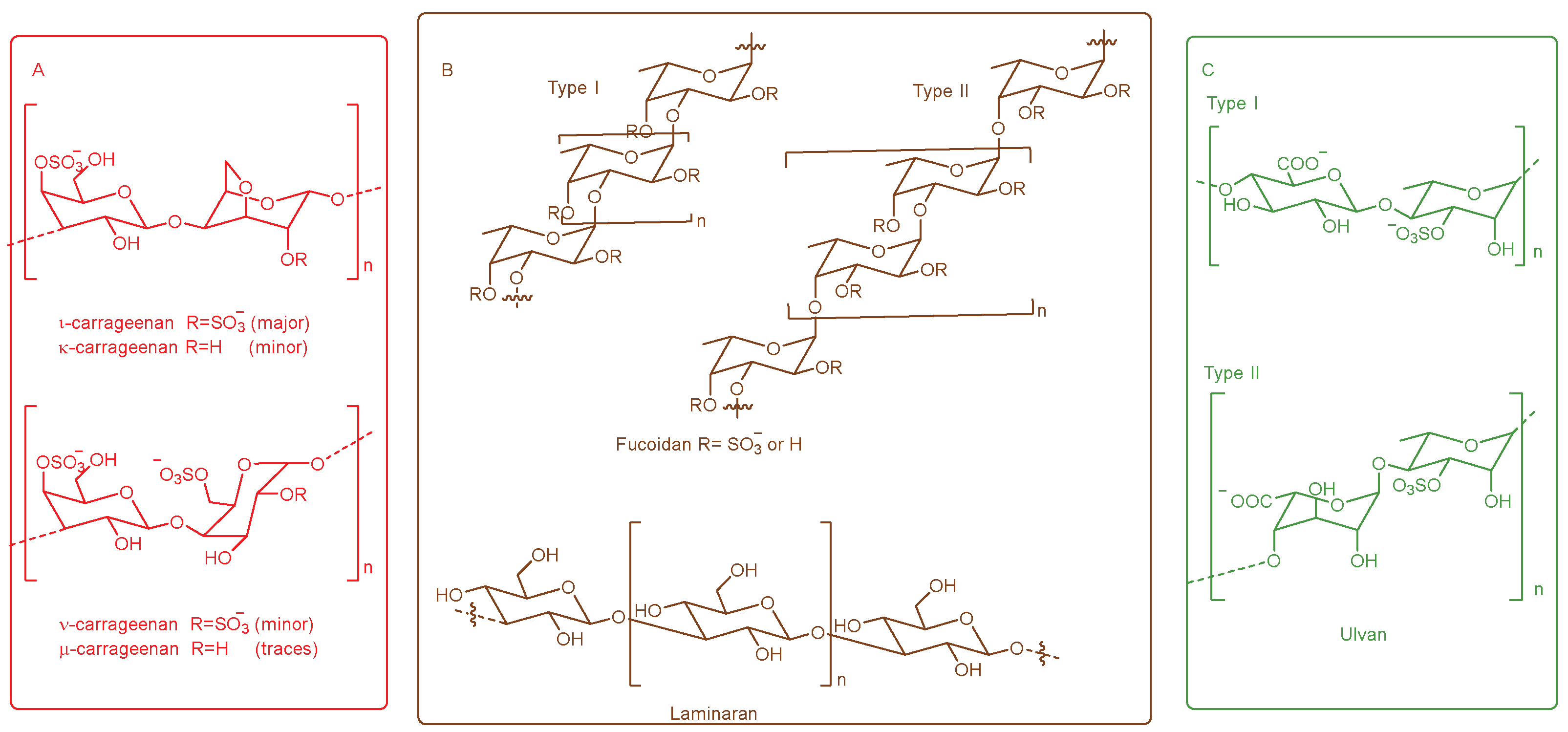
2.8. Marine Algae Polysaccharides
2.9. Other Plant Sources of Polysaccharides
2.9.1. Polysaccharides with Anti-Lung Cancer Activity
2.9.2. Polysaccharides with Anti-Pancreatic Cancer Activity
2.9.3. Polysaccharides with Anticancer Activity
3. Polysaccharides from Animals
3.1. Polysaccharides from Mammals
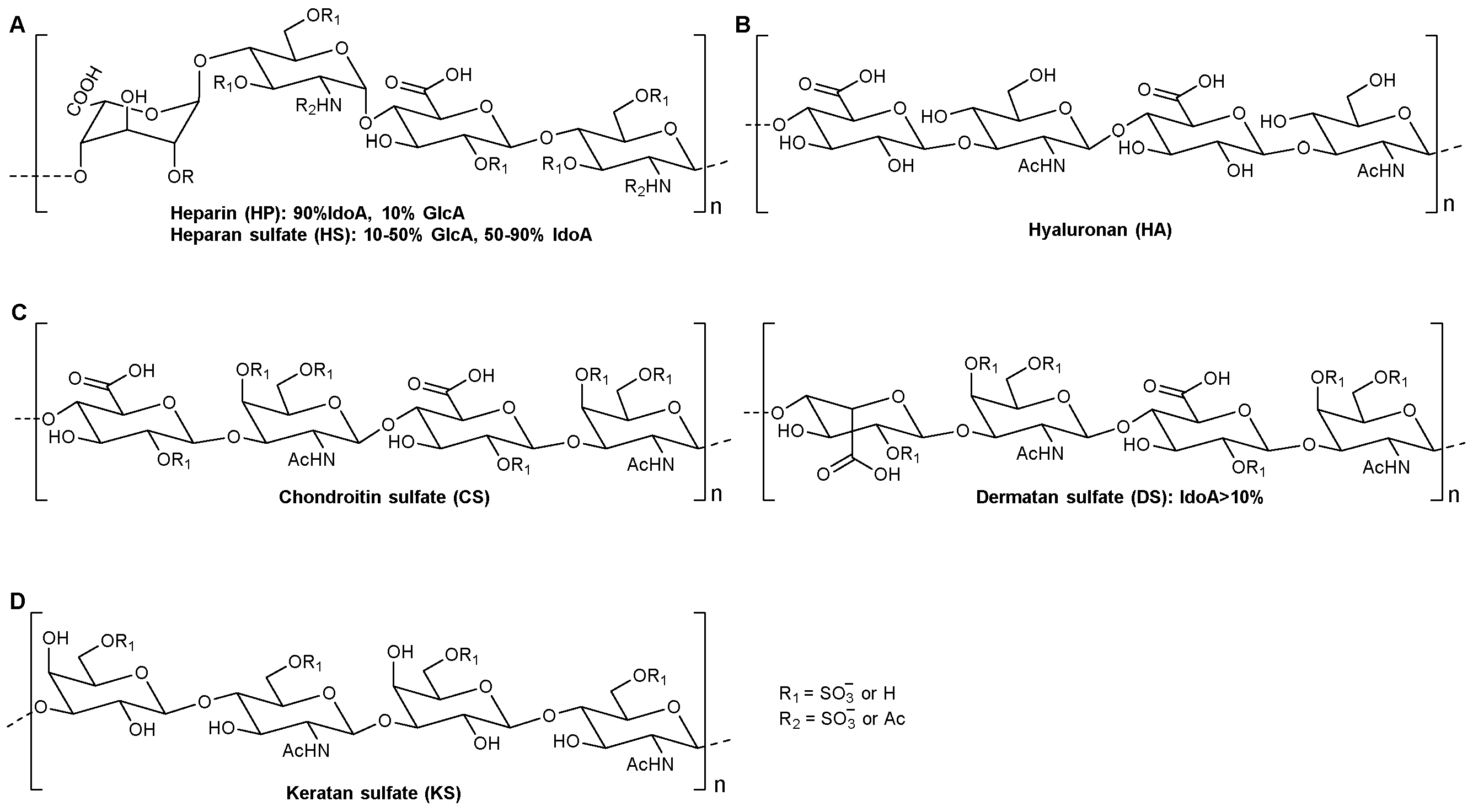
3.1.1. Heparin/Heparan Sulfate
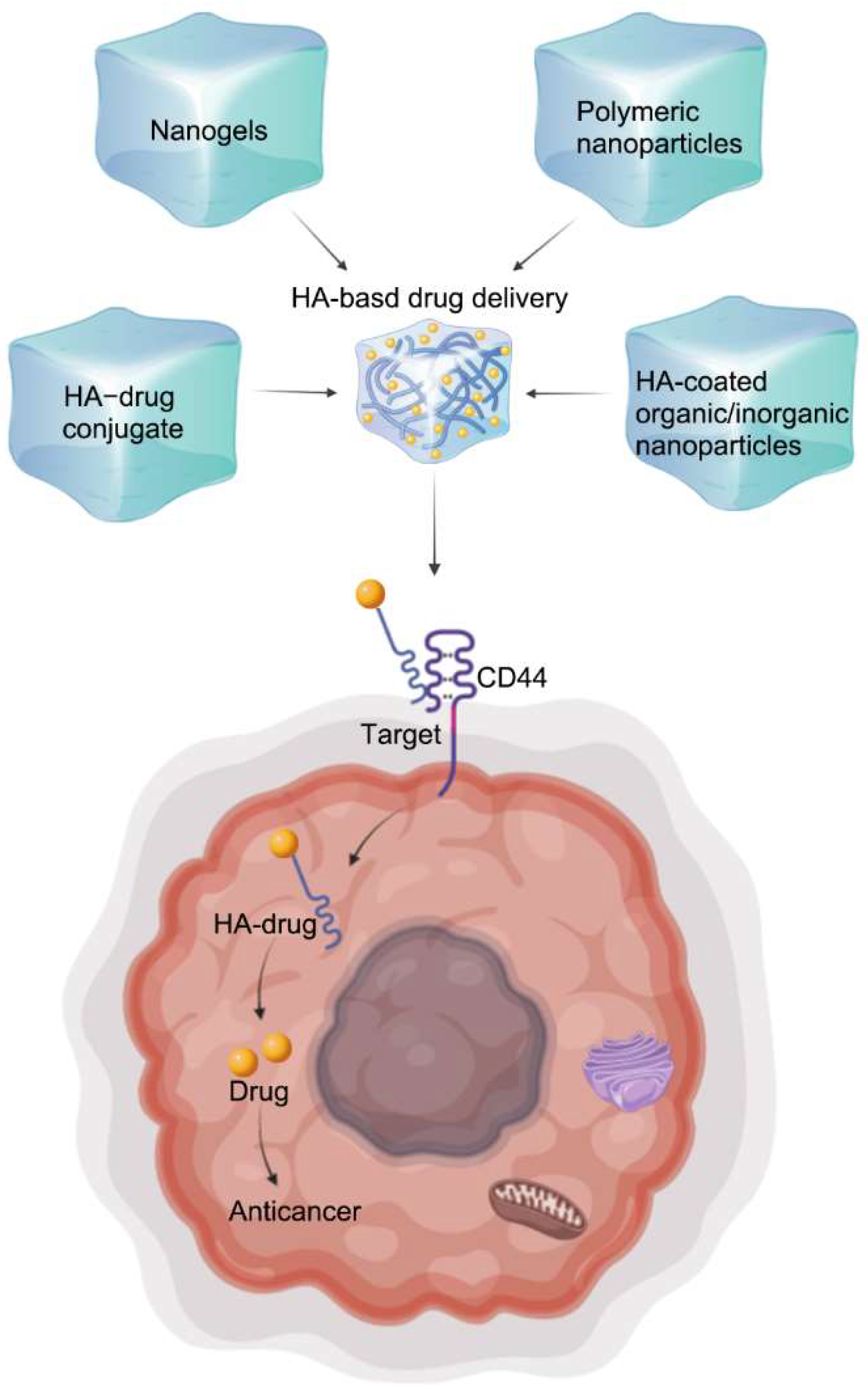
3.1.2. Hyaluronan
3.1.3. Chondroitin Sulfate/Dermatan Sulfate
3.1.4. Keratan Sulfate
3.2. Polysaccharides Derived from Marine Animals
3.2.1. Chondroitin Sulfate from Sturgeon and Cartilage
3.2.2. Sulfated Polysaccharides from Sea Cucumber
3.2.3. Polysaccharides from Common Cockles
4. Polysaccharides from Fungi
4.1. Lentinan
4.2. Ganoderma lucidum Polysaccharide
5. Conclusions
Author Contributions
Funding
Institutional Review Board Statement
Informed Consent Statement
Data Availability Statement
Conflicts of Interest
References
- Bray, F.; Ferlay, J.; Soerjomataram, I.; Siegel, R.L.; Torre, L.A.; Jemal, A. Global cancer statistics 2018: GLOBOCAN estimates of incidence and mortality worldwide for 36 cancers in 185 countries. CA A Cancer J. Clin. 2018, 68, 394–424. [Google Scholar] [CrossRef]
- Schjoldager, K.T.; Narimatsu, Y.; Joshi, H.J.; Clausen, H. Global view of human protein glycosylation pathways and functions. Nat. Rev. Mol. Cell Biol. 2020, 21, 729–749. [Google Scholar] [CrossRef]
- Li, N.; Wang, C.; Georgiev, M.I.; Bajpai, V.K.; Tundis, R.; Gandara, J.S.; Lu, X.; Xiao, J.; Tang, X.; Qiao, T. Advances in dietary polysaccharides as anticancer agents: Structure-activity relationship. Trends Food Sci. Technol. 2021, 111, 360–377. [Google Scholar] [CrossRef]
- Zong, A.; Cao, H.; Wang, F. Anticancer polysaccharides from natural resources: A review of recent research. Carbohydr. Polym. 2012, 90, 1395–1410. [Google Scholar] [CrossRef]
- Zeng, Y.; Xiang, Y.; Sheng, R.; Tomás, H.; Rodrigues, J.; Gu, Z.; Zhang, H.; Gong, Q.; Luo, K. Polysaccharide-based nanomedicines for cancer immunotherapy: A review. Bioact. Mater. 2021, 6, 3358–3382. [Google Scholar] [CrossRef] [PubMed]
- Liu, L.; Xu, F.-R.; Wang, Y.Z. Traditional uses, chemical diversity and biological activities of Panax, L. (Araliaceae): A review. J. Ethnopharmacol. 2020, 263, 112792. [Google Scholar] [CrossRef] [PubMed]
- Zhao, B.; Lv, C.; Lu, J. Natural occurring polysaccharides from Panax ginseng C. A. Meyer: A review of isolation, structures, and bioactivities. Int. J. Biol. Macromol. 2019, 133, 324–336. [Google Scholar] [CrossRef]
- Sun, L.; Wu, D.; Ning, X.; Yang, G.; Lin, Z.; Tian, M.; Zhou, Y. α-Amylase-assisted extraction of polysaccharides from Panax ginseng. Int. J. Biol. Macromol. 2015, 75, 152–157. [Google Scholar] [CrossRef]
- Sun, L.; Ropartz, D.; Cui, L.; Shi, H.; Ralet, M.C.; Zhou, Y. Structural characterization of rhamnogalacturonan domains from Panax ginseng C. A. Meyer. Carbohydr Polym. 2019, 203, 119–127. [Google Scholar] [CrossRef]
- Yue, F.; Xu, J.; Zhang, S.; Hu, X.; Wang, X.; Lü, X. Structural features and anticancer mechanisms of pectic polysaccharides: A review. Int. J. Biol. Macromol. 2022, 209, 825–839. [Google Scholar] [CrossRef]
- Maxwell, E.G.; Belshaw, N.J.; Waldron, K.W.; Morris, V.J. Pectinan emerging newbioactive food polysaccharide. Trends Food Sci. Technol. 2012, 24, 64–73. [Google Scholar] [CrossRef]
- Shakhmatov, E.G.; Makarova, E.N.; Belyy, V.A. Structural studies of biologically active pectin-containing polysaccharides of pomegranate Punica granatum. Int. J. Biol. Macromol. 2019, 122, 29–36. [Google Scholar] [CrossRef] [PubMed]
- Zhang, X.; Yu, L.; Bi, H.; Li, X.; Ni, W.; Han, H.; Li, N.; Wang, B.; Zhou, Y.; Tai, G. Total fractionation and characterization of the water-soluble polysaccharides isolated from Panax ginseng C.A. Meyer. Carbohydr. Polym. 2009, 77, 544–552. [Google Scholar] [CrossRef]
- Li, C.; Cai, J.; Geng, J.; Li, Y.; Wang, Z.; Li, R. Purification, characterization and an-ticancer activity of a polysaccharide from Panax ginseng. Int. J. Biol. Macromol. 2012, 51, 968–973. [Google Scholar] [CrossRef]
- Cai, J.-P.; Wu, Y.-J.; Li, C.; Feng, M.Y.; Shi, Q.T.; Li, R.; Wang, Z.Y.; Geng, J.S. Panax ginseng polysaccharide suppresses metastasis by modulating Twist expression in gastric cancer. Int. J. Biol. Macromol. 2013, 57, 22–25. [Google Scholar] [CrossRef]
- Li, C.; Tian, Z.-N.; Cai, J.P.; Chen, K.X.; Zhang, B.; Feng, M.Y.; Shi, Q.T.; Li, R.; Qin, Y.; Geng, J.S. Panax ginseng polysaccharide induces apoptosis by targeting Twist/AKR1C2/NF-1 pathway in human gastric cancer. Carbohydr. Polym. 2014, 102, 103–109. [Google Scholar] [CrossRef]
- Gao, X.; Zhi, Y.; Sun, L.; Peng, X.; Zhang, T.; Xue, H.; Tai, G.; Zhou, Y. The inhibitory effects of a rhamnogalacturonan I (RG-I) domain from ginseng pectin on galectin-3 and its structure-activity relationship. J. Biol. Chem. 2013, 288, 33953–33965. [Google Scholar] [CrossRef]
- Jia, H.; Zhao, B.; Zhang, F.; Santhanam, R.K.; Wang, X.; Lu, J. Extraction, structural characterization, and anti-hepatocellular carcinoma activity of polysaccharides from Panax ginseng meyer. Front. Oncol. 2021, 11, 4905. [Google Scholar] [CrossRef]
- Zhou, X.; Shi, H.; Jiang, G.; Zhou, Y.; Xu, J. Antitumour activities of ginseng polysaccharide in C57BL/6 mice with Lewis lung carcinoma. Tumor Biol. 2014, 35, 12561–12566. [Google Scholar] [CrossRef]
- Liao, K.; Bian, Z.; Xie, D.; Peng, Q. A selenium-modified ginseng polysaccharide promotes the apoptosis in human promyelocytic leukemia (HL-60) cells via a mitochondrial-mediated pathway. Biol. Trace Elem. Res. 2017, 177, 64–71. [Google Scholar] [CrossRef]
- Chen, X.-P.; Li, W.; Xiao, X.F.; Zhang, L.L.; Liu, C.X. Phytochemical and pharmacological studies on Radix Angelica sinensis. Chin. J. Nat. Med. 2013, 11, 577–587. [Google Scholar] [CrossRef] [PubMed]
- Younas, F.; Aslam, B.; Muhammad, F.; Mohsin, M.; Raza, A.; Faisal, M.N.; Hassan, S.; Majeed, W. Haematopoietic effects of Angelica sinensis root cap polysaccharides against lisinopril-induced anaemia in albino rats. Pharm. Biol. 2017, 55, 108–113. [Google Scholar] [CrossRef] [PubMed]
- Pan, S.; Jiang, L.; Wu, S. Stimulating effects of polysaccharide from Angelica sinensis on the nonspecific immunity of white shrimps (Litopenaeus vannamei). Fish Shellfish Immun. 2018, 74, 170–174. [Google Scholar] [CrossRef]
- Wang, Y.; Li, X.; Chen, X.; Zhao, P.; Qu, Z.; Ma, D.; Zhao, C.; Gao, W. Effect of stir-frying time during Angelica Sinensis Radix processing with wine on physicochemical, structure properties and bioactivities of polysaccharides. Process Biochem. 2019, 81, 188–196. [Google Scholar] [CrossRef]
- Zhou, W.-J.; Wang, S.; Hu, Z.; Zhou, Z.Y.; Song, C.J. Angelica sinensis polysaccharides promotes apoptosis in human breast cancer cells via CREB-regulated caspase-3 activation. Biochem. Biophys. Res. Commun. 2015, 467, 562–569. [Google Scholar] [CrossRef]
- Fu, Z.; Li, Y.; Yang, S.; Ma, C.; Zhao, R.; Guo, H.; Wei, H. Angelica sinensis polysaccharide promotes apoptosis by inhibiting JAK/STAT pathway in breast cancer cells. Trop. J. Pharm. Res. 2019, 18, 2247–2253. [Google Scholar]
- Yang, J.; Shao, X.; Jiang, J.; Sun, Y.; Wang, L.; Sun, L. Angelica sinensis polysaccharide inhibits proliferation, migration, and invasion by downregulating microRNA-675 in human neuroblastoma cell line SH-SY5Y. Cell Biol. Int. 2018, 42, 867–876. [Google Scholar] [CrossRef]
- Zhang, Y.; Cui, Z.; Mei, H.; Xu, J.; Zhou, T.; Cheng, F.; Wang, K. Angelica sinensis polysaccharide nanoparticles as a targeted drug delivery system for enhanced therapy of liver cancer. Carbohydr. Polym. 2019, 219, 143–154. [Google Scholar] [CrossRef]
- Tleubayeva, M.I.; Datkhayev, U.M.; Alimzhanova, M.; Ishmuratova, M.Y.; Korotetskaya, N.V.; Abdullabekova, R.M.; Flisyuk, E.V.; Gemejiyeva, N.G. Component composition and antimicrobial activity of CO2 extract of portulaca oleracea, growing in the territory of kazakhstan. Sci. World J. 2021, 2021, 1–10. [Google Scholar] [CrossRef]
- Kim, K.-H.; Park, E.-J.; Jang, H.J.; Lee, S.J.; Park, C.S.; Yun, B.S.; Lee, S.W.; Rho, M.C. 1-Carbomethoxy-β-Carboline, derived from portulaca oleracea L., ameliorates LPS-mediated inflammatory response associated with MAPK signaling and nuclear translocation of NF-κB. Molecules 2019, 24, 4042. [Google Scholar] [CrossRef]
- Tian, X.; Ding, Y.; Kong, Y.; Wang, G.; Wang, S.; Cheng, D. Purslane (Portulacae oleracea L.) attenuates cadmium-induced hepatorenal and colonic damage in mice: Role of chelation, antioxidant and intestinal microecological regulation. Phytomedicine 2021, 92, 153716. [Google Scholar] [CrossRef]
- Zhang, W.; Zheng, B.; Deng, N.; Wang, H.; Li, T.; Liu, R.H. Effects of ethyl acetate fractional extract from Portulaca oleracea L. (PO-EA) on lifespan and healthspan in Caenorhabditis elegans. J. Food Sci. 2020, 85, 4367–4376. [Google Scholar] [CrossRef]
- Shena, H.; Tang, G.; Zeng, G.; Yang, Y.; Cai, X.; Li, D.; Liu, H.; Zhou, N. Purification and characterization of an antitumour polysaccharide from Portulaca oleracea L. Carbohydr. Polym. 2013, 93, 395–400. [Google Scholar] [CrossRef] [PubMed]
- Zhao, R.; Gao, X.; Cai, Y.; Shao, X.; Jia, G.; Huang, Y.; Qin, X.; Wang, J.; Zheng, X. Antitumour activity of Portulaca oleracea L. polysaccharides against cervical carcinoma in vitro and in vivo. Carbohydr. Polym. 2013, 96, 376–383. [Google Scholar] [CrossRef] [PubMed]
- Zhao, R.; Zhang, T.; Ma, B.; Li, X. Antitumour activity of portulaca oleracea L. polysaccharide on heLa cells through inducing TLR4/NF-kB signaling. Nutr. Cancer 2017, 69, 131–139. [Google Scholar] [CrossRef] [PubMed]
- Zhao, R.; Shao, X.; Jia, G.; Huang, Y.; Liu, Z.; Song, B.; Hou, J. Anti-cervical carcinoma effect of Portulaca oleracea L. polysaccharides by oral administration on intestinal dendritic cells. BMC Complement. Altern. Med. 2019, 19, 1–10. [Google Scholar] [CrossRef]
- Park, Y.M.; Lee, H.Y.; Kang, Y.G.; Park, S.H.; Lee, B.G.; Park, Y.J.; Oh, H.G.; Moon, D.I.; Kim, Y.P.; Park, D.S.; et al. Immune-enhancing effects of Portulaca oleracea L.-based complex extract in cyclophosphamide-induced splenocytes and immunosuppressed rats. Food Agric. Immunol. 2018, 30, 13–24. [Google Scholar] [CrossRef]
- Jia, G.; Shao, X.; Zhao, R.; Zhang, T.; Zhou, X.; Yang, Y.; Li, T.; Chen, Z.; Liu, Y. Portulaca oleracea L. polysaccharides enhance the immune efficacy of dendritic cell vaccine for breast cancer. Food Function 2021, 12, 4046–4059. [Google Scholar] [CrossRef]
- Zhuang, S.; Ming, K.; Ma, N.; Sun, J.; Wang, D.; Ding, M.; Ding, Y. Portulaca oleracea L. polysaccharide ameliorates lipopolysaccharide-induced inflammatory responses and barrier dysfunction in porcine intestinal epithelial monolayers. J. Funct. Foods 2022, 91, 104997. [Google Scholar] [CrossRef]
- Masci, A.; Carradori, S.; Casadei, M.A.; Paolicelli, P.; Petralito, S.; Ragno, R.; Cesa, S. Lycium barbarum polysaccharides: Extraction, purification, structura characterization and evidence about hypoglycaemic and hypolipidaemic effects. A review. Food Chem. 2018, 254, 377–389. [Google Scholar] [CrossRef]
- Amagase, H.; Farnsworth, N.R. A review of botanical characteristics, phytochemistry, clinical relevance in efficacy and safety of Lycium barbarum fruit (GOJI). Food Res. Int. 2011, 44, 1702–1717. [Google Scholar] [CrossRef]
- Chen, F.; Ran, L.; Mi, J.; Yan, Y.; Lu, L.; Jin, B.; Li, X.; Cao, Y. Isolation, characterization and antitumour effect on DU145 cells of a main polysaccharide in pollen of chinese wolfberry. Molecules 2018, 23, 2430. [Google Scholar] [CrossRef] [PubMed]
- Ran, L.; Chen, F.; Zhang, J.; Mi, J.; Lu, L.; Yan, Y.; Cao, Y. Antitumour effects of pollen polysaccharides from Chinese wolfberry on DU145 cells via the PI3K/AKT pathway in vitro and in vivo. Int. J. Biol. Macromol. 2020, 152, 1164–1173. [Google Scholar] [CrossRef] [PubMed]
- Chen, D.; Sun, S.; Cai, D.; Kong, G. Induction of mitochondrial-dependent apoptosis in T24 cells by a selenium (Se)-containing polysaccharide from Ginkgo biloba L. leaves. Int. J. Biol. Macromol. 2017, 101, 126–130. [Google Scholar] [CrossRef] [PubMed]
- Zhang, F.; Shi, J.-J.; Thakur, K.; Hu, F.; Zhang, J.G.; Wei, Z.J. Anti-cancerous potential of polysaccharide fractions extracted from peony seed dreg on various human cancer cell lines via cell cycle arrest and apoptosis. Front. Pharmacol. 2017, 8, 102. [Google Scholar] [CrossRef]
- Hua, Y.; Zhang, J.; Zou, L.; Fu, C.; Li, P.; Zhao, G. Chemical characterization, antioxidant, immune-regulatin and anticancer activities of a novel bioactive polysaccharide from Chenopodium quinoa seeds. Int. J. Biol. Macromol. 2017, 99, 622–629. [Google Scholar] [CrossRef] [PubMed]
- Lin, H.-C.; Lin, J.-Y. GSF3, a polysaccharide from guava (Psidium guajava L.) seeds, inhibits MCF-7 breast cancer cell growth via increasing Bax/Bcl-2 ratio or Fas mRNA expression levels. Int. J. Biol. Macromol. 2020, 161, 1261–1271. [Google Scholar] [CrossRef]
- Kaya, M.; Sousa, A.G.; Crépeau, M.J.; Sørensen, S.O.; Ralet, M.C. Characterization of citrus pectin samples extracted under different conditions: Influence of acid type and pH of extraction. Ann. Bot. 2014, 114, 1319–1326. [Google Scholar] [CrossRef]
- Glinsky, V.V.; Raz, A. Modified citrus pectin anti-metastatic properties: One bullet, multiple targets. Carbohydr. Res. 2009, 344, 1788–1791. [Google Scholar] [CrossRef]
- Nangia-Makker, P.; Hogan, V.; Honjo, Y.; Baccarini, S.; Tait, L.; Bresalier, R.; Raz, A. Inhibition of human cancer cell growth and metastasis in nude mice by oral intake of modified citrus pectin. J. Natl. Cancer Inst. 2002, 94, 1854–1862. [Google Scholar] [CrossRef]
- Ahmed, H.; Alsadek, D.M.M. Galectin-3 as a potential target to prevent cancer metastasis. Clin. Med. Insights: Oncol. 2015, 9, 113–121. [Google Scholar] [CrossRef] [PubMed]
- Fang, T.; Liu, D.-D.; Ning, H.; Liu, D.; Sun, J.; Huang, X.; Dong, Y.; Geng, M.; Yun, S.; Yan, J.; et al. Modified citrus pectin inhibited bladder tumor growth through downregulation of galectin-3. Acta Pharmacol. Sin. 2018, 39, 1885–1893. [Google Scholar] [CrossRef] [PubMed]
- Wang, S.; Li, P.; Lu, S.M.; Ling, Z.Q. Chemoprevention of low-molecular-weight citrus pectin (LCP) in gastrointestinal cancer cells. Int. J. Biol. Sci. 2016, 12, 746–756. [Google Scholar] [CrossRef] [PubMed]
- Conti, S.; Vexler, A.; Hagoel, L.; Kalich-Philosoph, L.; Corn, B.W.; Honig, N.; Shtraus, N.; Meir, Y.; Ron, I.; Eliaz, I.; et al. Modified citrus pectin as a potential sensitizer for radiotherapy in prostate cancer. Integr. Cancer Ther. 2018, 17, 1225–1234. [Google Scholar] [CrossRef]
- do Prado, S.B.R.; Shiga, T.M.; Harazono, Y.; Hogan, V.A.; Raz, A.; Carpita, N.C.; Fabi, J.P. Migration and proliferation of cancer cells in culture are differentially affected by molecular size of modified citrus pectin. Carbohydr. Polym. 2019, 211, 141–151. [Google Scholar] [CrossRef]
- Pynam, H.; Dharmesh, S.M. A xylorhamnoarabinogalactan I from Bael (Aegle marmelos L.) modulates UV/DMBA induced skin cancer via galectin-3 & gut microbiota. J. Funct. Foods 2019, 60, 103425. [Google Scholar]
- do Prado, S.B.R.; Mourão, P.A.S.; Fabi, J.P. Chelate-soluble pectin fraction from papaya pulp interacts with galectin-3 and inhibits colon cancer cell proliferation. Int. J. Biol. Macromol. 2019, 126, 170–178. [Google Scholar] [CrossRef]
- Bermudez-Oria, A.; Rodriguez-Gutierrez, G.; Fátima, R.S.; Marta, S.C.; Juan, F.B. Antiproliferative activity of olive extract rich in polyphenols and modified pectin on bladder cancer cells. J. Med. Food 2020, 23, 719–727. [Google Scholar] [CrossRef]
- Tanna, B.; Mishra, A. Nutraceutical potential of seaweed polysaccharides: Structure, bioactivity, safety, and toxicity. Compr. Rev. Food Sci. Food Saf. 2019, 18, 817–831. [Google Scholar] [CrossRef]
- Zheng, L.-X.; Chen, X.-Q. Current trends in marine algae polysaccharides: The digestive tract, microbial catabolism, and prebiotic potential. Int. J. Biol. Macromol. 2020, 151, 344–354. [Google Scholar] [CrossRef] [PubMed]
- Jiao, G.; Yu, G.; Zhang, J.; Ewart, H.S. Chemical structures and bioactivities of sulfated polysaccharides from marine algae. Mar. Drugs 2011, 9, 196–223. [Google Scholar] [CrossRef] [PubMed]
- Tziveleka, L.-A.; Ioannou, E.; Roussis, V. Ulvan, a bioactive marine sulfated polysaccharide as a key constituent of hybrid biomaterials: A review. Carbohydr. Polym. 2019, 218, 355–370. [Google Scholar] [CrossRef] [PubMed]
- Liu, G.; Kuang, S.; Wu, S.; Jin, W.; Sun, C. A novel polysaccharide from Sargassum integerrimum induces apoptosis in A549 cells and prevents angiogensis in vitro and in vivo. Sci. Rep. 2016, 6, 1–12. [Google Scholar] [CrossRef] [PubMed]
- Vaikundamoorthy, R.; Krishnamoorthy, V.; Vilwanathan, R.; Rajendran, R. Structural characterization and anticancer activity (MCF7 and MDA-MB-231) of polysaccharides fractionated from brown seaweed Sargassum wightii. Int. J. Biol. Macromol. 2018, 111, 1229–1237. [Google Scholar] [CrossRef]
- Senthilkumar, K.; Manivasagan, P.; Venkatesan, J.; Kim, S.K. Brown seaweed fucoidan: Biological activity and apoptosis, growth signaling mechanism in cancer. Int. J. Biol. Macromol. 2013, 60, 366–374. [Google Scholar] [CrossRef]
- Wang, Z.-J.; Xu, W.; Liang, J.W.; Wang, C.S.; Kang, Y. Effect of fucoidan on B16 murine melanoma cell melanin formation and apoptosis. Afr. J. Tradit. Complement. Altern. Med. 2017, 14, 149–155. [Google Scholar] [CrossRef]
- Chen, J.; Hu, Y.; Zhang, L.; Wang, Y.; Wang, S.; Zhang, Y.; Guo, H.; Ji, D.; Wang, Y. Alginate oligosaccharide DP5 exhibits antitumor effects in osteosarcoma patients following surgery. Front. Pharmacol. 2017, 8, 623. [Google Scholar] [CrossRef]
- Anand, J.; Sathuvan, M.; Babu, G.V.; Sakthivel, M.; Palani, P.; Nagaraj, S. Bioactive potential and composition analysis of sulfated polysaccharide from Acanthophora spicifera (Vahl) Borgeson. Int. J. Biol. Macromol. 2018, 111, 1238–1244. [Google Scholar] [CrossRef]
- Chen, X.; Song, L.; Wang, H.; Liu, S.; Yu, H.; Wang, X.; Li, R.; Liu, T.; Li, P. Partial characterization, the immune modulation and anticancer activities of sulfated polysaccharides from filamentous microalgae tribonema sp. Molecules 2019, 24, 322. [Google Scholar] [CrossRef]
- Yang, S.; Wan, H.; Wang, R.; Hao, D. Sulfated polysaccharides from Phaeodactylum tricornutum: Isolation, structural characteristics, and inhibiting HepG2 growth activity in vitro. PeerJ 2019, 7, e6409. [Google Scholar] [CrossRef]
- Wanga, H.; Gao, T.; Du, Y.; Yang, H.; Wei, L.; Bi, H.; Ni, W. Anticancer and immunostimulating activities of a novel homogalacturonan from Hippophae rhamnoides L. berry. Carbohydr. Polym. 2015, 131, 288–296. [Google Scholar] [CrossRef] [PubMed]
- Han, K.; Jin, C.; Chen, H.; Wang, P.; Yu, M.; Ding, K. Structural characterization and anti-A549 lung cancer cells bioactivity of a polysaccharide from Houttuynia cordata. Int. J. Biol. Macromol. 2018, 120, 288–296. [Google Scholar] [CrossRef] [PubMed]
- Wu, J.; Gao, W.; Song, Z.; Xiong, Q.; Xu, Y.; Han, Y.; Yuan, J.; Zhang, R.; Cheng, Y.; Fang, J.; et al. Anticancer activity of polysaccharide from Glehnia littoralis on human lung cancer cell line A549. Int. J. Biol. Macromol. 2018, 106, 464–472. [Google Scholar] [CrossRef] [PubMed]
- Zhong, C.; Yang, J.; Lu, Y.; Xie, H.; Zhai, S.; Zhang, C.; Luo, Z.; Chen, X.; Fang, X.; Jia, L. Achyranthes bidentata polysaccharide can safely prevent NSCLC metastasis by targeting EGFR and EMT. Signal Transduct. Target. Ther. 2020, 5, 178. [Google Scholar] [CrossRef] [PubMed]
- Lina, L.; Wang, P.; Du, Z.; Wang, W.; Cong, Q.; Zheng, C.; Jin, C.; Ding, K.; Shao, C. Structural elucidation of a pectin from flowers of Lonicera japonica and its antipancreatic cancer activity. Int. J. Biol. Macromol. 2016, 88, 130–137. [Google Scholar] [CrossRef]
- Zhanga, S.; He, F.; Chen, X.; Ding, K. Isolation and structural characterization of a pectin from Lycium ruthenicum Murr and its anti-pancreatic ductal adenocarcinoma cell activity. Carbohydr. Polym. 2019, 223, 115104. [Google Scholar] [CrossRef]
- Xu, L.; Cao, J.; Chen, W. Structural characterization of a broccoli polysaccharide and evaluation of anticancer cell proliferation effects. Carbohydr. Polym. 2015, 126, 179–184. [Google Scholar] [CrossRef]
- Zhang, Z.F.; Lv, G.Y.; Jiang, X.; Cheng, J.H.; Fan, L.F. Extraction optimization and biological properties of a polysaccharide isolated from Gleoestereum incarnatum. Carbohydr. Polym. 2015, 117, 185–191. [Google Scholar] [CrossRef]
- Wang, Y.; Liu, X.; Zhang, J.; Liu, G.; Liu, Y.; Wang, K.; Yang, M.; Cheng, H.; Zhao, Z. Structural characterization and in vitro antitumour activity of polysaccharides from Zizyphus jujuba cv. Muzao. RSC Adv. 2015, 5, 7860–7867. [Google Scholar] [CrossRef]
- Zhao, C.; Li, Z.; Li, C.; Yang, L.; Yao, L.; Fu, Y.; He, X.; Shi, K.; Lu, Z. Optimized extraction of polysaccharides from Taxus chinensis var. mairei fruits and its antitumour activity. Int. J. Biol. Macromol. 2015, 75, 192–198. [Google Scholar] [CrossRef]
- Feng, Y.-N.; Zhang, X.-F. Polysaccharide extracted from Huperzia serrata using response surface methodology and its biological activity. Int. J. Biol. Macromol. 2020, 157, 267–275. [Google Scholar] [CrossRef] [PubMed]
- Ren, F.; Li, J.; Yuan, X.; Wang, Y.; Wu, K.; Kang, L.; Luo, Y.; Zhang, H.; Yuan, Z. Dandelion polysaccharides exert anticancer effect on Hepatocellular carcinoma by inhibiting PI3K/AKT/mTOR pathway and enhancing immune response. J. Funct. Foods 2019, 55, 263–274. [Google Scholar] [CrossRef]
- Ren, F.; Wu, K.; Yang, Y.; Yang, Y.; Wang, Y.; Li, J. Dandelion polysaccharide exerts anti-angiogenesis effect on hepatocellular carcinoma by regulating VEGF/HIF-1a expression. Front. Pharmacol. 2020, 11, 460. [Google Scholar] [CrossRef] [PubMed]
- Wang, J.-H.; Luo, J.-P. Comparison of antitumor activities of different polysaccharide fractions from the stems of Dendrobium nobile Lindl. Carbohydr. Polym. 2010, 79, 114–118. [Google Scholar] [CrossRef]
- Yeung, B.K.S.; Chong, P.Y.C.; Petillo, P.A. Synthesis of Glycosaminoglycans. J. Carbohydr. Chem. 2002, 21, 799–865. [Google Scholar] [CrossRef]
- Pomin, V.H.; Mulloy, B. Glycosaminoglycans and proteoglycans. Pharmaceuticals 2018, 11, 27. [Google Scholar] [CrossRef]
- Mende, M.; Bednarek, C.; Wawryszyn, M.; Sauter, P.; Biskup, M.B.; Schepers, U.; Bräse, S. Chemical synthesis of glycosaminoglycans. Chem. Rev. 2016, 116, 8193–8255. [Google Scholar] [CrossRef]
- Volpi, N. Therapeutic applications of glycosaminoglycans. Curr. Med. Chem. 2006, 13, 1799–1810. [Google Scholar] [CrossRef]
- Berdiaki, A.; Neagu, M.; Giatagana, E.M.; Kuskov, A.; Tsatsakis, A.M.; Tzanakakis, G.N.; Nikitovic, D. Glycosaminoglycans: Carriers and Targets for Tailored Anti-Cancer Therapy. Biomolecules 2021, 11, 395. [Google Scholar] [CrossRef]
- Li, W.; Johnson, D.J.D.; Esmon, C.T.; Huntington, J.A. Structure of the antithrombin-thrombin-heparin ternary complex reveals the antithrombotic mechanism of heparin. Nat. Struct. Mol. Biol. 2004, 11, 857–862. [Google Scholar] [CrossRef]
- Khorana, A.A.; Streiff, M.B.; Farge, D.; Mandala, M.; Debourdeau, P.; Cajfinger, F.; Marty, M.; Falanga, A.; Lyman, G.H. Venous thromboembolism prophylaxis and treatment in cancer: A consensus statement of major guidelines panels and call to action. J. Clin. Oncol. 2009, 27, 4919–4926. [Google Scholar] [CrossRef] [PubMed]
- Farge, D.; Bounameaux, H.; Brenner, B.; Cajfinger, F.; Debourdeau, P.; Khorana, A.A.; Pabinger, I.; Solymoss, S.; Douketis, J.; Kakkar, A. International clinical practice guidelines including guidance for direct oral anticoagulants in the treatment and prophylaxis of venous thromboembolism in patients with cancer. Lancet Oncol. 2016, 17, e452–e466. [Google Scholar] [CrossRef]
- Frere, C.; Benzidia, I.; Marjanovic, Z.; Farge, D. Recent advances in the management of cancer-associated thrombosis: New Hopes but New Challenges. Cancers 2019, 11, 71. [Google Scholar] [CrossRef] [PubMed]
- Gil-Bernabé, A.M.; Lucotti, S.; Muschel, R.J. Coagulation and metastasis: What does the experimental literature tell us? Br. J. Haematol. 2013, 162, 433–441. [Google Scholar] [CrossRef]
- Walenga, J.M.; Lyman, G.H. Evolution of heparin anticoagulants to ultralow-molecular-weight heparins: A review of pharmacologic and clinical differences and applications in patients with cancer. Crit. Rev. Oncol. Hematol. 2013, 88, 1–18. [Google Scholar] [CrossRef]
- Zhang, W.; Swanson, R.; Izaguirre, G.; Xiong, Y.; Lau, L.F.; Olson, S.T. The heparin-binding site of antithrombin is crucial for antiangiogenic activity. Blood 2005, 106, 1621–1628. [Google Scholar] [CrossRef]
- Rohloff, J.; Zinke, J.; Schoppmeyer, K.; Tannapfel, A.; Witzigmann, H.; Mössner, J.; Wittekind, C.; Caca, K. Heparanase expression is a prognostic indicator for postoperative survival in pancreatic adenocarcinoma. Br. J. Cancer 2002, 86, 1270–1275. [Google Scholar] [CrossRef]
- Ludwig, R.J.; Boehme, B.; Podda, M.; Henschler, R.; Jager, E.; Tandi, C.; Boehncke, W.H.; Zollner, T.M.; Kaufmann, R.; Gille, J. Endothelial P-selectin as a target of heparin action in experimental melanoma lung metastasis. Cancer Res. 2004, 64, 2743–2750. [Google Scholar] [CrossRef]
- Hwang, H.H.; Jeong, H.J.; Yun, S.; Byun, Y.; Okano, T.; Kim, S.W.; Lee, D.Y. Anticancer effect of heparin–taurocholate conjugate on orthotopically induced exocrine and endocrine pancreatic cancer. Cancers 2021, 13, 5775. [Google Scholar] [CrossRef]
- Kim, J.-y.; Al-Hilal, T.A.; Chung, S.W.; Kim, S.Y.; Ryu, G.H.; Son, W.C.; Byun, Y. Antiangiogenic and anticancer effect of an orally active low molecular weight heparin conjugates and its application to lung cancer chemoprevention. J. Control. Release 2015, 199, 122–131. [Google Scholar] [CrossRef]
- Pfankuchena, D.B.; Stölting, D.P.; Schlesinger, M.; Royer, H.D.; Bendas, G. Low molecular weight heparin tinzaparin antagonizes cisplatin resistance of ovarian cancer cells. Biochem. Pharmacol. 2015, 97, 147–157. [Google Scholar] [CrossRef] [PubMed]
- Park, J.; Jeong, J.-H.; Al-Hilal, T.A.; Kim, J.; Byun, Y. Size controlled heparin fragment−deoxycholic acid conjugate showed anticancer property by inhibiting VEGF165. Bioconjugate Chem. 2015, 26, 932–940. [Google Scholar] [CrossRef] [PubMed]
- Park, J.; Kim, J.-y.; Hwang, S.R.; Mahmud, F.; Byun, Y. Chemical conjugate of low molecular weight heparin and suramin fragment inhibits tumor growth possibly by blocking VEGF165. Mol. Pharm. 2015, 12, 3935–3942. [Google Scholar] [CrossRef]
- Parka, J.; Hwang, S.R.; Choi, J.U.; Alam, F.; Byun, Y. Self-assembled nanocomplex of PEGylated protamine and heparin–suramin conjugate for accumulation at the tumor site. Int. J. Pharm. 2018, 535, 38–46. [Google Scholar] [CrossRef]
- Yang, Y.-C.; Cai, J.; Yin, J.; Zhang, J.; Wang, K.L.; Zhang, Z.T. Heparin-functionalized Pluronic nanoparticles to enhance the antitumour efficacy of sorafenib in gastric cancers. Carbohydr. Polym. 2016, 136, 782–790. [Google Scholar] [CrossRef]
- Seib, F.P.; Tsurkan, M.; Freudenberg, U.; Kaplan, D.L.; Werner, C. Heparin-modified polyethylene glycol microparticle aggregates for focal cancer chemotherapy. ACS Biomater. Sci. Eng. 2016, 2, 2287–2293. [Google Scholar] [CrossRef]
- Li, J.; Pan, H.; Qiao, S.; Li, Y.; Wang, J.; Liu, W.; Pan, W. The utilization of lowmolecular weight heparin-poloxamer associated Laponite nanoplatform for safe and efficient tumor therapy. Int. J. Biol. Macromol. 2019, 134, 63–72. [Google Scholar] [CrossRef]
- Newlanda, B.; Varricchio, C.; Körner, Y.; Hoppe, F.; Taplan, C.; Newland, H.; Eigel, D.; Tornillo, G.; Pette, D.; Brancale, A.; et al. Focal drug administration via heparin-containing cryogel microcarriers reduces cancer growth and metastasis. Carbohydr. Polym. 2020, 245, 116504. [Google Scholar] [CrossRef] [PubMed]
- Wang, D.; Luo, W.; Wen, G.; Yang, L.; Hong, S.; Zhang, S.; Diao, J.; Wang, J.; Wei, H.; Li, Y.; et al. Synergistic effects of negatively charged nanoparticles assisted by ultrasound on the reversal multidrug resistance phenotype in breast cancer cells. Ultrason. Sonochem. 2017, 34, 448–457. [Google Scholar] [CrossRef] [PubMed]
- Tian, F.; Dahmani, F.Z.; Qiao, J.; Ni, J.; Xiong, H.; Liu, T.; Zhou, J.; Yao, J. A targeted nanoplatform codelivering chemotherapeutic and antiangiogenic drugs as a tool to reverse multidrug resistance in breast cancer. Acta Biomater. 2018, 75, 398–412. [Google Scholar] [CrossRef]
- Thi, T.T.H.; Tran, D.-H.N.; Bach, L.G.; Vu-Quang, H.; Nguyen, D.C.; Park, K.D.; Nguyen, D.H. Functional magnetic core-shell system-based iron oxide nanoparticle coated with biocompatible copolymer for anticancer drug delivery. Pharmaceutics 2019, 11, 120. [Google Scholar]
- Qiu, L.; Ge, L.; Long, M.; Mao, J.; Ahmed, K.S.; Shan, X.; Zhang, H.; Qin, L.; Lv, G.; Chen, J. Redox-responsive biocompatible nanocarriers based on novel heparosan polysaccharides for intracellular anticancer drug delivery. Asian J. Pharm. Sci. 2020, 15, 83–94. [Google Scholar] [CrossRef] [PubMed]
- Guo, R.; Long, Y.; Lu, Z.; Deng, M.; He, P.; Li, M.; He, Q. Enhanced stability and efficacy of GEM-TOS prodrug by coassembly with antimetastatic shell LMWH-TOS. Acta Pharm. Sin. B 2020, 10, 1977–1988. [Google Scholar] [CrossRef] [PubMed]
- Trana, T.H.; Bae, B.-c.; Lee, Y.; Na, K.; Huh, K.M. Heparin-folate-retinoic acid bioconjugates for targeted delivery of hydrophobic photosensitizers. Carbohydr. Polym. 2013, 92, 1615–1624. [Google Scholar] [CrossRef] [PubMed]
- Shi, X.; Wang, Y.; Sun, H.; Chen, Y.; Zhang, X.; Xu, J.; Zhai, G. Heparin-reduced graphene oxide nanocomposites for curcumin delivery: In vitro, in vivo and molecular dynamics simulation study. Biomater. Sci. 2019, 7, 1011. [Google Scholar] [CrossRef] [PubMed]
- Wu, Y.; Li, F.; Zhang, X.; Li, Z.; Zhang, Q.; Wang, W.; Pan, D.; Zheng, X.; Gu, Z.; Zhang, H.; et al. Tumor microenvironment-responsive PEGylated heparin-pyropheophorbide-a nanoconjugates for photodynamic therapy. Carbohydr. Polym. 2021, 255, 117490. [Google Scholar] [CrossRef]
- Chaudhry, G.-e.S.; Akim, A.; Zafar, M.N.; Safdar, N.; Sung, Y.Y.; Muhammad, T.S.T. Understanding hyaluronan receptor (CD44) interaction, HA-CD44 activated potential targets in cancer therapeutics. Adv. Pharm. Bull. 2021, 11, 426–438. [Google Scholar] [CrossRef]
- Dosio, F.; Arpicco, S.; Stella, B.; Fattal, E. Hyaluronic acid for anticancer drug and nucleic acid delivery. Adv. Drug Deliv. Rev. 2016, 97, 204–236. [Google Scholar] [CrossRef]
- Espejo-Román, J.M.; Rubio-Ruiz, B.; Cano-Cortés, V.; Cruz-López, O.; Gonzalez-Resines, S.; Domene, C.; Conejo-García, A.; Sánchez-Martín, R.M. Selective anticancer therapy based on a HA-CD44 interaction inhibitor loaded on polymeric nanoparticles. Pharmaceutics 2022, 14, 788. [Google Scholar] [CrossRef]
- Mo, Y.; Wang, H.; Liu, J.; Lan, Y.; Guo, R.; Zhang, Y.; Xue, W.; Zhang, Y. Controlled release and targeted delivery to cancer cells of doxorubicin from polysaccharide-functionalised single-walled carbon nanotubes. J. Mater. Chem. B 2015, 3, 1846–1855. [Google Scholar] [CrossRef]
- Chen, W.; Wang, F.; Zhang, X.; Hu, J.; Wang, X.; Yang, K.; Huang, L.; Xu, M.; Li, Q.; Fu, L. Overcoming ABCG2-mediated multidrug resistance by a mineralized hyaluronan-drug nanocomplex. J. Mater. Chem. B 2016, 4, 6652–6661. [Google Scholar] [CrossRef] [PubMed]
- Teong, B.; Lin, C.-Y.; Chang, S.J.; Niu, G.C.C.; Yao, C.H.; Chen, I.F.; Kuo, S.M. Enhanced anticancer activity by curcumin-loaded hydrogel nanoparticle derived aggregates on A549 lung adenocarcinoma cells. J. Mater. Sci. Mater. Med. 2015, 26, 1–15. [Google Scholar] [CrossRef] [PubMed]
- Hsiao, K.Y.; Wu, Y.-J.; Liu, Z.; Chuang, C.; Huang, H.; Kuo, S. Anticancer effects of sinulariolide-conjugated hyaluronan nanoparticles on lung adenocarcinoma cells. Molecules 2016, 21, 297. [Google Scholar] [CrossRef] [PubMed]
- Chen, D.; Dong, X.; Qi, M.; Song, X.; Sun, J. Dual pH/redox responsive and CD44 receptor targeting hybrid nanochrysalis based on new oligosaccharides of hyaluronan conjugates. Carbohydr. Polym. 2017, 157, 1272–1280. [Google Scholar] [CrossRef]
- Cai, Z.; Zhang, H.; Wei, Y.; Wei, Y.; Xie, Y.; Cong, F. Reduction- and pH-sensitive hyaluronan nanoparticles for delivery of iridium(III) anticancer drugs. Biomacromolecules 2017, 18, 2102–2117. [Google Scholar] [CrossRef]
- Zhang, W.; Tung, C.-H. Redox-responsive cisplatin nanogels for anticancer drug Delivery. Chem. Commun. 2018, 54, 8367–8370. [Google Scholar] [CrossRef] [PubMed]
- Sun, Z.; Yi, Z.; Cui, X.; Chen, X.; Su, W.; Ren, X.; Li, X. Tumor-targeted and nitric oxide-generated nanogels of keratin and hyaluronan for enhanced cancer therapy. Nanoscale 2018, 10, 12109–12122. [Google Scholar] [CrossRef]
- Amano, Y.; Ohta, S.; Sakura, K.L.; Ito, T. Pemetrexed-conjugated hyaluronan for the treatment of malignant pleural mesothelioma. Eur. J. Pharm. Sci. 2019, 138, 105008. [Google Scholar] [CrossRef]
- Yu, J.S.; Shin, D.H.; Kim, J.S. Repurposing of fluvastatin as an anticancer agent against breast cancer stem cells via encapsulation in a hyaluronan-conjugated liposome. Pharmaceutics 2020, 12, 1133. [Google Scholar] [CrossRef]
- Kang, Y.; Sun, W.; Li, S.; Li, M.; Fan, J.; Du, J.; Liang, X.J.; Peng, X. Oligo Hyaluronan-Coated Silica/Hydroxyapatite Degradable Nanoparticles for Targeted Cancer Treatment. Adv. Sci. 2019, 6, 1–11. [Google Scholar] [CrossRef]
- Cosco, D.; Mare, R.; Paolino, D.; Salvatici, M.C.; Cilurzo, F.; Fresta, M. Sclareol-loaded hyaluronan-coated PLGA nanoparticles: Physico-chemical properties and in vitro anticancer features. Int. J. Biol. Macromol. 2019, 132, 550–557. [Google Scholar] [CrossRef] [PubMed]
- Wang, J.; Muhammad, N.; Li, T.; Wang, H.; Liu, Y.; Liu, B.; Zhan, H. Hyaluronic acid-coated camptothecin nanocrystals for targeted drug delivery to enhance anticancer efficacy. Mol. Pharm. 2020, 17, 2411–2425. [Google Scholar] [CrossRef] [PubMed]
- Yang, H.; Miao, Y.; Chen, L.; Li, Z.; Yang, R.; Xu, X.; Liu, Z.; Zhang, L.M.; Jiang, X. Redox-responsive nanoparticles from disulfide bond-linked poly-(N-ε-carbobenzyloxy-L-lysine)-grafted hyaluronan copolymers as theranostic nanoparticles for tumor-targeted MRI and chemotherapy. Int. J. Biol. Macromol. 2020, 148, 483–492. [Google Scholar] [CrossRef] [PubMed]
- Poudel, K.; Gautam, M.; Maharjan, S.; Jeong, J.H.; Choi, H.G.; Khan, G.M.; Yong, C.S.; Kim, J.O. Dual stimuli-responsive ursolic acid-embedded nanophytoliposome for targeted antitumour therapy. Int. J. Pharm. 2020, 582, 119330. [Google Scholar] [CrossRef]
- Cong, Z.; Zhang, L.; Ma, S.Q.; Lam, K.S.; Yang, F.F.; Liao, Y.H. Size-transformable hyaluronan stacked self-assembling peptide nanoparticles for improved transcellular tumor penetration and photo−chemo combination therapy. ACS Nano 2020, 14, 1958–1970. [Google Scholar] [CrossRef]
- Gao, X.; Wei, M.; Ma, D.; Yang, X.; Zhang, Y.; Zhou, X.; Li, L.; Deng, Y.; Yang, W. Engineering of a hollow-structured Cu2−XS nano-homojunction platform for near infrared-triggered infected wound healing and cancer therapy. Adv. Funct. Mater. 2021, 31, 2106700. [Google Scholar] [CrossRef]
- Gao, S.; Islam, R.; Fang, J. Tumor environment-responsive hyaluronan conjugated zinc protoporphyrin for targeted anticancer photodynamic therapy. J. Pers. Med. 2021, 11, 136. [Google Scholar] [CrossRef]
- Kim, J.-E.; Park, Y.-J. Hyaluronan self-agglomerating nanoparticles for non-small cell lung cancer targeting. Cancer Nanotechnol. 2022, 13, 1–24. [Google Scholar] [CrossRef]
- Chang, Y.-L.; Liao, P.B.; Wu, P.H.; Chang, W.J.; Lee, S.Y.; Huang, H.M. Cancer cytotoxicity of a hybrid hyaluronan-superparamagnetic iron oxide nanoparticle material: An in-vitro evaluation. Nanomaterials 2022, 12, 496. [Google Scholar] [CrossRef]
- Jacquinet, J.C.; Lopin-Bon, C.; Vibert, A. From polymer to size-defined oligomers: A highly divergent and stereocontrolled construction of chondroitin sulfate A, C, D, E, K, L, and M oligomers from a single precursor: Part 2. Chem. Eur. 2009, 15, 9579–9595. [Google Scholar] [CrossRef]
- Jardim, K.V.; Joanitti, G.A.; Azevedo, R.B.; Parize, A.L. Physico-chemical characterization and cytotoxicity evaluation of curcumin loaded in chitosan/chondroitin sulfate nanoparticles. Mater. Sci. Eng. C 2015, 56, 294–304. [Google Scholar] [CrossRef] [PubMed]
- Bárbara, S.; Cátia, C.; Nunes, S.; Panice, M.R.; Scariot, D.B.; Nakamura, C.V.; Muniz, E.C. Manufacturing micro/nano chitosan/chondroitin sulfate curcumin-loaded hydrogel in ionic liquid: A new biomaterial effective against cancer cells. Int. J. Biol. Macromol. 2021, 180, 88–96. [Google Scholar]
- Yuan, Y.; Ma, M.; Zhang, S.; Liu, C.; Chen, P.; Li, H.; Wang, D.; Xu, Y. Effect of sophorolipid on the curcumin-loaded ternary composite nanoparticles self-assembled from zein and chondroitin sulfate. Food Hydrocoll. 2021, 113, 106493. [Google Scholar] [CrossRef]
- Soe, Z.C.; Poudel, B.K.; Nguyen, H.T.; Thapa, R.K.; Ou, W.; Gautam, M.; Poudel, K.; Jin, S.G.; Jeong, J.H.; Ku, S.K.; et al. Folate-targeted nanostructured chitosan/chondroitin sulfate complex carriers for enhanced delivery of bortezomib to colorectal cancer cells. Asian J. Pharm. Sci. 2019, 14, 40–51. [Google Scholar] [CrossRef] [PubMed]
- Liang, T.; Zhang, Z.; Jing, P. Black rice anthocyanins embedded in self-assembled chitosan/chondroitin sulfate nanoparticles enhance apoptosis in HCT-116 cells. Food Chem. 2019, 301, 125280. [Google Scholar] [CrossRef] [PubMed]
- Barkat, K.; Ahmad, M.; Minhas, M.U.; Khalid, I.; Malik, N.S. Chondroitin sulfate-based smart hydrogels for targeted delivery of oxaliplatin in colorectal cancer: Preparation, characterization and toxicity evaluation. Polym. Bull. 2020, 77, 6271–6297. [Google Scholar] [CrossRef]
- Li, M.; Sun, J.; Zhang, W.; Zhao, Y.; Zhang, S.; Zhang, S. Drug delivery systems based on CD44-targeted glycosaminoglycans for cancer therapy. Carbohydr. Polym. 2021, 251, 117103. [Google Scholar] [CrossRef]
- Zu, M.; Ma, L.; Zhang, X.; Xie, D.; Kang, Y.; Xiao, B. Chondroitin sulfate-functionalized polymeric nanoparticles for colon cancer-targeted chemotherapy. Colloids Surf. B: Biointerfaces 2019, 177, 399–406. [Google Scholar] [CrossRef]
- Chen, Y.; Li, B.; Chen, X.; Wu, M.; Ji, Y.; Tang, G.; Ping, Y. A supramolecular codelivery strategy for combined breast cancer treatment and metastasis prevention. Chin. Chem. Lett. 2020, 31, 1153–1158. [Google Scholar] [CrossRef]
- Singhai, N.J.; Maheshwari, R.; Jain, N.K.; Ramteke, S. Chondroitin sulfate and α-tocopheryl succinate tethered multiwalled carbon nanotubes for dual-action therapy of triple-negative breast cancer. J. Drug Deliv. Sci. Technol. 2020, 60, 102080. [Google Scholar] [CrossRef]
- Zhang, Z.; Ma, L.; Luo, J. Chondroitin sulfate-modified liposomes for targeted co-delivery of doxorubicin and retinoic acid to suppress breast cancer lung metastasis. Pharmaceutics 2021, 13, 406. [Google Scholar] [CrossRef] [PubMed]
- Shi, X.; Yang, X.; Liu, M.; Wang, R.; Qiu, N.; Liu, Y.; Yang, H.; Ji, J.; Zhai, G. Chondroitin sulfate-based nanoparticles for enhanced chemo-photodynamic therapy overcoming multidrug resistance and lung metastasis of breast cancer. Carbohydr. Polym. 2021, 254, 117459. [Google Scholar] [CrossRef] [PubMed]
- Wu, R.; Shang, N.; Gui, M.; Yin, J.; Li, P. Sturgeon (acipenser)-derived chondroitin sulfate suppresses human colon cancer HCT-116 both in vitro and in vivo by inhibiting proliferation and inducing apoptosis. Nutrients 2020, 12, 1130. [Google Scholar] [CrossRef] [PubMed]
- Peng, C.; Wang, Q.; Jiao, R.; Xu, Y.; Han, N.; Wang, W.; Zhu, C.; Li, F. A novel chondroitin sulfate E from Dosidicus gigas cartilage and its antitumour metastatic activity. Carbohydr. Polym. 2021, 262, 117971. [Google Scholar] [CrossRef]
- Thinh, P.D.; Ly, B.M.; Usoltseva, R.V.; Shevchenko, N.M.; Rasin, A.B.; Anastyuk, S.D.; Malyarenko, O.S.; Zvyagintseva, T.N.; San, P.T.; Ermakova, S.P. A novel sulfated fucan from Vietnamese sea cucumber Stichopus variegatus: Isolation, structure and anticancer activity in vitro. Int. J. Biol. Macromol. 2018, 117, 1101–1109. [Google Scholar] [CrossRef]
- Aldairi, A.F.; Ogundipe, O.D.; Pye, D.A. Antiproliferative Activity of Glycosaminoglycan-Like Polysaccharides Derived from Marine Molluscs. Mar. Drugs 2018, 16, 63. [Google Scholar] [CrossRef]
- Sasaki, T.; Takasuka, N. Further Study of the Structure of Lentinan, an Antitumour Polysaccharide from Lentinus Edodes. Carbohydr. Res. 1976, 47, 99–104. [Google Scholar] [CrossRef]
- Chen, Q.; Zheng, Y.; Chen, X.; Ge, P.; Wang, P.; Wu, B. Upregulation of miR-216a-5p by lentinan targeted inhibition of JAK2/STAT3 signaling pathway to reduce lung adenocarcinoma cell stemness, promote apoptosis, and slow down the lung adenocarcinoma, mechanisms. Front. Oncol. 2021, 11, 778096. [Google Scholar] [CrossRef]
- Liu, Z.; Yu, L.; Gu, P.; Bo, R. Preparation of lentinan-calcium carbonate microspheres and their application as vaccine adjuvants. Carbohydr. Polym. 2020, 245, 116520. [Google Scholar] [CrossRef]
- Yang, F.; Huang, J.; Liu, H.; Lin, W.; Li, X.; Zhu, X.; Chen, T. Lentinan-functionalized selenium nanosystems with high permeability infiltrate solid tumors by enhancing transcellular transport. Nanoscale 2020, 12, 14494–14503. [Google Scholar] [CrossRef]
- Song, Z.; Luo, W.; Zheng, H.; Zeng, Y.; Wang, J.; Chen, T. Translational nanotherapeutics reprograms immune microenvironment in malignant pleural effusion of lung adenocarcinoma. Adv. Healthc. Mater. 2021, 10, 2100149. [Google Scholar] [CrossRef]
- Lu, J.; He, R.; Sun, P.; Zhang, F.; Linhardt, R.J. Molecular mechanisms of bioactive polysaccharides from Ganoderma lucidum (Lingzhi). a review. Int. J. Biol. Macromol. 2020, 150, 765–774. [Google Scholar] [CrossRef] [PubMed]
- Dong, Q.; Wang, Y.; Shi, L.; Yao, J.; Li, J.; Ma, F.; Ding, K. A novel water-soluble β-D-glucan isolated from the spores of Ganoderma lucidum. Carbohydr. Res. 2012, 353, 100–105. [Google Scholar] [CrossRef] [PubMed]
- Bao, X.-F.; Zhen, Y.; Ruan, L.; Fang, J.N. Purification, characterization, and modification of T lymphocyte-stimulating polysaccharide from spores of Ganoderma lucidum. Chem. Pharm. Bull. 2002, 50, 623–629. [Google Scholar] [CrossRef] [PubMed]
- Fu, Y.; Shi, L.; Ding, K. Structure elucidation and antitumour activity in vivo of a polysaccharide from spores of Ganoderma lucidum (Fr.) Karst. Int. J. Biol. Macromol. 2019, 141, 693–699. [Google Scholar] [CrossRef]
- Liu, H.; Amakye, W.K.; Ren, J. Codonopsis pilosula polysaccharide in synergy with dacarbazine inhibits mouse melanoma by repolarizing M2-like tumor-associated macrophages into M1-like tumor-associated macrophages. Biomed. Pharmacother. 2021, 142, 112016. [Google Scholar] [CrossRef]
- da Silva Milhorini, S.; de Lima Bellan, D.; Zavadinack, M.; Simas, F.F.; Smiderle, F.R.; de Santana-Filho, A.P.; Sassaki, G.L.; Iacomini, M. Antimelanoma effect of a fucoxylomannan isolated from Ganoderma lucidum fruiting bodies. Carbohydr. Polym. 2022, 294, 119823. [Google Scholar] [CrossRef] [PubMed]
- Hsu, W.-H.; Qiu, W.-L.; Tsao, S.M.; Tseng, A.J.; Lu, M.K.; Hua, W.J.; Cheng, H.C.; Hsu, H.Y.; Lin, T.Y. Effects ofWSG, a polysaccharide from Ganoderma lucidum, on suppressing cell growth and mobility of lung cancer. Int. J. Biol. Macromol. 2020, 165, 1604–1613. [Google Scholar] [CrossRef]
- Hsu, W.-H.; Hua, W.-J.; Qiu, W.L.; Tseng, A.J.; Cheng, H.C.; Lin, T.Y. WSG, a glucose-enriched polysaccharide from Ganoderma lucidum, suppresses tongue cancer cells via inhibition of EGFR-mediated signaling and potentiates cisplatin-induced apoptosis. Int. J. Biol. Macromol. 2021, 193, 1201–1208. [Google Scholar] [CrossRef]
- Zhang, S.; Pang, G.; Chen, C.; Qin, J.; Yu, H.; Liu, Y.; Zhang, X.; Song, Z.; Zhao, J.; Wang, F.; et al. Effective cancer immunotherapy by Ganoderma lucidum polysaccharide-gold nanocomposites through dendritic cell activation and memory T-cell response. Carbohydr. Polym. 2019, 205, 192–202. [Google Scholar] [CrossRef]
- Yu, H.; Yang, Y.; Jiang, T.; Zhang, X.; Zhao, Y.; Pang, G.; Feng, Y.; Zhang, S.; Wang, F.; Wang, Y.; et al. Effective Radiotherapy in Tumor Assisted by Ganoderma lucidum Polysaccharide-Conjugated Bismuth Sulfide Nanoparticles through Radiosensitization and Dendritic Cell Activation. ACS Appl. Mater. Interfaces 2019, 11, 27536–27547. [Google Scholar] [CrossRef] [PubMed]
- Soerjomataram, I.; Bray, F. Planning for tomorrow: Global cancer incidence and the role of prevention 2020–2070. Nat. Rev. Clin. Oncol. 2021, 18, 663–672. [Google Scholar] [CrossRef] [PubMed]


| Natural Polysaccharides | Performances | Structural Features |
|---|---|---|
| Polysaccharides from plants | Target Twist/AKR1C2/NF-1 pathway | acidic protein–polysaccharide |
| Polysaccharides from animals | Antiangiogenic properties | GlcN-GlcA or GlcN-IdoA |
| Polysaccharides from fungi | Inhibiting JAK2/STAT3 signaling pathway | β-(1→3) glucose linkages |
| Compound | Structure Features | MW | Antitumor Mechanism | Ref. |
|---|---|---|---|---|
| PGPW1 | 97.4% carbohydrate and 1.2% uronic acid | ~3.5 × 10⁵ Da | Not been elucidated | [14,15] |
| PGP2a | Acidic protein–polysaccharide | ~3.2 × 10⁴ Da | Target Twist/AKR1C2/NF-1 pathway | [16] |
| RG-I | RG-I and side chains AG-I | ~6 × 10⁴ Da | Bound to galectin-3 | [17] |
| MCGP-1 | The ratio of Rha/GalA is 0.82 | 1.649 × 10⁵ Da | Might be related to the Ara residues linked to the surface of the polysaccharide | [18] |
| MCGP-2 | Mainly composed of GalA, Ara, Gal, Rha, and Glc | 1.644 × 10⁵ Da | The same mechanism as MCGP-1 | [18] |
| MCGP-3 | The characteristic compositions of RG-I pectin | 1.572 × 10⁵ Da | The same mechanism as MCGP-1 and contains disaccharide [-(1, 4)-α-D-GalAp-(1, 2. -α-L-Rhap-] | [18] |
| MCGP-4 | The characteristic compositions of RG-I pectin | 1.673 × 10⁵ Da | The same mechanism as MCGP-1 | [18] |
| MCGP-5 | The ratio of Rha/GalA is 0.24 | 1.600 × 10⁵ Da | The same mechanism as MCGP-1 | [18] |
| MCGP-6 | Mainly composed of GalA, Ara, Gal, Rha, and Glc | 1.592 × 10⁵ Da | The same mechanism as MCGP-1 | [18] |
| MCGP-7 | Mainly composed of GalA, Ara, Gal, Rha, and Glc | 1.520 × 10⁵ Da | The same mechanism as MCGP-1 | [18] |
| Plants Species | Types of Carcinoma Cell Lines | Ref. |
|---|---|---|
| Peony seeds | Pc-3/HCT-116/MCF-7/Hela | [45] |
| Chenopodium quinoa seeds | SMMC 7721/MCF-7 | [46] |
| Psidium guajava L. seeds | MCF-7 | [47] |
| Plants Species | Structure Features | Types of Carcinoma Cell Lines | Ref. |
|---|---|---|---|
| Broccoli | Comprised of Ara, Gal, and Rha with a molar ratio of 5.3:0.8:1.0 | HepG2, Siha cervical, MDA-MB-231 | [77] |
| Gleoestereum incarnatum | Composed of Gal, Glc, xylose, and Man at molar ratios of 1:4.25:1.14:1.85 | HepG2 | [78] |
| Zizyphus jujuba cv.Muzao | Presence of RG-I domains and typical pectic polysaccharides, with homogalacturonan (methyl and acetyl esterified) | HepG2 | [79] |
| Taxus chinensis var.mairei fruits | S180 | [80] | |
| Huperzia serrata | Composed of Gal, Glc, Ara, Rha, Man, GalA, and so on | Skov3 and A2780 | [81] |
| Dandelion | α-type polysaccharides, consisted of Glc, Gal, Ara, arabinose rhamnose, and GlcA | HepG2 | [82,83] |
| Dendrobium nobile Lindl | Composed of Gal, Glc, Ara, Rha, Man, and so on | Sarcoma 180 | [84] |
| Compound | HP Combination Types | Anticancer Mechanisms | Types of Cancer | Ref. |
|---|---|---|---|---|
| LHT | HP–drug conjugate | Antiangiogenic properties | Pancreatic cancer cells-bearing mice | [99] |
| Oral LMWH conjugate (LHTD4) | HP–drug conjugate | Antiangiogenic properties | A549 lung cancer cells | [100] |
| Tinzaparin, a LMWH | HP fragments | Reverses the cisplatin resistance in A2780cis cells | A2780cis cells | [101] |
| Deoxycholic acid conjugatedHP fragments (HFD) | HP–drug conjugate | Inhibiting VEGF165 | SCC7 cells | [102] |
| LMWH-Suramin | HP–drug conjugate | Inhibiting VEGF165 | SCC7-bearing mouse model | [103] |
| HP-suramin/PEGylated protamine | HP–drug conjugate | Antiangiogenic properties | SCC7-bearing mouse model | [104] |
| HP-functionalized Pluronic nanoparticles | Polymeric nanoparticles | Antiangiogenic properties and drug combination | Gastric cancers | [105] |
| Heparin/polyethyleneglycol (PEG) hydrogel | Nanogels | Antiangiogenic properties and drug combination | Breast cancer | [106] |
| LMWH-poloxamer | Nanogels | Enhancing the efficacies, minimizing the side effects ofdalteparin, and exhibiting a good thermosensitivity | Xenograft S180 sarcoma tumor | [107] |
| HP-containing cryogel microcarriers | Polyelectrolyte complex nanoparticles | Reversible strong electrostatic interaction | Metastatic breast cancer | [108] |
| HP-Folate-Tat-Taxol | Polyelectrolyte complex nanoparticles | Negatively charged nanoparticles may cause lower toxic effect | Breastcancer cells | [109] |
| LMWH–quercetin conjugate | HP–drug conjugate | Antiangiogenic properties | MCF-7 tumor cells | [110] |
| HP-Poloxamer | HP-coated inorganic nanoparticles | Antiangiogenic properties and drug combination | HeLa cells | [111] |
| Heparosan-cystamine-vitamin E succinate | Nanogels | Increase tumor selectivity and improve the therapeutic effect | MGC80-3 tumor cells | [112] |
| LMWH-TOS | Polyelectrolyte complex nanoparticles | Antiangiogenic properties and drug combination | 4T1 solid tumor model | [113] |
| HP–folate–retinoic acid bioconjugates | Polyelectrolyte complex nanoparticles | Drug combination | HeLa cells | [114] |
| HP-reduced graphene oxide nanocomposites | Polyelectrolyte complex nanoparticles | Combinational chemotherapy and photothermal therapy | MCF-7 and A549cells | [115] |
| PEGylated HP-based nanomedicines | Polyelectrolyte complex nanoparticles | Photodynamic therapy | 4T1 cells | [116] |
| Compound | HA Combination Types | Anticancer Mechanisms | Types of Cancer | Ref. |
|---|---|---|---|---|
| Carbon nanotubes-Chitosan (CHI)-HA-DOX | Polymeric nanoparticles | CD44-targeted, hydrophilic | HeLa cells | [120] |
| HA-DOX-afatinib-CaP | Polymeric nanoparticles | CD44-targeted, high-densitycarboxyl groups | A549 lung cancer cells | [121] |
| HA-Curcumin (Cur) | Nanogels | CD44-targeted | A549 lung cancer cells | [122] |
| HA-Sinulariolide | Polymeric nanoparticles | CD44-targeted | A549 lung cancer cells | [123] |
| HA-Cur-prodrug-CaP | Polymeric nanoparticles | CD44-targeted | MB-MDA-231 mouse model | [124] |
| HA-cystamin-pyrenyl-Ir(III) | Polymeric nanoparticles | CD44-targeted, hydrophilic | A549 tumor-bearing mice | [125] |
| HA-DOX-cisplatin | Nanogels | CD44-targeted | A2780 cell lines | [126] |
| HA-keratin-DOX | Nanogels | CD44-targeted, negative charge and good hydrophilicity | 4T1 and B16 cells | [127] |
| HA-Pemetrexed | HA–drug conjugate | CD44-targeted, as a prognostic marker in malignant pleural mesothelioma | Malignant pleuralmesothelioma model | [128] |
| HA-fluvastatin-encapsulating liposomes | Polymeric nanoparticles | CD44-targeted, hydrophilic barrier | Breast cancer stem cellxenografted mouse model | [129] |
| HA-coated silica/hydroxyapatite- DOX | HA-coated inorganic nanoparticles | CD44-targeted | 4T1 tumor-bearing mice | [130] |
| HA-sclareol/poly-lactic-co-glycolic acid | HA-coated inorganic nanoparticles | CD44-targeted, hydrophilic | MCF-7 and MDA-MB468 cell lines | [131] |
| HA-coated camptothecin | HA-coated inorganic nanoparticles | CD44-targeted | MDA-MB-231 cells | [132] |
| HA and poly-(N-ε-carbobenzyloxy-L-lysine) | Polymeric nanoparticles | CD44-targeted | HepG2 tumor-bearing mice | [133] |
| Ursolic acid-loadedin a poly-L-lysine coat and HA | HA-coated organic nanoparticles | CD44-targeted | SCC-7 xenograft tumor model | [134] |
| folic acid- and dopamine-decorated HA | HA-coated organic nanoparticles | CD44-targeted | B16 melanoma model | [135] |
| HA-Cu2−XS | HA-coated organic nanoparticles | CD44-targeted, biocompatibility | CT26.WT cells-bearing mice | [136] |
| HA Conjugated ZincProtoporphyrin | HA conjugated cincprotoporphyrin | CD44-targeted | C26 colon cancer cells | [137] |
| Irinotecan-loaded self-agglomerating HA | Polymeric nanoparticles | CD44-targeted | H23 non-small-cell lung cancer cells | [138] |
| HA-SuperparamagneticIron Oxide | Polyelectrolyte complex nanoparticles | CD44-targeted | U87MG cells | [139] |
| CS Types | Major Disaccharide Unit | Other Disaccharide Unit |
|---|---|---|
| CS-A | GlcA-GalNAc4S | GlcA-GalNAc/GlcA2S-GalNAc |
| CS-B(DS) | IdoA-GalNAc4S | IdoA2S-GalNAc4S/GlcA3S-GalNAc |
| CS-C | GlcA-GalNAc6S | IdoA-GalNAc4S6S/GlcA3S-GalNAc4S |
| CS-D | GlcA2S-GalNAc6S | IdoA2S-GalNAc4S6S/GlcA3S-GalNAc4S6S |
| CS-E | GlcA-GalNAc4S6S | IdoA2S-GalNAc/GlcA3S-GalNAc6S |
Publisher’s Note: MDPI stays neutral with regard to jurisdictional claims in published maps and institutional affiliations. |
© 2022 by the authors. Licensee MDPI, Basel, Switzerland. This article is an open access article distributed under the terms and conditions of the Creative Commons Attribution (CC BY) license (https://creativecommons.org/licenses/by/4.0/).
Share and Cite
Jin, H.; Li, M.; Tian, F.; Yu, F.; Zhao, W. An Overview of Antitumour Activity of Polysaccharides. Molecules 2022, 27, 8083. https://doi.org/10.3390/molecules27228083
Jin H, Li M, Tian F, Yu F, Zhao W. An Overview of Antitumour Activity of Polysaccharides. Molecules. 2022; 27(22):8083. https://doi.org/10.3390/molecules27228083
Chicago/Turabian StyleJin, Hongzhen, Maohua Li, Feng Tian, Fan Yu, and Wei Zhao. 2022. "An Overview of Antitumour Activity of Polysaccharides" Molecules 27, no. 22: 8083. https://doi.org/10.3390/molecules27228083
APA StyleJin, H., Li, M., Tian, F., Yu, F., & Zhao, W. (2022). An Overview of Antitumour Activity of Polysaccharides. Molecules, 27(22), 8083. https://doi.org/10.3390/molecules27228083