
Synthesis and Cytotoxic Activity of 1,2,4-Triazolo-Linked Bis-Indolyl Conjugates as Dual Inhibitors of Tankyrase and PI3K
 , , , , ,
  , , and
Abstract
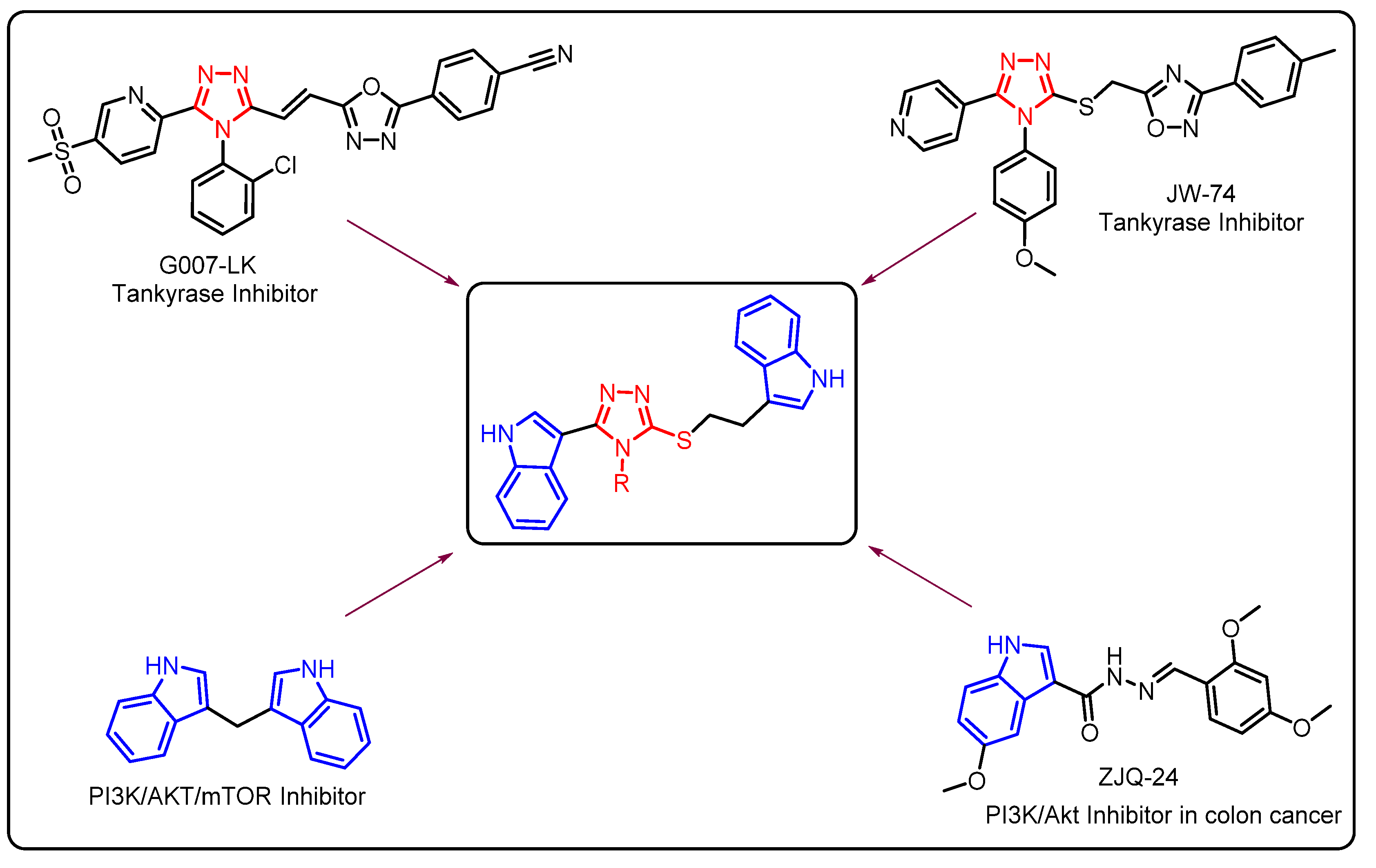
1. Introduction
2. Results and Discussion
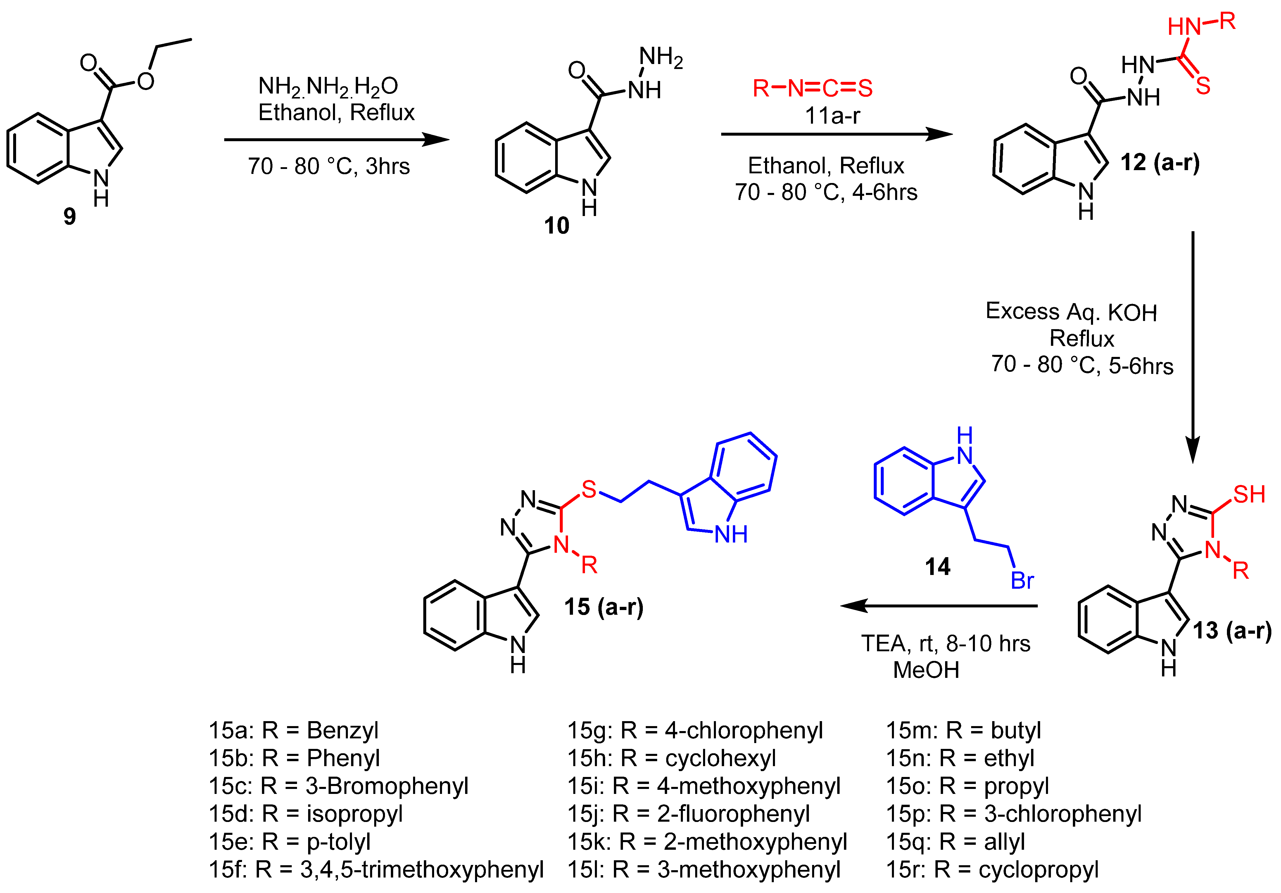
2.1. Chemistry
2.2. Biology
2.2.1. Cytotoxic Activity
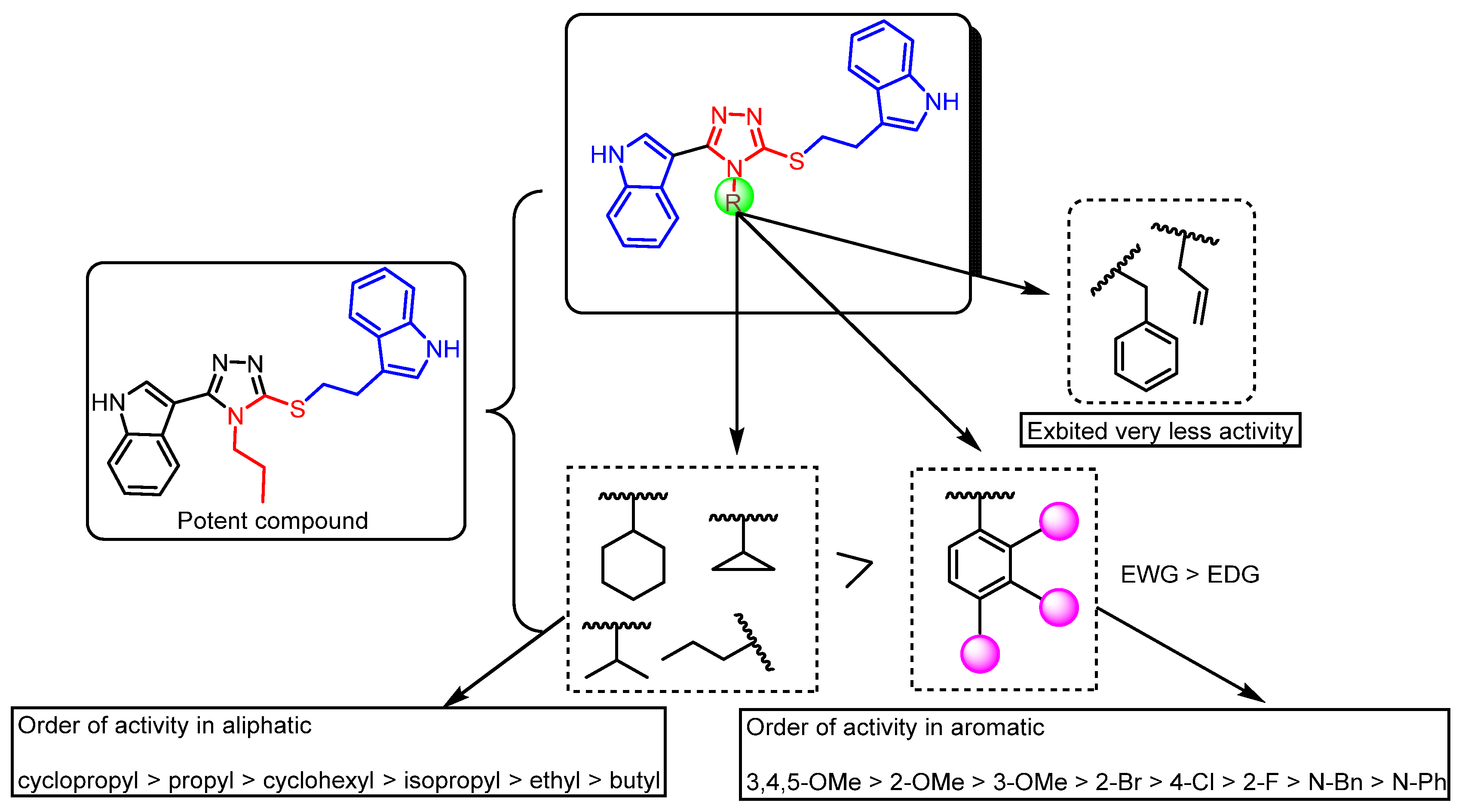
2.2.2. Structure–Activity Relationship (SAR)
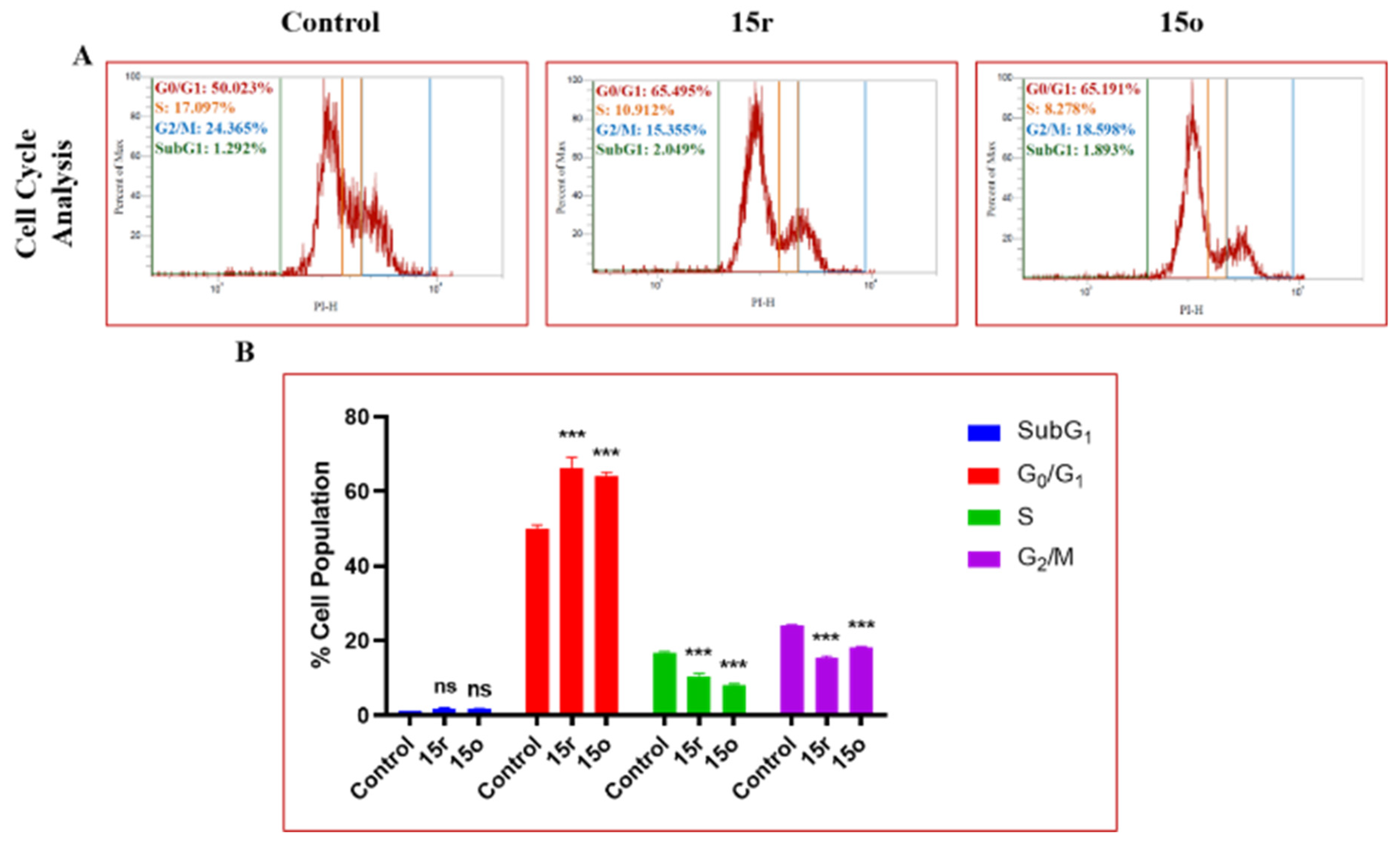
2.2.3. Cell Cycle Analysis
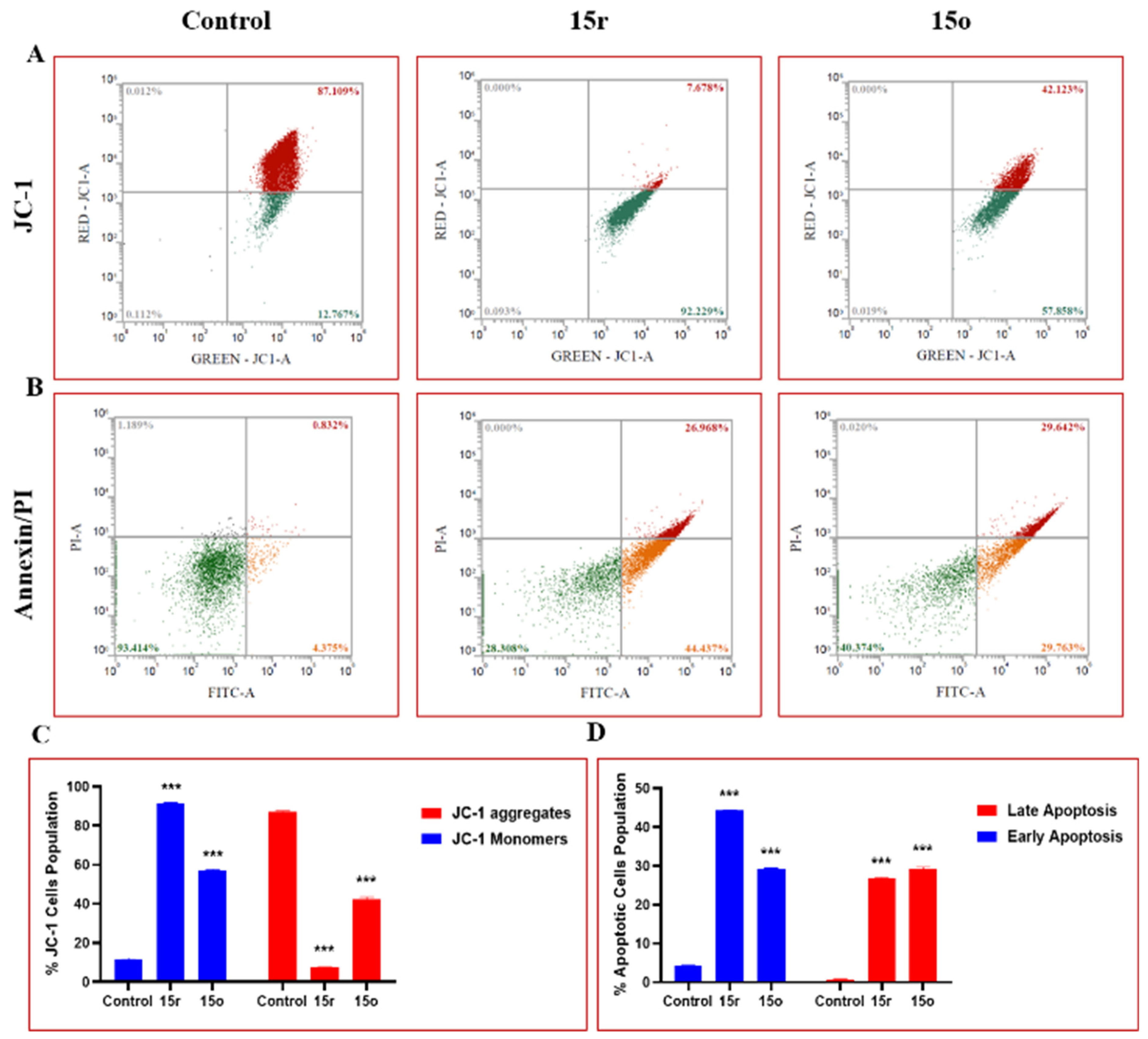
2.2.4. Mitochondrial Membrane Potential (MMP) Analysis
2.2.5. Measurement of Cellular Apoptosis in HT-29 Cells
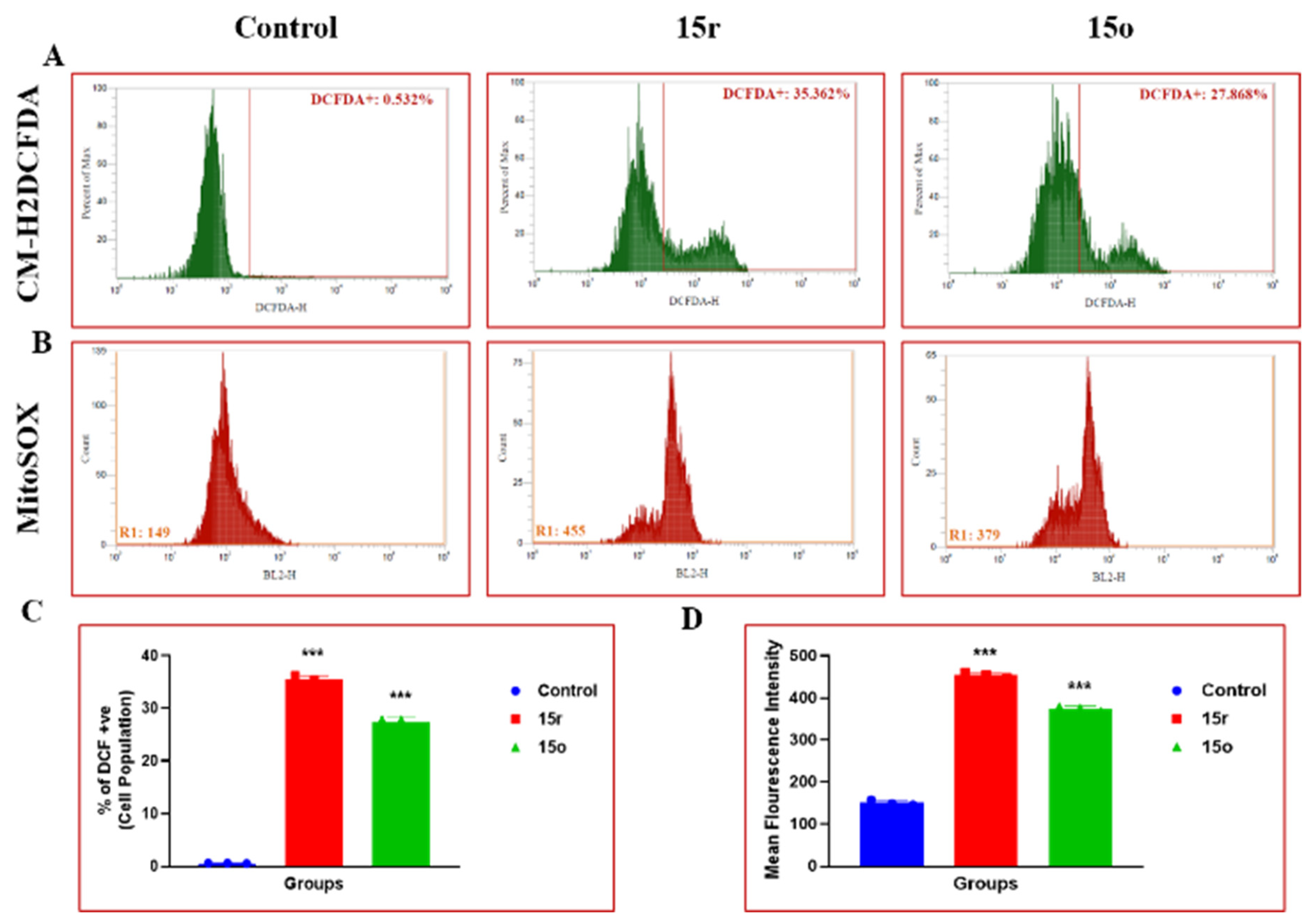
2.2.6. Effect on ROS Production
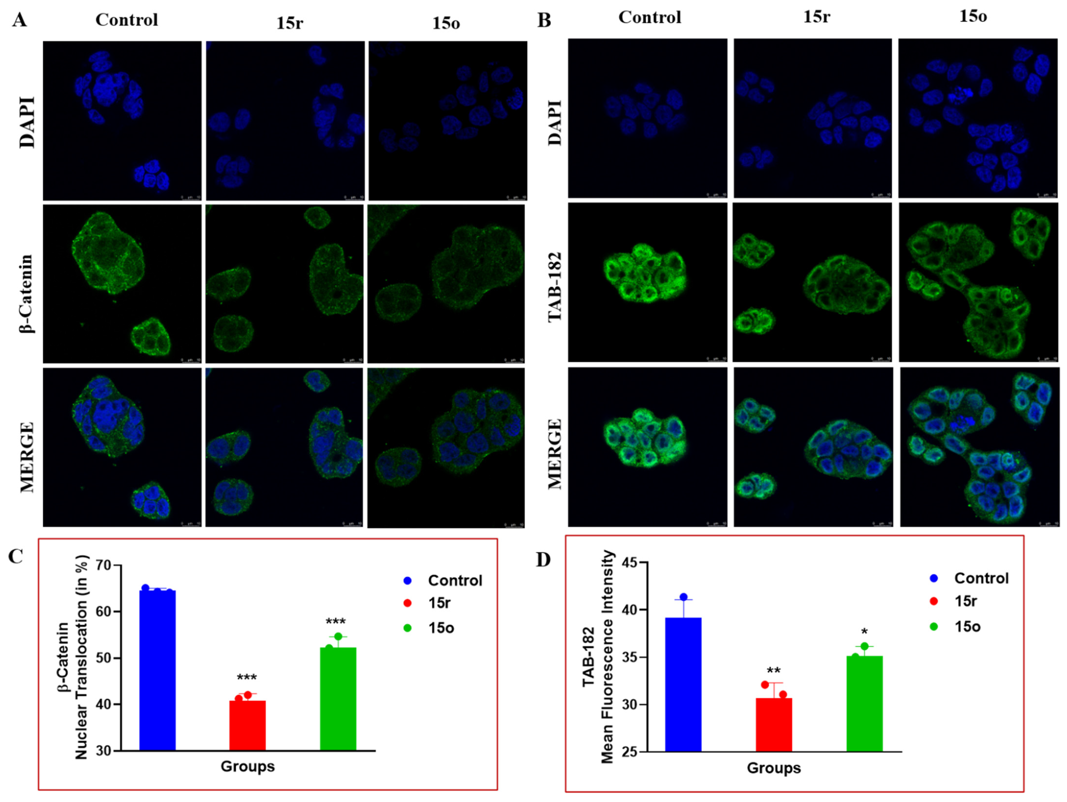
2.2.7. Immunofluorescence Analysis of TAB-182 Levels and β-Catenin Levels
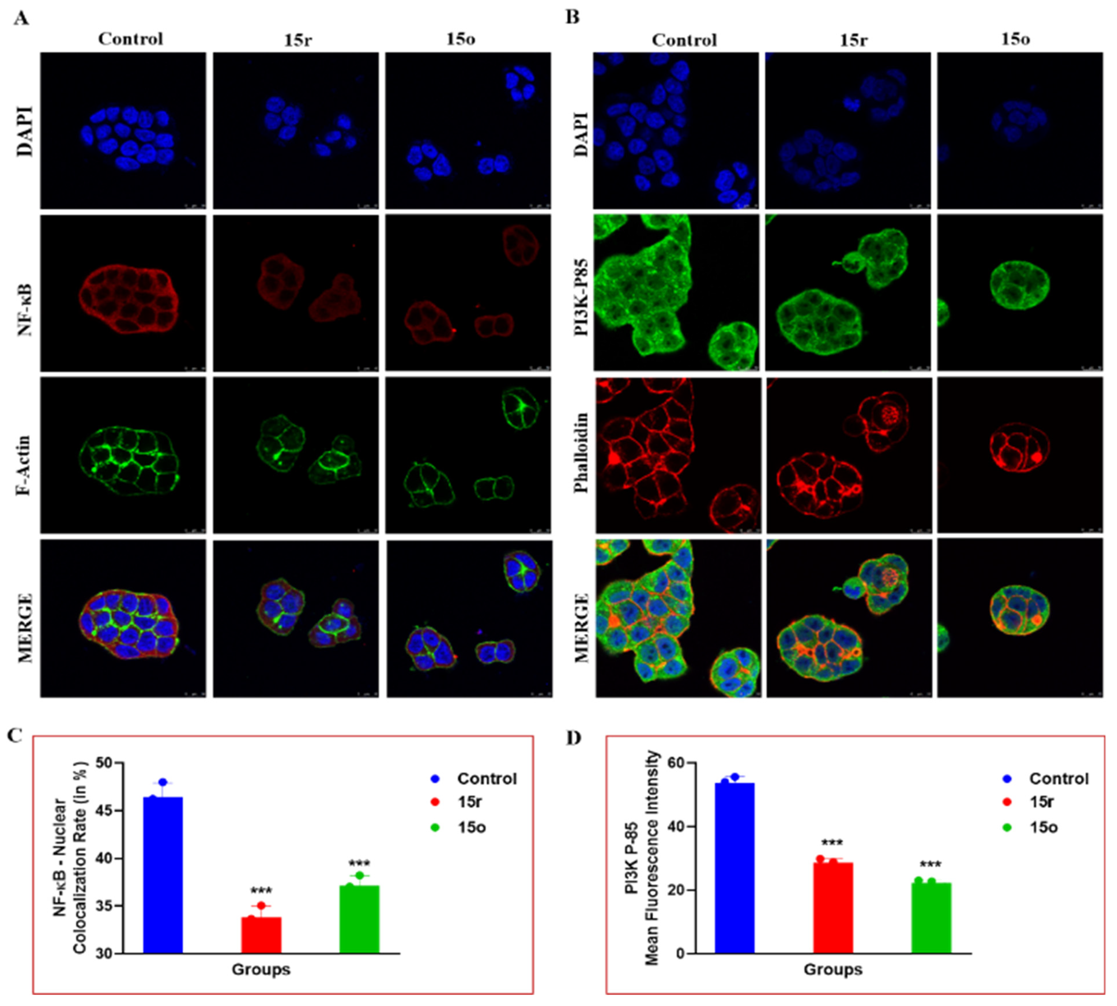
2.2.8. Immunofluorescence Analysis of NF-κB and PI3K-P85
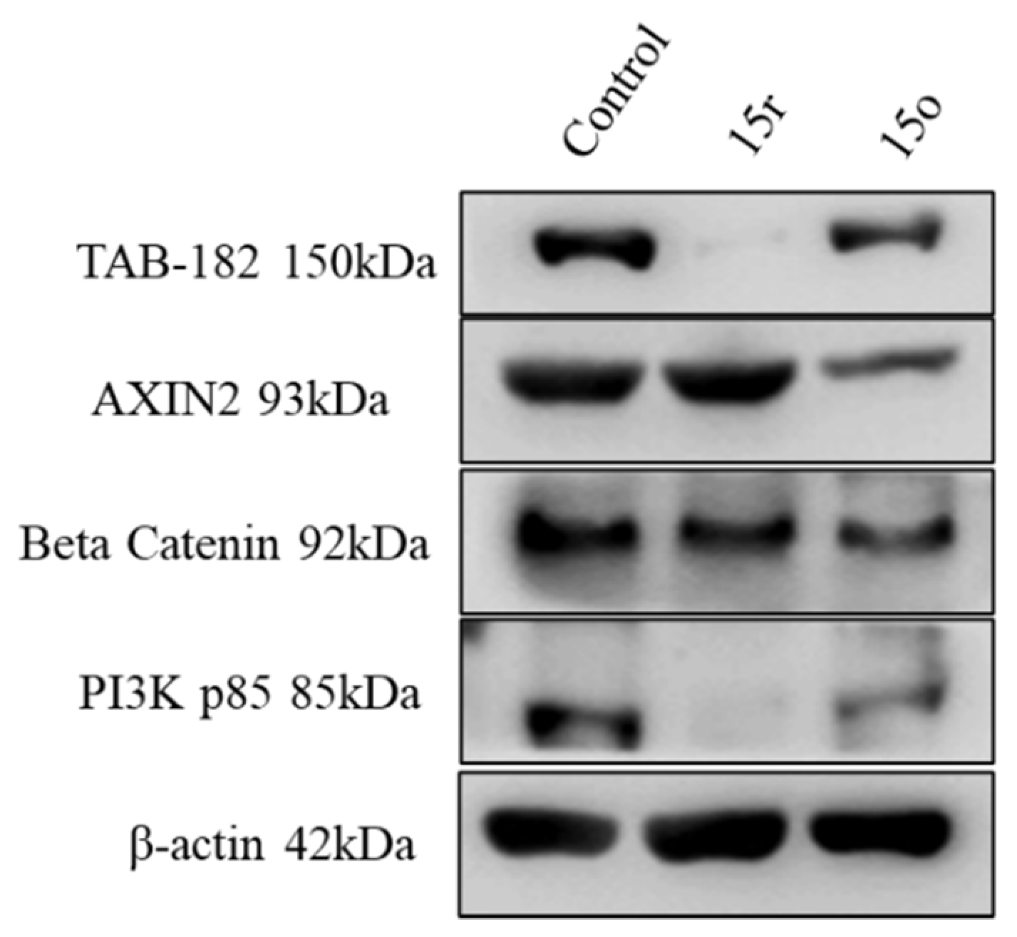
2.2.9. Western Blotting
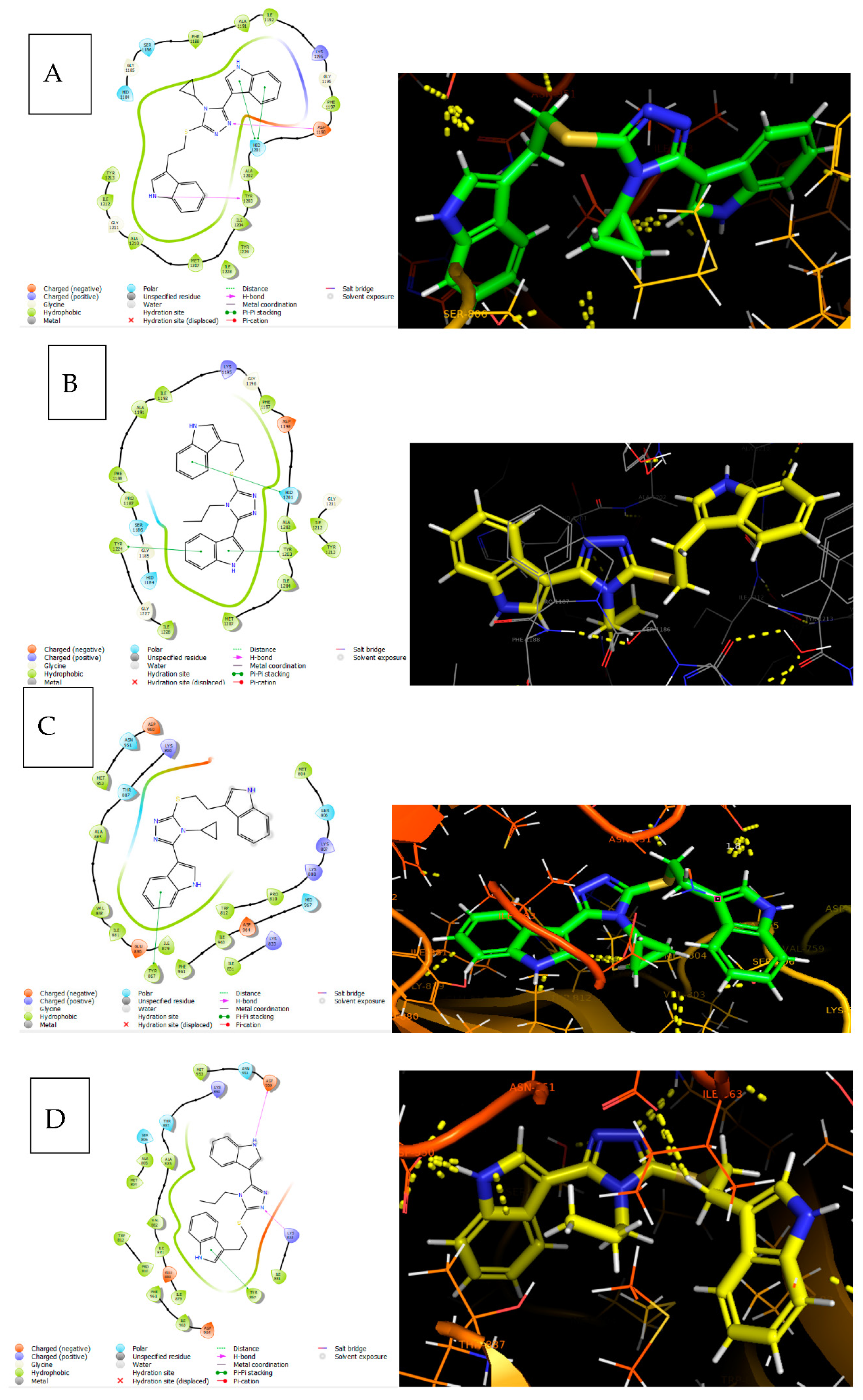
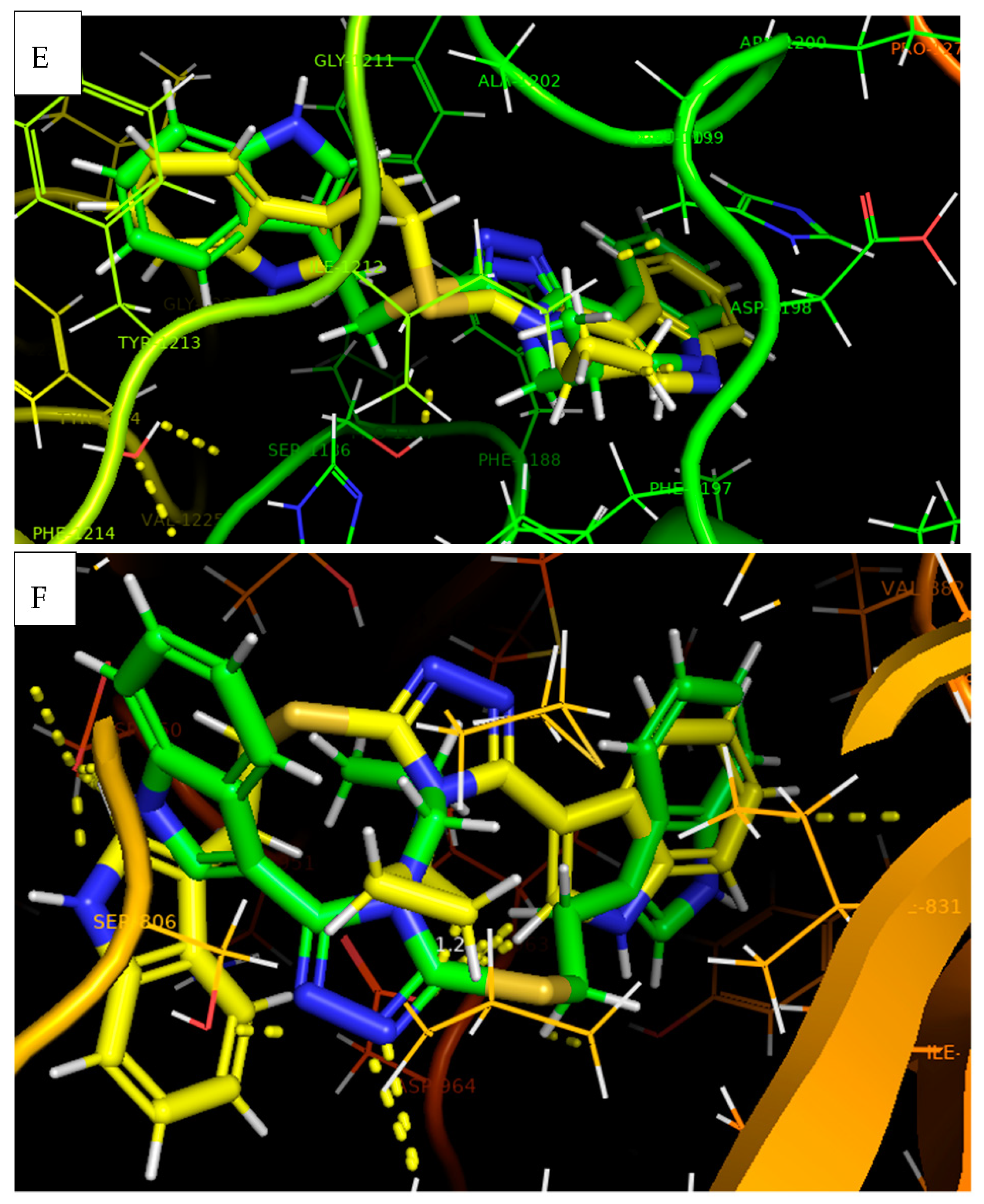
2.2.10. Molecular Docking Studies
2.2.11. Results of In Silico ADME Studies
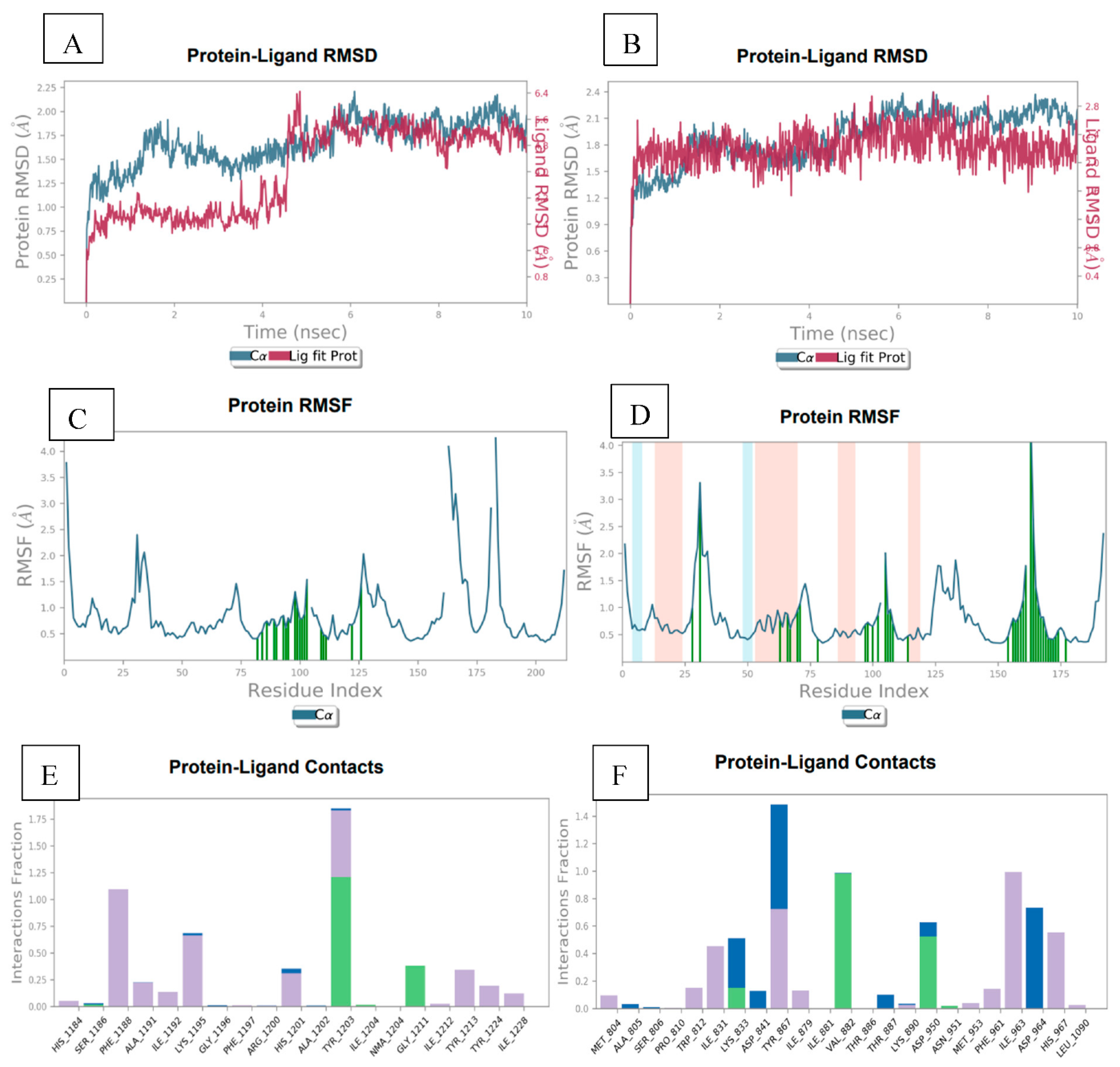
2.2.12. Molecular Dynamic Simulation Studies
3. Materials and Methods
3.1. General
3.2. Chemistry
3.2.1. General Procedure for Synthesis of 1H-indole-3-carbohydrazide (10)
3.2.2. General Procedure for Synthesis of 2-(1H-indole-3-carbonyl)-N-phenylhydrazine-1-carbothioamide (11a–r)
3.2.3. General Procedure for the Synthesis of 5-(1H-indol-3-yl)-4-phenyl-4H-1,2,4-triazole-3-thiol (13a–r)
3.2.4. General Procedure for the Synthesis of 3-(5-((2-(1H-indol-3-yl)ethyl)thio)-4-benzyl-4H-1,2,4-triazol-3-yl)-1H-indole (15a–r)
- 3-(5-((2-(1H-Indol-3-yl)ethyl)thio)-4-benzyl-4H-1,2,4-triazol-3-yl)-1H-indole (15a)
- 3-(2-((5-(1H-Indol-3-yl)-4-phenyl-4H-1,2,4-triazol-3-yl)thio) ethyl)-1H-indole (15b)
- 3-(5-((2-(1H-Indol-3-yl)ethyl)thio)-4-(3-bromophenyl)-4H-1,2,4-triazol-3-yl)-1H-indole (15c)
- 3-(2-((5-(1H-Indol-3-yl)-4-isopropyl-4H-1,2,4-triazol-3-yl)thio)ethyl)-1H-indole (15d)
- 3-(2-((5-(1H-Indol-3-yl)-4-(p-tolyl)-4H-1,2,4-triazol-3-yl)thio)ethyl)-1H-indole (15e)
- 3-(2-((5-(1H-Indol-3-yl)-4-(3,4,5-trimethoxyphenyl)-4H-1,2,4-triazol-3-yl)thio)ethyl)-1H-indole (15f)
- 3-(5-((2-(1H-Indol-3-yl)ethyl)thio)-4-(4-chlorophenyl)-4H-1,2,4-triazol-3-yl)-1H-indole (15g)
- 3-(5-((2-(1H-Indol-3-yl)ethyl)thio)-4-cyclohexyl-4H-1,2,4-triazol-3-yl)-1H-indole (15h)
- 3-(2-((5-(1H-Indol-3-yl)-4-(4-methoxyphenyl)-4H-1,2,4-triazol-3-yl)thio)ethyl)-1H-indole (15i)
- 3-(5-((2-(1H-Indol-3-yl)ethyl)thio)-4-(2-fluorophenyl)-4H-1,2,4-triazol-3-yl)-1H-indole (15j)
- 3-(2-((5-(1H-Indol-3-yl)-4-(2-methoxyphenyl)-4H-1,2,4-triazol-3-yl)thio)ethyl)-1H-indole (15k)
- 3-(2-((5-(1H-Indol-3-yl)-4-(3-methoxyphenyl)-4H-1,2,4-triazol-3-yl)thio)ethyl)-1H-indole (15l)
- 3-(5-((2-(1H-Indol-3-yl)ethyl)thio)-4-butyl-4H-1,2,4-triazol-3-yl)-1H-indole (15m)
- 3-(5-((2-(1H-Indol-3-yl)ethyl)thio)-4-ethyl-4H-1,2,4-triazol-3-yl)-1H-indole (15n)
- 3-(2-((5-(1H-Indol-3-yl)-4-propyl-4H-1,2,4-triazol-3-yl)thio)ethyl)-1H-indole (15o)
- 3-(5-((2-(1H-Indol-3-yl)ethyl)thio)-4-(3-chlorophenyl)-4H-1,2,4-triazol-3-yl)-1H-indole (15p)
- 3-(5-((2-(1H-Indol-3-yl)ethyl)thio)-4-allyl-4H-1,2,4-triazol-3-yl)-1H-indole (15q)
- 3-(5-((2-(1H-Indol-3-yl)ethyl)thio)-4-cyclopropyl-4H-1,2,4-triazol-3-yl)-1H-indole (15r)
3.3. Biology
3.3.1. MTT Assay
3.3.2. Cell Cycle Analysis
3.3.3. Evaluation of Mitochondrial Membrane Potential
3.3.4. Evaluation of Apoptosis by Annexin V/Propidium Iodide (PI)
3.3.5. Evaluation of Total Reactive Oxygen Species (ROS)
3.3.6. Evaluation of Mitochondrial ROS
3.3.7. Immunocytochemistry (ICC)
3.3.8. Western Blotting
3.3.9. Molecular Docking
3.3.10. ADMET Property Prediction
3.3.11. Molecular Dynamic Simulation
4. Conclusions
Supplementary Materials
Author Contributions
Funding
Institutional Review Board Statement
Informed Consent Statement
Data Availability Statement
Acknowledgments
Conflicts of Interest
Sample Availability
Abbreviations
| ROS | reactive oxygen species |
| MMP | mitochondrial membrane potential |
| IC50 | half-maximal inhibitory concentration |
| SAR | structure–activity relationship |
| PI3K | phosphoinositide 3-kinase |
References
- Sung, H.; Ferlay, J.; Siegel, R.L.; Laversanne, M.; Soerjomataram, I.; Jemal, A.; Bray, F. Global Cancer Statistics 2020: GLOBOCAN Estimates of Incidence and Mortality Worldwide for 36 Cancers in 185 Countries. CA Cancer J. Clin. 2021, 71, 209–249. [Google Scholar] [CrossRef] [PubMed]
- Jemal, A.; Bray, F.; Ferlay, J. Global Cancer Statistics: 2011. CA Cancer J. Clin. 1999, 49, 33–64. [Google Scholar] [CrossRef]
- Ferlay, J.; Soerjomataram, I.; Dikshit, R.; Eser, S.; Mathers, C.; Rebelo, M.; Parkin, D.M.; Forman, D.; Bray, F. Cancer Incidence and Mortality Worldwide: Sources, Methods and Major Patterns in GLOBOCAN 2012. Int. J. Cancer 2015, 136, E359–E386. [Google Scholar] [CrossRef] [PubMed]
- Ranasinghe, R.; Mathai, M.; Zulli, A. A Synopsis of Modern—Day Colorectal Cancer: Where We Stand. Biochim. Biophys. Acta Rev. Cancer 2022, 1877, 188699. [Google Scholar] [CrossRef]
- Siegel, R.L.; Miller, K.D.; Fuchs, H.E.; Jemal, A. Cancer Statistics, 2022. CA. Cancer J. Clin. 2022, 72, 7–33. [Google Scholar] [CrossRef]
- Ikediobi, O.N.; Davies, H.; Bignell, G.; Edkins, S.; Stevens, C.; O’Meara, S.; Santarius, T.; Avis, T.; Barthorpe, S.; Brakenbury, L.; et al. Mutation Analysis of 24 Known Cancer Genes in the NCI-60 Cell Line Set. Mol. Cancer Ther. 2006, 5, 2606–2612. [Google Scholar] [CrossRef]
- Shaik, A.B.; Rao, G.K.; Kumar, G.B.; Patel, N.; Reddy, V.S.; Khan, I.; Routhu, S.R.; Kumar, C.G.; Veena, I.; Chandra Shekar, K.; et al. Design, Synthesis and Biological Evaluation of Novel Pyrazolochalcones as Potential Modulators of PI3K/Akt/MTOR Pathway and Inducers of Apoptosis in Breast Cancer Cells. Eur. J. Med. Chem. 2017, 139, 305–324. [Google Scholar] [CrossRef]
- Kamal, A.; Lakshma Nayak, V.; Nagesh, N.; Vishnuvardhan, M.V.P.S.; Subba Reddy, N.V. Benzo[b]Furan Derivatives Induces Apoptosis by Targeting the PI3K/Akt/MTOR Signaling Pathway in Human Breast Cancer Cells. Bioorg. Chem. 2016, 66, 124–131. [Google Scholar] [CrossRef]
- Clevers, H. Wnt/β-Catenin Signaling in Development and Disease. Cell 2006, 127, 469–480. [Google Scholar] [CrossRef]
- Arqués, O.; Chicote, I.; Puig, I.; Tenbaum, S.P.; Argilés, G.; Dienstmann, R.; Fernández, N.; Caratù, G.; Matito, J.; Silberschmidt, D.; et al. Tankyrase Inhibition Blocks Wnt/b-Catenin Pathway and Reverts Resistance to PI3K and AKT Inhibitors in the Treatment of Colorectal Cancer. Clin. Cancer Res. 2016, 22, 644–656. [Google Scholar] [CrossRef]
- Solberg, N.T.; Waaler, J.; Lund, K.; Mygland, L.; Olsen, P.A.; Krauss, S. TANKYRASE Inhibition Enhances the Antiproliferative Effect of PI3K and EGFR Inhibition, Mutually Affecting β-CATENIN and AKT Signaling in Colorectal Cancer. Mol. Cancer Res. 2018, 16, 543–553. [Google Scholar] [CrossRef] [PubMed]
- Kamal, A.; Riyaz, S.; Srivastava, A.; Rahim, A. Tankyrase Inhibitors as Therapeutic Targets for Cancer. Curr. Top. Med. Chem. 2014, 14, 1967–1976. [Google Scholar] [CrossRef]
- Ma, L.; Wang, X.; Jia, T.; Wei, W.; Chua, M.S.; So, S. Tankyrase Inhibitors Attenuate WNT/ß-Catenin Signaling and Inhibit Growth of Hepatocellular Carcinoma Cells. Oncotarget 2015, 6, 25390–25401. [Google Scholar] [CrossRef] [PubMed]
- Ahmed, K.; Shaw, H.V.; Koval, A.; Katanaev, V.L. A Second WNT for Old Drugs: Drug Repositioning against WNT-Dependent Cancers. Cancers 2016, 8, 66. [Google Scholar] [CrossRef] [PubMed]
- Ahmad, A.; Biersack, B.; Li, Y.; Kong, D.; Bao, B.; Schobert, R.; Padhye, S.B.; Sarkar, F.H. Targeted Regulation of PI3K/Akt/MTOR/NF-ΚB Signaling by Indole Compounds and Their Derivatives: Mechanistic Details and Biological Implications for Cancer Therapy. Anti-Cancer Agents Med. Chem. Former. Curr. Med. Chem. Anti-Cancer Agent 2013, 13, 1002–1013. [Google Scholar] [CrossRef]
- Kim, D.J.; Reddy, K.; Kim, M.O.; Li, Y.; Nadas, J.; Cho, Y.-Y.; Kim, J.-E.; Shim, J.-H.; Song, N.R.; Carper, A.; et al. (3-Chloroacetyl)-Indole, a Novel Allosteric AKT Inhibitor, Suppresses Colon Cancer Growth In Vitro and In Vivo. Cancer Prev. Res. (Phila) 2011, 4, 1842–1851. [Google Scholar] [CrossRef]
- Zheng, S.; Liu, J.; Wu, Y.; Huang, T.L.; Wang, G. Small-Molecule Inhibitors of Wnt Signaling Pathway: Towards Novel Anticancer Therapeutics. Future Med. Chem. 2015, 7, 2485–2505. [Google Scholar] [CrossRef]
- Voronkov, A.; Krauss, S. Wnt/Beta-Catenin Signaling and Small Molecule Inhibitors. Curr. Pharm. Des. 2012, 19, 634–664. [Google Scholar] [CrossRef]
- Gustafson, K.; Roman, M.; Fenical, W. The Macrolactins, a Novel Class of Antiviral and Cytotoxic Macrolides from a Deep-Sea Marine Bacterium. J. Am. Chem. Soc. 1989, 111, 7519–7524. [Google Scholar] [CrossRef]
- James, R.G.; Davidson, K.C.; Bosch, K.A.; Biechele, T.L.; Robin, N.C.; Taylor, R.J.; Major, M.B.; Camp, N.D.; Fowler, K.; Martins, T.J.; et al. WIKI4, a Novel Inhibitor of Tankyrase and Wnt/ß-Catenin Signaling. PLoS ONE 2012, 7, e50457. [Google Scholar] [CrossRef]
- McGonigle, S.; Chen, Z.; Wu, J.; Chang, P.; Kolber-Simonds, D.; Ackermann, K.; Twine, N.C.; Shie, J.L.; Miu, J.T.; Huang, K.C.; et al. E7449: A Dual Inhibitor of PARP1/2 and Tankyrase1/2 Inhibits Growth of DNA Repair Deficient Tumors and Antagonizes Wnt Signaling. Oncotarget 2015, 6, 41307–41323. [Google Scholar] [CrossRef] [PubMed]
- Liu, J.; Liu, Y.; Zhang, J.; Liu, D.; Bao, Y.; Chen, T.; Tang, T.; Lin, J.; Luo, Y.; Jin, Y.; et al. Indole Hydrazide Compound ZJQ-24 Inhibits Angiogenesis and Induces Apoptosis Cell Death through Abrogation of AKT/MTOR Pathway in Hepatocellular Carcinoma. Cell Death Dis. 2020, 11, 926. [Google Scholar] [CrossRef]
- Waaler, J.; Leenders, R.G.G.; Sowa, S.T.; Alam Brinch, S.; Lycke, M.; Nieczypor, P.; Aertssen, S.; Murthy, S.; Galera-Prat, A.; Damen, E.; et al. Preclinical Lead Optimization of a 1,2,4-Triazole Based Tankyrase Inhibitor. J. Med. Chem. 2020, 63, 6834–6846. [Google Scholar] [CrossRef] [PubMed]
- Maira, S.M.; Pecchi, S.; Huang, A.; Burger, M.; Knapp, M.; Sterker, D.; Schnell, C.; Guthy, D.; Nagel, T.; Wiesmann, M.; et al. Identification and Characterization of NVP-BKM120, an Orally Available Pan-Class I PI3-Kinase Inhibitor. Mol. Cancer Ther. 2012, 11, 317–328. [Google Scholar] [CrossRef]
- Kang, Y.; Regmi, S.C.; Kim, M.Y.; Banskota, S.; Gautam, J.; Kim, D.H.; Kim, J.A. Anti-Angiogenic Activity of Macrolactin A and Its Succinyl Derivative Is Mediated through Inhibition of Class i PI3K Activity and Its Signaling. Arch. Pharm. Res. 2015, 38, 249–260. [Google Scholar] [CrossRef]
- Regmi, S.C.; Park, S.Y.; Kim, S.J.; Banskota, S.; Shah, S.; Kim, D.H.; Kim, J.A. The Anti-Tumor Activity of Succinyl Macrolactin a Is Mediated through the β-Catenin Destruction Complex via the Suppression of Tankyrase and PI3K/Akt. PLoS ONE 2015, 10, e0141753. [Google Scholar] [CrossRef] [PubMed]
- Rachakonda, S.; Naaz, F.; Ali, I.; Prasad, K.R.S.; Mandava, V.B.R.; Syed, S. Synthesis and Anti–microbial Activity of 1,2,3–triazole Tethered Nitroguiacol Ethers. Asian J. Pharm. Clin. Res. 2019, 12, 329–334. [Google Scholar] [CrossRef]
- Naaz, F.; Neha, K.; Haider, M.R.; Shafi, S. Indole Derivatives (2010–2020) as Versatile Tubulin Inhibitors: Synthesis and Structure-Activity Relationships. Future Med. Chem. 2021, 13, 1795–1828. [Google Scholar] [CrossRef]
- Naaz, F.; Ahmad, F.; Lone, B.A.; Pokharel, Y.R.; Fuloria, N.K.; Fuloria, S.; Ravichandran, M.; Pattabhiraman, L.; Shafi, S.; Shahar Yar, M. Design and Synthesis of Newer 1,3,4-Oxadiazole and 1,2,4-Triazole Based Topsentin Analogues as Anti-Proliferative Agent Targeting Tubulin. Bioorg. Chem. 2020, 95, 103519. [Google Scholar] [CrossRef]
- Pedada, S.R.; Yarla, N.S.; Tambade, P.J.; Dhananjaya, B.L.; Bishayee, A.; Arunasree, K.M.; Philip, G.H.; Dharmapuri, G.; Aliev, G.; Putta, S.; et al. Synthesis of New Secretory Phospholipase A2-Inhibitory Indole Containing Isoxazole Derivatives as Anti-Inflammatory and Anticancer Agents. Eur. J. Med. Chem. 2016, 112, 289–297. [Google Scholar] [CrossRef]
- Namballa, H.K.; Anchi, P.; Lakshmi Manasa, K.; Soni, J.P.; Godugu, C.; Shankaraiah, N.; Kamal, A. β-Carboline Tethered Cinnamoyl 2-Aminobenzamides as Class I Selective HDAC Inhibitors: Design, Synthesis, Biological Activities and Modelling Studies. Bioorg. Chem. 2021, 117, 105461. [Google Scholar] [CrossRef] [PubMed]
- Ahmed, S.; Kwatra, M.; Ranjan Panda, S.; Murty, U.S.N.; Naidu, V.G.M. Andrographolide Suppresses NLRP3 Inflammasome Activation in Microglia through Induction of Parkin-Mediated Mitophagy in In-Vitro and In-Vivo Models of Parkinson Disease. Brain. Behav. Immun. 2021, 91, 142–158. [Google Scholar] [CrossRef] [PubMed]
- Sakamuru, S.; Attene-Ramos, M.S.; Xia, M. Mitochondrial Membrane Potential Assay. Methods Mol. Biol. 2016, 1473, 17–22. [Google Scholar] [CrossRef] [PubMed]
- Rieger, A.M.; Barreda, D.R. Accurate Assessment of Cell Death by Imaging Flow Cytometry. Methods Mol. Biol. 2016, 1389, 209–220. [Google Scholar] [CrossRef]
- Wang, Q.; Zou, M.H. Measurement of Reactive Oxygen Species (ROS) and Mitochondrial ROS in AMPK Knockout Mice Blood Vessels. In Methods in Molecular Biology; Humana Press Inc.: Totowa, NJ, USA, 2018; Volume 1732, pp. 507–517. [Google Scholar]
- Zorov, D.B.; Juhaszova, M.; Sollott, S.J. Mitochondrial Reactive Oxygen Species (ROS) and ROS-Induced ROS Release. Physiol. Rev. 2014, 94, 909–950. [Google Scholar] [CrossRef]
- Ahmed, S.; Panda, S.R.; Kwatra, M.; Sahu, B.D.; Naidu, V. Perillyl Alcohol Attenuates NLRP3 Inflammasome Activation and Rescues Dopaminergic Neurons in Experimental In Vitro and In Vivo Models of Parkinson’s Disease. ACS Chem. Neurosci. 2022, 13, 53–68. [Google Scholar] [CrossRef]
- Naaz, F.; Ahmad, F.; Lone, B.A.; Khan, A.; Sharma, K.; Intzarali; Shaharyar, M.; Pokharel, Y.R.; Shafi, S. Apoptosis Inducing 1,3,4-Oxadiazole Conjugates of Capsaicin: Their in Vitro Antiproliferative and in Silico Studies. ACS Med. Chem. Lett. 2021, 12, 1694–1702. [Google Scholar] [CrossRef]
- Knight, S.D.; Adams, N.D.; Burgess, J.L.; Chaudhari, A.M.; Darcy, M.G.; Donatelli, C.A.; Luengo, J.I.; Newlander, K.A.; Parrish, C.A.; Ridgers, L.H.; et al. Discovery of GSK2126458, a Highly Potent Inhibitor of PI3K and the Mammalian Target of Rapamycin. ACS Med. Chem. Lett. 2010, 1, 39–43. [Google Scholar] [CrossRef]
- Kulak, O.; Chen, H.; Holohan, B.; Wu, X.; He, H.; Borek, D.; Otwinowski, Z.; Yamaguchi, K.; Garofalo, L.A.; Ma, Z.; et al. Disruption of Wnt/β-Catenin Signaling and Telomeric Shortening Are Inextricable Consequences of Tankyrase Inhibition in Human Cells. Mol. Cell. Biol. 2015, 35, 2425–2435. [Google Scholar] [CrossRef]
- Rathod, B.; Chak, S.; Patel, S.; Shard, A. Tumor Pyruvate Kinase M2 Modulators: A Comprehensive Account of Activators and Inhibitors as Anticancer Agents. RSC Med. Chem. 2021, 12, 1121–1141. [Google Scholar] [CrossRef]
- Lucas, A.J.; Sproston, J.L.; Barton, P.; Riley, R.J. Estimating Human ADME Properties, Pharmacokinetic Parameters and Likely Clinical Dose in Drug Discovery. Expert Opin. Drug Discov. 2019, 14, 1313–1327. [Google Scholar] [CrossRef] [PubMed]
- Kumar, A.; Rajappan, R.; Kini, S.G.; Rathi, E.; Dharmarajan, S.; Sreedhara Ranganath Pai, K. E-Pharmacophore Model-Guided Design of Potential DprE1 Inhibitors: Synthesis, in Vitro Antitubercular Assay and Molecular Modelling Studies. Chem. Pap. 2021, 75, 5571–5585. [Google Scholar] [CrossRef]
- Oda, A.; Saijo, K.; Ishioka, C.; Narita, K.; Katoh, T.; Watanabe, Y.; Fukuyoshi, S.; Takahashi, O. Predicting the Structures of Complexes between Phosphoinositide 3-Kinase (PI3K) and Romidepsin-Related Compounds for the Drug Design of PI3K/Histone Deacetylase Dual Inhibitors Using Computational Docking and the Ligand-Based Drug Design Approach. J. Mol. Graph. Model. 2014, 54, 46–53. [Google Scholar] [CrossRef]
- Feng, T.T.; Zhang, Y.J.; Chen, H.; Fan, S.; Han, J.G. The Binding Mechanism of a Novel Nicotinamide Isostere Inhibiting with TNKSs: A Molecular Dynamic Simulation and Binding Free Energy Calculation. J. Biomol. Struct. Dyn. 2016, 34, 517–528. [Google Scholar] [CrossRef] [PubMed]
- Ash, J.; Fourches, D. Characterizing the Chemical Space of ERK2 Kinase Inhibitors Using Descriptors Computed from Molecular Dynamics Trajectories. J. Chem. Inf. Model. 2017, 57, 1286–1299. [Google Scholar] [CrossRef] [PubMed]
- Kumar, A.; Rathi, E.; Kini, S.G. E-Pharmacophore Modelling, Virtual Screening, Molecular Dynamics Simulations and in-Silico ADME Analysis for Identification of Potential E6 Inhibitors against Cervical Cancer. J. Mol. Struct. 2019, 1189, 299–306. [Google Scholar] [CrossRef]


| S. No | Comp | IC50 (10 µM) against the Selected Cell Lines | ||||||||
|---|---|---|---|---|---|---|---|---|---|---|
| CaCo2 | DLD1 | HT-29 | HCT-15 | A172 | A549 | TERA-1 | MCF-7 | MDA-MB-231 | ||
| 1 | 15a | 4.9 | 20.6 | >20 | >20 | >20 | >20 | >20 | >20 | 14.5 |
| 2 | 15b | >20 | >20 | 3.71 | >20 | >20 | >20 | >20 | >20 | >20 |
| 3 | 15c | 11.9 | 7.3 | >20 | >20 | 16.6 | 7.8 | >20 | >20 | >20 |
| 4 | 15d | 4.5 | 16.5 | >20 | >20 | >20 | 17.9 | >20 | 3.36 | >20 |
| 5 | 15e | >20 | 18.9 | >20 | >20 | >20 | 17.5 | >20 | >20 | >20 |
| 6 | 15f | >20 | 8.1 | 2.98 | 2.64 | 12.3 | 5.02 | 4.8 | >20 | >20 |
| 7 | 15g | 6.9 | 7.2 | >20 | >20 | >20 | 7.0 | >20 | >20 | >20 |
| 8 | 15h | >20 | >20 | 5.85 | >20 | >20 | >20 | 17.4 | >20 | 1.35 |
| 9 | 15i | >20 | >20 | >20 | >20 | 10.7 | >20 | >20 | >20 | 3.16 |
| 10 | 15j | 8.9 | 13.3 | >20 | >20 | 17.3 | 10.0 | 17.5 | >20 | >20 |
| 11 | 15k | 12.3 | 8.2 | 3.73 | 1.37 | >20 | 8.9 | >20 | 3.85 | >20 |
| 12 | 15l | >20 | >20 | 6.44 | 3.20 | >20 | >20 | >20 | >20 | >20 |
| 13 | 15m | 8 | 8.9 | >20 | 12.42 | 14.7 | 13.7 | 17.7 | 2.29 | 6.11 |
| 14 | 15n | 12.1 | 9.3 | >20 | >20 | 15.9 | 10.5 | 14.1 | >20 | >20 |
| 15 | 15o | 5.1 | 5.6 | 2.04 | >20 | 4.4 | 5.7 | 5.1 | >20 | >20 |
| 16 | 15p | >20 | 19.1 | >20 | >20 | >20 | >20 | >20 | >20 | >20 |
| 17 | 15q | 14.4 | 8.9 | >20 | >20 | 18.9 | 9.3 | >20 | >20 | >20 |
| 18 | 15r | 6.5 | 7.9 | 0.85 | 4.04 | 8.8 | 7.4 | 8.4 | 3.03 | >20 |
| 19 | 5-FU | - | - | 5.31 | - | - | - | - | - | - |
| S. No. | Compound | Xp G Score (kcal/mol) 4OA7 | Interactions of Tankyrase1 in Complex with IWR1 at the Ligand Binding Site 4OA7 | Xp G Score (kcal/mol) 3L54 | Interactions of Pi3K Gamma at the Ligand Binding Site 3L54 |
|---|---|---|---|---|---|
| 1 | 15a | −8.021 | π-π: PHE 1188, HIE 1201 | −5.635 | π-π: TYR 867 π-Cation: LYS 890, LYS 802 |
| 2 | 15b | −6.399 | π-π: PHE 1208, TYR 1203, TYR 1224 | −4.279 | π-π: TRP 812, TYR 867 π-π-Cation: LYS 890 |
| 3 | 15c | −6.258 | π H-Bond: TYR 1203 π-π: PHE 1208, TYR 1224 Halogen bond: GLY 1185 | −5.845 | π-π: TRP 812 π-Cation: LYS 890 |
| 4 | 15d | −9.269 | H-Bond: GLY 1196 π-π: HIE 1201 | −4.776 | π-π: TYR 867 |
| 5 | 15e | −5.396 | π-π: HIE 1201, TYR 1213, TYR 1224 | −4.262 | π-π: TYR 867 |
| 6 | 15f | −5.396 | H-Bond: LYS 1195 π-π: PHE 1188, HIE 1201 | −4.96 | π-π: TRP 812, TYR 867 π-Cation: LYS 890 |
| 7 | 15g | −6.386 | π-π: PHE 1188, HIE 1201, TYR 1203 Halogen bond: LYS 1195 | −5.694 | H-Bond: VAL 882, LYS 890 π-π: TRP 812 π-Cation: LYS 890, LYS 802 |
| 8 | 15h | −8.919 | H-Bond: GLY 1196 π-π: HIE 1201, TYR 1213 | −3.213 | π-π: TRP 812 π-Cation: LYS 802 |
| 9 | 15i | −4.509 | H-Bond: TYR 1203 π-π: HIE 1201, PHE 1188, TYR 1224 | −5.363 | π-π: TYR 867, TRP 812 π-Cation: LYS 890 |
| 10 | 15j | −9.128 | H-Bond: TYR 1203 π-π: PHE 1208 | −3.829 | π-π: TYR 867, TRP 812 |
| 11 | 15k | −4.960 | H-Bond: TYR 1203 π-π: PHE 1208 | −6.257 | π-π: TYR 867 H-Bond: LYS 890 π-Cation: LYS 890, LYS 802 |
| 12 | 15l | −9.12 | H-Bond: ASN 1190 π-π: PHE 1188, HIE 1201 | −5.63 | H-Bond: ASP 950 π-Cation: LYS 890 |
| 13 | 15m | −8.633 | π-π: PHE 1188, TYR 1224 | −6.907 | π-π: TYR 867 π-Cation: LYS 890 |
| 14 | 15n | −9.724 | H-Bond: GLY 1196, TYR 1203 π-π: HIE 1201, TYR 1203, TYR 1224 | −5.768 | H-Bond: TYR 867 π-π: TRP 812 |
| 15 | 15o | −9.614 | H-Bond: ASP 1198, TYR 1203 π-π: HID 1201 | −6.833 | π-π: TYR 867 |
| 16 | 15p | −8.643 | H-Bond: GLY 1196, TYR 1203 π-π: HIE 1201, TYR 1203, TYR 1213 | −6.085 | H-Bond: VAL 882 π-π: TYR 867, TRP 812 |
| 17 | 15q | −6.243 | H-Bond: TYR 1203 π-π: HIE 1201, PHE 1188, TYR 1224 | −4.271 | H-Bond: VAL 882 π-π: TYR 867 π-Cation: LYS 890 |
| 18 | 15r | −9.223 | π-π: HID 1201, TYR 1203, TYR 1224 | −6.196 | H-Bond: LYS 833 π-π: TYR 867 |
| S. No. | Comp | M.Wt | H-Bond Accept | H-Bond Donor | LogP o/w | Absorption | Rule of 5 |
|---|---|---|---|---|---|---|---|
| 1 | 15a | 449.57 | 2 | 2 | 3.12 | 100 | 0 |
| 2 | 15b | 435.54 | 2 | 2 | 2.82 | 100 | 0 |
| 3 | 15c | 514.44 | 2 | 2 | 3.38 | 100 | 1 |
| 4 | 15d | 401.53 | 2 | 2 | 2.8 | 100 | 0 |
| 5 | 15e | 449.57 | 2 | 2 | 3.22 | 100 | 0 |
| 6 | 15f | 525.62 | 5 | 2 | 3.91 | 100 | 1 |
| 7 | 15g | 469.99 | 2 | 2 | 3.06 | 100 | 0 |
| 8 | 15h | 441.59 | 2 | 2 | 2.85 | 100 | 0 |
| 9 | 15i | 465.57 | 3 | 2 | 3 | 100 | 0 |
| 10 | 15j | 453.53 | 3 | 2 | 2.98 | 100 | 0 |
| 11 | 15k | 465.57 | 3 | 2 | 3.22 | 100 | 0 |
| 12 | 15l | 465.57 | 3 | 2 | 2.75 | 100 | 0 |
| 13 | 15m | 415.55 | 2 | 2 | 3.13 | 100 | 0 |
| 14 | 15o | 387.5 | 2 | 2 | 2.66 | 100 | 0 |
| 15 | 15n | 401.53 | 2 | 2 | 2.82 | 100 | 0 |
| 16 | 15p | 469.99 | 2 | 2 | 3.06 | 100 | 0 |
| 17 | 15q | 399.51 | 2 | 2 | 2.79 | 100 | 0 |
| 18 | 15r | 399.51 | 2 | 2 | 3.01 | 100 | 0 |
Publisher’s Note: MDPI stays neutral with regard to jurisdictional claims in published maps and institutional affiliations. |
© 2022 by the authors. Licensee MDPI, Basel, Switzerland. This article is an open access article distributed under the terms and conditions of the Creative Commons Attribution (CC BY) license (https://creativecommons.org/licenses/by/4.0/).
Share and Cite
Yakkala, P.A.; Panda, S.R.; Shafi, S.; Naidu, V.G.M.; Yar, M.S.; Ubanako, P.N.; Adeyemi, S.A.; Kumar, P.; Choonara, Y.E.; Radchenko, E.V.; et al. Synthesis and Cytotoxic Activity of 1,2,4-Triazolo-Linked Bis-Indolyl Conjugates as Dual Inhibitors of Tankyrase and PI3K. Molecules 2022, 27, 7642. https://doi.org/10.3390/molecules27217642
Yakkala PA, Panda SR, Shafi S, Naidu VGM, Yar MS, Ubanako PN, Adeyemi SA, Kumar P, Choonara YE, Radchenko EV, et al. Synthesis and Cytotoxic Activity of 1,2,4-Triazolo-Linked Bis-Indolyl Conjugates as Dual Inhibitors of Tankyrase and PI3K. Molecules. 2022; 27(21):7642. https://doi.org/10.3390/molecules27217642
Chicago/Turabian StyleYakkala, Prasanna A., Samir R. Panda, Syed Shafi, V. G. M. Naidu, M. Shahar Yar, Philemon N. Ubanako, Samson A. Adeyemi, Pradeep Kumar, Yahya E. Choonara, Eugene V. Radchenko, and et al. 2022. "Synthesis and Cytotoxic Activity of 1,2,4-Triazolo-Linked Bis-Indolyl Conjugates as Dual Inhibitors of Tankyrase and PI3K" Molecules 27, no. 21: 7642. https://doi.org/10.3390/molecules27217642
APA StyleYakkala, P. A., Panda, S. R., Shafi, S., Naidu, V. G. M., Yar, M. S., Ubanako, P. N., Adeyemi, S. A., Kumar, P., Choonara, Y. E., Radchenko, E. V., Palyulin, V. A., & Kamal, A. (2022). Synthesis and Cytotoxic Activity of 1,2,4-Triazolo-Linked Bis-Indolyl Conjugates as Dual Inhibitors of Tankyrase and PI3K. Molecules, 27(21), 7642. https://doi.org/10.3390/molecules27217642