


Polysorbates versus Hydroxypropyl Beta-Cyclodextrin (HPβCD): Comparative Study on Excipient Stability and Stabilization Benefits on Monoclonal Antibodies
Abstract
1. Introduction
2. Results
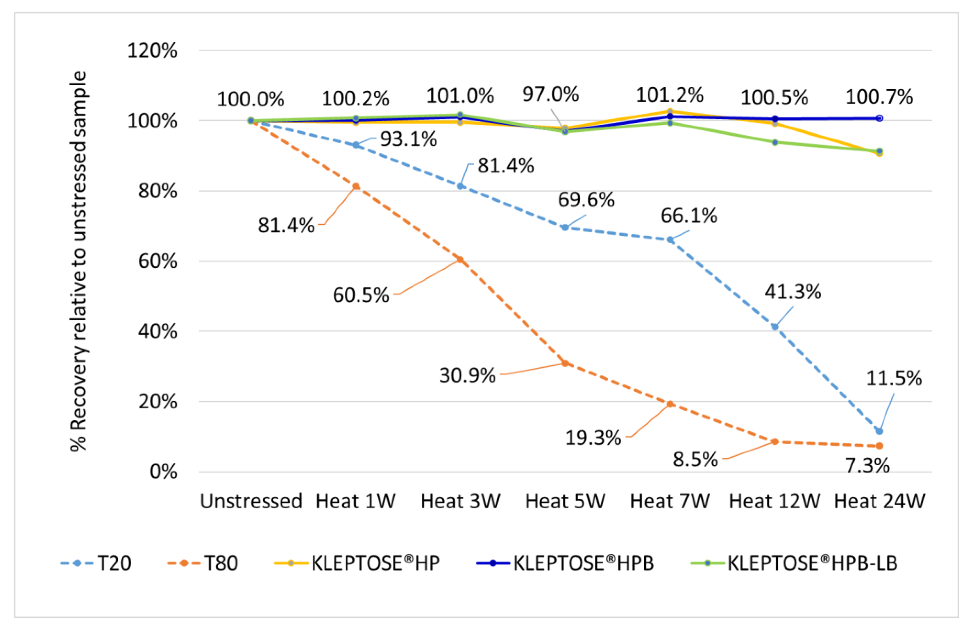
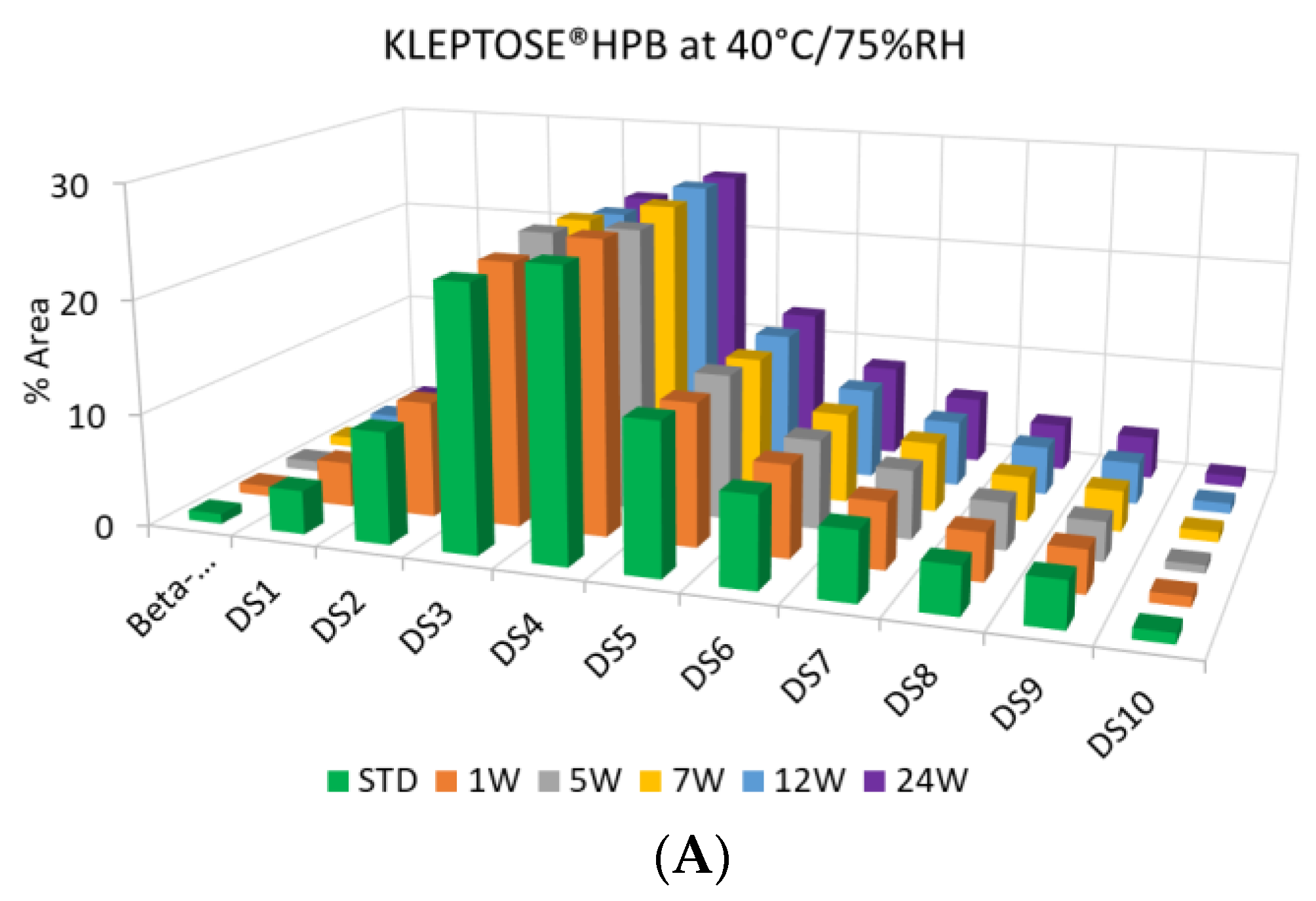
2.1. Accelerated Thermal Stability Studies
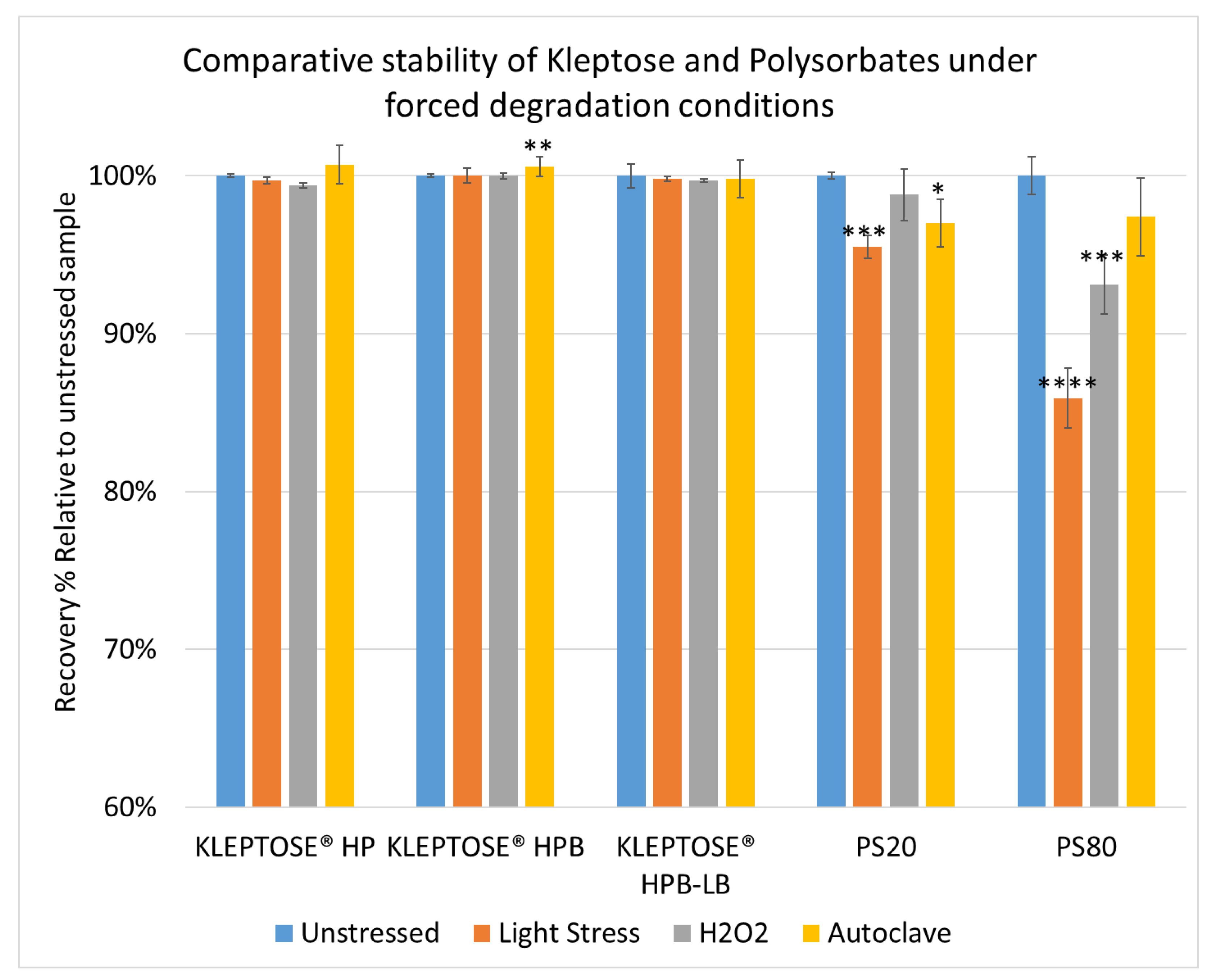
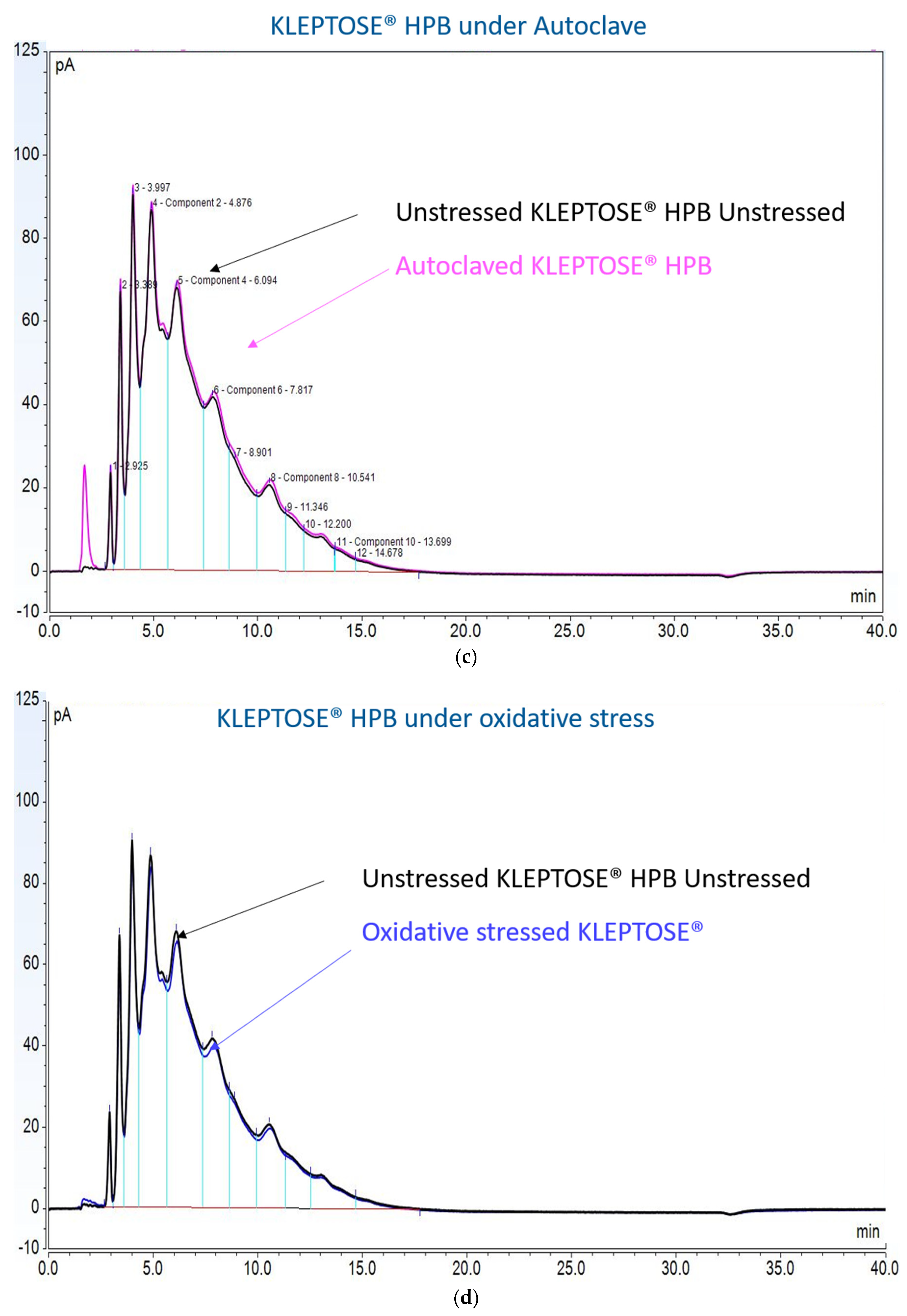
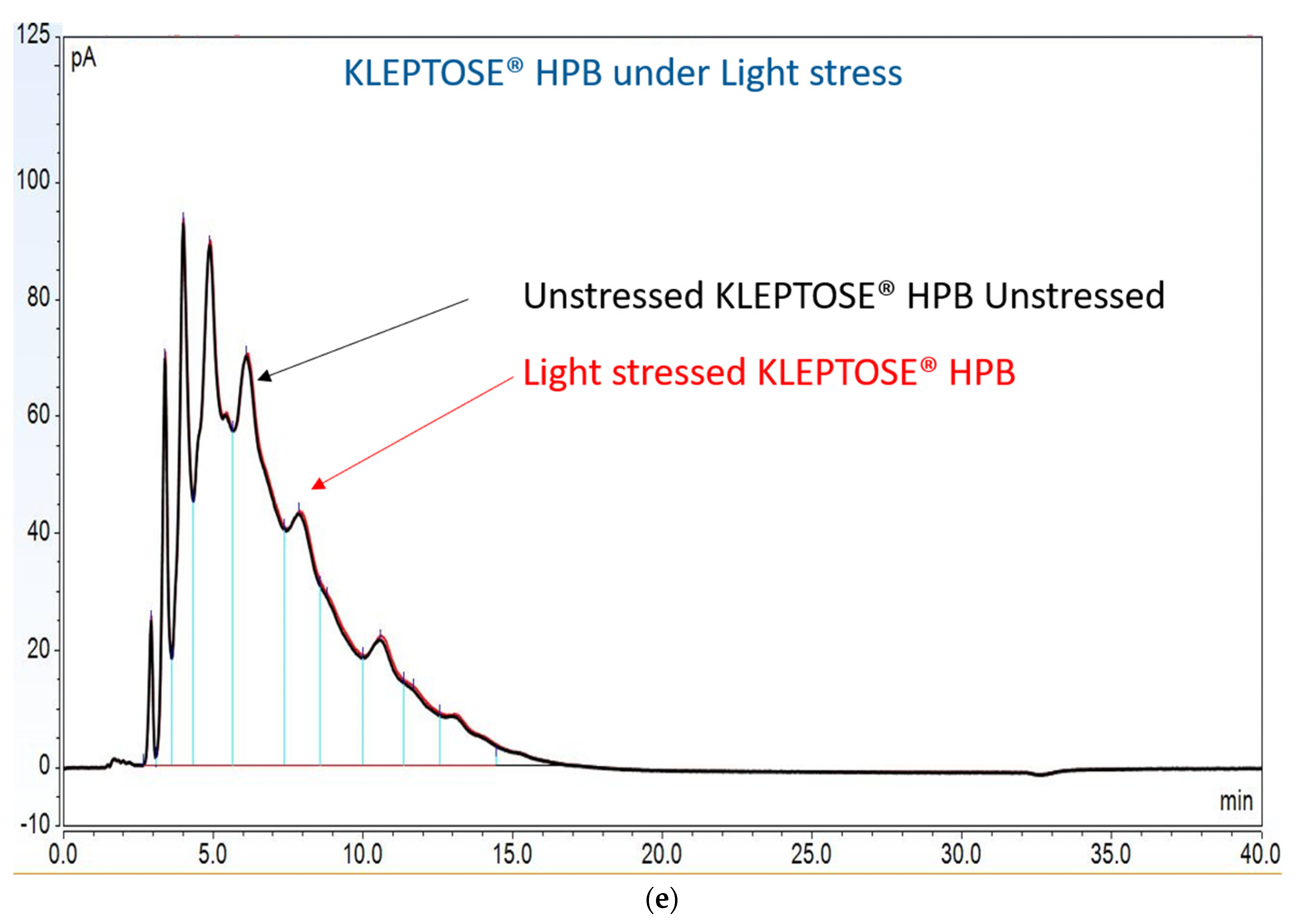
2.2. Forced Degradation Studies
2.3. Stabilization Benefits on Monoclonal Antibody
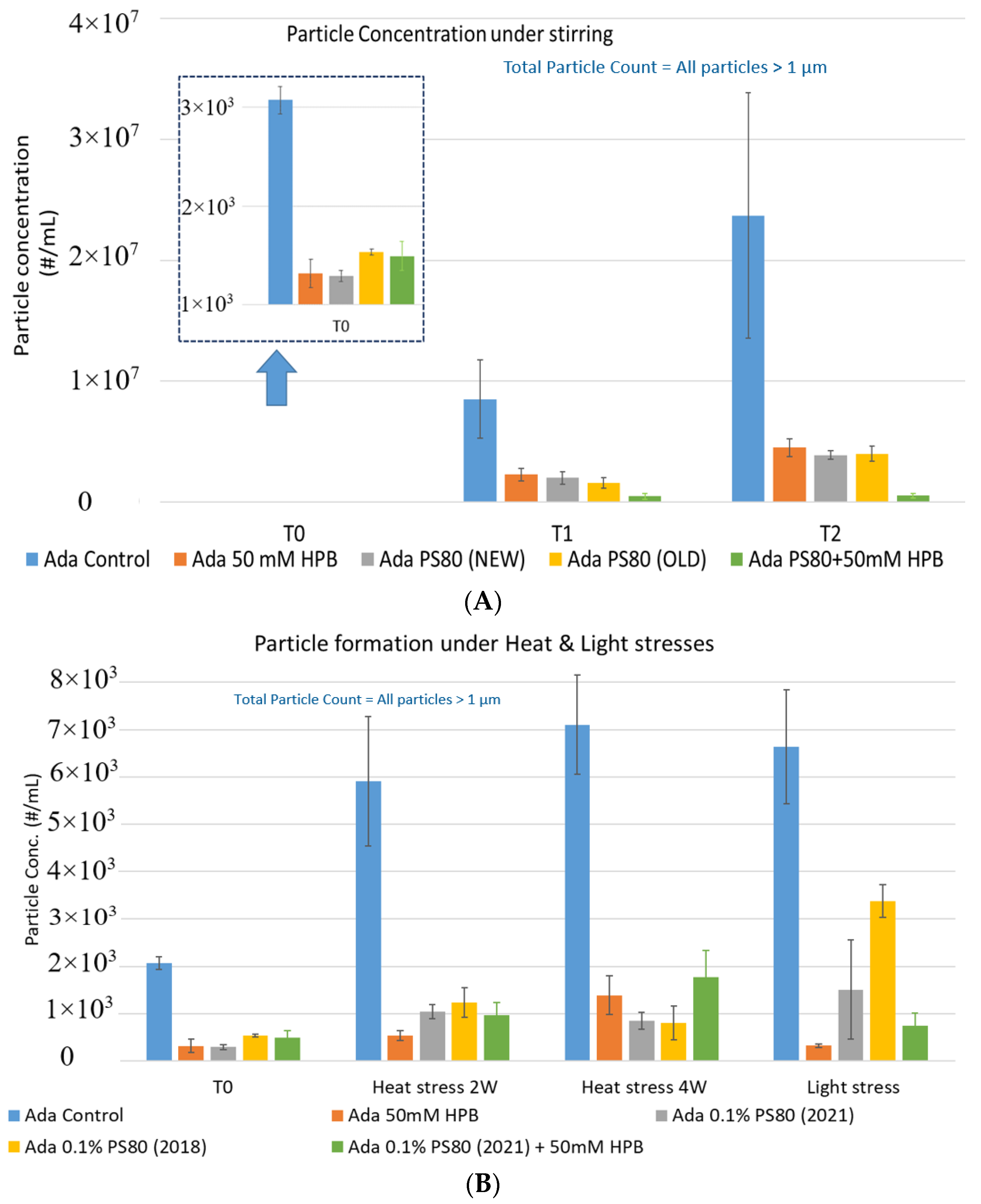
2.3.1. Subvisible Particle Formation of Adalimumab under Various Stresses
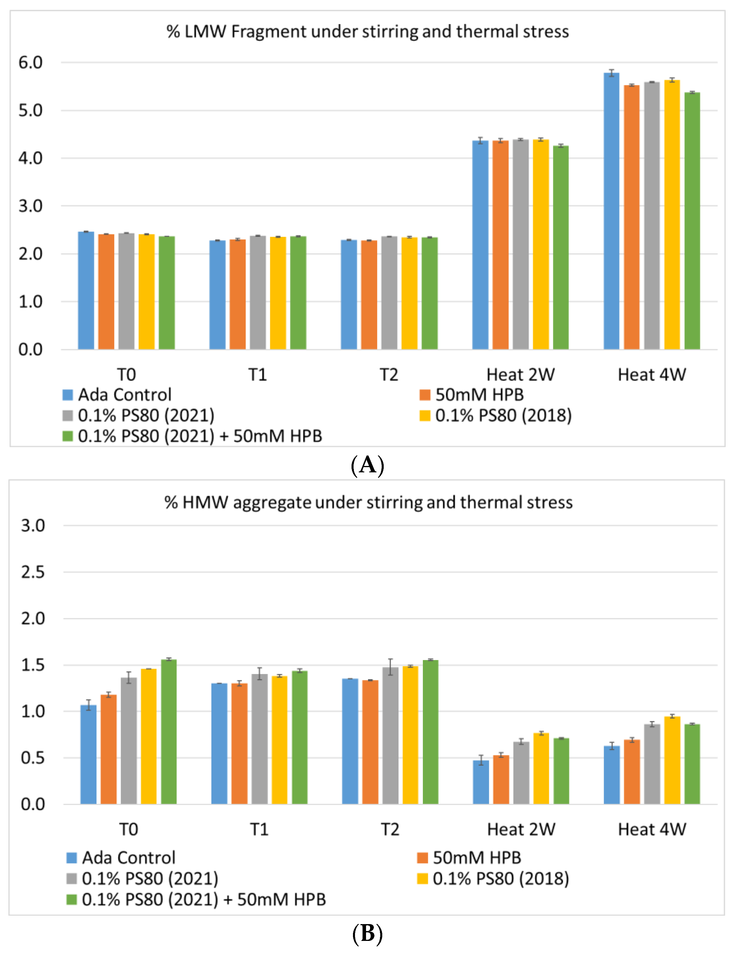
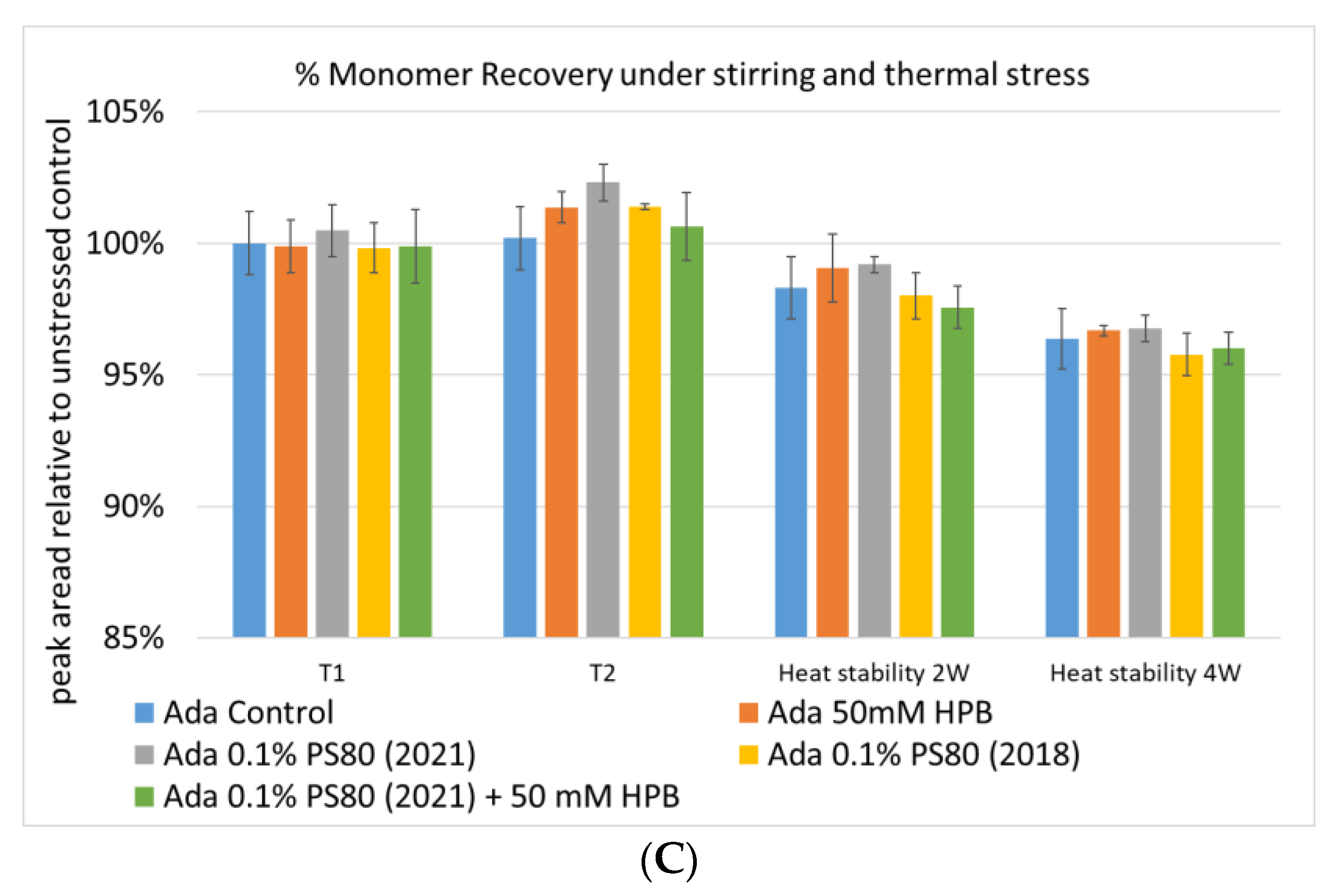
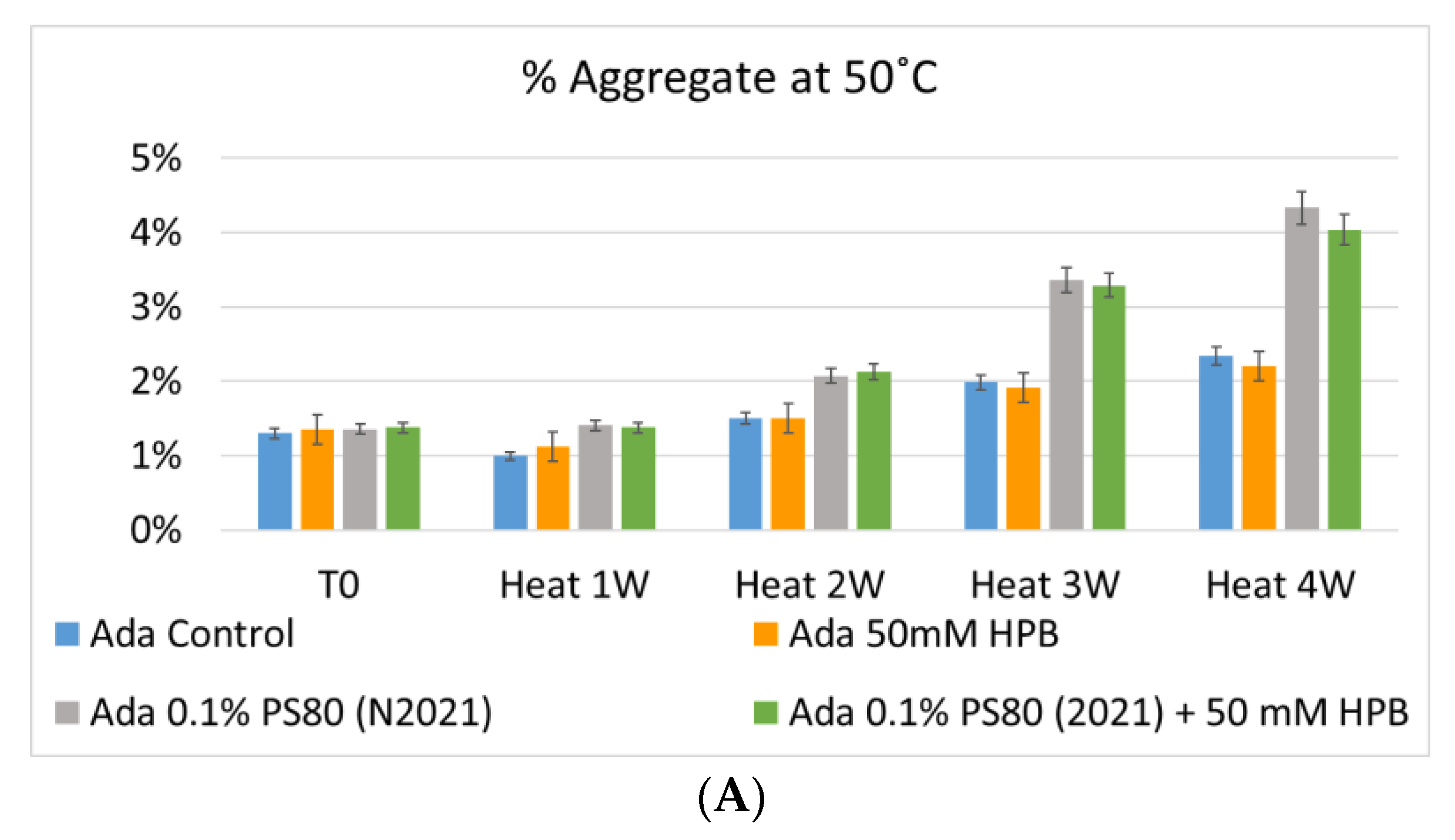
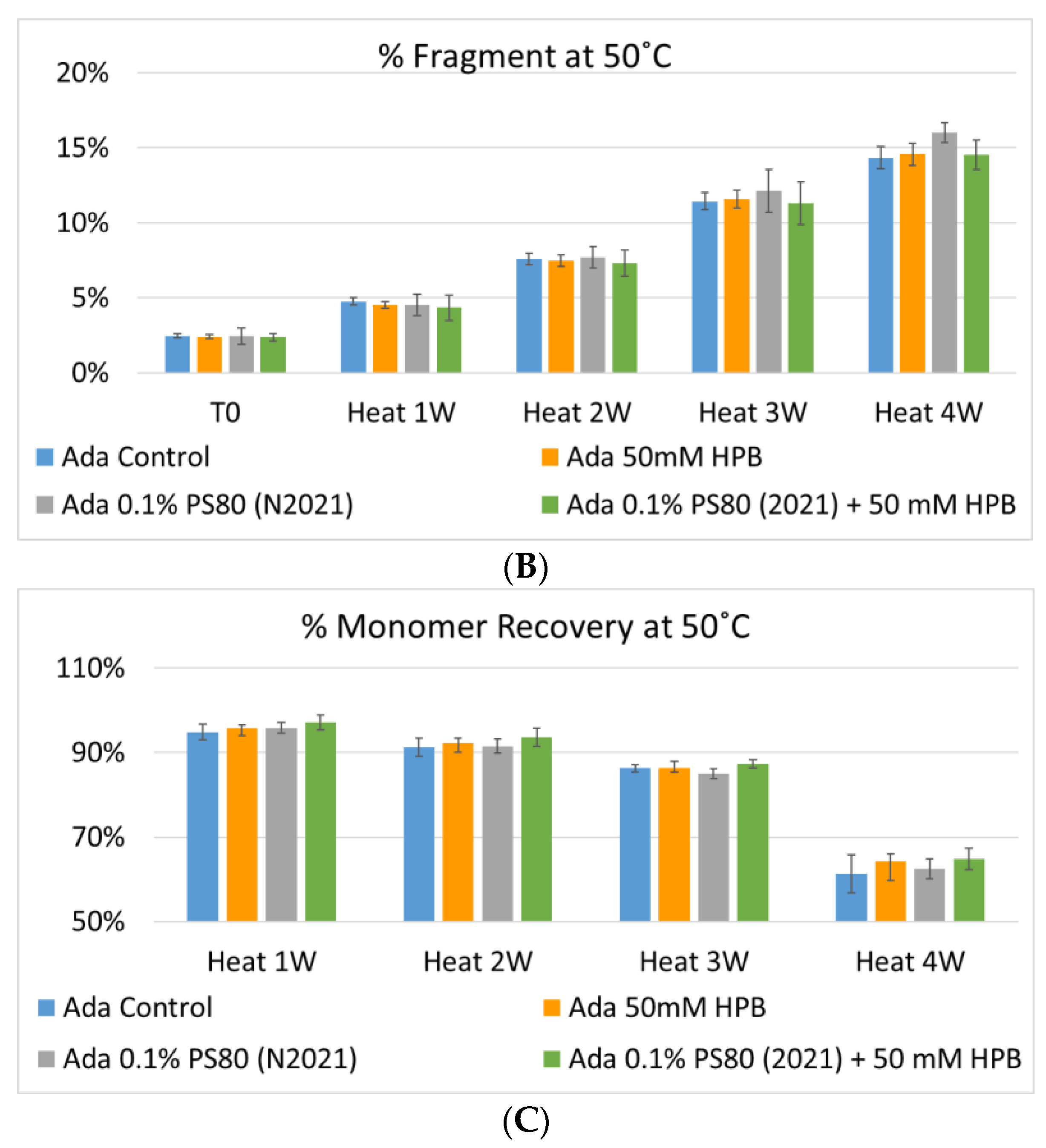
2.3.2. Adalimumab Aggregation and Fragmentation Profiles under Various Stresses
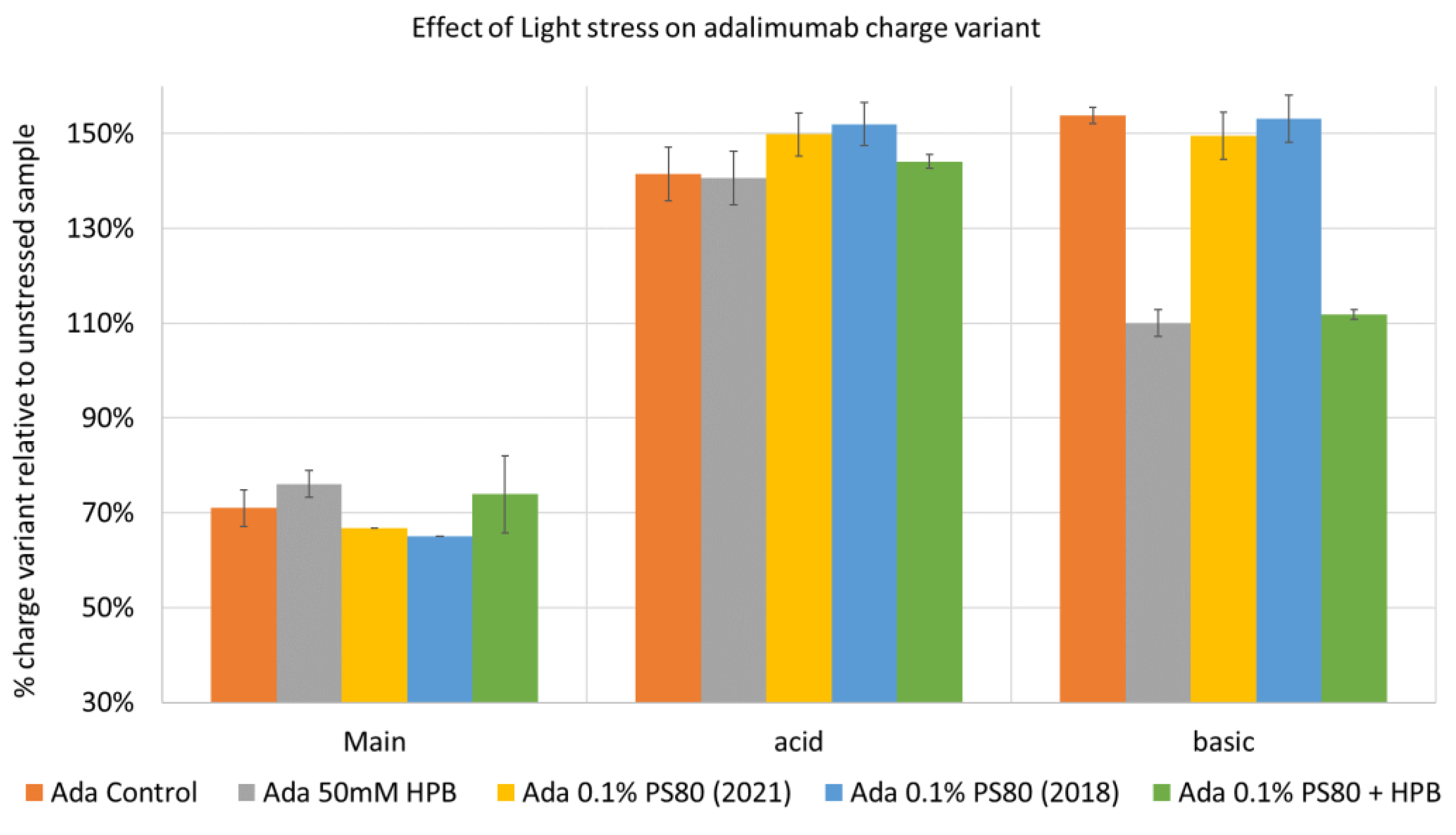
2.3.3. Adalimumab Charge Variant Profiles under Various Stresses
2.4. Discussion
3. Materials and Methods
3.1. Materials
3.2. Methods
3.2.1. Sample Preparation and Stability Studies Design
3.2.2. High Performance Liquid Chromatography with Refractive Index (RI) and Charged Aerosol Detection (HPLC–CAD)
3.2.3. Liquid Chromatography–Mass Spectrometry (LC–MS)
3.2.4. SEC–HPLC
3.2.5. Adalimumab Formulation Stability Studies
3.2.6. Determination of Peroxide Value
3.2.7. Subvisible Particles Analysis by Micro-Flow Imaging (MFI)
3.2.8. Antibody Particle Size Analysis
3.2.9. Cation-Exchange Chromatography (CEX) for Charge Heterogeneity Profiling of Adalimumab
3.2.10. Statistical Analysis
4. Conclusions
Supplementary Materials
Author Contributions
Funding
Institutional Review Board Statement
Informed Consent Statement
Data Availability Statement
Conflicts of Interest
Sample Availability
References
- Carpenter, J.; Cherney, B.; Lubinecki, A.; Ma, S.; Marszal, E.; Mire-Sluis, A.; Nikolai, T.; Novak, J.; Ragheb, J.; Simak, J. Meeting report on protein particles and immunogenicity of therapeutic proteins: Filling in the gaps in risk evaluation and mitigation. Biologicals 2010, 38, 602–611. [Google Scholar] [CrossRef] [PubMed]
- Katz, J.S.; Chou, D.K.; Christian, T.R.; Das, T.K.; Patel, M.; Singh, S.N.; Wen, Y. Emerging Challenges and Innovations in Surfactant-mediated Stabilization of Biologic Formulations. J. Pharm. Sci. 2022, 111, 919–932. [Google Scholar] [CrossRef] [PubMed]
- Li, J.; Krause, M.E.; Chen, X.; Cheng, Y.; Dai, W.; Hill, J.J.; Huang, M.; Jordan, S.; LaCasse, D.; Narhi, L.; et al. Interfacial Stress in the Development of Biologics: Fundamental Understanding, Current Practice, and Future Perspective. AAPS J. 2019, 21, 44. [Google Scholar] [CrossRef] [PubMed]
- Brovc, E.V.; Mravljak, J.; Sink, R.; Pajk, S. Rational design to biologics development: The polysorbates point of view. Int. J. Pharm. 2020, 581, 119285. [Google Scholar] [CrossRef] [PubMed]
- Lee, H.J.; McAuley, A.; Schilke, K.F.; McGuire, J. Molecular origins of surfactant-mediated stabilization of protein drugs. Adv. Drug Deliv. Rev. 2011, 63, 1160–1171. [Google Scholar] [CrossRef] [PubMed]
- Strickley, R.G.; Lambert, W.J. A review of Formulations of Commercially Available Antibodies. J. Pharm. Sci. 2021, 110, 2590–2608.e56. [Google Scholar] [CrossRef]
- Bollenbach, L.; Buske, J.; Mader, K.; Garidel, P. Poloxamer 188 as surfactant in biological formulations–An alternative for polysorbate 20/80? Int. J. Pharm. 2022, 620, 121706. [Google Scholar] [CrossRef]
- Dwivedi, M.; Blech, M.; Presser, I.; Garidel, P. Polysorbate degradation in biotherapeutic formulations: Identification and discussion of current root causes. Int. J. Pharm. 2018, 552, 422–436. [Google Scholar] [CrossRef]
- Kerwin, B.A. Polysorbates 20 and 80 used in the formulation of protein biotherapeutics: Structure and degradation pathways. J. Pharm. Sci. 2008, 97, 2924–2935. [Google Scholar] [CrossRef]
- Dixit, N.; Salamat-Miller, N.; Salinas, P.A.; Taylor, K.D.; Basu, S.K. Residual Host Cell Protein Promotes Polysorbate 20 Degradation in a Sulfatase Drug Product Leading to Free Fatty Acid Particles. J. Pharm. Sci. 2016, 105, 1657–1666. [Google Scholar] [CrossRef]
- Kishore, R.S.; Kiese, S.; Fischer, S.; Pappenberger, A.; Grauschopf, U.; Mahler, H.C. The degradation of polysorbates 20 and 80 and its potential impact on the stability of biotherapeutics. Pharm. Res. 2011, 28, 1194–1210. [Google Scholar] [CrossRef] [PubMed]
- Martos, A.; Koch, W.; Jiskoot, W.; Wuchner, K.; Winter, G.; Friess, W.; Hawe, A. Trends on Analytical Characterization of Polysorbates and Their Degradation Products in Biopharmaceutical Formulations. J. Pharm. Sci. 2017, 106, 1722–1735. [Google Scholar] [CrossRef]
- Tomlinson, A.; Demeule, B.; Lin, B.; Yadav, S. Polysorbate 20 Degradation in Biopharmaceutical Formulations: Quantification of Free Fatty Acids, Characterization of Particulates, and Insights into the Degradation Mechanism. Mol. Pharm. 2015, 12, 3805–3815. [Google Scholar] [CrossRef] [PubMed]
- Singh, S.K.; Afonina, N.; Awwad, M.; Bechtold-Peters, K.; Blue, J.T.; Chou, D.; Cromwell, M.; Krause, H.J.; Mahler, H.C.; Meyer, B.K.; et al. An industry perspective on the monitoring of subvisible particles as a quality attribute for protein therapeutics. J. Pharm. Sci. 2010, 99, 3302–3321. [Google Scholar] [CrossRef]
- Müller, B.W.; Brauns, U. Hydroxypropyl-beta-cyclodextrin derivatives: Influence of average degree of substitution on complexing ability and surface activity. J. Pharm. Sci. 1986, 75, 571–572. [Google Scholar] [CrossRef]
- Sa Couto, A.R.; Ryzhakov, A.; Loftsson, T. 2-Hydroxypropyl-beta-Cyclodextrin Aggregates: Identification and Development of Analytical Techniques. Materials 2018, 11, 1971. [Google Scholar] [CrossRef] [PubMed]
- Serno, T.; Carpenter, J.F.; Randolph, T.W.; Winter, G. Inhibition of agitation-induced aggregation of an IgG-antibody by hydroxypropyl-beta-cyclodextrin. J. Pharm. Sci. 2010, 99, 1193–1206. [Google Scholar] [CrossRef]
- Serno, T.; Hartl, E.; Besheer, A.; Miller, R.; Winter, G. The role of polysorbate 80 and HPbetaCD at the air-water interface of IgG solutions. Pharm. Res. 2013, 30, 117–130. [Google Scholar] [CrossRef]
- Hedges, A. Chapter 22–Cyclodextrins: Properties and Applications. In Starch, 3rd ed.; BeMiller, J., Whistler, R., Eds.; Academic Press: San Diego, CA, USA, 2009; pp. 833–851. [Google Scholar] [CrossRef]
- Sharma, N.; Baldi, A. Exploring versatile applications of cyclodextrins: An overview. Drug Deliv. 2016, 23, 739–757. [Google Scholar] [CrossRef]
- Irie, T.; Uekama, K. Pharmaceutical applications of cyclodextrins. III. Toxicological issues and safety evaluation. J. Pharm. Sci. 1997, 86, 147–162. [Google Scholar] [CrossRef]
- Serno, T.; Geidobler, R.; Winter, G. Protein stabilization by cyclodextrins in the liquid and dried state. Adv. Drug Deliv. Rev. 2011, 63, 1086–1106. [Google Scholar] [CrossRef] [PubMed]
- Wu, H.H.; Garidel, P.; Michaela, B. HP-beta-CD for the formulation of IgG and Ig-based biotherapeutics. Int. J. Pharm. 2021, 601, 120531. [Google Scholar] [CrossRef] [PubMed]
- Tavornvipas, S.; Tajiri, S.; Hirayama, F.; Arima, H.; Uekama, K. Effects of hydrophilic cyclodextrins on aggregation of recombinant human growth hormone. Pharm. Res. 2004, 21, 2369–2376. [Google Scholar] [CrossRef] [PubMed]
- Gokhale, R. Hydroxylpropyl ß-Cyclodextrin: A Promising Excipient for Protein Stabilization. In Proceedings of the Global Drug Delivery & Formulation Summit, Berlin, Germany, 11 March 2019. [Google Scholar]
- Hong, S. Extending Shelf Life of Therapeutic Proteins with Hydroxylpropyl ß-Cyclodextrins–Mechanistic Understanding of Bevacizumab Stabilization. In Proceedings of the Global Drug Delivery & Formulation Summit, Berlin, Germany, 11 March 2019. [Google Scholar]
- Haeuser, C.; Goldbach, P.; Huwyler, J.; Friess, W.; Allmendinger, A. Be Aggressive! Amorphous Excipients Enabling Single-Step Freeze-Drying of Monoclonal Antibody Formulations. Pharmaceutics 2019, 11, 616. [Google Scholar] [CrossRef] [PubMed]
- Haeuser, C.; Goldbach, P.; Huwyler, J.; Friess, W.; Allmendinger, A. Excipients for Room Temperature Stable Freeze-Dried Monoclonal Antibody Formulations. J. Pharm. Sci. 2020, 109, 807–817. [Google Scholar] [CrossRef]
- Callahan, D.J.; Stanley, B.; Li, Y. Control of protein particle formation during ultrafiltration/diafiltration through interfacial protection. J. Pharm. Sci. 2014, 103, 862–869. [Google Scholar] [CrossRef]
- Hong, S.; Peng, T.; Gokhale, R. Feasibility and Functionality of Hydroxypropyl Beta-Cyclodextrin in Ultrafiltration/Diafiltration: A Case Study of Adalimumab. In Proceedings of the American Association of Pharmaceutical Scientists (AAPS) PharmSci 360, Philadelphia, PA, USA, 17 October 2021. [Google Scholar]
- Hong, S.; Peng, T.; Gokhale, R. Hydroxypropyl Beta-Cyclodextrin as a Versatile Excipient in Mitigation Protein Particle Formation during Ultrafiltration/Diafiltration and in Formulation. In Proceedings of the American Association of Pharmaceutical Scientists (AAPS) 2021 PharmSci 360, Philadelphia, PA, USA, 17 October 2021. [Google Scholar]
- Hartl, E.; Dixit, N.; Besheer, A.; Kalonia, D.; Winter, G. Weak antibody-cyclodextrin interactions determined by quartz crystal microbalance and dynamic/static light scattering. Eur. J. Pharm. Biopharm. 2013, 85, 781–789. [Google Scholar] [CrossRef]
- Fusaro, E.; Durez, P.; Wohlrab, J.; Lee, S.; SangWook, Y.; Marotte, H. An update on the adalimumab biosimilar landscape following the approval of the first high-concentration biosimilar. Immunotherapy 2021, 14, 235–252. [Google Scholar] [CrossRef]
- Humira, INN- Adalimumab, Scientific Discussion, Europa. Available online: https://www.ema.europa.eu/en/documents/scientific-discussion/humira-epar-scientific-discussion_en.pdf (accessed on 25 August 2022).
- Chen, W.; Stolz, S.; Wegbecher, V.; Parakkattel, D.; Haeuser, C.; Oltra, N.S.; Kishore, R.S.K.; Bond, S.; Bell, C.; Kopf, R. The degradation of poloxamer 188 in buffered formulation conditions. AAPS Open 2022, 8, 5. [Google Scholar] [CrossRef]


| Sample | KLEPTOSE® HP, Biopharma | KLEPTOSE® HPB, Biopharma | KLEPTOSE® HPB-LB, Parental |
|---|---|---|---|
| Molecular Weight (g/mol) | 1501 (nominal) | 1387 (nominal) | 1338–1424 |
| Molar Substitution (MS) | 0.81–0.99 (0.9, nominal) | 0.58–0.68 (0.62, nominal) | 0.50–0.71 |
| Degree of Substitution (DS) | 6.7 (Nominal) | 4.3 (Nominal) | NA |
| Solubility in H2O | >50% (20 °C, w/w%) | ||
| Residual Beta-CD | 0.10% | 0.7–0.8% | 0.30% |
| Sample | Group | Identified Components (MS Detection) |
|---|---|---|
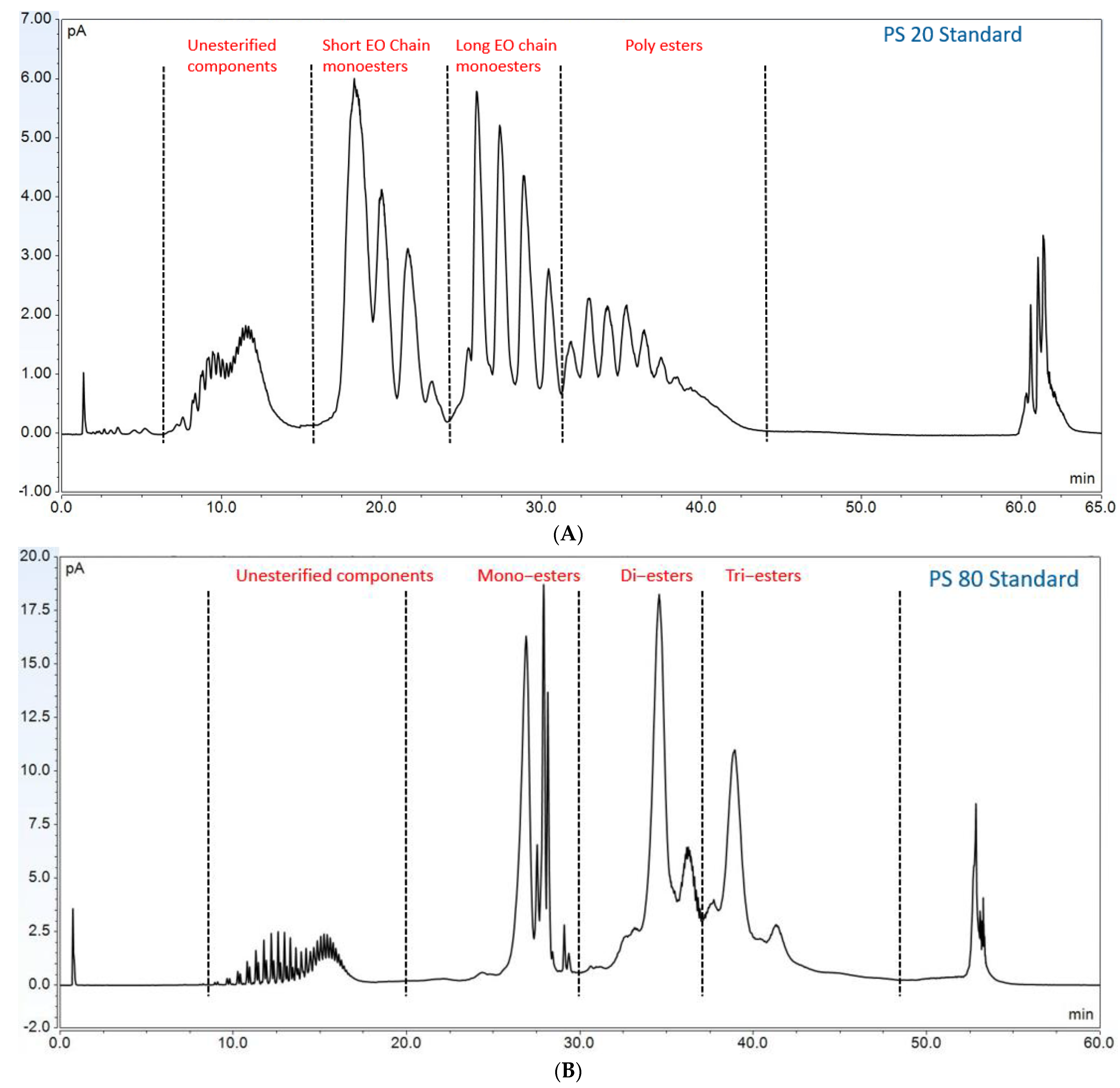
| PS 20 | Non-esterified components | Polyoxyethylene (POE), POE isosorbide, POE sorbitan |
| Short ethylene oxide chain monoesters | (POE isosorbide, POE sorbitan) mono (laurate, myristate) | |
| Long ethylene oxide chain monoesters | (POE isosorbide, POE sorbitan) mono (palmitate, sterate) | |
| Polyesters (di- and tri-esters) | (POE isosorbide, POE sorbitan)-di, tri, or mixed (laurate, myristate) | |
| PS 80 | Non-esterified components | POE, POE isosorbide, POE sorbitan |
| Monoesters | (POE isosorbide, POE sorbitan) mono (oleate, linoleate) | |
| Diesters | POE isosorbide and POE sorbitan dioleate | |
| Tri-esters | POE isosorbide and POE sorbitan trioleate | |
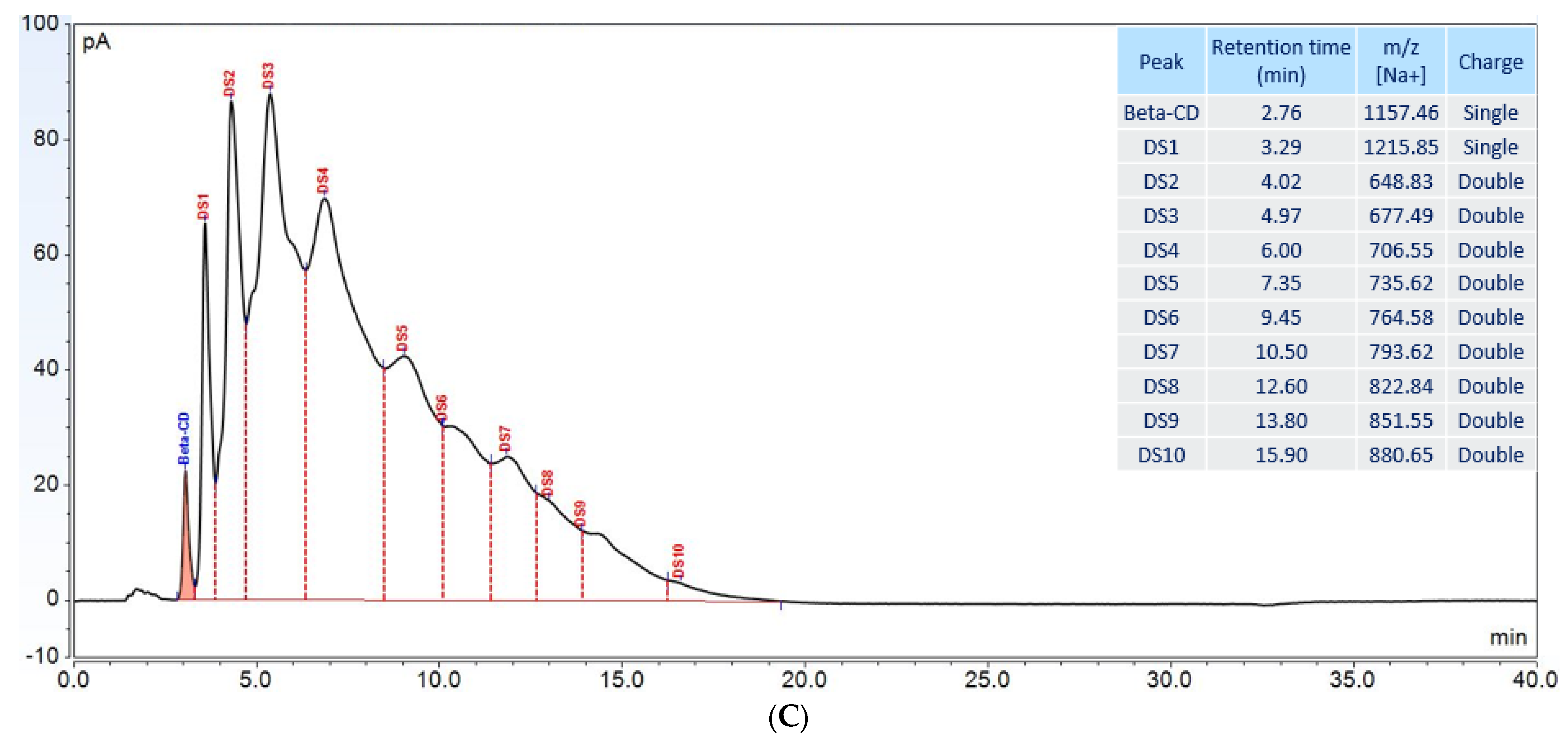
| KLEPTOSE® HPB | Beta-CD | Beta-CD |
| HP-β-CD | HP-DS1-β-CD | |
| HP-DS2-β-CD | ||
| HP-DS3-β-CD | ||
| HP-DS4-β-CD | ||
| HP-DS5-β-CD | ||
| HP-DS6-β-CD | ||
| HP-DS7-β-CD | ||
| HP-DS8-β-CD | ||
| HP-DS9-β-CD | ||
| HP-DS10-β-CD |
Publisher’s Note: MDPI stays neutral with regard to jurisdictional claims in published maps and institutional affiliations. |
© 2022 by the authors. Licensee MDPI, Basel, Switzerland. This article is an open access article distributed under the terms and conditions of the Creative Commons Attribution (CC BY) license (https://creativecommons.org/licenses/by/4.0/).
Share and Cite
Zhang, H.; Hong, S.; Tan, S.S.K.; Peng, T.; Goh, L.Y.H.; Lam, K.H.; Chow, K.T.; Gokhale, R. Polysorbates versus Hydroxypropyl Beta-Cyclodextrin (HPβCD): Comparative Study on Excipient Stability and Stabilization Benefits on Monoclonal Antibodies. Molecules 2022, 27, 6497. https://doi.org/10.3390/molecules27196497
Zhang H, Hong S, Tan SSK, Peng T, Goh LYH, Lam KH, Chow KT, Gokhale R. Polysorbates versus Hydroxypropyl Beta-Cyclodextrin (HPβCD): Comparative Study on Excipient Stability and Stabilization Benefits on Monoclonal Antibodies. Molecules. 2022; 27(19):6497. https://doi.org/10.3390/molecules27196497
Chicago/Turabian StyleZhang, Hailong, Shiqi Hong, Sarah Si Kai Tan, Tao Peng, Lucas Yuan Hao Goh, Kwan Hang Lam, Keat Theng Chow, and Rajeev Gokhale. 2022. "Polysorbates versus Hydroxypropyl Beta-Cyclodextrin (HPβCD): Comparative Study on Excipient Stability and Stabilization Benefits on Monoclonal Antibodies" Molecules 27, no. 19: 6497. https://doi.org/10.3390/molecules27196497
APA StyleZhang, H., Hong, S., Tan, S. S. K., Peng, T., Goh, L. Y. H., Lam, K. H., Chow, K. T., & Gokhale, R. (2022). Polysorbates versus Hydroxypropyl Beta-Cyclodextrin (HPβCD): Comparative Study on Excipient Stability and Stabilization Benefits on Monoclonal Antibodies. Molecules, 27(19), 6497. https://doi.org/10.3390/molecules27196497