
Current Analytical Strategies for Antibody–Drug Conjugates in Biomatrices


Abstract
:1. Introduction
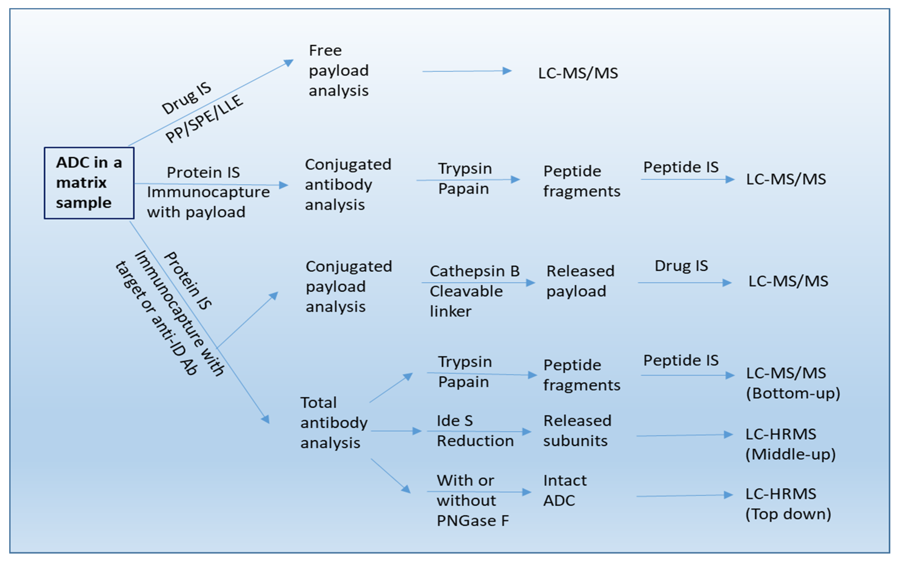
2. ADC Forms to Be Quantitated by PK Assays
3. Assay Platforms
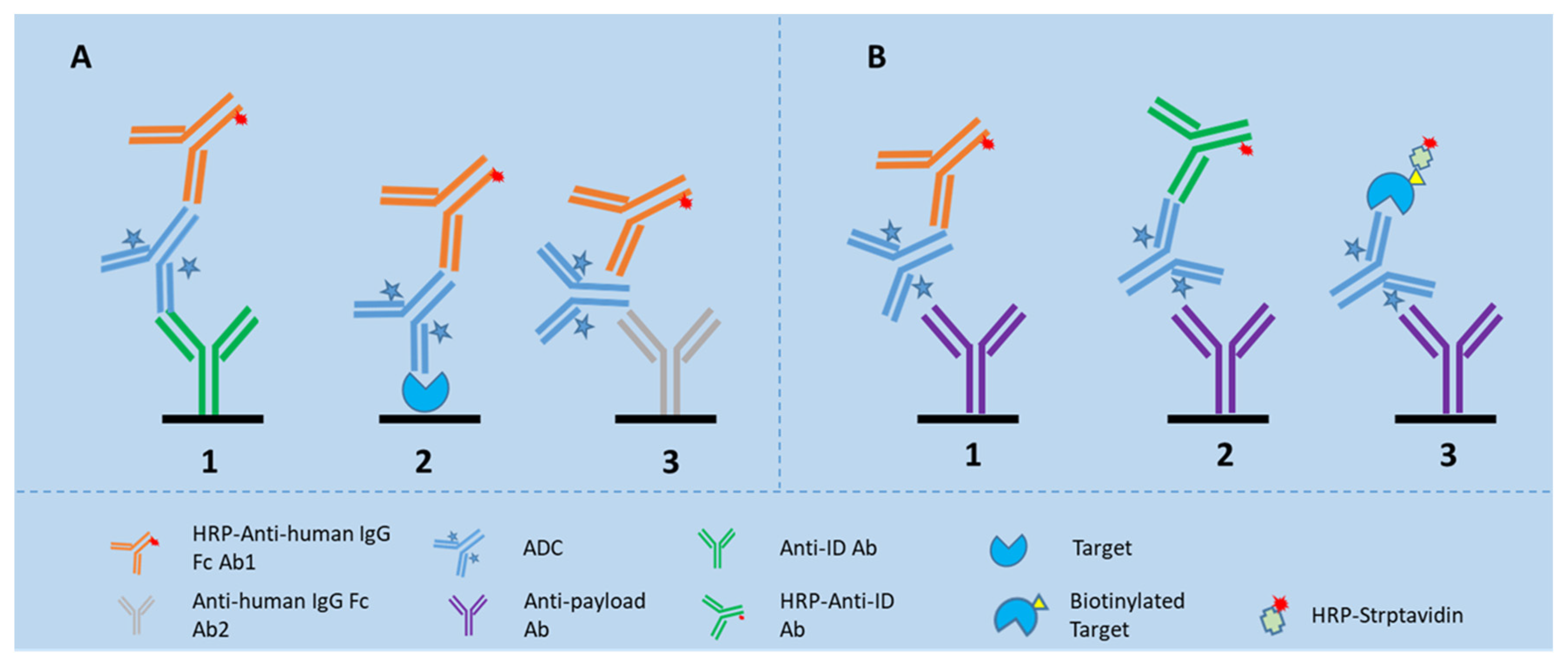
3.1. LBA Platform
3.2. LC-MS Platform
3.2.1. LC-MS/MS
3.2.2. LBA-LC-MS/MS
3.2.3. LBA-LC-HRMS (High-Resolution Mass Spectrometry)
4. LBA vs. LC-MS Based PK Assays
5. Detection of ADAs
5.1. Assay Platforms
5.1.1. LBA Platform
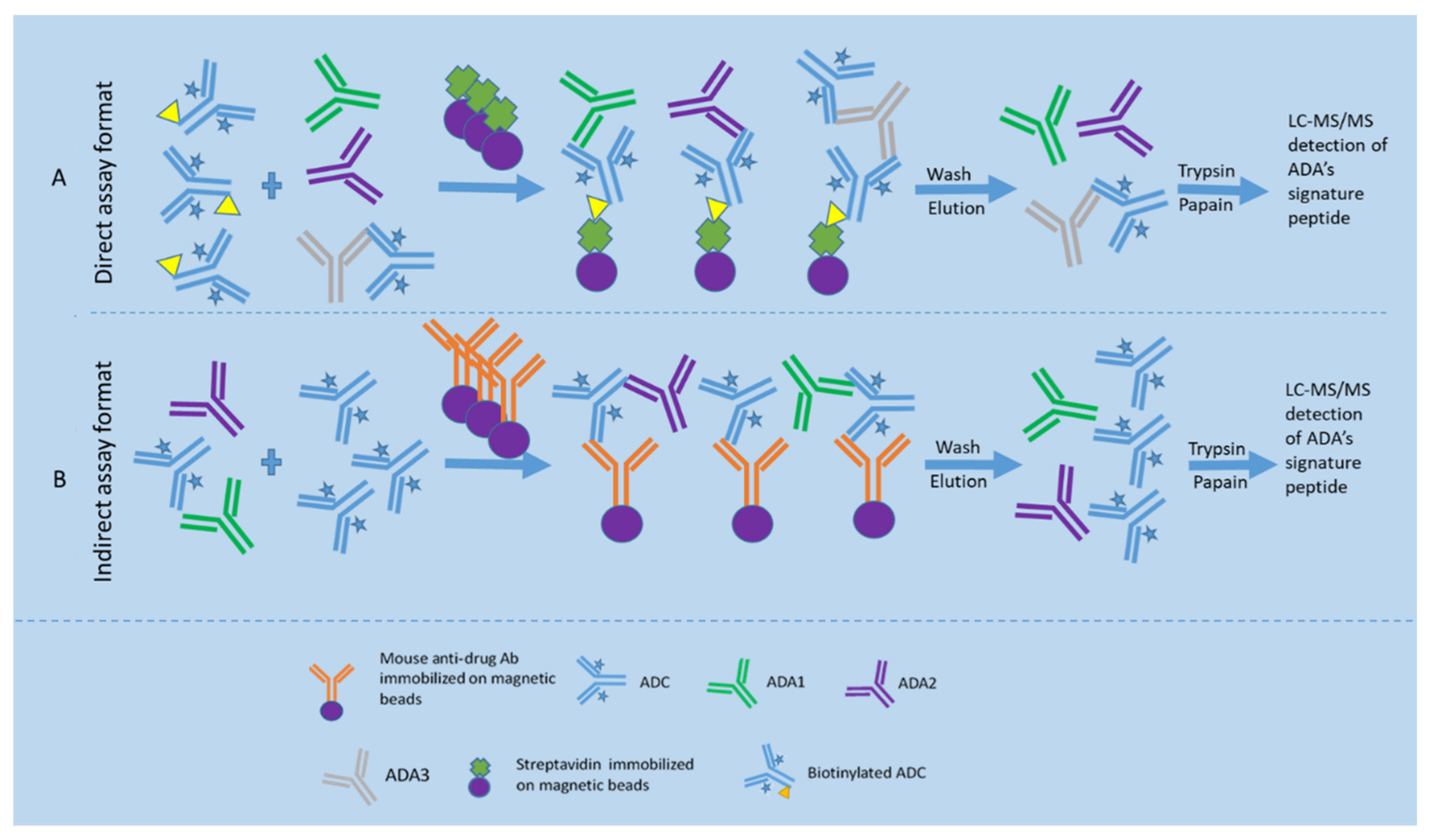
5.1.2. LBA-LC-MS/MS Platform
Direct LBA-LC-MS/MS Assay Format
Indirect LBA-LC-MS/MS Assay Format
6. LBA vs. LBA-LC-MS/MS Based ADA Assays
7. Factors That Interfere with the Assay Performance
7.1. PK Assays
7.1.1. DAR
7.1.2. ADA
7.1.3. Soluble Target or Extracellular Domain of the Target
7.1.4. Sample Preparation
7.2. ADA Assays
7.2.1. Drug Target
7.2.2. Drug
7.2.3. Modification of Critical Reagents
7.2.4. Preexisting Antibodies
7.2.5. Rheumatoid Factor (RF)
8. Conclusions
Author Contributions
Funding
Institutional Review Board Statement
Informed Consent Statement
Data Availability Statement
Conflicts of Interest
References
- Zhao, P.; Zhang, Y.; Li, W.; Jeanty, C.; Xiang, G.; Dong, Y. Recent advances of antibody drug conjugates for clinical applications. Acta Pharm. Sin. B 2020, 10, 1589–1600. [Google Scholar] [CrossRef] [PubMed]
- McPherson, M.J.; Hobson, A.D. Pushing the Envelope: Advancement of ADCs Outside of Oncology. Methods Mol. Biol. 2020, 2078, 23–36. [Google Scholar]
- Ford, C.H.; Newman, C.E.; Johnson, J.R.; Woodhouse, C.S.; Reeder, T.A.; Rowland, G.F.; Simmonds, R.G. Localisation and toxicity study of a vindesine-anti-CEA conjugate in patients with advanced cancer. Br. J. Cancer 1983, 47, 35–42. [Google Scholar] [CrossRef] [PubMed]
- Tsuchikama, K.; An, Z. Antibody-drug conjugates: Recent advances in conjugation and linker chemistries. Protein Cell 2018, 9, 33–46. [Google Scholar] [CrossRef] [PubMed]
- Fu, Z.; Li, S.; Han, S.; Shi, C.; Zhang, Y. Antibody drug conjugate: The “biological missile” for targeted cancer therapy. Signal. Transduct. Target. Ther. 2022, 7, 93. [Google Scholar] [CrossRef] [PubMed]
- Van de Merbel, N.C. Protein quantification by LC-MS: A decade of progress through the pages of Bioanalysis. Bioanalysis 2019, 11, 629–644. [Google Scholar] [CrossRef] [PubMed]
- Kaur, S.; Bateman, K.P.; Glick, J.; Jairaj, M.; Kellie, J.F.; Sydor, J.; Zeng, J. IQ consortium perspective: Complementary LBA and LC-MS in protein therapeutics bioanalysis and biotransformation assessment. Bioanalysis 2020, 12, 257–270. [Google Scholar] [CrossRef]
- Kang, L.; Weng, N.; Jian, W. LC–MS bioanalysis of intact proteins and peptides. Biomed. Chromatogra. 2019, 34, e4633. [Google Scholar] [CrossRef] [PubMed]
- Chen, L.Z.; Roos, D.; Philip, E. Development of Immunocapture-LC/MS Assay for Simultaneous ADA Isotyping and Semiquantitation. J. Immunol. Res. 2016, 2016, 7682472. [Google Scholar] [CrossRef] [PubMed]
- Wang, J.; Gu, H.; Liu, A.; Kozhich, A.; Rangan, V.; Myler, H.; Luo, L.; Wong, R.; Sun, H.; Wang, B.; et al. Antibody-drug conjugate bioanalysis using LB-LC-MS/MS hybrid assays: Strategies, methodology and correlation to ligand-binding assays. Bioanalysis 2016, 8, 1383–1401. [Google Scholar] [CrossRef] [PubMed]
- Cong, Y.; Zhang, Z.; Zhang, S.; Hu, L.; Gu, J. Quantitative MS analysis of therapeutic mAbs and their glycosylation for pharmacokinetics study. Proteom. Clin. Appl. 2016, 10, 303–314. [Google Scholar] [CrossRef]
- U.S. Food and Drug Administration. Bioanalytical Method Validation; U.S. Food and Drug Administration: Silver Spring, MD, USA, 2018. [Google Scholar]
- U.K. European Medicines Agency. Guideline on Bioanalytical Method Validation; U.K. European Medicines Agency: London, UK, 2011. [Google Scholar]
- ICH Harmonised Guideline. Bioanalytical Method Validation and Study Sample Analysis M10; ICH Harmonised Guideline: Geneva, Switzerland, 2022. [Google Scholar]
- U.K. European Medicines Agency. Guideline on Immunogenicity Assessment of Biotechnology—Derived Therapeutic Proteins; U.K. European Medicines Agency: London, UK, 2007. [Google Scholar]
- U.S. Food and Drug Administration. Immunogenicity Testing of Therapeutic Protein Products—Developing and Validating Assays for Anti-Drug Antibody Detection; U.S. Food and Drug Administration: Silver Spring, MD, USA, 2019. [Google Scholar]
- Hoofring, S.A.; Lopez, R.; Hock, M.B.; Kaliyaperumal, A.; Patel, S.K.; Swanson, S.J.; Chirmule, N.; Starcevic, M. Immunogenicity testing strategy and bioanalytical assays for antibody-drug conjugates. Bioanalysis 2013, 5, 1041–1055. [Google Scholar] [CrossRef] [PubMed]
- Devanarayan, V.; Smith, W.C.; Brunelle, R.L.; Seger, M.E.; Krug, K.; Bowsher, R.R. Recommendations for Systematic Statistical Computation of Immunogenicity Cut Points. AAPS J. 2017, 19, 1487–1498. [Google Scholar] [CrossRef]
- Neubert, H.; Song, A.; Lee, A.; Wei, C.; Duggan, J.; Xu, K.; Woolf, E.; Evans, C.; Palandra, J.; Laterza, O.; et al. 2017 White Paper: Rise of hybrid LBA/LCMS immunogenicity assays (Part 2: Hybrid LBA/LCMS biotherapeutics, biomarkers & immunogenicity assays and regulatory agencies’ inputs). Bioanalysis 2017, 9, 1895–1912. [Google Scholar]
- Huang, X.; Xu, X.; Partridge, M.A.; Chen, J.; Koehler-Stec, E.; Sumner, G.; Qiu, H.; Torri, A.; Li, N. Isotyping and Semi-Quantitation of Monkey Anti-Drug Antibodies by Immunocapture Liquid Chromatography-Mass Spectrometry. AAPS J. 2021, 23, 16. [Google Scholar] [CrossRef] [PubMed]
- Todoroki, K.; Mizuno, H.; Sugiyama, E.; Toyo’oka, T. Bioanalytical methods for therapeutic monoclonal antibodies and antibody–drug conjugates: A review of recent advances and future perspectives. J. Pharm. Biomed. Anal. 2020, 179, 112991. [Google Scholar] [CrossRef] [PubMed]
- Myler, H.; Rangan, V.S.; Wang, J.; Kozhich, A.; Cummings, J.A.; Neely, R.; Dail, D.; Liu, A.; Wang, B.; Vezina, H.E.; et al. An integrated multiplatform bioanalytical strategy for antibody-drug conjugates: A novel case study. Bioanalysis 2015, 7, 1569–1582. [Google Scholar] [CrossRef] [PubMed]
- Faria, M.; Peay, M.; Lam, B.; Ma, E.; Yuan, M.; Waldron, M.; Mylott Jr, W.R.; Liang, M.; Rosenbaum, A.I. Multiplex LC-MS/MS Assays for Clinical Bioanalysis of MEDI4276, an Antibody-Drug Conjugate of Tubulysin Analogue Attached via Cleavable Linker to a Biparatopic Humanized Antibody against HER-2. Antibodies 2019, 8, 11. [Google Scholar] [CrossRef]
- Zhu, L.; Glick, J.; Flarakos, J. Bioanalytical Challenges in Support of Complex Modalities of Antibody-Based Therapeutics. AAPS J. 2020, 22, 130. [Google Scholar] [CrossRef] [PubMed]
- Kaur, S.; Xu, K.; Saad, O.M.; Dere, R.C.; Carrasco-Triguero, M. Bioanalytical assay strategies for the development of antibody-drug conjugate biotherapeutics. Bioanalysis 2013, 5, 201–226. [Google Scholar] [CrossRef] [PubMed]
- Pei, M.; Liu, T.; Ouyang, L.; Sun, J.; Deng, X.; Sun, X.; Wu, W.; Huang, P.; Chen, Y.; Tan, X.; et al. Enzyme-linked immunosorbent assays for quantification of MMAE-Conjugated ADCs and total antibodies in cynomolgus monkey sera. J. Pharm. Anal. 2021, 12, 645–652. [Google Scholar] [CrossRef]
- Shen, B.; Bumbaca, D.; Saad, O.; Yue, Q.; Pastuskovas, C.V.; Khojasteh, S.C.; Tibbitts, J.; Kaur, S.; Wang, B.; Chu, Y.W.; et al. Catabolic fate and pharmacokinetic characterization of trastuzumab emtansine (T-DM1): An emphasis on preclinical and clinical catabolism. Curr. Drug Metab. 2012, 13, 901–910. [Google Scholar] [CrossRef] [PubMed]
- Liu, A.; Kozhich, A.; Passmore, D.; Gu, H.; Wong, R.; Zambito, F.; Rangan, V.S.; Myler, H.; Aubry, A.-F.; Arnold, M.E.; et al. Quantitative bioanalysis of antibody-conjugated payload in monkey plasma using a hybrid immuno-capture LC–MS/MS approach: Assay development, validation, and a case study. J. Chromatogr. B Analat. Biomed. Life Sci. 2015, 1002, 54–62. [Google Scholar] [CrossRef] [PubMed]
- Sanderson, R.J.; Nicholas, N.D.; Baker Lee, C.; Hengel, S.M.; Lyon, R.P.; Benjamin, D.R.; Alley, S.C. Antibody-conjugated drug assay for protease-cleavable antibody-drug conjugates. Bioanalysis 2016, 8, 55–63. [Google Scholar] [CrossRef]
- Rago, B.; Tumey, L.N.; Wei, C.; Barletta, F.; Clark, T.; Hansel, S.; Han, X. Quantitative Conjugated Payload Measurement Using Enzymatic Release of Antibody–Drug Conjugate with Cleavable Linker. Bioconjug. Chem. 2017, 28, 620–626. [Google Scholar] [CrossRef] [PubMed]
- Chen, T.; Su, D.; Gruenhagen, J.; Gu, C.; Li, Y.; Yehl, P.; Chetwyn, N.P.; Medley, C.D. Chemical de-conjugation for investigating the stability of small molecule drugs in antibody-drug conjugates. J. Pharm. Biomed. Anal. 2016, 117, 304–310. [Google Scholar] [CrossRef]
- Zhu, X.; Huo, S.; Xue, C.; An, B.; Qu, J. Current LC-MS-based strategies for characterization and quantification of antibody-drug conjugates. J. Pharm. Anal. 2020, 10, 209–220. [Google Scholar] [CrossRef] [PubMed]
- Huang, R.Y.C.; Chen, G. Characterization of antibody–drug conjugates by mass spectrometry: Advances and future trends. Drug Discov. Today 2016, 21, 850–855. [Google Scholar] [CrossRef] [PubMed]
- Kotapati, S.; Deshpande, M.; Jashnani, A.; Thakkar, D.; Xu, H.; Dollinger, G. The role of ligand-binding assay and LC-MS in the bioanalysis of complex protein and oligonucleotide therapeutics. Bioanalysis 2021, 13, 931–954. [Google Scholar] [CrossRef] [PubMed]
- Wei, C.; Su, D.; Wang, J.; Jian, W.; Zhang, D. LC–MS Challenges in Characterizing and Quantifying Monoclonal Antibodies (mAb) and Antibody-Drug Conjugates (ADC) in Biological Samples. Curr. Pharmacol. Rep. 2018, 4, 45–63. [Google Scholar] [CrossRef]
- Fung, E.N.; Bryan, P.; Kozhich, A. Techniques for quantitative LC-MS/MS analysis of protein therapeutics: Advances in enzyme digestion and immunocapture. Bioanalysis 2016, 8, 847–856. [Google Scholar] [CrossRef]
- Li, H.; Ortiz, R.; Tran, L.; Hall, M.; Spahr, C.; Walker, K.; Laudemann, J.; Miller, S.; Salimi-Moosavi, H.; Lee, J.W. General LC-MS/MS Method Approach to Quantify Therapeutic Monoclonal Antibodies Using a Common Whole Antibody Internal Standard with Application to Preclinical Studies. Anal. Chem. 2012, 84, 1267–1273. [Google Scholar] [CrossRef] [PubMed]
- Lee, J.W. Generic method approaches for monoclonal antibody therapeutics analysis using both ligand binding and LC-MS/MS techniques. Bioanalysis 2016, 8, 19–27. [Google Scholar] [CrossRef] [PubMed]
- Vasicek, L.A.; Zhu, X.; Spellman, D.S.; Bateman, K.P. Direct quantitation of therapeutic antibodies for pharmacokinetic studies using immuno-purification and intact mass analysis. Bioanalysis 2019, 11, 3. [Google Scholar] [CrossRef] [PubMed]
- Huang, Y.; Mou, S.; Wang, Y.; Mu, R.; Liang, M.; Rosenbaum, A.I. Characterization of Antibody-Drug Conjugate Pharmacokinetics and in Vivo Biotransformation Using Quantitative Intact LC-HRMS and Surrogate Analyte LC-MRM. Anal. Chem. 2021, 93, 6135–6144. [Google Scholar] [CrossRef]
- He, J.; Yu, S.F.; Yee, S.; Kaur, S.; Xu, K. Characterization of in vivo biotransformations for trastuzumab emtansine by high-resolution accurate-mass mass spectrometry. MAbs 2018, 10, 960–967. [Google Scholar] [PubMed]
- Mu, R.; Yuan, J.; Huang, Y.; Meissen, J.K.; Mou, S.; Liang, M.; Rosenbaum, A.I. Bioanalytical Methods and Strategic Perspectives Addressing the Rising Complexity of Novel Bioconjugates and Delivery Routes for Biotherapeutics. BioDrugs 2022, 36, 181–196. [Google Scholar] [CrossRef]
- Fiorotti, C.K. Immunogenicity considerations for antibody-drug conjugates: A focus on neutralizing antibody assays. Bioanalysis 2018, 10, 65–70. [Google Scholar] [CrossRef]
- Liu, T.; Tong, Y.; Gao, J.; Fang, W.; Wu, J.; Peng, X.; Fan, X.; Chen, X.; Sun, J.; Cao, S.; et al. Development of a bridging ELISA for detection of antibodies. J. Pharmacol. Toxicol. Met. 2022, in press. [Google Scholar] [CrossRef]
- Hock, M.B.; Thudium, K.E.; Carrasco-Triguero, M.; Schwabe, N.F. Immunogenicity of Antibody Drug Conjugates: Bioanalytical Methods and Monitoring Strategy for a Novel Therapeutic Modality. AAPS J. 2014, 17, 35–43. [Google Scholar] [CrossRef] [PubMed]
- Stubenrauch, K.; Mackeben, K.; Vogel, R.; Heinrich, J. Generic anti-drug antibody assay with drug tolerance in serum samples from mice exposed to human antibodies. Anal. Biochem. 2012, 430, 193–199. [Google Scholar] [CrossRef]
- Stubenrauch, K.; Wessels, U.; Essig, U.; Vogel, R.; Schleypen, J. Evaluation of a generic immunoassay with drug tolerance to detect immune complexes in serum samples from cynomolgus monkeys after administration of human antibodies. J. Pharm. Biomed. Anal. 2010, 52, 249–254. [Google Scholar] [CrossRef]
- Bautista, A.C.; Salimi-Moosavi, H.; Jawa, V. Universal immunoassay applied during early development of large molecules to understand impact of immunogenicity on biotherapeutic exposure. AAPS J. 2012, 14, 843–849. [Google Scholar] [CrossRef]
- Jiang, H.; Myler, H.; Zeng, J.; Mora, J.; Kolaitis, G.; Pillutla, R. Perspectives on exploring hybrid LBA/LC-MS approach for clinical immunogenicity testing. Bioanalysis 2019, 11, 1605–1617. [Google Scholar] [CrossRef]
- Jiang, H.; Xu, W.; Titsch, C.A.; Furlong, M.T.; Dodge, R.; Voronin, K.; Allentoff, A.; Zeng, J.; Aubry, A.F.; DeSilva, B.S.; et al. Innovative use of LC-MS/MS for simultaneous quantitation of neutralizing antibody, residual drug, and human immunoglobulin G in immunogenicity assay development. Anal. Chem. 2014, 86, 2673–2680. [Google Scholar] [CrossRef]
- Neubert, H.; Grace, C.; Rumpel, K.; James, I. Assessing immunogenicity in the presence of excess protein therapeutic using immunoprecipitation and quantitative mass spectrometry. Anal. Chem. 2008, 80, 6907–6914. [Google Scholar] [CrossRef]
- Stephan, J.P.; Kozak, K.R.; Wong, W.L. Challenges in developing bioanalytical assays for characterization of antibody-drug conjugates. Bioanalysis 2011, 3, 677–700. [Google Scholar] [CrossRef]
- Chirmule, N.; Jawa, V.; Meibohm, B. Immunogenicity to therapeutic proteins: Impact on PK/PD and efficacy. AAPS J. 2012, 14, 296–302. [Google Scholar] [CrossRef]
- Wei, D.; Sullivan, M.; Espinosa, O.; Yang, L. A sensitive LC–MS/MS method for the determination of free maytansinoid DM4 concentrations—Method development, validation, and application to the nonclinical studies of antitumor agent DM4 conjugated hu-anti-Cripto MAb B3F6 (B3F6DM4) in rats and monkeys. Int. J. Mass Spectrom. 2012, 312, 53–60. [Google Scholar] [CrossRef]
- Bargh, J.D.; Isidro-Llobet, A.; Parker, J.S.; Spring, D.R. Cleavable linkers in antibody-drug conjugates. Chem. Soc. Rev. 2019, 48, 4361–4374. [Google Scholar] [CrossRef]
- Liu, Y.; Zhou, F.; Sang, H.; Ye, H.; Chen, Q.; Yao, L.; Ni, P.; Wang, G.; Zhang, J. LC-MS/MS method for the simultaneous determination of Lys-MCC-DM1, MCC-DM1 and DM1 as potential intracellular catabolites of the antibody-drug conjugate trastuzumab emtansine (T-DM1). J. Pharm. Biomed. Anal. 2017, 137, 170–177. [Google Scholar] [CrossRef] [PubMed]
- Goldenberg, D.M.; Sharkey, R.M. Antibody-drug conjugates targeting TROP-2 and incorporating SN-38: A case study of anti-TROP-2 sacituzumab govitecan. MAbs 2019, 11, 987–995. [Google Scholar] [CrossRef] [PubMed]
- Zhong, Z.D.; Clements-Egan, A.; Gorovits, B.; Maia, M.; Sumner, G.; Theobald, V.; Wu, Y.; Rajadhyaksha, M. Drug Target Interference in Immunogenicity Assays: Recommendations and Mitigation Strategies. AAPS J. 2017, 19, 1564–1575. [Google Scholar] [CrossRef] [PubMed]
- Wang, Y.; Luong, M.; Guadiz, C.; Zhang, M.; Gorovits, B. Addressing soluble target interference in the development of a functional assay for the detection of neutralizing antibodies against a BCMA-CD3 bispecific antibody. J. Immunol. Methods 2019, 474, 112642. [Google Scholar] [CrossRef]
- Chen, J.; Kendra, K.; Torri, A.; Sumner, G. Overcoming multimeric target interference in a bridging immunogenicity assay with soluble target receptor, target immunodepletion and mild acidic assay pH. Bioanalysis 2020, 12, 1071–1085. [Google Scholar] [CrossRef]
- Chen, J.; Garlits, J.; Dhulipala, G.; Sirimanne, T.; Xu, C.; Lu, K.; Palackal, N.; Pyles, E.; Torri, A.; Sumner, G. Mitigating target interference in bridging immunogenicity assay with target-blocking reagents and mild basic pH. Bioanalysis 2019, 11, 1569–1580. [Google Scholar] [CrossRef]
- Carrasco-Triguero, M.; Mahood, C.; Milojic-Blair, M.; Amaya, C.; Ruppel, J.; Hong, K.; Yi, J.H.; Kaur, S. Overcoming soluble target interference in an anti-therapeutic antibody screening assay for an antibody-drug conjugate therapeutic. Bioanalysis 2012, 4, 2013–2026. [Google Scholar] [CrossRef]
- Dengler, A.F.; Weiss, R.; Truong, T.; Irvin, S.C.; Gadhia, N.; Hassanein, M.; Georgaros, C.; Taylor, J.A.; Paccaly, A.; Sumner, G.; et al. Bioanalytical Challenges due to Prior Checkpoint Inhibitor Exposure: Interference and Mitigation in Drug Concentration and Immunogenicity Assays. AAPS J. 2021, 23, 109. [Google Scholar] [CrossRef]
- Bütikofer, L.; Lemaillet, G.; Faust, H. Strategies to estimate and improve drug tolerance in anti-drug antibody assays. Bioanalysis 2012, 4, 1999–2012. [Google Scholar] [CrossRef]
- Patton, A.; Mullenix, M.C.; Swanson, S.J.; Koren, E. An acid dissociation bridging ELISA for detection of antibodies directed against therapeutic proteins in the presence of antigen. J. Immunol. Methods 2005, 304, 189–195. [Google Scholar] [CrossRef]
- Chen, Y.Q.; Pottanat, T.G.; Carter, Q.L.; Troutt, J.S.; Konrad, R.J.; Sloan, J.H. Affinity capture elution bridging assay: A novel immunoassay format for detection of anti-therapeutic protein antibodies. J. Immunol. Methods 2016, 431, 45–51. [Google Scholar] [CrossRef] [PubMed]
- Smith, H.W.; Butterfield, A.; Sun, D. Detection of antibodies against therapeutic proteins in the presence of residual therapeutic protein using a solid-phase extraction with acid dissociation (SPEAD) sample treatment prior to ELISA. Regul. Toxicol. Pharmacol. 2007, 49, 230–237. [Google Scholar] [CrossRef] [PubMed]
- Xu, W.; Sank, M.; Cummings, J.; Carl, S.; Juhel, M.; Gleason, C.; Dodge, R.; DeSilva, B.S.; Kolaitis, G.; Pillutla, R. Bead-extraction and heat-dissociation (BEHD): A novel way to overcome drug and matrix interference in immunogenicity testing. J. Immunol. Methods 2018, 462, 34–41. [Google Scholar] [CrossRef]
- Zoghbi, J.; Xu, Y.; Grabert, R.; Theobald, V.; Richards, S. A breakthrough novel method to resolve the drug and target interference problem in immunogenicity assays. J. Immunol. Methods 2015, 426, 62–69. [Google Scholar] [CrossRef] [PubMed]
- Niu, H.; Klem, T.; Yang, J.; Qiu, Y.; Pan, L. A biotin-drug extraction and acid dissociation (BEAD) procedure to eliminate matrix and drug interference in a protein complex anti-drug antibody (ADA) isotype specific assay. J. Immunol. Methods 2017, 446, 30–36. [Google Scholar] [CrossRef]
- Qin, Q.; Liu, T.; Gong, L.; Wu, S.; Ren, J. A device and method for removing free drugs from anti-drug antibody detection samples, the preparation method and application of the device. China Patent ZL2018101302350, 12 May 2020. [Google Scholar]
- Rocha, A.G.; Krynski, K.; Mancino, A.; Sciscione, M.; Beaver, C.J. New insights on critical reagent optimization for antidrug antibody assays. Bioanalysis 2019, 11, 815–823. [Google Scholar] [CrossRef] [PubMed]
- Oquendo, E.; Savoie, J.; Swenson, J.M.; Grimaldi, C. Critical reagent generation, characterization, handling and storage workflows: Impact on ligand binding assays. Bioanalysis 2021, 13, 847–860. [Google Scholar] [CrossRef]
- Ramsland, P.A.; Movafagh, B.F.; Reichlin, M.; Edmundson, A.B. Interference of rheumatoid factor activity by aspartame, a dipeptide methyl ester. J. Mol. Recognit. 1999, 12, 249–257. [Google Scholar] [CrossRef]
- Wagh, A.; Song, H.; Zeng, M.; Tao, L.; Das, T.K. Challenges and new frontiers in analytical characterization of antibody-drug conjugates. MAbs 2018, 10, 222–243. [Google Scholar] [CrossRef]
- Tong, J.T.W.; Harris, P.W.R.; Brimble, M.A.; Kavianinia, I. An Insight into FDA Approved Antibody-Drug Conjugates for Cancer Therapy. Molecules 2021, 26, 5847. [Google Scholar] [CrossRef]
- Khongorzul, P.; Ling, C.J.; Khan, F.U.; Ihsan, A.U.; Zhang, J. Antibody-Drug Conjugates: A Comprehensive Review. Mol. Cancer Res. 2020, 18, 3–19. [Google Scholar] [CrossRef] [PubMed]
- Cahuzac, H.; Devel, L. Analytical Methods for the Detection and Quantification of ADCs in Biological Matrices. Pharmaceuticals 2020, 13, 462. [Google Scholar] [CrossRef] [PubMed]
- Pu, J.; An, B.; Vazvaei, F.; Qu, J. Enrichment of protein therapeutics and biomarkers for LC-MS quantification. Bioanalysis 2018, 10, 979–982. [Google Scholar] [CrossRef] [PubMed]
- Myler, H.; Rangan, V.S.; Kozhich, A.; Hoffpauir, B.; Dail, D.; Cummings, J.; Saewert, M.; Manney, A.; Liu, A.; Rao, C.; et al. Validation of an integrated series of ligand-binding assays for the quantitative determination of antibody-drug conjugates in biological matrices. Bioanalysis 2016, 8, 519–531. [Google Scholar] [CrossRef] [PubMed]
- Tarantino, P.; Carmagnani Pestana, R.; Corti, C.; Modi, S.; Bardia, A.; Tolaney, S.M.; Cortes, J.; Soria, J.C.; Curigliano, G. Antibody-drug conjugates: Smart chemotherapy delivery across tumor histologies. CA Cancer J. Clin. 2022, 72, 165–182. [Google Scholar] [CrossRef]


Publisher’s Note: MDPI stays neutral with regard to jurisdictional claims in published maps and institutional affiliations. |
© 2022 by the authors. Licensee MDPI, Basel, Switzerland. This article is an open access article distributed under the terms and conditions of the Creative Commons Attribution (CC BY) license (https://creativecommons.org/licenses/by/4.0/).
Share and Cite
Qin, Q.; Gong, L. Current Analytical Strategies for Antibody–Drug Conjugates in Biomatrices. Molecules 2022, 27, 6299. https://doi.org/10.3390/molecules27196299
Qin Q, Gong L. Current Analytical Strategies for Antibody–Drug Conjugates in Biomatrices. Molecules. 2022; 27(19):6299. https://doi.org/10.3390/molecules27196299
Chicago/Turabian StyleQin, Qiuping, and Likun Gong. 2022. "Current Analytical Strategies for Antibody–Drug Conjugates in Biomatrices" Molecules 27, no. 19: 6299. https://doi.org/10.3390/molecules27196299
APA StyleQin, Q., & Gong, L. (2022). Current Analytical Strategies for Antibody–Drug Conjugates in Biomatrices. Molecules, 27(19), 6299. https://doi.org/10.3390/molecules27196299