Article

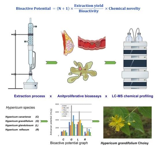
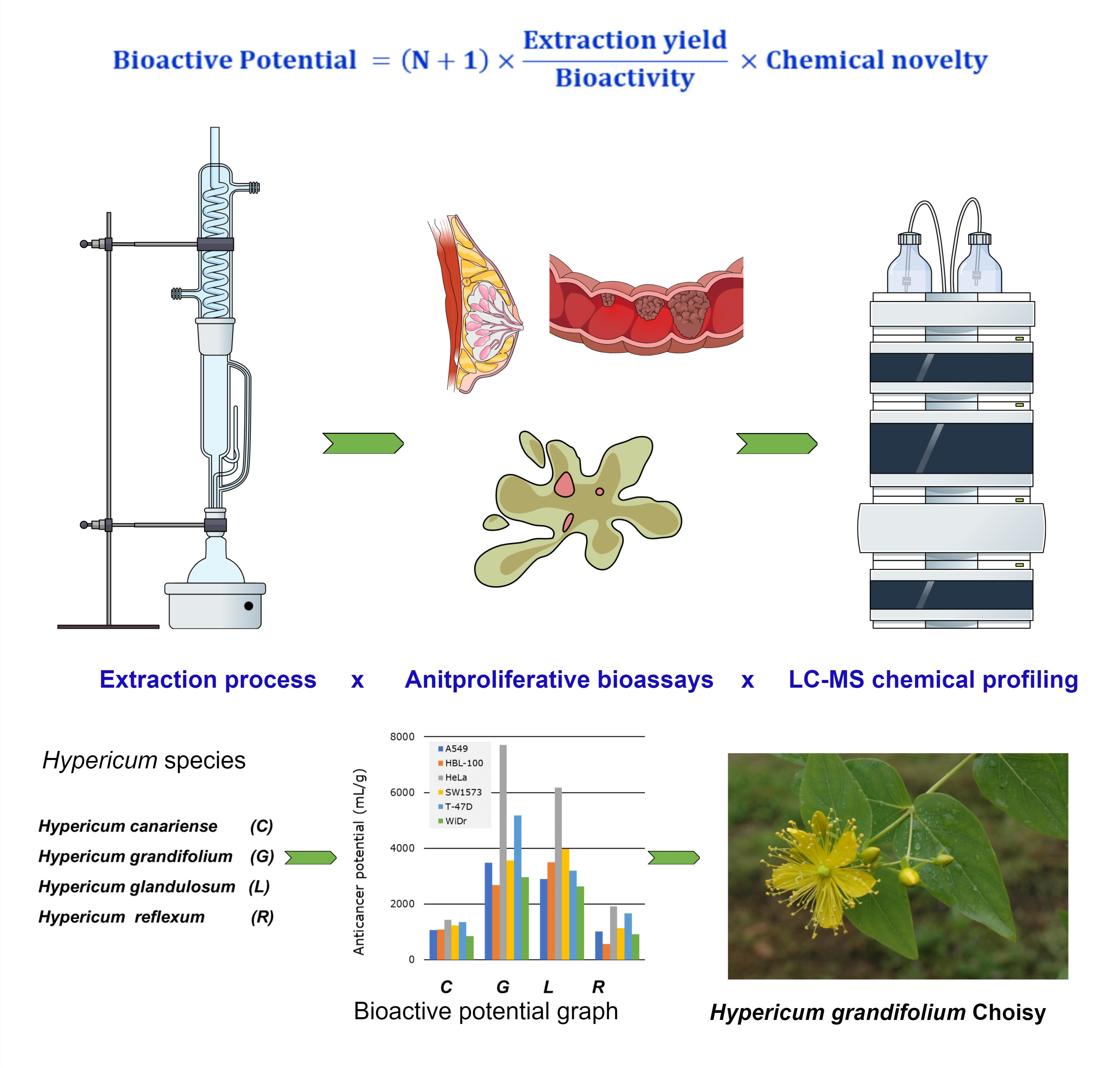
Bioactive Potential: A Pharmacognostic Definition through the Screening of Four Hypericum Species from the Canary Islands

by
Rodney Lacret
1,2,*,
Adrián Puerta
1,
Sebastian Granica
3,
Aday González-Bakker
1,
Danela Hevia
1,
Yiling Teng
1,
Candelaria C. Sánchez-Mateo
2,
Pedro Luis Pérez de Paz
4 and
José M. Padrón
1
1
BioLab, Instituto Universitario de Bio-Orgánica Antonio González (IUBO-AG), Universidad de La Laguna, Avda. Astrofísico Francisco Sánchez 2, 38206 La Laguna, Spain
2
Departamento de Medicina Física y Farmacología, Facultad de Farmacia, Universidad de La Laguna, Tenerife, 38200 La Laguna, Spain
3
Microbiota Lab, Centre of Preclinical Studies, Medical University of Warsaw, Banacha 1b, 02-097 Warsaw, Poland
4
Departamento de Botánica, Ecología y Fisiología Vegetal, Facultad de Farmacia, Universidad de La Laguna, Tenerife, 38200 La Laguna, Spain
*
Author to whom correspondence should be addressed.
Molecules 2022, 27(18), 6101; https://doi.org/10.3390/molecules27186101
Submission received: 12 August 2022 / Revised: 13 September 2022 / Accepted: 15 September 2022 / Published: 18 September 2022

Abstract

:
In this work, we propose a general methodology to assess the bioactive potential (BP) of extracts in the quest of vegetable-based drugs. To exemplify the method, we studied the anticancer potential (AP) of four endemic species of genus Hypericum (Hypericum canariense L, Hypericum glandulosum Aiton, Hypericum grandifolium Choisy and Hypericum reflexum L.f) from the Canary Islands. Microextracts were obtained from the aerial parts of these species and were tested against six human tumor cell lines, A549 (non-small-cell lung), HBL-100 (breast), HeLa (cervix), SW1573 (non-small-cell lung), T-47D (breast) and WiDr (colon). The methanol–water microextracts were evaluated further for cell migration, autophagy and cell death. The most promising bioactive polar microextracts were analyzed by UHPLC–DAD–MS. The extraction yield, the bioactivity evaluation and the chemical profiling by LC–MS suggested that H. grandifolium was the species with the highest AP. Label-free live-cell imaging studies on HeLa cells exposed to the methanol–water microextract of H. grandifolium enabled observing cell death and several apoptotic hallmarks. Overall, this study allows us to select Hypericum grandifolium Choisy as a source of new chemical entities with a potential interest for cancer treatment.

Graphical Abstract

1. Introduction

The chemical bioprospecting of new therapeutic agents, pesticides, nutraceuticals, and other products with biotechnological applications from plants is one of research lines with the greatest impact worldwide [1,2,3]. The chemical bioprospecting protocol includes (a) the selection and extraction of appropriate plants, (b) the evaluation of the bioactive potential of the extracts to select the species with the highest bioactive potential, and (c) the bioguided isolation of compounds from the best candidates [4]. Thus, the rationalization of each stage of the chemical bioprospecting contributes to accelerate the discovery of compounds with high added value [5,6].
Bioactive potential (BP) is a very often employed term in the search of bioactive compounds from living organisms (bacteria, fungi or plants) but its definition is not clear yet in the literature. BP can be subdivided into agrochemical or pharmacological potential depending on the field of the research, i.e., agrochemistry or pharmacology, respectively [3,7]. In drug discovery, the bioactive potential of extract from plants as a source of high-value compounds has mainly focused the bioactivity evaluation and dereplication of extracts [8,9,10]. Eloff and coworkers introduced a parameter called total activity, a mathematical relationship between extraction yield and the antimicrobial activity for the quantification of the bioactivity of plan extracts during the screening [11]. However, there are a few examples in the literature that use the total activity as a tool [12,13,14]. On the other hand, rules or recommendations of thumb for defining anti-infective potential in natural products were developed by Cos and coworkers [15]. It is worthwhile to mention that these recommendations did not take into account the total activity previously reported and they are only related to the anti-infective activity. From this state of art, we propose to define bioactive potential as a combination of three parameters, namely, the extraction process, the biological activity profiling (either agrochemical or pharmacological) and the early chemical profiling. The main goal of the evaluation of BP is to prioritize, through extracts from plants, the rational selection of the best vegetable species for the discovery of new chemical bioactive entities. BP can be classified into different types depending on the field of bioassays (antibacterial, anti-inflammatory, anticancer, biocidal, etc.). BP can be also applied to the bioguided isolation process.
The genus Hypericum (Hypericacea) comprises high-value medicinal plants, particularly Hypericum perforatum L., which is largely used in folk medicine [16]. Extensive pharmacological and chemical studies on the genus Hypericum have revealed its potential as a source of antibacterial, anti-inflammatory, antiproliferative and antiviral drugs [17,18,19,20,21,22]. In the Canary Islands, the genus Hypericum is comprised of eight species: H. canariense, H. coadunatum, H. glandulosum, H. grandifolium, H. humifusum, H. perfoliatum, H. perforatum and H. reflexum. Likewise, the aerial parts of most of them have been used in the traditional medicine as vulnerary, sedative, tranquilizer, vermifuge, diuretic, tinctorial and as food for goats [23,24,25]. The chemical and pharmacological studies on these Hypericum species from the Canary Islands have been conducted for more than 35 years [26,27,28,29,30,31,32,33,34,35,36,37,38,39,40,41,42]. From them, eleven works have been published related to the evaluation of pharmacological actitivities of extracts from H. canariense, H. glandulosum, H. grandifolium and H. reflexum [26,27,28,29,30,31,32,33,34,35,42] and six papers have adressed the isolation of compounds from H. canariense, H. coadunatum and H. reflexum [36,37,38,39,40,41]. Nevertheless, to date, there are no bioguided chemical studies on these Hypericum species from the Canary Islands that provide information about compounds responsible for the previous observed pharmacological activities.
Since 2019, our research interests focusses on the chemical bioprospecting of plants from the Canary Islands as a source of high-added-value compounds and the evaluation of their BP with a particular interest in the anticancer potential (AP). The aim of our approach is to identify the plants with the highest AP. In this step, the extraction of plants at a small scale (microextraction), the biological activity profiling of these microextracts, and their early chemical profiling by LC–MS are considered relevant factors as a whole. The outcome of this selection process is a sorted (prioritized) list of plants from the highest to the lowest AP. In this work, we will show the implementation of our strategy with the evaluation of the AP of four Hypericum species from the Canary Islands, namely, H. canariense L, H. glandulosum Aiton, H. grandifolium Choisy and H. reflexum L.f. Notably, this is the first time that these four species of Hypericum were profiled together both at the pharmacological and at the chemical level.

2. Results and Discussion

Our evaluation protocol of the AP of plants consists of three sequential steps. Every step provides relevant information to help in the assessment of the AP. In the next Sections, we will present our implementation of the methodology and we will discuss the results and the criteria we followed to prioritize the Hypericum species from the Canary Islands as a source of bioactive compounds.

2.1. Selection of Plants and Microextraction

H. canariense, H. glandulosum, H. grandifolium and H. reflexum were selected considering chemosystematic and ethnopharmacological approaches [7]. The extraction process of these species started with the maceration of their aerial part at a small scale (microextraction). For this purpose, to perform the extraction in a sequential maceration, we selected as solvents water, methanol and dichloromethane. The procedure we followed is an adapted methodology previously described by Macías in the search of phytotoxic compounds [43]. This methodology was designed regarding the vast range of specialized metabolites in the Hypericum genus (range of polarities and bioactivities) [22,44] and as a way to solve the difficulty to obtain them using a single extraction [45]. Therefore, each plant dried material (approximately 10 g) was sequentially macerated under the same conditions from polar to non polar solvents considering a sample ratio of 20:1 (v/w). Firstly, a methanol/water (1:1) mixture (MW), then methanol (MM), and finally a dichloromethane/methanol (1:1) mixture (DM) were used, as depicted in Figure 1.
After the extraction process, three microextracts from each plant were obtained (MW, MM and DM). Thus, a total of 12 microextracts were prepared. For each microextract, the dry mass was weighed and the extraction yield, expressed in percentage, was calculated. The results are shown in Table 1.
The extraction yield is a pharmacognostic parameter used for considering any vegetal as a source of leads or drugs [14]. It also enables the calculation of the total activity (TA) (Equation (1), ), a useful tool for the comparison of crude extracts during screening of plants as a source of bioactive compounds [11]. In our study, the MW and MM microextracts from the aerial parts of the four Hypericum species gave the highest yields. In contrast, the DM microextracts provided the lowest yields, except for H. glandulosum. In general, these data suggested that Hypericum species from the Canary Islands were rich in medium and polar compounds. These results also match with a recently published study on Hypericum species from the UK [14]. These 12 microextracts were submitted to the biological evaluation at two levels.

2.2. Biological Evaluation

The scientific literature holds several examples of non-mammalian and mammalian cell-based assays that have been used for the identification and characterization of bioactive natural products [46]. In the therapeutic area of cancer, screenings based on mammalian cell cultures have enabled the rapid study of a multitude of natural product extracts. In drug discovery programs of plant extracts (such as the NCI’s), the first biological assessment is an antiproliferative test against tumor cell lines (a general or primary bioassay) [47,48].

2.2.1. Antiproliferative Activity

Following the guidelines of the NCI, the antiproliferative activity of the 12 microextracts (MW, MM and DM) obtained from the aerial parts of H. canariense (CMW, CMM and CDM), H. glandulosum (GMW, GMM and GDM), H. grandifolium (LMW, LMM and LDM), and H. reflexum (RMW, RMM and RDM) were evaluated against six human tumor cell lines: A549, HBL-100, HeLa, SW1573, T-47D and WiDr. The maximum test concentration was set at 250 µg/mL, according to NCI guidelines [49,50]. The results, expressed as 50% growth inhibition (GI50), are given in Figure 2 (Table S1). According to NCI calculations, GI50 values represent approximate values. Taken as a whole, the GI50 values enabled an arrangement of the plants in the following decreasing order of activity: H. grandifolium > H. canariense > H. glandulosum > H. reflexum. Our results are in agreement with a previous study where the antiproliferative activity of essential oils and polar extracts (methanol—acetone 1:1) from the aerial parts of H. canariense, H. grandifolium and H. reflexum were evaluated against the human tumor cell lines A375 (malignant melanoma), MDA-MB-231 (breast adenocarcinoma) and HCT116 (colon carcinoma) [42]. In that study, the polar extract from H. grandifolium also exhibited a significant antiproliferative activity and resulted the most active one. Noteworthy, our work represents the first study reporting antiproliferative activity of H. glandulosum extracts.
When we are considering the solvent extraction, the sequence obtained (in decreasing activity) for our samples was DM > MM > MW. In the particular case of H. grandifolium, the DM and MM microextracts (i.e., GDM and GMM, respectively) were the most active ones against all the tumor cell lines tested. They displayed the lowest GI50 values (in the range 2.7–13 µg/mL) among the twelve assayed microextracts (Figure 2). However, these GI50 values denote a highly cytotoxic profile (GI50 ≤  20 µg/mL) according to the NCI’s guidelines [50]. Noteworthy, the MW microextracts induced moderate cytotoxicity (GI50 21–200 µg/mL). In our experience, highly cytotoxic samples induce large cell killing and biological effects are more difficult to be observed. In contrast, samples with moderate cytotoxicity allow to measure relevant phenotypic changes in treated cells. According to this criterion and our interest in the pharmacognostic study of polar extracts, the hydroalcoholic microextracts were the prioritized for further testing.

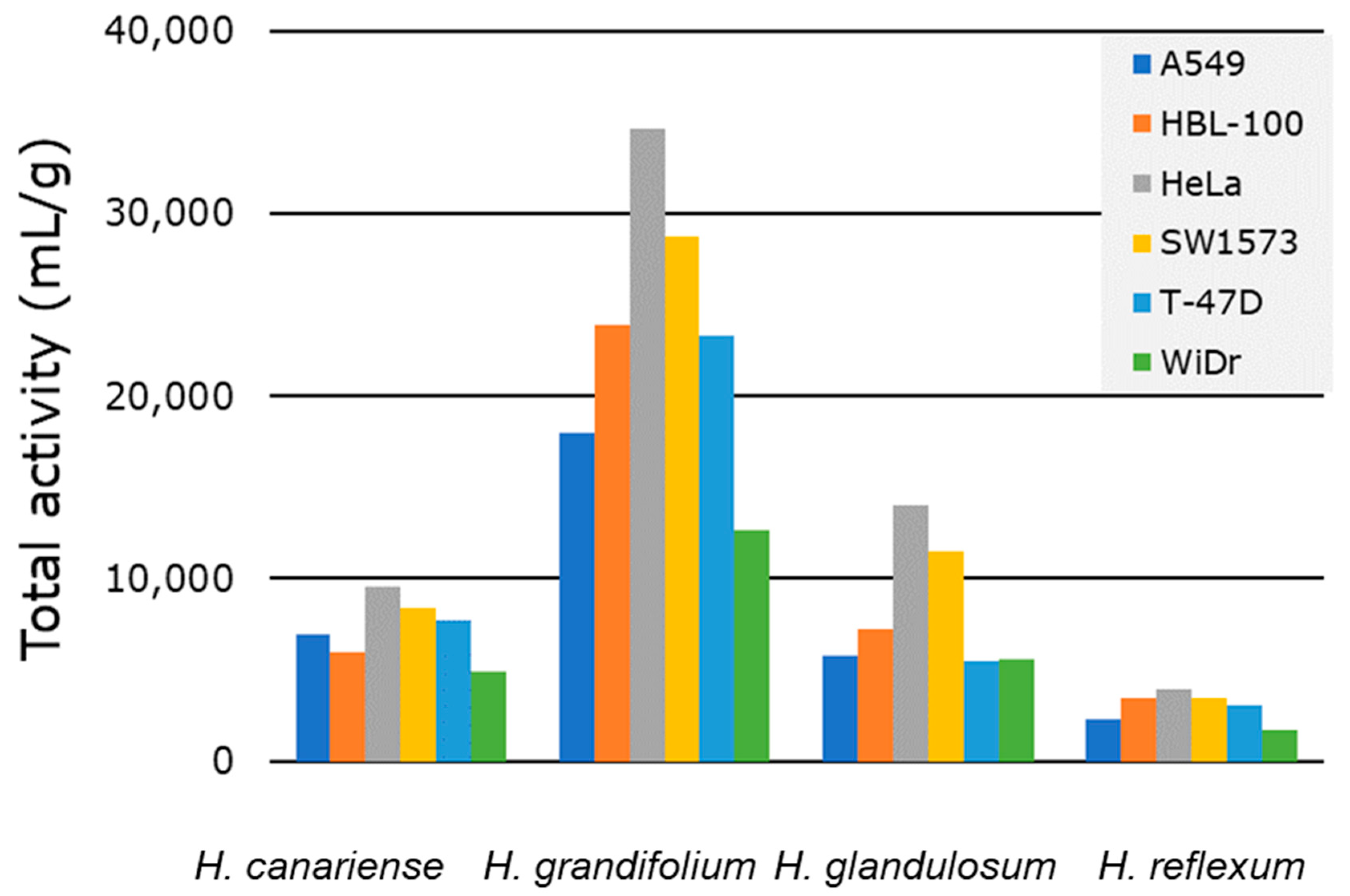
2.2.2. Total Activity

The TA (Equation (1)) is a significant pharmacognostic parameter for the quantification of the antimicrobial activity of plant extracts and it is very useful in the rational screening of diverse plants. This tool has been used as a part of the evaluation of the antibacterial activity of extracts from south African species [12]. TA, expressed in (mL/g), was defined as “the largest volume to which the bioactive compounds in one gram of plant material can be diluted and still inhibit the growth of the tested organisms” [11]. Analogously, we have envisioned the calculation of the TA when considering the antiproliferative activity of plant extracts against human solid tumor cells. From the pharmacological point of view, the growth inhibition (GI50 in µg/mL) indicates the potency of extracts, while TA represents the efficacy of the extracts [11].
T A   ( m L g ) = m e x t r a c t   ( m g ) m p l a n t   ( g ) × 1 G I 50   ( μ g / m L )
In this work, we calculated the TA of the Hypericum species to compare the antiproliferative efficacy of their microextracts. To the best of our knowledge, this is the first time that the TA is reported to assess the antiproliferative activity during the screening of the of plant species.
Since our protocol involves the sequential extraction using three solvent systems (Figure 1), we considered appropriate to compute the TA for each individual extract and sum all the TAs corresponding to a virtual total extract from each plant and each cell line. With this calculation, each TA value represents a theoretical activity of each theorical total extract against each cell line that complies with the definition provided by Eloff [11]. Therefore, Figure 3 (Table S2) collects the TA for each of the plant species against each of the six cell lines tested. The results enabled ordering the species according to decreasing values of TA: H. grandifolium > H. glandulosum > H. canariense > H. reflexum.
Based on the GI50 data (Figure 2), the TA values (Figure 3 and Table S2) and our interest in polar extracts, we selected for further studies the MW microextracts from H. canariense (CMW), H. glandulosum (LMW) and H. grandifolium (GMW). The sibling microextract of H. reflexum (i.e., RMW) were also tested for comparison purposes. At this point, many different phenotypic experiments can be considered to depict the overall mode of action of the microextracts and give information about possible cell targets. These experiments are generally termed as secondary bioassays. The literature reports diverse secondary bioassays for the potential anticancer activity of Hypericum sp. extracts [19,20,21]. Herein, we selected as secondary tests cell migration and vacuole formation, two relevant hallmarks for cancer treatment, which have not been reported for the (four) Hypericum sp. (included in this work). Cell migration experiments give an idea of the antimetastatic potential of the tested samples [51], while vacuole formation is an indicator of autophagy [52] and drug resistance mechanisms [53], among others.

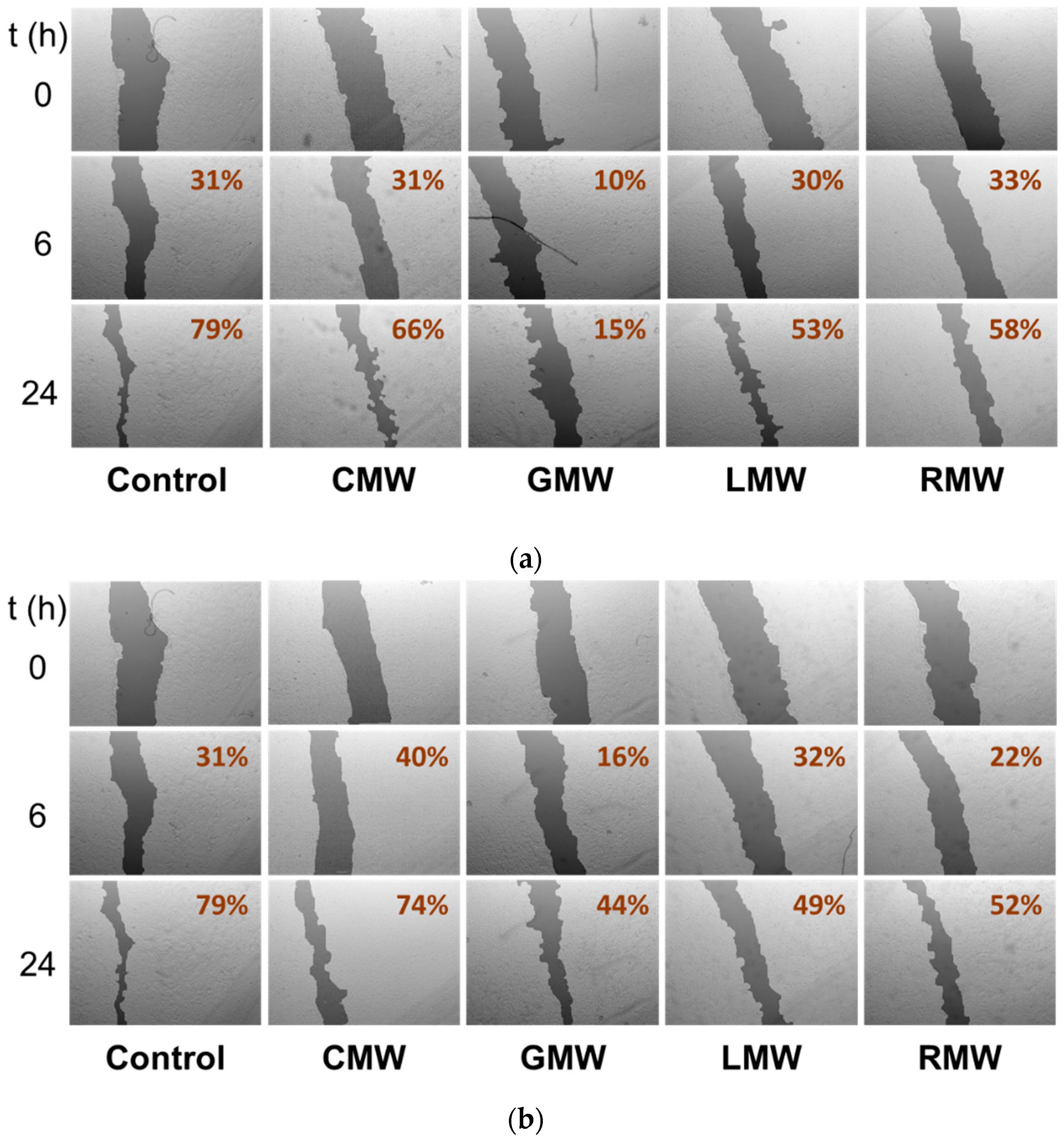
2.2.3. Cell Migration Disturbances in A549 Cells

Cell migration is the ability of a cancer cell to undergo movement and invasion allowing it to relocate within the tissues [51]. This process allows neoplastic cells to enter lymphatic and blood vessels for dissemination into the circulation metastasize in distant organs. The non-small-cell lung cancer (NSCLC) cell line A549 is considered as a highly metastatic cancer cell line [54] and represents a good in vitro model to assess the antimigratory properties using the wound healing assay [55].
To study the effects on A549 cell migration of the MW microextracts of the four Hypericum species, we selected two doses, a high (100 µg/mL) and a low dose (50 µg/mL), and two time points, 6 and 24 h (Figure 4). The use of this early time point enables avoiding, at minimum, the effect of cell proliferation in the assay, limiting the response to the antimigratory activity of the microextract. With the exception of CMW all microextracts, were able to reduce the closure of the wound when compared to control wells after 6 and 24 h. GMW was able to inhibit cell migration after six hours of treatment in a concentration dependent manner, being the most active of all microextracts assayed. Interestingly, CMW, which showed antiproliferative activity, failed to reduce the migration of A549 cells. Meanwhile, both LMW and RMW did not show correlation between dose and inhibition of cell mobility.

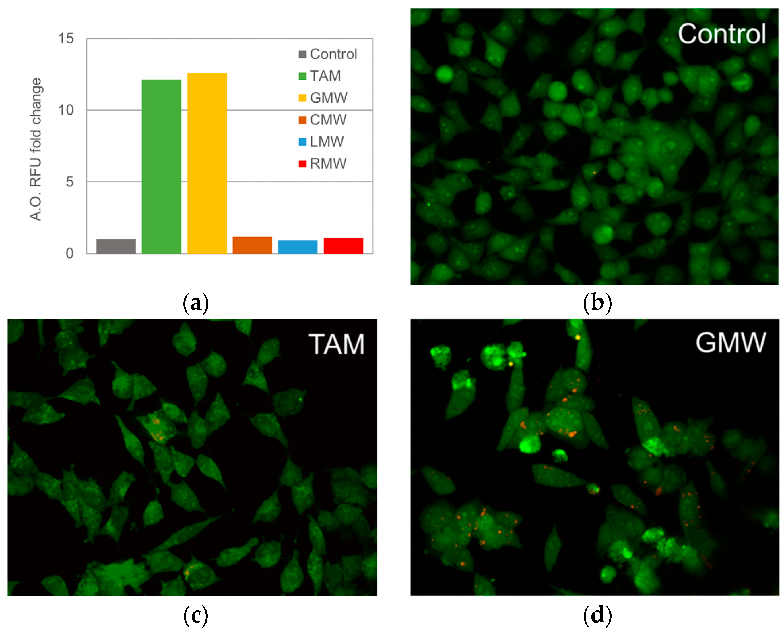
2.2.4. Formation of Acidic Vacuoles in HeLa Cells

Considering the antiproliferative profile of the Hypericum extracts, CMW, GMW, LMW and RMW were selected to perform acridine orange (AO) staining in order to evaluate the formation of acidic vacuoles in the cells after the treatment. AO is a fluorescent dye that binds to acidic compartments. Differences in fluorescence intensity could represent changes in cell response to the plant microextract, of particular interest to the formation of acidic vacuoles [56]. As a positive control we used 10 µM of tamoxifen (TAM), an established anticancer drug whose ability to enhance cell vacuolization has been reported previously [57]. As depicted in Figure 5a, GMW showed the highest fold change in RFU over control, similar to the effect produced by TAM, whilst the rest of the microextracts did not produce a representative fold change when treated with 100 µg/mL for 24 h. Taking this into account and considering the previous result, we proceeded to evaluate GMW microextract effects on HeLa cells under fluorescence microscopy. Images taken agreed with the data obtained by spectrofluorimetric assays, with this microextract enhancing the accumulation of the dye inside clearly visible vacuoles (Figure 5b).The accumulation of AO inside acidic vacuoles produces a metachromatic shift from green to red fluorescence [58].
To sum, the biological tests (primary and secondary bioassays) of the Hypericum species resembled the aforementioned order based on the TA values. Thus, the relevance of the plants was established as H. grandifolium > H. glandulosum > H. canariense > H. reflexum. The lower TA values together with neglected effects observed in the biological tests (Figure 4 and Figure 5) allow us do not consider the MW microextract H. reflexum for the chemical analysis by LC–MS. Accordingly, the MW microextracts of H. canariense, H. glandulosum and H. grandifolium were submitted to the study of their chemical profiling. Overall, the results might anticipate a different chemical profile of these microextracts.

2.3. Early Chemical Profiling of MW Microextracts

The final step for the evaluation of the anticancer potential was to analyze the chemical composition of the selected bioactive MW microextracts from the aerial parts of H. canariense, H. glandulosum and H. grandifolium (i.e., CMW, LMW and GMW, respectively). In an effort to estimate their chemical qualitative composition in terms of specialized metabolites, they were analyzed by UHPLC–DAD–MS (Table 2). Methanol and acetone–methanol extracts from H. canariense and H. grandifolium were analyzed by LC–MS and reported previously [33,42]. This work represents the first preliminary chemical study of H. glandulosum extract.
Table 2 shows the chemical profiles of the three Hypericum MW microextracts at 280 nm. These chromatograms also show the UHPLC fingerprint of these species and the substantial differences in their chemical composition, both in terms of the number of peaks and in the proportion of common peaks (Figure S1). We speculate that this differential profile is responsible for the differences observed in the biological activity of the microextracts (Figure 2, Figure 4 and Figure 5).
According to these preliminary results, a total of forty one main compounds in terms of specialized metabolites were detected in the methanol–water microextracts CMW, GMW and LMW, respectively. Twenty four were known compounds tentatively identified based on their retention times, spectral properties (UV-Vis and MS/MS patterns) and from the comparison of literature. These known compounds belonged to the family of phenolic secondary metabolites and they have previously been described or isolated from Hypericum extracts [19,42,60,62,63,64]. Among these known compounds 6, 9, 14, 15, 19, 20, 28 and 33 were ubiquitous in all MW microextracts. A careful analysis of the UV trace at 280 nm of CMW, GMW and LMW microextracts enabled the detection of at least the presence of eighteen, nineteen and twenty seven specialized metabolites, respectively. Additionally, these MW microextracts also contained a total of fifteen unidentified compounds whose spectral data suggested their novelty as natural products from Hypericum species since they were not found in the literature.
In the case of CMW microextract (from H. canariense), six major compounds were detected and identified as chlorogenic acid isomers (6 and 11), mangiferin isomers (16), quercetin-3-O-glucouronide (24) and quercetin (28). Only two detected compounds in CMW (4 and 25) were not associated to a described natural product previous detected in Hypericum species. Following the same analysis for GMW, we found three outstanding peaks, including p-coumaroylquinic acid isomer (19), quercetin 3-O-rhamnoside (327) and quercetin (28). The chromatogram obtained for GMW (from H. grandifolium), showed the presence of several minor compounds, four of them (5, 18, 31 and 32) were not possible to identify it, considering their UV spectra and their MS/MS fragmentations. Likewise the analysis of LMW microextract (H. glandulosum) by LC–MS revealed the presence of twenty seven compounds, including seven major compounds, among them, chlorogenic acid isomers (6, 11 and 12), catechin (15), rutin (22), kaempferol -3-O-rhamnoglucoside(26) and an unidentified component (40). This analysis also enables detecting another ten unidentified compounds (3, 7, 32, 3439 and 41). These unidentified secondary metabolites in LMW are mostly less polar compounds. This microextract is also characterized by the presence of several minor compounds.
In spite of this preliminary analysis by UHPLC–DAD–MS of MW microextracts from H. canariense, H. glandulosum and H. grandifolium, several minor detected compounds cannot be identified due to many of them being co-eluted with major compounds. In this framework, further bioguided isolation studies might be required to clarify their identity.

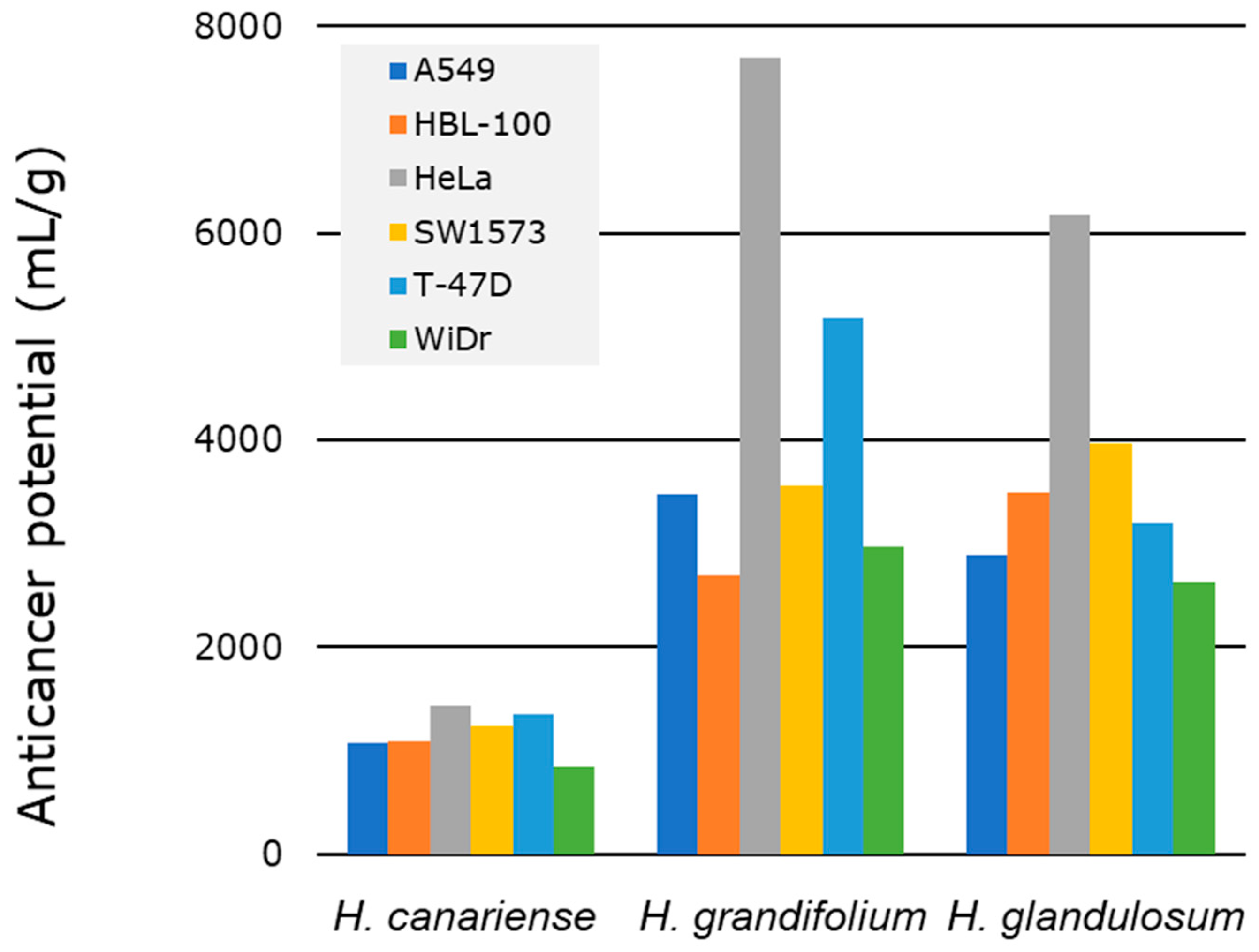
2.4. Bioactive and Anticancer Potential

In this work, we discussed the microextraction with three different solvents, the evaluation of bioactivity of the obtained microextracts, and the chemical analysis of the most polar microextracts from the aerial parts of four Hypericum species from Canary Islands. These experiments might address the selection of the species with the highest bioactive potential. Therefore, we propose an empirical equation for the calculation of the bioactive potential (BP, Equation (2)) based on the previous works by Eloff and Cos [11,15], our experience on the screening of plants [65,66] and the results described in previous sections. Basically, BP and TA have similar definitions, but BP refers to the efficacy of the plant material in several related bioassays and the possibility to find new compounds as the responsible of the initial observed biological activity.
( B P ) = ( N + 1 )   ×   T o t a l   A c t i v i t y     ×   C h e m i c a l   n o v e l t y
C h e m i c a l   n o v e l t y = t o t a l   n u m b e r   o f   d e t e c t e d   c o m p o u n d s n u m b e r   o f   k n o w n   c o m p o u n d s  
( B P ) = ( N + 1 )   ×   Y i e l d     e x t r a c t i o n   ×   1 B i o a c t i v i t y     ×   C h e m i c a l   n o v e l t y
In summary, the calculation of BP involves three experimental components (Equation (4)): the extraction process (yield extraction), the bioactivity evaluation (bioactivity in primary bioassays), and the chemical analysis by LC–MS (chemical novelty, Equation (3)); and a fourth factor defined as N + 1, where N means how many times a sample becomes the most active one in a bioassay. This fourth factor (N + 1) implies that BP never could be zero since an extract might be bioactive in any sort of biological test (intrinsic bioactivity). Therefore, after applying Equation (4), the resulting value of BP enables the sorting and prioritizing a list of plants from the highest to the lowest BP during a screening. agrochemical or pharmacological studies) related to the search of high-value products from plants.
To exemplify the calculation of BP, considering our special interest in the anticancer activity in the polar extracts from of the aerial parts of Hypericum species, we applied and modified BP formulae to calculate the anticancer potential (AP) of methanol–water microextracts (MW) from H. canariense (CMW), H. grandifolium (GMW) and H. glandulosum (LMW) (Equation (5)). From the biological tests and regarding GI50 values, the MW microextract from H. reflexum (RMW) was not subjected to the chemical analysis by LC–MS, and consequently it was considered the sample with the less AP.
A n t i c a n c e r   P o t e n t i a l   ( A P ) = ( N + 1 )   ×   T o t a l   A c t i v i t y     ×   C t o t a l C k n o w n  
The anticancer potential values from the MW were obtained based on one primary and two secondary bioassays related to anticancer activity (Figure 6). From the first biological test we conclude that none of the MW microextracts were the most active one. Finally, GMW was selected as the most active one in two of the secondary bioassays: cell migration disturbances in A549 cells and formation of acidic vacuoles in HeLa cells. Table 3 shows the N and chemical novelty values considered for the calculation of the AP of MW microextracts from H. grandifolium, H. glandulosum and H. canariense. The TA for these MW microextracts were computed in Section 2.2.2 (Table S2). Overall, the AP values permit to sort the microextracts: GMW > LMW> CMW (H. grandifolium > H. glandulosum > H. canariense). These results also allow select H. grandifolium Choisy as the species with the highest AP as a source of polar potentially bioactive compounds.

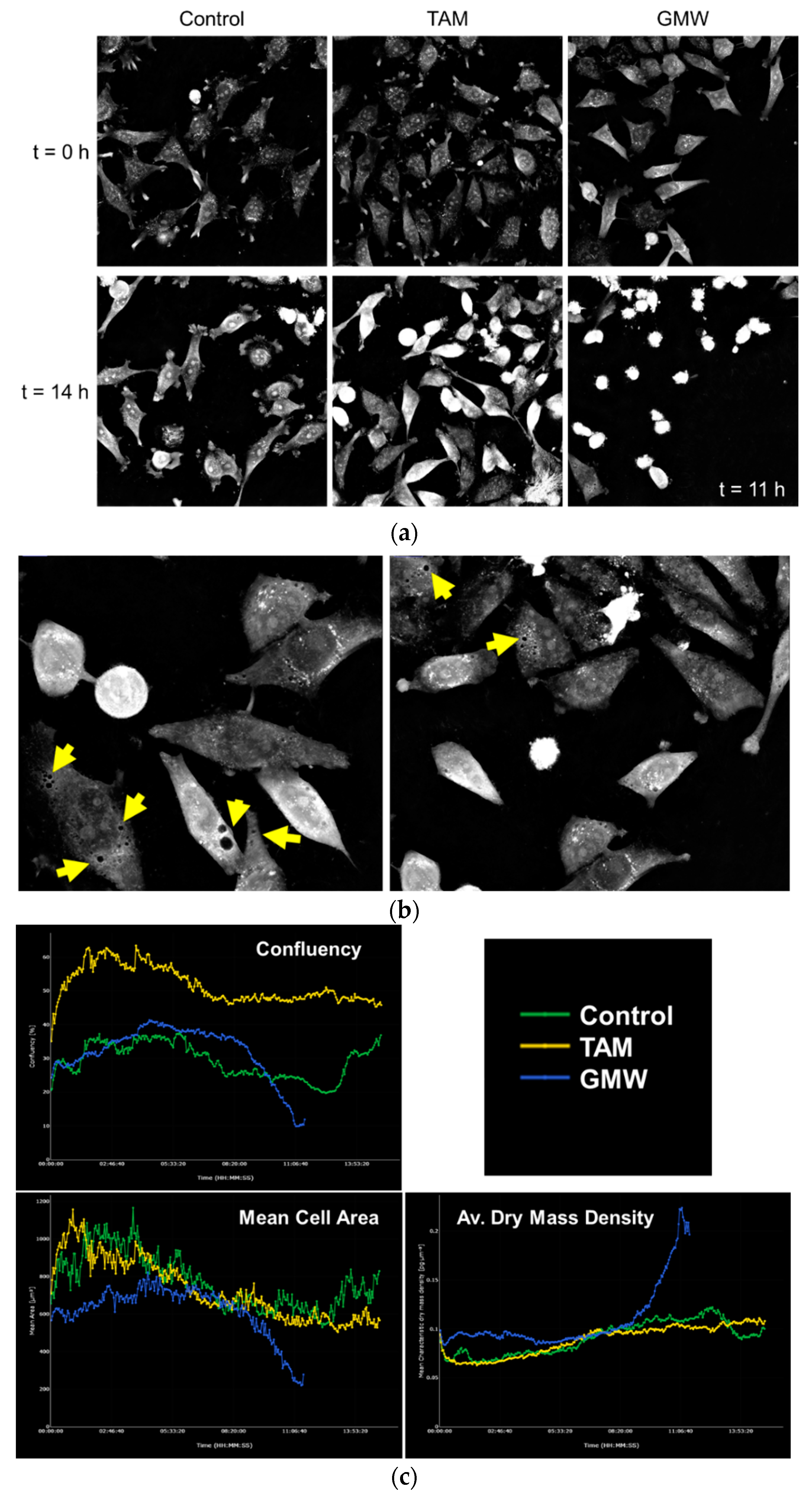
2.5. MW Microextracts of H. Grandifolium Induce Cell Death in HeLa Cells

Based on this, we selected the MW microextract of H. grandifolium (GMW) to run a more detailed study on HeLa cells using label-free continuous live-cell imaging. Live-cell imaging opens a new window in the study of cell response to potential bioactive compounds. The ability to evaluate the behavior of a population at single cell level facilitates the study of phenotypic changes between subgroups of individuals. Moreover, live imaging enables checking the effects at continuous time points, changing the concept of dose–response relationship at fixed times.
HeLa cells were exposed to GMW and the effects were monitored for 15 h. For comparison purposes, we used TAM. Figure 7 depicts how GMW is able to induce death to HeLa cells earlier and in extended populations than TAM (Figure 7a). In consonance with AO staining results (Figure 5), cells are highly vacuolated (Figure 7b) in the GMW-treated group. This feature could be induced not only by the pharmacological activity of the extract but as a cell response to an external agent, trying to isolate the exogenous substances. Since live imaging has enabled us to observe cell death progressively, we can differentiate several apoptotic hallmarks. Treatment with GMW induced nuclear condensation and cell shrinkage after 10 h of incubation in a broader way than TAM. For the same time period, cell death was not observed in the control group (Figure 7a, Supplementary material V1). The observed effects were analyzed and quantified with STEVE software (Figure 7c, Figures S2–S4). Cell death is represented as a drastic decrease in the percentage of confluence in the GMW-treated samples. The formation of apoptotic bodies by cell shrinkage is depicted in the diminished cell area. In agreement with this last result, dry mass density is increased in the GMW-treated cells when compared to the control, reflecting the collapsing of cells caused by the extract.

3. Materials and Methods

3.1. General Experimental Procedures

Acetonitrile, methanol, and formic acid for UHPLC were purchased from Merck (Darmstadt, Germany). Water for UHPLC was purified with a Millipore Simplicity System (Bedford, MA, USA). Solvents for the extraction of plant material were of analytical grade.

3.2. Plant Material

The aerial parts of H. canariense, H. grandifolium, and H. reflexum (6 to 8 kg of fresh material) were collected in La Palma and Tenerife (Canary Islands, Spain), during the period between June and July 2019. 7 kg of the aerial parts of H. glandulosum, were collected in La Palma and Tenerife (Canary Islands, Spain), in June 2021. All the specimens were collected and identified by Dr. Pedro Luis Pérez de Paz, University of La Laguna. A voucher specimen of species was deposited at TFC Herbarium, University of La Laguna (Tenerife, Spain) (Table 4). Plant material was dried in airy oven at 38 °C temperature for one week. Finally, plant material was stored at room temperature in the darkness.

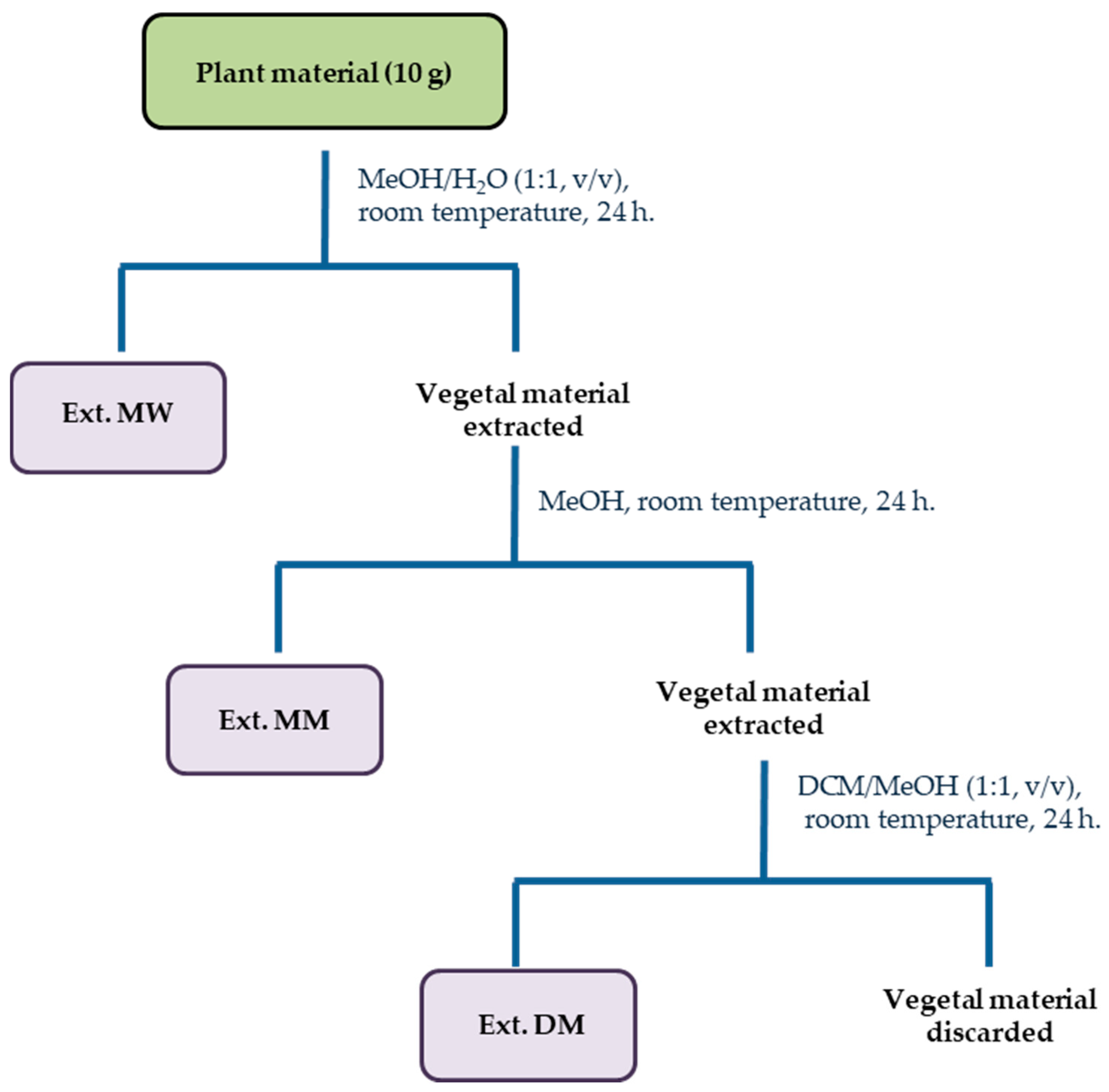
3.3. Extraction of Plant Material

Ground aerial parts (10 g) from H. canariense, H. glandulosum, H. grandifolium, and H. reflexum, were sequentially extracted with the same volume (200 mL) of methanol–water (1:1, v/v, MW), methanol (MM), and dichloromethane–methanol (1:1, v/v, DM) for 24 h at room temperature. The resulting extracts were filtered and concentrated under reduced pressure to evaporate the less volatile solvent. All extracts were resuspended in water and lyophilized affording MW, MM and DM extracts, respectively. Thereafter, the microextracts were weighed and stored at 4 °C until further processing.

3.4. UHPLC–DAD–MS3 Analysis

UHPLC–DAD–MS analyses of raw and aqueous extracts were conducted using a previously established method [67] with some modifications. Thus, 10 mg of polar microextracts (CMW, GMW and LMW) were dissolved in 1 mL of methanol–water (1:1, v/v) and subjected to UHPLC analyses. The separation was carried out on a Kinetex XB-C18 (150 mm × 2.1 mm × 1.7 μm, Phenomenex, Torrance, CA, USA). The mobile phases were 0.1% formic acid in water (A), and 0.1% formic acid in acetonitrile (B), and elution was conducted with the following gradient: 0 min–3% B, 60 min–26% B, 120 min–95% B. The flow rates were 0.3 mL/min. The UV–Vis spectra of the detected compounds were recorded over the 190–600 nm range. The chromatograms of these microextracts) were recorded at 280 nm. Mass spectra were recorded in the positive and negative ion modes. Compounds were characterized based on the maxima observed in their UV–Vis spectra and on their MS spectra. Additionally, the Reaxys database was searched for compounds previously detected and identified in Hypericum species.

3.5. Cell, Culture and Plating

The human solid tumor cell lines A549 (lung), HBL-100 (breast), HeLa (cervix), SW1573 (non-small-cell lung), T-47D (breast), and WiDr (colon) were used in this study. These cell lines were a kind gift from Prof. Godefridus J. Peters (VU Medical Center, Amsterdam, The Netherlands). Cells were maintained in 25 cm2 culture flasks in RPMI 1640 supplemented with 5% FBS and 2 mM L-glutamine in a 37 °C, 5% CO2, 95% humidified air incubator. For all tests, exponentially growing cells were trypsinogen and resuspended in an antibiotic-containing medium (100 units penicillin G and 0.1 mg of streptomycin per mL). Single-cell suspensions were counted with Moxi Z. After counting, dilutions were made to give the appropriate cell densities for inoculation onto the appropriate (microtiter) plates.

3.6. Antiproliferative Tests

The tests were performed in 96-well plates using the SRB assay [68] with the following specifications. Cell seeding density was 2500 cells/well for A549, HBL-100, HeLa, and SW1573, and 5000 cells/well for T-47D and WiDr. Stock samples were dissolved in DMSO at an initial concentration of 100 mg/mL. Test samples were prepared by decimal serial dilutions of the stock sample to give a final test concentration of 250, 25 and 2.5 µg/mL. The incubation time was 48 h. The optical density of each well was measured at 530 (primary) and 620 nm (secondary) using a microplate reader (Power Wave XS, BioTek, Winooski, VT, USA). The antiproliferative activity, expressed as 50% growth inhibition (GI50) values, was calculated according to the NCI formulas [49]. GI50 represent approximate values. Following NCI guidelines, the concentrations giving PG values above and below PG = 50 were used to make interpolations on the concentration axis.
The criteria used to categorize the cytotoxicity of the microextracts on the NCI’s protocol was as follows: GI50  ≤  20 µg/mL = highly cytotoxic, GI50 ranged 21–200 µg/mL = moderately cytotoxic, and GI50 > 201 µg/mL = weakly cytotoxic [69].

3.7. Cell Migration Assay

To study cell migration, we used the wound healing (scratch) assay [70]. Single-cell suspensions of A549 cells were seeded onto a 24 well plate at a density of 50,000 cells/well. Cells were incubated until they reached >90% confluence. Afterwards, a mark was drawn on the outside bottom of each well. This enabled finding the reference point when taking pictures. For each well, a scratch on the cell culture was made perpendicularly to the mark using a sterile p200 tip. Then, the medium was replaced with fresh medium with 2.5% FBS. This allows us to diminished the interference caused by cell proliferation during the time of assay, evaluating only the cell migration. Pictures were taken with a brightfield microscope (Axiovert 40 CFL, Zeiss, Germany) at one magnification (5X) using the software ZEN 2012 (blue edition v1.1.0.0) (accessed on 10 August 2022) at different time intervals (0, 6 and 24 h since the formation of the scratch). For the quantification of cell migration, TScratch, an image software based in MATLAB, was used adapting threshold of images to measure the area of the wound made [71]. Results are represented as percentage of the closed area as defined by Equation (6).
W o u n d   C l o s u r e   % = ( A t = 0 A t = Δ t A t = 0   )   ×   100

3.8. Acridine Orange Staining Assay

To evaluate the induction of acidic vacuoles by the plant extracts the acridine orange staining assay was used. Briefly, single-cell suspensions of HeLa were seeded onto a 6 well plate at a density of 100,000 cells/well. After 24 h extracts diluted in DMSO were added and incubated for an additional period of 24 h. Then, the medium was removed, cells were detached with trypsin, resuspended with PBS (200 µL), transferred to 1.5 mL tubes and centrifuge 1200 rpm × 5 min (Centrifuge 5418 R, Eppendorf). Each group of cell suspensions were incubated with 1 mL of acridine orange solution (20 µM in PBS) for 30 min at room temperature under dark conditions. After centrifuge at 1200 rpm for 5 min, cells were washed twice with PBS to remove the excess of staining, centrifuged one last time and resuspended in PBS. Then, 100 µL of each cell suspension were added onto a black-walled, clear-bottom, flat bottom 96 well plate and measured fluorescence intensity at 502/528 excitation/emission wavelengths using a microplate reader (Varioskan LUX VLBL0TD0, Thermo Fisher Scientific, Waltham, MA, USA). The number of cells was normalized with crystal violet staining and suspending the dye with 1% SDS in PBS, and finally measuring the optical density at 595 nm using a microplate reader as aforementioned.
For fluorescence microscopy images, cells were seeded onto a coverslip into a p6 well at the aforementioned seeding density. Treatments were made exactly the same as previously described and then staining was performed directly on the wells after removal of the medium. Two washes with PBS were made and coverslips were mounted fresh over glass slides to observe under fluorescence microscopy using SP5 Leica apparatus with LAS-AF software (Leica Microsystems) equipped with I3 (450–490) and N2.1 (515–560) excitation filters for green and red channels, respectively. Both channels were merged into individual images using ImageJ.

3.9. Live-Cell Imaging

A CX-A imaging platform microscope (Nanolive SA, Lausanne, Switzerland) was used to measure refractive indices, creating a holotomographic 3D image of the cells. HeLa cells were cultured at a density of 50,000 cells/well onto a IBIDI μ-Dish, 35 mm high (Martinsried, IBIDI, Germany) and treated with or without 100 µg/mL of extract right before the acquisition of the images. Data obtained were transferred to FIJI (NIH, USA) for image analysis. STEVE software (Nanolive SA) was used for the analysis of the refractive indices and obtention of the Confluency, Mean Cell Area, and Mean Average Dry Mass Density.

4. Conclusions

In summary, we have proposed a general methodology and empirical equation to assess the BP during the screening of extracts from plants. We applied the said protocol to four Hypericum species from the Canary Islands (H. canariense L., H. glandulosum Aiton, H. grandifolium Choisy and H. reflexum L.f.), specifically to evaluate the anticancer potential (AP) of their methanol–water microextracts. The assessment of AP involved the evaluation of the yield of extraction, the assessment of the antiproliferative activity of the microextracts and their dereplication by LC–MS. After the application of this pharmacognostic protocol, it was possible to assess the AP of the methanol–water microextracts and to select H. grandifolium as the best candidate to carry out the bioguided isolation of polar natural products with high added value.

Supplementary Materials

The following supporting information can be downloaded at: https://www.mdpi.com/article/10.3390/molecules27186101/s1, Figure S1: LC–PDA–MSn chromatograms for Hypericum MW microextracts; Figure S2: Confluency obtained with STEVE software based on refractive indexes resulting from CX-A observation over time; Figure S3: Mean Cell Area obtained with STEVE software based on refractive indexes resulting from CX-A observation over time; Figure S4: Average dry Mass Density obtained with STEVE software based on refractive indexes resulting from CX-A observation over time; Table S1: Antiproliferative activity of Hypericum microextracts; Table S2: Total activity of Hypericum microextracts; Video S1: Live-cell imaging of MW microextract of H. grandifolium.

Author Contributions

Conceptualization, R.L., C.C.S.-M. and J.M.P.; methodology, R.L., S.G. and J.M.P.; software, R.L., A.P., S.G. and J.M.P.; validation, R.L., A.P., S.G. and J.M.P.; formal analysis, A.P. and J.M.P.; investigation, R.L., A.P., A.G.-B., D.H., Y.T., S.G. and J.M.P.; resources, P.L.P.d.P.; data curation, R.L., A.P., S.G. and J.M.P.; writing—original draft preparation, R.L., A.P. and J.M.P.; writing—review and editing, R.L., S.G. and J.M.P.; supervision, R.L. and J.M.P.; project administration, R.L. and J.M.P.; funding acquisition, R.L. and J.M.P. All authors have read and agreed to the published version of the manuscript.

Funding

This work was supported by University of La Laguna grant 1192_2020 Hyperpharma project. It was also funded by the Canary Islands Government ACIISI/FEDER, UE, grant numbers ProID2020010101 and EIS 2020 06_ULL.

Institutional Review Board Statement

Not applicable.

Informed Consent Statement

Not applicable.

Data Availability Statement

The data presented in this study are available on request from the corresponding author. The data are not publicly available due to privacy or ethical restrictions.

Acknowledgments

R.L. thanks Miriam Pimienta Álvarez and Noelia Pimienta Álvarez, for their support and encouragement throughout all his research career. A.P. thanks the EU Social Fund (FSE) and the Canary Islands ACIISI for a predoctoral grant TESIS2020010055. The authors thank Silvia González and Víctor S. Martín for unvaluable support.

Conflicts of Interest

The authors declare no conflict of interest.

References

  1. Aburjai, T.; Natsheh, F.M. Plants Used in Cosmetics. Phytother. Res. 2003, 17, 987–1000. [Google Scholar] [CrossRef] [PubMed]
  2. Atanasov, A.G.; Zotchev, S.B.; Dirsch, V.M.; Orhan, I.E.; Banach, M.; Rollinger, J.M.; Barreca, D.; Weckwerth, W.; Bauer, R.; Bayer, E.A.; et al. Natural Products in Drug Discovery: Advances and Opportunities. Nat. Rev. Drug Discov. 2021, 20, 200–216. [Google Scholar] [CrossRef] [PubMed]
  3. Macías, F.A.; Mejías, F.J.; Molinillo, J.M. Recent Advances in Allelopathy for Weed Control: From Knowledge to Applications. Pest Manag. Sci. 2019, 75, 2413–2436. [Google Scholar] [CrossRef] [PubMed]
  4. Bhatia, P.; Chugh, A. Role of Marine Bioprospecting Contracts in Developing Access and Benefit Sharing Mechanism for Marine Traditional Knowledge Holders in the Pharmaceutical Industry. Glob. Ecol. Conserv. 2015, 3, 176–187. [Google Scholar] [CrossRef]
  5. Dixit, S.; Shukla, A.; Singh, V.; Upadhyay, S.K. Bioprospecting of Natural Compounds for Industrial and Medical Applications. In Bioprospecting of Plant Biodiversity for Industrial Molecules; Wiley: Hoboken, NJ, USA, 2021; pp. 53–71. [Google Scholar]
  6. Kewalanand; Prajapati, B. Cultivation and Bioprospecting of Medicinal Plants; Springer: Singapore, 2018; ISBN 9789811082917. [Google Scholar]
  7. Atanasov, A.G.; Waltenberger, B.; Pferschy-Wenzig, E.M.; Linder, T.; Wawrosch, C.; Uhrin, P.; Temml, V.; Wang, L.; Schwaiger, S.; Heiss, E.H.; et al. Discovery and Resupply of Pharmacologically Active Plant-Derived Natural Products: A Review. Biotechnol. Adv. 2015, 33, 1582–1614. [Google Scholar] [CrossRef] [PubMed]
  8. Rodríguez, A.O.; Ochoa-Pacheco, A.; Daniel, M.; Ortiz-Beat, E.; Font-salmo, O.; Guisado-bourzac, F.; Molina-bertr, S.; Monzote, L.; Cos, P.; Foubert, K.; et al. Cultivars of Plectranthus Neochilus Schltr. Plants 2022, 11, 134. [Google Scholar] [CrossRef]
  9. Schultz, F.; Osuji, O.F.; Nguyen, A.; Anywar, G.; Scheel, J.R.; Caljon, G.; Pieters, L.; Garbe, L.A. Pharmacological Assessment of the Antiprotozoal Activity, Cytotoxicity and Genotoxicity of Medicinal Plants Used in the Treatment of Malaria in the Greater Mpigi Region in Uganda. Front. Pharmacol. 2021, 12, 678535. [Google Scholar] [CrossRef]
  10. Amoo, S.O.; Finnie, J.F.; Van Staden, J. In Vitro Pharmacological Evaluation of Three Barleria Species. J. Ethnopharmacol. 2009, 121, 274–277. [Google Scholar] [CrossRef]
  11. Eloff, J.N. Quantification the Bioactivity of Plant Extracts during Screening and Bioassay Guided Fractionation. Phytomedicine 2004, 11, 370–371. [Google Scholar] [CrossRef]
  12. Gado, D.A.; Abdalla, M.A.; Ahmed, A.S.; Madikizela, B.; Nkadimeng, S.M.; Ehlers, M.M.; McGaw, L.J. In Vitro Antibacterial Activity of Loxostylis Alata Extracts and Isolated Compounds against Salmonella Species. BMC Complement. Med. Ther. 2021, 21, 121. [Google Scholar] [CrossRef]
  13. Bisi-Johnson, M.A.; Obi, C.L.; Hattori, T.; Oshima, Y.; Li, S.; Kambizi, L.; Eloff, J.N.; Vasaikar, S.D. Evaluation of the Antibacterial and Anticancer Activities of Some South African Medicinal Plants. BMC Complement. Altern. Med. 2011, 11, 14. [Google Scholar] [CrossRef] [PubMed]
  14. Saddiqe, Z.; Naeem, I.; Hellio, C.; Patel, A.V.; Abbas, G. Phytochemical Profile, Antioxidant and Antibacterial Activity of Four Hypericum Species from the UK. South African J. Bot. 2020, 133, 45–53. [Google Scholar] [CrossRef]
  15. Cos, P.; Vlietinck, A.J.; Berghe, D.V.; Maes, L. Anti-Infective Potential of Natural Products: How to Develop a Stronger in Vitro “Proof-of-Concept”. J. Ethnopharmacol. 2006, 106, 290–302. [Google Scholar] [CrossRef] [PubMed]
  16. Galeotti, N. Hypericum Perforatum (St John’s Wort) beyond Depression: A Therapeutic Perspective for Pain Conditions. J. Ethnopharmacol. 2017, 200, 136–146. [Google Scholar] [CrossRef]
  17. Zhao, J.; Liu, W.; Wang, J.C. Recent Advances Regarding Constituents and Bioactivities of Plants from the Genus Hypericum. Chem. Biodivers. 2015, 12, 309–349. [Google Scholar] [CrossRef] [PubMed]
  18. Sanna, C.; Scognamiglio, M.; Fiorentino, A.; Corona, A.; Graziani, V.; Caredda, A.; Cortis, P.; Montisci, M.; Ceresola, E.R.; Canducci, F.; et al. Prenylated Phloroglucinols from Hypericum Scruglii, an Endemic Species of Sardinia (Italy), as New Dual HIV-1 Inhibitors Effective on HIV-1 Replication. PLoS ONE 2018, 13, e0195168. [Google Scholar] [CrossRef]
  19. Zhang, R.; Ji, Y.; Morcol, T.; Lin, F.; Gu, R.; Kennelly, E.J.; Long, C. UPLC-QTof-MS Chemical Profiling and Characterization of Antiproliferative and Anti-Inflammatory Compounds from Seven Hypericum Species in China. Ind. Crops Prod. 2021, 173, 114156. [Google Scholar] [CrossRef]
  20. Cenić-Miloŝević, D.; Tambur, Z.; Ivanĉajić, S.; Stanojković, T.; Grozdanić, N.; Kuliŝić, Z.; Juranić, Z. Antiproliferative Effects of Tanaceti Partheni, Hypericum Perforatum and Propolis on HeLa Cells. Arch. Biol. Sci. 2014, 66, 705–712. [Google Scholar] [CrossRef]
  21. Gaid, M.; Füller, J.; Müller-Goymann, C. The Petroleum Ether Extract from Hypericum Perforatum Root Cultures Exhibits Potent Antiproliferative Activity in Human Keratinocytes and Fibroblasts. Planta Med. 2019, 85, 591–598. [Google Scholar] [CrossRef]
  22. Mokoro Vincent, O.; Mwanzia Nguta, J.; Mutie Musila, F.; Matara Nyak, D.; Hashim Mohammed, A.; Apiri Gervason, M.; Simon Mitema, E. Ethnopharmacology, Pharmacological Activities, and Chemistry of the Hypericum Genus. J. Phytopharm. 2021, 10, 105–113. [Google Scholar] [CrossRef]
  23. De Paz, P.L.P.; Padrón, C.E.H. Plantas Medicinales o Útiles En La Flora Canaria: Aplicaciones Populares; Francisco Lemus: La Laguna, Spain, 1999; ISBN 8487973124. [Google Scholar]
  24. Pérez de Paz, P.L.; Medina Medina, I. Catálogo de Las Plantas Medicinales de La Flora Canaria: Aplicaciones Populares, 1st ed.; Viceconsejería de Cultura y Deportes del Gobierno de Canarias, Instituto de Estudios Canarios: Santa Cruz de Tenerife, Spain, 1988. [Google Scholar]
  25. Pardo de Santayana, M.; Morales, R.; Tardío, J.; Aceituna, L.; Molina, M. Conocimientos Tradicionales Relativos a La Biodiversidad; Ministerio de Agricultura, Alimentación y Medio Ambiente: Madrid, Spain, 2018; Volume Segunda Fa, ISBN 9788449114724. [Google Scholar]
  26. Herrera, R.M.; Pérez, M.; Martín-Herrera, D.A.; López-García, R.; Rabanal, R.M.; Arias, A. Antimicrobial Activity of Extracts from Plants Endemic to the Canary Islands. Phyther. Res. 1996, 10, 364–366. [Google Scholar] [CrossRef]
  27. Rabanal, R.M.; Arias, A.; Prado, B.; Hernández-Pérez, M.; Sánchez-Mateo, C.C. Antimicrobial Studies on Three Species of Hypericum from the Canary Islands. J. Ethnopharmacol. 2002, 81, 287–292. [Google Scholar] [CrossRef]
  28. Luz Cardona, M.; Pedro, J.R.; Seoane, E.; Vidal, R. Xanthone Constituents of Hypericum Canariensis. J. Nat. Prod. 1985, 48, 467–469. [Google Scholar] [CrossRef]
  29. Cardona, M.L.; Fernandez, M.I.; Pedro, J.R.; Seoane, E.; Vidal, R. Additional New Xanthones and Xanthonolignoids from Hypericum Canariensis. J. Nat. Prod. 1986, 49, 95–100. [Google Scholar] [CrossRef]
  30. Cardona, M.L.; Fernandez, M.I.; Pedro, J.R.; Serrano, Á. Xanthones From Hypericum Reflexum. Phytochemestry 1990, 29, 3003–3006. [Google Scholar] [CrossRef]
  31. Cardona, L.; Pedro, J.R.; Serrano, A.; Muñoz, M.C.; Solans, X. Spiroterpenoids from Hypericum Reflexum. Phytochemistry 1993, 33, 1185–1187. [Google Scholar] [CrossRef]
  32. Díaz, J.G.; De Paz, P.P.; Herz, W. New Water Soluble Flavone and Xanthone Glycosides from Hypericum Canariense L. Phytochem. Lett. 2010, 3, 171–175. [Google Scholar] [CrossRef]
  33. Díaz, J.G. Chemical Composition of Hypericum Coadunatum Chr. from the Canary Islands. J. Mol. Struct. 2022, 1248, 131447. [Google Scholar] [CrossRef]
  34. Zorzetto, C.; Sánchez-Mateo, C.C.; Rabanal, R.M.; Lupidi, G.; Petrelli, D.; Vitali, L.A.; Bramucci, M.; Quassinti, L.; Caprioli, G.; Papa, F.; et al. Phytochemical Analysis and in Vitro Biological Activity of Three Hypericum Species from the Canary Islands (Hypericum Reflexum, Hypericum Canariense and Hypericum Grandifolium). Fitoterapia 2015, 100, 95–109. [Google Scholar] [CrossRef]
  35. Prado, B.; Rabanal, R.M.; Sanchez-Mateo, C.C. Evaluation of the Central Properties of Several Hypericum Species from the Canary Islands. Phyther. Res. 2002, 16, 740–744. [Google Scholar] [CrossRef]
  36. Sánchez-Mateo, C.C.; Bonkanka, C.X.; Prado, B.; Rabanal, R.M. Antidepressant Properties of Some Hypericum Canariense L. and Hypericum Glandulosum Ait. Extracts in the Forced Swimming Test in Mice. J. Ethnopharmacol. 2005, 97, 541–547. [Google Scholar] [CrossRef]
  37. Rabanal, R.M.; Bonkanka, C.X.; Hernández-Pérez, M.; Sánchez-Mateo, C.C. Analgesic and Topical Anti-Inflammatory Activity of Hypericum Canariense L. and Hypericum Glandulosum Ait. J. Ethnopharmacol. 2005, 96, 591–596. [Google Scholar] [CrossRef] [PubMed]
  38. Sánchez-Mateo, C.C.; Bonkanka, C.X.; Hernández-Pérez, M.; Rabanal, R.M. Evaluation of the Analgesic and Topical Anti-Inflammatory Effects of Hypericum Reflexum L. Fil. J. Ethnopharmacol. 2006, 107, 1–6. [Google Scholar] [CrossRef] [PubMed]
  39. Sánchez-Mateo, C.C.; Bonkanka, C.X.; Prado, B.; Rabanal, R.M. Antidepressant Activity of Some Hypericum Reflexum L. Fil. Extracts in the Forced Swimming Test in Mice. J. Ethnopharmacol. 2007, 112, 115–121. [Google Scholar] [CrossRef] [PubMed]
  40. Bonkanka, C.X.; Smelcerovic, A.; Zuehlke, S.; Rabanal, R.M.; Spiteller, M.; Sánchez-Mateo, C.D.C. HPLC-MS Analysis and Anti-Oedematogenic Activity of Hypericum Grandifolium Choisy (Hypericaceae). Planta Med. 2008, 74, 719–725. [Google Scholar] [CrossRef]
  41. Sánchez-Mateo, C.C.; Bonkanka, C.X.; Rabanal, R.M. Hypericum Grandifolium Choisy: A Species Native to Macaronesian Region with Antidepressant Effect. J. Ethnopharmacol. 2009, 121, 297–303. [Google Scholar] [CrossRef]
  42. Bonkanka, C.X.; Sánchez-Mateo, C.D.C.; Rabanal, R.M. Antinociceptive Activity of Hypericum Grandifolium Choisy in Mice. J. Nat. Med. 2011, 65, 122–128. [Google Scholar] [CrossRef]
  43. Macías, F.A.; Lacret, R.; Varela, R.M.; Nogueiras, C.; Molinillo, J.M.G. Bioactive Apocarotenoids from Tectona Grandis. Phytochemistry 2008, 69, 2708–2715. [Google Scholar] [CrossRef]
  44. Ion, V.; Ielciu, I.; Cârje, A.G.; Muntean, D.L.; Crişan, G.; Păltinean, R. Hypericum Spp.—An Overview of the Extraction Methods and Analysis of Compounds. Separations 2022, 9, 17. [Google Scholar] [CrossRef]
  45. Dresler, S.; Kováčik, J.; Strzemski, M.; Sowa, I.; Wójciak-Kosior, M. Methodological Aspects of Biologically Active Compounds Quantification in the Genus Hypericum. J. Pharm. Biomed. Anal. 2018, 155, 82–90. [Google Scholar] [CrossRef]
  46. Wilson, B.A.P.; Thornburg, C.C.; Henrich, C.J.; Grkovic, T.; O’Keefe, B.R. Creating and Screening Natural Product Libraries. Nat. Prod. Rep. 2020, 37, 893–918. [Google Scholar] [CrossRef] [PubMed]
  47. Thornburg, C.C.; Britt, J.R.; Evans, J.R.; Akee, R.K.; Whitt, J.A.; Trinh, S.K.; Harris, M.J.; Thompson, J.R.; Ewing, T.L.; Shipley, S.M.; et al. NCI Program for Natural Product Discovery: A Publicly-Accessible Library of Natural Product Fractions for High-Throughput Screening. ACS Chem. Biol. 2018, 13, 2484–2497. [Google Scholar] [CrossRef] [PubMed]
  48. Grkovic, T.; Akee, R.K.; Thornburg, C.C.; Trinh, S.K.; Britt, J.R.; Harris, M.J.; Evans, J.R.; Kang, U.; Ensel, S.; Henrich, C.J.; et al. National Cancer Institute (NCI) Program for Natural Products Discovery: Rapid Isolation and Identification of Biologically Active Natural Products from the NCI Prefractionated Library. ACS Chem. Biol. 2020, 15, 1104–1114. [Google Scholar] [CrossRef] [PubMed]
  49. Monks, A.; Scudiero, D.; Skehan, P.; Shoemaker, R.; Paull, K.; Vistica, D.; Hose, C.; Langley, J.; Cronise, P.; Vaigro-wolff, A.; et al. Feasibility of a High-Flux Anticancer Drug Screen Using a Diverse Panel of Cultured Human Tumor Cell Lines. J. Natl. Cancer Inst. 1991, 83, 757–766. [Google Scholar] [CrossRef]
  50. Geran, R.I.; Greenberg, N.H.; MacDonald, M.M. Protocols for Screening Chemical Agents and Natural Products against Animal Tumors and Other Biological Systems. Cancer Chemother. Rep. 1972, 3, 17–19. [Google Scholar]
  51. Friedl, P.; Wolf, K. Tumour-Cell Invasion and Migration: Diversity and Escape Mechanisms. Nat. Rev. Cancer 2003, 3, 362–374. [Google Scholar] [CrossRef]
  52. Hu, Y.; Reggiori, F. Molecular Regulation of Autophagosome Formation. Biochem. Soc. Trans. 2022, 50, 55–69. [Google Scholar] [CrossRef]
  53. Alvarez-Meythaler, J.G.; Garcia-Mayea, Y.; Mir, C.; Kondoh, H.; LLeonart, M.E. Autophagy Takes Center Stage as a Possible Cancer Hallmark. Front. Oncol. 2020, 10, 6069. [Google Scholar] [CrossRef]
  54. Shindo-Okada, N.; Takeuchi, K.; Han, B.S.; Nagamachi, Y. Establishment of Cell Lines with High and Low Metastatic Potential from A549 Human Lung Adenocarcinoma. Japanese J. Cancer Res. 2002, 93, 50–60. [Google Scholar] [CrossRef]
  55. Vaid, B.; Chopra, B.S.; Raut, S.; Sagar, A.; Badmalia, M.D.; Ashish; Khatri, N. Antioxidant and Wound Healing Property of Gelsolin in 3T3-L1 Cells. Oxid. Med. Cell. Longev. 2020, 2020, 4045365. [Google Scholar] [CrossRef]
  56. Karna, P.; Zughaier, S.; Pannu, V.; Simmons, R.; Narayan, S.; Aneja, R. Induction of Reactive Oxygen Species-Mediated Autophagy by a Novel Microtubule-Modulating Agent. J. Biol. Chem. 2010, 285, 18737–18748. [Google Scholar] [CrossRef] [Green Version]
  57. Majumdar, S.K.; Valdellon, J.A.; Brown, K.A. In Vitro Investigations on the Toxicity and Cell Death Induced by Tamoxifen on Two Non-Breast Cancer Cell Types. J. Biomed. Biotechnol. 2001, 2001, 99–107. [Google Scholar] [CrossRef]
  58. Thomé, M.P.; Filippi-Chiela, E.C.; Villodre, E.S.; Migliavaca, C.B.; Onzi, G.R.; Felipe, K.B.; Lenz, G. Ratiometric Analysis of Acridine Orange Staining in the Study of Acidic Organelles and Autophagy. J. Cell Sci. 2016, 129, 4622–4632. [Google Scholar] [CrossRef] [PubMed]
  59. Alahmad, A.; Alghoraibi, I.; Zein, R.; Kraft, S.; Dräger, G.; Walter, J.G.; Scheper, T. Identification of Major Constituents of Hypericum Perforatum L. Extracts in Syria by Development of a Rapid, Simple, and Reproducible HPLC-ESI-Q-TOF MS Analysis and Their Antioxidant Activities. ACS Omega 2022, 7, 13475–13493. [Google Scholar] [CrossRef] [PubMed]
  60. Tatsis, E.C.; Boeren, S.; Exarchou, V.; Troganis, A.N.; Vervoort, J.; Gerothanassis, I.P. Identification of the Major Constituents of Hypericum Perforatum by LC/SPE/NMR and/or LC/MS. Phytochemistry 2007, 68, 383–393. [Google Scholar] [CrossRef] [PubMed]
  61. Hong, D.; Yin, F.; Hu, L.H.; Lu, P. Sulfonated Xanthones from Hypericum Sampsonii. Phytochemistry 2004, 65, 2595–2598. [Google Scholar] [CrossRef]
  62. Ploss, O.; Petereit, F.; Nahrstedt, A. Procyanidins from the Herb of Hypericum Perforatum. Pharmazie 2001, 56, 509–511. [Google Scholar]
  63. Ersoy, E.; Eroglu Ozkan, E.; Boga, M.; Yilmaz, M.A.; Mat, A. Anti-Aging Potential and Anti-Tyrosinase Activity of Three Hypericum Species with Focus on Phytochemical Composition by LC–MS/MS. Ind. Crops Prod. 2019, 141, 111735. [Google Scholar] [CrossRef]
  64. Camas, N.; Radusiene, J.; Ivanauskas, L.; Jakstas, V.; Kayikci, S.; Cirak, C. Chemical Composition of Hypericum Species from the Taeniocarpium and Drosanthe Sections. Plant Syst. Evol. 2014, 300, 953–960. [Google Scholar] [CrossRef]
  65. Macías, F.A.; Lacret, R.; Varela, R.M.; Nogueiras, C.; Molinillo, J.M.G. Isolation and Phytotoxicity of Terpenes from Tectona Grandis. J. Chem. Ecol. 2010, 36, 396–404. [Google Scholar] [CrossRef]
  66. González-Coloma, A.; Reina, M.; Sáenz, C.; Lacret, R.; Ruiz-Mesia, L.; Arán, V.J.; Sanz, J.; Martínez-Díaz, R.A. Antileishmanial, Antitrypanosomal, and Cytotoxic Screening of Ethnopharmacologically Selected Peruvian Plants. Parasitol. Res. 2012, 110, 1381–1392. [Google Scholar] [CrossRef] [PubMed]
  67. Pawłowska, K.A.; Hałasa, R.; Dudek, M.K.; Majdan, M.; Jankowska, K.; Granica, S. Antibacterial and Anti-Inflammatory Activity of Bistort (Bistorta Officinalis) Aqueous Extract and Its Major Components. Justification of the Usage of the Medicinal Plant Material as a Traditional Topical Agent. J. Ethnopharmacol. 2020, 260, 113077. [Google Scholar] [CrossRef] [PubMed]
  68. Orellana, E.A.; Kasinski, A.L. Sulforhodamine B (SRB) Assay in Cell Culture to Investigate Cell Proliferation. Bio-protocol 2016, 6, e1984. [Google Scholar] [CrossRef] [PubMed]
  69. Niksic, H.; Becic, F.; Koric, E.; Gusic, I.; Omeragic, E.; Muratovic, S.; Miladinovic, B.; Duric, K. Cytotoxicity Screening of Thymus Vulgaris L. Essential Oil in Brine Shrimp Nauplii and Cancer Cell Lines. Sci. Rep. 2021, 11, 13178. [Google Scholar] [CrossRef]
  70. Liang, C.C.; Park, A.Y.; Guan, J.L. In Vitro Scratch Assay: A Convenient and Inexpensive Method for Analysis of Cell Migration in Vitro. Nat. Protoc. 2007, 2, 329–333. [Google Scholar] [CrossRef] [PubMed]
  71. Gebäck, T.; Schulz, M.M.P.; Koumoutsakos, P.; Detmar, M. TScratch: A Novel and Simple Software Tool for Automated Analysis of Monolayer Wound Healing Assays. Biotechniques 2009, 46, 265–274. [Google Scholar] [CrossRef]
Figure 1. Workflow procedure for the preparation of microextracts from aerial parts of Hypericum species from the Canary Islands.
Molecules 27 06101 g001
Figure 2. Antiproliferative activity (GI50) of microextracts from Hypericum species. MW = methanol–water; MM = methanol; DM = dichloromethane–methanol.
Molecules 27 06101 g002
Figure 3. Total activity (GI50) of microextracts from Hypericum species.
Molecules 27 06101 g003
Figure 4. Wound closure assay results on A549 cells when using 100 µg/mL (a) and 50 µg/mL (b) of MW microextracts. Wound closure percentage with respect to time = 0 is given on the right top corner of each image.
Molecules 27 06101 g004
Figure 5. (a) Relative fluorescence units (RFU) compared to control. HeLa cells were treated for 24 h with 100 µg/mL of each extract. (bd). Representative images of fluorescence microscopy after 24 h of incubation with 10 µM of TAM and 100 µg/mL of GMW. Captured images in green and red were merged using ImageJ.
Molecules 27 06101 g005
Figure 6. Anticancer potential of MW microextracts from Hypericum species.
Molecules 27 06101 g006
Figure 7. Live-cell imaging study on HeLa cells. (a) Representative images of the experiments for untreated (control) cells and cells exposed to 10 µM TAM or 100 µg/mL GMW. (b) Images of cell vacuolization after treatment with GMW for 5 h. Yellow arrows indicate cytoplasmic vacuoles observed. (c) Confluency, Mean Cell Area and Average Dry Mass Density obtained with STEVE software based on refractive indices resulting from CX-A observation over time. Green: untreated cells. Yellow: TAM (10 µM). Blue: GMW (100 µg/mL).
Molecules 27 06101 g007
Table 1. Yield of microextracts from Hypericum species from the Canary Islands.
Yield (%)
SpeciesMWMMDMTotal
C8.59.52.420.4
G7.58.01.917.4
L13.97.69.130.6
R14.36.91.222.1
C = H. canariense; G = H. grandifolium; L = H. glandulosum; R = H. reflexum; MW = methanol/water microextracts; MM = methanol microextracts; DM = dichloromethane/methanol microextracts.
Table 2. Qualitative analysis of polar microextracts by liquid chromatography photodiode array detection mass spectrometry (LC–PDA–MSn).
PeakRT (min)(max)MS [M+H]+MS [M-H]CMWGMWLMWPutative Compound
13.3269171.03169.03--gallic acid [59]
26.6259, 293154.99153.07-3,4-dihydroxybenzoic acid [60]
37.6214, 256205.30203.18--unidentified compound
47.8202, 279-315.22--unidentified compound
58.1205, 262, 294343.19341.18--unidentified compound
611.3214, 325355.2353.25neochlorogenic acid [61]
712.9287, 316343.21341.28--unidentified compound
814.4209, 311339.27337.27-p-coumaroylquinic acid isomer [62,63]
915.4217, 310339.20337.21p-coumaroylquinic acid isomer [62,63]
1016.5279579.19577.19--dimeric procyanidin type B [64]
1117.9216, 325355.20353.25-chlorogenic acid [42]
1220.0214, 325355.19353.24--cryptochlorogenic acid [63]
1321.7280579.20577.19--dimeric procyanidin type B [64]
1423.3279579.17577.19dimeric procyanidin type B
1524.6279291.13289.17catechin [64]
1625.9203, 257, 318, 369423.19421.16--mangiferin/isomangiferin [63]
1727.2203407.18405.28--neolancerin/xanthohypericosider [60]
1829.2279865.23863.23--unidentified procyanidin
1930.2279867.24865.26trimeric procyanidin type C [64]
2033.2279-576.17 atetrameric procyanidin B type [64]
2135.9279-720.22 b-pentameric procyanidn B type [64]
2237.6209, 255, 354611.21609.20--rutin [19,42]
2338.5256, 353465.18463.14-quercetin 3-O-glucoside [63]
2439.8203, 255, 353479.16477.18--quercetin 3-O-glucuronide [63]
2541.4286-361.17--unidentified compound
2642.4203, 265, 344595.20593.19--kaempferol 3-O-rhamnoglucoside
2743.7203, 255, 350449.19447.17--quercetin 3-O-rhamnoside [19,42]
2855.5203, 254, 368303.11301.1quercetin [19,42]
2960.0217, 310, 353611.17609.20-quercetin O-p-coumaroylhexoside [63]
3065.1218, 368287.10285.08--kaempferol [19]
3167.7219, 342749.16747.18--unidentified compound
3268.4265, 340749.15747.17-unidentified compound
3371.1268, 335539.16537.30amentoflavone [19]
3478.8284, 369, 483, 565367.34365.30--unidentified compound
3579.2287, 369, 483381.32379.36--unidentified compound
3686.7221, 291363.32361.32--unidentified compound
3787.4287, 369349.31347.33--unidentified compound
3891.9221, 291363.35361.35--unidentified compound
3997.3221, 290333.31331.27--unidentified compound
40100.0221, 291347.33345.33--unidentified compound
41101.0221, 287333.34331.34--unidentified compound
a [M-2H]2−. b [M-2H]2−.
Table 3. Anticancer potential for the most bioactive MW Hypericum extracts.
Chemical NoveltyAnticancer Potential (mL/g)
MicroextractsNCtotalCknownCtotal/CknownA549HBL-100HeLaSW1573T-47DWiDr
CMW018161.12510741087142712421347846
GMW219151.266347526897699355951802966
LMW027161.688289735036175397732072637
Table 4. Collection data of four Hypericum species.
Plant SpeciesCommon NameCollection SiteVoucher
Hypericum canariense L.granadilloBreña Baja. Las Ledas (La Palma island)53,351
Hypericum glandulosum Aitonmalfurada del monteIn between Mirador de Gallegos and Roque Faro (La Palma island)53,775
Hypericum grandifolium ChoisymalfuradaVilla de Mazo. La Tablada (La Palma island)53,350
Hypericum reflexum L.fcruzadillaArico-Granadilla (Tenerife island)53,346
Publisher’s Note: MDPI stays neutral with regard to jurisdictional claims in published maps and institutional affiliations.

