Article

Synthesis and In Silico Docking Study towards M-Pro of Novel Heterocyclic Compounds Derived from Pyrazolopyrimidinone as Putative SARS-CoV-2 Inhibitors

1
Laboratory of Heterocyclic Chemistry, Natural Products and Reactivity, Medicinal Chemistry and Natural Products (LR11ES39), Faculty of Sciences of Monastir, University of Monastir, Monastir 5000, Tunisia
2
Department of Organic Chemistry, Martin-Luther-University Halle-Wittenberg, Kurt-Mothes-Str. 2, D-06120 Halle, Germany
3
Department of Zoology, College of Science, King Saud University, Riyad 11451, Saudi Arabia
*
Authors to whom correspondence should be addressed.
Molecules 2022, 27(16), 5303; https://doi.org/10.3390/molecules27165303
Submission received: 30 July 2022 / Revised: 11 August 2022 / Accepted: 15 August 2022 / Published: 19 August 2022
(This article belongs to the Section Medicinal Chemistry)

Abstract

:
In addition to vaccines, antiviral drugs are essential in order to suppress COVID-19. Although some inhibitor candidates have been determined to target the SARS-CoV-2 protein, there is still an urgent need to continue researching novel inhibitors of the SARS-CoV-2 main protease ‘Omicron P132H’, a protein that has recently been discovered. In the present study, in the search for therapeutic alternatives to treat COVID-19 and its recent variants, we conducted a structure-based virtual screening using docking studies for a new series of pyrazolo[3,4-d]pyrimidin-4(5H)-one derivatives 513, which were synthesized from the condensation reaction of pyrazolopyrimidinone-hydrazide (4) with a series of electrophiles. Some significant ADMET predictions–in addition to the docking results–were obtained based on the types of interactions formed and the binding energy values were compared to the reference anti- SARS-CoV-2 redocked drug nirmatrelvir.

Graphical Abstract

1. Introduction

At the end of 2019, a new coronavirus named SARS-CoV-2 appeared in Wuhan, China, and caused an unusual outbreak of viral pneumonia. Highly contagious, this infection was caused by the novel coronavirus, also known as coronavirus disease 2019 (COVID-19) [1]. According to the World Health Organization (WHO), about 14.9 million excess deaths were associated with the COVID-19 pandemic in 2020 and 2021 [2]. The main clinical symptoms of this virus are fever, fatigue, headache, dry cough, and runny nose. These problems are specifically associated with difficulties in breathing, which often lead to the patient’s death. The WHO declared this pandemic to be one of the most dangerous health catastrophes in human history [3] as this virus accelerated faster than experts predicted [4].
Nowadays, many variants of SARS-CoV-2 are appearing; these variants hold altered spike proteins, hence allowing the virus to nullify the effectiveness of the vaccines. The Centers for Disease Control and Prevention (CDC) has classified SARS-CoV-2 variants as viruses of concern, interest, and high importance. Thus, several variants of SARS-CoV-2 have been identified and deemed a source of long-term risk of infection in immunocompromised individuals [5]. The SARS-CoV-2 main protease (Mpro) is a cysteine protease that is critical for the treatment of the two polyproteins (pp1a and pp1ab) encoded by the SARS-CoV-2 genome. Mpro plays an indispensable role in the replication cycle of the coronavirus. In more detail, the Mpro operates at more than 11 cleavage sites on the pp1ab [6]. The recognition sequence is Leu-Gln for most of the 11 sites [7]. Inhibiting the role of Mpro would essentially block any viral replication [8]. There are no known homologs of Mpro in humans with an identical cleavage specificity. Thereby, its inhibition is unlikely to show any side effects, making it an important target for COVID-19 drugs.
On 26 November 2021, the World Health Organization’s Technical Advisory Group on Virus Evolution (TAG-VE) proposed that the variant B.1.1.529, commonly known as Omicron, was associated with a substantial ability to evade immunity from prior infections [9]. The TAG-VE made this decision after finding that the Omicron variant has several mutations that might influence how quickly it spreads or the danger of the disease it causes. Further, the Omicron variant is considered as the most divergent strain seen in large numbers so far during the COVID-19 pandemic, raising concerns that it may be related to greater transmissibility, lower vaccine efficacy, and an increased risk of reinfection [10,11,12,13]. As known, the whole world is experiencing a difficult time at many economic and social levels because of COVID-19.Therefore, many studies have been carried out in order to end this pandemic [14,15,16].
Unfortunately over time, SARS-CoV-2, which is the viral strain behind the ongoing global pandemic of COVID-19, is still considered a global health issue [17,18] and its menace keeps growing with the emergence of many newly evolved strains [19,20,21,22]. The highly contagious nature of this life-threatening virus makes it imperative to find therapies to counteract its spread. It is known that vaccinations are considered a preventive measure, but the search for effective treatments remains of paramount importance [23,24,25].
For a long time, nitrogen heterocycles attracted the interest of both synthetic and biological researchers because of their various chemical, biological, and pharmacological properties [26,27]. Many of them, in particular those incorporating a pyrimidine scaffold fused with one or more heterocyclic fragments, exhibited a broad range of biological activities such as cytotoxic [28], anti-microbial [29], anti-tyrosinase [30], and anti-cholinesterase activities [31].
Furthermore, several synthetic molecules containing heterocyclic moieties are currently known for their antiviral potentials, such as pyrimidinone [32] (Figure 1A), pyrazole [33] (Figure 1B), pyrazole fused pyrimidinone [34] (Figure 1C), acid: COVID-19-inhibitor candidate [35] (Figure 1D), and the 2,5-pyrrolidinedione [36] (Figure 1E) systems.
Encouraged by these findings, in this study, we have designed and synthesized a new series of heterocyclic compounds via the combination of a pyrazolopyrimidinone moiety with different pharmacophores, such as pyrazole, 2,5-pyrrolidinedione, and the carboxylic acid function. Furthermore, using a combined two-step track of virtual screening and molecular docking methods, we have screened our collection of synthesized compounds. Thereby, molecules holding a high structural diversity were selected in order to cover a broad range of chemical spaces in the enzyme pockets. So, the virtual screening experiments were performed by applying the blind docking mode of the AutoDock Vina software in order to predict the inhibition of the target proteins corresponding to Omicron. Furthermore, chemoinformatic tools were used to examine the ADMET properties, pharmacokinetic parameters, and toxicological characteristics of the synthesized analogues.

2. Results and Discussion
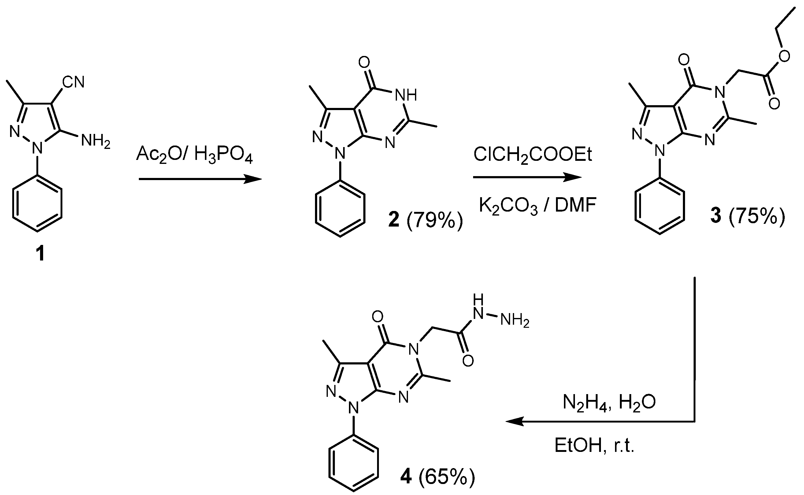
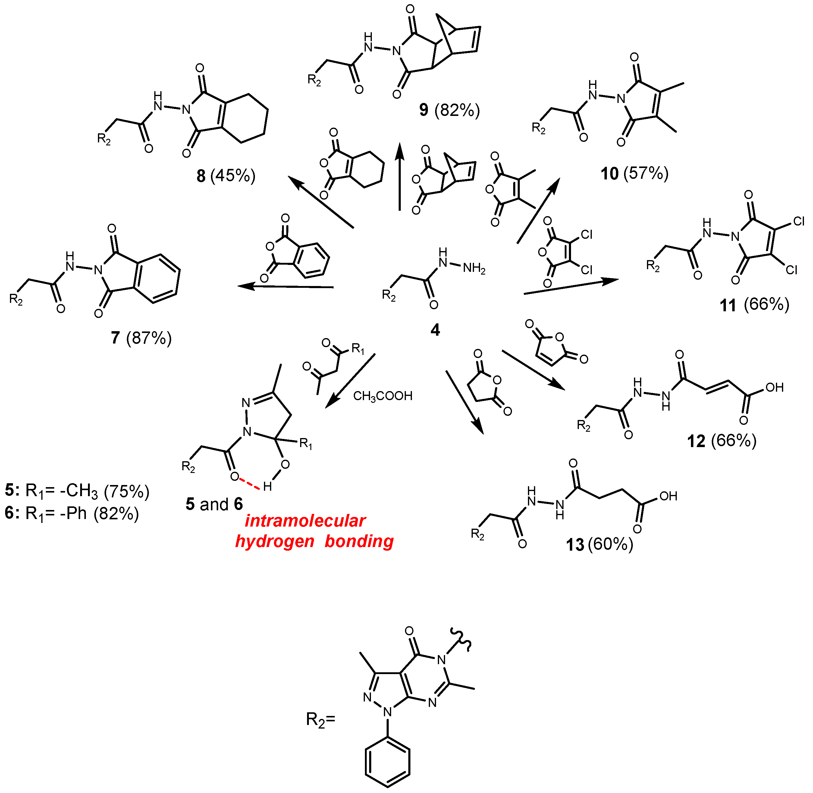
2.1. Chemistry

The justification of a large part of the chemistry directed towards the access to new compounds containing nitrogen at the fusion of the rings, is due to the exploitation of numerous molecules that have interesting biological potentials, such as antiviral potentials, in the field of medicinal chemistry.
In order to access our target molecules 513, we started by preparing 5-amino-3-methyl-1-phenyl-1H-pyrazole-4-carbonitrile (1), in accordance with our previously reported method [28]. The hydrazides constitute an essential intermediate for the synthesis of several heterocyclic compounds [37,38,39] and are known for their high reactivity in addition to their utility as building blocks. This stirred our interest for their use in the preparation of the desired compounds 513. Our approach to the target molecules started with the synthesis of the key intermediate (4). Herein, we are reporting a scalable methodology for the synthesis of hydrazide (4) (65% yield). The synthetic route is illustrated in Scheme 1. The reaction sequence began with the preparation of pyrazolopyrimidinone (2), by heating a solution of the precursor 1 in an acetic anhydride in the presence of phosphoric acid under reflux. The reaction of the compound (2) with ethyl chloroacetate gave the corresponding ester (3). The latter was then submitted to a nucleophilic substitution with hydrazine monohydrate affording the 2-(3,6-dimethyl-4-oxo-1-phenyl-1,4-dihydro-5H-pyrazolo[3,4-d]pyrimidin-5-yl)acetohydrazide (4). The structures of compounds 14 were established on the basis of their 1H and 13C-NMR and MS spectra, as well as their elemental analysis data after purification; their complete spectral data are given in the experimental section.
The condensation of the precursor 4 with pentane-2,4-dione or 1-phenyl-1,3-butanedione in the presence of a catalytic amount of acetic acid in refluxing dioxane was carried out for 5 h. This afforded the new hydroxylated pyrazole derivatives 5 and 6, respectively, via the Knorr pyrazole synthesis mechanism [30]. Furthermore, the hydrazide 4 was reacted with a series of cyclic anhydrides in refluxing 1,4-dioxane, in the presence of a catalytic amount of acetic acid over a period of7–9 h in order to yield the compounds 713 (Scheme 2).
The structures of the target compounds were characterized by 1H and 13C-NMR and MS spectra, as well as by their microanalytical data. Indeed, the 1H-NMR spectra of the compounds 513 showed the disappearance of the signals at δH 4.45 corresponding to the –NH2 function of the hydrazide 4 and the appearance of new signals assigned to the hydrogen introduced by the different electrophilic species used. In addition, the 1H NMRanalysis wasenriched by some 2D experiments. For example, in the case of the compound 5, the Knorr-pyrazole’s structure was evidenced with the help of the HMBC spectrum that exhibited the correlations between H-20/C-18, H-20/C-17, and H-19/C-17. This sequence is consolidated by COSY and NOESY experiments that show the homonuclear correlations between H-20/H-17 and H-19/H-17. The analysis of the 13C-NMR spectra also confirmed the structure of the compounds. In addition, the ESI-MS spectra of the examined compounds 513 showed the correct pseudo-molecular ions [M − H]; in addition, the elemental analysis data were consistent with the molecular formulas.

2.2. Molecular Docking Studies

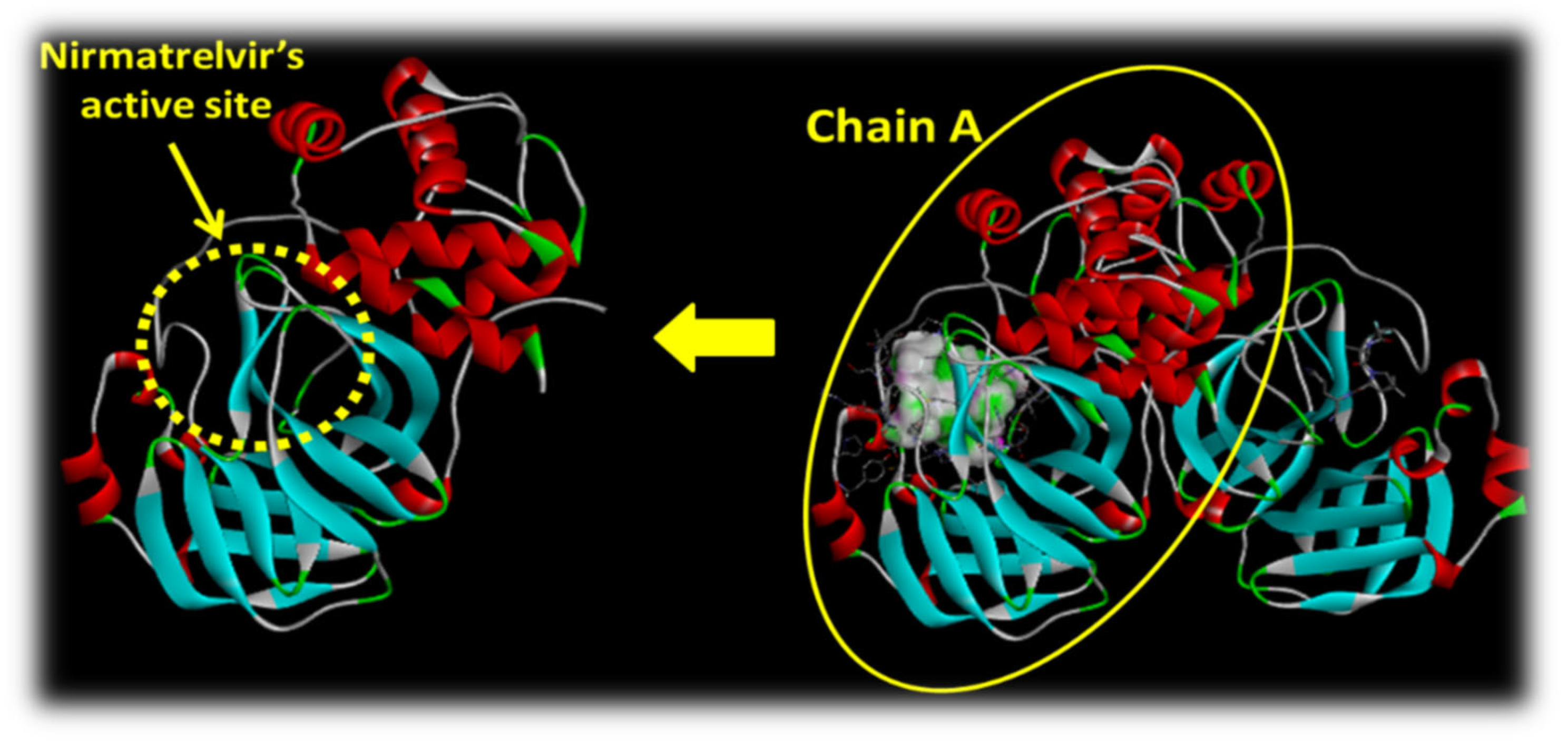
2.2.1. Insight into the Protein Used

Thanks to the efforts of many structural biology and crystallography research groups, we actually have access to a significant number of SARS-CoV-2-related protein structures (spike protein, papain-like protease, and main protease) in the Protein Data Bank. Based on the importance of M-pro detailed in the introduction and which is considered as an optimal target for the search of inhibitors potentially usable as drugs in the curing of SARS-CoV-2 infections, the SARS-CoVs-2: Omicron (pdb:7tll, chain A) was considered and selected as a good COVID-19 target for this study (Figure 2). Our goal was to determine the modes of interaction of the protein-ligand complexes, thereby looking for favorable orientations for the binding of the ligand to the receptor in the active site of nirmatrelvir.

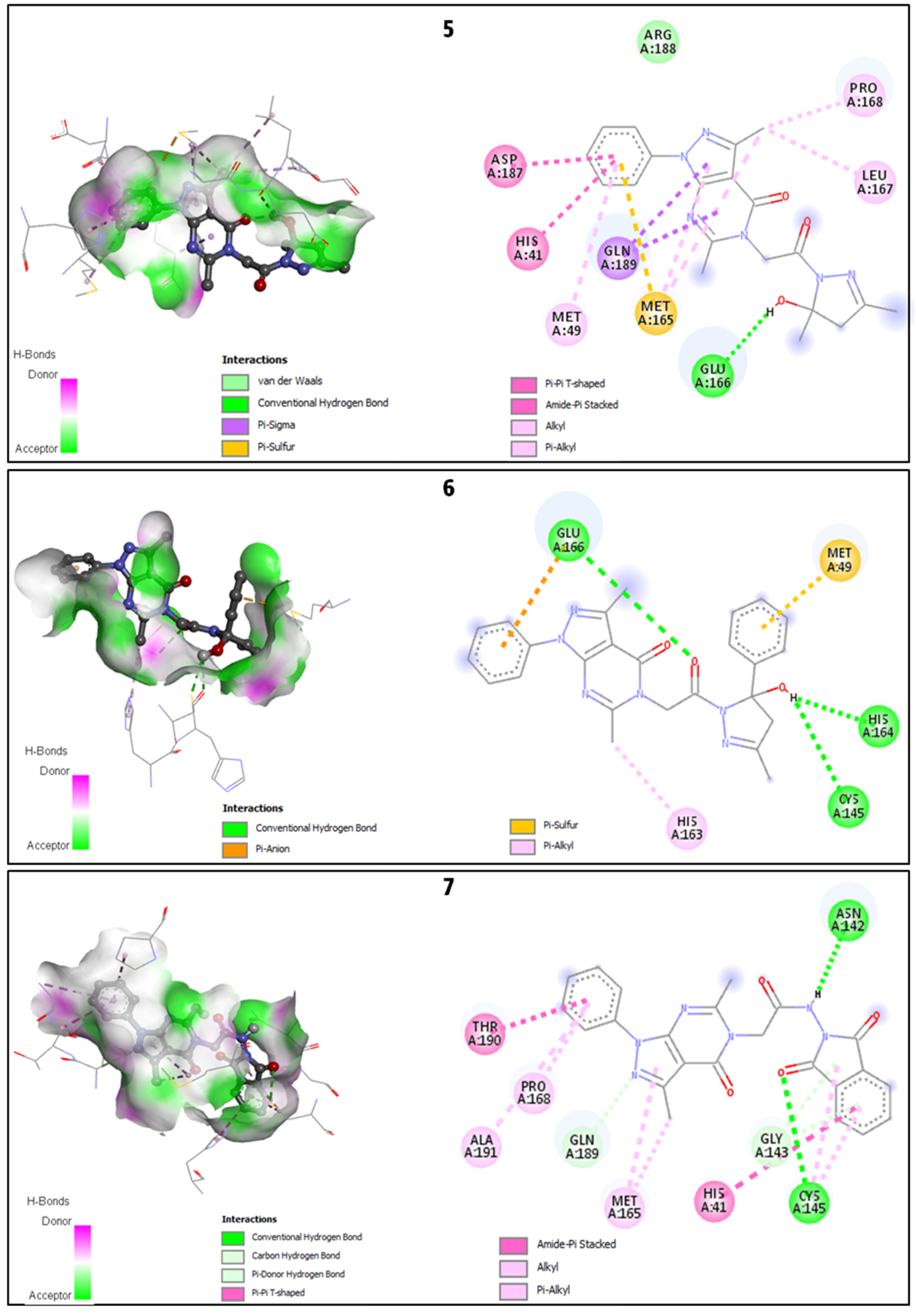
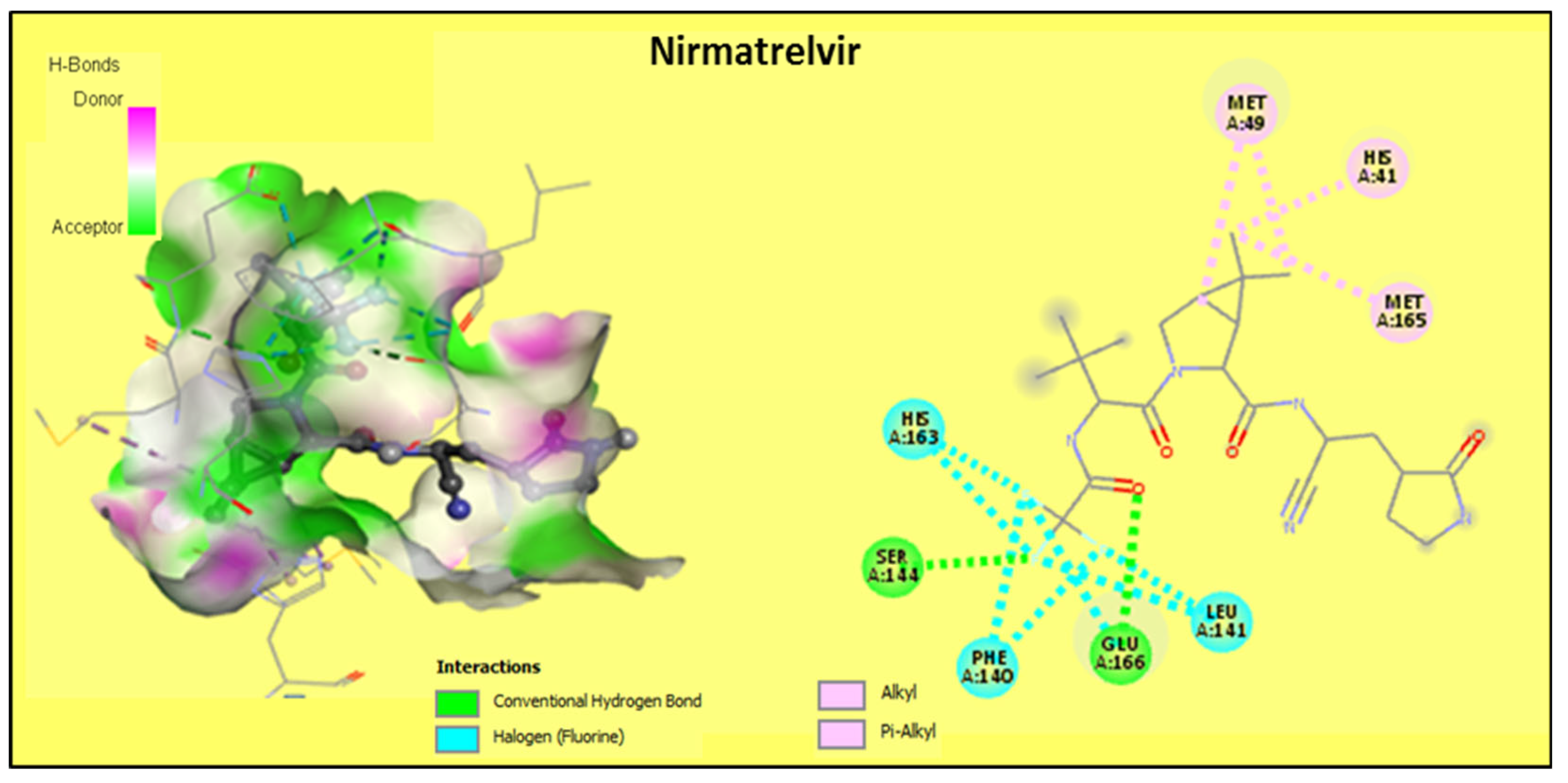
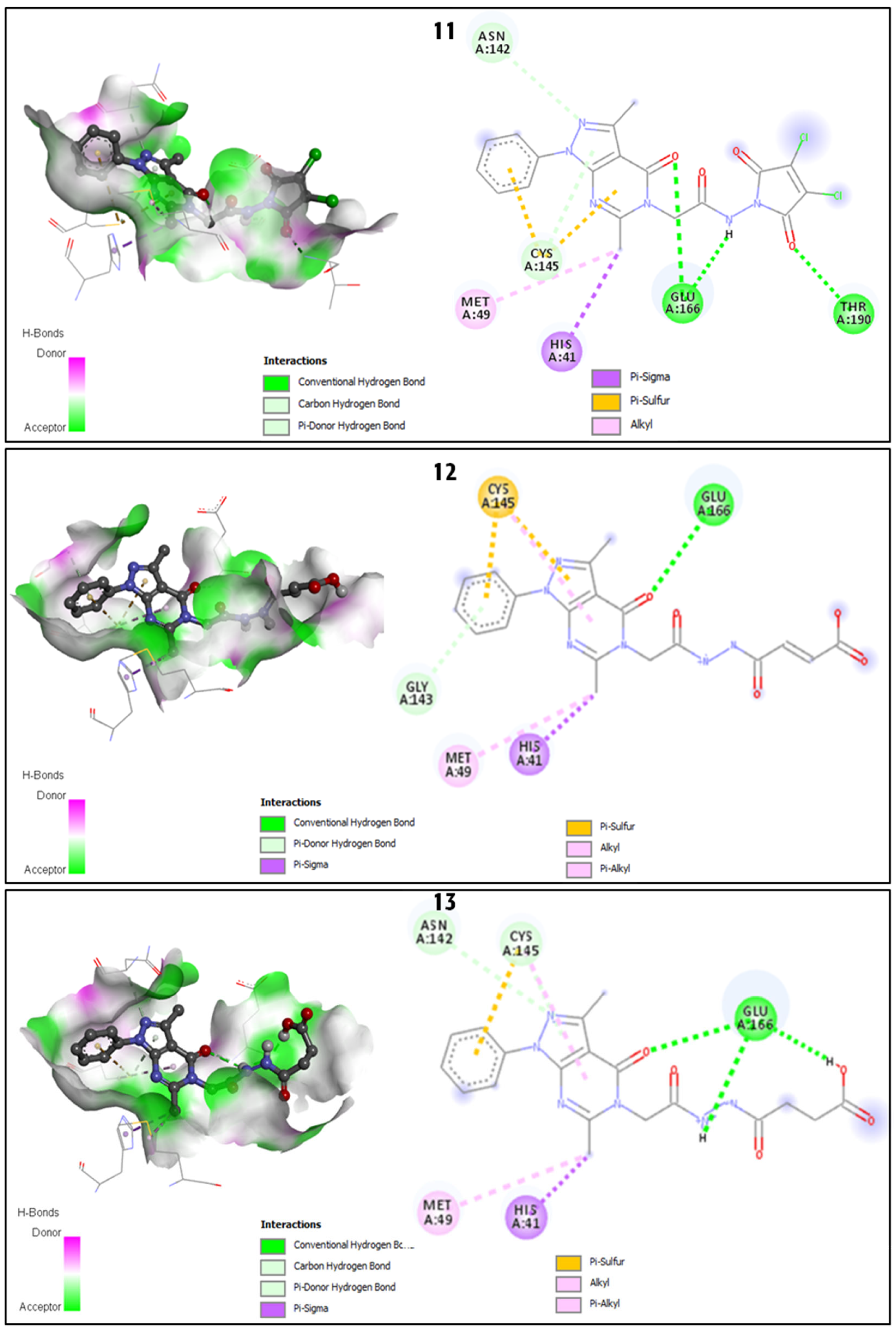
2.2.2. Discussion of the Different Interactions Formed

The molecular docking studies of the synthesized compounds 513, towards the ‘SARS-CoV-2 Mpro Omicron P132H’ receptor were investigated and the obtained results are summarized in Table 1. It is essential to mention that all of the docked molecules against the target enzyme COVID-19 are ranked according to their binding energy, and a quick check of each molecule’s total interactions with nrmatrelvir binding site was performed effectively by counting the total number of hydrogen bonds. Figure 3, Figure 4, Figure 5 and Figure 6 revealed that the docked ligands are involved in many interesting hydrogen bonds and hydrophobic interactions which demonstrate a good protein inhibition of some synthesized molecules. Indeed, as a detailed description of all docked compounds, it can be noted that the compound 5 which contains, in its structure, a hydroxylated pyrazole fragment that formed a conventional H-bond with GLU166 via the hydroxyl group. Furthermore, its importance as an inhibitor is perceptible through many hydrophobic interactions such as: Pi-Sigma with GLN189, Pi-Sulfur with MET165, Pi-Pi T-shaped with HIS41, Amide-Pi stacked with ASP187, Pi-Alkyl with MET49, MET165 and Alkyl with MET165, LEU167, and PRO168 via the phenylpyrazolopyrimidinone moiety, while the analogue 6 engages mainly by the three H-bonds displayed with residues: CYS145, HIS164, and GLU166. Furthermore, the compounds 711 that consist of pyrazolopyrimidine linked to 2,5-pyrrolidinediones, showed interesting docking results by at least two hydrogen bonds by the NH and CO groups; we cite the case of the compound 7, which showed H-bonds with ASN142, CYS145, and likewise the compound 9 with GLU166, in addition to other hydrophobic interactions. The compounds 8, 10, and 11 each presented three H-bonds in addition to several other interactions. For more detail, the compound 8 formed, through its phenylpyrazolopyrimidinone pharmacophore, a Pi-Sulfur with MET49, Pi-Alkyl with CYS145 and MET165, and Alkyl interactions with HIS41, MET49, and HIS163. Furthermore, compounds 10 and 11 showed the same types of interactions: three H-bonds via the NH and CO groups with amino acids: GLU166 and THR190, Pi-Sigma with HIS41, alkyl with MET49, Pi-Sulfur with CYS145, carbon hydrogen bond with CYS145, and the Pi-donor hydrogen bond with ASN142. Regarding the acid derivatives, the compound 12 displayed a single H-bond with GLU166 in addition to other hydrophobic bonds similar to those achieved by the compounds 10 and 11, while 13 is involved in three conventional hydrogen bonds with the residue GLU166. Some of the above results are more significant than those given by the reference drug nirmatrelvir which only formed two H bonds with SER144 and GLU166. The present in silico results showed an interesting anti-Mpro potential for most of our synthesized pyrazolopyrimidinone derivatives (binding affinity= −7.5 to −8.2 kcal/mol) compared to the redocked nirmatrelvir (binding affinity= −7.7 kcal/mol), and also to previous works showing similar results with the repurposed drugs darunavir (−7.4 kcal/mol), lopinavir (−7.3 kcal/mol), as well as to some polyphenol compounds (binding energy = −7.2 to −8.2 kcal/mol) [40]. This comparison is supported by some data from the same study and is based on the importance of the H-bonds for the inhibition of Mpro, that showed that some of our compounds showed 3 hydrogen bonds with the residues Glu166, Gly143, and sometimes also with Cys145, while darunavir showed only two (with Glu166, Gly143) and lopinavir showed only one with Cys145.

2.2.3. Binding Energies Analysis

The nature and number of the interactions exerted by the synthesized compounds with the different amino acids are not on their own sufficient in order to predict their anti-SARS CoV-2 potency. It is also very useful to discuss the value of the energy of the complex (compound-protein) resulting from these interactions: the lower the energy, the more stable the complex and hence, the better the activity. The compound 8 that holds a 4,5,6,7-hexahydro-2H-isoindol-2-yl)acetamide fragment, exhibited the lowest energy value (8.2 kcal/mol) followed by its analogue 7 (R = phthalic) with an energy value of 8.2 kcal/mol. Then, the compound 13 with its acid function, displayed an energy value of 7.8 kcal/mol. These findings suggest that these three derivatives may have the most significant activity against the Omicron variant of SARS-CoV-2 compared to the rest of the analogues and the reference drug nirmatrelvir (7.7 kcal/mol) (Table 1). Regarding the above results, we assume that some of the synthesized pyrazolopyrimidine derivatives could possibly have a promising effect against COVID-19, but this eventuality obviously requires serious in-depth and experimental scientific assays.

2.3. Prediction of Drug-Likeness Descriptors and Toxicity

2.3.1. Predicted Molecular Properties and Drug-Likeness

The potential inhibitors or the docked ligands (tested molecules) can be selected based on their binding energies. Nevertheless, the ADMET (Absorption, Distribution, Metabolism, Excretion, and Toxicity) properties are considered as key steps and the appropriate methodologies employed in drug development and discovery processes [41]. In addition, the physicochemical properties are important parameters of a molecule that influence efficacy, safety, or metabolism and can be predicted by using Lipinski’s rule of five (RO5),as summarized in Table 2 and Table 3. The rule of five (RO5) deals with the dependency of active compounds and defines the twelve simple pharmacokinetics parameters named as the molecular weight, log P, H-bond donors, H-bond acceptors, octanol/water partition coefficient, number of rotatable bonds, topological polar surface area, molecular volume, percentage of absorption, N violation Lipinski, Lipinski Rule, and Pfizer Rule. These pharmacokinetic parameters are associated with the acceptable aqueous solubility and the intestinal permeability and comprise the first steps in oral bioavailability. The RO5 helps in predicting the medicinal and combinatorial chemistry of the selected class of molecules. The predicted values of various properties categorized as molecular weight describe the size of the molecule. Further, the value of log P corresponds to the lipophilicity of the molecules which is related to the solubility of the drug molecule in the aqueous medium. Therefore, the higher the solubility, the higher the activity of the therapeutic agents. Furthermore, the parameters of the H-bond donor and H-bond acceptor indicate the quantification of all atoms (N, O) and their efficiency in the formation of the hydrogen bond. The results obtained from the ADMET analysis with ADMETlab 2.0 software revealed that the predicted compounds 5 to 11 fully obeyed Lipinski’s rule of five while the compounds 12 and 13, with an acid function, gave lightly elevated values of nHA, TPSA, and relatively low %Abs (Table 2). These findings suggest that the synthesized compounds are feasible in order to serve as a future potential antiviral candidate in the treatment of COVID-19 infections.

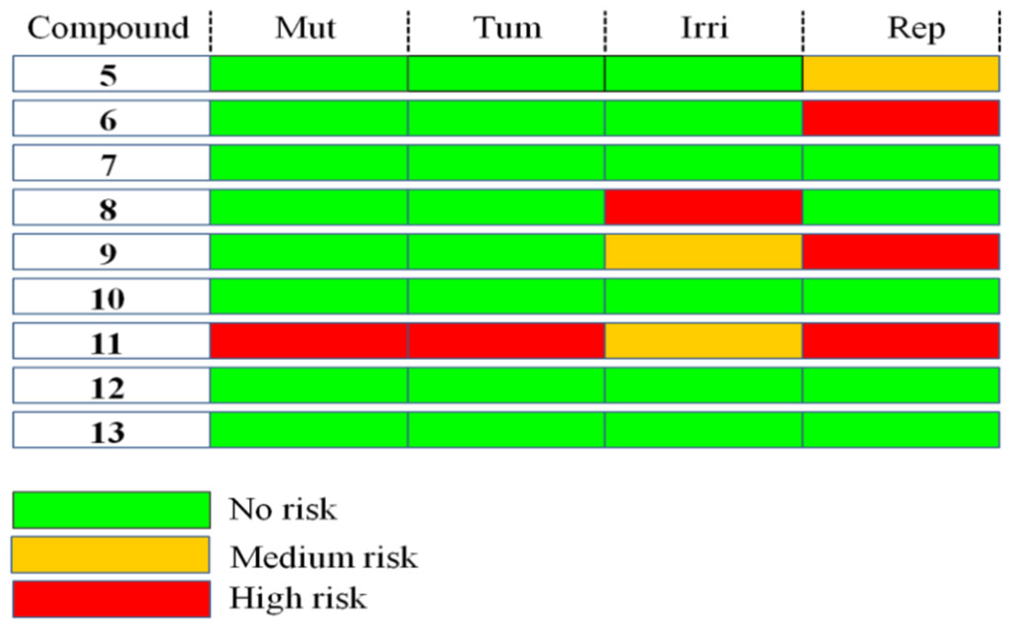
2.3.2. Toxicity Analysis

The toxicity determination of the selected class of compounds is another significant property in drug designing. Indeed, the computational approach to evaluate toxicity helps to determine the toxic level of doses. Therefore, it helps in the reduction of the risk of failure in the experimental procedure [42]. As depicted in Figure 7, it was found that almost all of the predicted compounds presented no risk of toxicity (mutagenicity, tumorigenicity, irritation, and reproductive effect), thereby suggesting the strong applicability of these compounds as antivirals to treat SARS-CoV-2, with the exception of the ligand 11 due, perhaps, to the presence of two chlorine substituents in its structure.

3. Materials and Methods

3.1. Chemistry

3.1.1. General Procedure for the Synthesis of 3,6-dimethyl-1-phenyl-1,5-dihydro-4H-pyrazolo [3,4-d]pyrimidin-4-one (2)

In a 50 mL three-necked flask, 0.39 g (2 mmol) of aminopyrazole 1 is introduced in 10 mL of acetic anhydride. Then, 5 mL of phosphoric acid is added dropwise. The mixture is then brought to reflux for 3 h with magnetic stirring. The progress of the reaction is monitored by TLC (eluent 2: 8 ethyl acetate/chloroform), which shows the appearance of a new stain more polar than that of aminopyrazole1. Once cooled, ice water is added to the reaction crude, the precipitate is recovered by filtration, then it is washed with cold ether, dried, and then recrystallized from ethanol in order to give pyrimidinone 2.
Yield: 79%; Rf = 0.52 (SiO2, CHCl3/MeOH, 95:5); m.p. 316–318 °C; UV-Vis (MeOH): λmax(log ε) = 234 nm (4.46), 272 nm (3.99); IR (ATR): ν ˜ = 2850m, 1668s, 1612m, 1591s, 1545w, 1531m, 1504s, 1435m, 1379m, 1321m, 1257m, 1181w, 1118m, 1079w, 1024w, 917m, 869m, 788m, 754s, 732s, 701w, 685s, 651s, 642s, 619m, 577s, 512m, 489m, 448m, 418w cm−1; 1H-NMR (500 MHz, DMSO-d6): δ = 12.22 (s, 1H, NH), 8.04 (dd, J = 7.7, 1.0 Hz, 2H, 2-H, 6-H), 7.53 (ddd, J = 8.6, 7.4, 1.1 Hz, 2H, 3-H, 5-H), 7.35 (tt, J = 7.2, 1.0 Hz, 1H, 4-H), 2.51 (d, J = 1.2 Hz, 3H, 13-H), 2.40 (d, J = 1.1 Hz, 3H, 12-H) ppm; 13C-NMR (126 MHz, DMSO-d6): δ = 158.6 (C-9), 158.6 (C-8), 152.9 (C-11), 145.7 (C-7), 138.4 (C-1), 129.0 (C-3, C-5), 126.4 (C-4), 121.3 (C-2, C-6), 103.7 (C-10), 21.5 (C-12), 13.3 (C-13) ppm; MS (ESI, negative mode, MeOH/DMSO (4:1)): m/z = 239 (100%, [M − H]); analysis calcd for C13H12N4O (240.10): C 64.99, H 5.03, N 23.32; found: C 64.71, H 23.56, N 23.08.

3.1.2. General Procedure for the Synthesis of Ethyl 2-(3,6-dimethyl-4-oxo-1-phenyl-1,4-dihydro-5H-pyrazolo[3,4-d]pyrimidin-5-yl)acetate (3)

The equimolar solution of 0.24 g (1 mmol) of pyrazolopyrimidinone 2, 0.14 g (1 mmol) of anhydrous potassium carbonate and 0.12 g (1 mmol) of ethyl chloroacetate was refluxed in dry dimethylformamide (DMF) (60 mL) and stirred for 6 h, in a 100 mL three-necked flask. Once the starting material had disappeared, the reaction mixture was then cooled and poured into cold water and the formed precipitate was filtered off, then washed with water, dried, and finally recrystallized from ethanol in order to give the ester 3.
Yield: 75%; Rf = 0.91 (SiO2, CHCl3/MeOH, 95:5); m.p. 122–124 °C; UV-Vis (MeOH): λmax(log ε) = 239 nm (4.49), 283 nm (3.79); IR (ATR): ν ˜   = 1762m, 1615m, 1509m, 1406m, 1377m, 1327m, 1296w, 1212s, 1151s, 1118m, 1053m, 791m, 758s, 716m, 683m, 512m cm−1; 1H-NMR (500 MHz, DMSO-d6): δ = 8.15 (d, J = 7.9 Hz, 2H, 6-H, 2-H), 7.55 (t, J = 7.8 Hz, 2H, 3-H, 5-H), 7.35 (t, J = 7.3 Hz, 1H, 4-H), 5.15 (s, 2H, 14-H), 4.20 (q, J = 7.1 Hz, 2H, 16-H), 2.61 (d, J = 2.9 Hz, 3H, 13-H), 2.58 (d, J = 2.6 Hz, 3H, 12-H), 1.22 (t, J = 7.1 Hz, 3H, 17-H) ppm; 13C-NMR (126 MHz, DMSO-d6): δ = 167.7 (C-15), 165.4 (C-9), 162.5 (C-11), 155.8 (C-8), 142.3 (C-7), 138.5 (C-1), 129.1 (C-3, C-5), 126.1 (C-4), 120.6 (C-2, C-6), 100.5 (C-10), 62.8 (C-14), 60.7 (C-16), 25.9 (C-12), 14.0 (C-17), 13.8 (C-13) ppm; MS (ASAP, positive mode, MeOH/DMSO (4:1)): m/z = 327 (100%, [M + H]+); analysis calcd for C17H18N4O3 (326.14): C 62.57, H 5.56, N 17.17; found: C 62.49, H 5.74, N 16.98.

3.1.3. General Procedure for the Synthesis of 2-(3,6-dimethyl-4-oxo-1-phenyl-1,4-dihydro-5H-pyrazolo[3,4-d]pyrimidin-5-yl)acetohydrazide (4)

The previously synthesized ester-pyrazolopyrimidinone 3 (0.2 g, 0.6 mmol) was treated with an excess of hydrazine hydrate in a minimum amount of ethanol (5 mL) at room temperature for 1–2 h until a white precipitate formed. Then, the solid obtained was filtered off, washed with ethanol, and dried in order to obtain hydrazide 4.
Yield: 65%; Rf = 0.14 (SiO2, CHCl3/MeOH, 95:5); m.p. 258–260 °C; UV-Vis (MeOH): λmax(log ε) = 234 nm (4.50), 279 nm (4.09); IR (ATR): ν ˜ = 3283m, 1706s, 1661s, 1566s, 1552s, 1497m, 1434w, 1383w, 1325w, 1277w, 1190w, 1127w, 994m, 950w, 869w, 786m, 758s, 725m, 705m, 687s, 654m, 618w, 570m, 551w, 513w, 498w cm−1; 1H-NMR (500 MHz, DMSO-d6): δ = 9.43 (s, 1H, NH), 8.05 (d, J = 8.1 Hz, 2H, 2-H, 6-H), 7.53 (t, J = 7.8 Hz, 2H, 3-H, 5-H), 7.35 (t, J = 7.4 Hz, 1H, 4-H), 4.72 (s, 2H, 14-H), 4.45–4.18 (m, 2H, NH2), 2.53 (s, 3H, 13-H), 2.50 (s, 3H, 12-H) ppm; 13C-NMR (126 MHz, DMSO-d6): δ = 166.1 (C-15), 159.8 (C-8), 157.7 (C-9), 150.8 (C-11), 145.9 (C-7), 138.3 (C-1), 129.1 (C-3, C-5), 126.5 (C-4), 121.1 (C-2, C-6), 102.9 (C-10), 44.2 (C-14), 23.6 (C-12), 13.2 (C-13) ppm; MS (ASAP, positive mode, MeOH/DMSO (4:1)): m/z = 313 (100%, [M + H]+); analysis calcd for C15H16N6O2 (312.12): C 57.68, H 5.16, N 26.91; found: C 57.55, H 5.39, N 26.77.

3.1.4. General Procedure for the Synthesis of the Compounds 5 and 6

A mixture of the appropriate hydrazide-pyrazolopyrimidinone 4 (0.31 g, 1 mmol) and 2,4-pentanedione (0.10 g, 1 mmol) or 1-phenyl-1,3-butanedione (0.16 g, 1 mmol) was stirred under reflux of dry dioxane (10 mL) in the presence of an a catalytic amount of acetic acid (0.001 mmol, 0.06 mL). Following a period of 8 h, the reaction mixture was cooled to room temperature, the dioxane was removed in vacuo, and the residue was filtered off, washed with ethanol, and dried in order to yield the compounds 5 and 6, respectively.
(5RS)-5-(2-(5-hydroxy-3,5-dimethyl-4,5-dihydro-1H-pyrazol-1-yl)-2-oxoethyl)-3,6-dimethyl-1-phenyl-1,5-dihydro-4H-pyrazolo[3,4-d]pyrimidin-4-one (5). Yield: 75%; Rf = 0.52 (SiO2, CHCl3/MeOH, 95:5); m.p. 249–251 °C; UV-Vis (MeOH): λmax(log ε) = 235 nm (4.67), 278 nm (4.14); IR (ATR): ν ˜ = 3428w, 1700m, 1665s, 1595w, 1569m, 1555m, 1514w, 1435m, 1406w, 1379m, 1295m, 1197w, 1125w, 1094w, 1017w, 967w, 930w, 869m, 801w, 770m, 753w, 726w, 692m, 654m, 620w, 489w, 416w cm−1; 1H-NMR (500 MHz, DMSO-d6): δ = 8.07 (td, J = 7.7, 1.8 Hz, 2H, 2-H, 6-H), 7.55 (t, J = 8.0 Hz, 2H, 3-H, 5-H), 7.37 (tt, J = 7.7, 1.2 Hz, 1H, 4-H), 5.22–5.04 (m, 2H, 14-H), 3.00 (d, J = 18.6 Hz, 1H, 17-Ha), 2.85 (d, J = 18.7 Hz, 1H, 17-Hb), 2.53 (s, 3H, 13-H), 2.51 (s, 3H, 12-H), 2.05 (s, 3H, 19-H), 1.77 (s, 3H, 20-H) ppm; 13C-NMR (126 MHz, DMSO-d6): δ = 163.6 (C-15), 159.9 (C-16), 157.7 (C-9), 156.3 (C-8), 150.9 (C-11), 145.9 (C-7), 138.3 (C-1), 129.2 (C-3, C-5), 126.5 (C-4), 121.2 (C-2, C-6), 102.8 (C-10), 90.7 (C-18), 52.0 (C-17), 45.6 (C-14), 25.7 (C-20), 23.4 (C-12), 15.9 (C-19), 13.2 (C-13) ppm; MS (ESI, negative mode, MeOH/DMSO (4:1)): m/z = 393 (100%, [M − H]); analysis calcd for C20H22N6O3 (394.18): C 60.90, H 5.62, N 21.31; found: C 60.43, H 5.81, N 21.19.
(5RS)-5-(2-(5-hydroxy-3-methyl-5-phenyl-4,5-dihydro-1H-pyrazol-1-yl)-2-1-phenyl-1,5-dihydro-4H-pyrazolo[3,4-d]pyrimidin-4-one (6). Yield: 82%; Rf = 0.65 (SiO2, CHCl3/MeOH, 95:5); m.p. 126–129 °C; UV-Vis (MeOH): λmax(log ε) = 235 nm (4.55), 279 nm (4.01); IR (ATR): ν ˜ = 1692m, 1672s, 1556m, 1511w, 1446m, 1385w, 1301m, 1209w, 1075w, 960w, 885w, 807w, 763m, 727w, 703m, 654w, 546w, 527w cm−1; 1H-NMR (500 MHz, DMSO-d6): δ = 8.02 (d, J = 7.8 Hz, 2H, 6-H), 7.53 (td, J = 8.5, 7.3 Hz, 2H, 5-H), 7.40–7.30 (m, 5H, 4-H, 21-H, 22-H, 24-H, 25-H), 7.25 (tt, J = 6.6, 1.4 Hz, 1H, 23-H), 7.03 (s, 1H, OH), 5.24 (s, 2H, 14-H), 3.24–3.09 (m, 2H, 17-H), 2.51 (s, 3H, 13-H), 2.45 (s, 3H, 12-H), 2.12 (s, 3H, 19-H) ppm; 13C-NMR (126 MHz, DMSO-d6): δ = 163.0 (C-15), 159.8 (C-16), 157.7 (C-9), 155.9 (C-11), 150.8 (C-8), 145.9 (C-20), 143.3 (C-7), 138.2 (C-1), 129.1 (C-3, C-5), 127.9 (C-22, C-24), 127.1 (C-4), 126.5 (C-23), 124.5 (C-21, C-25), 121.2 (C-2, C-6), 102.7 (C-10), 92.3 (C-18), 55.3 (C-17), 45.4 (C-14), 23.4 (C-12), 15.8 (C-19), 13.2 (C-13) ppm; MS (ESI, negative mode, MeOH/DMSO (4:1)): m/z = 455 (100%, [M − H]); analysis calcd for C25H24N6O3 (456.19): C 65.78, H 5.30, N 18.41; found: C 65.47, H 5.51, N 18.23.

3.1.5. General Procedure for the Synthesis of the Compounds 713

A mixture of the appropriate hydrazide-pyrazolopyrimidinone 4 (0.31 g, 1 mmol) and cyclic anhydride (1 eq) was stirred under reflux of dry dioxane (10 mL) in the presence of a catalytic amount of acetic acid (0.001 mmol, 0.06 mL). Following a period of 8 h, the reaction mixture was cooled to room temperature, the dioxane was removed in vacuo and the residue was washed with ethanol, and dried in order to yield the compounds 713, respectively.
(3,6-dimethyl-4-oxo-1-phenyl-1,4-dihydro-5H-pyrazolo[3,4-d]pyrimidin-5-yl)-N-(1,3-dioxoisoindolin-2-yl)acetamide (7). Yield: 87%; Rf = 0.34 (SiO2, CHCl3/MeOH, 95:5); m.p. 310–315 °C; UV-Vis (MeOH): λmax(log ε) = 218 nm (4.80), 280 nm (4.17); IR (ATR): ν ˜   = 3334w, 1795w, 1734s, 1714s, 1693s, 1566m, 1554m, 1510m, 1463w, 1439w, 1407w, 1368w, 1324w, 1219m, 1122m, 1086w, 1028w, 954w, 882m, 869w, 790w, 762m, 726m, 713m, 692m, 653w, 617w, 576w, 526w, 479s cm−1; 1H-NMR (500 MHz, DMSO-d6): δ = 11.26 (s, 1H, NH), 8.03 (d, J = 8.0 Hz, 2H, 2-H, 6-H), 7.98–7.87 (m, 4H, 18-H, 19-H), 7.52 (t, J = 7.8 Hz, 2H, 3-H, 5-H), 7.35 (t, J = 7.4 Hz, 1H, 4-H), 5.10 (s, 2H, 14-H), 2.61 (s, 3H, 13-H), 2.53 (s, 3H, 12-H) ppm; 13C-NMR (126 MHz, DMSO-d6): δ = 166.8 (C-15), 164.8 (C-16), 159.5 (C-9), 157.6 (C-8), 150.7 (C-11), 146.0 (C-7), 138.2 (C-1), 135.3 (C-19), 129.4 (C-17), 129.1 (C-3, C-5), 126.6 (C-4), 123.8 (C-18), 121.2 (C-6), 102.8 (C-10), 43.6 (C-14), 23.3 (C-12), 13.2 (C-13) ppm; MS (ESI, negative mode, MeOH/DMSO (4:1)): m/z = 441 (100%, [M − H]); analysis calcd for C23H18N6O4 (442.14): C 62.44, H 4.10, N 19.00; found: C 62.15, H 4.37, N 18.76.
2-(3,6-dimethyl-4-oxo-1-phenyl-1,4-dihydro-5H-pyrazolo[3,4-d]pyrimidin-5-yl)-N-(1,3-dioxo-1,3,4,5,6,7-hexahydro-2H-isoindol-2-yl)acetamide (8). Yield: 45%; Rf = 0.38 (SiO2, CHCl3/MeOH, 95:5); m.p. 266 °C; UV-Vis (MeOH): λmax(log ε) = 230 nm (4.55), 279 nm (4.03); IR (ATR): ν ˜ = 3316w, 2943w, 1732s, 1709s, 1679s, 1596w, 1557s, 1512m, 1429m, 1361w, 1327w, 1270w, 1228m, 1192m, 1122w, 1070w, 966w, 870w, 796m, 774m, 714m, 704m, 697m, 620w, 543m, 467m cm−1; 1H-NMR (500 MHz, DMSO-d6): δ = 10.93 (s, 1H, NH), 8.04 (d, J = 7.8 Hz, 2H, 2-H, 6-H), 7.54 (t, J = 7.8 Hz, 2H, 3-H, 5-H), 7.37 (t, J = 7.4 Hz, 1H, 4-H), 5.03 (s, 2H, 14-H), 2.57 (s, 3H, 13-H), 2.54 (s, 3H, 12-H), 2.33–2.23 (m, 4H, 18-H), 1.74–1.67 (m, 4H, 19-H) ppm; 13C-NMR (126 MHz, DMSO-d6): δ = 167.7 (C-8), 166.7 (C-15), 159.5 (C-16), 157.6 (C-9), 150.7 (C-11), 146.0 (C-7), 140.5 (C-17), 138.2 (C-1), 129.1 (C-3, C-5), 126.6 (C-4), 121.2 (C-2, C-6), 102.8 (C-10), 43.5 (C-14), 23.2 (C-12), 20.6 (C-19), 19.6 (C-18), 13.2 (C-13) ppm; MS (ESI, negative mode, MeOH/DMSO (4:1)): m/z = 445 (100%, [M − H]); analysis calcd for C23H22N6O4 (446.17): C 61.88, H 4.97, N 18.82; found: C 61.60, H 5.18, N 18.64.
2-(3,6-dimethyl-4-oxo-1-phenyl-1,4-dihydro-5H-pyrazolo[3,4-d]pyrimidin-5-yl)-N-((3aR,4R,7S,7aS)-1,3-dioxo-1,3,3a,4,7,7a-hexahydro-2H-4,7-methanoisoindol-2-yl)acetamide (9). Yield: 82%; Rf = 0.25 (SiO2, CHCl3/MeOH, 95:5); m.p. 235–238 °C; UV-Vis (MeOH): λmax(log ε) = 234 nm (4.34), 279 nm (3.96); IR (ATR): ν ˜ = 3195w, 3023w, 1783w, 1723s, 1664s, 1594w, 1554m, 1510m, 1441w, 1419w, 1376w, 1324w, 1214m, 1194s, 1120w, 1083w, 1033w, 972w, 870m, 843w, 796w, 751m, 730m, 688m, 653w, 612w, 540w, 464w cm−1; 1H-NMR (500 MHz, DMSO-d6): δ = 8.05 (d, J = 7.4 Hz, 2H, 2-H, 6-H), 7.55 (t, J = 8.0 Hz, 2H, 3-H, 5-H), 7.37 (tt, J = 7.0, 1.1 Hz, 1H, 4-H), 6.18–5.95 (m, 2H, 19-H), 3.58 (s, 2H, 14-H), 3.47 (m, 2H, 18-H), 3.30 (m, 2H, 17-H), 2.55 (s, 3H, 12-H), 2.53 (s, 3H, 13-H), 1.56 (t, J = 10.6 Hz, 2H, 20-H) ppm; 13C-NMR (126 MHz, DMSO-d6): δ = 174.2 (C-8), 165.6 (C-15), 160.0 (C-16), 158.0 (C-9), 151.2 (C-11), 146.5 (C-7), 138.6 (C-1), 134.9 (C-19), 129.6 (C-5), 127.1 (C-4), 121.7 (C-6), 103.3 (C-10), 66.8 (C-14), 51.7 (C-20), 44.8 (C-17), 44.0 (C-18), 23.7 (C-12), 13.7 (C-13) ppm; MS (ESI, negative mode, MeOH/DMSO (4:1)): m/z = 457 (100%, [M − H]); analysis calcd for C24H22N6O4 (458.17): C 62.87, H 4.84, N 18.33; found: 62.73, H 5.03, N 18.04.
2-(3,4-dimethyl-2,5-dioxo-2,5-dihydro-1H-pyrrol-1-yl)-2-(3,6-dimethyl-4-oxo-1-phenyl-1,4-dihydro-5H-pyrazolo[3,4-d]pyrimidin-5-yl)acetamide (10). Yield: 57%; Rf = 0.36 (SiO2, CHCl3/MeOH, 95:5); m.p. 303–306 °C; UV-Vis (MeOH): λmax(log ε) = 229 nm (4.62), 278 nm (4.10); IR (ATR): ν ˜ = 3242w, 3038w, 1731s, 1696s, 1589w, 1565m, 1555m, 1539m, 1494m, 1436m, 1387m, 1321w, 1243m, 1232m, 1191m, 1089m, 1071w, 982w, 952m, 869m, 793m, 759s, 721s, 691s, 661m, 652m, 542w, 525m, 516m, 508m, 405m cm−1; 1H-NMR (500 MHz, DMSO-d6): δ = 10.93 (s, 1H, NH), 8.02 (dt, J = 7.6, 1.6 Hz, 2H, 2-H, 6-H), 7.51 (t, J = 8.0 Hz, 2H, 3-H, 5-H), 7.34 (t, J = 7.5 Hz, 1H, 4-H), 5.00 (s, 2H, 14-H), 2.54 (s, 3H, 13-H), 2.51 (s, 3H, 12-H), 1.93 (s, 6H, 18-H) ppm; 13C-NMR (126 MHz, DMSO-d6): δ = 168.8 (C-8), 166.7 (C-15), 159.5 (C-16), 157.6 (C-9), 150.7 (C-11), 146.0 (C-7), 138.2 (C-1), 136.6 (C-17), 129.1 (C-5), 126.6 (C-4), 121.2 (C-6), 102.8 (C-10), 43.5 (C-14), 23.2 (C-12), 13.2 (C-13), 8.6 (C-18) ppm; MS (ESI, negative mode, MeOH/DMSO (4:1)): m/z = 419 (100%, [M − H]); analysis calcd for C21H20N6O4 (420.15): C 59.99, H 4.80, N 19.99; found: C 59.75, H 5.02, N 19.85.
(3,4-dichloro-2,5-dioxo-2,5-dihydro-1H-pyrrol-1-yl)-2-(3,6-dimethyl-4-oxo-1-phenyl-1,4-dihydro-5H-pyrazolo[3,4-d]pyrimidin-5-yl)acetamide (11). Yield: 66%; Rf = 0.05 (SiO2, CHCl3/MeOH, 9:1); m.p. > 260 °C (slow decomp.); UV-Vis (MeOH): λmax(log ε) = 234 nm (4.50), 380 nm (4.08); IR (ATR): ν ˜ = 3239br, 1677m, 1561s, 1510m, 1374m, 1325w, 1234w, 1192w, 1093w, 1029w, 870w, 791m, 757m, 689m, 653m, 508w cm−1; 1H-NMR (300 MHz, DMSO-d6): δ = 10.65 (s, 1H, NH), 8.05 (d, J = 7.77Hz,2H, 2-H, 6-H), 7.54 (t, J = 7.47 Hz, 2H, 3-H, 5-H), 7.36 (t, J = 7.02 Hz, 1H, 4-H), 4.90 (s, 2H, 14-H), 2.56–2.52 (m, 6H, 12-H, 13-H) ppm; 13C-NMR (126 MHz, DMSO-d6): δ = 168.4 (C-8), 164.4 (C-15), 159.8 (C-16), 157.7 (C-9), 150.9 (C-11), 146.0 (C-7), 138.3 (C-1), 129.2 (C-3, C-5), 126.6 (C-4), 124.2 (C-17), 121.3 (C-2, C-6), 102.9 (C-10), 43.9 (C-14), 23.4 (C-12), 13.2 (C-13) ppm; MS (ESI, negative mode, MeOH/DMSO (4:1)): m/z = 433 (100%, [M − HCN]); analysis calcd for C19H14N6O4Cl2 (460.05): C 49.48, H 3.06, N 18.22; found: C 49.20, H 3.32, N 17.95.
(E)-4-(2-(2-(3,6-dimethyl-4-oxo-1-phenyl-1,4-dihydro-5H-pyrazolo[3,4-d]pyrimidin-5-yl)acetyl)hydrazinyl)-4-oxobut-2-enoic acid (12). Yield: 66%; Rf = 0.03 (SiO2, CHCl3/MeOH, 9:1); m.p. 265–270 °C; UV-Vis (MeOH): λmax(log ε) = 234 nm (4.43), 279 nm (4.09); IR (ATR): ν ˜ = 3240br, 1682m, 1557s, 1505m, 1433m, 1384w, 1325w, 1235w, 1192w, 1126w, 1030w, 960w, 910w, 869w, 790w, 756m, 689w, 653w, 618w cm−1; 1H-NMR (300 MHz, DMSO-d6): δ = 15.86 (s, 1H, OH),10.31 (s, 1H, NH), 9.33 (s, 1H, NH), 8.07–8.05 (m, 2H, 2-H, 6-H), 7.56–7.51 (m, 2H, 3-H, 5-H), 7.35 (t, J = 7.2 Hz, 1H, 4-H), 6.20–5.86 (m, 2H, 17-H, 18-H), 4.92–5.12 (m, 2H, 14-H), 2.55–2.52 (m, 6H, 12-H, 13-H) ppm; 13C-NMR (126 MHz, DMSO-d6): δ = 168.0 (C-15), 160.0 (C-16), 160.0 (C-19), 157.8 (C-8), 157.8 (C-9), 150.9 (C-11), 146.0 (C-7), 138.3 (C-1), 136.1 (C-17), 129.2 (C-3, C-5), 129.0 (C-18), 126.5 (C-4), 121.2 (C-6), 102.9 (C-10), 44.5 (C-14), 23.6 (C-12), 13.2 (C-13) ppm; MS (ESI, negative mode, MeOH/DMSO (4:1)): m/z = 409 (100%, [M − H]); analysis calcd for C19H18N6O5 (410.13): C 55.61, H 4.42, N 20.48; found: C 55.44, H 4.69, N 20.16.
4-(2-(2-(3,6-dimethyl-4-oxo-1-phenyl-1,4-dihydro-5H-pyrazolo[3,4-d]pyrimidin-5-yl)acetyl)hydrazinyl)-4-oxobutanoic acid (13). Yield: 60%; Rf = 0.2 (SiO2, CHCl3/MeOH, 9:1); m.p. 234–237 °C; UV-Vis (MeOH): λmax(log ε) = 234 nm (4.44), 279 nm (4.01); IR (ATR): ν ˜ = 3208w, 1703s, 1611s, 1551m, 1496m, 1439m, 1406m, 1379w, 1350w, 1263w, 1188m, 1030w, 987w, 951m, 866w, 789m, 756m, 688w, 651m, 586w, 506w, 448m cm−1; 1H-NMR (500 MHz, DMSO-d6): δ = 15.85 (s, 1H, OH), 10.26 (s, 1H, NH), 9.91 (s, 1H, NH), 8.02 (d, J = 8.0 Hz, 2H, 2-H, 6-H), 7.51 (t, J = 7.8 Hz, 2H, 3-H, 5-H), 7.33 (t, J = 7.5 Hz, 1H, 4-H), 4.82 (s, 2H, 14-H), 2.53 (s, 3H, 13-H), 2.48 (s, 3H, 12-H), 2.45–2.33 (m, 4H, 17-H, 18-H) ppm; 13C-NMR (126 MHz, DMSO-d6): δ = 174.0 (C-19), 170.6 (C-16), 166.3 (C-15), 160.2 (C-8), 158.1 (C-9), 151.3 (C-11), 146.4 (C-7), 138.7 (C-1), 129.6 (C-3, C-5), 127.0 (C-4), 121.6 (C-2, C-6), 103.3 (C-10), 44.3 (C-14), 29.2 (C-18), 28.4 (C-17), 23.9 (C-12), 13.6 (C-13) ppm; MS (ESI, negative mode, MeOH/DMSO (4:1)): m/z = 411 (100%, [M − H]); analysis calcd for C19H20N6O5 (412.15): C 55.34, H 4.89, N 20.38; found: C 55.06, H 5.14, N 20.03.
The NMR spectra of the compounds 213 are given as Supplementary Materials.

3.2. Molecular Docking Procedure

The molecular docking simulations were performed using the Auto Dock 4.2 program package [43]. The optimization of the geometries of the compounds was carried out using ACD (3D viewer) software (http://www.filefacts.com/acd3d-viewer-freeware-info (accessed on 10 July 2022)). The three dimensional structure of PDB (PDB: 7TLL) was obtained from the RSCB Protein Data Dank [44]. First, the water molecules were eliminated and the missing hydrogens and Gasteiger charges were then added to the system during the preparation of the receptor input file. Once completed, the AutoDock Tools were used for the preparation of the corresponding ligand and protein files (PDBQT). Next, the pre-calculation of the grid maps was performed using Auto Grid in order to save a lot of time during the docking. Subsequently, the docking calculation was carried out using a grid per map with 40 × 40 × 40 Å points of (PDB: 7TLL) in addition to a grid-point spacing of 0.375 Å, which was centered on the receptor in order to determine the active site. Finally, the visualization and analysis of the interactions were performed using Discovery Studio 2017R2 (https://www.3dsbiovia.com/products/collaborative-science/biovia-discovery-studio/ (accessed on 10 July 2022)).

3.3. Chemoinformatics

3.3.1. ADMET Properties

In order to predict the pharmacokinetic properties such as: molecular weight, number of hydrogen-bond acceptors, number of hydrogen-bond donors, octanol/water partition coefficient, number of rotatable bonds, topological polar surface area, molecular volume, percentage of absorption, N violation Lipinski, Lipinski Rule, and Pfizer Rule of all synthesized compounds, the chemoinformatics tools (database): ADMETlab 2.0 (https://admetmesh.scbdd.com/service/evaluation/cal/ (accessed on 15 July 2022)) and Molinspiration (https://www.molinspiration.com/ (accessed on 15 July 2022)) were used.

3.3.2. Toxicity Prediction

Toxicity issues remain the important aspect to be resolved, therefore, the toxicity profile in terms of the various parameters such as mutagenicity, tumorigenicity, irritation, and reproductive or developmental toxicity of the synthesized compounds were determined using the free online available software named the OSIRIS Property Explorer (https://www.organic-chemistry.org/prog/peo/(accessed on 15 July 2022)).

4. Conclusions

In summary, a new series of pyrazole, 2,5-pyrrolidinedione, and carboxylic acid linked pyrazolopyrimidinones were synthesized and evaluated theoretically in a silico study of molecular docking and ADMET properties in order to predict their inhibition of Mpro which characterizes the Omicron variant designed for SARS-CoV-2. Significant results were obtained, and some of these compounds, such as 7, 8, and 13, showed interesting binding energies and types of interactions compared to nirmatrelvir that was employed as a reference drug. These results showed that pyrazolopyrimidinone linked to 2,5-pyrrolidinedione and the unconjugated carboxylic acid could have interesting applications against the Omicron variant of SARS-CoV-2, and therefore encourages to expand this series via the synthesis of more analogues, and to test them all experimentally in order to draw the right conclusions.

Supplementary Materials

The following are available online at https://www.mdpi.com/article/10.3390/molecules27165303/s1, depicted NMR (1H, 13C) spectra of the compounds 213.

Author Contributions

Conceptualization, M.H. and N.V.H.; methodology, A.R.; software, M.H.; validation, H.B.J., R.C. and A.H.H.; formal analysis, M.H.; investigation, M.H. and N.V.H.; data curation, A.R.; writing—original draft preparation, M.H., N.V.H. and A.R.; writing—review and editing, H.B.J. and R.C.; visualization, H.B.J. and R.C.; supervision, H.B.J.; project administration, H.B.J. and R.C. All authors have read and agreed to the published version of the manuscript.

Funding

The authors express their thanks to the Ministry of Higher Education and Scientific Research of Tunisia for financial support (LR11ES39), and they are also grateful to Researchers Supporting Project number (RSP-2021/17) at King Saud University, Riyadh, Saudi Arabia.

Institutional Review Board Statement

Not applicable.

Informed Consent Statement

Not applicable.

Data Availability Statement

The source data for the underlying tables and figures are available from the authors upon request.

Acknowledgments

We would like to thank D. Ströhl, Y. Schiller and S. Ludwig for the NMR spectra and T. Schmidt for the numerous MS spectra; IR, UV/vis spectra and elemental analyses were measured by M. Schneider (Martin Luther University Halle-Wittenberg). We are also grateful to Researchers Supporting Project number (RSP-2021/17) at King Saud University, Riyadh, Saudi Arabia.

Conflicts of Interest

The authors declare no conflict of interest.

Sample Availability

Samples of the compounds are available from the authors.

References

  1. Hu, B.; Guo, H.; Zhou, P.; Shi, Z.L. Characteristics of SARS-CoV-2 and COVID-19. Nat. Rev. Microbiol. 2021, 19, 141–154. [Google Scholar] [CrossRef]
  2. World Health Organization (WHO). 14.9 Million Excess Deaths Associated with the COVID-19 Pandemic in 2020 and 2021. Available online: https://www.who.int/news/item/05-05-2022-14.9-million-excess-deaths-were-associated-with-the-covid-19-pandemic-in-2020-and-2021 (accessed on 7 July 2022).
  3. Bheenaveni, R.S. India’s Indigenous Idea of Herd Immunity: The Solution for COVID-19? Tradit. Med. Res. 2020, 5, 182. [Google Scholar]
  4. Al-Shamsi, H.O.; Alhazzani, W.; Alhuraiji, A.; Coomes, E.A.; Chemaly, R.F.; Almuhanna, M.; Wolff, R.A.; Ibrahim, N.K.; Chua, M.L.K.; Hotte, S.J.; et al. A Practical Approach to the Management of Cancer Patients During the Novel Coronavirus Disease 2019 (COVID-19) Pandemic: An International Collaborative Group. Oncologist 2020, 25, e936–e945. [Google Scholar] [CrossRef] [PubMed]
  5. Corey, L.; Beyrer, C.; Cohen, M.S.; Michael, N.L.; Bedford, T.; Rolland, M. SARS-CoV-2 Variants in Patients with Immunosuppression. N. Engl. J. Med. 2021, 385, 562–566. [Google Scholar] [CrossRef]
  6. Ibrahim, M.; Abdelrahman, A.; Hussien, T.; Badr, E.; Mohamed, T.; El-Seedi, H.; Pare, P.; Efferth, T.; Hegazy, M.E. In silico drug discovery of major metabolites from spices as SARS-CoV-2 main protease inhibitors. Comput. Biol. Med. 2020, 126, 104046. [Google Scholar] [CrossRef] [PubMed]
  7. Zhang, L.; Lin, D.; Sun, X.; Curth, U.; Drosten, C.; Sauerhering, L.; Becker, S.; Rox, K.; Hilgenfeld, R. Crystal structure of SARS-CoV-2 main protease provides a basis for design of improved a-ketoamide inhibitors. Science 2020, 368, 409–412. [Google Scholar] [CrossRef]
  8. Bhardwaj, V.; Singh, R.; Sharma, J.; Rajendran, V.; Purohit, R.; Kumar, S. Identification of bioactive molecules from Tea plant as SARS-CoV-2 main protease inhibitors. J. Biomol. Struct. Dyn. 2020, 10, 3449–3458. [Google Scholar] [CrossRef]
  9. Kumar, S.; Thambiraja, T.S.; Karuppanan, K.; Subramaniam, G. Omicron and Delta Variant of SARS-CoV-2: A Comparative Computational Study of Spike Protein. J. Med. Virol. 2022, 94, 1641–1649. [Google Scholar] [CrossRef]
  10. Chen, J.; Wang, R.; Gilby, N.B.; Wei, G.-W. Omicron Variant (B.1.1.529): Infectivity, Vaccine Breakthrough, and Antibody Resistance. J. Chem. Inf. Model. 2022, 62, 412–422. [Google Scholar] [CrossRef]
  11. Pulliam, J.R.C.; van Schalkwyk, C.; Govender, N.; von Gottberg, A.; Cohen, C.; Groome, M.J.; Dushoff, J.; Mlisana, K.; Moultrie, H. Increased Risk of SARS-CoV-2 Reinfection Associated with Emergence of the Omicron Variant in South Africa. Science 2022, 376, eabn4947. [Google Scholar] [CrossRef]
  12. Graham, F. Daily Briefing: Omicron Coronavirus Variant Puts Scientists on Alert. Nature 2021. Available online: https://www.nature.com/articles/d41586-021-03564-6 (accessed on 7 July 2022). [CrossRef] [PubMed]
  13. Karim, S.S.A.; Karim, Q.A. Omicron SARS-CoV-2 Variant: A New Chapter in the COVID-19 Pandemic. Lancet 2021, 398, 2126–2128. [Google Scholar] [CrossRef]
  14. Commissioner O. of the Coronavirus (COVID-19) Update: FDA Issues Emergency Use Authorization for Potential COVID-19 Treatment. Available online: https://www.fda.gov/news-events/press-announcements/coronavirus-covid-19-update-fda-issues-emergency-use-authorization-potential-covid-19-treatment (accessed on 6 June 2022).
  15. EMA Treatments and Vaccines for COVID-19. Available online: https://www.ema.europa.eu/en/human-regulatory/overview/public-health-threats/coronavirus-disease-covid-19/treatments-vaccines-covid-19 (accessed on 6 June 2022).
  16. Wang, Z.; Chen, X.; Lu, Y.; Chen, F.; Zhang, W. Clinical Characteristics and Therapeutic Procedure for Four Cases with 2019 Novel Coronavirus Pneumonia Receiving Combined Chinese and Western Medicine Treatment. Biosci. Trends 2020, advpub. [Google Scholar] [CrossRef] [PubMed]
  17. Eastin, C.; Eastin, T. Epidemiological Characteristics of 2143 Pediatric Patients with 2019 Coronavirus Disease in China. J. Emerg. Med. 2020, 58, 712–713. [Google Scholar] [CrossRef]
  18. Wang, C.; Horby, P.W.; Hayden, F.G.; Gao, G.F. A Novel Coronavirus Outbreak of Global Health Concern. Lancet 2020, 395, 470–473. [Google Scholar] [CrossRef]
  19. UniProt Consortium UniProt: A Worldwide Hub of Protein Knowledge. Nucleic Acids Res. 2019, 47, D506–D515. [CrossRef]
  20. Pickett, B.E.; Greer, D.S.; Zhang, Y.; Stewart, L.; Zhou, L.; Sun, G.; Gu, Z.; Kumar, S.; Zaremba, S.; Larsen, C.N.; et al. Virus Pathogen Database and Analysis Resource (ViPR): A Comprehensive Bioinformatics Database and Analysis Resource for the Coronavirus Research Community. Viruses 2012, 4, 3209–3226. [Google Scholar] [CrossRef]
  21. Shu, Y.; McCauley, J. GISAID: Global Initiative on Sharing All Influenza Data—From Vision to Reality. Eurosurveilliance 2017, 22, 30494. [Google Scholar] [CrossRef]
  22. Gasteiger, E.; Gattiker, A.; Hoogland, C.; Ivanyi, I.; Appel, R.D.; Bairoch, A. ExPASy: The Proteomics Server for in-Depth Protein Knowledge and Analysis. Nucleic Acids Res. 2003, 31, 3784–3788. [Google Scholar] [CrossRef]
  23. Kumar, S.; Mathavan, S.; Jin, W.J.; Azman, N.A.B.; Subramanaiam, D.; Zainalabidin, N.A.B.; Lingadaran, D.; Sattar, Z.B.A.; Manickam, D.L.; Anbananthan, P.S.; et al. COVID-19 Vaccine Candidates by Identification of B and T Cell Multi-Epitopes Against SARS-COV-2. Preprints 2020, 2020080092. [Google Scholar] [CrossRef]
  24. Graham, B.S. Rapid COVID-19 Vaccine Development. Science 2020, 368, 945–946. [Google Scholar] [CrossRef] [PubMed]
  25. Heaton, P.M. The Covid-19 Vaccine-Development Multiverse. N. Engl. J. Med. 2020, 383, 1986–1988. [Google Scholar] [CrossRef] [PubMed]
  26. Tan, J.-N.; Li, H.; Gu, Y. Water Mediated Trapping of Active Methylene Intermediates Generated by IBX-Induced Oxidation of Baylis–Hillman Adducts with Nucleophiles. Green Chem. 2010, 12, 1772–1782. [Google Scholar] [CrossRef]
  27. Thomas, N.; Zachariah, S.M. Pharmacological activities of chromene derivatives: An overview. Asian J. Pharm. Clin. Res. 2013, 6, 11–15. [Google Scholar]
  28. Horchani, M.; Heise, N.V.; Hoenke, S.; Csuk, R.; Harrath, A.H.; Jannet, H.B.; Romdhane, A. Synthesis and In Silico Docking of New Pyrazolo[4,3-e]Pyrido[1,2-a]Pyrimidine-Based Cytotoxic Agents. Int. J. Mol. Sci. 2021, 22, 10258. [Google Scholar] [CrossRef]
  29. Horchani, M.; Hajlaoui, A.; Harrath, A.H.; Mansour, L.; Ben Jannet, H.; Romdhane, A. New Pyrazolo-Triazolo-Pyrimidine Derivatives as Antibacterial Agents: Design and Synthesis, Molecular Docking and DFT Studies. J. Mol. Struct. 2020, 1199, 127007. [Google Scholar] [CrossRef]
  30. Chortani, S.; Nimbarte, V.D.; Horchani, M.; Ben Jannet, H.; Romdhane, A. Synthesis, Biological Evaluation and Molecular Docking Analysis of Novel Benzopyrimidinone Derivatives as Potential Anti-Tyrosinase Agents. Bioorg. Chem. 2019, 92, 103270. [Google Scholar] [CrossRef]
  31. Cherif, M.; Horchani, M.; Al-Ghamdi, Y.O.; Almalki, S.G.; Alqurashi, Y.E.; Ben Jannet, H.; Romdhane, A. New Pyrano-1,2,3-Triazolopyrimidinone Derivatives as Anticholinesterase and Antibacterial Agents: Design, Microwave-Assisted Synthesis and Molecular Docking Study. J. Mol. Struct. 2020, 1220, 128685. [Google Scholar] [CrossRef]
  32. Mamdouh, A.A.; Galal, H.E.; Nashwa, M.M. Anti-Covid-19 Drug Analogues: Synthesis of Novel Pyrimidine Thioglycosides as Antiviral Agents Against SARS-COV-2 and Avian Influenza H5N1 Viruses. ACS Omega 2021, 6, 16890–16904. [Google Scholar] [CrossRef]
  33. El-Sabbagh, O.I.; Baraka, M.M.; Ibrahim, S.M.; Pannecouque, C.; Andrei, G.; Snoeck, R.; Balzarini, J.; Rashad, A.A. Synthesis and Antiviral Activity of New Pyrazole and Thiazole Derivatives. Eur. J. Med. Chem. 2009, 44, 3746–3753. [Google Scholar] [CrossRef]
  34. Rashad, A.E.; Hegab, M.I.; Abdel-Megeid, R.E.; Micky, J.A.; Abdel-Megeid, F.M.E. Synthesis and Antiviral Evaluation of Some New Pyrazole and Fused Pyrazolopyrimidine Derivatives. Bioorg. Med. Chem. 2008, 16, 7102–7106. [Google Scholar] [CrossRef] [PubMed]
  35. Elebeedy, D.; Elkhatib, W.F.; Kandeil, A.; Ghanem, A.; Kutkat, O.; Alnajjar, R.; Saleh, M.A.; Maksoud, A.I.A.E.; Badawy, I.; Al-Karmalawy, A.A. Anti-SARS-CoV-2 Activities of Tanshinone IIA, Carnosic Acid, Rosmarinic Acid, Salvianolic Acid, Baicalein, and Glycyrrhetinic Acid between Computational and in Vitro Insights. RSC Adv. 2021, 11, 29267–29286. [Google Scholar] [CrossRef] [PubMed]
  36. Mandić, L.; Benčić, P.; Mlinarić-Majerski, K.; Liekens, S.; Snoeck, R.; Andrei, G.; Kralj, M.; Basarić, N. Substituted Adamantylphthalimides: Synthesis, Antiviral and Antiproliferative Activity. Arch. Pharm. 2020, 353, 2000024. [Google Scholar] [CrossRef]
  37. Gan, X.; Hu, D.; Chen, Z.; Wang, Y.; Song, B. Synthesis and Antiviral Evaluation of Novel 1,3,4-Oxadiazole/Thiadiazole-Chalcone Conjugates. Bioorg. Med. Chem. Lett. 2017, 27, 4298–4301. [Google Scholar] [CrossRef]
  38. Sławiński, J.; Szafrański, K.; Pogorzelska, A.; Żołnowska, B.; Kawiak, A.; Macur, K.; Belka, M.; Bączek, T. Novel 2-Benzylthio-5-(1,3,4-Oxadiazol-2-Yl)Benzenesulfonamides with Anticancer Activity: Synthesis, QSAR Study, and Metabolic Stability. Eur. J. Med. Chem. 2017, 132, 236–248. [Google Scholar] [CrossRef] [PubMed]
  39. Naresh Kumar, R.; Poornachandra, Y.; Nagender, P.; Santhosh Kumar, G.; Krishna Swaroop, D.; Ganesh Kumar, C.; Narsaiah, B. Synthesis of Novel Nicotinohydrazide and (1,3,4-Oxadiazol-2-Yl)-6-(Trifluoromethyl)Pyridine Derivatives as Potential Anticancer Agents. Bioorg. Med. Chem. Lett. 2016, 26, 4829–4831. [Google Scholar] [CrossRef]
  40. Ghosh, R.; Chakraborty, A.; Biswas, A.; Chowdhuri, S. Identification of polyphenols from Broussonetiapapyrifera as SARS CoV-2 main protease inhibitors using in silico docking and molecular dynamics simulation approaches. J. Biomol. Struct. Dyn. 2021, 39, 6747–6760. [Google Scholar] [CrossRef]
  41. Ye, Z.; Yang, Y.; Li, X.; Cao, D.; Ouyang, D. An Integrated Transfer Learning and Multitask Learning Approach for Pharmacokinetic Parameter Prediction. Mol. Pharm. 2019, 16, 533–541. [Google Scholar] [CrossRef]
  42. Saraswat, J.; Singh, P.; Patel, R. A Computational Approach for the Screening of Potential Antiviral Compounds against SARS-CoV-2 Protease: Ionic Liquid vs Herbal and Natural Compounds. J. Mol. Liq. 2021, 326, 115298. [Google Scholar] [CrossRef]
  43. Trott, O.; Olson, A.J. AutoDock Vina: Improving the Speed and Accuracy of Docking with a New Scoring Function, Efficient Optimization and Multithreading. J. Comput. Chem. 2010, 31, 455–461. [Google Scholar] [CrossRef]
  44. Greasley, S.E.; Noell, S.; Plotnikova, O.; Ferre, R.A.; Liu, W.; Bolanos, B.; Fennell, K.; Nicki, J.; Craig, T.; Zhu, Y.; et al. Structural Basis for Nirmatrelvir in Vitro Efficacy against SARS-CoV-2 Variants. J. Biol. Chem. 2022, 298, 101972. [Google Scholar] [CrossRef] [PubMed]
Figure 1. Previously reported antiviral compounds (AE).
Molecules 27 05303 g001
Scheme 1. Synthetic pathway to the precursors 24.
Molecules 27 05303 sch001
Scheme 2. Synthetic pathway to the compounds 513.
Molecules 27 05303 sch002
Figure 2. (Right): the structure of SARS-CoV-2 Mpro Omicron P132H in complex with nirmatrelvir. (Left): chain A of main protease enzyme.
Molecules 27 05303 g002
Figure 3. Representation of the 3D structure of the docked compounds 5, 6, and 7 bound to the pocket region of SARS-CoV-2 Mpro with the hydrogen bond (left) and the corresponding 2D diagram of the interactions (right).
Molecules 27 05303 g003
Figure 4. Representation of the 3D structure of the docked compounds 8, 9, and 10 bound to the pocket region of SARS-CoV-2 Mpro with the hydrogen bond (left) and the corresponding 2D diagram of the interactions (right).
Molecules 27 05303 g004
Figure 5. Representation of the 3D structure of the docked compounds 11, 12, and 13 bound to the pocket region of SARS-CoV-2 Mpro with the hydrogen bond (left) and the corresponding 2D diagram of the interactions (right).
Molecules 27 05303 g005
Figure 6. Representation of the 3D structure of the docked nirmatrelvir bound to the pocket region of SARS-CoV-2 Mpro with the hydrogen bond (left) and the corresponding 2D diagram of the interactions (right).
Molecules 27 05303 g006
Figure 7. Computational Toxicity Risk plots of compounds 513. Mut: Mutagenicity, Tum: Tumorigenicity, Irri: Irritation, Rep: Reproductive or developmental toxicity.
Molecules 27 05303 g007
Table 1. Ligand and receptor (Omicron) interactions: binding affinity, number of conventional hydrogen bonds and interacting amino acid residues.
LigandBinding Affinity
(kcal/mol)
Intermolecular Interactions
Conventional
Hydrogen Bonds
Interacting Amino Acid Residues
5−7.51HIS41, MET49, MET165, GLU166 *, LEU167, PRO168, ASP187, GLN189
6−7.73MET49, CYS145 *, HIS163, HIS164 *, GLU166 *
7−8.02HIS41, ASN142 *, GLY143, CYS145 *, MET165, PRO168, GLN189, THR190, ALA191
8−8.23THR26 *, HIS41, MET49, GLY143 *, CYS145, HIS163, MET165, GLU166 *
9−7.42HIS41, MET49, ASN142, CYS145, MET165, GLU166 **, PRO168
10−7.73HIS41, MET49, ASN142, CYS145, GLU166 **, THR190 *
11−7.63HIS41, MET49, GLY143, CYS145, GLU166 **, THR190 *
12−7.01HIS41, MET49, GLY143, CYS145, GLU166 *
13−7.83HIS41, MET49, ASN142, CYS145, GLU166 ***
Nirmatrelvir R−7.72HIS41, MET49, PHE140, LEU141, SER144 *, HIS163, MET165, GLU166 *
R: Reference drug; *: One H-bond; **: Two H-bonds; ***: Three H-bonds.
Table 2. Predicted physicochemical properties for the target compounds 513.
Compd. No.MW anHA bnHD clogP(o/w) dNrotbo eTPSA fMV g%Abs h
Acceptable Value<500<10<5<5≤10<140500100%
5394.180910.6544105.610388.87372.56
6456.190911.5155105.610458.88872.56
7442.1401011.1015119.190430.44967.87
8446.1701011.3275127.200435.72265.11
9458.1701011.0145127.200444.46265.11
10420.1501010.9025119.190409.68767.87
11460.0501011.2635119.190405.51767.87
12410.130113−0.5058148.870392.44157.63
13412.150113−0.5629148.210395.07857.86
But this a Molecular weight, b Number of hydrogen-bond acceptors, c Number of hydrogen-bond donors, d Octanol/water partition coefficient, e Number of rotatable bonds, f Topological polar surface area, g Molecular volume, and h Percentage of absorption (%Abs = 109 − [0.345 × TPSA]).
Table 3. Predicted medicinal chemistry properties and the Lipinski parameters for the target compounds 513.
Compd. No.5678910111213
Pfizer Rule+++++++++
Lipinski Rule+++++++++
i Nviolation Lipinski
(Acceptable Value ≤ 1)
000000011
The pred +: Accepted; i Nviolation Lipinski: number of violations according to the Lipinski “rule of five”.
Publisher’s Note: MDPI stays neutral with regard to jurisdictional claims in published maps and institutional affiliations.

