Review

Hydrogen Sulfide Biology and Its Role in Cancer

1
Henan International Joint Laboratory for Nuclear Protein Regulation, School of Basic Medical Sciences, Henan University, Kaifeng 475004, China
2
Department of Surgery, Miller School of Medicine, University of Miami, Miami, FL 33136, USA
3
Henan-Macquarie University Joint Centre for Biomedical Innovation, School of Life Sciences, Henan University, Kaifeng 475004, China
4
Department of Epidemic Disease Research, Institute for Research & Medical Consultations (IRMC), Imam Abdulrahman Bin Faisal University, P.O. Box 1982, Dammam 31441, Saudi Arabia
5
National Centre for Biotechnology, King Abdulaziz City for Science and Technology (KACST), P.O. Box 6086, Riyadh 11442, Saudi Arabia
6
Department of Psychiatric and Mental Health, Central South University, Changsha 410078, China
7
Department of Thoracic Surgery, The First Affiliated Hospital of Zhengzhou University, Zhengzhou 450052, China
8
Department of General Thoracic Surgery, Hami Central Hospital, Hami 839000, China
9
Kaifeng Key Laboratory of Infection and Biological Safety, School of Basic Medical Sciences, Henan University, Kaifeng 475004, China
10
School of Stomatology, Henan University, Kaifeng 475004, China
*
Authors to whom correspondence should be addressed.
These authors contributed equally to this work.
Molecules 2022, 27(11), 3389; https://doi.org/10.3390/molecules27113389
Submission received: 8 March 2022 / Revised: 27 April 2022 / Accepted: 1 May 2022 / Published: 25 May 2022
(This article belongs to the Special Issue Supramolecular Cancer Nanotheranostics)

Abstract

:
Hydrogen sulfide (H2S) is an endogenous biologically active gas produced in mammalian tissues. It plays a very critical role in many pathophysiological processes in the body. It can be endogenously produced through many enzymes analogous to the cysteine family, while the exogenous source may involve inorganic sulfide salts. H2S has recently been well investigated with regard to the onset of various carcinogenic diseases such as lung, breast, ovaries, colon cancer, and neurodegenerative disorders. H2S is considered an oncogenic gas, and a potential therapeutic target for treating and diagnosing cancers, due to its role in mediating the development of tumorigenesis. Here in this review, an in-detail up-to-date explanation of the potential role of H2S in different malignancies has been reported. The study summarizes the synthesis of H2S, its roles, signaling routes, expressions, and H2S release in various malignancies. Considering the critical importance of this active biological molecule, we believe this review in this esteemed journal will highlight the oncogenic role of H2S in the scientific community.

1. Introduction

Like nitrogen oxide (NO) and carbon monoxide (CO), hydrogen sulfide (H2S) is a biologically active gas found in mammalian tissues. H2S plays an essential role in mediating the many molecular, physiological, and pathophysiological functions in living systems [1,2]. H2S is considered to be involved in developing and progressing various diseases, ranging from non-malignant [3,4] to carcinogenic [5,6,7].
In its carcinogenic roles, H2S is involved in both the inhibition and advancement of cancer [5,8,9,10], while in its non-carcinogenic function, it has been found that H2S plays a significant role in the development of oral diseases [11,12], respiratory diseases [13,14], cardiovascular diseases [15,16,17], and common kidney diseases [18,19,20]. Consequently, these molecules have a tremendous impact on, and regulate the function of the mammalian system [1,21]. H2S is the third-most abundant naturally occurring gas, after NO and CO [21,22,23,24], and profoundly affects the body’s production and regulation of enzymes.
H2S has become widely accepted as a critical signaling molecule in cancer biology due to its unique chemistry, molecular reactivity mechanisms, capacity to change proteins, and active participation in numerous redox processes with metal. H2S has been implicated in a variety of physiological processes linked to the cell cycle and tumor progression, including angiogenesis, tumor growth, cellular and mitochondrial biogenesis, tumor blood flow, migration and invasion, metastasis, protein sulfhydration, epithelial-mesenchymal transition, DNA repair, and chemotherapy resistance [25,26,27,28,29,30,31]. There are numerous publications describing the potential roles of H2S in cancer [32], such as Cao et al. [32] and Shackelford et al. [27]. Cao et al. explain the synthesis, metabolism, measurement, and modulation of H2S as a novel treatment in cancer [32], and Shackelford et al. investigated the role of H2S in cancer, with an emphasis on the molecular processes through which H2S promotes cancer development, dedifferentiation, metastasis, and proliferation. However, considering the bloom and growth of knowledge on H2S biology, an up-to-date and exclusive publication is worth considering.
The present review outlines advancements in the understanding of H2S in cancer management by highlighting its functional involvement in critical cellular process such as programmed cell death, DNA repair, ferroptosis, immunomodulatory, and downstream impacts on cellular activities, with its role in signaling and mediating a dual role in cancer. The review also emphasizes the therapeutic potential of H2S donors alone or in combination with other therapeutics. Although H2S is the hallmark for many processes at the cellular level in mammalian systems, physiological functions are entirely understandable and clarified. However, to better understand its significant role and function in cancer-related processes, the review also describes in-depth knowledge on the synthesis of H2S.

2. Physiological and Pathological Roles of H2S

Previous studies have shown that H2S takes part in an extensive range of physiological and pathological situations, such as vascular relaxation, neuronal activity, angiogenesis, glucose metabolism, energy production, atherosclerosis, ischemia-reperfusion (I/R) injury [33,34,35], vasodilatation [36,37], anti-inflammation [38,39], anticancer [40], and cardioprotection [41]. However, there are several arguments on the function of H2S in cancer growth and development. In recent years, several reports have recommended that the endogenous or exogenous production of H2S may establish two different roles in forming cancer cells [42,43].
According to Chiku et al.’s study, human CSE profligacy occurs in various reactions that produce H2S from cysteine and homocysteine [44]. In the presence of alpha-ketoglutarate, it can also be generated via platelet-rich plasma (PRP)-independent 3-mercapto-pyruvate sulphate transferase (3-MST) or cysteine aminotransferase (CAT) [45]. In mitochondria, free H2S may be oxidized by sulfhydryl reductase (SQR) and may be methylated through sulfhydryl-S-methyl transferase in the cytoplasm [46,47]. Additionally, free H2S is emitted by biological liquids when it joins molecules with metal and methemoglobin [48]. H2S production increased in vivo in the presence of phosphodiesterase inhibitors, inorganic sulfide salts, and organic H2S donors [49]. Sodium hydrosulfide and P-(4-methoxyphenyl)-p-4morpholinodithiophosphoric acid (GYY4137) are two common H2S donors [50]; SG-1002 [51], NOSH-aspirin, ACS67 (a mixed compound of latanoprost and H2S releasing moiety) [52,53], and L-cysteine are substrates for the endogenous formation of H2S [21,54]. Although a lack of confirmation exists on the normal range of H2S in altered cells and the events that lead to its variation in tumor cells, existing research proposes that only a small amount of H2S is required for maintaining cellular activities, and that any change in its level, whether increasing or decreasing, has a significant impact on cancer-modulating cellular activities.

3. Endogenous Production of H2S

Recent experimental research has shown that all mammals, including humans, produce H2S enzymatically [55]. The commonly observed enzymatic pathway includes CSE and CBS, two pyridoxal-5-phosphates (PLP). Further, another enzyme, 3-MST, which is non-PLP-dependent, act in unison with CAT and in the presence of α-ketoglutarate to produce H2S from L-cysteine. Both enzymes are co-localized in the cytosol and mitochondria [56,57,58]. Additionally, a study has shown that D-amino acid oxidase can catalyze D-cysteine to form an achiral α-ketoacid, 3-mercaptopyruvate, which is further processed through 3-MST into H2S in both brain and kidneys [6] (Figure 1). The produced H2S is instantly released or converted to acid-labile sulfur or bound sulfane sulfur and stored in mammalian cells [33,59,60]. The catabolism of H2S can occur through mitochondrial oxidation to sulfate and thiosulfate, excretion from the kidneys or lungs, hemoglobin-mediated scavenging, and thiol methyltransferase and rhodanese-mediated methylation to generate methanethiol and dimethyl sulfide [33,61,62]. CBS and CSE are present in the fluid portion of the cytoplasm, and are also called cytosolic enzymes with certain tissue distributions. CBS is mainly present in the central nervous system (CNS) and occurs in the kidney, liver, uterus, pancreatic islets, placenta, and ileum.
In contrast, CSE can be generated in the heart, kidney, uterus, ileum, vascular smooth muscle, and liver. CSE is the mainly applicable H2S-producing enzyme in the cardiovascular system [55,63]. 3-MST has been detected in mitochondria and in the cytosol, whereas CBS and CSE are predominantly cytosolic [64]. They have been found in the kidney, liver, heart, lung, brain, thoracic aorta, thymus, and testis, which is very important for H2S production in the vasculature brain [55,63].
H2S may be directly discharged, released, or accumulated in acid-labile sulfur, or bound during the cells’ enzymatic pathway [63]. Moreover, H2S can be endogenously produced via either enzymatic or non-enzymatic pathways [55,63]. The endogenously non-enzymatic production of H2S occurs via glucose, organic and inorganic polysulfides present in garlic, and elemental sulfur and glutathione [63].
Non-enzymatically, H2S can be generated from glucose by a different process such as glycolysis (>90%) or phosphogluconate through nicotinamide adenine dinucleotide phosphate (NADPH) oxidase (<10%) [55,63]. When glucose interacts with methionine, homocysteine, or cysteine, gaseous sulfur compounds—methanethiol and H2S—are formed. The direct reduction in glutathione and elemental sulfur also produces H2S. Elemental sulfur is reduced to H2S by reducing equivalents of the glucose oxidation pathway such as NADH or NADPH [65,66]. Furthermore, garlic and garlic-derived organic polysulfides might influence H2S formation in a thiol-dependent manner; for example, diallyl trisulfide (DATS), diallyl disulfide (DADS), S-allyl cysteine (SAC), and diallyl sulfide (DAS), respectively [35,63,67,68]. Similarly, L-cysteine is produced by two pyridoxal-5′-phosphate-dependent enzymes such as CBS and CSE, and with the joined action of 3-MST and CAT, H2S is produced [57,69] (Figure 1).
The biosynthesis of cysteine in mammals is the primary pathway that plays an essential role in producing H2S. CSE and CBS are pyridoxals-5′-phosphate-reliant enzymes in the cytosol. Thus, CBS is expressed mainly in the central nervous system [70], catalyzing H2S and cystathionine from homocysteine by cysteine declination and degradation [71]. Simultaneously, CSE is most abundantly expressed in the cardiovascular system and produces cysteine, ammonia, and α-ketobutyrate from cystathionine, and H2S from cysteine metabolism. The third pathway consists of 3-MST and CAT, which are mainly present in the cytosol and mitochondria of different tissues such as neuroglia cells, kidneys, cardiac, and liver [71]. In the presence of α-ketoglutarate, CAT catalyzes the alteration of cysteine to 3-MST, which can generate H2S and pyruvate from 3-MP degradation and breakdown [72].
The production and biogenesis of H2S in the human body mainly occurs via two main routes, via particular endogenous enzymes, and as a result or a by-product of microbial metabolic pathways inside the gut microbiota, especially in sulfate-reducing bacteria. Amusingly, it was found from the evaluation of germ-free against conservative mice that intestinal microbes control homeostasis, not just in the gut, but also systemically in different organs and tissues [59]. Specifically, microbiota occurrence has been attached to upper levels of free H2S, not just in the colon and cecum (intestinal tracts), but also in plasma. Furthermore, in germ-free mice compared to conventional animals, the traditional animals showed higher levels of bound sulfane sulfur in the fat, lungs, and plasma, with lower cysteine levels and higher CSE activity in most tissue organs [59]. Additionally, persulfides and polysulfides are considered to be secondary sources of H2S, which may be produced or generated endogenously, or from nutritional intake [73]. The main three human enzymes studied to produce H2S endogenously are CBS, CSE, and 3-MST [45].

4. Upregulation of Different H2S-Producing Enzymes in Cancer

Different studies have revealed the altered expression and role of these three enzymes in developing several cancer cells, as explained below and summarized in Table 1. Szabo et al. revealed that CBS is highly expressed in colon cancer cells [34]. Later on, several other studies reported on the upregulation of CBS, CSE, and 3-MST in various cancers [74,75,76,77]. The functional role of H2S-producing enzymes in cancer has been studied comprehensively, and it has been revealed that cancer cells upregulate the ability of H2S-producing enzymes to assist with bioenergetics functions, in order to exploit ATP generation for the purpose of increased growth, migration, and proliferation [34,76,77,78,79,80,81,82,83,84,85].

4.1. CBS Expression in Cancer

In 2013, a study comparing human colon cancer specimens with healthy mucosa tissue presented selective up-regulation of CBS in cancer tissue, whereas low CBS expression levels were observed in non-cancerous peri-tumor tissue. The other H2S-producing enzymes, CSE and 3-MST, showed no upregulation in tumor cells. With the consequent testing of HCT-116, HT-29, and Lovo (colon adenocarcinoma-derived cell lines), it was observed that CBS is selectively upregulated, in concert with NCM356 (a non-malignant cell line of colonic epithelial cells). Cell fractionation studies were also conducted to investigate CBS location in colon cancer cells, with the results showing that CBS is habitually localized to the cytosol, but mitochondrial translocation is also possible [86,87]. A study using HCT116 cancer cells revealed that CBS is present in cytosol and mitochondria. As per an estimate, homogenates of specimens collected from patient-derived colon tumors and cancer cell lines derived from colon homogenates showed an increase in the production rate of H2S. This response was inhibited using the prototypical CBS inhibitor compound amino-oxy-acetic acid (AOAA) [86].
CBS was overexpressed in primary epithelial tissues, ovarian cancer, and multiple ovarian cancer cell line specimens similar to colon cancer cells. After examining 200 primary epithelial ovarian cancer patients’ tissues using microarrays, Bhattacharyya and colleagues found high levels of CBS expression, predominantly in a common histological variant of serous carcinoma. Tumors with higher-grade cancers and serous histology contain higher levels of CBS. Previous studies have shown strong levels of CBS expression in the FIGO stages (I and II) of ovarian cancer [88]. In further experiments using quantitative real-time PCR (RT-PCR) along with immunoblotting, CBS mRNA and protein expression were compared with the control in a variety of cell lines of ovarian cancer, which showed high levels of expression for CBS mRNA and protein in a non-malignant ovarian surface epithelial cell line (OSE) [88].
In melanoma cancer cells that express more CBS than usual, Panza and colleagues identified the various types of congenital nevi (combinational, functional, and dysplastic). According to a study of primitive human melanoma, CBS and 3-MST were also present at a high but variable level. In contrast, different human melanoma cell lines (Sk-Mel-5, Sk-Mel-28, A375, and PES 43) with normal epidermal melanocytes (NHEM) do not exhibit CBS expression [89]. Despite these facts, minimal literature is available to identify significant changes in the expression levels of various H2S-producing enzymes for multiple cancers. Guo et al. reported that CBS and CSE could be found in prostatic epithelium normal tissue, while only CSE was found per acinar stroma cells. An androgen-dependent prostate cancer cell line (LNCaP) demonstrated noticeable degrees of CSE and CBS expression.
In contrast, an epithelial cell line of the normal prostatic peripheral zone (RWPE-1) showed low observable expression of both CBS and CSE [90]. Similarly, in many other prostatic cancer cell lines, both CBS and CSE expression and reduced expression of CBS and CSE were observed. Both CBS and CSE are primarily localized to the cytoplasm. Guo and colleagues tested the effect of dihydrotestosterone (DHT) on CBS and CSE expression, and found that DHT initiated an increase in CBS and CSE expression in LNCaP cells [90]. Additional cancer cell types showing increased CBS expression consist majorly of cells in the NC160 collection [91], myeloma, and biliary tract carcinoma [92]; breast cancers consistently showed the highest increase, as did renal tumors [93]. The functional effect of variation in H2S synthesis in the cancer cell lines was not investigated.
CBS knockdown of cancer cells inhibited xenograft development and neovessel density, indicating a function for endogenous H2S in tumor angiogenesis. Unlike CBS, suppressing CSE (whose expression was intact in colon cancer) did not affect tumor development or bioenergetics. In conclusion, H2S produced from CBS serves to (i) maintain colon cancer cellular bioenergetics, thereby supporting tumor growth and proliferation, and (ii) promote angiogenesis and vasorelaxation, consequently providing the tumor with blood and nutrients. CBS-derived H2S has been identified as a tumor growth factor and anticancer medication target [34]. CBS detection can also be observed in several other cells. CBS expression is low or absent in the normal prostate peripheral zone epithelial cell line RWPE-1, while CBS expression is high in the androgen-dependent prostate cancer cell line LNCaP [90].
Like non-malignant colon mucosa cells, epithelial cell lines from colon cancer exhibit selective CBS upregulation and increased H2S development [36]. The expression of CBS was significantly increased in protein and mRNA levels in ovarian cancer cells [12,94]. In addition, human breast cancer cells MDA-MB-468, MCF-7, and Hs578T showed significantly higher CBS levels than normal breast cells [95]. Recent studies have shown that estrogen-related receptor a1 (ERRa1) activates the transcription factor Sp1 and plays a critical role in controlling CBS expression in different cell types [96]. Further analyses were needed to determine whether ERR1 plays a role in expressing CBS in cancer cells, such as the expression of CBS mRNA in hepatocellular carcinoma (HCC). On the other hand, hypoxia and radiation conditions can dramatically increase the amount of CBS in the human hepatoma cell line, HepG2 [97]. CBS is inhibited in colon and colorectal cancer by promoter methylation, and CBS, mediated by methylation, can be reversed genetically or pharmacologically [98].
CBS expression can also contribute to the development and progression of human glioblastomas [99]. CBS expression was not detected in leukemia cells, indicating that CBS is more prevalent in solid tumors [35,93]. Therefore, the expression of CBS is significantly altered in several human tumor types, and the expression of CBS has a few characteristics. CBS mechanisms must be characterized to determine their specific roles in tumor cell proliferation, invasion, and metastasis.

4.2. CBS Function in Cancer

Szabo et al. compared human colon cancers with normal tissue from the patient’s lining. Further, several cell lines derived from the colon’s adenocarcinoma were examined [34], including the selective regulation of CBS, which was contrasted with the non-malignant epithelial cell line [34]. Szabo et al. explored the localization of colon cancer cells using CBS cell fractionation [34]. In addition to its translocation from the cytosol to the mitochondria, CBS is considered a cytosolic enzyme [86,87]. According to Szabo et al. [34], a study on the localization of HCT116 cancer showed that CBS is in the cytosol and the mitochondria. It is predicted that colon cancer patients’ serum homogenates and cell lines produce significantly more H2S suppressed by AOAA, a prototypical CBS inhibitor [34]. Szabo et al. examined the role of HBS derived from CBS in colon reproduction, passage, and in vitro invasion. CBS can be genetically silenced or pharmacologically inhibited to prevent the proliferation, migration, and invasion of HCT116 cells [34].
Moreover, S-Adenylyl-L-methionine (SAM), an allosteric CBS activator of this compound, enhances the proliferation of HCT116 cells at low concentrations [34,86]. By combining genetic and pharmacological approaches, AOAA inhibits CBS. The genetic silencing of CBS or the pharmacological inhibition of CBS inhibits the proliferation, migration, and invasion of HCT116 cells [34]. Additionally, S-Adenylyl-L-methionine (SAM), a CBS allosteric activator of this compound, stimulates HCT116 cell proliferation at low concentrations [34,86]. CBS-derived H2S proliferative and pro-migratory characteristics are likely suitable for Akt/PI3K signaling stimulation, as early studies have established that H2S exogenous donors excite the migration of HCT116 cells via pathways activation [100]. In summary, the H2S produced by CBS results from its stimulation of mitochondrial and bioenergetic actions. Additionally, Szabo et al. revealed that silencing CBS and inhibiting AOAA decreased the bioenergetic activity of HCT116, which requires basal electron transport. The respiratory reserve capacity bioenergetic parameter is an increase in mitochondrial oxygen consumption in response to a mitochondrial uncoupling agent [34]. CBS inhibition obscures mitochondrial function and the glycolytic function of HCT116 cells [34], a finding that can be explained by H2S’s documented stimulation effect on GAPDH activity [101], a key enzyme in the glycolytic pathway. SAM increased the bioenergetic activity of HCT116 cells at low concentrations, similar to the CBS activator SAM’s allosteric effect on proliferation [86].
Experiments with nude xenografts of HCT116 cells or patient tumor tissue (PDTX) confirmed the results in vivo. A combination of the pharmacological inhibition of CBS with AOAA and CBS silencing significantly reduced the formation of tumor xenografts. Szabo et al. have reported CBS inhibition effects in vivo [34]. In some cases, it may have paracrine effects on the tumor microenvironment. Moreover, the absence of CBS blocked the density and convolution of CD31-positive vessels between the tumor tissues, indicating reduced tumor angiogenesis. In addition, AOAA was shown to condense blood flow per tumor and act as a local vasodilator when injected directly into the tumor parenchyma [34]. CBS inhibition with AOAA inhibited the metastatic spread of HCT116 cells from the cecum to the liver, and AOAA oxaliplatin inhibited the metastatic spread in the same model [102]. Primary epithelial ovarian cancer tissues and several ovarian cancer cell lines are over-expressed with CBS. Bhattacharya et al., proposed that primary ovarian tumors expressed high levels of CBS. CBS levels are typically higher in tumors with serous histology and a higher degree of cancer [103].
The expression of CBS is already identifiable in most of the early stages (FIGO Stage I and II) of the ovarian cancers studied [88]. Another study by a group used quantitative RT-PCR and immunoblotting to compare the expression of CBS mRNA and protein levels in different ovarian cancer cell lines with a non-malignant superficial ovarian cell line as a control (OSE) [88]. Bhattacharyya and colleagues investigated the functional role of CBS-derived H2S in inhibiting the proliferation, migration, and invasion of ovarian cancer in vitro, using a combination of genetic and pharmacological approaches. The regulation or inhibition of CBS has been shown to inhibit cell proliferation, and treatment with AOAA, particularly at higher concentrations, also reduces cell viability. As Bhattacharyya and colleagues investigated the intracellular mechanisms underlying these acts, they discovered that controlling or inhibiting CBS reduces the essential antioxidant glutathione’s (GSH + GSSG) intracellular content and causes apoptotic cascades. The absorption of the intracellular antioxidant may cause this latter effect after CBS inhibition/suppression. Another inevitable result of CBS cessation or inhibition is an increase in reactive oxygen species in the cells; this may be secondary to antioxidant depletion or correlated with mitochondrial function changes. Finally, CBS styling impacts on intracellular signaling in A2780 cells: Bhattacharyya and colleagues discovered that CBS suppression increased the expression of p53 while reducing the expression of the NF-kB RelA/p65 subunit [88].
H2S increases mitochondrial function and cell bioenergy in ovarian cancer cells, as seen in colon cancer cells. According to Bhattacharya and colleagues, CBS reduced oxygen consumption in mitochondria, and similar effects were observed when ovarian cancer cell lines were treated with a CBS, AOAA inhibitor. In addition to CBS suppression, CBS inhibition has been shown to increase mitochondrial ROS production, decrease NAD/NADH ratios, decrease ATP synthesis, and increase ADP/ATP ratios [88]. The results of these experiments were confirmed in nude mice transplanted with A2780/CP-20 xenografts. There was a significant reduction in tumor weight (approximately 40%) and a consistent, more pronounced (approximately 70%) decline in tumor nodules with CBS. Based on Ki-67 staining, CBS silencing decreased cancer cell proliferation. As well as inhibiting the angiogenesis surrounding the tumor, CBS silencing inhibited angiogenesis (as in the colon cancer study mentioned above) [88]. Bhattacharyya and colleagues demonstrated that CBS inhibition sensitizes cancer cells to chemotherapy, both in vitro and in vivo. CBS silencing reduced cisplatin’s IC50 rate in A2780 cells by more than half. Using cisplatin alone, 80–90% of patients felt distressing symptoms after being treated with cisplatin independently. CBS siRNA and cisplatin, on the other hand, significantly reduced tumor weight and nodules [88].

4.3. CSE Expression in Cancer

CSE has been shown to play a critical role in various types of cancer cells, as indicated by a study inhibiting CSE by shRNA, or a study inhibiting DL propargyl glycine and cancer cell proliferation and invasion in the human colon SW480 [103]. Human colon cancer HCT116 cells express mRNA and protein levels of CSE [34,104]. H2S/CSE is also involved in hepatocytes, as they correlate with H2S output and are critical for cell proliferation [105]. CSE’s expression and functional activity have also been determined in C6 glioma cells [106]. A CSE expression analysis revealed that PC-3 prostate carcinoma cells are the primary source of endogenous H2S [107]. Molecular mechanisms of CSE may be crucial for cancer development and progression, and this requires further investigation. Cancer prevention and treatment can be enhanced by identifying and producing specific CSE inhibitors [35].
The expression of CSE and CBS is also reduced in prostatic cancer cell lines [107]. The cytoplasm is the primary location of CBS and CSE. A study conducted by Guo and colleagues found that DHT increased the expression of CBS and CSE in LNCaP cells [90]. Drugs targeting CSE or CSE inhibition do not affect the proliferation, migration, or growth of HCT116 cells [34]. Additionally, it was found in SW480 that CSE expression is high in the cells. The levels were further enhanced by activating the Wnt pathway in the cells. Pharmacological or genetic inhibition of CSE impairs cell proliferation in vitro, and blocks CSE with propargylglycine (PAG). Using SW480 cells inhibited by CSE, tumor growth in mice bearing tumors is slowed down [103]. CBS and other CSE cell lines develop H2S in larger amounts than other colon cancer cell lines, which is consistent with the conclusion that H2S promotes cell proliferation.

4.4. CSE Function in Cancer

Researchers have indicated that CSE may be the main H2S-producing enzyme in peripheral tissues, based on their studies of mice lacking CSE [108]. CSE has different evidence-based arguments to support its involvement in diverse cancers. Researchers have investigated CSEs in prostate cancer by studying LNCaP cells [90], late-onset in PC-3, and LNcaP-B [109]. The over-expression of CSE with NAHS stimulates the proliferation of prostate cancer cells [109]. This suggests a role for CSE/H2S pathways in prostate cancer. There have been reports of CSE-induced outcomes in melanoma [110] and gastric cancer [40,111], and H2S donors foremost to cell apoptosis in both types of cancer. However, it is still unclear whether CSE enhances or prevents cancer. A recent study did not explain what CSE inhibition or removal means in these cells. Studies have shown that CSE helps cells to survive in hepatocellular carcinomas [105,112]. Yin et al. have found that the PI3K/AKT pathways control CSE in QGY-7703 and SMMC-7721 hepatoma cell lines [112]. In HepG2 cells, the knockdown of CSE by siRNA significantly inhibits cell proliferation, as found in another study [105]. Several vital pathways are involved in this type of cancer, including the Wnt/B-catenin pathways [103]. It also suggested that siRNA knockdown or the pharmacological inhibition of CSE has pro-cancer effects on colon cancer. This is supported by evidence from experiments with SW480 and HCT116 [103].
NaHS stimulated cancer cell proliferation by activating extracellular signal-regulated kinase pathways in HCT116 and SW480 [104]. In recent research, the expression level of CSE correlates positively with urothelial cell carcinoma of the bladder [113]. However, the exact contribution of CSE remnants to bladder cancer development needs to be explored.

4.5. Expression of 3-MST in Cancer

3-MST is expressed by all somatic cells. The expression of 3-MST by tumor cells is not unexpected. Human neoplastic cell lines have also been reported to express and activate 3-MST, with activities that are higher than CSE. Therefore, it is presumed that 3-MST is the primary source of H2S, rather than CSE [114,115]. Additionally, 3-MST expression and catalytic activity have been reported in hepatoma [116,117], glioblastoma and astrocytoma [115], colon cancer [10,34,118], renal carcinoma [119], urothelial cancer [113,120], lungs adenocarcinoma [121], and melanoma cell lines [89] with marginal concentrations. Cancer cell lines with stem-like properties and multidrug resistance express 3-MST induced by stress or cytotoxic stimuli [117,118]. Despite this observation, 3-MST may not offer any cytoprotective or valuable benefits in advanced or drug-resistant cancer. 3-MST expression is also evident in primary tumors, including human gliomas, with higher malignant grades [122]. Accordingly, glioblastoma containing ipsilateral hemispheres produced higher amounts of H2S than controls [123]. A relatively low proportion of 3-MST was expressed in human melanoma (25–50%), but a significant proportion of the subunit E was not available for analysis [89]. The expression of 3-MST in resections of bladder cancer [113,120], colon cancer [10], oral squamous cell carcinoma [124], lung carcinoma [125], and adenoid cystic carcinoma [126] was significantly higher than in healthy tissues.

4.6. The Function of 3-MST in Cancer

In 1960, Kun’s group reported an initial investigation using the metabolic reaction of 3-mercaptopyruvate in tumors and healthy cells. During their investigations, the author claimed that the 3-MST system in cancer cells appears to have reduced enzymatic activity than normal tissues [127]. Later on, Wlodek et al. observed similar observations of 3-MST activity and cysteine aminotransferase activity [128]. However, this perception was changed because the tumor cells displayed an increased oxidative stress burden. 3-MST might be inactivated in the oxidized state, while an ex-vivo enzymatic assay cannot duplicate living cells’ conditions. Noticeable differences in the results of these new papers may also have occurred because the 3-MST activity of the cancer cells was compared with homogenates of liver and kidney; amongst all of the parenchymal organs, the liver and kidney have the highest expression of 3-MST. Even if the 3-MST activity was lower than the actual level, there was still substantial activity to confer major functional roles by focusing on the 3-mercaptopyruvate functional effects and the effects of 3-MST silencing Hepa1c1c7 (murine hepatoma cell line). The 3-MST substrate, in lower concentrations, induced a bio-energetic stimulatory impact, whereas an inhibitory effect was higher. With the silencing of 3-MST, it has been observed that the cell’s basal bio-energetic function was slight, nonetheless, it was considerably suppressed; the 3-mercaptopyruvate stimulatory effect was undetectable [116]. After 3-MST silencing, the mitochondria of the isolated cells, in an estimation for their ability to generate H2S in reaction from 3-mercaptopyruvate, were prominently suppressed [116]. The stimulatory bio-energetic effects facilitated by 3-mercaptopyruvate are associated with directing electron donation to the electron transport system in mitochondria, because SQR silencing of an enzyme obligatory for this electron donation reduced the 3-mercaptopyruvate bio-energetic stimulatory effect [116]. An additional study to check the 3-MST silencing functional effect used a lung adenocarcinoma cell line (A549). After 3-MST silencing, a reduction in the cells’ proliferation rate was observed, suppressing mitochondrial DNA repair frequency [125].
Instead of reducing 3-MST, silencing enhanced the bio-energetic function of these cells. Due to the effects of 3-MST silencing, the bio-energetic variations may change in a specific direction. There may also be differences in cell types [129]. Interestingly, renal cell carcinoma (RCC4) silencing of 3-MST did not alter its effects [119]. Recently, it has been found that 3-MST inhibitors play a predominant role in many health conditions, including cancers [74]. The 3-MST system plays a role in the microenvironment of tumor cells. The production of 3-MST-derived H2S by endothelial cells has been shown to occur during cell proliferation, migration, vascular relaxation, and angiogenesis, specifically under hypoxic conditions [130]. However, bioenergetics and metabolism play a critical role in regulating endothelial cells with the 3-MST system [130].
Furthermore, experiments will be required to determine the value of these findings and the function of the tumor microenvironment. For a better understanding of tumor growth and angiogenesis, more information is needed on the possible role of 3-MST in in vivo studies [131]. Recently, its effects on proliferation, migration, and bioenergetics have been confirmed in murine colon cancer cells [81]. According to the published literature, the role of the 3-MST system extends beyond producing H2S. Studies with 3-MST inhibitors and 3MT silencing should be anticipated in terms of its effects on the redox processes that 3-MST regulates, in combination with the H2S effects that 3-MST induces. Simulating 3-MST would impair interactions between 3-MST-mediated proteins, possibly resulting in functional consequences. 3-MST is physically associated with enzymes such as L-cysteine de-sulfurase NFS1 and the rhodanese-like protein MOCS3 [132].

5. Dual Role of H2S in Cancer

In cancer, the function of H2S depends on the donor’s supplementation, cancer types, and concentration. Figure 2 illustrates how these donors promote and inhibit cancer, as shown here.

5.1. The Cancer-Promoting Effect of H2S

Mammal cells are currently envisaged to respond to H2S as the bioenergetics stimulator at low concentrations. According to Goubern et al., sulfides are have a high affinity to mitochondria in mammals, making them a suitable energetic substrate, even at low concentrations [133]. Thus, the mitochondrial enzyme SQOR is an electron donor that self-regulates parallel to coenzyme Q, in addition to complexes I and II [116] for various cellular bioenergetic functions. However, 3MP can enhance the process [116]. Consequently, H2S-mediated mitochondrial respiration can only aid cancer development in the presence of sufficient oxygenation. Additionally, H2S, as a substrate for mitochondrial respiration, increases intramitochondrial cAMP levels and induces the persulfidation of mitochondrial ATP synthase and lactate dehydrogenase [116,118]. Glibenclamide inhibits p38 phosphorylation and the migration of endothelial cells after exposure to H2S [134], which indicates that p38 is involved in the proangiogenic process induced by H2S. H2S mediates persulfidation by stimulating the KATP channel and influencing downstream effects, as demonstrated by Mustafa et al. [135]. H2 promotes ischemia-induced angiogenesis in several experiments by upregulating HIF-1a expression [136]. The hypothesis that H2S contributes to hypoxia-induced angiogenesis during cancer development is highly plausible, although insufficient evidence supports it. The pro-angiogenic activity of H2S was first discovered in the late 2000s. H2S from NaHS, when used experimentally, promotes the proliferation of endothelial cells, migration, and tubular formation [134]. NaHS also stimulated blood vessel growth and branching when exposed to chicken chorioallantoic membranes. The proangiogenic effects of H2S have also been established in rat models [134]. The inhibition of CSE by pharmacological agents or genetic deletion restricted VEGF-induced angiogenesis [134], implicates H2S as a physiological angiogenic agent. The PI3K/AKT pathway, mitogen-activated protein kinase (MAPK), and ATP-sensitive potassium (KATP) channels have all been shown to mediate the proangiogenic effects of H2S [72,134]. H2S has also been shown to promote angiogenesis in endothelial tumors. Using a well-established model of tumor angiogenesis, Puppo et al. discovered that NaHS enhances the migration of endothelial cells isolated from breast carcinomas (B-TECs). In the absence of VEGF, the inhibition of CSE inhibits the migration of B-TECs, suggesting H2S is a crucial contributor to both exogenous and endogenous breast cancer angiogenesis. In rat models of colon cancer [34] and ovarian cancer [88], CBS silencing inhibited tumor growth and neovessel density. H2S can promote tumor growth by promoting angiogenesis and by transporting nutrients and oxygen to cancer cells. However, in cancer biology, it is essential to note that high concentrations or doses of NaHS may also suppress angiogenesis [137]. This indicates that the pro-angiogenic movement may occur only in low or endogenous H2S concentrations. H2S has an anti-apoptotic effect, including a defensive effect against apoptotic stimuli [138,139]. Additionally, anti-apoptotic activity has been reported in several cancer cells, including colon cancer [140], hepatocellular carcinoma [141], and neuroblastoma [142]. Additional research has shown possible underlying mechanisms, including the activation of the nuclear factor kappa-light-chain enhancer in activated B cells [143], the activation of the keap1 transcription factor NF-E2-related nuclear factor 2 (Nrf2) [144], and the activation of the MEK1-ERK pathway [140], mediated by H2S-linked persulfidation.
The cell cycle is a series of highly well-defined events that control the transition from cellular quiescence (GO) to proliferation, and attest to the high loyalty of the genomic transcript. In eukaryotes, the cell cycle is divided into four phases: gap phase 1 (G1), DNA synthesis phase (S phase), and gap phase 2 (G2), in which the cell prepares for division and the mitosis phase (M phase). Distinct chromosome separation and cell division occur during the M process [145]. The breakdown of normal cell cycle regulation is a common feature of human cancer [146,147]. A recent study has shown that NaHS can function as a proliferative factor by increasing PKB/AKT and ERK expression in oral squamous carcinoma [94]. Although the treatment of HCT 116 cells with NaHS for 24 h effectively and significantly reduced the GO–G1 population and increased the S-phase cell population, treatment with H2S effectively and significantly reduced the expression of p21 proteins, which are considered CDK inhibitory proteins [104]. Similarly, the downregulation of CSE disrupts the G1/GO process and reduces cell number in the S phase while increasing cell population in the S phase in the human hepatoma cells BEL-704 [112]. These studies concluded that H2S acts as a proliferative factor in cancer by encouraging cell cycle progression.
Cancer cells can proliferate indefinitely by evading arrest in the cell cycle. Recent evidence shows that H2S can accelerate or prolong the cell cycle in diverse cell types, including cardiomyocytes, cancer cells, and endothelial cells [107,148]. Here, exogenous H2S inhibits the expression of regulatory genes in the cell cycle and increases proliferating nuclear antigens and cyclin-dependent kinase 4 [94]. H2S was investigated for its acceleration effect on the cell cycle in colon cancer [104] and hepatoma cells [105]. This fundamental signaling mechanism may be linked to the activation of the ERK and AKT pathways [94,104,105], as the inhibition of ERK or AKT phosphorylation has been shown to inhibit the cell cycle significantly, thereby accelerating the effect of H2S on squamous cell carcinomas and colon cancer cell lines [94,104]. Although not explicitly reported, the persulfidation of MEK1 demonstrates the fundamental mechanisms of H2S-induced ERK activation. However, the molecular mechanisms of the critical role of AKT in the development of human cancer are still unclear [149]. An explanation and clarification of this would have a significant effect.

5.2. Anti-Cancer Effect of H2S

The prolonged exposure of cancer cells to high H2S concentrations will lead to their death, while normal fibroblasts are unaffected. Figure 3 illustrates the possible mechanisms underlying the antagonization of cancer growth by H2S. H2S regulation determines the normal functioning of the cells, and depending on the cell type, any dysregulation (upregulation and downregulation) in endogenous H2S levels is associated with the development and metastasis of cancer [150,151,152]. Researchers have found that H2S-synthesizing enzymes are increased in various human malignancies, including colon cancer, prostate cancer, breast cancer, urothelial, ovarian, oral squamous cells, and thyroid cancers, and a worse prognosis mediates the tumor to advance stages [124,153,154]. To reveal these associations between H2S levels and cancer progression, many novel types of research have recently been published in reputed journals [155,156,157,158,159,160]. Recently, two novel studies from China, published in a cancer letter, unknotted this highly recommended and awaited work, and evaluated the potential impact of H2S inhibition and H2S donation, respectively, on cancer cells [5,56]. Exposure studies have indicated that 5-(4-hydroxyphenyl)-3H-1, 2-dithiol-3-thione (ADT-OH) is a commonly used H2S donor for breast cancer cells. HA-ADT suppresses the growth of human breast cancer cells by inhibiting the PI3K/AKT/mTOR and RAS/RAF/MEK/ERK signaling pathways [56].
Similarly, promising results were achieved by inhibiting the CBS, a significant contributor to H2S synthesis out of three essential enzymes; its expression heals cancer by reversing acquired resistance to 5-FU in colon cancer cell lines [5]. These two studies are of utmost importance for paving insights into the physiological workings of H2S in cancer cells and providing a baseline with implications on the prospect of developing cancer therapies by targeting H2S levels in the human body. In adherence to the results of increased expression of Bax and Bcl-2, which mediate apoptosis-related cancer cell death and the destruction of signaling pathways upon the exposure of H2S donors achieved in [56], in recent experiments, it has been evaluated that co-treatment with DATS (Dially thiosulfate, a H2S donor) and Dex (dexamethasone) significantly inhibits sphere formation, colony formation, and the proliferation of multiple myeloma cells by inducing apoptosis and cell cycle arrest. In addition, treatment mediated an increase in the expression of miR-127-3p and inhibited PI3K, p-AKT, and p-mTOR pathways [161]. In another aspect of the H2S role against cancer, a novel study published on the pharmacological inhibition of H2S-producing enzymes, showed that the induction of significant changes in gene and protein expression has the potential to pharmacologically induce the mesenchymal-to-epithelia transition (MET) and disturb the EMT/MET balance in colon cancer, which evaluates and designates H2S as a potential contributor in anti-metastatic mechanisms against cancer [85].While according to recent study, I194496, a new CSE inhibitor, suppresses human TNBC development and metastasis by downregulating numerous signalling pathways [162]. In follow-up investigations on [49], it has been further elucidated that ADT-OH prevents IκBɑ breakdown, leading to decreased NF-B activation and subsequent downregulation of the anti-apoptotic proteins XIAP and Bcl-2. More crucially, it prevents FADD from being degraded by ubiquitin by suppressing the production of MKRN1, a FADD E3 ubiquitin ligase [53].
The metabolic process of glycolysis is found within cancer cells [163], and it is designed to increase glucose production and convert lactate into energy. Lactate accumulation can cause inflammation and stress in the cells. An acidic microenvironment may enhance angiogenesis and metastases in cancer cells derived from intracellular acid synthesis [164]. Therefore, it is a promising strategy for treating cancer to target intracellular pH regulators [165]. GYY4137 (200 to 1000 nM) increased cancer cell glycolysis via cumulative glucose uptake. It temporarily inhibits the export of intracellular acids by blocking the function of anion exchangers (AE) and sodium/proton exchangers (NHS) [166]. H2S catabolism to H2SO4 must not be abandoned entirely, as it can also lead to subsequent intracellular acidification. Consequently, uncontrollable intracellular acidification occurs in a panel of cancer cell lines, leading to cell death [166]. GYY4137 did not show such an effect when tested with ZYJ1122, a sulfur-free control compound [8,166], which means that H2S alone was responsible for its behavior.
In mouse models, GYY4137 has significant antitumor activity. In their study, researchers studied the impact of GYY 4137 on non-cancerous fibroblast cells such as Wi-38 and MCF10A [8,166]. They discovered that GYY4137 did not cause intracellular acidification. By stimulating the activity of the Cl¯/HCO3¯ transporter, NaHS (10 µM–1 mM), in contrast, reduces the intracellular pH of vascular muscle cells [167]. It has been demonstrated that the same findings were replicated in primary cultured glial cells but not in SH-SY5Y neuroblastoma cells [168].
Cell cycle dysregulation proved to be involved in cancer progression [169]. Thus, cell cycle arrest induction is an effective way to treat cancer cells. Several studies have reported the H2S suppressive effect on the cell cycle switch. Sproargyl-cysteine (SPRC) acts as an H2S donor, causing G1/S phase cell cycle arrest and subsequent apoptosis in vitro and in vivo in the gastric cancer cell line SGC-7901 [40]. In a group of colon cancer cell lines (HT-29, SW116, and HCT116), NaHS (0.4 to 1 mM) induces cell cycle arrest at G1/S, likely by upregulating the cyclin-dependent kinase inhibitor p21Cip1 [170]. Furthermore, the inductive effect of GYY4137 on cell cycle arrest in many cancer types has been suggested [8,171]. For example, Lu et al. discovered that GYY4137 inhibited the transition of the G1/S cell cycle by downregulating cyclin D1, thus inhibiting tumor growth in the subcutaneous HepG2 xenograft model [171]. GYY4137 induced a partial arrest of G2/M in a breast cancer cell line (MCF7), but the underlying mechanism is unknown [8]. H2S induces cell cycle arrest in cancer cells, since neither NaHS nor GYY4137 induces cell cycle arrest in normal fibroblast cells in the above studies [8,170].
Dysregulation of the cell cycle has been shown to play a crucial role in cancer progression [169]. Therefore, the inhibition of cell cycle arrest is beneficial in cancer treatment [147]. Although many research studies have shown that the H2S suppressive effect is crucial for cell cycle transition, H2S donors and Sproargyl-cysteine both cause cell cycle arrest at the G1/S step and subsequent apoptosis in the gastric cancer cell line SGC-7901, in vitro and in vivo [40]. NaHS-induced cell cycle arrest at G1/S in HCT116, SW116, and HT-29 colon cancer lines may be due to the upregulation of the cyclin-dependent kinase inhibitor p21Cip1 [105]. However, GYY4137 was also used to investigate the effects of the cell cycle on various cancer types [8,171]. Lu et al. demonstrated that GYY4137 inhibited the transition of the G1/S cell cycle through downregulation of cyclin D1, suppressing tumor development in the subcutaneous HepG2 xenograft model [171]. Additionally, it has been shown that GYY4137 causes a partial arrest of G2/M in breast cancer, but the primary mechanism has not been identified [8]. To our delight, H2S tends to precisely arrest the cell cycle in cancer cells, as NaHS or GYY4137 do in normal fibroblast cells [8,170]. However, the molecular mechanisms by which H2S causes those effects remain unknown. In eukaryotes, the cell cycle is divided primarily into three phases: G1 to S, G2 to M, and M to G1 [172]. The precise transition from the G1 to the S phase is crucial for regulating cell proliferation, and failure to do so can lead to oncogenesis [172]. SPRC treatment of SGC-7901 gastric cancer cells for 24 h will significantly inhibit proliferation and migration by blocking the cell cycle in the G1/S phase [173]. The administration of GYY4137 for 24 h inhibits the cyclin D1, inhibiting the transition of the G1/S cell cycle and tumor growth in the Xenograft model of the subcutaneous HepG2 [171].
Several studies have found that NaHS can arrest the cell cycle and promote the expression of the p21Cip1 protein in colon cells treated for 12 to 24 h [170]. According to recent research, the G2/M checkpoint may be a potential target for anticancer drugs [174]. The treatment of breast cancer cells with 400 mM GYY4137 for 5 to 8 days results in the G2/M cell cycle arrest, accompanied by an increase in the G1 cell population. Consequently, H2S appears to inhibit the proliferative activity of cancer cells by specifically blocking the cell cycle and protecting non-cancer cells from death [175]. There could be other mechanisms responsible for H2S’s anticancer activity. For example, H2S can increase E-cadherin levels, which have anti-metastatic effects [176], and inhibit histone deacetylase, resulting in the epigenetic reactivation of tumor suppressor genes [177]. The molecular targets responsible for the pleiotropic effects of H2S on biological processes remain unknown, because H2S is responsible for a plethora of biological processes. Cancer cells disturb the balance between apoptosis and survival by activating pro-survival pathways in persistently growing cancer cells [178]. NF-κB, a signaling pathway, has been implicated in the development of several cancers, including non-small cell lung cancer, breast cancer, and prostate cancer. In addition to activating NF-κB by persulfidating the p65 subunit, H2S has also been shown to inhibit its activation by TNF and lipopolysaccharide [179,180]. Thus, it is not surprising that chronic exposure to H2S [89] or donation hybrids [181] causes detrimental effects, including NF-κB inhibition and apoptosis.
In contrast, the molecular mechanism by which H2S inhibits NF-κB activity is not well understood, and further research is needed to better understand the mechanism of H2S anticancer action. For instance, GYY4137 induces apoptosis in hepatocellular carcinoma cell lines by inhibiting STAT3 activators and downregulating B cell lymphoma 2 through STAT3 [171]. Additionally, chronic H2S exposure causes apoptosis in oral cancer cells, probably due to the downregulation of pleckstrin homology-like Domain-A1, an apoptotic suppressor found in this type of cancer [182]. The importance of identifying and discussing the H2S-target proteins involved in cell survival pathways must be addressed in the future.

6. H2S Production and Programmed Cell Death

Programmed cell death (so-called apoptosis) plays a fundamental role in controlling oncogene initiation, unrestrained proliferation, and chemotherapy. Recently, H2S production and programmed cell death have been well-investigated in many studies. H2S prevents apoptosis in colon cancer cells induced by β-phenyl ethyl isothiocyanate [167]. 6-hydroxydopamine management also contributes to apoptosis in a human neuroblastoma cell line (SH-SY5Y), while NaHS treatment and CBS over-expression decreased cell death [110]. It has also been shown that the H2S signaling pathway is crucial to maintaining the proliferation of hepatoma cells. While the inhibition of these pathways prevents these cells from developing, this may be due to mitochondrial apoptosis. However, treatment with NaHS increases cell viability in the hepatic cells, PLC/PRF/5 [111]. Additionally, NaHS treatment alleviates mitochondrial oxidative stress and restores the protective effect of NaHS against mitochondrial toxicity (Figure 4) [112]. Although H2S is necessary for increasing the apoptotic ratio of cancer cells (CA9-22), apoptotic markers in normal keratinocytes are unknown [113]. H2S sulfhydrates the NF-κB p65 subunit, facilitating its attachment to the co-activator ribosomal protein S3. The anti-apoptotic capabilities of NF-κB are drastically diminished in CSE mutant mice. H2S that is released via CSE improves DNA binding and NF-κB gene activation, both being abolished in CSE-deficient animals. H2S sulfhydrates the NF-κB p65 subunit, facilitating its attachment to the co-activator ribosomal protein S3. The anti-apoptotic capabilities of NF-κB are drastically diminished in CSE mutant mice [114,122]. Due to these studies, H2S appears to mediate anti-apoptosis in the progression of diseases that are associated with extreme cell development and division, including cancer. However, the exact mechanism is unknown.

7. H2S as a Signaling Molecule and Role in Signaling Pathways

H2S acts as a signal molecule in various structures and tissues, including the circulation, nervous system, and organs [117,118]. Endothelial cells, smooth muscle cells, mitochondria, endoplasmic reticulum, and transcription factors play a role in using H2S in inflammatory cells [104,183]. Hong et al. demonstrated that H2S promotes the proliferation and migration of SW480 cells derived from human colon cancer in vitro, and can contribute to SIRT1 upregulation. CBS increased H2S synthesis in ovarian and colorectal cancers, which are critical for the bioenergetics, proliferation, and migration of cancer cells [119]. By activating NF-κB signals, H2S increases the expression of IL-6 and IL-8 in periodontal fibroblasts, which can contribute to the stimulation and production of periodontitis [120].
TNF functions as an activator of NF-κB pathways, resulting in increased CSE expression and H2S generation. CSE enhanced p65 DNA binding and downstream gene expression in mice lacking CSE. NF-anti-apoptotic B’s activity is dramatically diminished in mice lacking CSE [88]. As a result of this finding, H2S has been proposed as an endogenous mediator of inflammation via its increase in the activity of the NF-κB pathway [121]. Similarly, the inhibition of CSE can moderate melanoma cells’ proliferation by inhibiting NF-κB pathways [122]. However, it is worth noting that exogenous H2S can suppress the activation of the NF-κB pathway in inflammatory conditions [46,47,123,124]. In particular, Yang et al. suggested that H2S acts as an endogenous stimulator for the Keap1pNrf2 pathways [125]. Activating this pathway makes it possible to cause the production of oxidants such as ferritin, S-transferases, and epoxy hydrolase, leading to chronic oxidative stress as the disease develops [126,184]. Zhao et al. proposed that H2S assists with DNA repair by triggering the MEK1/REK/PARP1 pathway [127].
By inhibiting the signaling pathway PI3K/AKT/mTOR, treatment with 10−3 M NaHS, a donor of H2S for 24 h, inhibits the migration, proliferation, and division of human hepatocellular carcinoma cells, inducing cell autophagy [185]. A recent study showed that 24 h treatment with 30 µM NaHS induces autophagy and regulates matrix metabolism in high-glucose mouse glomerular endothelial cells through the LKB1/STAD/MO25 signaling pathway [186]. Numerous studies suggest that H2S activates the AMPK-activated protein kinase (AMPK) in rat glomerular epithelial cells, BV2 mouse microglial cells, and C2C12 mouse skeletal muscle cells via calmodulin kinase beta (CamKK) [129,130]. This process may act as a checkpoint for signaling pathways such as PI3K/SGK1/GSK3 and PI3K/AKT [132]. Excessive autophagy may contribute to the vascular endothelial dysfunction associated with diabetes when induced by severe oxidative stress. For example, 12 weeks of therapy with 100 mol/kg NaHS (i.v. or i.p. injection) could protect the mouse’s arterial endothelial cells from oxidative stress by blocking the Nrf2-ROS-AMPK signaling cascade [133]. The findings indicate a novel therapeutic strategy for diabetes-induced endothelial damage to the arterial wall [133]. According to a recent report, intragastric administration of the NaHS solution at a dose of 8 mol/kg/day for four months can minimize the death of smoking-induced autophagy cells in rats by modulating the AMPK/mTOR signaling pathway [169]. NaHS (0.2 mg/kg injected over 10 s, followed by a 2 mg/kg/h infusion) can provide biochemical myocardial defense in cardioplegia and cardiopulmonary bypass by activating ERK/1 and attenuating caspase-independent apoptosis and autophagy [187].

8. Protein Sulfhydration and Cancer

Protein sulfhydration is a post-translational protein alteration in which a sulfur atom is added to a reactive protein cysteine, resulting in a -SSH or a persulfide group being created. Cysteine persulfide is formed when an oxidized cysteine derivative combines with a sulfide or sulfide oxidation product. Protein sulfhydration predominantly inhibits, with most activating events being driven by the persulfidation-induced inhibition of a negative regulator [75]. This section will look at a few cancer-related self-hydrated proteins that have received much attention.
Similarly, polysulfides are also considered to be the key players in mediating the different oncogenesis pathways. A recent study indicates that a high CBS and CSE expression level indicates a poor prognosis [188,189]. A study of 186 stage III or IV ovarian tumors using surface-enhanced Raman spectroscopy discovered elevated CSE expression associated with cisplatin resistance, a poor prognosis, and higher tumor polysulfides. Moreover, enhanced polysulfide production boosted cisplatin resistance in ovarian carcinoma cell lines with high CSE expression. CSE suppression improved ovarian tumor cell susceptibility to cisplatin, which was caused by increasing the phosphorylation of histone H2AX and reducing polysulfides. In vitro, hydrogen polysulfides reduced cisplatin-induced DNA damage, with minor damage being seen as the number of sulfur atoms in every polysulfide increased [188]. Polysulfides inactivate PTEN, a tumor suppressor gene product, by adding sulfate sulfur to the cysteine’s active site, decreasing PTEN phosphatase activity [190]. This, and many other polysulfide-related procedures, is likely to result in a thankless function in cancer.

9. H2S-Mediated Persulfidation of NF-kB

NF-kB is a dimeric transcription factor family that is triggered through a wide range of stimuli and is involved in immunological responses, inflammation, and cancer [191]. A modified biotin switch assay revealed that NaHS administration increased cell invasion and NF-kB p65 cysteine 39 sulfhydration in the prostate cancer PC3 cell line. Maximum p65 cysteine 39 sulfhydration was seen at NaHS concentrations ranging from 10 nM to 10 M, indicating that sulfhydration occurred at physiologically relevant donor dosages [191]. In contrast, CSE knockdown inhibited sulfhydration and cell invasion. PC3 cells harboring the p65C38S mutant had decreased NaHS-induced invasive ability, but no sulfhydration. These occurrences were avoided by re-expressing wild-type p65 expression. In a murine xenograft animal model, the p65C38S mutant had fewer metastases than the wild-type p65. These data imply that p65 subunit Cys sulfhydration is vital in prostate cancer spread [192].
The NF-kB pathway inhibits apoptosis by increasing the production of anti-apoptotic proteins such as TNFR-associated factor (TRAF)-1, TRAF-2, caspase-8-c-FLP, and cellular inhibitors of apoptosis [193], signifying that H2S can operate as an endogenous activator of the Keap/Nrf2 pathway. By stimulating the Keap1-Nrf2 pathway, H2S can stimulate the expression of enzymes such as glutathione S transferases, epoxide hydrolase, and ferritin, allowing cancer cells to adapt to prolonged oxidative stress and progress [194,195]. Even so, the mechanism by which the persulfidation of Keap1 leads to the liberation of Nrf2 is still indistinguishable, and more research is required. By stimulating the MEK1-ERK- PARP1 pathway, H2S can aid in DNA repair, as in Zhao et al. [109]. H2S persulfates MEK1, in particular, in cysteine-341, and thereby, it influences downstream effects. Many cell types are affected by H2S, including cardiomyocytes, cancer cells, and endothelial cells [48,196]. For example, exogenous H2S (NaHS, 200 to 500 lM) lowers the expression of cell cycle control genes, such as replication protein A70 and retinoblastoma protein 1. However, in certain oral cancer cell lines, it increases the production of proliferating nuclear antigen and cyclin-dependent kinase 4, which leads to cell proliferation [94]. Studies have found that H2S can speed up the cell cycle in colon cancer cells [104] and hepatoma cells [105]. Cell lines from squamous cell carcinoma and colon cancer [94,104,105], ERK or AKT phosphorylation partially inhibit the H2S-induced acceleration of the cell cycle [94,104]. Although not explicitly confirmed, it has been suggested the persulfidation of MEK1 may cause H2S-induced ERK activation. The molecular mechanism by which H2S phosphorylates AKT is, however, unknown. In light of AKT’s crucial role in creating human cancer [149], deciphering this will be significant.

10. H2S and DNA Repair

ATR kinase suppression reduced cellular H2S levels, indicating a function for H2S in DNA repair. Low cellular H2S levels enhanced ATR kinase activity, as determined through CHK1 phosphorylation; high amounts of H2S, on the other hand, prevented ATR ser-435 phosphorylation, which is a hallmark of ATR kinase activity [31,197]. ATRCHK1 pathway activation is increased in various tumor types [198,199]. Furthermore, elevated ATR protein, phospho-ATR, and phospho-CHK1 expression are associated with a poor prognosis in bladder, ovarian, and breast cancers [199,200,201]. Because the ATR kinase controls H2S concentrations, increases in ATR and CHK1 may increase H2S production. Furthermore, targeted ATR inhibition is being investigated in cancer treatment [202]. This cancer treatment method might perhaps partially suppress H2S production [203]. These potential cancer-related occurrences should be investigated.

11. H2S and Immunomodulation in Cancer

H2S has complex and robust immune system-regulating effects in normal and pathologic conditions, with decreased functioning being frequently observed at low and high H2S concentrations [204]. Considerable amounts of data suggest that H2S-induced immune modulation has a role in cancer [95]. CBS, as previously indicated, is found at the cancer cell membrane in breast cancer, where CBS-derived H2S protects the cancer cells from activated macrophage-generated ROS [95]. Melanoma-bearing mice were also injected with a vehicle or a vehicle + DATS, and melanoma development, splenic myeloid-derived suppressor cells (MDSCs), dendritic cells, and T cells were assessed [150]. MDSCs contribute to cancer development by inhibiting tumor-specific T cells. DATS injection suppressed melanoma development and decreased the number of MDSCs in the spleen, blood, and tumor microenvironment, while boosting CD8 T-cells and dendritic cells. DATS administration also dramatically reduced the MDSCs’ immuno-suppressive activity, restoring T cell function and T cell-mediated tumor growth suppression, suggesting that H2S donation controls tumor development via immune system regulation [103,150]. These findings suggest that H2S can stimulate and prevent tumor development by modulating the immune system.

12. H2S and Ferroptosis

Cell death is crucial for mammalian growth and homeostasis, and it is thoroughly interwoven with an organism’s physiological function and pathological condition [205]. Cell death orchestration, either geographically or temporally, is crucial for the growth of numerous human illnesses [206]. Cell death may be classified into four forms for most of the cells in the body: apoptosis, necrosis, autophagy, and pyroptosis [207]. A novel non-apoptotic cell death mechanism mediated through an iron-dependent lipid peroxidation damage was called “ferroptosis” in 2012 [208]. Ferroptosis is a type of cell death that is caused by cell membrane damage, via glutathione peroxidase (GPX) activity failure and intracellular lipid peroxide, which is accompanied by iron-dependent reactive oxygen species production (ROS) [208,209]. Its physiology, genetics, and biochemical properties are distinct from apoptosis, necrosis, autophagy, and pyroptosis [210].
Ferroptosis manifests in cells primarily as decreased mitochondrial volume, increased bilayer membrane density, and the reduction or disappearance of mitochondrial cristae, with no nuclear concentration or chromatin marginalization [211]. In general, the mitochondrion regulates ROS generation, ferroptosis, and the cell cycle, and it has been linked to a variety of cancers, including lung cancer. Furthermore, irradiation and hypoxia stimulate the activity of mitochondrial stress pathways to survive in a harsh environment. Compared to normal cells, tumor cells consume more ROS and iron due to their increased metabolic rate [212,213]. As a result, the changes above inhibit ferroptosis in tumor cells. Many studies on ferroptosis and cancer are currently being conducted. CSE generates H2S endogenously, acting as a cardiovascular protective enzyme and as the key enzyme for l-cysteine (a precursor of GSH) [214]. H2S has an antioxidative effect by increasing GSH content and reducing ROS [215]. In addition, to reduce ferroptosis, GPX4 activity is inhibited while the Xc system is kept stable [216,217]. GSH depletion is an essential feature of ferroptosis. The homocysteine/methionine cycle produces GSH, an intracellular antioxidant [218]. L-cysteine, a precursor of GSH, is also a significant generator of H2S. Growing amounts of data suggest that H2S increases GSH synthesis to reduce oxidative damage. In a neurocyte, mitochondrial H2S synthesis raises the amount of GSH and encourages its redistribution to the mitochondria, therefore protecting the neurocyte from oxidative stress [219]. H2S increases GSH production in a myotube to ameliorate impaired glucose homeostasis [220]. H2S donor NaHS treatment boosts GSH synthesis, reducing oxidative stress and postponing cell senescence [144]. According to findings, exogenous (NaHS injection), H2S generation considerably recovered GSH loss in response to HHP. As a result, H2S downregulation-mediated GSH reduction might constitute a unique mechanism of ferroptosis in HHP [221]. The beneficial effects of H2S on ferroptosis suppression have recently been demonstrated in research. H2S inhibits ferroptosis by suppressing ALOX12 acetylation and controlling the stability of the xCT (the functional submit of the Xc system), according to Wang and Chen et al. [216,222]. Therapy with the H2S donor GYY4137 reduces ferroptosis, which helps to reduce acute lung damage [223]. In hepatocellular carcinoma, inhibiting H2S generation with the CBS inhibitor CH004 supplement worsens ferroptosis [224]. These investigations show that H2S might inhibit ferroptosis, and so provide protection. The study found that administering the H2S donor NaHS boosted GPX4 expression while decreasing ROS generation and lipid peroxidation, correcting high hydrostatic-induced ferroptosis.
Furthermore, NaHS reversed RLS3-induced ferroptosis. Altogether, H2S reduces ferroptosis, which helps to reduce HHP-induced VSMC dysfunction [221]. Zinc oxide nanospheres (VZnO) can effectively reduce H2S content in colorectal cancer, thereby inhibiting the growth of CT26 and HCT116 colorectal cancer cells. Furthermore, removing H2S from colorectal cancer cells inhibits tumor growth by activating ferroptosis, a non-apoptotic form of cell death. Biosafety-related toxicological and pathological analyses demonstrated the low toxicity and high safety of VZnO in the treatment of colorectal cancer [225]. Traditional treatment programs employ ferroptosis inducers, and new adjuvants effectively treat lung cancer. As a result, inducing ferroptosis in lung cancer cells has emerged as a novel anti-cancer treatment strategy [226,227]. Ferroptosis, overall, performs a significant role in the development and treatment of cancer.

13. H2S-Donating Compounds

The cytoprotective properties of H2S are being increasingly recognized. Recent studies have identified novel gas sources to restore physiological function to diseased cells or organs. To date, many individuals have been identified as donors. H2S compounds occur naturally in garlic [67,228], sulforaphane, and iberin [229,230]. Cysteine-activated H2S donors [231] are cysteine analogs [40,180,232] such as S-propyl cysteine, S-allyl cysteine (SAC), S-propargyl cysteine (S-SPRC) and N-acetyl cysteine (NAC), H2S-releasing NSAID derivatives [233], and GYY4137 [8,179,234,235]. H2S donors, including ADT-OH, NaHS, thiobenzamide, DADS, and DATS, have recently been used for indefinite endogenous H2S development [236,237,238,239]. ATB-346 and GIC-1001, two H2S donor-based therapies, are being tested in phase II clinical trials [240,241]. Furthermore, aside from promoting coronary and chronic diseases [242,243,244,245,246,247], endogenous H2S promotes angiogenesis, accelerates the cell cycle, prevents apoptosis, and promotes the expression of oncogenes separately [34,248]. Tumor growth is inhibited by promoting intracellular acidification, PTEN/Akt, PI3K/Akt/mTOR, and NF-κB pathways, with no discernible adverse effects on animal health [76,100,181,249,250,251]. According to research, isothiocyanates (ITCs) in cruciferous vegetables have been linked to a lower cancer risk or cancer incidence [252]. ITCs are a defensive strategy against infections by glucosinolate hydrolysis triggered by the myrosinase family of enzymes [253]. ITCs that have received the greatest attention include allyl isothiocyanate (AITC), benzyl isothiocyanate (BITC), phenethyl isothiocyanate (PEITC), and sulforaphane (SFN) [254]. AITC (5, 10, 15, and 20 M) induced oxidative stress, as well as the ERK signaling pathway in a human breast carcinoma cell model, which contributed to apoptosis activation (e.g., the upregulation of caspases 3 and 9) and the growth arrest of cells in the G2/M phase (e.g., the increased expression of p21 and the suppression of cyclin B and CDK1), mitochondrial depolarization, and mitochondria-associated protein dysregulation (e.g., reduced Bcl-2 expression and elevated cytochrome c and Apaf1) [255]. In keeping with these findings, Wu et al. (2011) showed that higher ROS levels occur in osteogenic sarcoma cells treated with BITC (7.5 M) and PEITC (10 M) that resulted in an increase in nitric oxide (NO) generation, the dysregulation of mitochondria potential, cell cycle suppression, and death [256]. Exposure to BITC (2.5–20 M) resulted in a substantial reduction in cell viability caused by ROS generation, mitochondrial malfunction, the dysregulation of pro- and antiapoptotic genes, and the activation of multiple caspases in a breast cancer cell model comprising MDAMB231 and MCF7 cells [257]. Furthermore, PEITC (0.5–5 M) was an efficient inhibitor of oral squamous carcinoma cell growth by cell cycle arrest and mitochondrial-dependent apoptosis caused by ROS generation and Ca2+ buildup [258]. AITC (1–40 M), on the other hand, inhibited the survival of human A549 and H1299 non-small cell lung cancer (NSCLC) cells in a dose-dependent manner by generating replication stress and sensitizing tumor cells to radiation [259]. Furthermore, it has been demonstrated that even modest doses of PEITC (0.1–10 M) can inhibit cell growth and proliferation in prostate cancer (LNCaP) cells [260]. Finally, the treatment of human colon cancer cell lines with SFN and PEITC (0.1–100 M) resulted in a dose-dependent decrease in proliferation and apoptotic induction [261].
PHI (5–40 M) inhibited cell cycle development in human leukemia cells by altering chromatin histones’ acetylated and methylation states [262]. In comparison, SFN (15 M) treatment produced a decrease in HDAC3 and six activity levels while increasing p21 expression levels in human embryonic kidney 293 cells and human colorectal cancer (HCT116) cells, indicating that SFN might operate as an efficient tumor suppressor agent [263,264]. SFN (15 M) was also an efficient HDAC inhibitor in BPH1, LnCaP, and PC3 prostate epithelial cells, producing growth arrest and apoptosis activation [265].
Furthermore, GSTP1 methylation is crucial in tumor initiation in prostate cancer. In this context, PEITC (0.5–20 M) has been shown to reduce the deacetylation and methylation of the GSTP1 gene, hence reducing the oncogenic process [266]. PEITC (5 M and 7.5 M) and SFN (20 M and 30 M) significantly inhibited the phosphorylation of IKK/IB kinases and p65, as well as NFB subunit nuclear translocation, thereby suppressing the expression of NFB-related genes (e.g., VEGF, cyclin D1, and B-cell lymphoma extra-large (BclXL), causing angiogenesis. Alternatively, it has been observed that the signal transducer and activator of transcription 3 (STAT3) factor are overexpressed in several cancers, encouraging tumor formation and progression [267]. Boreddy et al. (2011) discovered that BITC (5–20 M) decreased the phosphorylation of STAT3 in pancreatic cancer cell lines, which was followed by a reduction in VEGF and MMP2 production, hence inhibiting angiogenesis [268]. Furthermore, it has been postulated that ITCs defend against tumorigenesis by increasing the ubiquitination of oncogenes, therefore favoring their destruction by the proteasome. Both BITC and PEITC have been shown to target USP9x (ubiquitin specific peptidase 9 X-linked), a member of the deubiquitinating enzymes (DUB), promoting the degradation of the antiapoptotic protein Mcl1 (myeloid cell leukaemia1) and the oncogenic fusion protein BcrAbl in various tumorigenic cell lines [269]. Lastly, tubulin, which is recognized to impair microtubule polymerization and, as a result, induce mitotic arrest and death, is another target of ITCs’ antiproliferative impact [270,271,272]. As a result, it is clear that numerous studies confirm the different impacts of ITCs in many malignancies, including ovarian [273], glioma [274], bladder [275], breast [276,277], myeloma [278], prostate [279,280], and colon [281].
BITC induces G2/M phase arrest and apoptosis in human melanoma A375.S2 cells through ROS and multiple mitochondrial and death receptor-mediated signaling pathways [282], and also via the in vitro inhibition of murine melanoma B16F10 cell motility and invasion [283]. NF-κB sensitives colorectal cancer cells to BITC-induced antiproliferation [284]. Recent work shows that STAT3 is an Sp-regulated gene in pancreatic cancer cells that can be targeted by BITC and other ROS inducers, establishing a potential therapeutic strategy for targeting STAT3 [285]. BITC causes apoptosis through increasing ROS, altering Ca2+ concentrations, and decreasing mitochondrial membrane potential. A few of these mechanisms have been noticed in glioblastoma GBM8401 cells [286,287], cisplatin-resistant oral cancer CAR cells [287], gefitinib-resistant lung cancer NCI-H460/G cells [288], estrogen-responsive (MCF-7) and estrogen-independent (MDA-MB-231) human breast cancer cells [289], murine WEHI-3 leukemia cells [290], and human melanoma A375.S2 cells. ROS generation caused mitochondrial malfunction by disrupting mitochondrial membrane integrity and causing oxidative damage, which led to apoptosis [291]. Furthermore, BITC has been shown to increase the expression of the pro-apoptotic proteins Bax and Bad while decreasing the expression of anti-apoptotic proteins Bcl-2 and Bcl-xL in breast cancer cells. Furthermore, BITC modulates mitochondrial dynamics in both estrogen-responsive (MCF-7) and estrogen-independent (MDA-MB-231) human breast cancer cells by modulating the proteins involved in mitochondrial fusion–fission [292]. In vivo, the BITC-mediated downregulation of proteins involved in mitochondrial dynamics regulation was discovered in the mammary tumors of MMTV-neu mice fed a 3 mol BITC/kg diet, and BITC oral administration increased the expression of pro-apoptotic proteins caspase-3 and Bax development in GBM 8401 tumor-bearing nude mice [293]. BITC exposure resulted in a considerable increase in ERK phosphorylation in human breast MCF-7 cells [294]. BITC was discovered to inhibit proliferation, induce apoptosis, and halt the cell cycle in U87MG cells. Furthermore, it inhibited SOD and GSH expression and produced oxidative stress in tumor cells. As a result, it is thought that BITC can stop the development of U87MG cells outside of the body [295]. BITC could limit HCC cell growth and cause cell cycle G2/M phase arrest by downregulating the level of cyclin B1, CDK1, and Cdc25c, and upregulating the expression of Weel; AFP was an antagonist in BITC-mediated cell cycle arrest in HCC cells [289]. In bladder cancer cells, BITC stimulates miR-99a production via an ERK/AP-1-dependent mechanism [296], while in another study, Moringin produced from the myrosinase hydrolysis of GMG displayed anticancer effectiveness in human malignant astrocytoma cells [297]. BITC suppresses human oral cancer cells by inhibiting the redox stress–DNA damage response [298]. In vitro, benzyl isothiocyanate inhibits murine WEHI-3 leukemia cells and enhances phagocytosis in BALB/c mice [290].
Therefore, it is essential that antitumor therapies are developed that use H2S donors that are less likely to cause side effects [299]. The role of H2S donors in cancer is described in Table 2.

14. The Potential of H2S in Cancer Therapy in Comparison to Other Complex Compounds

Cancer is one of the most significant threats to human existence, and vast sums of money have been committed to its treatment. Traditionally, the discovery of cytotoxic compounds has resulted in the development of anti-cancer drugs. Over several decades, these drugs, linked to alkylating agents and nitrogen mustard, have been beneficial against various cancers. However, they have substantial side effects, since they cannot distinguish between cancer cells and normal cells.
Advances in molecular biology and genomics have revealed the genetic basis of cancer and potential new targets. As a result, the anticancer drug development paradigm has shifted toward molecularly targeted therapy [319]. The introduction of molecularly targeted medicines such as imatinib, gefitinib, and bortezomib demonstrates the paradigm’s efficacy. However, several limitations have emerged in recent years, including (i) cancer cells that can develop resistance to these drugs; (ii) the treatment can be lost if the target changes; (iii) drugs may be challenging to develop for some targets; (iv) due to the heterogeneity of tumor populations, one drug can hardly abolish tumor growth; and (v) the drug may be unable to penetrate solid tumors adequately. This is represented in the emergence of a changing paradigm. Several targets are covered via pharmaceutical cocktails or multiple-targeted treatments, especially for complex disorders like cancer, diabetes, and acquired immune deficiency syndrome [320,321]. Since its licensure, Regorafenib’s effectiveness and safety have been investigated in various clinical trials and real-world studies, giving a wealth of experience and significant insights into its optimal usage in clinical practice. It is critical to understand that the survival benefit of regorafenib is achieved through disease control rather than through tumor shrinkage, and through the proactive management of adverse events, dose optimization, and patient treatments. At the same time, they are critical for patients who are still undergoing therapies [322,323]. Previously, a patent review on efficient complete synthesis methodologies for pazopanib, regorafenib, and lenvatinib as innovative anti-angiogenesis receptor tyrosine kinase inhibitors for cancer therapy has been published by Shiri et al. [324]. Previously, regorafenib dosage management has been reactive. However, the benefits of proactive first-cycle dose optimization have lately been evident, such as the ReDOS method [325]. HFSR is one of the most prevalent side effects linked with TKIs, including regorafenib [326,327,328]. A published meta-analysis of regorafenib studies found a clinically meaningful difference in all-grade regorafenib-related HFSR incidence across tumor types, with more excellent rates in patients with GIST (60%) vs. HCC (50%) and mCRC (47%). Numerous earlier publications have thoroughly discussed well-established guidelines for preventing and managing HFSR (including therapy and dosage changes) [329,330]. Importantly, regorafenib-related HFSR typically develops during Cycles 1–2 and is thus addressed proactively with dosage adjustments rather than therapy termination [322,331]. In CORRECT and CONCUR, dose modifications were used in 67% and 71% of regorafenib-treated patients to manage all AEs, including HFSR. Yet, the overall rate of discontinuation in CORRECT and CONCUR was relatively low (17% and 14%, respectively), with just 1% and 1% of patients ultimately quitting regorafenib following HFSR [322,332]. Post hoc exploratory analyses of the CORRECT and RESOURCE trials show that patients with treatment-related HFSR received more regorafenib benefits than those who did not; notably, a significant OS benefit was observed when HFSR occurred during the first treatment cycle, supporting continued treatment with dose adjustments [333,334]. Similar results have been observed for regorafenib in the REBECCA real-world trial [335], the Japanese mCRC post-marketing monitoring study [336], and the TKIs sorafenib and sunitinib in HCC and renal cell carcinoma [328,337]. Early HFSR after sorafenib therapy in HCC has recently been linked to enhanced treatment response [338]. However, so that these occurrences are identified prospectively, this technique does not influence the preliminary choice of patients most likely to benefit from regorafenib. The search for baseline prognostic biomarkers is continuing.
Numerous studies have been performed to investigate the effects of pazopanib, which also include hematological, hepatotoxicity, gastrointestinal, cardiovascular, metabolic illnesses, and endocrine and dermatological disorders [339,340]. In clinical investing options, pazopanib has been connected to the development of grade 3 or 4 toxicities [341,342]. According to one meta-analysis, there was a 1.4% incidence of fatal adverse events with pazopanib (FAE). Ischemic stroke, impaired liver function, and rectal bleeding had a relative risk of 4.52 [343]. Pazopanib has been linked to hypertension, myocardial infarction, chest discomfort, ischemia, and transient ischemic attack [344]. Lin et al. (2013) found that pazopanib significantly enhanced the chance of hypertension development in cancer patients. In a phase I study of pazopanib patients with advanced cancer, hypertension was the most common adverse effect, affecting 29% of participants [345]. According to a meta-analysis and another study published in 2012, pazopanib can raise the chance of developing hypertension by 40% [346]. According to one report, up to 52% of participants in a phase 2 trial of breast cancer patients receiving pazopanib suffered hypertension [347]. HTN was seen in 40% of patients with advanced RCC treated with pazopanib in a more extensive randomized, double-blind phase 3 study. MI or ischemia occurred in 3% of participants in this same research. Similarly, in a phase 2 study of pazopanib for recurrent glioblastoma, the incidence of HTN was reported to be 37% [348]. In cancer patients, the risk of all-grade hypertension with pazopanib was comparable to that of axitinib [349]. According to Ghatalia et al., pazopanib has a lower incidence of extended QT intervals [226]. Torsade de Pointes has been observed in less than 2% of individuals treated with pazopanib [350]. According to a meta-analysis and another systematic review, pazopanib has also been connected to venous thromboembolism (VTE).
Furthermore, when compared to controls, the risk of VTE is not statistically significant Min et al., 2013. Hepatotoxicity is yet another severe side effect of pazopanib [351]. Pazopanib treatment increased the considerable risk of severe hepatotoxicity in cancer patients, one of the most prevalent reasons for pazopanib termination [352]. A clinical investigation also showed that the combination medication of pazopanib and simvastatin could cause a rise in ALT, with the incidence of ALT elevation being 7% greater in patients treated with the combination therapy than those treated with pazopanib monotherapy [353]. Acute pancreatitis is an uncommon pazopanib consequence [354]. Proteinuria has also been connected to the use of pazopanib. According to Hurwitz et al.’s phase I trial, 5% of people treated with pazopanib had proteinuria, with 3% having grade 3 or 4 proteinuria. Proteinuria was not observed in the phase II research, which comprised 225 individuals with mRCC.
Proteinuria and grade 3 or 4 proteinuria were found in patients with mRCC who received pazopanib as treatment, with an incidence rate of 9% and 1%, respectively, in a larger population (435 persons) in phase III research [355]. In a community setting, the most common adverse effects of pazopanib were nausea (40%), vomiting (44%), diarrhea (52%), and tiredness (56%) [356,357]. A randomized, phase II study of pazopanib excluded 72% of patients with castrate-sensitive prostate cancer owing to grade 1 or grade 2 toxicities such as diarrhea, fatigue, hypertension, and a rise in ALT and AST levels [358]. However, it is associated with a lower incidence and a relative risk of high-grade and all-grade weariness when compared to sunitinib and sorafenib [359,360]. The use of pazopanib in conjunction with other cytotoxic drugs may result in severe and unbearable side effects. As a result, patients should be closely monitored to avoid toxicity [361]. Because both pazopanib and docetaxel are CYP3A4 substrates, the dosage of pazopanib must be lowered to 400 mg when taken together [362].
Lenvatinib was initially characterized as a multitargeted RTK inhibitor that is capable of inhibiting several kinases at nanomole doses (half-maximal inhibitory concentration, IC50) of 4–100 nM in 2008 [363]. In animal tests, lenvatinib significantly reduced angiogenesis, causing tumors to decrease in a mouse model. A further study in a breast cancer model discovered that targeting vascular endothelial growth factor receptor (VEGFR) 3 during angiogenesis and lymphangiogenesis decreased breast cancer spread to the lymph nodes and lungs [364]. An orthotopic malignant mesothelioma mouse model, which has previously been proven to respond to angiogenesis inhibitors, has also shown efficacy. Lenvatinib extended the lives of mice treated with three mesothelioma cell lines [365] and animals with a sarcoma xenograft [366]. Lenvatinib was eventually developed as an orally administered TKI in the tumor. In healthy volunteers or in patients with solid tumors, lenvatinib is easily absorbed and frequently reaches its peak concentration between 1 and 4 h after oral administration [367]. The absorption followed first and zero-order kinetics unaffected by the higher pH of the stomach. Absorption in patients with solid tumors followed a dose-dependent linear pharmacokinetic pattern, with no drug accumulation after once-daily dosing (maximum concentrations after many doses were the same as those after a single dosage) [368]. Dose modification occurs in people who have toxicities.
The initial phase I study, which included 27 patients [369], was conducted in solid tumors on a two-week, one-week-off regimen. Starting at 0.5 mg b.i.d., the dose was gradually increased to 13, 16, and 20 mg b.i.d. No G3 or four toxicities were seen in individuals taking up to 13 mg b.i.d. during cycle 1. When patients were given greater dosages, dose-limiting toxicities (DLTs) emerged. G3 aspartate aminotransferase/alanine aminotransferase rose in one patient at 16 mg b.i.d., and G3 platelet count dropped in two individuals at 20 mg b.i.d. It was determined that lenvatinib at a dose of up to 13 mg b.i.d. given twice a week for two weeks and once a week for one week would most likely have effects. This trial determined that lenvatinib up to 13 mg b.i.d. in a 2-weeks-on and 1-week-off regimen would have a good toxicity profile.
Another phase I trial with 87 individuals was carried out. In a 28 day cycle, the dosage of lenvatinib was gradually increased from 0.2 mg to 32 mg once a day. Analyses of pharmacokinetics were conducted on days 1, 8, 15, 22, and 28. DLT was identified as G3 proteinuria, with an MTD of 25 mg daily [370]. As a result, the current recommended dose of lenvatinib, when taken alone in patients with solid tumors and retained liver function, is 24 mg daily. It was revealed in a dose-finding study based on population pharmacodynamics and exposure-response analysis in patients with HCC treated with CPA that as body weight declined in individuals with HCC, AUC rose. There was an exposure–response relationship, with higher lenvatinib AUC and lower body weight resulting in faster drug withdrawal or dose reduction. The optimum cutoff values for body weight and lenvatinib AUC to predict the group at high risk for early drug discontinuation or dose reduction were 57.8 kg and 2430 ngh/mL, respectively.
Consequently, for patients with HCC CPA 49, initial doses of 12 mg and 8 mg once daily for persons weighing 60 kg were recommended. A phase Ib study combining lenvatinib and everolimus was conducted to establish the safe dosage for RCC. Starting with 12 mg of lenvatinib once daily (n = 7) in a three-plus-three pattern, lenvatinib was increased to 18 mg (n = 11) and 24 mg (n = 2) in conjunction with 5 mg of everolimus, both given once daily. The MTD was calculated to be 18 mg of lenvatinib and 5 mg of everolimus per day [371]. After years of study, we were able to identify a multitargeted TKI that is active in a variety of solid tumor malignancies. However, it is well recognized that treatment-emergent side effects are common and usually result in dose interruption or therapy discontinuation [372]. Dose interruptions of more than 10% had a worse effect than dose interruptions of less than 10% [373]. A lower dose is being studied to determine whether it can reduce toxicity while maintaining efficacy [374].
Furthermore, biomarkers that predict therapeutic efficacy and toxicity must be researched further so that patients are not put in danger. Finally, lenvatinib appears to be helpful in the treatment of brain cancers [375,376], and its ability to inhibit cancers that have progressed to the brain should be studied further.
H2S is a potential therapeutic drug with many biological targets and different properties. Unlike damaging chemotherapeutic drugs, H2S has favorable effects in different organs, even at concentrations that are capable of preventing tumor formation, as revealed by the slowly releasing donor GYY4137 [235,319,377,378]. As a result, it is thought that the presence of H2S is essential for the maintenance of cellular homeostasis in both standard and malignant cells. This is supported by evidence that H2S is essential for modulating redox [379] and thiol homeostasis [380]. As a result, H2S modulation may disrupt the cellular equilibrium of cancer as a whole, ultimately leading to death. As a result, H2S-based therapy has been effective in cancer types [166,380].
Notably, the differences in endogenous H2S levels between cancerous and non-cancerous cells and other factors may allow them to tolerate H2S supplementation or inhibition differently. This is reinforced by the therapeutic window demonstrated by the H2S-based strategy for cancer treatment [166]. Furthermore, H2S is a small lipophilic molecule that may easily pass through all cell membranes and become physiologically active [381]. This might have at least two outcomes: (i) H2S may significantly affect the tumor microenvironment, which has been associated with tumor development [382]; (ii) H2S might be able to enter solid tumors quickly. In comparison to molecularly focused treatments, H2S has been demonstrated to influence many targets in cancer cells, potentially overcoming the limits of the molecularly targeted medications stated above. As a result, H2S-based therapy may constitute a novel and distinct technique for cancer treatment, despite its infancy.

15. Tumor Markers Associated with H2S in Bodily Gas and Fluids

Numerous studies have detected higher levels of H2S and associated sulfur compounds in cancer-related controls. Higher H2S, for instance, has been found in the headspace vapor of stomach contents in patients with gastroesophageal cancer. Higher H2S and methanethiol levels have been discovered in colon and lung cancer patients’ flatulence and exhaled air [383,384,385]. Urine thiosulfate concentrations were 50-fold higher in men with prostate cancer than in men without the condition, indicating that urine thiosulfate may help diagnose prostatic cancer in men with low PSA and negative digital rectal exams. Men with benign prostatic hypertrophy had a 5-fold increase in urine thiosulfate, distinguishing hypertrophy from cancer [386]. The amounts of cystathionine and sarcosine in urine have been linked to prostate cancer [93,387,388]. Cysteine, homocysteine, and cystathionine levels were similarly raised in males with recurrent prostate cancer. Patients with several cancers, including endometrial, esophageal, SCC, prostate, colorectal, and breast cancer, had high plasma homocysteine levels [389]. Ultimately, endogenous H2S has been used to detect cancer cells and as a cancer biomarker in mice [203,390]. These findings suggest that H2S and similar sulfur compounds may be found in high concentrations in body fluids and gases, and may be helpful in cancer diagnosis. H2S and similar sulfur compounds in particular might be utilized in cancer diagnosis by measuring substances such as thiols in the blood and urine to track the efficacy of cancer treatment, induce remission, and identify recurrence.

16. Conclusions and Future Directions

As the conclusion of this review, we have stated that CSE, CBS, and 3-MST are three enzymes that play an essential role in producing H2S in mammals. These enzymes are over-expressed in all cancer types, and show cancer-related properties. The current understanding of H2S research firmly reveals that these enzymes are a key player in regulating the proliferation, migration, and the invasion of cancer cells. In other words, CBS, CSE, and 3-MST may serve as new molecular markers and biomarkers for the diagnosis and treatment of cancer. Considering the double role of H2S in cancer, H2S donors releasing high levels of H2S and other pharmacological designed H2S inhibitors are attaining much attention, both in research stations and clinical settings. Being functionally active biomolecules, H2S has become the most highly investigated molecular target in cancer biology across the globe.
Although this review has summarized the potential involvement of H2S in important cellular events that directly and indirectly mediate cell fate, there is still much to do before using H2S-based anticancer drugs in pre-clinical trials. Firstly, no study has investigated the pathways involved in the beneficial effects of H2S donors on cancer, such as NaHS, ADT-OH, DATS, and GYY4137. Secondly, extensive research is needed on the link between the production of endogenous H2S through CBS, CSE, and 3-MST, and the activation of cyclin-dependent kinases (CDKs), as we know that CDKs have a vital role in the regulation. Thirdly, most studies are evaluating the function of H2S in conjunction with other drugs and messengers, including NO, which moderates many physiological and pathological processes. These messenger molecules offer new perspectives on cancer treatment. Thus, further research is needed to clarify its impact on different cancers.
Cancer cell signals, survival, and bioenergetics, and perhaps also angiogenesis, depend on the H2S system (Figure 5). With the availability of H2S pharmacological inhibitors, one might assume these effects translate into functionally detectable in vivo models. Mice bearing tumors could be studied.

Author Contributions

D.-D.W. and X.-Y.J. conceived the concept of the review and supervised the project. S.K., M.A.R., N.H.K., Q.-Q.Z., H.-J.C., P.M., M.A.A., M.N.A., M.J. and C.-Y.Z. reviewed literatures and extracted data. SK and MAR drafted the manuscript. C.-Y.Z., X.-Y.J. and D.-D.W. revised the manuscript and provided intellectual input on the review. All authors have read and agreed to the published version of the manuscript. The final version of the work was reviewed and approved by all authors.

Funding

This work was supported by grants from the National Natural Science Foundation of China (Nos. 81802718, U1504817), the Training Program for Young Backbone Teachers of Institutions of Higher Learning in Henan Province, China (No. 2020GGJS038), the Natural Science Foundation of Education Department of Henan Province, China (No. 21A310003), and the Foundation of Science & Technology Department of Henan Province, China (Nos. 222102310490, 222102310495).

Data Availability Statement

Not applicable.

Acknowledgments

The authors wish to thank Xin-Ying Ji and Dong-Dong Wu for their cooperation in finalizing the manuscript. Furthermore, we are thankful to the website database for allowing us permission to utilize their data.

Conflicts of Interest

The authors declare no conflict of interest.

Abbreviations

H2S: hydrogen sulfide; NO: nitric oxide; CO: carbon monoxide; CBS: cystathionine-β-synthase; CSE: cystathionine-γ-lyase; 3-MST: 3-mercaptopyruvate sulfurtransferase; CAT: Cysteine aminotransferase; GYY4137: P-(4-methoxyphenyl)-p-4-morpholinodithiophosphoric acid; 4CPI: 4-carboxyphenyl-isothiocyanate acid esters; CNS: central nervous system; CVS: cardiovascular system; GIS: gastrointestinal system; I/R: ischemia-reperfusion injury; SQR: sulfhydryl reductase; DATS: diallyl trisulfide; DADS: diallyl disulfide; SAC: S-allyl cysteine; DAS: diallyl sulfide; ATP: adenosine triphosphate; TNF: tumor necrosis factor; AMPK: AMPK-activated protein kinase; AOAA: amino-oxy-acetic acid; DHT: dihydrotestosterone; HCC: hepatocellular carcinoma; SAM: S-Adenylyl-L-methionine; SPRC: Sproargyl-cysteine; STAT-3: signal transducer and activator of transporter-1; Nrf-2: nuclear factor erythroid-2 related factor; NF-κβ: nuclear factor-kappa B; PI3K: phosphoinositide 3-kinase; ERK: extra cellular signal-regulated kinase; AMPK: AMP-activated protein kinase; TNF-α: tumor necrosis factor-α; TGF-β1: transforming growth factor beta 1; SOD: superoxide dismutase; IL: interleukin; IKK: IκB kinase; Keap1: kelch-like-ECH-associated protein.

References

  1. Zaorska, E.; Tomasova, L.; Koszelewski, D.; Ostaszewski, R.; Ufnal, M. Hydrogen Sulfide in Pharmacotherapy, Beyond the Hydrogen Sulfide-Donors. Biomolecules 2020, 10, 323. [Google Scholar] [CrossRef] [PubMed] [Green Version]
  2. PPowell, C.R.; Dillon, K.; Matson, J.B. A review of hydrogen sulfide (H2S) donors: Chemistry and potential therapeutic applications. Biochem. Pharmacol. 2018, 149, 110–123. [Google Scholar] [CrossRef] [PubMed]
  3. Askari, H.; Seifi, B.; Kadkhodaee, M.; Sanadgol, N.; Elshiekh, M.; Ranjbaran, M.; Ahghari, P. Protective effects of hydrogen sulfide on chronic kidney disease by reducing oxidative stress, inflammation and apoptosis. EXCLI J. 2018, 17, 14–23. [Google Scholar] [CrossRef] [PubMed]
  4. John, A.M.S.P.; Kundu, S.; Pushpakumar, S.; Fordham, M.; Weber, G.; Mukhopadhyay, M.; Sen, U. GYY4137, a Hydrogen Sulfide Donor Modulates miR194-Dependent Collagen Realignment in Diabetic Kidney. Sci. Rep. 2017, 7, 10924. [Google Scholar] [CrossRef] [PubMed]
  5. Dong, Q.; Yang, B.; Han, J.-G.; Zhang, M.-M.; Liu, W.; Zhang, X.; Yu, H.-L.; Liu, Z.-G.; Zhang, S.-H.; Li, T.; et al. A novel hydrogen sulfide-releasing donor, HA-ADT, suppresses the growth of human breast cancer cells through inhibiting the PI3K/AKT/mTOR and Ras/Raf/MEK/ERK signaling pathways. Cancer Lett. 2019, 455, 60–72. [Google Scholar] [CrossRef] [PubMed]
  6. Zhen, Y.; Wu, Q.; Ding, Y.; Zhang, W.; Zhai, Y.; Lin, X.; Weng, Y.; Guo, R.; Zhang, Y.; Feng, J.; et al. Exogenous hydrogen sulfide promotes hepatocellular carcinoma cell growth by activating the STAT3-COX-2 signaling pathway. Oncol. Lett. 2018, 15, 6562–6570. [Google Scholar] [CrossRef] [Green Version]
  7. Ngowi, E.E.; Afzal, A.; Sarfraz, M.; Khattak, S.; Zaman, S.U.; Khan, N.H.; Li, T.; Jiang, Q.-Y.; Zhang, X.; Duan, S.-F.; et al. Role of hydrogen sulfide donors in cancer development and progression. Int. J. Biol. Sci. 2021, 17, 73–88. [Google Scholar] [CrossRef]
  8. Lee, Z.W.; Zhou, J.; Chen, C.-S.; Zhao, Y.; Tan, C.-H.; Li, L.; Moore, P.K.; Deng, L.-W. The Slow-Releasing Hydrogen Sulfide Donor, GYY4137, Exhibits Novel Anti-Cancer Effects In Vitro and In Vivo. PLoS ONE 2011, 6, e21077. [Google Scholar] [CrossRef] [Green Version]
  9. Sakuma, S.; Minamino, S.; Takase, M.; Ishiyama, Y.; Hosokura, H.; Kohda, T.; Ikeda, Y.; Fujimoto, Y. Hydrogen sulfide donor GYY4137 suppresses proliferation of human colorectal cancer Caco-2 cells by inducing both cell cycle arrest and cell death. Heliyon 2019, 5, e02244. [Google Scholar] [CrossRef] [Green Version]
  10. Oláh, G.; Módis, K.; Törö, G.; Hellmich, M.R.; Szczesny, B.; Szabo, C. Role of endogenous and exogenous nitric oxide, carbon monoxide and hydrogen sulfide in HCT116 colon cancer cell proliferation. Biochem. Pharmacol. 2018, 149, 186–204. [Google Scholar] [CrossRef]
  11. Wu, D.-D.; Ngowi, E.E.; Zhai, Y.K.; Wang, Y.Z.; Khan, N.H.; Kombo, A.F.; Khattak, S.; Li, T.; Ji, X.-Y. Role of Hydrogen Sulfide in Oral Disease. Oxidative Med. Cell. Longev. 2022, 2022, 1886277. [Google Scholar] [CrossRef] [PubMed]
  12. Zhang, S.; Bian, H.; Li, X.; Wu, H.; Bi, Q.; Yan, Y.; Wang, Y. Hydrogen sulfide promotes cell proliferation of oral cancer through activation of the COX2/AKT/ERK1/2 axis. Oncol. Rep. 2016, 35, 2825–2832. [Google Scholar] [CrossRef] [PubMed] [Green Version]
  13. Khattak, S.; Zhang, Q.-Q.; Sarfraz, M.; Muhammad, P.; Ngowi, E.; Khan, N.; Rauf, S.; Wang, Y.-Z.; Qi, H.-W.; Wang, D.; et al. The Role of Hydrogen Sulfide in Respiratory Diseases. Biomolecules 2021, 11, 682. [Google Scholar] [CrossRef]
  14. Bates, M.N.; Crane, J.; Balmes, J.R.; Garrett, N. Investigation of Hydrogen Sulfide Exposure and Lung Function, Asthma and Chronic Obstructive Pulmonary Disease in a Geothermal Area of New Zealand. PLoS ONE 2015, 10, e0122062. [Google Scholar] [CrossRef] [PubMed] [Green Version]
  15. Wang, Y.-Z.; Ngowi, E.; Wang, D.; Qi, H.-W.; Jing, M.-R.; Zhang, Y.-X.; Cai, C.-B.; He, Q.-L.; Khattak, S.; Khan, N.; et al. The Potential of Hydrogen Sulfide Donors in Treating Cardiovascular Diseases. Int. J. Mol. Sci. 2021, 22, 2194. [Google Scholar] [CrossRef] [PubMed]
  16. Aghagolzadeh, P.; Radpour, R.; Bachtler, M.; van Goor, H.; Smith, E.R.; Lister, A.; Odermatt, A.; Feelisch, M.; Pasch, A. Hydrogen sulfide attenuates calcification of vascular smooth muscle cells via KEAP1/NRF2/NQO1 activation. Atherosclerosis 2017, 265, 78–86. [Google Scholar] [CrossRef] [PubMed] [Green Version]
  17. Baskar, R.; Radpour, R.; Bachtler, M.; van Goor, H.; Smith, E.R.; Lister, A.; Odermatt, A.; Feelisch, M.; Pasch, A. Effect of S-diclofenac, a novel hydrogen sulfide releasing derivative inhibit rat vascular smooth muscle cell proliferation. Eur. J. Pharmacol. 2008, 594, 1–8. [Google Scholar] [CrossRef]
  18. Ngowi, E.E.; Sarfraz, M.; Afzal, A.; Khan, N.H.; Khattak, S.; Zhang, X.; Li, T.; Duan, S.-F.; Ji, X.-Y.; Wu, D.-D. Roles of Hydrogen Sulfide Donors in Common Kidney Diseases. Front. Pharmacol. 2020, 11, 564281. [Google Scholar] [CrossRef]
  19. Koniukh, S.; Voloshchuk, N.; Melnyk, A.; Domin, I. Hydrogen Sulfide Metabolism and Its Role in Kidney Function in a Rat Model of Chronic Kidney Disease. Health Probl. Civiliz. 2020, 14, 289–297. [Google Scholar] [CrossRef]
  20. Qian, X.; Li, X.; Ma, F.; Luo, S.; Ge, R.; Zhu, Y. Novel hydrogen sulfide-releasing compound, S-propargyl-cysteine, prevents STZ-induced diabetic nephropathy. Biochem. Biophys. Res. Commun. 2016, 473, 931–938. [Google Scholar] [CrossRef]
  21. Zhang, L.; Wang, Y.; Li, Y.; Li, L.; Xu, S.; Feng, X.; Liu, S. Hydrogen Sulfide (H2S)-Releasing Compounds: Therapeutic Potential in Cardiovascular Diseases. Front. Pharmacol. 2018, 9, 1066. [Google Scholar] [CrossRef] [PubMed] [Green Version]
  22. Szabo, C. Gasotransmitters in cancer: From pathophysiology to experimental therapy. Nat. Rev. Drug Discov. 2015, 15, 185–203. [Google Scholar] [CrossRef] [PubMed] [Green Version]
  23. Hartle, M.D.; Pluth, M.D. A practical guide to working with H2S at the interface of chemistry and biology. Chem. Soc. Rev. 2016, 45, 6108–6117. [Google Scholar] [CrossRef] [PubMed] [Green Version]
  24. Wang, R. Two’s company, three’s a crowd: Can H2S be the third endogenous gaseous transmitter? FASEB J. 2002, 16, 1792–1798. [Google Scholar] [CrossRef] [PubMed] [Green Version]
  25. Cai, W.-J.; Wang, M.-J.; Moore, P.K.; Jin, H.-M.; Yao, T.; Zhu, Y.-C. The novel proangiogenic effect of hydrogen sulfide is dependent on Akt phosphorylation. Cardiovasc. Res. 2007, 76, 29–40. [Google Scholar] [CrossRef]
  26. Katsouda, A.; Bibli, S.-I.; Pyriochou, A.; Szabo, C.; Papapetropoulos, A. Regulation and role of endogenously produced hydrogen sulfide in angiogenesis. Pharmacol. Res. 2016, 113, 175–185. [Google Scholar] [CrossRef] [Green Version]
  27. Shackelford, R.E.; Mohammad, I.Z.; Meram, A.T.; Kim, D.; Alotaibi, F.; Patel, S.; Ghali, G.E.; Kevil, C.G. Molecular Functions of Hydrogen Sulfide in Cancer. Pathophysiology 2021, 28, 437–456. [Google Scholar] [CrossRef]
  28. Paul, B.D.; Snyder, S.H.; Kashfi, K. Effects of hydrogen sulfide on mitochondrial function and cellular bioenergetics. Redox Biol. 2021, 38, 101772. [Google Scholar] [CrossRef]
  29. Wang, M.; Yan, J.; Cao, X.; Hua, P.; Li, Z. Hydrogen sulfide modulates epithelial-mesenchymal transition and angiogenesis in non-small cell lung cancer via HIF-1α activation. Biochem. Pharmacol. 2020, 172, 113775. [Google Scholar] [CrossRef]
  30. Mao, Z.; Yang, X.; Muzutani, S.; Huang, Y.; Zhang, Z.; Shinmori, H.; Gao, K.; Yao, J. Hydrogen Sulfide Mediates Tumor Cell Resistance to Thioredoxin Inhibitor. Front. Oncol. 2020, 10, 252. [Google Scholar] [CrossRef] [Green Version]
  31. Shackelford, R.; Ozluk, E.; Islam, M.Z.; Hopper, B.; Meram, A.; Ghali, G.; Kevil, C.G. Hydrogen sulfide and DNA repair. Redox Biol. 2021, 38, 101675. [Google Scholar] [CrossRef] [PubMed]
  32. Cao, X.; Ding, L.; Xie, Z.-Z.; Yang, Y.; Whiteman, M.; Moore, P.K.; Bian, J.-S. A review of hydrogen sulfide synthesis, metabolism, and measurement: Is modulation of hydrogen sulfide a novel therapeutic for cancer? Antioxid. Redox Signal. 2019, 31, 1–38. [Google Scholar] [CrossRef] [PubMed]
  33. Wu, D.; Wang, H.; Teng, T.; Duan, S.; Ji, A.; Li, Y. Hydrogen sulfide and autophagy: A double edged sword. Pharmacol. Res. 2018, 131, 120–127. [Google Scholar] [CrossRef] [PubMed]
  34. Szabo, C.; Coletta, C.; Chao, C.; Módis, K.; Szczesny, B.; Papapetropoulos, A.; Hellmich, M.R. Tumor-derived hydrogen sulfide, produced by cystathionine-β-synthase, stimulates bioenergetics, cell proliferation, and angiogenesis in colon cancer. Proc. Natl. Acad. Sci. USA 2013, 110, 12474–12479. [Google Scholar] [CrossRef] [PubMed] [Green Version]
  35. Wu, D.; Wang, J.; Li, H.; Xue, M.; Ji, A.; Li, Y. Role of Hydrogen Sulfide in Ischemia-Reperfusion Injury. Oxidative Med. Cell. Longev. 2015, 2015, 186908. [Google Scholar] [CrossRef] [Green Version]
  36. Köhn, C.; Dubrovska, G.; Huang, Y.; Gollasch, M. Hydrogen Sulfide: Potent Regulator of Vascular Tone and Stimulator of Angiogenesis. Int. J. Biomed. Sci. IJBS 2012, 8, 81–86. [Google Scholar]
  37. Ariyaratnam, P.; Loubani, M.; Morice, A.H. Hydrogen sulphide vasodilates human pulmonary arteries: A possible role in pulmonary hypertension? Microvasc. Res. 2013, 90, 135–137. [Google Scholar] [CrossRef]
  38. Wallace, J.L. Hydrogen sulfide-releasing anti-inflammatory drugs. Trends Pharmacol. Sci. 2007, 28, 501–505. [Google Scholar] [CrossRef]
  39. Wallace, J.L.; Vong, L.; McKnight, W.; Dicay, M.; Martin, G.R. Endogenous and exogenous hydrogen sulfide promotes resolution of colitis in rats. Gastroenterology 2009, 137, 569–578.e1. [Google Scholar] [CrossRef] [Green Version]
  40. Kaium, M.; Liu, Y.; Zhu, Q.; Liu, C.-h.; Duan, J.-L.; Tan, B.K.-H.; Zhu, Y.Z. H2S donor, S-propargyl-cysteine, increases CSE in SGC-7901 and cancer-induced mice: Evidence for a novel anti-cancer effect of endogenous H2S? PLoS ONE 2011, 6, e20525. [Google Scholar]
  41. Zhao, Y.; Yang, C.; Organ, C.; Li, Z.; Bhushan, S.; Otsuka, H.; Pacheco, A.; Kang, J.; Aguilar, H.C.; Lefer, D.J. Design, synthesis, and cardioprotective effects of N-mercapto-based hydrogen sulfide donors. J. Med. Chem. 2015, 58, 7501–7511. [Google Scholar] [CrossRef] [PubMed]
  42. Calvert, J.; Coetzee, W.; Lefer, D.J. Novel Insights into Hydrogen Sulfide–Mediated Cytoprotection. Antioxid. Redox Signal. 2010, 12, 1203–1217. [Google Scholar] [CrossRef] [PubMed]
  43. Faro, M.L.L.; Fox, B.; Whatmore, J.L.; Winyard, P.G.; Whiteman, M. Hydrogen sulfide and nitric oxide interactions in inflammation. Nitric Oxide 2014, 41, 38–47. [Google Scholar] [CrossRef] [PubMed]
  44. Chiku, T.; Padovani, D.; Zhu, W.; Singh, S.; Vitvitsky, V.; Banerjee, R. H2S Biogenesis by Human Cystathionine γ-Lyase Leads to the Novel Sulfur Metabolites Lanthionine and Homolanthionine and Is Responsive to the Grade of Hyperhomocysteinemia. J. Biol. Chem. 2009, 284, 11601–11612. [Google Scholar] [CrossRef] [Green Version]
  45. Kabil, O.; Banerjee, R. Redox Biochemistry of Hydrogen Sulfide. J. Biol. Chem. 2010, 285, 21903–21907. [Google Scholar] [CrossRef] [Green Version]
  46. Bouillaud, F.; Blachier, F. Mitochondria and Sulfide: A Very Old Story of Poisoning, Feeding, and Signaling? Antioxid. Redox Signal. 2011, 15, 379–391. [Google Scholar] [CrossRef]
  47. Levitt, M.D.; Abdel-Rehim, M.S.; Furne, J. Free and acid-labile hydrogen sulfide concentrations in mouse tissues: Anomalously high free hydrogen sulfide in aortic tissue. Antioxid. Redox Signal. 2011, 15, 373–378. [Google Scholar] [CrossRef]
  48. Yang, G.; Cao, K.; Wu, L.; Wang, R. Cystathionine γ-lyase overexpression inhibits cell proliferation via a H2-dependent modulation of ERK1/2 phosphorylation and p21Cip/WAK-1. J. Biol. Chem. 2004, 279, 49199–49205. [Google Scholar] [CrossRef] [Green Version]
  49. Bełtowski, J. Hydrogen sulfide in pharmacology and medicine–An update. Pharmacol. Rep. 2015, 67, 647–658. [Google Scholar] [CrossRef]
  50. Bankhele, P.; Salvi, A.; Jamil, J.; Njie-Mbye, F.; Ohia, S.; Opere, C.A. Comparative Effects of Hydrogen Sulfide-Releasing Compounds on [3H]D-Aspartate Release from Bovine Isolated Retinae. Neurochem. Res. 2018, 43, 692–701. [Google Scholar] [CrossRef]
  51. Kondo, K.; Bhushan, S.; King, A.L.; Prabhu, S.D.; Hamid, T.; Koenig, S.; Murohara, T.; Predmore, B.L.; GojonSr, G.; Wang, R.; et al. H2S Protects Against Pressure Overload–Induced Heart Failure via Upregulation of Endothelial Nitric Oxide Synthase. Circulation 2013, 127, 1116–1127. [Google Scholar] [CrossRef] [PubMed] [Green Version]
  52. Kashfi, K.; Olson, K.R. Biology and therapeutic potential of hydrogen sulfide and hydrogen sulfide-releasing chimeras. Biochem. Pharmacol. 2013, 85, 689–703. [Google Scholar] [CrossRef] [PubMed] [Green Version]
  53. Salvi, A.; Bankhele, P.; Jamil, J.M.; Kulkarni-Chitnis, M.; Njie-Mbye, Y.F.; Ohia, S.E.; Opere, C.A. Pharmacological Actions of Hydrogen Sulfide Donors on Sympathetic Neurotransmission in the Bovine Anterior Uvea, In Vitro. Neurochem. Res. 2016, 41, 1020–1028. [Google Scholar] [CrossRef] [PubMed]
  54. Salvi, A.; Bankhele, P.; Jamil, J.; Chitnis, M.K.; Njie-Mbye, Y.F.; Ohia, S.E.; Opere, C.A. Effect of Hydrogen Sulfide Donors on Intraocular Pressure in Rabbits. J. Ocul. Pharmacol. Ther. 2016, 32, 371–375. [Google Scholar] [CrossRef] [PubMed] [Green Version]
  55. Polhemus, D.J.; Lefer, D.J. Emergence of Hydrogen Sulfide as an Endogenous Gaseous Signaling Molecule in Cardiovascular Disease. Circ. Res. 2014, 114, 730–737. [Google Scholar] [CrossRef] [PubMed]
  56. Wallace, J.L.; Wang, R. Hydrogen sulfide-based therapeutics: Exploiting a unique but ubiquitous gasotransmitter. Nat. Rev. Drug Discov. 2015, 14, 329–345. [Google Scholar] [CrossRef]
  57. Szabó, C. Hydrogen sulphide and its therapeutic potential. Nat. Rev. Drug Discov. 2007, 6, 917–935. [Google Scholar] [CrossRef]
  58. Wang, R. Physiological Implications of Hydrogen Sulfide: A Whiff Exploration That Blossomed. Physiol. Rev. 2012, 92, 791–896. [Google Scholar] [CrossRef] [Green Version]
  59. Shen, X.; Carlstrom, M.; Borniquel, S.; Jädert, C.; Kevil, C.G.; Lundberg, J.O. Microbial regulation of host hydrogen sulfide bioavailability and metabolism. Free Radic. Biol. Med. 2013, 60, 195–200. [Google Scholar] [CrossRef] [Green Version]
  60. Shen, X.; Peter, E.A.; Bir, S.; Wang, R.; Kevil, C.G. Analytical measurement of discrete hydrogen sulfide pools in biological specimens. Free Radic. Biol. Med. 2012, 52, 2276–2283. [Google Scholar] [CrossRef] [Green Version]
  61. Wu, D.-D.; Wang, D.-Y.; Li, H.-M.; Guo, J.-C.; Duan, S.-F.; Ji, X.-Y. Hydrogen Sulfide as a Novel Regulatory Factor in Liver Health and Disease. Oxidative Med. Cell. Longev. 2019, 2019, 3831713. [Google Scholar] [CrossRef] [PubMed]
  62. Mani, S.; Untereiner, A.; Wu, L.; Wang, R. Hydrogen Sulfide and the Pathogenesis of Atherosclerosis. Antioxid. Redox Signal. 2014, 20, 805–817. [Google Scholar] [CrossRef] [PubMed]
  63. Kimura, H. Production and Physiological Effects of Hydrogen Sulfide. Antioxid. Redox Signal. 2014, 20, 783–793. [Google Scholar] [CrossRef] [PubMed] [Green Version]
  64. Donnarumma, E.; Trivedi, R.K.; Lefer, D.J. Protective Actions of H2S in Acute Myocardial Infarction and Heart Failure. Compr. Physiol. 2011, 7, 583–602. [Google Scholar] [CrossRef]
  65. Searcy, D.G.; Lee, S.H. Sulfur reduction by human erythrocytes. J. Exp. Zool. 1998, 282, 310–322. [Google Scholar] [CrossRef]
  66. Kolluru, G.K.; Shen, X.; Bir, S.C.; Kevil, C.G. Hydrogen sulfide chemical biology: Pathophysiological roles and detection. Nitric Oxide 2013, 35, 5–20. [Google Scholar] [CrossRef] [Green Version]
  67. Benavides, G.A.; Squadrito, G.L.; Mills, R.W.; Patel, H.D.; Isbell, T.S.; Patel, R.P.; Darley-Usmar, V.M.; Doeller, J.E.; Kraus, D.W. Hydrogen sulfide mediates the vasoactivity of garlic. Proc. Natl. Acad. Sci. USA 2007, 104, 17977–17982. [Google Scholar] [CrossRef] [Green Version]
  68. Predmore, B.L.; Lefer, D.J.; Gojon, G. Hydrogen Sulfide in Biochemistry and Medicine. Antioxid. Redox Signal. 2012, 17, 119–140. [Google Scholar] [CrossRef] [Green Version]
  69. Kimura, H. Hydrogen sulfide: Its production, release and functions. Amino Acids 2011, 41, 113–121. [Google Scholar] [CrossRef]
  70. Maclean, K.N.; Kraus, E.; Kraus, J.P. The Dominant Role of Sp1 in Regulating the Cystathionine β-Synthase –1a and –1b Promoters Facilitates Potential Tissue-specific Regulation by Kruppel-like Factors. J. Biol. Chem. 2004, 279, 8558–8566. [Google Scholar] [CrossRef] [Green Version]
  71. Huang, C.W.; Moore, P.K. H2S synthesizing enzymes: Biochemistry and molecular aspects. Chem. Biochem. Pharmacol. Hydrog. Sulfide 2015, 3–25. [Google Scholar]
  72. Li, L.; Rose, P.; Moore, P.K. Hydrogen sulfide and cell signaling. Annu. Rev. Pharmacol. Toxicol. 2011, 51, 169–187. [Google Scholar] [CrossRef] [PubMed] [Green Version]
  73. Rose, P.; Moore, P.K.; Zhu, Y.Z. H2S biosynthesis and catabolism: New insights from molecular studies. Cell Mol. Life Sci. 2017, 74, 1391–1412. [Google Scholar] [CrossRef] [PubMed] [Green Version]
  74. Augsburger, F.; Szabo, C. Potential role of the 3-mercaptopyruvate sulfurtransferase (3-MST)—Hydrogen sulfide (H2S) pathway in cancer cells. Pharmacol. Res. 2020, 154, 104083. [Google Scholar] [CrossRef]
  75. Nagy, P. Mechanistic Chemical Perspective of Hydrogen Sulfide Signaling. Methods Enzymol. 2015, 554, 3–29. [Google Scholar] [CrossRef]
  76. Akbari, M.; Sogutdelen, E.; Juriasingani, S.; Sener, A. Hydrogen Sulfide: Emerging Role in Bladder, Kidney, and Prostate Malignancies. Oxidative Med. Cell. Longev. 2019, 2019, 2360945. [Google Scholar] [CrossRef]
  77. Giuffrè, A.; Tomé, C.S.; Fernandes, D.G.F.H.; Zuhra, K.; Vicente, J.B. Hydrogen Sulfide Metabolism and Signaling in the Tumor Microenvironment. In Advances in Experimental Medicine and Biology; Springer: Berlin/Heidelberg, Germany, 2020; Volume 1219, pp. 335–353. [Google Scholar] [CrossRef]
  78. Zuhra, K.; Tomé, C.S.; Masi, L.; Giardina, G.; Paulini, G.; Malagrinò, F.; Forte, E.; Vicente, J.B.; Giuffrè, A. N-Acetylcysteine Serves as Substrate of 3-Mercaptopyruvate Sulfurtransferase and Stimulates Sulfide Metabolism in Colon Cancer Cells. Cells 2019, 8, 828. [Google Scholar] [CrossRef] [Green Version]
  79. Libiad, M.; Vitvitsky, V.; Bostelaar, T.; Bak, D.; Lee, H.-J.; Sakamoto, N.; Fearon, E.; Lyssiotis, C.A.; Weerapana, E.; Banerjee, R. Hydrogen sulfide perturbs mitochondrial bioenergetics and triggers metabolic reprogramming in colon cells. J. Biol. Chem. 2019, 294, 12077–12090. [Google Scholar] [CrossRef] [Green Version]
  80. Malagrinò, F.; Zuhra, K.; Mascolo, L.; Mastronicola, D.; Vicente, J.B.; Forte, E.; Giuffrè, A. Hydrogen Sulfide Oxidation: Adaptive Changes in Mitochondria of SW480 Colorectal Cancer Cells upon Exposure to Hypoxia. Oxidative Med. Cell. Longev. 2019, 2019, 8102936. [Google Scholar] [CrossRef]
  81. Augsburger, F.; Randi, E.B.; Jendly, M.; Ascencao, K.; Dilek, N.; Szabo, C. Role of 3-Mercaptopyruvate Sulfurtransferase in the Regulation of Proliferation, Migration, and Bioenergetics in Murine Colon Cancer Cells. Biomolecules 2020, 10, 447. [Google Scholar] [CrossRef] [Green Version]
  82. Yue, T.; Zuo, S.; Bu, D.; Zhu, J.; Chen, S.; Ma, Y.; Ma, J.; Guo, S.; Wen, L.; Zhang, X.; et al. Aminooxyacetic acid (AOAA) sensitizes colon cancer cells to oxaliplatin via exaggerating apoptosis induced by ROS. J. Cancer 2020, 11, 1828–1838. [Google Scholar] [CrossRef] [PubMed]
  83. Ye, F.; Li, X.; Sun, K.; Xu, W.; Shi, H.; Bian, J.; Lu, R.; Ye, Y. Inhibition of endogenous hydrogen sulfide biosynthesis enhances the anti-cancer effect of 3,3′-diindolylmethane in human gastric cancer cells. Life Sci. 2020, 261, 118348. [Google Scholar] [CrossRef] [PubMed]
  84. Karim, Z.; Panagaki, T.; Randi, E.B.; Augsburger, F.; Blondel, M.; Friocourt, G.; Herault, Y.; Szabo, C. Mechanism of cystathionine-β-synthase inhibition by disulfiram: The role of bis (N, N-diethyldithiocarbamate)-copper (II). Biochem. Pharmacol. 2020, 182, 114267. [Google Scholar]
  85. Ascenção, K.; Dilek, N.; Augsburger, F.; Panagaki, T.; Zuhra, K.; Szabo, C. Pharmacological induction of mesenchymal-epithelial transition via inhibition of H2 biosynthesis and consequent suppression of ACLY activity in colon cancer cells. Pharmacol. Res. 2021, 165, 105393. [Google Scholar] [CrossRef] [PubMed]
  86. Szabo, C.; Ransy, C.; Módis, K.; Andriamihaja, M.; Murghes, B.; Coletta, C.; Olah, G.; Yanagi, K.; Bouillaud, F. Regulation of mitochondrial bioenergetic function by hydrogen sulfide. Part I. Biochemical and physiological mechanisms. J. Cereb. Blood Flow Metab. 2014, 171, 2099–2122. [Google Scholar] [CrossRef] [PubMed] [Green Version]
  87. Teng, H.; Wu, B.; Zhao, K.; Yang, G.; Wu, L.; Wang, R. Oxygen-sensitive mitochondrial accumulation of cystathionine β-synthase mediated by Lon protease. Proc. Natl. Acad. Sci. USA 2013, 110, 12679–12684. [Google Scholar] [CrossRef] [Green Version]
  88. Bhattacharyya, S.; Saha, S.; Giri, K.; Lanza, I.R.; Nair, K.S.; Jennings, N.B.; Rodriguez-Aguayo, C.; Lopez-Berestein, G.; Basal, E.; Weaver, A.L.; et al. Cystathionine Beta-Synthase (CBS) Contributes to Advanced Ovarian Cancer Progression and Drug Resistance. PLoS ONE 2013, 8, e79167. [Google Scholar] [CrossRef]
  89. Panza, E.; De Cicco, P.; Armogida, C.; Scognamiglio, G.; Gigantino, V.; Botti, G.; Germano, D.; Napolitano, M.; Papapetropoulos, A.; Bucci, M.; et al. Role of the cystathionine γ lyase/hydrogen sulfide pathway in human melanoma progression. Pigment. Cell Melanoma Res. 2015, 28, 61–72. [Google Scholar] [CrossRef]
  90. Guo, H.; Gai, J.-W.; Wang, Y.; Jin, H.-F.; Du, J.-B.; Jin, J. Characterization of Hydrogen Sulfide and Its Synthases, Cystathionine β-Synthase and Cystathionine γ-Lyase, in Human Prostatic Tissue and Cells. Urology 2012, 79, 483.e1–483.e5. [Google Scholar] [CrossRef]
  91. De Vos, J.; Thykjær, T.; Tarte, K.; Ensslen, M.; Raynaud, P.; Requirand, G.; Pellet, F.; Pantesco, V.; Rème, T.; Jourdan, M.; et al. Comparison of gene expression profiling between malignant and normal plasma cells with oligonucleotide arrays. Oncogene 2002, 21, 6848–6857. [Google Scholar] [CrossRef] [Green Version]
  92. Hansel, D.E.; Rahman, A.; Hidalgo, M.; Thuluvath, P.J.; Lillemoe, K.D.; Shulick, R.; Ku, J.-L.; Park, J.-G.; Miyazaki, K.; Ashfaq, R.; et al. Identification of Novel Cellular Targets in Biliary Tract Cancers Using Global Gene Expression Technology. Am. J. Pathol. 2003, 163, 217–229. [Google Scholar] [CrossRef] [Green Version]
  93. Zhang, W.; Braun, A.; Bauman, Z.; Olteanu, H.; Madzelan, P.; Banerjee, R. Expression Profiling of Homocysteine Junction Enzymes in the NCI60 Panel of Human Cancer Cell Lines. Cancer Res. 2005, 65, 1554–1560. [Google Scholar] [CrossRef] [PubMed] [Green Version]
  94. Ma, Z.; Bi, Q.; Wang, Y. Hydrogen sulfide accelerates cell cycle progression in oral squamous cell carcinoma cell lines. Oral Dis. 2014, 21, 156–162. [Google Scholar] [CrossRef] [PubMed]
  95. Sen, S.; Kawahara, B.; Gupta, D.; Tsai, R.; Khachatryan, M.; Roy-Chowdhuri, S.; Bose, S.; Yoon, A.; Faull, K.; Farias-Eisner, R.; et al. Role of cystathionine β-synthase in human breast Cancer. Free Radic. Biol. Med. 2015, 86, 228–238. [Google Scholar] [CrossRef]
  96. Wu, N.; Siow, Y.L.; Karmin, O. Ischemia/Reperfusion Reduces Transcription Factor Sp1-mediated Cystathionine β-Synthase Expression in the Kidney. J. Biol. Chem. 2010, 285, 18225–18233. [Google Scholar] [CrossRef] [Green Version]
  97. Zhang, J.; Xie, Y.; Xu, Y.; Pan, Y.; Shao, C. Hydrogen sulfide contributes to hypoxia-induced radioresistance on hepatoma cells. J. Radiat. Res. 2011, 52, 622–628. [Google Scholar] [CrossRef] [Green Version]
  98. Zhao, H.; Li, Q.; Wang, J.; Su, X.; Ng, K.M.; Qiu, T.; Shan, L.; Ling, Y.; Wang, L.; Cai, J.; et al. Frequent Epigenetic Silencing of the Folate-Metabolising Gene Cystathionine-Beta-Synthase in Gastrointestinal Cancer. PLoS ONE 2012, 7, e49683. [Google Scholar] [CrossRef]
  99. Takano, N.; Sarfraz, Y.; Gilkes, D.M.; Chaturvedi, P.; Xiang, L.; Suematsu, M.; Zagzag, D.; Semenza, G.L. Decreased expression of cystathionine β-synthase promotes glioma tumorigenesis. Mol. Cancer Res. 2014, 12, 1398–1406. [Google Scholar] [CrossRef] [Green Version]
  100. Hellmich, M.R.; Szabo, C. Hydrogen sulfide and cancer. Chem. Biochem. Pharmacol. Hydrog. Sulfide 2015, 230, 233–241. [Google Scholar]
  101. Mustafa, A.K.; Gadalla, M.M.; Sen, N.; Kim, S.; Mu, W.; Gazi, S.K.; Barrow, R.K.; Yang, G.; Wang, R.; Snyder, S.H. H2S signals through protein S-sulfhydration. Sci. Signal. 2009, 2, ra72. [Google Scholar] [CrossRef] [Green Version]
  102. Bohanon, F.J.; Mrazek, A.A.; Porro, L.J.; Spratt, H.; Hellmich, M.R.; Chao, C. Aminooxyacetic Acid in Combination with Oxaliplatin Significantly Decreases Colorectal Liver Metastasis in vivo. J. Am. Coll. Surg. 2014, 219, S132. [Google Scholar] [CrossRef]
  103. Fan, K.; Li, N.; Qi, J.; Yin, P.; Zhao, C.; Wang, L.; Li, Z.; Zha, X. Wnt/β-catenin signaling induces the transcription of cystathionine-γ-lyase, a stimulator of tumor in colon cancer. Cell. Signal. 2014, 26, 2801–2808. [Google Scholar] [CrossRef] [PubMed]
  104. Cai, W.; Wang, M.; Ju, L.; Wang, C.; Zhu, Y. Hydrogen sulfide induces human colon cancer cell proliferation: Role of Akt, ERK and p21. Cell Biol. Int. 2010, 34, 565–572. [Google Scholar] [CrossRef] [PubMed]
  105. Pan, Y.; Ye, S.; Yuan, D.; Zhang, J.; Bai, Y.; Shao, C. Hydrogen sulfide (H2)/cystathionine γ-lyase (CSE) pathway contributes to the proliferation of hepatoma cells. Mutat. Res./Fundam. Mol. Mech. Mutagen. 2014, 763, 10–18. [Google Scholar] [CrossRef] [PubMed]
  106. Kandil, S.; Brennan, L.; McBean, G.J. Glutathione depletion causes a JNK and p38MAPK-mediated increase in expression of cystathionine-γ-lyase and upregulation of the transsulfuration pathway in C6 glioma cells. Neurochem. Int. 2010, 56, 611–619. [Google Scholar] [CrossRef]
  107. Pei, Y.; Wu, B.; Cao, Q.; Wu, L.; Yang, G. Hydrogen sulfide mediates the anti-survival effect of sulforaphane on human prostate cancer cells. Toxicol. Appl. Pharmacol. 2011, 257, 420–428. [Google Scholar] [CrossRef]
  108. Yang, G.; Wu, L.; Jiang, B.; Yang, W.; Qi, J.; Cao, K.; Meng, Q.; Mustafa, A.K.; Mu, W.; Zhang, S.; et al. H2 as a physiologic vasorelaxant: Hypertension in mice with deletion of cystathionine γ-lyase. Science 2008, 322, 587–590. [Google Scholar] [CrossRef] [Green Version]
  109. Zhao, K.; Ju, Y.; Li, S.; Altaany, Z.; Wang, R.; Yang, G. S-sulfhydration of MEK 1 leads to PARP-1 activation and DNA damage repair. EMBO Rep. 2014, 15, 792–800. [Google Scholar] [CrossRef] [Green Version]
  110. Pálinkás, Z.; Furtmüller, P.G.; Nagy, A.; Jakopitsch, C.; Pirker, K.F.; Magierowski, M.; Jasnos, K.; Wallace, J.L.; Obinger, C.; Nagy, P. Interactions of hydrogen sulfide with myeloperoxidase. J. Cereb. Blood Flow Metab. 2015, 172, 1516–1532. [Google Scholar] [CrossRef] [Green Version]
  111. Zhang, L.; Qi, Q.; Yang, J.; Sun, D.; Li, C.; Xue, Y.; Jiang, Q.; Tian, Y.; Xu, C.; Wang, R. An Anticancer Role of Hydrogen Sulfide in Human Gastric Cancer Cells. Oxidative Med. Cell. Longev. 2015, 2015, 1–8. [Google Scholar] [CrossRef]
  112. Yin, P.; Zhao, C.; Li, Z.; Mei, C.; Yao, W.; Liu, Y.; Li, N.; Qi, J.; Wang, L.; Shi, Y.; et al. Sp1 is involved in regulation of cystathionine γ-lyase gene expression and biological function by PI3K/Akt pathway in human hepatocellular carcinoma cell lines. Cell. Signal. 2012, 24, 1229–1240. [Google Scholar] [CrossRef] [PubMed]
  113. Gai, J.-W.; Qin, W.; Liu, M.; Wang, H.-F.; Zhang, M.; Li, M.; Zhou, W.-H.; Ma, Q.-T.; Liu, G.-M.; Song, W.-H.; et al. Expression profile of hydrogen sulfide and its synthases correlates with tumor stage and grade in urothelial cell carcinoma of bladder. Urol. Oncol. Semin. Orig. Investig. 2016, 34, 166.e15–166.e20. [Google Scholar] [CrossRef] [PubMed]
  114. Jurkowska, H.; Placha, W.; Nagahara, N.; Wróbel, M. The expression and activity of cystathionine-γ-lyase and 3-mercaptopyruvate sulfurtransferase in human neoplastic cell lines. Amino Acids 2011, 41, 151–158. [Google Scholar] [CrossRef] [PubMed]
  115. Wróbel, M.; Bronowicka-Adamska, P.; Bentke, A. Hydrogen sulfide generation from L-cysteine in the human glioblastoma-astrocytoma U-87 MG and neuroblastoma SHSY5Y cell lines. Acta Biochim. Pol. 2017, 64, 171–176. [Google Scholar] [CrossRef]
  116. Módis, K.; Coletta, C.; Erdélyi, K.; Papapetropoulos, A.; Szabo, C. Intramitochondrial hydrogen sulfide production by 3-mercaptopyruvate sulfurtransferase maintains mitochondrial electron flow and supports cellular bioenergetics. FASEB J. 2013, 27, 601–611. [Google Scholar] [CrossRef]
  117. Ostrakhovitch, E.A.; Akakura, S.; Sanokawa-Akakura, R.; Goodwin, S.; Tabibzadeh, S. Dedifferentiation of cancer cells following recovery from a potentially lethal damage is mediated by H2–Nampt. Exp. Cell Res. 2015, 330, 135–150. [Google Scholar] [CrossRef] [Green Version]
  118. Untereiner, A.; Pavlidou, A.; Druzhyna, N.; Papapetropoulos, A.; Hellmich, M.R.; Szabo, C. Drug resistance induces the upregulation of H2-producing enzymes in HCT116 colon cancer cells. Biochem. Pharmacol. 2018, 149, 174–185. [Google Scholar] [CrossRef]
  119. Breza, J., Jr.; Soltysova, A.; Hudecova, S.; Penesova, A.; Szadvari, I.; Babula, P.; Chovancova, B.; Lencesova, L.; Pos, O.; Ondrias, K.; et al. Endogenous H2S producing enzymes are involved in apoptosis induction in clear cell renal cell carcinoma. BMC Cancer 2018, 18, 591. [Google Scholar] [CrossRef]
  120. Wahafu, W.; Gai, J.; Song, L.; Ping, H.; Wang, M.; Yang, F.; Niu, Y.; Xing, N. Increased H2 and its synthases in urothelial cell carcinoma of the bladder, and enhanced cisplatin-induced apoptosis following H2 inhibition in EJ cells. Oncol. Lett. 2018, 15, 8484–8490. [Google Scholar] [CrossRef]
  121. Bai, Y.W.; Ye, M.J.; Yang, D.L.; Yu, M.P.; Zhou, C.F.; Shen, T. Hydrogen sulfide attenuates paraquat—Induced epithelial-mesenchymal transition of human alveolar epithelial cells through regulating transforming growth factor—β1/Smad2/3 signaling pathway. J. Appl. Toxicol. 2019, 39, 432–440. [Google Scholar] [CrossRef]
  122. Wróbel, M.; Czubak, J.; Bronowicka-Adamska, P.; Jurkowska, H.; Adamek, D.; Papla, B. Is development of high-grade gliomas sulfur-dependent? Molecules 2014, 19, 21350–21362. [Google Scholar] [CrossRef] [PubMed] [Green Version]
  123. Shiota, M.; Naya, M.; Yamamoto, T.; Hishiki, T.; Tani, T.; Takahashi, H.; Kubo, A.; Koike, D.; Itoh, M.; Ohmura, M.; et al. Gold-nanofève surface-enhanced Raman spectroscopy visualizes hypotaurine as a robust anti-oxidant consumed in cancer survival. Nat. Commun. 2018, 9, 1561. [Google Scholar] [CrossRef] [PubMed]
  124. Meram, A.T.; Chen, J.; Patel, S.; Kim, D.D.; Shirley, B.; Covello, P.; Coppola, D.; Wei, E.X.; Ghali, G.; Kevil, C.G.; et al. Hydrogen Sulfide Is Increased in Oral Squamous Cell Carcinoma Compared to Adjacent Benign Oral Mucosae. Anticancer Res. 2018, 38, 3843–3852. [Google Scholar] [CrossRef] [PubMed] [Green Version]
  125. Szczesny, B.; Marcatti, M.; Zatarain, J.R.; Druzhyna, N.; Wiktorowicz, J.E.; Nagy, P.; Hellmich, M.R.; Szabo, C. Inhibition of hydrogen sulfide biosynthesis sensitizes lung adenocarcinoma to chemotherapeutic drugs by inhibiting mitochondrial DNA repair and suppressing cellular bioenergetics. Sci. Rep. 2016, 6, 36125. [Google Scholar] [CrossRef] [Green Version]
  126. Dongsoo, K.; Chen, J.; Wei, E.; Ansari, J.; Meram, A.; Patel, S.; Ghali, G.; Kevil, C.; Shackelford, R.E. Hydrogen Sulfide and Hydrogen Sulfide-Synthesizing Enzymes Are Altered in a Case of Oral Adenoid Cystic Carcinoma. Case Rep. Oncol. 2018, 11, 585–590. [Google Scholar] [CrossRef]
  127. Kun, E.; Klausner, C.; Fanshier, D. The rate of cleavage of β-mercaptopyruvate by rapidly dividing cells. Experientia 1960, 16, 55–56. [Google Scholar] [CrossRef]
  128. Włodek, L.; Wróbel, M.; Czubak, J. Transamination and transsulphuration of l-cysteine in ehrlich ascites tumor cells and mouse liver: The nonenzymatic reaction of l-cysteine with pyruvate. Int. J. Biochem. 1993, 25, 107–112. [Google Scholar] [CrossRef]
  129. Panagaki, T.; Randi, E.B.; Szabo, C. Role of 3-Mercaptopyruvate Sulfurtransferase in the Regulation of Proliferation and Cellular Bioenergetics in Human Down Syndrome Fibroblasts. Biomolecules 2020, 10, 653. [Google Scholar] [CrossRef] [Green Version]
  130. Govar, A.A.; Törő, G.; Szaniszlo, P.; Pavlidou, A.; Bibli, S.; Thanki, K.; Resto, V.A.; Chao, C.; Hellmich, M.R.; Szabo, C.; et al. 3-Mercaptopyruvate sulfurtransferase supports endothelial cell angiogenesis and bioenergetics. J. Cereb. Blood Flow Metab. 2020, 177, 866–883. [Google Scholar] [CrossRef] [Green Version]
  131. Nagahara, N. Multiple role of 3-mercaptopyruvate sulfurtransferase: Antioxidative function, H2 and polysulfide production and possible SOx production. Br. J. Pharmacol. 2018, 175, 577–589. [Google Scholar] [CrossRef] [Green Version]
  132. Fräsdorf, B.; Radon, C.; Leimkühler, S. Characterization and Interaction Studies of Two Isoforms of the Dual Localized 3-Mercaptopyruvate Sulfurtransferase TUM1 from Humans. J. Biol. Chem. 2014, 289, 34543–34556. [Google Scholar] [CrossRef] [PubMed] [Green Version]
  133. Goubern, M.; Andriamihaja, M.; Nübel, T.; Blachier, F.; Bouillaud, F. Sulfide, the first inorganic substrate for human cells. FASEB J. 2007, 21, 1699–1706. [Google Scholar] [CrossRef] [PubMed]
  134. Papapetropoulos, A.; Pyriochou, A.; Altaany, Z.; Yang, G.; Marazioti, A.; Zhou, Z.; Jeschke, M.G.; Branski, L.K.; Herndon, D.N.; Wang, R.; et al. Hydrogen sulfide is an endogenous stimulator of angiogenesis. Proc. Natl. Acad. Sci. USA 2009, 106, 21972–21977. [Google Scholar] [CrossRef] [PubMed] [Green Version]
  135. Mustafa, A.K.; Sikka, G.; Gazi, S.K.; Steppan, J.; Jung, S.M.; Bhunia, A.K.; Barodka, V.M.; Gazi, F.K.; Barrow, R.K.; Wang, R.; et al. Hydrogen Sulfide as Endothelium-Derived Hyperpolarizing Factor Sulfhydrates Potassium Channels. Circ. Res. 2011, 109, 1259–1268. [Google Scholar] [CrossRef]
  136. Liu, Y.-H.; Lu, M.; Hu, L.-F.; Wong, P.T.-H.; Webb, G.D.; Bian, J. Hydrogen Sulfide in the Mammalian Cardiovascular System. Antioxid. Redox Signal. 2012, 17, 141–185. [Google Scholar] [CrossRef]
  137. Wu, D.; Luo, N.; Wang, L.; Zhao, Z.; Bu, H.; Xu, G.; Yan, Y.; Che, X.; Jiao, Z.; Zhao, T.; et al. Hydrogen sulfide ameliorates chronic renal failure in rats by inhibiting apoptosis and inflammation through ROS/MAPK and NF-κB signaling pathways. Sci. Rep. 2017, 7, 455. [Google Scholar] [CrossRef]
  138. Zhou, H.; Ding, L.; Wu, Z.; Cao, X.; Zhang, Q.; Lin, L.; Bian, J.-S. Hydrogen sulfide reduces RAGE toxicity through inhibition of its dimer formation. Free Radic. Biol. Med. 2017, 104, 262–271. [Google Scholar] [CrossRef]
  139. Yin, J.; Tu, C.; Zhao, J.; Ou, D.; Chen, G.; Liu, Y.; Xiao, X. Exogenous hydrogen sulfide protects against global cerebral ischemia/reperfusion injury via its anti-oxidative, anti-inflammatory and anti-apoptotic effects in rats. Brain Res. 2013, 1491, 188–196. [Google Scholar] [CrossRef]
  140. Rose, P.; Moore, P.K.; Ming, S.H.; Nam, O.C.; Armstrong, J.S.; Whiteman, M. Hydrogen sulfide protects colon cancer cells from chemopreventative agent β-phenylethyl isothiocyanate induced apoptosis. World J. Gastroenterol. 2005, 11, 3990–3997. [Google Scholar] [CrossRef]
  141. Zhen, Y.; Pan, W.; Hu, F.; Wu, H.; Feng, J.; Zhang, Y.; Chen, J. Exogenous hydrogen sulfide exerts proliferation/anti-apoptosis/angiogenesis/migration effects via amplifying the activation of NF-κB pathway in PLC/PRF/5 hepatoma cells. Int. J. Oncol. 2015, 46, 2194–2204. [Google Scholar] [CrossRef] [Green Version]
  142. Tiong, C.X.; Lu, M.; Bian, J.-S. Protective effect of hydrogen sulphide against 6-OHDA-induced cell injury in SH-SY5Y cells involves PKC/PI3K/Akt pathway. J. Cereb. Blood Flow Metab. 2010, 161, 467–480. [Google Scholar] [CrossRef] [PubMed] [Green Version]
  143. Sen, N.; Paul, B.D.; Gadalla, M.M.; Mustafa, A.K.; Sen, T.; Xu, R.; Kim, S.; Snyder, S.H. Hydrogen sulfide-linked sulfhydration of NF-κB mediates its antiapoptotic actions. Mol. Cell 2012, 45, 13–24. [Google Scholar] [CrossRef] [PubMed] [Green Version]
  144. Yang, G.; Zhao, K.; Ju, Y.; Mani, S.; Cao, Q.; Puukila, S.; Khaper, N.; Wu, L.; Wang, R. Hydrogen Sulfide Protects Against Cellular Senescence via S-Sulfhydration of Keap1 and Activation of Nrf2. Antioxid. Redox Signal. 2013, 18, 1906–1919. [Google Scholar] [CrossRef] [PubMed]
  145. Francia, M.; Striepen, B. Cell division in apicomplexan parasites. Nat. Rev. Genet. 2014, 12, 125–136. [Google Scholar] [CrossRef]
  146. Schwartz, G.K.; Shah, M.A. Targeting the Cell Cycle: A New Approach to Cancer Therapy. J. Clin. Oncol. 2005, 23, 9408–9421. [Google Scholar] [CrossRef]
  147. Malumbres, M.; Barbacid, M. Cell cycle, CDKs and cancer: A changing paradigm. Nat. Cancer 2009, 9, 153–166. [Google Scholar] [CrossRef]
  148. Bartkova, J.; Hořejší, Z.; Koed, K.; Krämer, A.; Tort, F.; Zieger, K.; Guldberg, P.; Sehested, M.; Nesland, J.M.; Lukas, C.; et al. DNA damage response as a candidate anti-cancer barrier in early human tumorigenesis. Nature 2005, 434, 864–870. [Google Scholar] [CrossRef]
  149. Osaki, M.; Oshimura, M.; Ito, H. PI3K-Akt pathway: Its functions and alterations in human cancer. Apoptosis 2004, 9, 667–676. [Google Scholar] [CrossRef]
  150. De Cicco, P.; Ercolano, G.; Rubino, V.; Terrazzano, G.; Ruggiero, G.; Cirino, G.; Ianaro, A. Modulation of the functions of myeloid-derived suppressor cells: A new strategy of hydrogen sulfide anti-cancer effects. J. Cereb. Blood Flow Metab. 2020, 177, 884–897. [Google Scholar] [CrossRef]
  151. Szabo, C. Hydrogen Sulfide, an Endogenous Stimulator of Mitochondrial Function in Cancer Cells. Cells 2021, 10, 220. [Google Scholar] [CrossRef]
  152. Li, H.; Xu, F.; Gao, G.; Gao, X.; Wu, B.; Zheng, C.; Wang, P.; Li, Z.; Hua, H.; Li, D. Hydrogen sulfide and its donors: Novel antitumor and antimetastatic therapies for triple-negative breast cancer. Redox Biol. 2020, 34, 101564. [Google Scholar] [CrossRef] [PubMed]
  153. Li, M.; Liu, Y.; Deng, Y.; Pan, L.; Fu, H.; Han, X.; Li, Y.; Shi, H.; Wang, T. Therapeutic potential of endogenous hydrogen sulfide inhibition in breast cancer (Review). Oncol. Rep. 2021, 45, 1–9. [Google Scholar] [CrossRef] [PubMed]
  154. Lechuga, T.J.; Qi, Q.; Magness, R.R.; Chen, D.-B. Ovine uterine artery hydrogen sulfide biosynthesis in vivo: Effects of ovarian cycle and pregnancy. Biol. Reprod. 2019, 100, 1630–1636. [Google Scholar] [CrossRef] [PubMed]
  155. Panagaki, T.; Lozano-Montes, L.; Janickova, L.; Zuhra, K.; Szabo, M.P.; Majtan, T.; Rainer, G.; Maréchal, D.; Herault, Y.; Szabo, C. Overproduction of hydrogen sulfide, generated by cystathionine β-synthase, disrupts brain wave patterns and contributes to neurobehavioral dysfunction in a rat model of down syndrome. Redox Biol. 2022, 51, 102233. [Google Scholar] [CrossRef] [PubMed]
  156. Peng, J.; Zhang, D. Potentials of CCL21 and CBS as therapeutic approaches for breast cancer. Eur. Surg. Res. 2022. [Google Scholar] [CrossRef]
  157. Arif, H.M.; Qian, Z.M.; Wang, R. Signaling Integration of Hydrogen Sulfide and Iron on Cellular Functions. Antioxid. Redox Signal. 2022, 36, 275–293. [Google Scholar] [CrossRef]
  158. Yao, M.; Lu, Y.; Shi, L.; Huang, Y.; Zhang, Q.; Tan, J.; Hu, P.; Zhang, J.; Luo, G. A ROS-responsive, self-immolative and self-reporting hydrogen sulfide donor with multiple biological activities for the treatment of myocardial infarction. Bioact. Mater. 2022, 9, 168–182. [Google Scholar] [CrossRef]
  159. Panda, A.K.; Keerthi, M.; Sakthivel, R.; Dhawan, U.; Liu, X.; Chung, R.-J. Biocompatible Electrochemical Sensor Based on Platinum-Nickel Alloy Nanoparticles for In Situ Monitoring of Hydrogen Sulfide in Breast Cancer Cells. Nanomaterials 2022, 12, 258. [Google Scholar] [CrossRef]
  160. Pozzi, G.; Gobbi, G.; Masselli, E.; Carubbi, C.; Presta, V.; Ambrosini, L.; Vitale, M.; Mirandola, P. Buffering Adaptive Immunity by Hydrogen Sulfide. Cells 2022, 11, 325. [Google Scholar] [CrossRef]
  161. He, W.; Fu, Y.; Zheng, Y.; Wang, X.; Liu, B.; Zeng, J. Diallyl thiosulfinate enhanced the anti-cancer activity of dexamethasone in the side population cells of multiple myeloma by promoting miR-127-3p and deactivating the PI3K/AKT signaling pathway. BMC Cancer 2021, 21, 125. [Google Scholar] [CrossRef]
  162. Liu, Y.; Wang, L.; Zhang, X.; Deng, Y.; Pan, L.; Li, H.; Shi, X.; Wang, T. A novel cystathionine γ-lyase inhibitor, I194496, inhibits the growth and metastasis of human TNBC via downregulating multiple signaling pathways. Sci. Rep. 2021, 11, 8963. [Google Scholar] [CrossRef] [PubMed]
  163. Hanahan, D.; Weinberg, R.A. Hallmarks of cancer: The next generation. Cell 2011, 144, 646–674. [Google Scholar] [CrossRef] [PubMed] [Green Version]
  164. Schulze, A.; Harris, A.L. How cancer metabolism is tuned for proliferation and vulnerable to disruption. Nature 2012, 491, 364–373. [Google Scholar] [CrossRef] [PubMed]
  165. Webb, B.A.; Chimenti, M.; Jacobson, M.P.; Barber, D.L. Dysregulated pH: A perfect storm for cancer progression. Nat. Cancer 2011, 11, 671–677. [Google Scholar] [CrossRef] [PubMed]
  166. Lee, Z.-W.; Teo, X.-Y.; Tay, E.Y.-W.; Tan, C.-H.; Hagen, T.; Moore, P.K.; Deng, L.-W. Utilizing hydrogen sulfide as a novel anti-cancer agent by targeting cancer glycolysis and pH imbalance. J. Cereb. Blood Flow Metab. 2014, 171, 4322–4336. [Google Scholar] [CrossRef] [Green Version]
  167. Lee, S.W.; Cheng, Y.; Moore, P.K.; Bian, J.-S. Hydrogen sulphide regulates intracellular pH in vascular smooth muscle cells. Biochem. Biophys. Res. Commun. 2007, 358, 1142–1147. [Google Scholar] [CrossRef]
  168. Lu, M.; Choo, C.H.; Hu, L.-F.; Tan, B.H.; Hu, G.; Bian, J.-S. Hydrogen sulfide regulates intracellular pH in rat primary cultured glia cells. Neurosci. Res. 2010, 66, 92–98. [Google Scholar] [CrossRef]
  169. Evan, G.I.; Vousden, K.H. Proliferation, cell cycle and apoptosis in cancer. Nature 2001, 411, 342–348. [Google Scholar] [CrossRef]
  170. Wu, Y.C.; Wang, X.J.; Yu, L.; Chan, F.K.L.; Cheng, A.; Yu, J.; Sung, J.J.Y.; Wu, W.K.K.; Cho, C.H. Hydrogen Sulfide Lowers Proliferation and Induces Protective Autophagy in Colon Epithelial Cells. PLoS ONE 2012, 7, e37572. [Google Scholar] [CrossRef] [Green Version]
  171. Lu, S.; Gao, Y.; Huang, X.; Wang, X. GYY4137, a hydrogen sulfide (H2) donor, shows potent anti-hepatocellular carcinoma activity through blocking the STAT3 pathway. Int. J. Oncol. 2014, 44, 1259–1267. [Google Scholar] [CrossRef] [Green Version]
  172. Bertoli, C.; Skotheim, J.M.; de Bruin, R.A.M. Control of cell cycle transcription during G1 and S phases. Nat. Rev. Mol. Cell Biol. 2013, 14, 518–528. [Google Scholar] [CrossRef] [PubMed] [Green Version]
  173. Lucarini, E.; Micheli, L.; Trallori, E.; Citi, V.; Martelli, A.; Testai, L.; De Nicola, G.R.; Iori, R.; Calderone, V.; Ghelardini, C.; et al. Effect of glucoraphanin and sulforaphane against chemotherapy-induced neuropathic pain: Kv7 potassium channels modulation by H2 S release in vivo. Phytother. Res. 2018, 32, 2226–2234. [Google Scholar] [CrossRef] [PubMed] [Green Version]
  174. Hong, M.; Tang, X.; He, K. Effect of hydrogen sulfide on human colon cancer SW480 cell proliferation and migration in vitro. Nan Fang Yi Ke Da Xue Xue Bao = J. South. Med. Univ. 2014, 34, 699–703. [Google Scholar]
  175. Vandiver, M.S.; Paul, B.D.; Xu, R.; Karuppagounder, S.; Rao, F.; Snowman, A.M.; Ko, H.S.; Lee, Y.I.; Dawson, V.L.; Dawson, T.M.; et al. Sulfhydration mediates neuroprotective actions of parkin. Nat. Commun. 2013, 4, 1626. [Google Scholar] [CrossRef] [Green Version]
  176. Howard, E.W.; Ling, M.-T.; Chua, C.W.; Cheung, H.W.; Wang, X.-H.; Wong, Y.C. Garlic-Derived S-allylmercaptocysteine Is a Novel In vivo Antimetastatic Agent for Androgen-Independent Prostate Cancer. Clin. Cancer Res. 2007, 13, 1847–1856. [Google Scholar] [CrossRef] [Green Version]
  177. Nian, H.; Delage, B.; Ho, E.; Dashwood, R.H. Modulation of histone deacetylase activity by dietary isothiocyanates and allyl sulfides: Studies with sulforaphane and garlic organosulfur compounds. Environ. Mol. Mutagen. 2009, 50, 213–221. [Google Scholar] [CrossRef] [Green Version]
  178. Cotter, T.G. Apoptosis and cancer: The genesis of a research field. Nat. Cancer 2009, 9, 501–507. [Google Scholar] [CrossRef]
  179. Whiteman, M.; Li, L.; Rose, P.; Tan, C.-H.; Parkinson, D.B.; Moore, P.K. The Effect of Hydrogen Sulfide Donors on Lipopolysaccharide-Induced Formation of Inflammatory Mediators in Macrophages. Antioxid. Redox Signal. 2010, 12, 1147–1154. [Google Scholar] [CrossRef]
  180. Gong, Q.H.; Wang, Q.; Pan, L.L.; Liu, X.H.; Xin, H.; Zhu, Y.Z. S-propargyl-cysteine, a novel hydrogen sulfide-modulated agent, attenuates lipopolysaccharide-induced spatial learning and memory impairment: Involvement of TNF signaling and NF-κB pathway in rats. Brain Behav. Immun. 2011, 25, 110–119. [Google Scholar] [CrossRef]
  181. Kashfi, K. Anti-Cancer Activity of New Designer Hydrogen Sulfide-Donating Hybrids. Antioxi. Redox Signal. 2014, 20, 831–846. [Google Scholar] [CrossRef] [Green Version]
  182. Murata, T.; Sato, T.; Kamoda, T.; Moriyama, H.; Kumazawa, Y.; Hanada, N. Differential susceptibility to hydrogen sulfide-induced apoptosis between PHLDA1-overexpressing oral cancer cell lines and oral keratinocytes: Role of PHLDA1 as an apoptosis suppressor. Exp. Cell Res. 2014, 320, 247–257. [Google Scholar] [CrossRef] [PubMed]
  183. Manna, P.; Jain, S.K. Hydrogen sulfide and L-cysteine increase phosphatidylinositol 3, 4, 5-trisphosphate (PIP3) and glucose utilization by inhibiting phosphatase and tensin homolog (PTEN) protein and activating phosphoinositide 3-kinase (PI3K)/serine/threonine protein kinase (AKT)/protein kinase Cζ/λ (PKCζ/λ) in 3T3l1 adipocytes. J. Biol. Chem. 2011, 286, 39848–39859. [Google Scholar] [PubMed] [Green Version]
  184. Xu, Y.; Ma, N.; Wei, P.; Zeng, Z.; Meng, J. Expression of hydrogen sulfide synthases and Hh signaling pathway components correlate with the clinicopathological characteristics of papillary thyroid cancer patients. Int. J. Clin. Exp. Pathol. 2018, 11, 1818–1824. [Google Scholar] [PubMed]
  185. Wang, S.S.; Chen, Y.H.; Chen, N.; Wang, L.J.; Chen, D.X.; Weng, H.L.; Dooley, S.; Ding, H.G. Hydrogen sulfide promotes autophagy of hepatocellular carcinoma cells through the PI3K/Akt/mTOR signaling pathway. Cell Death Dis. 2017, 8, e2688. [Google Scholar] [CrossRef] [Green Version]
  186. Kundu, S.; Pushpakumar, S.; Khundmiri, S.J.; Sen, U. Hydrogen sulfide mitigates hyperglycemic remodeling via liver kinase B1-adenosine monophosphate-activated protein kinase signaling. Biochim. Biophys. Acta (BBA)-Mol. Cell Res. 2014, 1843, 2816–2826. [Google Scholar] [CrossRef] [Green Version]
  187. Zheng, D.; Chen, Z.; Chen, J.; Zhuang, X.; Feng, J.; Li, J. Exogenous hydrogen sulfide exerts proliferation, anti-apoptosis, migration effects and accelerates cell cycle progression in multiple myeloma cells via activating the Akt pathway. Oncol. Rep. 2016, 36, 1909–1916. [Google Scholar] [CrossRef] [Green Version]
  188. Honda, K.; Hishiki, T.; Yamamoto, S.; Yamamoto, T.; Miura, N.; Kubo, A.; Itoh, M.; Chen, W.-Y.; Takano, M.; Yoshikawa, T.; et al. On-tissue polysulfide visualization by surface-enhanced Raman spectroscopy benefits patients with ovarian cancer to predict post-operative chemosensitivity. Redox Biol. 2021, 41, 101926. [Google Scholar] [CrossRef]
  189. Kumar Chakraborty, P.; Murphy, B.; Banerjee Mustafi, S.; Dey, A.; Xiong, X.; Rao, G.; Naz, S.; Zhang, M.; Yang, D.; Dhanasekaran, D.N.; et al. Cystathionine β-synthase regulates mitochondrial morphogenesis in ovarian cancer. FASEB J. 2018, 32, 4145–4157. [Google Scholar] [CrossRef] [Green Version]
  190. Greiner, R.; Pálinkás, Z.; Bäsell, K.; Becher, D.; Antelmann, H.; Nagy, P.; Dick, T.P. Polysulfides link H2 to protein thiol oxidation. Antioxid. Redox Signal. 2013, 19, 1749–1765. [Google Scholar] [CrossRef] [Green Version]
  191. Mitchell, S.; Vargas, J.; Hoffmann, A. Signaling via the NFκB system. Wiley Interdiscip. Rev. Syst. Biol. Med. 2016, 8, 227–241. [Google Scholar] [CrossRef] [Green Version]
  192. Wang, Y.; Huang, J.; Chen, W.; Wang, R.; Kao, M.; Pan, Y.; Chan, S.-H.; Tsai, K.-W.; Kung, H.; Lin, K.; et al. Dysregulation of cystathionine γ-lyase promotes prostate cancer progression and metastasis. EMBO Rep. 2019, 20, e45986. [Google Scholar] [CrossRef] [PubMed]
  193. O’Neill, L.A.; Kaltschmidt, C. NF-kB: A crucial transcription factor for glial and neuronal cell function. Trends Neurosci. 1997, 20, 252–258. [Google Scholar] [CrossRef]
  194. Calabrese, E.J.; Dhawan, G.; Kapoor, R.; Iavicoli, I.; Calabrese, V. HORMESIS: A Fundamental Concept with Widespread Biological and Biomedical Applications. Gerontology 2015, 62, 530–535. [Google Scholar] [CrossRef] [PubMed]
  195. Calabrese, V.; Giordano, J.; Signorile, A.; Ontario, M.L.; Castorina, S.; De Pasquale, C.; Eckert, G.; Calabrese, E.J. Major pathogenic mechanisms in vascular dementia: Roles of cellular stress response and hormesis in neuroprotection. J. Neurosci. Res. 2016, 94, 1588–1603. [Google Scholar] [CrossRef]
  196. Baskar, R.; Bian, J. Hydrogen sulfide gas has cell growth regulatory role. Eur. J. Pharmacol. 2011, 656, 5–9. [Google Scholar] [CrossRef]
  197. Chen, J.I.; Shen, X.; Pardue, S.; Meram, A.T.; Rajendran, S.; Ghali, G.E.; Kevil, C.G.; Shackelford, R.E. The Ataxia telangiectasia-mutated and Rad3-related protein kinase regulates cellular hydrogen sulfide concentrations. DNA Repair 2019, 73, 55–63. [Google Scholar] [CrossRef]
  198. Parikh, R.; Appleman, L.; Bauman, J.E.; Sankunny, M.; Lewis, D.W.; Vlad, A.; Gollin, S.M. Upregulation of the ATR-CHEK1 pathway in oral squamous cell carcinomas. Genes Chromosom. Cancer 2014, 53, 25–37. [Google Scholar] [CrossRef] [Green Version]
  199. Li, C.-C.; Yang, J.-C.; Lu, M.-C.; Lee, C.-L.; Peng, C.-Y.; Hsu, W.-Y.; Dai, Y.-H.; Chang, F.-R.; Zhang, D.-Y.; Wu, W.-J.; et al. ATR-Chk1 signaling inhibition as a therapeutic strategy to enhance cisplatin chemosensitivity in urothelial bladder cancer. Oncotarget 2015, 7, 1947–1959. [Google Scholar] [CrossRef] [Green Version]
  200. Abdel-Fatah, T.M.; Middleton, F.K.; Arora, A.; Agarwal, D.; Chen, T.; Moseley, P.M.; Perry, C.; Doherty, R.; Chan, S.; Green, A.R.; et al. Untangling the ATR-CHEK1 network for prognostication, prediction and therapeutic target validation in breast cancer. Mol. Oncol. 2015, 9, 569–585. [Google Scholar] [CrossRef]
  201. Feng, W.; Dean, D.C.; Hornicek, F.J.; Wang, J.; Jia, Y.; Duan, Z.; Shi, H. ATR and p-ATR are emerging prognostic biomarkers and DNA damage response targets in ovarian cancer. Ther. Adv. Med. Oncol. 2020, 12. [Google Scholar] [CrossRef]
  202. Sundar, R.; Brown, J.; Russo, A.I.; Yap, T.A. Targeting ATR in cancer medicine. Curr. Probl. Cancer 2017, 41, 302–315. [Google Scholar] [CrossRef] [PubMed]
  203. Zhang, K.; Zhang, J.; Xi, Z.; Li, L.-Y.; Gu, X.; Zhang, Q.-Z.; Yi, L. A new H2S-specific near-infrared fluorescence-enhanced probe that can visualize the H2S level in colorectal cancer cells in mice. Chem. Sci. 2017, 8, 2776–2781. [Google Scholar] [CrossRef] [PubMed] [Green Version]
  204. Dilek, N.; Papapetropoulos, A.; Toliver-Kinsky, T.; Szabo, C. Hydrogen sulfide: An endogenous regulator of the immune system. Pharmacol. Res. 2020, 161, 105119. [Google Scholar] [CrossRef]
  205. Kim, M.; Yun, G.; Kim, S. Metabolic Regulation of Ferroptosis in Cancer. Biology 2021, 10, 83. [Google Scholar] [CrossRef] [PubMed]
  206. Li, S.; Huang, Y. Ferroptosis: An iron-dependent cell death form linking metabolism, diseases, immune cell and targeted therapy. Clin. Transl. Oncol. 2021, 24, 1–12. [Google Scholar] [CrossRef] [PubMed]
  207. Aguirre, J.; Castillo, E.; Kimmel, D. Biologic and pathologic aspects of osteocytes in the setting of medication-related osteonecrosis of the jaw (MRONJ). Bone 2021, 153, 116168. [Google Scholar] [CrossRef] [PubMed]
  208. Dixon, S.J.; Lemberg, K.M.; Lamprecht, M.R.; Skouta, R.; Zaitsev, E.M.; Gleason, C.E.; Patel, D.N.; Bauer, A.J.; Cantley, A.M.; Yang, W.S.; et al. Ferroptosis: An Iron-Dependent Form of Nonapoptotic Cell Death. Cell 2012, 149, 1060–1072. [Google Scholar] [CrossRef] [Green Version]
  209. Kinowaki, Y.; Taguchi, T.; Onishi, I.; Kirimura, S.; Kitagawa, M.; Yamamoto, K. Overview of Ferroptosis and Synthetic Lethality Strategies. Int. J. Mol. Sci. 2021, 22, 9271. [Google Scholar] [CrossRef]
  210. Mazhar, M.; Din, A.U.; Ali, H.; Yang, G.; Ren, W.; Wang, L.; Fan, X.; Yang, S. Implication of ferroptosis in aging. Cell Death Discov. 2021, 7, 149. [Google Scholar] [CrossRef]
  211. Li, J.; Cao, F.; Yin, H.L.; Huang, Z.J.; Lin, Z.T.; Mao, N.; Sun, B.; Wang, G. Ferroptosis: Past, present and future. Cell Death Dis. 2020, 11, 88. [Google Scholar] [CrossRef]
  212. Lu, R.; Jiang, Y.; Lai, X.; Liu, S.; Sun, L.; Zhou, Z.-W. A Shortage of FTH Induces ROS and Sensitizes RAS-Proficient Neuroblastoma N2A Cells to Ferroptosis. Int. J. Mol. Sci. 2021, 22, 8898. [Google Scholar] [CrossRef] [PubMed]
  213. Yao, F.; Cui, X.; Zhang, Y.; Bei, Z.; Wang, H.; Zhao, D.; Wang, H.; Yang, Y. Iron regulatory protein 1 promotes ferroptosis by sustaining cellular iron homeostasis in melanoma. Oncol. Lett. 2021, 22, 1–12. [Google Scholar] [CrossRef] [PubMed]
  214. Kimura, H. Hydrogen sulfide (H2) and polysulfide (H2n) signaling: The first 25 years. Biomolecules 2021, 11, 896. [Google Scholar] [CrossRef] [PubMed]
  215. Tabassum, R.; Jeong, N.Y. Potential for therapeutic use of hydrogen sulfide in oxidative stress-induced neurodegenerative diseases. Int. J. Med. Sci. 2019, 16, 1386–1396. [Google Scholar] [CrossRef] [PubMed] [Green Version]
  216. Chen, S.; Bu, D.; Zhu, J.; Yue, T.; Guo, S.; Wang, X.; Pan, Y.; Liu, Y.; Wang, P. Endogenous hydrogen sulfide regulates xCT stability through persulfidation of OTUB1 at cysteine 91 in colon cancer cells. Neoplasia 2021, 23, 461–472. [Google Scholar] [CrossRef] [PubMed]
  217. Wang, Y.; Wang, S.; Xin, Y.; Zhang, J.; Wang, S.; Yang, Z.; Liu, C. Hydrogen sulfide alleviates the anxiety-like and depressive-like behaviors of type 1 diabetic mice via inhibiting inflammation and ferroptosis. Life Sci. 2021, 278, 119551. [Google Scholar] [CrossRef]
  218. Rodrigues, C.; Percival, S.S. Immunomodulatory Effects of Glutathione, Garlic Derivatives, and Hydrogen Sulfide. Nutrients 2019, 11, 295. [Google Scholar] [CrossRef] [Green Version]
  219. Kimura, Y.; Goto, Y.-I.; Kimura, H. Hydrogen Sulfide Increases Glutathione Production and Suppresses Oxidative Stress in Mitochondria. Antioxid. Redox Signal. 2010, 12, 1–13. [Google Scholar] [CrossRef]
  220. Parsanathan, R.; Jain, S.K. Hydrogen sulfide increases glutathione biosynthesis, and glucose uptake and utilisation in C2C12 mouse myotubes. Free. Radic. Res. 2018, 52, 288–303. [Google Scholar] [CrossRef]
  221. Jin, R.; Yang, R.; Cui, C.; Zhang, H.; Cai, J.; Geng, B.; Chen, Z. Ferroptosis due to Cystathionine γ Lyase/Hydrogen Sulfide Downregulation Under High Hydrostatic Pressure Exacerbates VSMC Dysfunction. Front. Cell Dev. Biol. 2022, 10, 79. [Google Scholar] [CrossRef]
  222. Wang, Y.; Yu, R.; Wu, L.; Yang, G. Hydrogen sulfide guards myoblasts from ferroptosis by inhibiting ALOX12 acetylation. Cell. Signal. 2021, 78, 109870. [Google Scholar] [CrossRef] [PubMed]
  223. Li, J.; Li, M.; Li, L.; Ma, J.; Yao, C.; Yao, S. Hydrogen sulfide attenuates ferroptosis and stimulates autophagy by blocking mTOR signaling in sepsis-induced acute lung injury. Mol. Immunol. 2022, 141, 318–327. [Google Scholar] [CrossRef] [PubMed]
  224. Wang, L.; Cai, H.; Hu, Y.; Liu, F.; Huang, S.; Zhou, Y.; Yu, J.; Xu, J.; Wu, F. A pharmacological probe identifies cystathionine β-synthase as a new negative regulator for ferroptosis. Cell Death Dis. 2018, 9, 1005. [Google Scholar] [CrossRef] [PubMed] [Green Version]
  225. Pan, X.; Qi, Y.; Du, Z.; He, J.; Yao, S.; Lu, W.; Ding, K.; Zhou, M. Zinc oxide nanosphere for hydrogen sulfide scavenging and ferroptosis of colorectal cancer. J. Nanobiotechnol. 2021, 19, 392. [Google Scholar] [CrossRef]
  226. Chai, M.; Li, X.; Zhang, Y.; Tang, Y.; Shu, P.; Lin, J.; Shi, K.; Wang, L.; Huang, X. A Nomogram Integrating Ferroptosis- and Immune-Related Biomarkers for Prediction of Overall Survival in Lung Adenocarcinoma. Front. Genet. 2021, 12, 706814. [Google Scholar] [CrossRef]
  227. Liu, T.; Yang, Q.; Zheng, H.; Jia, H.; He, Y.; Zhang, X.; Zheng, J.; Xi, Y.; Zhang, H.; Sun, R.; et al. Multifaceted roles of a bioengineered nanoreactor in repressing radiation-induced lung injury. Biomaterials 2021, 277, 121103. [Google Scholar] [CrossRef]
  228. Tsubura, A.; Lai, Y.C.; Kuwata, M.; Uehara, N.; Yoshizawa, K. Anticancer effects of garlic and garlic-derived compounds for breast cancer control. Anti-Cancer Agents Med. Chem. (Former. Curr. Med. Chem. Anti-Cancer Agents) 2011, 11, 249–253. [Google Scholar] [CrossRef]
  229. Calabrese, V.; Cornelius, C.; Dinkova-Kostova, A.; Calabrese, E.J.; Mattson, M.P. Cellular Stress Responses, The Hormesis Paradigm, and Vitagenes: Novel Targets for Therapeutic Intervention in Neurodegenerative Disorders. Antioxid. Redox Signal. 2010, 13, 1763–1811. [Google Scholar] [CrossRef]
  230. Mukherjee, S.; Lekli, I.; Ray, D.; Gangopadhyay, H.; Raychaudhuri, U.; Das, D.K. Comparison of the protective effects of steamed and cooked broccolis on ischaemia–reperfusion-induced cardiac injury. Br. J. Nutr. 2010, 103, 815–823. [Google Scholar] [CrossRef] [Green Version]
  231. Zhao, Y.; Wang, H.; Xian, M. Cysteine-activated hydrogen sulfide (H2) donors. J. Am. Chem. Soc. 2011, 133, 15–17. [Google Scholar] [CrossRef] [Green Version]
  232. Wang, Q.; Wang, X.-L.; Liu, H.-R.; Rose, P.; Zhu, Y.-Z. Protective Effects of Cysteine Analogues on Acute Myocardial Ischemia: Novel Modulators of Endogenous H2 Production. Antioxid. Redox Signal. 2010, 12, 1155–1165. [Google Scholar] [CrossRef] [PubMed] [Green Version]
  233. Switzer, C.H.; Cheng, R.Y.S.; Ridnour, L.A.; Murray, M.C.; Tazzari, V.; Sparatore, A.; Del Soldato, P.; Hines, H.B.; Glynn, S.A.; Ambs, S.; et al. Dithiolethiones inhibit NF-κB activity via covalent modification in human estrogen receptor–negative breast cancer. Cancer Res. 2012, 72, 2394–2404. [Google Scholar] [CrossRef] [PubMed] [Green Version]
  234. Li, L.; Moore, P.K. Could hydrogen sulfide be the next blockbuster treatment for inflammatory disease? Expert Rev. Clin. Pharmacol. 2013, 6, 593–595. [Google Scholar] [CrossRef] [PubMed] [Green Version]
  235. Li, L.; Salto-Tellez, M.; Tan, C.H.; Whiteman, M.; Moore, P.K. GYY4137, a novel hydrogen sulfide-releasing molecule, protects against endotoxic shock in the rat. Free Radic. Biol. Med. 2009, 47, 103–113. [Google Scholar] [CrossRef]
  236. Li, L.; Whiteman, M.; Guan, Y.Y.; Neo, K.L.; Cheng, Y.; Lee, S.W.; Zhao, Y.; Baskar, R.; Tan, C.H.; Moore, P.K. Characterization of a novel, water-soluble hydrogen sulfide-releasing molecule (GYY4137). Circulation 2008, 117, 2351–2360. [Google Scholar]
  237. Busquet, M.; Calsamiglia, S.; Ferret, A.; Carro, M.D.; Kamel, C. Effect of Garlic Oil and Four of its Compounds on Rumen Microbial Fermentation. J. Dairy Sci. 2005, 88, 4393–4404. [Google Scholar] [CrossRef] [Green Version]
  238. Lai, K.C.; Hsu, S.C.; Kuo, C.L.; Yang, J.S.; Ma, C.Y.; Lu, H.F.; Tang, N.Y.; Hsia, T.C.; Ho, H.C.; Chung, J.G. Diallyl sulfide, diallyl disulfide, and diallyl trisulfide inhibit migration and invasion in human colon cancer colo 205 cells through the inhibition of matrix metalloproteinase-2, -7, and -9 expressions. Environ. Toxicol. 2013, 28, 479–488. [Google Scholar] [CrossRef]
  239. Hasegawa, U.; van der Vlies, A.J. Design and Synthesis of Polymeric Hydrogen Sulfide Donors. Bioconjug. Chem. 2014, 25, 1290–1300. [Google Scholar] [CrossRef]
  240. Wallace, J.L.; Caliendo, G.; Santagada, V.; Cirino, G. Markedly reduced toxicity of a hydrogen sulphide-releasing derivative of naproxen (ATB-346). J. Cereb. Blood Flow Metab. 2010, 159, 1236–1246. [Google Scholar] [CrossRef] [Green Version]
  241. Cenac, N.; Castro, M.; Desormeaux, C.; Colin, P.; Sie, M.; Ranger, M.; Vergnolle, N. A novel orally administered trimebutine compound (GIC-1001) is anti-nociceptive and features peripheral opioid agonistic activity and Hydrogen Sulphide-releasing capacity in mice. Eur. J. Pain 2016, 20, 723–730. [Google Scholar] [CrossRef] [Green Version]
  242. Nagpure, B.; Bian, J.-S. Brain, learning, and memory: Role of H 2 S in neurodegenerative diseases. Chem. Biochem. Pharmacol. Hydrog. Sulfide 2015, 230, 193–215. [Google Scholar]
  243. Levinn, C.M.; Cerda, M.M.; Pluth, M.D. Activatable Small-Molecule Hydrogen Sulfide Donors. Antioxid. Redox Signal. 2020, 32, 96–109. [Google Scholar] [CrossRef] [PubMed]
  244. Lee, M.; Tazzari, V.; Giustarini, D.; Rossi, R.; Sparatore, A.; Del Soldato, P.; McGeer, E.; McGeer, P.L. Effects of Hydrogen Sulfide-releasing l-DOPA Derivatives on Glial Activation: Potential for treating Parkinson disease. J. Biol. Chem. 2010, 285, 17318–17328. [Google Scholar] [CrossRef] [PubMed] [Green Version]
  245. Lee, M.; Sparatore, A.; Del Soldato, P.; Mcgeer, E.; McGeer, P.L. Hydrogen sulfide-releasing NSAIDs attenuate neuroinflammation induced by microglial and astrocytic activation. Glia 2010, 58, 103–113. [Google Scholar] [CrossRef] [PubMed]
  246. Guo, W.; Cheng, Z.-Y.; Zhu, Y.-Z. Hydrogen sulfide and translational medicine. Acta Pharmacol. Sin. 2013, 34, 1284–1291. [Google Scholar] [CrossRef]
  247. Yang, N.; Liu, Y.; Li, T.; Tuo, Q. Role of Hydrogen Sulfide in Chronic Diseases. DNA Cell Biol. 2020, 39, 187–196. [Google Scholar] [CrossRef]
  248. Wu, D.; Si, W.; Wang, M.; Lv, S.; Ji, A.; Li, Y. Hydrogen sulfide in cancer: Friend or foe? Nitric Oxide 2015, 50, 38–45. [Google Scholar] [CrossRef]
  249. Reis, A.K.C.A.; Stern, A.; Monteiro, H.P. S-nitrosothiols and H2 donors: Potential chemo-therapeutic agents in cancer. Redox Biol. 2019, 27, 101190. [Google Scholar] [CrossRef]
  250. Wu, D.; Li, M.; Tian, W.; Wang, S.; Cui, L.; Li, H.; Wang, H.; Ji, A.; Li, Y. Hydrogen sulfide acts as a double-edged sword in human hepatocellular carcinoma cells through EGFR/ERK/MMP-2 and PTEN/AKT signaling pathways. Sci. Rep. 2017, 7, 5134. [Google Scholar] [CrossRef] [Green Version]
  251. Citi, V.; Piragine, E.; Pagnotta, E.; Ugolini, L.; Di Cesare Mannelli, L.; Testai, L.; Ghelardini, C.; Lazzeri, L.; Calderone, V.; Martelli, A. Anticancer properties of erucin, an H2-releasing isothiocyanate, on human pancreatic adenocarcinoma cells (AsPC-1). Phytother. Res. 2019, 33, 845–855. [Google Scholar] [CrossRef] [Green Version]
  252. Lam, T.K.; Gallicchio, L.; Lindsley, K.; Shiels, M.; Hammond, E.; Tao, X.; Chen, L.; Robinson, K.A.; Caulfield, L.E.; Herman, J.G.; et al. Cruciferous Vegetable Consumption and Lung Cancer Risk: A Systematic Review. Cancer Epidemiol. Biomark. Prev. 2009, 18, 184–195. [Google Scholar] [CrossRef] [PubMed] [Green Version]
  253. Wolf, M.A.; Claudio, P.P. Benzyl Isothiocyanate Inhibits HNSCC Cell Migration and Invasion, and Sensitizes HNSCC Cells to Cisplatin. Nutr. Cancer 2013, 66, 285–294. [Google Scholar] [CrossRef] [PubMed] [Green Version]
  254. De Gianni, E.; Fimognari, C. Anticancer Mechanism of Sulfur-Containing Compounds. In The Enzymes; Elsevier: Amsterdam, The Netherlands, 2015; pp. 167–192. [Google Scholar]
  255. Tsai, S.C.; Huang, W.W.; Huang, W.C.; Lu, C.C.; Chiang, J.H.; Peng, S.F.; Chung, J.G.; Lin, Y.H.; Hsu, Y.M.; Amagaya, S.; et al. ERK-modulated intrinsic signaling and G2/M phase arrest contribute to the induction of apoptotic death by allyl isothiocyanate in MDA-MB-468 human breast adenocarcinoma cells. Int. J. Oncol. 2012, 41, 2065–2072. [Google Scholar] [CrossRef] [PubMed]
  256. Wu, C.L.; Huang, A.C.; Yang, J.S.; Liao, C.L.; Lu, H.F.; Chou, S.T.; Ma, C.Y.; Hsia, T.C.; Ko, Y.C.; Chung, J.G. Benzyl isothiocyanate (BITC) and phenethyl isothiocyanate (PEITC)-mediated generation of reactive oxygen species causes cell cycle arrest and induces apoptosis via activation of caspase-3, mitochondria dysfunction and nitric oxide (NO) in human osteogenic sarcoma U-2 OS cells. J. Orthop. Res. 2011, 29, 1199–1209. [Google Scholar]
  257. Xiao, D.; Vogel, V.; Singh, S.V. Benzyl isothiocyanate–induced apoptosis in human breast cancer cells is initiated by reactive oxygen species and regulated by Bax and Bak. Mol. Cancer Ther. 2006, 5, 2931–2945. [Google Scholar] [CrossRef] [Green Version]
  258. Chen, P.Y.; Lin, K.C.; Lin, J.P.; Tang, N.Y.; Yang, J.S.; Lu, K.W.; Chung, J.G. Phenethyl Isothiocyanate (PEITC) inhibits the growth of human oral squamous carcinoma HSC-3 cells through G0/G1 phase arrest and mitochondria-mediated apoptotic cell death. Evid. Based Complement. Altern. Med. 2012, 2012, 718320. [Google Scholar]
  259. Tripathi, K.; Hussein, U.K.; Anupalli, R.; Barnett, R.; Bachaboina, L.; Scalici, J.; Rocconi, R.P.; Owen, L.B.; Piazza, G.; Palle, K. Allyl isothiocyanate induces replication-associated DNA damage response in NSCLC cells and sensitizes to ionizing radiation. Oncotarget 2015, 6, 5237–5252. [Google Scholar] [CrossRef]
  260. Hwang, E.-S.; Lee, H.J. Effects of phenylethyl isothiocyanate and its metabolite on cell-cycle arrest and apoptosis in LNCaP human prostate cancer cells. Int. J. Food Sci. Nutr. 2010, 61, 324–336. [Google Scholar] [CrossRef]
  261. Pappa, G.; Lichtenberg, M.; Iori, R.; Barillari, J.; Bartsch, H.; Gerhäuser, C. Comparison of growth inhibition profiles and mechanisms of apoptosis induction in human colon cancer cell lines by isothiocyanates and indoles from Brassicaceae. Mutat. Res. Mol. Mech. Mutagen. 2006, 599, 76–87. [Google Scholar] [CrossRef]
  262. Ma, X.; Fang, Y.; Beklemisheva, A.; Dai, W.; Feng, J.; Ahmed, T.; Liu, D.; Chiao, J.W. Phenylhexyl isothiocyanate inhibits histone deacetylases and remodels chromatins to induce growth arrest in human leukemia cells. Int. J. Oncol. 2006, 28, 1287–1293. [Google Scholar] [CrossRef] [Green Version]
  263. Rajendran, P.; Delage, B.; Dashwood, W.M.; Yu, T.W.; Wuth, B.; Williams, D.E.; Ho, E.; Dashwood, R.H. Histone deacetylase turnover and recovery in sulforaphane-treated colon cancer cells: Competing actions of 14-3-3 and Pin1 in HDAC3/SMRT corepressor complex dissociation/reassembly. Mol. Cancer 2011, 10, 68. [Google Scholar] [CrossRef] [PubMed] [Green Version]
  264. Myzak, M.C.; Karplus, P.A.; Chung, F.L.; Dashwood, R.H. A novel mechanism of chemoprotection by sulforaphane: Inhibition of histone deacetylase. Cancer Res. 2004, 64, 5767–5774. [Google Scholar] [CrossRef] [PubMed] [Green Version]
  265. Myzak, M.C.; Hardin, K.; Wang, R.; Dashwood, R.H.; Ho, E. Sulforaphane inhibits histone deacetylase activity in BPH-1, LnCaP and PC-3 prostate epithelial cells. Carcinogenesis 2006, 27, 811–819. [Google Scholar] [CrossRef] [PubMed]
  266. Wang, L.G.; Beklemisheva, A.; Liu, X.M.; Ferrari, A.C.; Feng, J.; Chiao, J.W. Dual action on promoter demethylation and chromatin by an isothiocyanate restored GSTP1 silenced in prostate cancer. Mol. Carcinog. 2007, 46, 24–31. [Google Scholar] [CrossRef]
  267. Xu, C.; Shen, G.; Chen, C.; Gelinas, C.; Kong, A.N.T. Suppression of NF-κB and NF-κB-regulated gene expression by sulforaphane and PEITC through IκBα, IKK pathway in human prostate cancer PC-3 cells. Oncogene 2005, 24, 4486–4495. [Google Scholar] [CrossRef] [Green Version]
  268. Boreddy, S.R.; Sahu, R.P.; Srivastava, S.K. Benzyl Isothiocyanate Suppresses Pancreatic Tumor Angiogenesis and Invasion by Inhibiting HIF-α/VEGF/Rho-GTPases: Pivotal Role of STAT-3. PLoS ONE 2011, 6, e25799. [Google Scholar] [CrossRef]
  269. Lawson, A.P.; Long, M.; Coffey, R.T.; Qian, Y.; Weerapana, E.; El Oualid, F.; Hedstrom, L. Naturally Occurring Isothiocyanates Exert Anticancer Effects by Inhibiting Deubiquitinating Enzymes. Cancer Res. 2015, 75, 5130–5142. [Google Scholar] [CrossRef] [Green Version]
  270. Mi, L.; Xiao, Z.; Hood, B.L.; Dakshanamurthy, S.; Wang, X.; Govind, S.; Conrads, T.P.; Veenstra, T.D.; Chung, F.-L. Covalent binding to tubulin by isothiocyanates: A mechanism of cell growth arrest and apoptosis. J. Biol. Chem. 2008, 283, 22136–22146. [Google Scholar] [CrossRef] [Green Version]
  271. Smith, T.K.; Lund, E.K.; Parker, M.L.; Clarke, R.G.; Johnson, I.T. Allyl-isothiocyanate causes mitotic block, loss of cell adhesion and disrupted cytoskeletal structure in HT29 cells. Carcinogenesis 2004, 25, 1409–1415. [Google Scholar] [CrossRef] [Green Version]
  272. Jackson, S.J.T.; Singletary, K.W. Sulforaphane Inhibits Human MCF-7 Mammary Cancer Cell Mitotic Progression and Tubulin Polymerization. J. Nutr. 2004, 134, 2229–2236. [Google Scholar] [CrossRef] [Green Version]
  273. Bryant, C.S.; Kumar, S.; Chamala, S.; Shah, J.; Pal, J.; Haider, M.; Seward, S.; Qazi, A.M.; Morris, R.; Semaan, A.; et al. Sulforaphane induces cell cycle arrest by protecting RB-E2F-1 complex in epithelial ovarian cancer cells. Mol. Cancer 2010, 9, 47. [Google Scholar] [CrossRef] [PubMed] [Green Version]
  274. Lee, C.-S.; Cho, H.-J.; Jeong, Y.-J.; Shin, J.-M.; Park, K.-K.; Park, Y.-Y.; Bae, Y.-S.; Chung, I.-K.; Kim, M.; Kim, C.-H.; et al. Isothiocyanates inhibit the invasion and migration of C6 glioma cells by blocking FAK/JNK-mediated MMP-9 expression. Oncol. Rep. 2015, 34, 2901–2908. [Google Scholar] [CrossRef] [PubMed] [Green Version]
  275. Tang, L.; Zhang, Y. Dietary Isothiocyanates Inhibit the Growth of Human Bladder Carcinoma Cells. J. Nutr. 2004, 134, 2004–2010. [Google Scholar] [CrossRef] [PubMed]
  276. Pledgie-Tracy, A.; Sobolewski, M.D.; Davidson, N.E. Sulforaphane induces cell type–specific apoptosis in human breast cancer cell lines. Mol. Cancer Ther. 2007, 6, 1013–1021. [Google Scholar] [CrossRef] [Green Version]
  277. Xiao, D.; Powolny, A.A.; Singh, S.V. Benzyl Isothiocyanate Targets Mitochondrial Respiratory Chain to Trigger Reactive Oxygen Species-dependent Apoptosis in Human Breast Cancer Cells. J. Biol. Chem. 2008, 283, 30151–30163. [Google Scholar] [CrossRef] [Green Version]
  278. Lu, Q.; Lin, X.; Feng, J.; Zhao, X.; Gallagher, R.; Lee, M.Y.; Chiao, J.W.; Liu, D. Phenylhexyl isothiocyanate has dual function as histone deacetylase inhibitor and hypomethylating agent and can inhibit myeloma cell growth by targeting critical pathways. J. Hematol. Oncol. 2008, 1, 1–10. [Google Scholar] [CrossRef] [Green Version]
  279. Chen, H.-E.; Lin, J.-F.; Lin, Y.-C.; Tsai, T.-F.; Chou, K.-Y.; Hwang, T. 44 Allyl isothiocyanate induces reactive oxygen species-mediated autophagy through beclin-1 in human prostate cancer cells. Eur. Urol. Suppl. 2016, 3, e44. [Google Scholar] [CrossRef]
  280. Kim, J.H.; Xu, C.; Keum, Y.-S.; Reddy, B.; Conney, A.; Kong, A.-N.T. Inhibition of EGFR signaling in human prostate cancer PC-3 cells by combination treatment with β-phenylethyl isothiocyanate and curcumin. Carcinogenesis 2006, 27, 475–482. [Google Scholar] [CrossRef]
  281. Gamet-Payrastre, L.; Li, P.; Lumeau, S.; Cassar, G.; Dupont, M.A.; Chevolleau, S.; Gasc, N.; Tulliez, J.; Tercé, F. Sulforaphane, a naturally occurring isothiocyanate, induces cell cycle arrest and apoptosis in HT29 human colon cancer cells. Cancer Res. 2000, 60, 1426–1433. [Google Scholar]
  282. Huang, S.-H.; Wu, L.-W.; Huang, A.-C.; Yu, C.-C.; Lien, J.-C.; Huang, Y.-P.; Yang, J.-S.; Yang, J.-H.; Hsiao, Y.-P.; Wood, W.G.; et al. Benzyl Isothiocyanate (BITC) Induces G2/M Phase Arrest and Apoptosis in Human Melanoma A375.S2 Cells through Reactive Oxygen Species (ROS) and both Mitochondria-Dependent and Death Receptor-Mediated Multiple Signaling Pathways. J. Agric. Food Chem. 2012, 60, 665–675. [Google Scholar] [CrossRef]
  283. Lai, K.-C.; Hsiao, Y.-T.; Yang, J.-L.; Ma, Y.-S.; Huang, Y.-P.; Chiang, T.-A.; Chung, J.-G. Benzyl isothiocyanate and phenethyl isothiocyanate inhibit murine melanoma B16F10 cell migration and invasion in vitro. Int. J. Oncol. 2017, 51, 832–840. [Google Scholar] [CrossRef] [PubMed] [Green Version]
  284. Abe, N.; Hou, D.X.; Munemasa, S.; Murata, Y.; Nakamura, Y. Nuclear factor-kappaB sensitizes to benzyl isothiocyanate-induced antiproliferation in p53-deficient colorectal cancer cells. Cell Death Dis. 2014, 5, e1534. [Google Scholar] [CrossRef] [PubMed] [Green Version]
  285. Kasiappan, R.; Jutooru, I.; Karki, K.; Hedrick, E.; Safe, S.H. Benzyl Isothiocyanate (BITC) Induces Reactive Oxygen Species-dependent Repression of STAT3 Protein by Down-regulation of Specificity Proteins in Pancreatic Cancer. J. Biol. Chem. 2016, 291, 27122–27133. [Google Scholar] [CrossRef] [PubMed] [Green Version]
  286. Tang, N.-Y.; Chueh, F.-S.; Yu, C.-C.; Liao, C.-L.; Lin, J.-J.; Hsia, T.-C.; Wu, K.-C.; Liu, H.-C.; Lu, K.-W.; Chung, J.-G. Benzyl isothiocyanate alters the gene expression with cell cycle regulation and cell death in human brain glioblastoma GBM 8401 cells. Oncol. Rep. 2016, 35, 2089–2096. [Google Scholar] [CrossRef]
  287. Kim, S.-H.; Singh, S.V. p53-Independent Apoptosis by Benzyl Isothiocyanate in Human Breast Cancer Cells Is Mediated by Suppression of XIAP Expression. Cancer Prev. Res. 2010, 3, 718–726. [Google Scholar] [CrossRef] [Green Version]
  288. Zhou, T.; Li, G.; Cao, B.; Liu, L.; Cheng, Q.; Kong, H.; Shan, C.; Huang, X.; Chen, J.; Gao, N. Downregulation of Mcl-1 through inhibition of translation contributes to benzyl isothiocyanate-induced cell cycle arrest and apoptosis in human leukemia cells. Cell Death Dis. 2013, 4, e515. [Google Scholar] [CrossRef] [Green Version]
  289. Zhu, M.; Li, W.; Guo, J.; Lu, Y.; Dong, X.; Lin, B.; Chen, Y.; Zhang, X.; Li, M. Alpha fetoprotein antagonises benzyl isothiocyanate inhibition of the malignant behaviors of hepatocellular carcinoma cells. Oncotarget 2016, 7, 75749–75762. [Google Scholar] [CrossRef] [Green Version]
  290. Tsou, M.-F.; Peng, C.-T.; Shih, M.-C.; Yang, J.-S.; Lu, C.-C.; Chiang, J.-H.; Wu, C.-L.; Lin, J.-P.; Lo, C.; Fan, M.-J.; et al. Benzyl isothiocyanate inhibits murine WEHI-3 leukemia cells in vitro and promotes phagocytosis in BALB/c mice in vivo. Leuk. Res. 2009, 33, 1505–1511. [Google Scholar] [CrossRef]
  291. Han, K.W.W.; Po, W.W.; Sohn, U.D.; Kim, H.-J. Benzyl Isothiocyanate Induces Apoptosis via Reactive Oxygen Species-Initiated Mitochondrial Dysfunction and DR4 and DR5 Death Receptor Activation in Gastric Adenocarcinoma Cells. Biomolecules 2019, 9, 839. [Google Scholar] [CrossRef] [Green Version]
  292. Sehrawat, A.; Croix, C.S.; Baty, C.J.; Watkins, S.; Tailor, D.; Singh, R.P.; Singh, S.V. Inhibition of mitochondrial fusion is an early and critical event in breast cancer cell apoptosis by dietary chemopreventative benzyl isothiocyanate. Mitochondrion 2016, 30, 67–77. [Google Scholar] [CrossRef] [Green Version]
  293. Ma, Y.-S.; Lin, J.-J.; Lin, C.-C.; Lien, J.-C.; Peng, S.-F.; Fan, M.-J.; Hsu, F.-T.; Chung, J.-G. Benzyl isothiocyanate inhibits human brain glioblastoma multiforme GBM 8401 cell xenograft tumor in nude mice in vivo. Environ. Toxicol. 2018, 33, 1097–1104. [Google Scholar] [CrossRef] [PubMed]
  294. Xie, B.; Nagalingam, A.; Kuppusamy, P.; Muniraj, N.; Langford, P.; Győrffy, B.; Saxena, N.K.; Sharma, D. Benzyl Isothiocyanate potentiates p53 signaling and antitumor effects against breast cancer through activation of p53-LKB1 and p73-LKB1 axes. Sci. Rep. 2017, 7, 40070. [Google Scholar] [CrossRef] [PubMed] [Green Version]
  295. Zhu, Y.; Zhuang, J.-X.; Wang, Q.; Zhang, H.-Y.; Yang, P. Inhibitory effect of benzyl isothiocyanate on proliferation in vitro of human glioma cells. Asian Pac. J. Cancer Prev. 2013, 14, 2607–2610. [Google Scholar] [CrossRef] [PubMed] [Green Version]
  296. Tsai, T.F.; Chen, P.C.; Lin, Y.C.; Chou, K.Y.; Chen, H.E.; Ho, C.Y.; Lin, J.F.; Hwang, T.I.S. Benzyl isothiocyanate promotes miR-99a expression through ERK/AP-1-dependent pathway in bladder cancer cells. Environ. Toxicol. 2020, 35, 47–54. [Google Scholar] [CrossRef]
  297. Rajan, T.S.; De Nicola, G.R.; Iori, R.; Rollin, P.; Bramanti, P.; Mazzon, E. Anticancer activity of glucomoringin isothiocyanate in human malignant astrocytoma cells. Fitoterapia 2016, 110, 1–7. [Google Scholar] [CrossRef]
  298. Yeh, Y.-T.; Hsu, Y.-N.; Huang, S.-Y.; Lin, J.-S.; Chen, Z.-F.; Chow, N.-H.; Su, S.-H.; Shyu, H.-W.; Lin, C.-C.; Huang, W.-T.; et al. Benzyl isothiocyanate promotes apoptosis of oral cancer cells via an acute redox stress-mediated DNA damage response. Food Chem. Toxicol. 2016, 97, 336–345. [Google Scholar] [CrossRef]
  299. Lee, Z.-W.; Deng, L.-W. Role of H2S donors in cancer biology. Chem. Biochem. Pharmacol. Hydrog. Sulfide 2015, 230, 243–265. [Google Scholar]
  300. Lee, Z.-W.; Teo, X.-Y.; Song, Z.J.; Nin, D.S.; Novera, W.; Choo, B.A.; Dymock, B.W.; Moore, P.K.; Huang, R.Y.-J.; Deng, L.-W. Intracellular Hyper-Acidification Potentiated by Hydrogen Sulfide Mediates Invasive and Therapy Resistant Cancer Cell Death. Front. Pharmacol. 2017, 8, 763. [Google Scholar] [CrossRef] [Green Version]
  301. Kalkunte, S.; Swamy, N.; Dizon, D.S.; Brard, L. Benzyl isothiocyanate (BITC) induces apoptosis in ovarian cancer cells in vitro. J. Exp. Ther. Oncol. 2006, 5, 287–300. [Google Scholar]
  302. Lv, M.; Li, Y.; Ji, M.-H.; Zhuang, M.; Tang, J.-H. Inhibition of invasion and epithelial-mesenchymal transition of human breast cancer cells by hydrogen sulfide through decreased phospho-p38 expression. Mol. Med. Rep. 2014, 10, 341–346. [Google Scholar] [CrossRef] [Green Version]
  303. Kim, E.J.; Hong, J.E.; Eom, S.J.; Lee, J.-Y.; Park, J.H.Y. Oral administration of benzyl-isothiocyanate inhibits solid tumor growth and lung metastasis of 4T1 murine mammary carcinoma cells in BALB/c mice. Breast Cancer Res. Treat. 2010, 130, 61–71. [Google Scholar] [CrossRef] [PubMed]
  304. Kim, S.-H.; Nagalingam, A.; Saxena, N.K.; Singh, S.V.; Sharma, D. Benzyl isothiocyanate inhibits oncogenic actions of leptin in human breast cancer cells by suppressing activation of signal transducer and activator of transcription 3. Carcinogenesis 2010, 32, 359–367. [Google Scholar] [CrossRef] [PubMed]
  305. Ma, Y.; Yan, Z.; Deng, X.; Guo, J.; Hu, J.; Yu, Y.; Jiao, F. Anticancer effect of exogenous hydrogen sulfide in cisplatin-resistant A549/DDP cells. Oncol. Rep. 2018, 39, 2969–2977. [Google Scholar] [CrossRef] [PubMed]
  306. Ye, M.; Yu, M.; Yang, D.; Li, J.; Wang, H.; Chen, F.; Yu, H.; Shen, T.; Zhu, Q.; Zhou, C. Exogenous hydrogen sulfide donor NaHS alleviates nickel-induced epithelial-mesenchymal transition and the migration of A549 cells by regulating TGF-β1/Smad2/Smad3 signaling. Ecotoxicol. Environ. Saf. 2020, 195, 110464. [Google Scholar] [CrossRef] [PubMed]
  307. Zhang, Q.-C.; Pan, Z.; Liu, B.-N.; Meng, Z.-W.; Wu, X.; Zhou, Q.-H.; Xu, K. Benzyl isothiocyanate induces protective autophagy in human lung cancer cells through an endoplasmic reticulum stress-mediated mechanism. Acta Pharmacol. Sin. 2017, 38, 539–550. [Google Scholar] [CrossRef] [PubMed]
  308. Huang, Y.-P.; Jiang, Y.-W.; Chen, H.-Y.; Hsiao, Y.-T.; Peng, S.-F.; Chou, Y.-C.; Yang, J.-L.; Hsia, T.-C.; Chung, J.-G. Benzyl Isothiocyanate Induces Apoptotic Cell Death through Mitochondria-dependent Pathway in Gefitinib-resistant NCI-H460 Human Lung Cancer Cells In Vitro. Anticancer Res. 2018, 38, 5165–5176. [Google Scholar] [CrossRef]
  309. Wu, D.; Li, J.; Zhang, Q.; Tian, W.; Zhong, P.; Liu, Z.; Wang, H.; Wang, H.; Ji, A.; Li, Y. Exogenous Hydrogen Sulfide Regulates the Growth of Human Thyroid Carcinoma Cells. Oxidative Med. Cell. Longev. 2019, 2019, 6927298. [Google Scholar] [CrossRef] [Green Version]
  310. Xu, S.; Pan, J.; Cheng, X.; Zheng, J.; Wang, X.; Guan, H.; Yu, H.; Bao, J.; Zhang, L. Diallyl trisulfide, a H2 donor, inhibits cell growth of human papillary thyroid carcinoma KTC-1 cells through a positive feedback loop between H2 and cystathionine-gamma-lyase. Phytother. Res. 2020, 34, 1154–1165. [Google Scholar] [CrossRef]
  311. Zhao, L.; Wang, Y.; Yan, Q.; Lv, W.; Zhang, Y.; He, S. Exogenous hydrogen sulfide exhibits anti-cancer effects though p38 MAPK signaling pathway in C6 glioma cells. Biol. Chem. 2015, 396, 1247–1253. [Google Scholar] [CrossRef]
  312. Zhen, Y.; Zhang, W.; Liu, C.; He, J.; Lu, Y.; Guo, R.; Feng, J.; Zhang, Y.; Chen, J. Exogenous hydrogen sulfide promotes C6 glioma cell growth through activation of the p38 MAPK/ERK1/2-COX-2 pathways. Oncol. Rep. 2015, 34, 2413–2422. [Google Scholar] [CrossRef] [Green Version]
  313. Zhu, Y.; Zhang, L.; Zhang, G.-D.; Wang, H.-O.; Liu, M.-Y.; Jiang, Y.; Qi, L.-S.; Ling, Z.; Yang, P. Potential Mechanisms of Benzyl Isothiocyanate Suppression of Invasion and Angiogenesis by the U87MG Human Glioma Cell Line. Asian Pac. J. Cancer Prev. 2014, 15, 8225–8228. [Google Scholar] [CrossRef] [PubMed] [Green Version]
  314. Li, Y.; Liu, H.; Chang, J.; Zhao, Z.; Hou, J. Effects of exogenous hydrogen sulfide on the proliferation and invasion of human Bladder cancer cells. J. Cancer Res. Ther. 2017, 13, 829–832. [Google Scholar] [CrossRef] [PubMed]
  315. Fernandes, V.S.; Xin, W.; Petkov, G.V. Novel mechanism of hydrogen sulfide-induced guinea pig urinary bladder smooth muscle contraction: Role of BK channels and cholinergic neurotransmission. Am. J. Physiol. Physiol. 2015, 309, C107–C116. [Google Scholar] [CrossRef] [PubMed] [Green Version]
  316. Lazarević, M.; Mazzon, E.; Momčilović, M.; Basile, M.S.; Colletti, G.; Petralia, M.C.; Bramanti, P.; Nicoletti, F.; Miljković, D. The H2S Donor GYY4137 Stimulates Reactive Oxygen Species Generation in BV2 Cells While Suppressing the Secretion of TNF and Nitric Oxide. Molecules 2018, 23, 2966. [Google Scholar] [CrossRef] [PubMed] [Green Version]
  317. Ma, L.; Chen, Y.; Han, R.; Wang, S. Benzyl isothiocyanate inhibits invasion and induces apoptosis via reducing S100A4 expression and increases PUMA expression in oral squamous cell carcinoma cells. Braz. J. Med. Biol. Res. 2019, 52, e8409. [Google Scholar] [CrossRef] [PubMed]
  318. Lei, Y.Y.; Feng, Y.F.; Zeng, B.; Zhang, W.; Xu, Q.; Cheng, F.; Lan, J.; Luo, H.H.; Zou, J.Y.; Chen, Z.G.; et al. Exogenous H2S promotes cancer progression by activating JAK2/STAT3 signaling pathway in esophageal EC109 cells. Int. J. Clin. Exp. Pathol. 2018, 11, 3247. [Google Scholar]
  319. Boyd, M.R.; Paull, K.D. Some practical considerations and applications of the national cancer institute in vitro anticancer drug discovery screen. Drug Dev. Res. 1995, 34, 91–109. [Google Scholar] [CrossRef]
  320. Cornell, S. Differentiating among incretin therapies: A multiple-target approach to type 2 diabetes. J. Clin. Pharm. Ther. 2012, 37, 510–524. [Google Scholar] [CrossRef]
  321. Vanneman, M.; Dranoff, G. Combining immunotherapy and targeted therapies in cancer treatment. Nat. Cancer 2012, 12, 237–251. [Google Scholar] [CrossRef] [Green Version]
  322. Grothey, A.; Van Cutsem, E.; Sobrero, A.; Siena, S.; Falcone, A.; Ychou, M.; Humblet, Y.; Bouché, O.; Mineur, L.; Barone, C.; et al. Regorafenib monotherapy for previously treated metastatic colorectal cancer (CORRECT): An international, multicentre, randomised, placebo-controlled, phase 3 trial. Lancet 2013, 381, 303–312. [Google Scholar] [CrossRef]
  323. Bekaii-Saab, T.; Kim, R.; Kim, T.W.; O’Connor, J.M.; Strickler, J.H.; Malka, D.; Sartore-Bianchi, A.; Bi, F.; Yamaguchi, K.; Yoshino, T.; et al. Third- or Later-line Therapy for Metastatic Colorectal Cancer: Reviewing Best Practice. Clin. Color. Cancer 2019, 18, e117–e129. [Google Scholar] [CrossRef] [PubMed] [Green Version]
  324. Shiri, P.; Ramezanpour, S.; Amani, A.M.; Dehaen, W. A patent review on efficient strategies for the total synthesis of pazopanib, regorafenib and lenvatinib as novel anti-angiogenesis receptor tyrosine kinase inhibitors for cancer therapy. Mol. Divers. 2022, 1–22. [Google Scholar] [CrossRef] [PubMed]
  325. Bekaii-Saab, T.S.; Ou, F.-S.; Ahn, D.H.; Boland, P.M.; Ciombor, K.K.; Heying, E.N.; Dockter, T.J.; Jacobs, N.L.; Pasche, B.C.; Cleary, J.M.; et al. Regorafenib dose-optimisation in patients with refractory metastatic colorectal cancer (ReDOS): A randomised, multicentre, open-label, phase 2 study. Lancet Oncol. 2019, 20, 1070–1082. [Google Scholar] [CrossRef]
  326. Rosenbaum, S.E.; Wu, S.; Newman, M.A.; West, D.; Kuzel, T.; Lacouture, M.E. Dermatological reactions to the multitargeted tyrosine kinase inhibitor sunitinib. Support. Care Cancer 2008, 16, 557–566. [Google Scholar] [CrossRef] [PubMed]
  327. Li, J.; Gu, J. Hand-foot skin reaction with vascular endothelial growth factor receptor tyrosine kinase inhibitors in cancer patients: A systematic review and meta-analysis. Crit. Rev. Oncol. 2017, 119, 50–58. [Google Scholar] [CrossRef] [PubMed]
  328. Díaz-González, A.; Sanduzzi-Zamparelli, M.; Sapena, V.; Torres, F.; Llarch, N.; Iserte, G.; Forner, A.; Da Fonseca, L.; Ríos, J.; Bruix, J.; et al. Systematic review with meta-analysis: The critical role of dermatological events in patients with hepatocellular carcinoma treated with sorafenib. Aliment. Pharmacol. Ther. 2019, 49, 482–491. [Google Scholar] [CrossRef]
  329. McLellan, B.; Ciardiello, F.; Lacouture, M.E.; Segaert, S.; Van Cutsem, E. Regorafenib-associated hand–foot skin reaction: Practical advice on diagnosis, prevention, and management. Ann. Oncol. 2015, 26, 2017–2026. [Google Scholar] [CrossRef] [Green Version]
  330. Grothey, A.; George, S.; van Cutsem, E.; Blay, J.-Y.; Sobrero, A.; Demetri, G.D. Optimizing Treatment Outcomes with Regorafenib: Personalized Dosing and Other Strategies to Support Patient Care. Oncologist 2014, 19, 669–680. [Google Scholar] [CrossRef] [Green Version]
  331. Bruix, J.; Qin, S.; Merle, P.; Granito, A.; Huang, Y.-H.; Bodoky, G.; Pracht, M.; Yokosuka, O.; Rosmorduc, O.; Breder, V.; et al. Regorafenib for patients with hepatocellular carcinoma who progressed on sorafenib treatment (RESORCE): A randomised, double-blind, placebo-controlled, phase 3 trial. Lancet 2017, 389, 56–66. [Google Scholar] [CrossRef] [Green Version]
  332. Li, J.; Qin, S.; Xu, R.; Yau, T.C.C.; Ma, B.; Pan, H.; Xu, J.; Bai, Y.; Chi, Y.; Wang, L.; et al. Regorafenib plus best supportive care versus placebo plus best supportive care in Asian patients with previously treated metastatic colorectal cancer (CONCUR): A randomised, double-blind, placebo-controlled, phase 3 trial. Lancet Oncol. 2015, 16, 619–629. [Google Scholar] [CrossRef]
  333. Grothey, A.; Huang, L.; Wagner, A.; Van Cutsem, E. Hand-foot skin reaction (HFSR) and outcomes in the phase 3 CORRECT trial of regorafenib for metastatic colorectal cancer (mCRC). J. Clin. Oncol. 2017, 35, 3551. [Google Scholar] [CrossRef]
  334. Bruix, J.; Merle, P.; Granito, A.; Huang, Y.H.; Bodoky, G.; Yokosuka, O.; Rosmorduc, O.; Breder, V.V.; Gerolami, R.; Masi, G.; et al. Hand-foot skin reaction (HFSR) and overall survival (OS) in the phase 3 RESORCE trial of regorafenib for treatment of hepatocellular carcinoma (HCC) progressing on sorafenib. Am. Soc. Clin. Oncol. 2018. [Google Scholar] [CrossRef]
  335. Adenis, A.; de la Fouchardiere, C.; Paule, B.; Burtin, P.; Tougeron, D.; Wallet, J.; Dourthe, L.M.; Etienne, P.L.; Mineur, L.; Clisant, S.; et al. Survival, safety, and prognostic factors for outcome with Regorafenib in patients with metastatic colorectal cancer refractory to standard therapies: Results from a multicenter study (REBECCA) nested within a compassionate use program. BMC Cancer 2016, 16, 1–8. [Google Scholar]
  336. Yamaguchi, K.; Komatsu, Y.; Satoh, T.; Uetake, H.; Yoshino, T.; Nishida, T.; Yamazaki, N.; Takikawa, H.; Morimoto, T.; Chosa, M.; et al. Large-Scale, Prospective Observational Study of Regorafenib in Japanese Patients with Metastatic Colorectal Cancer in a Real-World Clinical Setting. Oncologist 2019, 24, e450–e457. [Google Scholar] [CrossRef] [PubMed] [Green Version]
  337. Reig, M.; Torres, F.; Rodriguez-Lope, C.; Forner, A.; Llarch, N.; Rimola, J.; Darnell, A.; Ríos, J.; Ayuso, C.; Bruix, J. Early dermatologic adverse events predict better outcome in HCC patients treated with sorafenib. J. Hepatol. 2014, 61, 318–324. [Google Scholar] [CrossRef]
  338. Rimola, J.; Díaz-González, Á.; Darnell, A.; Varela, M.; Pons, F.; Hernandez-Guerra, M.; Delgado, M.; Castroagudin, J.; Matilla, A.; Sangro, B.; et al. Complete response under sorafenib in patients with hepatocellular carcinoma: Relationship with dermatologic adverse events. Hepatology 2018, 67, 612–622. [Google Scholar] [CrossRef] [Green Version]
  339. Faruque, L.I.; Lin, M.; Battistella, M.; Wiebe, N.; Reiman, T.; Hemmelgarn, B.; Thomas, C.; Tonelli, M. Systematic Review of the Risk of Adverse Outcomes Associated with Vascular Endothelial Growth Factor Inhibitors for the Treatment of Cancer. PLoS ONE 2014, 9, e101145. [Google Scholar] [CrossRef] [Green Version]
  340. Mousa, S.A.; Mousa, S.S. Current status of vascular endothelial growth factor inhibition in age-related macular degeneration. BioDrugs 2010, 24, 183–194. [Google Scholar] [CrossRef]
  341. Schmidinger, M. Understanding and managing toxicities of vascular endothelial growth factor (VEGF) inhibitors. Eur. J. Cancer Suppl. 2013, 11, 172–191. [Google Scholar] [CrossRef] [Green Version]
  342. Vrdoljak, E.; Ciuleanu, T.E.; Kharkevich, G.; Mardiak, J.; Mego, M.; Padrik, P.; Petruzelka, L.; Purkalne, G.; Shparyk, Y.; Škrbinc, B.; et al. Optimizing treatment for patients with metastatic renal cell carcinoma in the central and Eastern European region. Expert Opin. Pharmacother. 2012, 13, 159–174. [Google Scholar] [CrossRef]
  343. Schutz, F.A.; Je, Y.; Richards, C.J.; Choueiri, T.K. Meta-Analysis of Randomized Controlled Trials for the Incidence and Risk of Treatment-Related Mortality in Patients with Cancer Treated with Vascular Endothelial Growth Factor Tyrosine Kinase Inhibitors. J. Clin. Oncol. 2012, 30, 871–877. [Google Scholar] [CrossRef] [PubMed]
  344. Crawford, E.D.; Schally, A.V.; Pinthus, J.H.; Block, N.L.; Rick, F.G.; Garnick, M.B.; Eckel, R.H.; Keane, T.E.; Shore, N.D.; Dahdal, D.N.; et al. The potential role of follicle-stimulating hormone in the cardiovascular, metabolic, skeletal, and cognitive effects associated with androgen deprivation therapy. Urol. Oncol. Semin. Orig. Investig. 2017, 35, 183–191. [Google Scholar] [CrossRef] [PubMed]
  345. Patel, P.; Srinivas, S. Toxicities of targeted agents in advanced renal cell carcinoma. Curr. Clin. Pharmacol. 2011, 6, 181–188. [Google Scholar] [CrossRef]
  346. Svoboda, M.; Poprach, A.; Dobes, S.; Kiss, I.; Vyzula, R. Cardiac Toxicity of Targeted Therapies Used in the Treatment for Solid Tumours: A Review. Cardiovasc. Toxicol. 2012, 12, 191–207. [Google Scholar] [CrossRef] [PubMed]
  347. Dickler, M.N.; Rugo, H.S.; Eberle, C.A.; Brogi, E.; Caravelli, J.F.; Panageas, K.S.; Boyd, J.; Yeh, B.; Lake, D.E.; Dang, C.T.; et al. A Phase II Trial of Erlotinib in Combination with Bevacizumab in Patients with Metastatic Breast Cancer. Clin. Cancer Res. 2008, 14, 7878–7883. [Google Scholar] [CrossRef] [Green Version]
  348. Rees, M.L.; Khakoo, A.Y. Molecular Mechanisms of Hypertension and Heart Failure Due to Antiangiogenic Cancer Therapies. Hear. Fail. Clin. 2011, 7, 299–311. [Google Scholar] [CrossRef] [PubMed]
  349. Qi, W.-X.; Lin, F.; Sun, Y.-J.; Tang, L.-N.; He, A.-N.; Yao, Y.; Shen, Z. Incidence and risk of hypertension with pazopanib in patients with cancer: A meta-analysis. Cancer Chemother. Pharmacol. 2012, 71, 431–439. [Google Scholar] [CrossRef]
  350. Ghatalia, P.; Je, Y.; Kaymakcalan, M.; Sonpavde, G.; Choueiri, T.K. QTc interval prolongation with vascular endothelial growth factor receptor tyrosine kinase inhibitors. Br. J. Cancer 2015, 112, 296–305. [Google Scholar] [CrossRef] [Green Version]
  351. De Marinis, F.; Bria, E.; Baas, P.; Tiseo, M.; Camerini, A.; Favaretto, A.G.; Gridelli, C. Treatment of Unfit Patients with Advanced Non–Small-Cell Lung Cancer: Definition Criteria According an Expert Panel. Clin. Lung Cancer 2015, 16, 399–405. [Google Scholar] [CrossRef]
  352. Teo, Y.L.; Ho, H.K.; Chan, A. Risk of tyrosine kinase inhibitors-induced hepatotoxicity in cancer patients: A meta-analysis. Cancer Treat. Rev. 2013, 39, 199–206. [Google Scholar] [CrossRef]
  353. Xu, C.-F.; Xue, Z.; Bing, N.; King, K.S.; McCann, L.A.; de Souza, P.L.; Goodman, V.L.; Spraggs, C.F.; Mooser, V.E.; Pandite, L.N. Concomitant use of pazopanib and simvastatin increases the risk of transaminase elevations in patients with cancer. Ann. Oncol. 2012, 23, 2470–2471. [Google Scholar] [CrossRef] [PubMed]
  354. Abramson, R.G.; Abramson, V.G.; Chan, E.; Horn, L.; Keedy, V.L.; Pao, W.; Sosman, J.A. Complications of Targeted Drug Therapies for Solid Malignancies: Manifestations and Mechanisms. Am. J. Roentgenol. 2013, 200, 475–483. [Google Scholar] [CrossRef] [PubMed] [Green Version]
  355. Kandula, P.; Agarwal, R. Proteinuria and hypertension with tyrosine kinase inhibitors. Kidney Int. 2011, 80, 1271–1277. [Google Scholar] [CrossRef] [PubMed] [Green Version]
  356. Escudier, B.; Szczylik, C.; Porta, C.; Gore, M. Treatment selection in metastatic renal cell carcinoma: Expert consensus. Nat. Rev. Clin. Oncol. 2012, 9, 327–337. [Google Scholar] [CrossRef]
  357. Vogelzang, N.J.; Hackshaw, M.D.; Hutson, T.E.; Bhowmik, D.; Yap, M.; Rembert, D.; Jonasch, E. First-Line and Sequential Use of Pazopanib Followed by Mammalian Target of Rapamycin Inhibitor Therapy Among Patients with Advanced Renal Cell Carcinoma in a US Community Oncology Setting. Clin. Genitourin. Cancer 2015, 13, 210–217. [Google Scholar] [CrossRef]
  358. Ward, J.E.; Karrison, T.; Chatta, G.; Hussain, M.; Shevrin, D.; Szmulewitz, R.Z.; O’Donnell, P.H.; Stadler, W.M.; Posadas, E.M. A randomized, phase II study of pazopanib in castrate-sensitive prostate cancer: A University of Chicago Phase II Consortium/Department of Defense Prostate Cancer Clinical Trials Consortium study. Prostate Cancer Prostatic Dis. 2012, 15, 87–92. [Google Scholar] [CrossRef] [Green Version]
  359. Cella, D.; Jensen, S.E.; Hahn, E.A.; Beaumont, J.; Korytowsky, B.; Bhattacharyya, H.; Motzer, R. Fatigue in patients with advanced renal cell carcinoma receiving sunitinib on an intermittent versus continuous dosing schedule in a randomized phase II trial. Cancer Med. 2014, 3, 1353–1358. [Google Scholar] [CrossRef]
  360. Santoni, M.; Conti, A.; Massari, F.; Arnaldi, G.; Iacovelli, R.; Rizzo, M.; De Giorgi, U.; Trementino, L.; Procopio, G.; Tortora, G.; et al. Treatment-related fatigue with sorafenib, sunitinib and pazopanib in patients with advanced solid tumors: An up-to-date review and meta-analysis of clinical trials. Int. J. Cancer 2015, 136, 1–10. [Google Scholar] [CrossRef]
  361. Diéras, V.; Bachelot, T.; Campone, M.; Isambert, N.; Joly, F.; Le Tourneau, C.; Cassier, P.; Bompas, E.; Fumoleau, P.; Noal, S.; et al. A Phase I, Dose-Escalation Trial of Pazopanib in Combination with Cisplatin in Patients with Advanced Solid Tumors: A UNICANCER Study. Oncol. Ther. 2016, 4, 211–223. [Google Scholar] [CrossRef] [Green Version]
  362. Hamberg, P.; Mathijssen, R.H.J.; De Bruijn, P.; Leonowens, C.; Van Der Biessen, D.; Eskens, F.A.L.M.; Sleijfer, S.; Verweij, J.; De Jonge, M.J.A. Impact of pazopanib on docetaxel exposure: Results of a phase I combination study with two different docetaxel schedules. Cancer Chemother. Pharmacol. 2015, 75, 365–371. [Google Scholar] [CrossRef]
  363. Matsui, J.; Yamamoto, Y.; Funahashi, Y.; Tsuruoka, A.; Watanabe, T.; Wakabayashi, T.; Uenaka, T.; Asada, M. E7080, a novel inhibitor that targets multiple kinases, has potent antitumor activities against stem cell factor producing human small cell lung cancer H146, based on angiogenesis inhibition. Int. J. Cancer 2008, 122, 664–671. [Google Scholar] [CrossRef] [PubMed]
  364. Matsui, J.; Funahashi, Y.; Uenaka, T.; Watanabe, T.; Tsuruoka, A.; Asada, M. Multi-Kinase Inhibitor E7080 Suppresses Lymph Node and Lung Metastases of Human Mammary Breast Tumor MDA-MB-231 via Inhibition of Vascular Endothelial Growth Factor-Receptor (VEGF-R) 2 and VEGF-R3 Kinase. Clin. Cancer Res. 2008, 14, 5459–5465. [Google Scholar] [CrossRef] [PubMed] [Green Version]
  365. Ikuta, K.; Yano, S.; Trung, V.T.; Hanibuchi, M.; Goto, H.; Li, Q.; Wang, W.; Yamada, T.; Ogino, H.; Kakiuchi, S.; et al. E7080, a Multi-Tyrosine Kinase Inhibitor, Suppresses the Progression of Malignant Pleural Mesothelioma with Different Proangiogenic Cytokine Production Profiles. Clin. Cancer Res. 2009, 15, 7229–7237. [Google Scholar] [CrossRef] [PubMed] [Green Version]
  366. Bruheim, S.; Kristian, A.; Uenaka, T.; Suo, Z.; Tsuruoka, A.; Nesland, J.M.; Fodstad, Ø. Antitumour activity of oral E7080, a novel inhibitor of multiple tyrosine kinases, in human sarcoma xenografts. Int. J. Cancer 2011, 129, 742–750. [Google Scholar] [CrossRef] [PubMed]
  367. Shumaker, R.; Aluri, J.; Fan, J.; Martinez, G.; Ren, M.; Chen, K. Evaluation of the effects of formulation and food on the pharmacokinetics of lenvatinib (E7080) in healthy volunteers. Int. J. Clin. Pharmacol. Ther. 2014, 52, 284–291. [Google Scholar] [CrossRef]
  368. Gupta, A.; Jarząb, B.; Capdevila, J.; Shumaker, R.; Hussein, Z. Population pharmacokinetic analysis of lenvatinib in healthy subjects and patients with cancer. Br. J. Clin. Pharmacol. 2016, 81, 1124–1133. [Google Scholar] [CrossRef] [Green Version]
  369. Nakamichi, S.; Nokihara, H.; Yamamoto, N.; Yamada, Y.; Honda, K.; Tamura, Y.; Wakui, H.; Sasaki, T.; Yusa, W.; Fujino, K.; et al. A phase 1 study of lenvatinib, multiple receptor tyrosine kinase inhibitor, in Japanese patients with advanced solid tumors. Cancer Chemother. Pharmacol. 2015, 76, 1153–1161. [Google Scholar] [CrossRef]
  370. Boss, D.S.; Glen, H.; Beijnen, J.H.; Keesen, M.; Morrison, R.; Tait, B.; Copalu, W.; Mazur, A.; Wanders, J.; O’Brien, J.P.; et al. A phase I study of E7080, a multitargeted tyrosine kinase inhibitor, in patients with advanced solid tumours. Br. J. Cancer 2012, 106, 1598–1604. [Google Scholar] [CrossRef] [Green Version]
  371. Molina, A.M.; Hutson, T.E.; Larkin, J.; Gold, A.M.; Wood, K.; Carter, D.; Motzer, R.; Michaelson, M.D. A phase 1b clinical trial of the multi-targeted tyrosine kinase inhibitor lenvatinib (E7080) in combination with everolimus for treatment of metastatic renal cell carcinoma (RCC). Cancer Chemother. Pharmacol. 2014, 73, 181–189. [Google Scholar] [CrossRef] [Green Version]
  372. Jasim, S.; Iniguez-Ariza, N.M.; Hilger, C.R.; Chintakuntlawar, A.V.; Ryder, M.M.; Morris, J.C.; Bible, K.C. Optimizing Lenvatinib Therapy in Patients with Metastatic Radioactive Iodine-resistant Differentiated Thyroid Cancers. Endocr. Pract. 2017, 23, 1254–1261. [Google Scholar] [CrossRef]
  373. Tahara, M.; Brose, M.S.; Wirth, L.J.; Suzuki, T.; Miyagishi, H.; Fujino, K.; Dutcus, C.E.; Gianoukakis, A. Impact of dose interruption on the efficacy of lenvatinib in a phase 3 study in patients with radioiodine-refractory differentiated thyroid cancer. Eur. J. Cancer 2019, 106, 61–68. [Google Scholar] [CrossRef] [PubMed] [Green Version]
  374. Yamazaki, H.; Iwasaki, H.; Takasaki, H.; Suganuma, N.; Sakai, R.; Masudo, K.; Nakayama, H.; Rino, Y.; Masuda, M. Efficacy and tolerability of initial low-dose lenvatinib to treat differentiated thyroid cancer. Medicine 2019, 98, e14774. [Google Scholar] [CrossRef] [PubMed]
  375. Arai, N.; Sasaki, H.; Tamura, R.; Ohara, K.; Yoshida, K. Unusual Magnetic Resonance Imaging Findings of a Glioblastoma Arising During Treatment with Lenvatinib for Thyroid Cancer. World Neurosurg. 2017, 107, 1047.e9–1047.e15. [Google Scholar] [CrossRef] [PubMed]
  376. Wang, R.; Yamada, T.; Arai, S.; Fukuda, K.; Taniguchi, H.; Tanimoto, A.; Nishiyama, A.; Takeuchi, S.; Yamashita, K.; Ohtsubo, K.; et al. Distribution and Activity of Lenvatinib in Brain Tumor Models of Human Anaplastic Thyroid Cancer Cells in Severe Combined Immune Deficient Mice. Mol. Cancer Ther. 2019, 18, 947–956. [Google Scholar] [CrossRef] [PubMed] [Green Version]
  377. Meng, G.; Wang, J.; Xiao, Y.; Bai, W.; Xie, L.; Shan, L.; Moore, P.K.; Ji, Y. GYY4137 protects against myocardial ischemia and reperfusion injury by attenuating oxidative stress and apoptosis in rats. J. Biomed. Res. 2015, 29, 203–213. [Google Scholar] [CrossRef] [Green Version]
  378. Meng, G.; Zhu, J.; Xiao, Y.; Huang, Z.; Zhang, Y.; Tang, X.; Xie, L.; Chen, Y.; Shao, Y.; Ferro, A.; et al. Hydrogen Sulfide Donor GYY4137 Protects against Myocardial Fibrosis. Oxidative Med. Cell. Longev. 2015, 2015, 1–14. [Google Scholar] [CrossRef] [Green Version]
  379. Xie, Z.-Z.; Liu, Y.; Bian, J.-S. Hydrogen Sulfide and Cellular Redox Homeostasis. Oxidative Med. Cell. Longev. 2016, 2016, 6043038. [Google Scholar] [CrossRef] [Green Version]
  380. Giustarini, D.; Del Soldato, P.; Sparatore, A.; Rossi, R. Modulation of thiol homeostasis induced by H2-releasing aspirin. Free Radic. Biol. Med. 2010, 48, 1263–1272. [Google Scholar] [CrossRef]
  381. Cao, X.; Bian, J.-S. The Role of Hydrogen Sulfide in Renal System. Front. Pharmacol. 2016, 7, 385. [Google Scholar] [CrossRef] [Green Version]
  382. Trédan, O.; Galmarini, C.M.; Patel, K.; Tannock, I.F. Drug Resistance and the Solid Tumor Microenvironment. J. Natl. Cancer Inst. 2007, 99, 1441–1454. [Google Scholar] [CrossRef] [Green Version]
  383. Kumar, S.; Huang, J.; Cushnir, J.R.; Španěl, P.; Smith, D.; Hanna, G.B. Selected Ion Flow Tube-MS Analysis of Headspace Vapor from Gastric Content for the Diagnosis of Gastro-Esophageal Cancer. Anal. Chem. 2012, 84, 9550–9557. [Google Scholar] [CrossRef] [PubMed]
  384. Bhatt, A.; Parsi, M.A.; Stevens, T.; Gabbard, S.; Kumaravel, A.; Jang, S.; Grove, D.; Lopez, R.; Murthy, S.; Vargo, J.J.; et al. Volatile organic compounds in plasma for the diagnosis of esophageal adenocarcinoma: A pilot study. Gastrointest. Endosc. 2015, 84, 597–603. [Google Scholar] [CrossRef] [PubMed]
  385. Yamagishi, K.; Onuma, K.; Chiba, Y.; Yagi, S.; Aoki, S.; Sato, T.; Sugawara, Y.; Hosoya, N.; Saeki, Y.; Takahashi, M.; et al. Generation of gaseous sulfur-containing compounds in tumour tissue and suppression of gas diffusion as an antitumour treatment. Gut 2012, 61, 554–561. [Google Scholar] [CrossRef] [PubMed]
  386. Chwatko, G.; Forma, E.; Wilkosz, J.; Głowacki, R.; Jóźwiak, P.; Różański, W.; Bryś, M.; Krześlak, A. Thiosulfate in urine as a facilitator in the diagnosis of prostate cancer for patients with prostate-specific antigen less or equal 10 ng/mL. Clin. Chem. Lab. Med. (CCLM) 2013, 51, 1825–1831. [Google Scholar] [CrossRef] [PubMed]
  387. Stabler, S.; Koyama, T.; Zhao, Z.; Martinez-Ferrer, M.; Allen, R.H.; Luka, Z.; Loukachevitch, L.V.; Clark, P.E.; Wagner, C.; Bhowmick, N.A. Serum Methionine Metabolites Are Risk Factors for Metastatic Prostate Cancer Progression. PLoS ONE 2011, 6, e22486. [Google Scholar] [CrossRef] [Green Version]
  388. Stephan, C.; Jung, K.; Miller, K.; Ralla, B. New biomarkers in serum and urine for detection of prostate cancer. Aktuelle Urol. 2015, 46, 129–143. [Google Scholar]
  389. Hasan, T.; Arora, R.; Bansal, A.; Bhattacharya, R.; Sharma, G.S.; Singh, L.R. Disturbed homocysteine metabolism is associated with cancer. Exp. Mol. Med. 2019, 51, 1–13. [Google Scholar] [CrossRef]
  390. Zhang, C.; Zhang, Q.-Z.; Zhang, K.; Li, L.-Y.; Pluth, M.D.; Yi, L.; Xi, Z. Dual-biomarker-triggered fluorescence probes for differentiating cancer cells and revealing synergistic antioxidant effects under oxidative stress. Chem. Sci. 2019, 10, 1945–1952. [Google Scholar] [CrossRef]
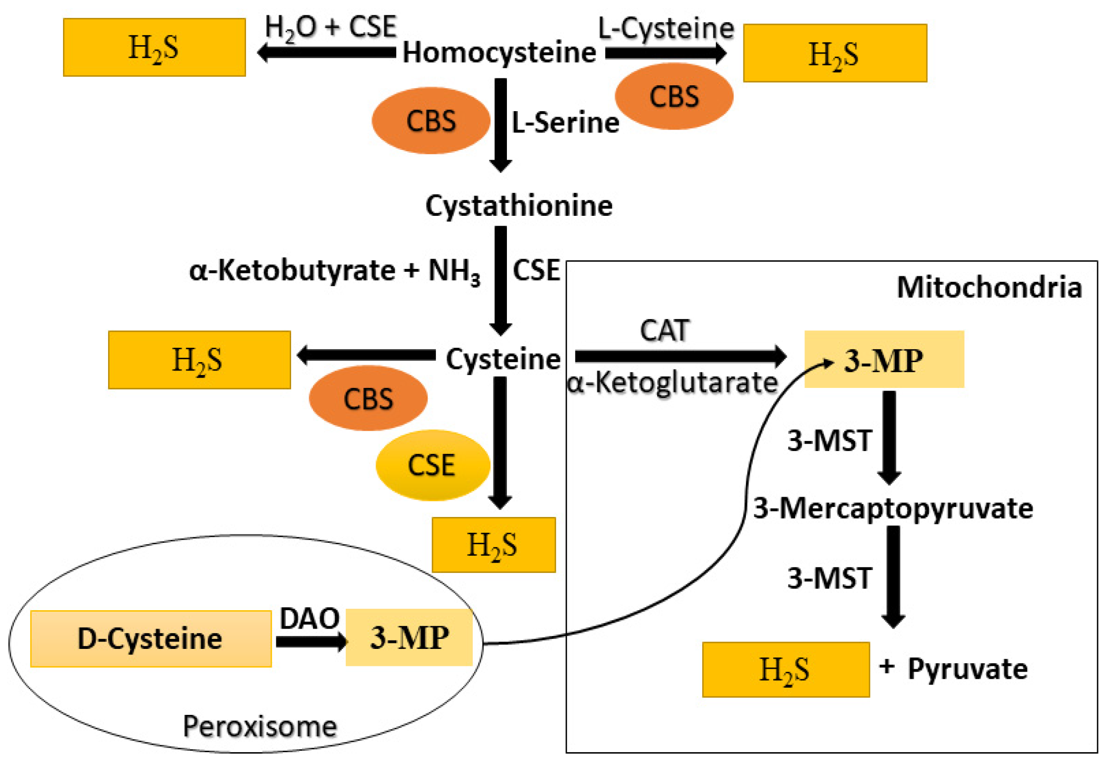
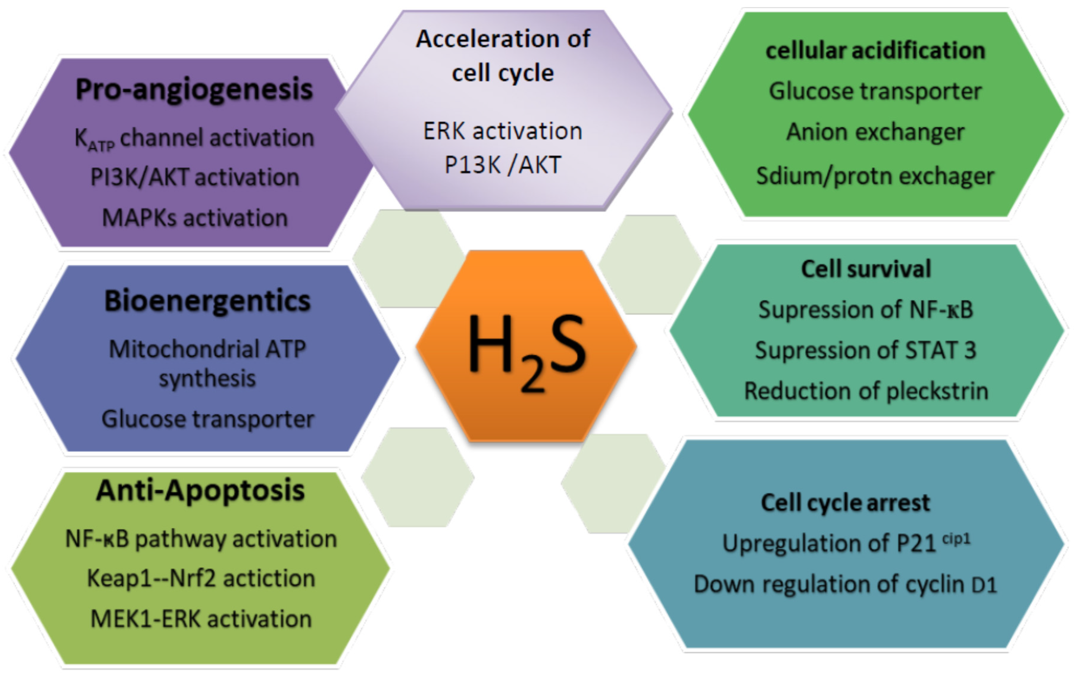
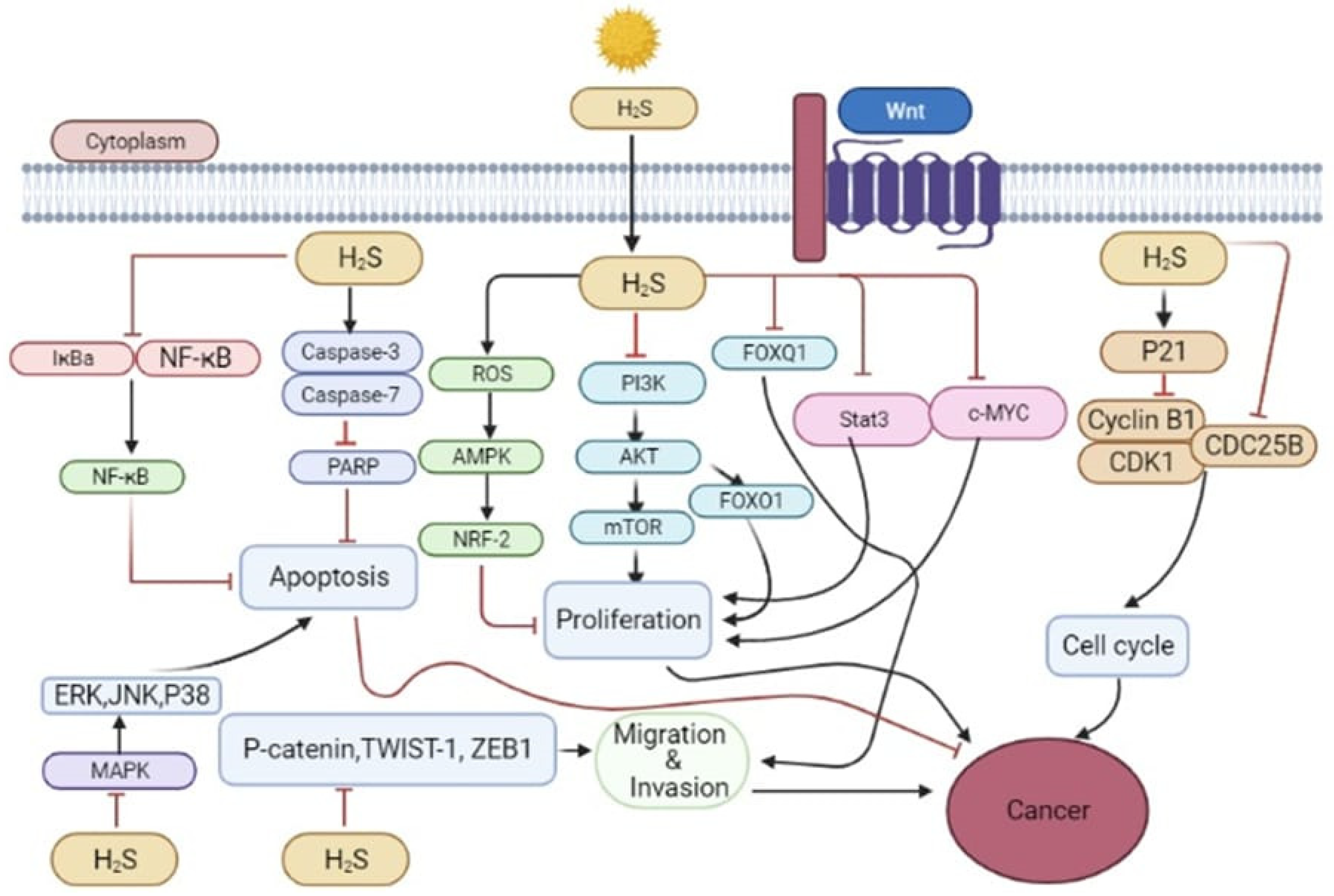
 [Green Version]
Figure 1. A schematic illustration of the biosynthesis of endogenous H2S in mammals. H2S, hydrogen sulfide; H2O, water; CBS, cystathionine β-synthase; CSE, cystathionine γ-lyase; NH3, ammonia; α-ketoglutarate; 3-MST, 3-mercaptopyruvate sulfurtransferase; CAT, cysteine aminotransferase; 3-MP, 3-mercaptopyruvate; DAO, D-amino acid oxidase.
Molecules 27 03389 g001
Figure 2. Dual role of H2S in cancer.
Molecules 27 03389 g002
Figure 3. Schematic diagram of pathways of H2S and H2S donors and their derivatives on cancer. H2S and H2S donors participate in regulating several pathways to induce apoptosis and proliferation.
Molecules 27 03389 g003
Figure 4. Proposed H2S-induced cytotoxicity pathways in mitochondria. H2S inhibits cytochrome c oxidase, resulting in reduced ATP generation. H2S also impairs calcium homeostasis, resulting in elevated intracellular calcium levels. Reduced glutathione depletion results in reactive oxygen species (ROS). H2S causes DNA damage, protein and lipid persulfidation, and ion channel dysregulation, exacerbated by high intracellular ROS levels. These H2S-induced actions, taken together, may result in programmed cell death. ↑: Increase or Generation; ↓: Decreased or Reduced.
Molecules 27 03389 g004
Figure 5. Potential role of H2S in function and mechanisms of action in cancer.
Molecules 27 03389 g005
Table 1. Change in H2S-producing enzymes in different kinds of cancers.
S/No.Cancer TypesCell LinesH2S Producing Enzyme
CSECBS3-MST
1MelanomaA375, WM35, SK-Mel-5, Sk-Mel-28, PES 43NT
2Colon cancerHCT116, HT29
3Prostate cancerLNCaP, PC3, LNCaP-BNT
4Gastric cancerSGC-7901NT
5OvarianOV202, SKOV3, A2780, OVCAR3, OVCAR4, OVCAR5NCNT
6BreastHs578T, MCF7, MDA-MB-428NT
7RenalRCC4
8ThyroidTPC1, TT, ARONC
9GliomasC6, U87MGNTNT
10Hepatocellular CarcinomaHepG2, PLC/PRF/5NT
11Urothelial carcinoma5637, EJ, UM-UC-3
12AstrocytomaU373NTNT
13NeuroblastomaSH-SY5YNTNT
14Oral squamous cell carcinomaOSCC
15LeukaemiaHL-60, MV4-11NTNT
16Biliary tract carcinomaEDI-1, TFK-1, HUCCT-1, SNU308, GB-D1, GB-H3NTNT
NT: Not tested; ↑: Upregulation; NC: No change.
Table 2. Role of H2S donors in cancer promotion and inhibition.
S/NoCancer TypesCell LinesH2S DonorsEffects on CancerReferences
1MelanomaNCI-H929NaHSPromotion[187]
SKMel 5,
SKMel 28
B16F10
A375
S2
GYY4137
 
 
BITC
 
Promotion
 
Inhibition
[89]
 
[282,283]
2Colon cancerHCT 116
SW480,
NaHSPromotion
Inhibition
[104]
[174]
HCT 116GYY4137Inhibition[8]
BITCInhibition[284]
3Prostate cancerPC-3NaHSInhibition[107,109]
PC-3, Rv1, DU145BITCInhibition[285]
LnCaP, DU145GYY4137Inhibition[300]
4Gastric cancerSGC 7901NaHSInhibition[111]
AGS Inhibition[291]
5OvarianA2780, HeyA8, PEA1, PEA2GYY4137Inhibition[300]
OCBITCInhibition[301]
6BreastMCF-7NaHSInhibition[8,302]
MCF-7, MDA-MB-231GYY4137Inhibition[8,300]
MCF-7, MDA-MB-231BITCInhibition[303,304]
7LungA549NaHSInhibition[305,306]
IMR90, WI-38, A549, H1299GYY4137Inhibition[8,300]
A549, H661, NCI-H460/GBITCInhibition[307,308]
8ThyroidTPC-1
ARO
KTC-1
NaHS

NaHS
Promotion
Inhibition
Inhibition
[309]

[310]
KTC-1GYY4137Inhibition[310]
9GliomasC6NaHSPromotion/Inhibition[311,312]
U87MGBITCInhibition[313]
10Hepatocellular CarcinomaHepG2, HLE PLC/PRF/5,
SMMC-7721
NaHSInhibition
Promotion
[8,185]
[6]
HepG2GYY4137Inhibition[8]
Bel 7402,HLEBITCInhibition[289]
11Urothelial carcinomaEJNaHSPromotion[314]
DSM cellGYY4137Promotion[315]
5637,T24BITCPromotion[296]
12AstrocytomaU373NaHSInhibition[244]
BV2 CellGYY4137Inhibition[316]
CCF-STTG1 BITCInhibition[297]
13NeuroblastomaSH-SY5YNaHSInhibition[244]
14Oral squamous cell carcinomaCal-27, WSU-HN6NaHSPromotion[12,94]
OG2BITCInhibition[298]
SCC9BITCInhibition[317]
15LeukemiaMV4-11NaHSInhibition[244]
HL-60, MV4-11GYY4137Inhibition[8]
WEHI-3BITCInhibition[290]
16Esophageal carcinomaEC-109NaHSPromotion[318]
Publisher’s Note: MDPI stays neutral with regard to jurisdictional claims in published maps and institutional affiliations.

