
Cannabinoid Quinones—A Review and Novel Observations


Abstract
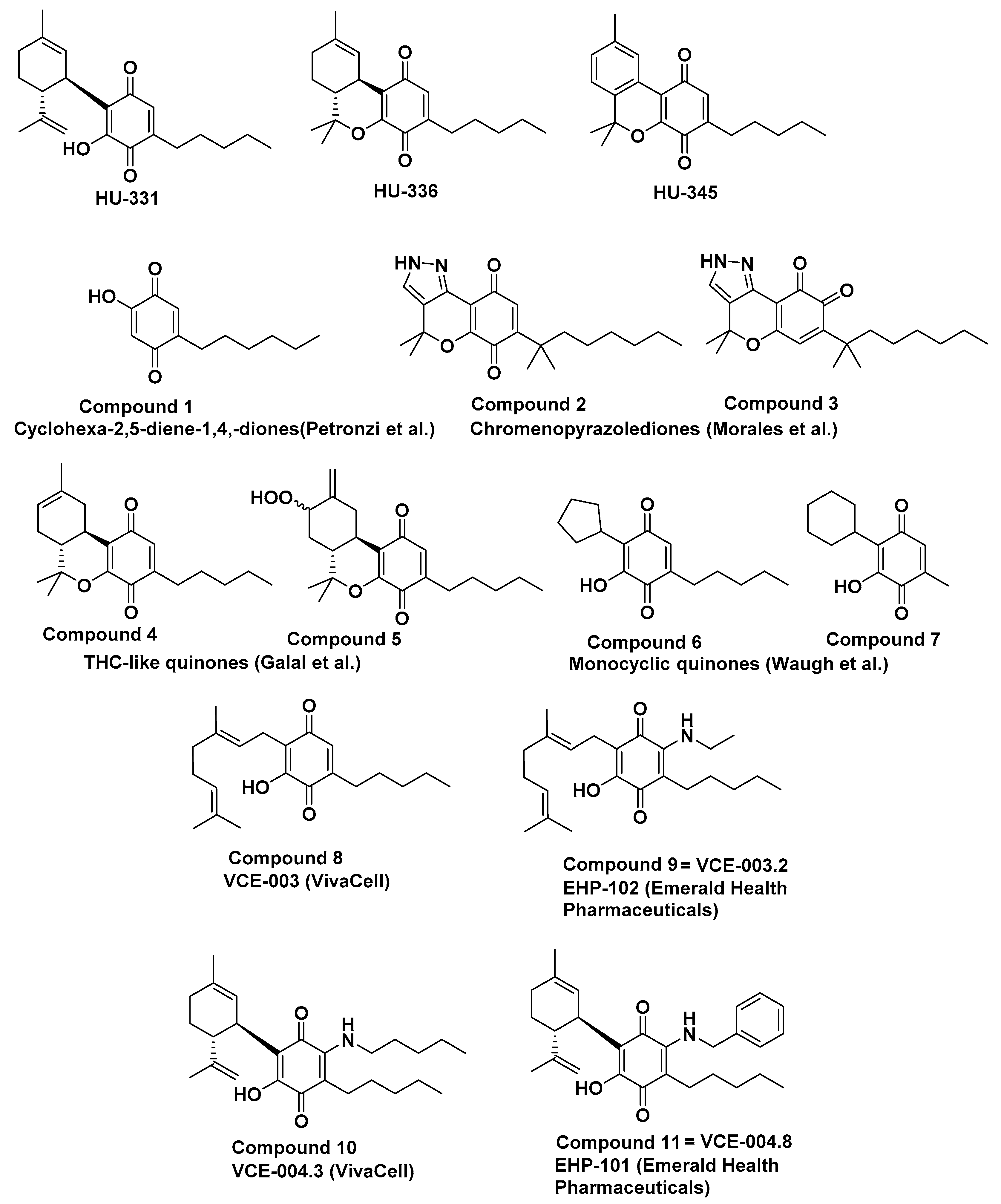
1. Introduction
2. Results
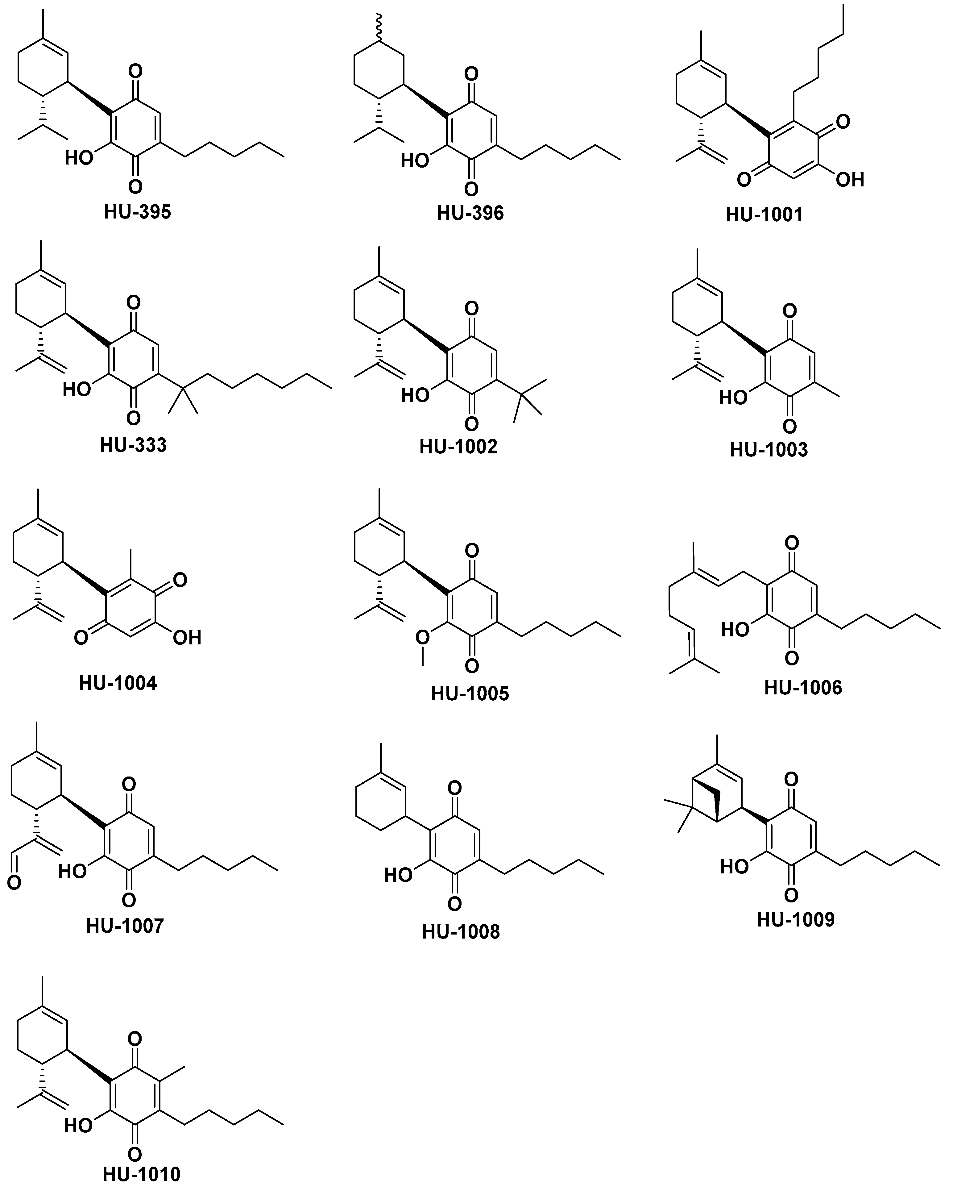
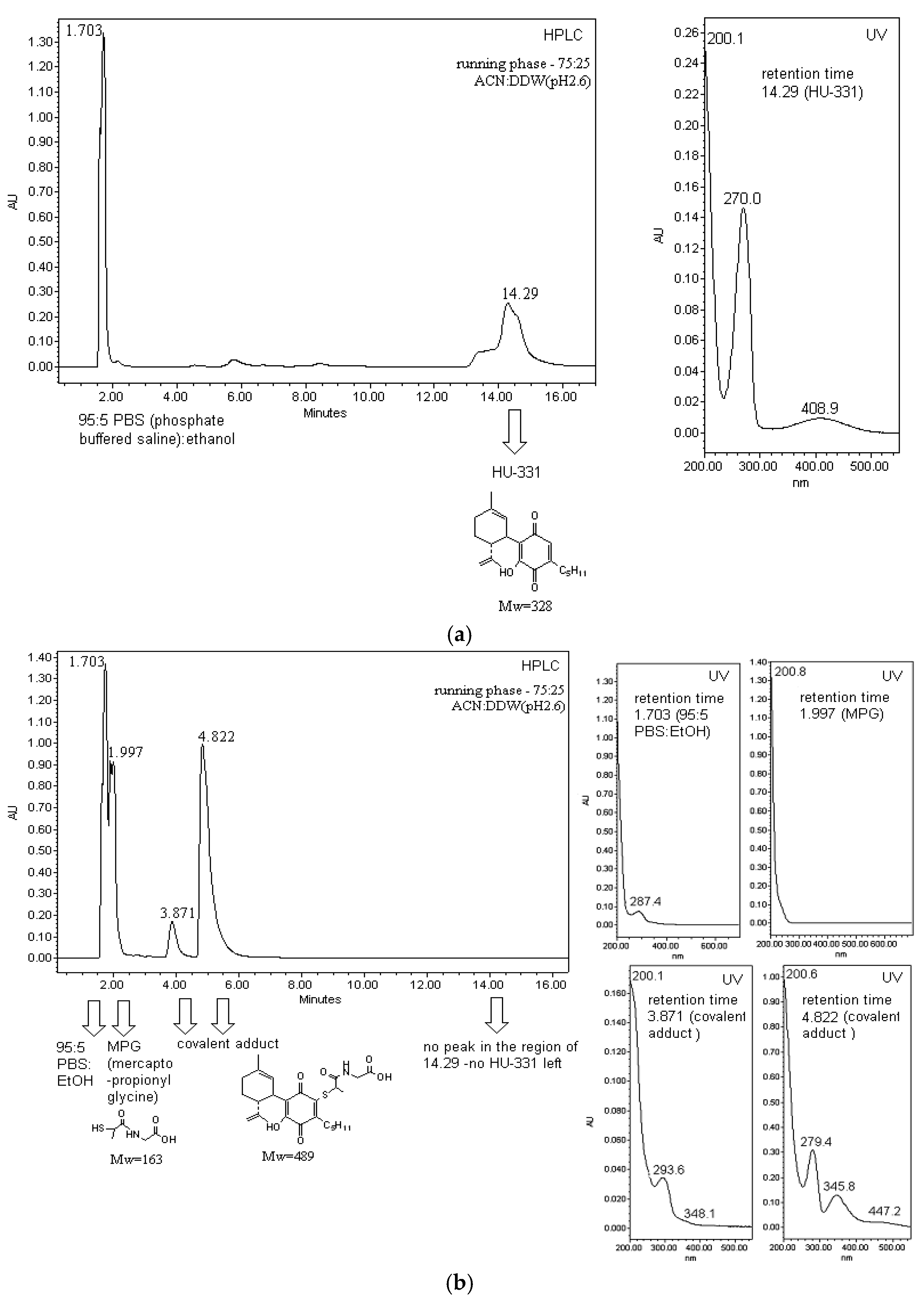
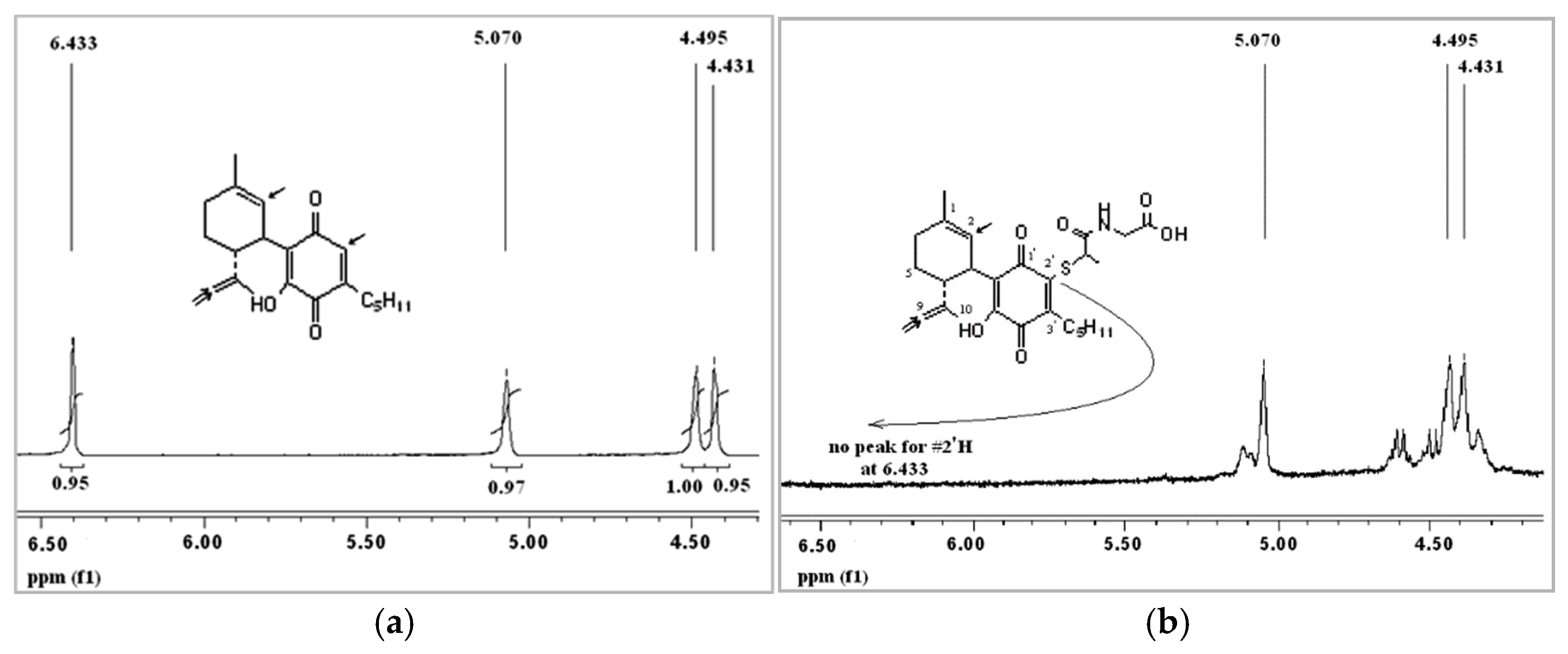
2.1. Chemistry
2.2. Biological Evaluation
2.3. Antioxidant Evaluation
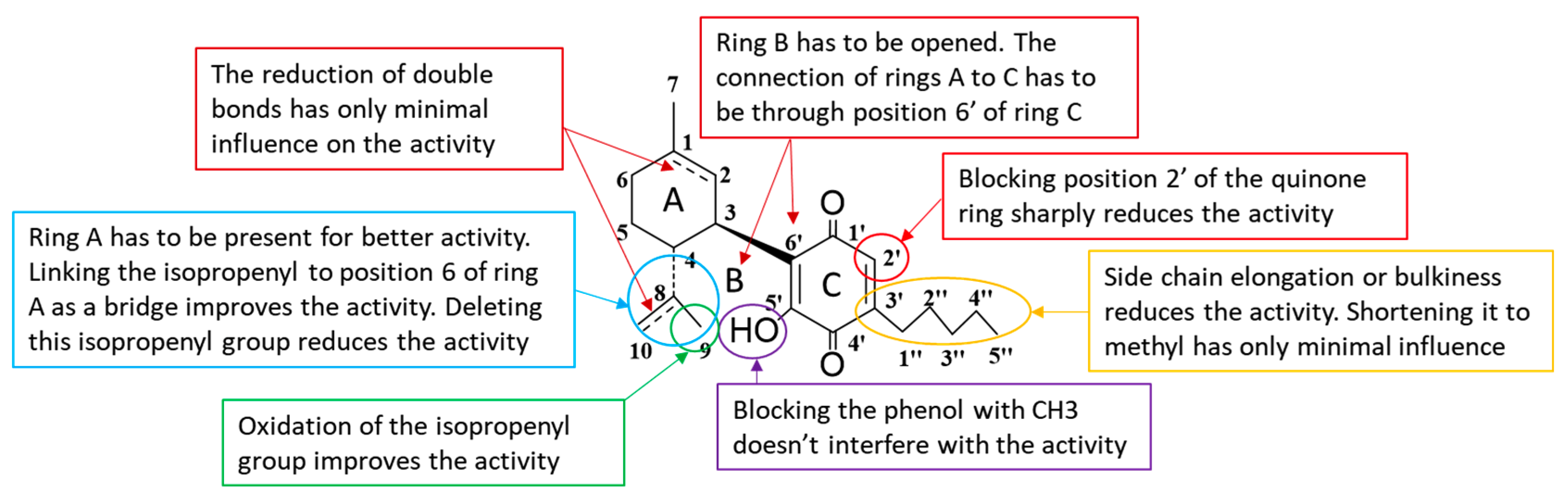
3. Discussion
- The double bonds on ring A have only a minor effect on the anticancer activity.
- The position at which rings A and C are attached one to another is of importance—ring C has to be attached through the “normal”, CBD-like position.
- The hydroxyl on ring C does not have to be free.
- Ring B has to be open.
- Ring A is of importance for the anticancer activity.
- The isopropenyl attached to position C4 of ring A is quite important for the anticancer activity; when absent the activity drops; the connection of isopropenyl to ring A as a bridge further improves the activity, as well as oxidation of the isopropenyl group attached to this ring.
- Position C2 of ring C is crucial for anticancer activity. If blocked or hindered, the activity sharply drops. This is probably a position through which a molecule binds to its intracellular target.
4. Materials and Methods
4.1. NMR Spectroscopy
4.2. Mass Spectrometry
4.3. Chemical Synthesis
4.4. Cell Culture
4.5. Cell Proliferation Test
4.6. Statistical Analyses
Author Contributions
Funding
Data Availability Statement
Acknowledgments
Conflicts of Interest
Sample Availability
References
- Thomson, R.H. Naturally Occurring Quinones IV; Springer: Dordrecht, The Netherlands, 1997. [Google Scholar]
- Meganathan, R. Biosynthesis of Menaquinone (Vitamin K2) and Ubiquinone (Coenzyme Q): A Perspective on Enzymatic Mechanisms. Vitam. Horm. 2001, 61, 173–218. [Google Scholar] [CrossRef]
- McIntire, W.S. Newly Discovered Redox Cofactors: Possible Nutritional, Medical, and Pharmacological Relevance to Higher Animals. Annu. Rev. Nutr. 1998, 18, 145–177. [Google Scholar] [CrossRef] [PubMed]
- Lee, K. Novel Antitumor Agents from Higher Plants. Med. Res. Rev. 1999, 19, 569–596. [Google Scholar] [CrossRef]
- Begleiter, A. Clinical Applications of Quinone-Containing Alkylating Agents. Front. Biosci. J. Virtual Libr. 2000, 5, E153–E171. [Google Scholar] [CrossRef]
- Di Marco, A.; Cassinelli, G.; Arcamone, F. The Discovery of Daunorubicin. Cancer Treat. Rep. 1981, 65, 3–8. [Google Scholar]
- Arcamone, F.; Cassinelli, G. Biosynthetic Anthracyclines. Curr. Med. Chem. 1998, 5, 391–419. [Google Scholar] [CrossRef] [PubMed]
- Zucchi, R.; Danesi, R. Cardiac Toxicity of Antineoplastic Anthracyclines. Curr. Med. Chem. Anti-Cancer Agents 2003, 3, 151–171. [Google Scholar] [CrossRef] [PubMed]
- Rosa, G.M.; Gigli, L.; Tagliasacchi, M.I.; di Iorio, C.; Carbone, F.; Nencioni, A.; Montecucco, F.; Brunelli, C. Update on Cardiotoxicity of Anti-Cancer Treatments. Eur. J. Clin. Investig. 2016, 46, 264–284. [Google Scholar] [CrossRef]
- Thomas, X.; Le, Q.H.; Fiere, D. Anthracycline-Related Toxicity Requiring Cardiac Transplantation in Long-Term Disease-Free Survivors with Acute Promyelocytic Leukemia. Ann. Hematol. 2002, 81, 504–507. [Google Scholar] [CrossRef] [PubMed]
- Razdan, R.K. Structure-Activity Relationships in Cannabinoids. Pharmacol. Rev. 1986, 38, 75–149. [Google Scholar] [PubMed]
- Mechoulam, R.; Hanuš, L.; Fride, E. Towards Cannabinoid Drugs—Revisited. Prog. Med. Chem. 1998, 35, 199–243. [Google Scholar] [CrossRef] [PubMed]
- Barth, F.; Rinaldi-Carmona, M. The Development of Cannabinoid Antagonists. Curr. Med. Chem. 1999, 6, 745–755. [Google Scholar] [CrossRef]
- Bagshaw, S.M.; Hagen, N.A. Medical Efficacy of Cannabinoids and Marijuana: A Comprehensive Review of the Literature. J. Palliat. Care 2002, 18, 111–122. [Google Scholar] [CrossRef] [PubMed]
- Mechoulam, R.; Ben-Zvi, Z.; Gaoni, Y. Hashish-XIII. On the Nature of the Beam Test. Tetrahedron 1968, 24, 5615–5624. [Google Scholar] [CrossRef]
- Watanabe, K.; Usami, N.; Yamamoto, I.; Yoshimura, H. Inhibitory Effect of Cannabidiol Hydroxy-Quinone, an Oxidative Product of Cannabidiol, on the Hepatic Microsomal Drug-Metabolizing Enzymes of Mice. J. Pharm. Dyn. 1991, 14, 421–427. [Google Scholar] [CrossRef] [PubMed]
- Bornheim, L.M.; Grillo, M.P. Characterization of Cytochrome P450 3a Inactivation by Cannabidiol: Possible Involvement of Cannabidiol-Hydroxyquinone as a P450 Inactivator. Chem. Res. Toxicol. 1998, 11, 1209–1216. [Google Scholar] [CrossRef] [PubMed]
- Kogan, N.M.; Rabinowitz, R.; Levi, P.; Gibson, D.; Sandor, P.; Schlesinger, M.; Mechoulam, R. Synthesis and Antitumor Activity of Quinonoid Derivatives of Cannabinoids. J. Med. Chem. 2004, 47, 3800–3806. [Google Scholar] [CrossRef] [PubMed]
- Kogan, N.M.; Schlesinger, M.; Priel, E.; Rabinowitz, R.; Berenshtein, E.; Chevion, M.; Mechoulam, R. HU-331, a Novel Cannabinoid-Based Anticancer Topoisomerase II Inhibitor. Mol. Cancer Ther. 2007, 6, 173–183. [Google Scholar] [CrossRef]
- Regal, K.M.; Mercer, S.L.; Deweese, J.E. HU-331 Is a Catalytic Inhibitor of Topoisomerase IIα. Chem. Res. Toxicol. 2014, 27, 2044–2051. [Google Scholar] [CrossRef]
- Waugh, T.M.; Masters, J.; Aliev, A.E.; Marson, C.M. Monocyclic Quinone Structure-Activity Patterns: Synthesis of Catalytic Inhibitors of Topoisomerase II with Potent Antiproliferative Activity. ChemMedChem 2020, 15, 114–124. [Google Scholar] [CrossRef]
- Kogan, N.M.; Blázquez, C.; Álvarez, L.; Gallily, R.; Schlesinger, M.; Guzmán, M.; Mechoulam, R. A Cannabinoid Quinone Inhibits Angiogenesis by Targeting Vascular Endothelial Cells. Mol. Pharmacol. 2006, 70, 51–59. [Google Scholar] [CrossRef] [PubMed]
- Kogan, N.M.; Schlesinger, M.; Peters, M.; Marincheva, G.; Beeri, R.; Mechoulam, R. A Cannabinoid Anticancer Quinone, HU-331, Is More Potent and Less Cardiotoxic than Doxorubicin: A Comparative in Vivo Study. J. Pharmacol. Exp. Ther. 2007, 322, 646–653. [Google Scholar] [CrossRef] [PubMed]
- Peters, M.; Kogan, N.M. HU-331: A Cannabinoid Quinone, with Uncommon Cytotoxic Properties and Low Toxicity. Expert Opin. Investig. Drugs 2007, 16, 1405–1413. [Google Scholar] [CrossRef]
- Usami, N.; Yamamoto, I.; Watanabe, K. Generation of Reactive Oxygen Species during Mouse Hepatic Microsomal Metabolism of Cannabidiol and Cannabidiol Hydroxy-Quinone. Life Sci. 2008, 83, 717–724. [Google Scholar] [CrossRef]
- Wu, H.Y.; Jan, T.R. Cannabidiol Hydroxyquinone-Induced Apoptosis of Splenocytes Is Mediated Predominantly by Thiol Depletion. Toxicol. Lett. 2010, 195, 68–74. [Google Scholar] [CrossRef] [PubMed]
- Caprioglio, D.; Mattoteia, D.; Pollastro, F.; Negri, R.; Lopatriello, A.; Chianese, G.; Minassi, A.; Collado, J.A.; Munoz, E.; Taglialatela-Scafati, O.; et al. The Oxidation of Phytocannabinoids to Cannabinoquinoids. J. Nat. Prod. 2020, 83, 1711–1715. [Google Scholar] [CrossRef]
- Petronzi, C.; Festa, M.; Peduto, A.; Castellano, M.; Marinello, J.; Massa, A.; Capasso, A.; Capranico, G.; la Gatta, A.; de Rosa, M.; et al. Cyclohexa-2,5-Diene-1,4-Dione-Based Antiproliferative Agents: Design, Synthesis, and Cytotoxic Evaluation. J. Exp. Clin. Cancer Res. 2013, 32, 24–35. [Google Scholar] [CrossRef]
- Morales, P.; Vara, D.; Goméz-Cañas, M.; Zúñiga, M.C.; Olea-Azar, C.; Goya, P.; Fernández-Ruiz, J.; Díaz-Laviada, I.; Jagerovic, N. Synthetic Cannabinoid Quinones: Preparation, In Vitro Antiproliferative Effects and In Vivo Prostate Antitumor Activity. Eur. J. Med. Chem. 2013, 70, 111–119. [Google Scholar] [CrossRef]
- Morales, P.; Blasco-Benito, S.; Andradas, C.; Gómez-Cañas, M.; Flores, J.M.; Goya, P.; Fernández-Ruiz, J.; Sánchez, C.; Jagerovic, N. Selective, Nontoxic CB2 Cannabinoid o-Quinone with in Vivo Activity against Triple-Negative Breast Cancer. J. Med. Chem. 2015, 58, 2256–2264. [Google Scholar] [CrossRef]
- Galal Osman, A.; Elokely, K.M.; Yadav, V.K.; Carvalho, P.; Radwan, M.; Slade, D.; Gul, W.; Khan, S.; Dale, O.R.; Husni, A.S.; et al. Bioactive Products from Singlet Oxygen Photooxygenation of Cannabinoids. Eur. J. Med. Chem. 2018, 143, 983–996. [Google Scholar] [CrossRef]
- Casares, L.; Unciti-Broceta, J.D.; Prados, M.E.; Caprioglio, D.; Mattoteia, D.; Higgins, M.; Apendino, G.; Dinkova-Kostova, A.T.; Muñoz, E.; de la Vega, L. Isomeric O-Methyl Cannabidiolquinones with Dual BACH1/NRF2 Activity. Redox Biol. 2020, 37, 101689. [Google Scholar] [CrossRef]
- Macieja, A.; Kopa, P.; Galita, G.; Pastwa, E.; Majsterek, I.; Poplawski, T. Comparison of the Effect of Three Different Topoisomerase II Inhibitors Combined with Cisplatin in Human Glioblastoma Cells Sensitized with Double Strand Break Repair Inhibitors. Mol. Biol. Rep. 2019, 46, 3625–3636. [Google Scholar] [CrossRef] [PubMed]
- Granja, A.G.; Carrillo-Salinas, F.; Pagani, A.; Gómez-Cañas, M.; Negri, R.; Navarrete, C.; Mecha, M.; Mestre, L.; Fiebich, B.L.; Cantarero, I.; et al. A Cannabigerol Quinone Alleviates Neuroinflammation in a Chronic Model of Multiple Sclerosis. J. Neuroimmune Pharmacol. 2012, 7, 1002–1016. [Google Scholar] [CrossRef]
- Carrillo-Salinas, F.J.; Navarrete, C.; Mecha, M.; Feliú, A.; Collado, J.A.; Cantarero, I.; Bellido, M.L.; Muñoz, E.; Guaza, C. A Cannabigerol Derivative Suppresses Immune Responses and Protects Mice from Experimental Autoimmune Encephalomyelitis. PLoS ONE 2014, 9, e94733. [Google Scholar] [CrossRef]
- Díaz-Alonso, J.; Paraíso-Luna, J.; Navarrete, C.; del Río, C.; Cantarero, I.; Palomares, B.; Aguareles, J.; Fernández-Ruiz, J.; Bellido, M.L.; Pollastro, F.; et al. VCE-003.2, a Novel Cannabigerol Derivative, Enhances Neuronal Progenitor Cell Survival and Alleviates Symptomatology in Murine Models of Huntington’s Disease. Sci. Rep. 2016, 6, 29789. [Google Scholar] [CrossRef] [PubMed]
- García, C.; Gómez-Cañas, M.; Burgaz, S.; Palomares, B.; Gómez-Gálvez, Y.; Palomo-Garo, C.; Campo, S.; Ferrer-Hernández, J.; Pavicic, C.; Navarrete, C.; et al. Benefits of VCE-003.2, a Cannabigerol Quinone Derivative, against Inflammation-Driven Neuronal Deterioration in Experimental Parkinson’s Disease: Possible Involvement of Different Binding Sites at the PPARγ Receptor. J. Neuroinflamm. 2018, 15, 19. [Google Scholar] [CrossRef] [PubMed]
- Burgaz, S.; García, C.; Gómez-Cañas, M.; Navarrete, C.; García-Martín, A.; Rolland, A.; del Río, C.; Casarejos, M.J.; Muñoz, E.; Gonzalo-Consuegra, C.; et al. Neuroprotection with the Cannabigerol Quinone Derivative VCE-003.2 and Its Analogs CBGA-Q and CBGA-Q-Salt in Parkinson’s Disease Using 6-Hydroxydopamine-Lesioned Mice. Mol. Cell. Neurosci. 2021, 110, 103583. [Google Scholar] [CrossRef]
- Burgaz, S.; García, C.; Gómez-Cañas, M.; Muñoz, E.; Fernández-Ruiz, J. Development of an Oral Treatment with the PPAR-γ-Acting Cannabinoid VCE-003.2 against the Inflammation-Driven Neuronal Deterioration in Experimental Parkinson’s Disease. Molecules 2019, 24, 2702. [Google Scholar] [CrossRef]
- Rodríguez-Cueto, C.; Santos-García, I.; García-Toscano, L.; Espejo-Porras, F.; Bellido, M.; Fernández-Ruiz, J.; Muñoz, E.; de Lago, E. Neuroprotective Effects of the Cannabigerol Quinone Derivative VCE-003.2 in SOD1G93A Transgenic Mice, an Experimental Model of Amyotrophic Lateral Sclerosis. Biochem. Pharmacol. 2018, 157, 217–226. [Google Scholar] [CrossRef] [PubMed]
- Del Río, C.; Navarrete, C.; Collado, J.A.; Bellido, M.L.; Gómez-Cañas, M.; Pazos, M.R.; Fernández-Ruiz, J.; Pollastro, F.; Appendino, G.; Calzado, M.A.; et al. The Cannabinoid Quinol VCE-004.8 Alleviates Bleomycin-Induced Scleroderma and Exerts Potent Antifibrotic Effects through Peroxisome Proliferator-Activated Receptor-γ and CB2 Pathways. Sci. Rep. 2016, 6, 21703. [Google Scholar] [CrossRef]
- Del Rio, C.; Cantarero, I.; Palomares, B.; Gómez-Cañas, M.; Fernández-Ruiz, J.; Pavicic, C.; García-Martín, A.; Luz Bellido, M.; Ortega-Castro, R.; Pérez-Sánchez, C.; et al. VCE-004.3, a Cannabidiol Aminoquinone Derivative, Prevents Bleomycin-Induced Skin Fibrosis and Inflammation through PPARγ- and CB2 Receptor-Dependent Pathways. Br. J. Pharmacol. 2018, 175, 3813–3831. [Google Scholar] [CrossRef]
- Palomares, B.; Ruiz-Pino, F.; Navarrete, C.; Velasco, I.; Sánchez-Garrido, M.A.; Jimenez-Jimenez, C.; Pavicic, C.; Vazquez, M.J.; Appendino, G.; Bellido, M.L.; et al. VCE-004.8, A Multitarget Cannabinoquinone, Attenuates Adipogenesis and Prevents Diet-Induced Obesity. Sci. Rep. 2018, 8, 16092. [Google Scholar] [CrossRef] [PubMed]
- Navarrete, C.; Carrillo-Salinas, F.; Palomares, B.; Mecha, M.; Jiménez-Jiménez, C.; Mestre, L.; Feliú, A.; Bellido, M.L.; Fiebich, B.L.; Appendino, G.; et al. Hypoxia Mimetic Activity of VCE-004.8, a Cannabidiol Quinone Derivative: Implications for Multiple Sclerosis Therapy. J. Neuroinflamm. 2018, 15, 64. [Google Scholar] [CrossRef] [PubMed]
- García-Martín, A.; Garrido-Rodríguez, M.; Navarrete, C.; del Río, C.; Bellido, M.L.; Appendino, G.; Calzado, M.A.; Muñoz, E. EHP-101, an Oral Formulation of the Cannabidiol Aminoquinone VCE-004.8, Alleviates Bleomycin-Induced Skin and Lung Fibrosis. Biochem. Pharmacol. 2018, 157, 304–313. [Google Scholar] [CrossRef] [PubMed]
- Navarrete, C.; García-Martin, A.; Garrido-Rodríguez, M.; Mestre, L.; Feliú, A.; Guaza, C.; Calzado, M.A.; Muñoz, E. Effects of EHP-101 on Inflammation and Remyelination in Murine Models of Multiple Sclerosis. Neurobiol. Dis. 2020, 143, 104994. [Google Scholar] [CrossRef]
- Razdan, R.K.; Dalzell, H.C.; Handrick, G.R. Hashish. X. Simple One-Step Synthesis of (-)-Δ1-Tetrahydrocannabinol (THC) from p-Mentha-2,8-Dien-1-Ol and Olivetol. J. Am. Chem. Soc. 1974, 96, 5860–5865. [Google Scholar] [CrossRef]
- Martin, B.R.; Compton, D.R.; Prescott, W.R.; Barrett, R.L.; Razdan, R.K. Pharmacological Evaluation of Dimethylheptyl Analogs of Δ9-THC: Reassessment of the Putative Three-Point Cannabinoid-Receptor Interaction. Drug Alcohol Depend. 1995, 37, 231–240. [Google Scholar] [CrossRef]
- Guimarães, F.S.; de Aguiar, J.C.; Mechoulam, R.; Breuer, A. Anxiolytic Effect of Cannabidiol Derivatives in the Elevated Plus-Maze. Gen. Pharmacol. Vasc. Syst. 1994, 25, 161–164. [Google Scholar] [CrossRef]
- Aung, M.M.; Griffin, G.; Huffman, J.W.; Wu, M.J.; Keel, C.; Yang, B.; Showalter, V.M.; Abood, M.E.; Martin, B.R. Influence of the N-1 Alkyl Chain Length of Cannabimimetic Indoles upon CB1 and CB2 Receptor Binding. Drug Alcohol Depend. 2000, 60, 133–140. [Google Scholar] [CrossRef]
- Edery, H.; Grunfeld, Y.; Porath, G.; Ben-Zvi, Z.; Shani, A.; Mechoulam, R. Structure-Activity Relationships in the Tetrahydrocannabinol Series. Modifications on the Aromatic Ring and It the Side-Chain. Arzneim. Forsch. Drug Res. 1972, 22, 1995–2003. [Google Scholar]


| Comp. Name | Raji (IC50, µM) | Jurkat (IC50, µM) |
|---|---|---|
| HU-331 | 0.603 ± 0.047 | 0.470 ± 0.057 |
| HU-395 | 0.227 ± 0.037 | 0.154 ± 0.036 |
| HU-396 | 0.617 ± 0.035 | 0.549 ± 0.066 |
| HU-1001 | 25.14 ± 0.962 *** | 39.07 ± 3.102 *** |
| HU-333 | 5.859 ± 0.794 ** | 17.54 ± 3.025 *** |
| HU-1002 | 2.044 ± 0.125 * | 6.373 ± 0.386 ** |
| HU-1003 | 0.820 ± 0.022 | 0.772 ± 0.075 |
| HU-1004 | 11.01 ± 1.182 *** | 9.984 ± 1.016 *** |
| HU-1005 | 0.634 ± 0.073 | 0.499 ± 0.045 |
| HU-1006 | 4.270 ± 0.149 * | 4.301 ± 0.168 * |
| HU-1007 | 0.179 ± 0.01 | 0.203 ± 0.041 |
| HU-1008 | 8.474 ± 0.982 *** | 9.229 ± 1.03 *** |
| HU-1009 | 0.220 ± 0.038 | 0.245 ± 0.076 |
| HU-1010 | 34.01 ± 2.665 *** | 17.31 ± 0.506 *** |
Publisher’s Note: MDPI stays neutral with regard to jurisdictional claims in published maps and institutional affiliations. |
© 2021 by the authors. Licensee MDPI, Basel, Switzerland. This article is an open access article distributed under the terms and conditions of the Creative Commons Attribution (CC BY) license (http://creativecommons.org/licenses/by/4.0/).
Share and Cite
Kogan, N.M.; Peters, M.; Mechoulam, R. Cannabinoid Quinones—A Review and Novel Observations. Molecules 2021, 26, 1761. https://doi.org/10.3390/molecules26061761
Kogan NM, Peters M, Mechoulam R. Cannabinoid Quinones—A Review and Novel Observations. Molecules. 2021; 26(6):1761. https://doi.org/10.3390/molecules26061761
Chicago/Turabian StyleKogan, Natalya M., Maximilian Peters, and Raphael Mechoulam. 2021. "Cannabinoid Quinones—A Review and Novel Observations" Molecules 26, no. 6: 1761. https://doi.org/10.3390/molecules26061761
APA StyleKogan, N. M., Peters, M., & Mechoulam, R. (2021). Cannabinoid Quinones—A Review and Novel Observations. Molecules, 26(6), 1761. https://doi.org/10.3390/molecules26061761