Review

Allicin, the Odor of Freshly Crushed Garlic: A Review of Recent Progress in Understanding Allicin’s Effects on Cells §

by
Jan Borlinghaus
1,*,
Jana Foerster (née Reiter)
1,
Ulrike Kappler
2,
Haike Antelmann
3,
Ulrike Noll
1,
Martin C. H. Gruhlke
1 and
Alan J. Slusarenko
1,*
1
Department of Plant Physiology, RWTH Aachen University, 52056 Aachen, Germany
2
School of Chemistry and Molecular Biosciences, University of Queensland, St. Lucia, Qld 4072, Australia
3
Institute of Biology-Microbiology, Freie Universität Berlin, 14195 Berlin, Germany
*
Authors to whom correspondence should be addressed.
§
Dedication: This article is dedicated to Professor emeritus Dr. Eric Block on his 79th birthday in recognition of his lifelong contributions to, and enthusiastic support of, research on the chemistry and biological effects of sulfur-containing natural compounds.
Molecules 2021, 26(6), 1505; https://doi.org/10.3390/molecules26061505
Submission received: 16 February 2021 / Revised: 5 March 2021 / Accepted: 6 March 2021 / Published: 10 March 2021
(This article belongs to the Special Issue Fragrant Molecules with Pharmacological Activities)

Abstract

:
The volatile organic sulfur compound allicin (diallyl thiosulfinate) is produced as a defense substance when garlic (Allium sativum) tissues are damaged, for example by the activities of pathogens or pests. Allicin gives crushed garlic its characteristic odor, is membrane permeable and readily taken up by exposed cells. It is a reactive thiol-trapping sulfur compound that S-thioallylates accessible cysteine residues in proteins and low molecular weight thiols including the cellular redox buffer glutathione (GSH) in eukaryotes and Gram-negative bacteria, as well as bacillithiol (BSH) in Gram-positive firmicutes. Allicin shows dose-dependent antimicrobial activity. At higher doses in eukaryotes allicin can induce apoptosis or necrosis, whereas lower, biocompatible amounts can modulate the activity of redox-sensitive proteins and affect cellular signaling. This review summarizes our current knowledge of how bacterial and eukaryotic cells are specifically affected by, and respond to, allicin.

1. Introduction

Garlic (Allium sativum) is an internationally appreciated culinary ingredient in many dishes and an economically important agricultural crop in several countries. It is well known that when an intact garlic clove is chopped or crushed it instantly releases a typical pungent bouquet or odor. The characteristic odor of garlic is due to volatile organic sulfur-containing compounds (VOSCs) that are rapidly produced upon tissue damage and diffuse into the air. The major contributor to the odor of freshly crushed garlic is allicin (diallyl thiosulfinate, preferred IUPAC name = S-prop-2-en-1-yl prop-2-ene-1-sulfinothioate). Garlic releases VOSCs to defend itself against attacks by pathogens and pests and its antibacterial principle was identified as allicin by Cavallito in 1944 [1,2]. In vivo allicin forms after cellular damage when the enzyme alliinase, which in intact cells is localized in the vacuole, mixes with the substrate alliin which is located in the cytosol (Scheme 1) [3,4,5]. Alliin (S-allylcysteine sulfoxide) is an odorless non-protein amino acid. It has been reported that alliinase is predominantly located in bundle sheath cells in bulbs, leaves, and inflorescence axes in the garlic plant [6]. Other alkyl-cysteine sulfoxides are also present in the cytoplasm of garlic cells and can serve as alliinase substrates, yielding amongst others, dimethyl, dipropyl and mixed alkyl thiosulfinates. Nevertheless, allicin accounts for 60–80% of the total thiosulfinates formed by damaged garlic [3]. Alliinase (S-alkyl-l-cysteine S-oxide alkyl-sulfenate-lyase, E.C. 4.4.1.4) readily converts its substrates to the corresponding sulfenic acids, which condense spontaneously to produce thiosulfinates (Scheme 1) [7]. The reactions are very rapid and are 97% complete within 30 s at 23 °C. Furthermore, a single clove of garlic (approximately 10 g) can produce up to 5 mg of allicin, illustrating the large evolutionary investment the plant places in this defense molecule [8]. An easy and efficient in vitro synthesis protocol for >98% pure allicin and approximately 90% yield has been published [9].
The odor of crushed garlic is usually described as ‘pungent’, which the shorter Oxford English dictionary defines as “biting, caustic or piquant, and affects the skin and the organs of taste and smell with a prickling sensation” [10]. VOSCs are known for their intense and unique olfactory properties, in many cases being detectable by some animal species in ppb. Even the relatively insensitive human nose can detect VOSCs in the low ppm range [7,11].
Garlic-derived preparations were shown to have insect-repellent effects and to discourage the feeding of some birds, for example European starlings. The garlic preparations tested against starlings contained mixed diallyl, dimethyl and allyl-methyl polysulfanes but no allicin [12]. Nevertheless, allicin has repellent effects on herbivores and has been shown to stimulate a subset of TRPA1 (transient receptor potential) non-selective ion channels in pain-sensing (nociceptive) neurons [13,14]. The TRPA1 ion channel, also known as the ‘wasabi receptor’, is activated by a wide range of electrophiles including allyl isothiocyanate, the active ingredient of wasabi [15]. Allyl isothiocyanate activates TRPA1 by covalent modification of a cysteine residue within the cytoplasmic N terminus in TRPA1 [16]. Due to its thiol-reactivity (see later), it seems likely that allicin acts similarly when it stimulates TRPA1-bearing neurons. Furthermore, it was shown to be the same particular subset of TRPA1 neurons that were stimulated by both allyl isothiocyanate and allicin [13]. Activation of TRPA1 results in local inflammation and pain and can evoke innate fear behavior in the sensory host, and it is no surprise therefore that allicin acts as an effective deterrent of herbivory [11,17]. In addition, plants can influence one another and other organisms by using volatile signals which act as allelochemicals, i.e., secondary products that affect the growth, survival or reproduction of other organisms [18]. In this sense, it could be argued that allicin, which has been shown experimentally to inhibit root growth of Arabidopsis seedlings (see Section 2.4.1), might also be considered to be an allelochemical [19,20].
Allicin is a colorless liquid at room temperature, but is sometimes described as pale yellow likely due to contamination with trace amounts of elemental sulfur. Because of its relatively high Mr (162) and the internal dipole within the molecule, allicin would not be expected to have a high vapor pressure. Nevertheless, despite its predicted low volatility allicin is a very odoriferous molecule and small quantities entering the gas phase are easily detected by smell. This becomes obvious every time garlic is chopped or crushed in the kitchen. Unfortunately, the actual vapor pressure of allicin, like its boiling point, is so far not measurable experimentally because upon heating allicin decomposes into a plethora of other VOSCs. For example, heating allicin or garlic, e.g., during cooking, leads to the production of polysulfanes and other physiologically active molecules like ajoene (see Section 4) [21,22]. The dynamic chemistry of the low molecular weight sulfur compounds in biological extracts makes their accurate quantitative and qualitative analysis difficult, and the effects of heating are particularly important. This is often overlooked and is a frequently met flaw in reports purporting to analyze the composition of Allium extracts or vapors by gas chromatography (GC) rather than, for example, by liquid chromatography coupled with mass spectrometry (LC-MS). The unavoidable heating step associated with GC inevitably degrades allicin and leads to false composition profiles not reflecting the natural situation.
An important descriptor for a pharmacologically active molecule is its logP or Kow value. This is an indicator of how a molecule partitions between the non-polar solvent octanol and the polar solvent water. A logP value of 1 reflects a 10-fold higher concentration in octanol than water and a value of 0.1 reflects the opposite. Allicin’s lipophilic nature is indicated by its calculated logP of 1.35 and it is readily membrane permeable and easily taken up by cells [23]. Similar to the structurally related dimethyl sulfoxide (DMSO) molecule, allicin has been shown to create transient pores as it crosses membranes. Temporary pore formation leads to a transient depolarization, i.e., a decrease in membrane potential (∆Em) [24]. This effect was observed both in artificial lipid bilayers, where proteins and other potentially allicin-reactive molecules are absent, as well as in biological membranes. Therefore, membrane permeabilization appears to be a physical effect, like that shown for DMSO [25], and is unrelated to any chemical reactivity of allicin [24].
Allicin is a reactive sulfur species (RSS) and a potent thiol-trapping reagent, rapidly reacting with glutathione (GSH) to yield S-allylmercaptoglutathione (GSSA). Thus, allicin depletes the cellular GSH pool and reacts with accessible cysteine thiols in proteins by S-thioallylation (Scheme 2) [21,26,27,28,29,30,31]. This reaction is key to allicin’s biological activity. Reversible oxidation and reduction of protein-thiols is central to the regulation of many processes in cells [32].
Effects of allicin on living cells can range from its being a dose-dependent biocide to it subtly modifying important cell-signaling pathways and cellular functions at lower, biocompatible doses. The thiol-disulfide exchange reaction (TDER) of a thiol with a disulfide is well described and is important in cellular protein chemistry and signaling [33]. The S-thioallylation of a protein thiol by allicin (Scheme 2) can be viewed as a form of TDER. Both the divalent sulfur atom and the sulfinyl sulfur atom are electrophilic but most of the chemistry occurs by nucleophilic attack on the divalent sulfur, with the sulfinyl sulfur being the leaving group when the O-atom is protonated. The polarized bond between the O- and S-atoms of the sulfinyl group significantly weakens the disulfide bond in allicin and makes it more reactive than a simple disulfide towards target nucleophilic thiol groups. Unlike in a standard TDER, the electrons end up in water. Nevertheless, the newly formed disulfide bond can be reduced back to a thiol just like any other protein disulfide, for example by thioredoxins or glutaredoxins, or potentially even by glutathione reductase. In support of this notion, we have recently shown that S-allylmercaptoglutathione, produced when allicin reacts with GSH, is a substrate for glutathione reductase [34]. Because allicin is such an effective thiol-trapping reagent it is interesting to know which proteins are S-thioallylated in cells by allicin. This is important not only in relation to allicin’s well documented antimicrobial effects [35], but also because the consumption of uncooked garlic, as well as breathing in garlic fumes, exposes cells in the body to allicin. Identifying which proteins become S-thioallylated and to what degree, is potentially of great significance in light of the manifold claims of the health benefits of consuming garlic (reviewed in [21]), and its increasing use in plant protection [36].
The purpose of this review is to summarize recent progress in our knowledge of the cellular effects of allicin in bacteria and eukaryotes and builds on our earlier 2014 review [19].

2. Allicin and Oxidative Stress in Cells and Organisms

Chemically, allicin clearly reacts as an oxidant when it S-thioallylates -SH groups. However, allicin is often described as an antioxidant, particularly in the diet. There are two aspects of allicin’s properties and mode of action that need to be considered in relation to this apparent contradiction. Firstly, from the chemical point of view, an antioxidant describes a substance that prevents or slows down the oxidation of another molecule, and in this regard, it has been shown that allicin readily undergoes a Cope elimination reaction at room temperature to form 2-propenesulfenic acid, a very potent antioxidant [37]. Interestingly, 2-propenesulfenic acid (allylsulfenic acid) is thus both a precursor (Scheme 1) and a decomposition product of allicin [37]. Secondly, from a nutritional and physiological point of view, cells respond to mild oxidative stress by activating oxidative stress protection responses, which prime the cells to be more resistant to subsequent, greater oxidative insults [38,39,40,41,42]. The detection of an oxidative threat to cells usually occurs via specialized sensor proteins, like Keap-1 in mammals, containing oxidation-sensitive cysteine residues [43]. Oxidation of these sensor cysteines leads to a cascade of phase II oxidative stress protective cellular responses. Detailed examples of how allicin interacts with the oxidative-stress-sensing machinery in some model organisms are given in the next section.

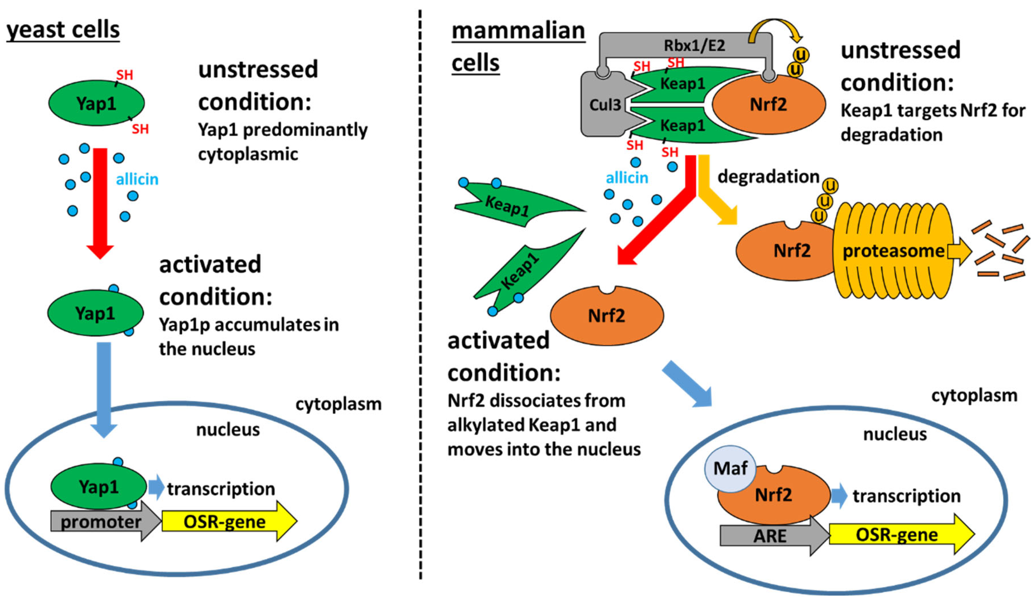
2.1. The Yeast Yap1 Oxidative Stress Sensor System

The oxidative stress response (OSR) in yeast (Saccharomyces cerevisiae), a model fungus, is centrally regulated by the Yap1 transcription factor which senses H2O2 in a Gpx3-dependent mechanism via the oxidation of redox-sensitive cysteines [44]. Yap1 regulates the expression of genes important for antioxidant metabolism, e.g., genes on the GSH biosynthetic pathway, glutathione reductase, glutaredoxins, thioredoxins and thioredoxin reductases [45,46]. Yap1 has both a nuclear localization sequence (NLS) and a nuclear export sequence (NES) where the export protein Crm1 binds. In unstressed yeast the cellular dynamics are such that, although Yap1 continually shuttles between the nucleus and the cytoplasm, it is predominantly located in the cytoplasm and Yap1-dependent genes are not transcribed [47,48]. Allicin directly oxidizes the Yap1 Cys598 and Cys620 residues in the C-terminal nuclear export sequence (NES), blocking the binding of the Crm1 export protein, thus allowing Yap1 to accumulate in the nucleus and to activate the expression of target OSR genes (Figure 1) [49]. Allicin-induced Yap1 activation also occurs in a ∆gpx3 mutant. This confirms that activation is direct and independent of H2O2. At the moment it is unclear to what extent allicin’s oxidative activity might contribute to by any downstream production of reactive oxygen species (ROS) like H2O2 [49].

2.2. The Mammalian Keap1-Nrf2/ARE Oxidative Stress Sensor System

The OSR in mammalian cells is coordinated by the Keap1-Nrf2/ARE system which responds to reactive electrophilic species [50,51]. The Keap1 (Kelch-like ECH-associated protein) sensor protein has free cysteine thiols in its reduced state that are sensitive oxidation targets, making the oxidative stress sensor mechanism in mammals similar to that of Yap1 in yeast. In contrast to Yap1, which is itself a transcription factor, the reduced Keap1 oxidation sensor sequesters the Nrf2 transcription factor in a protein complex. In this complex Nrf2 is a substrate for ubiquitination and therefore degradation in the cytosol, making Nrf2 unavailable for transcription. However, when the redox-sensing cysteines in Keap1 are alkylated by electrophiles, Nrf2 dissociates from the complex and is no longer targeted for degradation (Figure 1). The Nrf2 transcriptional regulator, freed from the complex, migrates to the nucleus, associates with its co-transcription factor Maf, and facilitates the transcription of genes which have an antioxidant response element (ARE) in their promoters. The products of these ARE genes, which include antioxidative enzymes, giving rise to so-called phase II responses to oxidative stress [52]. Allicin, as a low Mr electrophilic oxidant is detected by, and activates, the Keap1-Nrf2/ARE system in mammalian cells [41,53].
Thiol oxidation chemistry is the common denominator employed in the OS-sensing systems of very different organisms (Figure 1). Taking advantage of this, we developed a very sensitive reporter system which can detect thiol-reactive low Mr electrophiles like allicin. We took the YKL071w gene (Oxidative Stress Induced 1, OSI1) from yeast that was reported to be the gene most highly induced by allicin [54] and fused its promoter to a luciferase reporter. The expression of OSI1 is absolutely Yap1-dependent [49] making the OSI1::luciferase fusion a highly sensitive reporter of Yap1 activation in yeast. Comparing the yeast reporter system with an Nrf2-activated promoter-luciferase reporter system in mammalian cells showed congruence of effect. Moreover, the yeast-based system offers a lot of advantages over the human cell based assay, particularly in terms of sensitivity, the dynamic response, and ease of handling—making it advantageous in large scale screening procedures such as the detection of skin irritants or electrophilic activity of pharmaceuticals, reducing the need for animal testing [53].

2.3. Bacterial Redox Sensors

The OSR in bacteria is controlled by different redox sensitive regulators depending on the species. For example, in Gram-negative Escherichia coli, the transcriptional regulator OxyR has cysteine residues that can be directly oxidized by H2O2, leading to intramolecular disulfide formation between Cys199 and Cys208 in each subunit of the OxyR tetramer and subsequent activation of OxyR [55]. In Gram-positive firmicutes like Bacillus subtilis and Staphylococcus aureus, transcriptional repressors such as PerR are released from DNA by Fe-mediated oxidation of redox sensitive histidines, thus enabling the expression of genes involved in the H2O2 stress response [56,57,58]. Interestingly, genes that are controlled by these transcription factors were shown to be upregulated by allicin [31,59,60], perhaps indirectly by possible downstream H2O2 generation, or directly by oxidation of the structural Zn-binding cysteines that are necessary for PerR dimerization and DNA-binding. However, allicin can also directly alter the function of transcriptional regulators. For the redox-sensitive transcriptional repressors of B. subtilis like OhrR, YodB, HypR, and Spx, it was shown that their redox-sensitive Cys residues can be modified directly by allicin (Section 3.1., Table S1), thus connecting their regulon activity with an allicin-dependent mechanism for DNA release and derepression of response genes [60]. Numerous transcriptional regulators were shown to be targets of allicin stress either directly via demonstrated S-thioallylation, and/or indirectly by observing their regulon activity. In this way, it was shown that allicin stress in S. aureus induced the PerR, HypR, QsrR, MhqR, CstR, CtsR, HrcA and CymR regulons [59], and the OhrR, PerR, Spx, YodB, CatR, HypR, AdhR, HxlR, LexA, CymR, CtsR, and HrcA regulons were induced upon allicin stress in B. subtilis [60]. A description of these transcriptional regulators is given in supplementary Table S1. Several of these regulators are redox sensitive and are involved in the redox stress response and thiol homeostasis, or in stress responses that often occur simultaneously with oxidative stress, e.g., protein damage or metal stress. Like their eukaryotic counterparts in mammals (Nrf2/Keap1) and yeast (Yap1), these allicin-activated bacterial transcription regulators are important for the oxidative stress response.

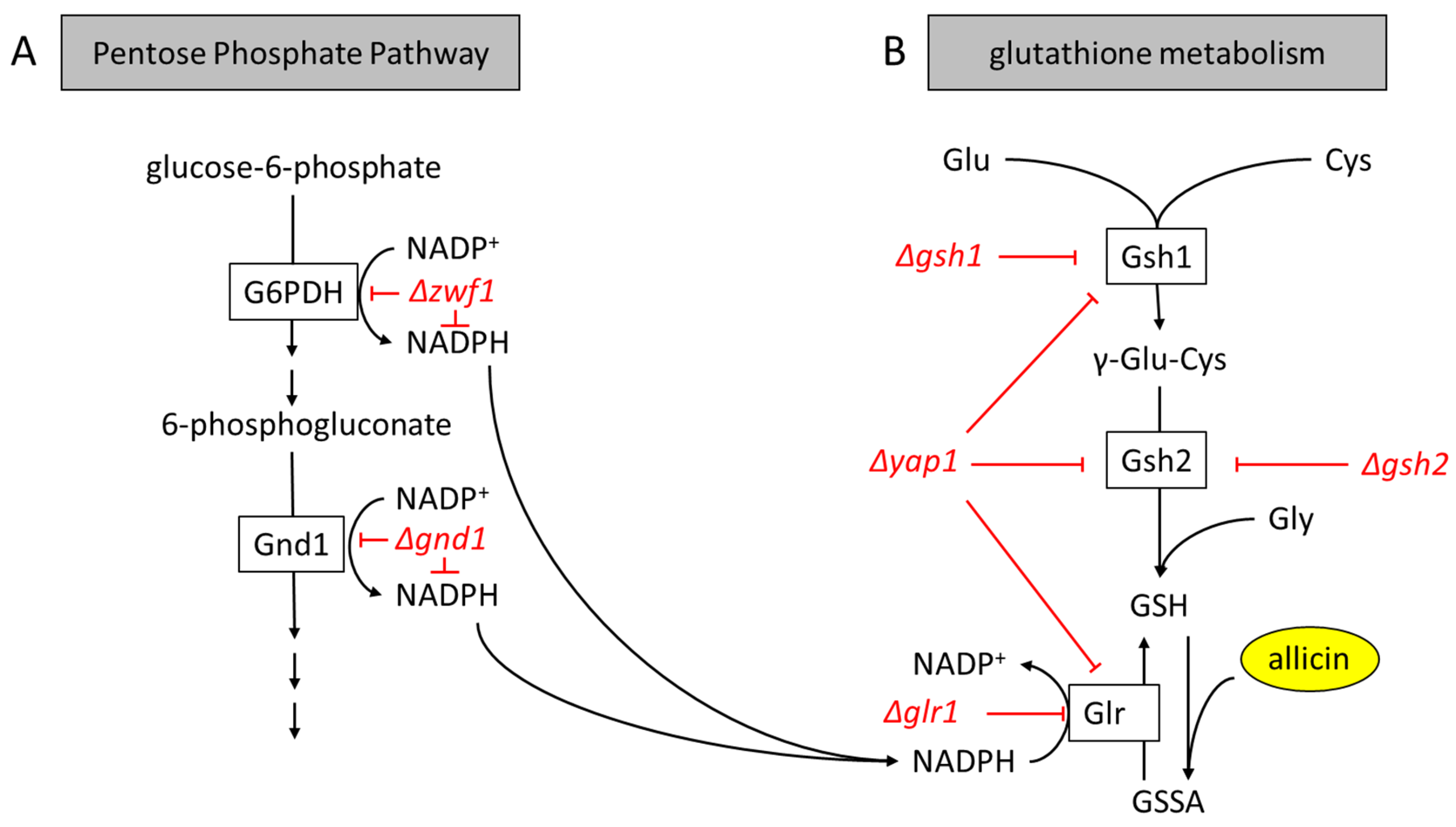
2.4. The Central Role of GSH Metabolism in Allicin Resistance

Glutathione (γ-l-Glutamyl-l-cysteinylglycine, GSH) is present at up to 10 mM in the cytosol of many organisms and is recognized as a major cellular antioxidant that protects cells in a first line of defense against oxidative stress [39,61,62,63,64]. Although the total concentration of other free thiol groups in a cell can exceed that contributed by GSH, the latter is the most highly abundant LMW thiol in most eukaryotes and many Gram-negative bacteria. Because allicin is such a potent thiol trapping reagent, GSH is a major reactant for allicin in cells and is in direct competition for allicin with other less abundant thiols in individual proteins. Because of its molecular abundance, GSH can titrate out and thus protect cells against allicin. GSH is synthesized in two steps from cysteine by the enzymes γ-glutamylcysteine synthase (abbreviated to Gsh1 in yeast, GshA in bacteria, GSH1 in plants) and glutathione synthase (abbreviated to Gsh2 in yeast, GshB in bacteria, GSH2 in plants). Both glutathione disulfide (GSSG) and S-allylmercaptoglutathione (GSSA), the product formed when allicin reacts with GSH, are reduced back to GSH and GSH plus allylmercaptan, respectively by the NADPH-dependent enzyme glutathione reductase (abbreviated as Glr in yeast, Gor in bacteria, and GR in plants). Glutathione reductase is dependent upon NADPH as a reductant which is produced in the Pentose Phosphate Pathway (Figure 2A,B). A number of recent studies with mutants affected in GSH synthesis and metabolism in plants, fungi, and bacteria have reinforced the central role of GSH in enabling cells to withstand the oxidative stress caused by allicin treatment.

2.4.1. Plants (Arabidopsis thaliana)

Allicin does not pass easily through the wax plates and cuticle layers of the leaf surface. However, if Arabidopsis leaf discs are floated on an allicin solution, which diffuses into the discs at the edges, oxidative stress occurs and chlorophyll bleaching is observed [65]. In contrast, young seedling roots are not covered by wax or cuticular layers and are more sensitive to allicin. Seedling roots are recognized as a suitable model to investigate the redox control of plant development [66,67] and similarly offer a good experimental system to study allicin effects on redox in plants. GSH is responsible for maintaining the root meristem and is important to enable root growth [68]. Treatment of plant roots with butathionine sulfoximine, a GSH biosynthesis inhibitor, led to a concentration-dependent suppression of root development (ibid), which phenocopied the rml1 (root-meristem less) mutant. The rml1 mutant, also known as cad2 because it confers cadmium sensitivity, was shown to be unable to synthesize GSH due to a mutation in the GSH1 gene [69]. Accordingly, root length in Arabidopsis correlates with the root glutathione content. Furthermore, root development depends not only on the total amount of GSH, but also on the redox state of the glutathione pool. Thus, the development of root hairs depends on the ratio of oxidized to reduced glutathione, i.e., the GSH half-cell redox status [70,71]. Allicin causes a concentration-dependent reduction in root growth because allicin oxidizes GSH directly to GSSA (and indirectly to GSSG), thus not only reducing the absolute GSH concentration but also by increasing the GSH half-cell redox potential. Whereas wild type roots are sensitive to allicin at 50 µM, root growth of GSH-deficient mutants was affected at 12.5 µM. We showed that other GSH-reacting thiosulfinates and thiosulfinate analogues also led to a considerable growth inhibition of Arabidopsis roots [19,72,73]. Furthermore, GSH-deficient pad2 mutants of Arabidopsis, which contain only about 20% of the wild type levels of GSH, were shown to be more susceptible to allicin than wild type seedlings [19,72]. These results support the hypothesis that GSH abundance and the redox state of the GSH pool are important in protecting cells against the effects of allicin.
In addition to depleting the GSH pool, allicin also affects the plant cytoskeleton organization. Using transgenic Arabidopsis lines expressing GFP-tagged tubulin [74], it was shown that the tubulin cytoskeleton loses its filamentous organization and becomes more amorphous after allicin treatment (Figure 2). We suspect that the treatment may affect the auxin gradient in the root, perhaps by affecting turnover of the PIN auxin exporters, leading to a phenotype similar to that seen in pin mutants [71,75].

2.4.2. Yeast (Saccharomyces cerevisiae)

A chemogenetic screen of a yeast deletion mutant library revealed a group of mutants which showed hypersusceptibility to allicin compared to the wild type [49,72,76]. The mutants identified (Δzwf1, Δgnd1, Δyap1, Δgsh1, Δgsh2, Δglr1) are positioned in yeast metabolism in Figure 3A,B. All the mutants were either affected in their ability to synthesize GSH or to reduce GSSG. Yap1 is the oxidative stress activated transcription factor which transcribes GSH metabolism genes, including gsh1, the gene for the first committed and rate-limiting enzyme for GSH synthesis. Furthermore, gsh2 is also Yap1-dependent, as is glr1. Glutathione reductase (Glr1) reduces GSSG back to GSH and is dependent on the activities of glucose-6-phosphate dehydrogenase (G6PDH, encoded by the zwf1 gene) and 6-phosphogluconate dehydrogenase (Gnd1), respectively, to provide the necessary NADPH reducing equivalents from the Pentose Phosphate Pathway (Figure 3A,B). These results confirm the mode of action of allicin in targeting cellular thiols and show the major protective role of GSH in allicin-stressed cells.

2.4.3. Glutathione in Gram-Negative Bacteria (E. coli, Pseudomonas spp.) and Bacillithiol in Gram-Positive Firmicutes (B. subtilis, S. aureus)

Like in eukaryotes, GSH in Gram-negative bacteria, and the functional analog bacillithiol (BSH) in Gram-positive firmicutes, are major allicin targets and key players when it comes to defense against allicin. Sublethal doses led to a significant decrease in GSH in E. coli from 2.6 mM to 1.7 mM [31]. In S. aureus a bacilliredoxin (Brx)-roGFP2-coupled redox biosensor demonstrated that allicin treatment led to a strong oxidative shift in the BSH redox potential [59]. Mutants unable to synthesize BSH were significantly more susceptible to allicin in both B. subtilis and S. aureus [59,60]. Transcriptomics data showed that genes required for BSH synthesis were upregulated 1.5- to 3-fold in B. subtilis upon sublethal allicin treatment and 2- to 5-fold in S. aureus. Other BSH recycling-related genes, like ypdA encoding bacillithiol disulfide (BSSB) reductase, were also shown to be upregulated by allicin and involved in the defense against allicin in S. aureus [59,60]. Furthermore, YpdA was shown to catalyze reduction of S-allylmercapto-BSH (BSSA) to recycle reduced BSH in vitro [59]. In a screen for allicin-tolerant bacteria we identified a Pseudomonas isolate which we named Pseudomonas fluorescens Allicin Resistant-1 (PfAR-1). The strain and its resistance-features will be discussed in more detail in Section 3, however, the strain had three glutathione reductase (gor) genes compared to the single copy in the closely related but less tolerant Pf0-1 strain. The higher gene copy number reflected a 2-fold higher Gor enzyme activity in PfAR-1 compared to Pf0-1 [77]. In the same study it was demonstrated that an E. coli Δgor mutant, which showed an increased susceptibility to allicin compared to the wild type, could be complemented by expression of a heterologous PfAR-1 gor gene. An analogous experiment was performed with a ΔypdA mutant of S. aureus, whereby the higher allicin susceptibility was complemented by plasmid-based expression of the wild type ypdA gene [59]. In conclusion, LMW thiols GSH and BSH, together with their regenerating enzymes Gor and YpdA, are the first line of defense against allicin in bacteria, similar to all other biological kingdoms investigated so far.

3. Transcriptomic and Proteomic Studies

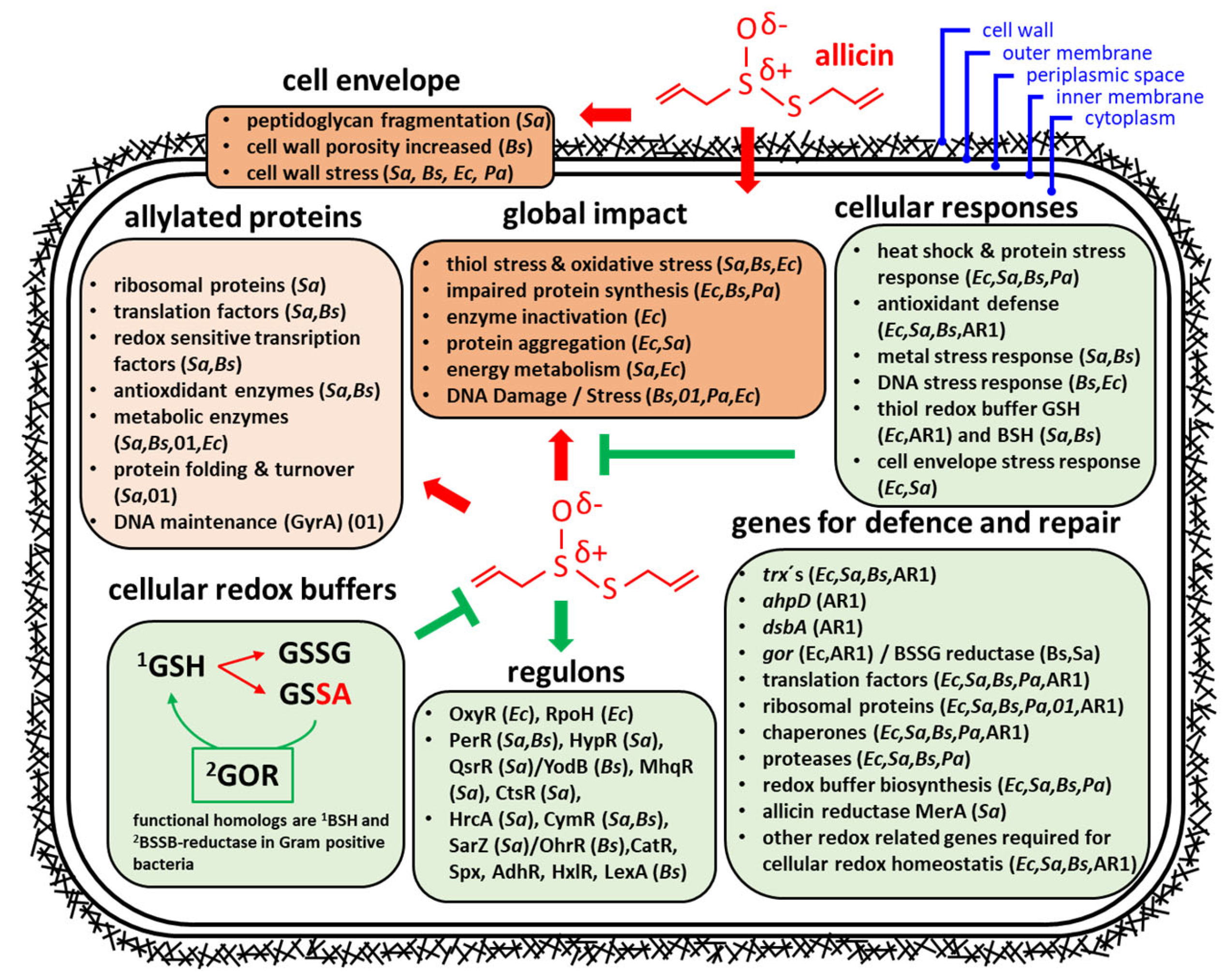
3.1. Effects of Allicin on the Transcriptome and Proteome in Bacteria

Apart from LMW thiols like GSH or BSH, the cellular thiol pool also contains proteins with thiol groups as targets for allicin. Using label-free MS/MS proteomic studies or the isotope-encoded affinity tag (ICAT) based OxICAT procedure, it was demonstrated that a broad spectrum of cellular proteins in E. coli, Pseudomonas, B. subtilis, and S. aureus was indeed S-thioallylated within minutes after sublethal allicin treatment [31,59,60]. The S-thioallyl proteomes of the Gram-positive bacteria B. subtilis and S. aureus were characterized by shotgun linear trap quadrupol (LTQ)-Orbitrap mass spectrometry of the tryptic peptides after treating the cells with allicin to identify peptides with a mass shift of 72 Da [59,60]. In B. subtilis, 79 proteins were identified after 30 min of allicin treatment and major targets were translation elongation factors, surfactin synthetase, antioxidant enzymes, redox-sensitive transcription factors, and biosynthetic enzymes for amino acids and nucleotides [60]. In S. aureus, 57 redox-sensitive proteins were found S-thioallylated, and the allylated proteins were functionally related to those allylated in B. subtilis [59,60]. In summary, detailed analysis of the targets revealed that allicin modified conserved redox-sensitive Cys residues in abundant cellular proteins as well as in lower abundant transcription factors in the proteomes of S. aureus and B. subtilis [59,60]. In this regard it is important to note that diallyl tetrasulfane reacted similarly with thiols and S-thioallylated a similar spectrum of proteins in B. subtilis to allicin [60].
The S-thioallylated allicin proteome has also been characterized in Gram-negative E. coli cells. Here again highly abundant cellular proteins were targeted, predominantly enzymes of primary metabolism, elongation factors, and ribosomal proteins [31]. Thus, interestingly, it seems that not only the accessibility/redox-sensitivity of a cysteine thiol makes it a preferred target for S-thioallylation but also the protein abundance within the cell. This likely reflects simple concentration-dependent competition between the reactants in a complex mixture and reinforces why GSH, which can be present at up to 10 mM intracellular concentration, is very effective at titrating out allicin and protecting cellular proteins against its action.
Upregulated transcripts and proteins, newly synthesized upon allicin treatment, reveal cellular responses to deal with allicin as well as indicating the damage it causes. Thus, a shared response to allicin in E. coli, B. subtilis, S. aureus, P. aeruginosa, and P. fluorescens Pf0-1 is the upregulation of transcripts and synthesis of proteins to alleviate the consequences of protein damage. For example, allicin (>1 mM, 15 min at 30 °C) caused aggregation of E. coli proteins in cell lysate. Assuming this effect also occurs in intact cells, it could be addressed by the observed allicin-induction of chaperones and proteases which are part of the heat shock response. Likewise, the CtsR and HrcA controlled regulons encoding Clp proteases and DnaK/GroESL chaperones were highly induced in the transcriptomes of B. subtilis and S. aureus [59,60]. Similarly, oxidative unfolding of proteins by allicin was countered by the induction of amino acid biosynthesis genes, ribosomal proteins, and translation factors in several bacteria tested [31,59,60,78,79]. It was shown that the ability to induce the heat shock response in E. coli was essential to overcome the effects of allicin stress. This was demonstrated with an rpoH mutant which was not able to induce the heat shock response, and unlike the wild type was unable to recover from allicin treatment [31]. Additionally, allicin also induced oxidative stress response genes controlled by the OxyR and PerR regulons, e.g., katA, ahpCF, and thioredoxins in B. subtilis, E. coli, and S. aureus [31,59,60,78].
An interesting effect of allicin highlighted by the data on upregulated transcripts or proteins, respectively, is damage of the cellular envelope. For example, in S. aureus, it was observed that allicin led to peptidoglycan fragmentation [80] and to the induction of the GraS cell wall stress regulon [60]. In E. coli, the outer membrane protein A (OmpA) and lipopolysaccharide protein P (Lpp), which are structural components in the cell envelope, were both induced after allicin treatment [31]. In B. subtilis, glucosamine-6-phosphate deaminase 2 (nagBB/gamA) and the oxidoreductase dltE, both responsible for cell wall restructuring via peptidoglycan degradation and teichoic acid modification, were also induced [78]. In P. aeruginosa alanine-tRNA can alter permeability in response to oxidative stress and membrane damage by aminoacylation of the phosphatidyl headgroups in the membrane. Thus, newly synthesized alanine-tRNA ligase might indicate involvement of cell envelope stress [78]. In addition to transcriptomic and proteomic studies, plasma membrane bubbles in the form of extrusions at the cell wall in B. subtilis were observed under the microscope after allicin treatment. This suggested that the plasma membrane might have extruded through holes in the cell wall created by allicin [78].
In addition to common responses to allicin, specific bacteria also showed unique responses. For example, although some aspects of the response of P. aeruginosa, e.g., the synthesis of various chaperones, matched the responses of other bacteria, Wüllner et al. [78] showed that P. aeruginosa does not synthesize additional proteins for increased antioxidants or other redox-related proteins upon sublethal allicin treatment. This might reflect the lesser effectiveness of allicin against P. aeruginosa PA01, which is seen in the much higher minimal inhibitory concentration (MIC ≥ 512 µg/mL allicin) compared to E. coli K12 (128 µg/mL allicin) and B. subtilis 168 (64 µg/mL allicin) [31,78]. Another so far unique aspect of allicin stress resistance was discovered in S. aureus. When Loi et al. investigated the allicin-induced changes in the transcriptome of S. aureus, they observed that the genes of the hypR-merA operon were strongly induced (195- to 228-fold change) [59]. It was already known that the transcription factor HypR was oxidized under strong oxidative and disulfide stress conditions, e.g., by hypochlorite or diamide stress, leading to de-repression of the merA gene encoding a flavin disulfide reductase. The NADPH-dependent MerA enzyme enabled increased survival of S. aureus during macrophage infection and hypochlorite stress [59], presumably by protecting S. aureus against the oxidative burst from activated macrophages [81]. The possible role of MerA in allicin protection was investigated in phenotype assays with merA mutants, which showed a decreased survival during allicin stress. In addition, purified MerA protein consumed NADPH when mixed with allicin, indicating that MerA was able to use allicin as a substrate [59]. Despite this specific resistance mechanism, allicin remains a potent, dose-dependent natural antimicrobial substance that is quite effective against methicillin resistant S. aureus (MRSA) [82].
Since allicin targets such a broad range of different molecules simultaneously within cells, spontaneous mutation to resistance is rarely observed in the laboratory [19]. We reasoned that we would most likely be able to find high allicin tolerance in nature in microorganisms living in close association with the garlic plant. We isolated and investigated a P. fluorescens isolate from garlic which showed an exceptionally high level of allicin resistance compared to other bacteria. We named this isolate P. fluorescens Allicin Resistant-1 (PfAR-1) [77]. A PfAR-1 genomic library was prepared and shotgun transferred into an allicin-sensitive P. syringae strain. Genomic clones which conferred resistance were found in a gain-of-function screen. Surprisingly, these clones were also able to confer allicin resistance to phylogenetically distant E. coli cells, suggesting a universal role in allicin protection for the respective genes. Genomic sequencing revealed that each genomic clone represented a small part (in average 10 kbp) of larger genomic regions present in three copies in the PfAR-1 genome. GC content and codon usage indicated that the genomic regions were genomic islands (GIs) that had been acquired by horizontal gene transfer. Most of the genes which the clones had in common (disulfide bond protein A (dsbA), thioredoxin (trx), potassium/proton antiporter KefC (kefC), old yellow enzyme (oye), and alkyhydroperoxide D (ahpD)) were shown by transposon mutagenesis to be needed for allicin resistance because mutants showed increased sensitivity. However, over-expression of each gene separately showed that ahpD and dsbA were the only single genes able to confer allicin resistance to the allicin-susceptible isolates P. syringae 4612 or P. syringae 1448A. This suggests that the other genes work incrementally together to enhance tolerance to the effects of allicin. The resistance-conferring clones made up only a part of the total length of the GIs. The congruent sets of genes from the GIs outside of the resistance-conferring clones were highly sequence related and largely showed redox related functions. For example, a copy of glutathione reductase (gor) was present in two of the three repeats. This adds up to three copies of the gor gene in the PfAR-1 genome. As mentioned above, the unusually high gor copy number and enzyme activity was shown to be linked to allicin resistance by genetic complementation studies [77]. In this way, PfAR-1 exhibits multiple mechanisms of resistance to allicin encoded in several genes of varying major or minor incremental effect. This is an example of the evolutionary investment, involving gene duplication and horizontal gene transfer, which has enabled this bacterium to thrive under the strong selection pressure of its ecological niche.
A proteomic study showed that in non-stressed cells of PfAR-1 and Pf0-1, cellular proteins were largely in a reduced state with only 76% and 77% respectively showing up to 20% cysteine oxidation. Comparing the degree of protein oxidation after allicin treatment showed that whereas 65% of cellular proteins remained less than 20% oxidized in PfAR-1, in Pf0-1 this was only 50%, suggesting a better global protection machinery for proteins against oxidation by allicin in PfAR-1 than in Pf0-1 [79]. This enhanced protective background is due to the function of the genes on the genomic islands gained by horizontal gene transfer by PfAR-1 that are absent from Pf0-1.
One of the differentially S-thioallylated proteins in Pf0-1 but not in PfAR-1 was DNA gyrase subunit A (GyrA). DNA gyrase is an essential bacterial enzyme and for this reason it is a target for de novo antibiotic development [79]. In unstressed cells the degree of Cys433 oxidation in GyrA was approximately 6% in both PfAR-1 and Pf0-1. After allicin treatment the degree of Cys433 oxidation increased to approximately 56% in Pf0-1 compared to only 11% in PfAR-1. Since DNA gyrase activity is essential for DNA replication it is likely that its protection from allicin oxidation is an important component of PfAR-1 resistance. It was possible that either the PfAR-1 GyrA was intrinsically less oxidizable by allicin, or that the PfAR-1 genetic background protected it from S-thioallylation more effectively than the Pf0-1 background. Both PfAR-1 and Pf0-1 GyrA amino acid sequences were highly conserved but the PfAR-1 GyrA showed an additional internal stretch of 40 amino acids compared to Pf0-1 GyrA. It was speculated that this extra domain might alter the protein conformation and protect Cys433 from allicin access, which could have been responsible for an intrinsic allicin resistance of this enzyme. However, by reciprocal genomic replacement of the gyrA subunits between PfAR-1 and Pf0-1, it was found that the allicin susceptibility of Pf0-1 cells with the PfAR-1 gyrA gene replacement was not decreased but actually increased slightly (but not significantly), while PfAR-1 cells with the Pf0-1 gyrA gene replacement remained allicin resistant. Because GyrA is essential for DNA replication in bacteria, this means that the Pf0-1 GyrA protein must have been protected from S-thioallylation in PfAR-1 cells. Furthermore, PfAR-1 GyrA enzyme activity was inhibited by allicin in vitro to a greater extent than Pf0-1 GyrA activity. In conclusion, this study showed that the PfAR‑1 genetic background was most likely responsible for the protection of GyrA against S-thioallylation by allicin, rather than an intrinsic difference in the two GyrA sequences [79].
Significantly, the allicin-resistance-conferring genomic clones of PfAR-1 did not confer general oxidative stress resistance. Thus, protection was not enhanced against either cumene hydroperoxide or H2O2. This indicates that the resistance mechanisms conferred by the genomic clones addressed specifically the types of stress caused by allicin, rather than ROS (reactive oxygen species) in general. This agrees with the hypothesis that the cost to the organism of maintaining multiple copies of the genomic islands in PfAR-1 is due to selection pressure enabling it to exploit its ecological niche on garlic. Normally bacteria do not support large genomic repeats because of the danger of intragenomic homologous recombination leading to loss of intervening essential genetic material. DNA syntenic to the PfAR-1 genomic repeats was also present in the genomes of other plant-associated bacteria—for example P. syringae pv. tomato DC3000 (one syntenic region) and P. salomonii ICMP14252 (two syntenic regions). Both species had elevated allicin resistance compared to related strains without the syntenic region [77]. Since P. salomonii is a garlic pathogen [83], one can conclude that the genes are needed to help it colonize its biological niche, while the presence of such a region in Pst. DC3000, that is pathogenic to plants from the Brassica family [84], remains a curiosity up to this point. The bacterium of origin for the horizontal gene transfer has not be identified so far. A plasmid-borne syntenic region was reported, however, in the onion pathogen Pantoea ananatis. This plasmid variant was shown to confer resistance against allicin, and it was necessary for efficient colonization of the onion host, even though onions do not produce allicin at all [21] and only relatively small amounts of other thiosulfinates [85,86].
It is interesting to note that there is some functional overlap between the genes that are induced in response to allicin stress in E. coli, B. subtilis, and S. aureus and the core set of genes from the allicin resistance-conferring regions in PfAR-1, as well as genes not present on the genomic clones but in the remaining parts of the genomic islands. For example, members of the thioredoxin family as well as proteins belonging to the chaperone class can be found among the upregulated genes as well as in the genomic repeats. However, the grouping of triplicated, horizontally transferred genes in PfAR-1 is quite distinctive, underlining the evolutionary matchmaking of the genome to the ecological niche of the garlic plant.
An overview of the genes relevant to allicin resistance found in studies with different bacteria is given in Figure 4 and Table S1.

3.2. Effects of Allicin on the Proteome of Human Cells (Jurkat T Lymphocytes)

The Jurkat cell line was established from the blood of a patient with T cell leukemia in the late 1970s and the cells are widely used to study T-cell signaling and the effects of anti-cancer treatments [89]. Within 10 min of exposure to 100 µM allicin, 332 proteins were shown to have been S-thioallylated, including several which are recognized targets for cancer therapy [90]. This included 8 out of the 10 glycolytic enzymes, including enolase, and several cytoskeletal proteins. Again, as with bacterial cells, many of the target proteins were highly abundant cellular proteins, for example the cytoskeletal proteins tubulin, actin, cofilin, filamin, and the actin-binding protein plastin-2, as well as heat shock chaperones (Figure 5) [90]. This again emphasizes the somewhat indiscriminate nature of allicin’s action in targeting accessible, oxidizable thiols per se in cells and the law of mass action meaning that abundant molecular species have a competitive advantage over quantitatively minor species. The 100 µM allicin concentration used in the above study was ‘biocompatible’, that is to say the treated cells showed no reduction in metabolic activity over a 24 h period compared to untreated controls. Nevertheless, after 10 min exposure to 100 µM allicin the cells showed an approximately 50% decrease in free thiols. This early time point was chosen to identify primary allicin targets rather than later protein modifications due to cell adaptation to allicin stress [90]. The efficacy of allicin as a thiol trapping reagent is shown clearly in these experiments, highlighting the potential for allicin to influence cell physiology at sublethal, biocompatible doses. In this context see Section 4 for a consideration of the bioavailability of allicin in the body. Furthermore, it is important to note that different cell lines have been shown to respond in certain ways in common, but in other ways differently to allicin [91].

4. Allicin in Medicine

There is a plethora of information in the literature, based mainly on studies with cell cultures, attesting to the potential positive effects of allicin, not only in relation to several diseases, but also on the immune system. Thus, allicin at sublethal doses was reported to reduce NO production in macrophages by inhibiting inducible NO-synthase (iNOS), and furthermore, by increasing the cytosolic [Zn2+] to stimulate the immune response in general [90,92]. Furthermore, there is a wealth of ancient and modern anecdotal material regarding the beneficial health effects of garlic consumption. However, evidence-based scientific studies, especially those including rigorous double blind, statistically adequate, independently funded, clinical studies are generally lacking. Nowhere has the conundrum of the promise that allicin offers and the difficulties associated with its use, been better reviewed than in Chapter 5 of the excellent book ‘Garlic and Other Alliums’, and the reader is referred to this text for a detailed treatment of numerous published studies [21]. In the opinion of the present authors, the potential for allicin, and for that matter some of the other sulfur-containing compounds found in Alliums, to be developed for therapeutic uses is real. A major problem that needs to be solved in order to move to the next stage for that potential to be realized is to develop a delivery system in the body that can achieve a therapeutically relevant concentration in the target tissues without damaging non-target cells. Much more fundamental research is necessary before allicin might be used safely in treating diseases.
In essence two fundamentally different therapeutic situations exist. One would be to use allicin’s antimicrobial activity against infection and the other relates to allicin’s physiological effects on body cells in combatting non-infectious conditions, such as coronary disease and cancer.
Before looking in detail at specific examples, it is useful to consider the fate of allicin in the body, a topic that is relevant to its bioavailability. Cooking garlic quickly destroys any allicin. On heating, allicin spontaneously decomposes to 2-propenesulfenic acid and 2-propenethial (thioacrolein) which react further to produce alkyl disulfides, polysulfanes, vinyl dithiins, and ajoene [21,22]. Many of these compounds are physiologically active in their own right, but when considering garlic in the diet as a source of allicin, only raw garlic counts. Under the acidic conditions in the stomach allicin hydrolyzes rapidly to 2-propenethiol (PT), which is metabolized to allyl methyl sulfide (AMS). Both PT and AMS are excreted in the urine and are recognizable in the unpleasant “garlic breath”, which often makes consumers of garlic unpopular amongst those who have not similarly participated [93,94,95,96]. These reactions reduce the amount of allicin available to react with cellular thiols. Nevertheless, organisms are exposed to allicin in varying amounts in different situations, not least via the pulmonary route, and any allicin entering cells can have profound effects on cellular metabolism.
Allicin taken up by cells, or entering the bloodstream, reacts readily with GSH in competition with cysteine residues in proteins. For this reason, it is difficult to envisage being able to achieve a therapeutically relevant concentration of allicin in cells anywhere in the body by simply swallowing it. Furthermore, as mentioned in the introduction, allicin is an irritant which stimulates pain-sensing neurons, and self-medication has led to a spate of reports of self-inflicted harm [21]. Thus, while small amounts are prized in culinary contexts, over-exposure to allicin is clearly harmful. Having issued that precautionary note, it is also true that there is a lack of volatile antibiotics available for use in medicine, and this possibility for allicin deserves further exploration [88,97,98,99]. Perhaps, after further carefully controlled research and all necessary precautions, a pulmonary exposure route for allicin vapor might be able to be developed under appropriate circumstances to achieve therapeutically effective concentrations in the lungs. A simple experiment to demonstrate the effectivity of allicin vapor against microorganisms is shown in Figure 6A,B. The example with E. coli shows that allicin vapor does not just exert bacteriostatic effects to prevent growth, but can also actively kill cells, i.e., there is a clear bactericidal effect of the vapor. The lawn of E. coli on the left Petri plate was exposed to allicin vapor and then replica-plated using a sterile velvet pad to pick up inoculum from the existing colonies and transfer it to a new plate. The pattern of cells on the original plate was replicated onto the second plate—shown on the right in Figure 6B. It can be seen that no living E. coli cells were transferred from the central region of the original plate where the cells were killed by the allicin vapor, and were therefore no longer able to grow even in the absence of allicin.

4.1. Allicin Activity against Respiratory Tract Pathogens

Because allicin vapor is bactericidal, the idea to use it to combat lung infections is attractive. Indeed, there is a precedent for this in a historical report from the pre-antibiotic-era [97]. Several patients with pulmonary tuberculosis were reported to have been successfully treated by inhaling the vapor from garlic preparations. Patients were fitted with a face mask with a pouch containing juice from pulverized garlic mixed with ethanol and eucalyptus oil to help to mask the smell. They breathed in the fumes for two one-hour periods daily, and a good success rate against pulmonary TB was reported [97]. Interestingly, more recent experiments have suggested that the allicin and ethanol vapors work together synergistically to kill microbes, which was probably unknown to Dr. Minchin at the time [98].
Antibiotics are usually taken orally or injected as solutions, whereas gas-phase antibiotics administered via the pulmonary route are not generally known. A recent survey showed that several lung-pathogenic bacteria, including MDR strains, could be inhibited in vitro by allicin vapor and the aerodynamic behavior of allicin aerosols and vapor has been investigated in a lung model, thus allowing some preliminary determination of dosage and synergy without the need for animal experiments [88,98,99]. Nevertheless, progression to the next stage will require carefully controlled, ethically sound animal experiments before therapeutic protocols for humans can become feasible.
An example showing that allicin can inhibit the growth of the opportunistic lung-pathogenic bacterium Haemophilus influenzae in a straightforward agar plate diffusion assay, and importantly via the gas phase in the inverted Petri plate assay, is illustrated in Figure 6C,D. The size of the inhibition zones shows a clear dose-dependent relationship to allicin exposure. To our knowledge allicin has not previously been reported to be active against H. influenzae, which is recalcitrant to amoxicillin and ampicillin and resistance to β-lactam antibiotics specifically, and is rising strongly in Nontypeable H. influenzae (NTHi) isolates.
Currently, the world is in the grip of a COVID-19 pandemic caused by the SARS-CoV-2 virus. Cysteine-containing proteins of this respiratory tract-infecting virus may be a direct target for S-thioallylation, either in the extracellular free virus particle or during replication in host cells. At present this remains unclear, however, there is a report using in silico analysis that the SARS-CoV-2 main protease may be a target for S-thioallylation [100]. Aside from this in silico speculation, experimental data suggest that garlic may be able to have a beneficial effect on helping to prevent severe COVID-19 via stimulation of the immune system by increasing intracellular Zn2+ levels [90]. Thus, it was shown that at biocompatible doses, allicin stimulated the interleukin-1 (IL-1) dependent release of IL-2 by mouse EL-4 T-cells. This effect is zinc dependent and allicin increases [Zn2+]cyt by causing intracellular Zn2+ release from proteins by oxidizing the Cys residues which chelate it, e.g., from SOD-1, shown to be a major target for S-thioallylation in human Jurkat cells [90]. The importance of Zn2+ for the immune system in relation to preventing severe COVID-19 has recently been reviewed [101].

4.2. Allicin vs. Cancer

It has been reported many times that allicin has pro-apoptotic and antitumorigenic properties against several cancer types, mostly in studies using cultured cells [41,91,102,103,104,105,106,107,108,109,110]. However, osteosarcoma and cholangiosarcoma in mice have for example been reported to respond to intraperitoneal injection of allicin in vivo [103,108].
As mentioned above, allicin has multiple cellular targets in human cells, including for example cytoskeletal proteins and glycolytic enzymes that are also candidates for cancer treatments [90]. Several studies have shown that signaling pathways important in tumor cell lines can be interfered with by allicin treatment [41,102,103,104,105,106]. Furthermore, synergism between allicin and some anticancer drugs, e.g., cyclophosphamidum, artesunate, and all-trans retinoic acid (ATRA) has been demonstrated [107,108,109]. It is also important to note that high GSH levels are associated with many tumor types. High GSH protects the tumor cells against oxidative stress and interferes with the activity of some therapeutic agents [111,112]. In this regard, allicin’s activity as a thiol trapping reagent that readily reacts with and depletes the GSH pool may be very helpful in combating tumor cells. Indeed, strategies to lower GSH levels have been combined with tumor therapies, for example the GSH synthesis inhibitor buthionine sulfoximine (BSO) was used in conjunction with the electrophile melphalan to treat neuroblastoma [113,114]. Another example is the use of BSO with azathioprine to treat human hepatocarcinoma and colon carcinoma [115]. Thus, the broad-ranging effects of allicin in cells could be advantageous rather than disadvantageous when it comes to cancer treatments. In addition to the documented effects of allicin on the cytoskeleton which directly affect cell division, and glycolytic enzymes relevant to the Warburg effect, a recently published study showed that allicin was 23,000 times more effective at inhibiting the enzyme ornithine decarboxylase (ODC) than was α-difluoromethylornithine (DFMO) [110]. ODC is especially important for neuroblastoma cell proliferation and DFMO is being tested in phase I/II human neuroblastoma trials with encouraging results. If a specific delivery system can be found, the potential advantages of allicin for treating neuroblastoma are obvious, as it not only targets ODC but concomitantly reduces GSH levels.
In this regard, the major problem of targeting allicin to tumor tissues (while leaving surrounding healthy cells unstressed) urgently needs to be overcome, and there is a need in the future for carefully controlled studies in animal models and clinical trials involving humans [21].

Supplementary Materials

The following are available online. Table S1. Overview of the characteristics of prokaryotic transcriptional regulators whose activities were shown to be influenced by allicin treatment.

Author Contributions

J.F., U.K., U.N. provided unpublished results, J.B., M.C.H.G. and A.J.S. prepared the Schemes and Figures. J.B., M.C.H.G., H.A. and A.J.S. wrote and edited the manuscript. All authors have read and agreed to the published version of the manuscript.

Funding

Financial support from the RWTH Aachen University (JB, JR, MCHG, AJS) is gratefully acknowledged. JF née Reiter was supported by an RFwN Ph.D. stipendium. K. Ueda, Nara Institute of Science and Technology, Japan is thanked for providing a GFP-tubulin construct. This work was further supported by an ERC Consolidator Grant (GA 615585) MYCOTHIOLOME and by grants from the Deutsche Forschungsgemeinschaft (SPP1710 project AN746/4-2) to HA. This work was supported by National Health and Medical Research Council grant GNT1158451 to UK.

Institutional Review Board Statement

Not applicable.

Informed Consent Statement

Not applicable.

Data Availability Statement

Not applicable.

Conflicts of Interest

The authors declare no conflict of interest.

References

  1. Cavallito, C.J.; Bailey, H.J. Allicin, the antibacterial principle of Allium sativum I. Isolation, physical properties and antibacterial action. J. Am. Chem. Soc. 1944, 66, 1950–1951. [Google Scholar] [CrossRef]
  2. Cavallito, C.J.; Buck, J.S.; Suter, C.M. Allicin, the antibacterial principle of Allium sativum II. Determination of the chemical structure. J. Am. Chem. Soc. 1944, 66, 1952–1954. [Google Scholar] [CrossRef]
  3. Lawson, L.D.; Wood, S.G.; Hughes, B.G. HPLC analysis of allicin and other thiosulfinates in garlic clove homogenates. Planta Med. 1991, 57, 263–270. [Google Scholar] [CrossRef] [PubMed]
  4. Lancaster, J.E.; Collin, H.A. Presence of alliinase in isolated vacuoles and of alkyl cysteine sulphoxides in the cytoplasm of bulbs of onion (Allium cepa). Plant Sci. Lett. 1981, 22, 169–176. [Google Scholar] [CrossRef]
  5. Block, E.; Naganathan, S.; Putman, D.; Zhao, S.H. Allium chemistry: HPLC Analysis of Thiosulfinates from Onion, Garlic, Wild Garlic (Ramsons), Leek, Scallion, Shallot, Elephant (Great-Headed) Garlic, Chive, and Chinese Chive. Uniquely High Allyl to Methyl Ratios in Some Garlic Samples. J. Agric. Food Chem. 1992, 40, 2418–2430. [Google Scholar] [CrossRef]
  6. Yamazaki, M.; Sugiyama, M.; Saito, K. Intercellular Localization of Cysteine Synthase and Alliinase in Bundle Sheaths of Allium Plants. Plant Biotechnol. 2002, 19, 7–10. [Google Scholar] [CrossRef] [Green Version]
  7. Block, E. Fifty years of smelling sulfur. J. Sulfur Chem. 2013, 34, 158–207. [Google Scholar] [CrossRef]
  8. Lawson, L.D.; Hughes, B.G. Characterization of the formation of allicin and other thiosulfinates from garlic. Planta Med. 1992, 58, 345–350. [Google Scholar] [CrossRef]
  9. Albrecht, F.; Leontiev, R.; Jacob, C.; Slusarenko, A.J. An Optimized Facile Procedure to synthesize and purify Allicin. Molecules 2017, 22, 770. [Google Scholar] [CrossRef] [Green Version]
  10. Fowler, F.G.; Fowler, H.W. The Concise Oxford Dictionary of Current English, 5th ed.; Oxford Clarendon Press: Oxford, UK, 1964. [Google Scholar]
  11. Block, E.; Batista, V.S.; Matsunami, H.; Zhuang, H.; Ahmed, L. The role of metals in mammalian olfaction of low molecular weight organosulfur compounds. Nat. Prod. Rep. 2017, 34, 529–597. [Google Scholar] [CrossRef] [Green Version]
  12. Hile, A.G.; Shan, Z.; Zhang, S.Z.; Block, E. Aversion of European Starlings (Sturnus vulgaris) to Garlic Oil Treated Granules: Garlic Oil as an Avian Repellent. Garlic Oil Analysis by Nuclear Magnetic Resonance Spectroscopy. Agric. Food Chem. 2004, 52, 2192–2196. [Google Scholar] [CrossRef] [PubMed]
  13. Bautista, D.M.; Movahed, P.; Hinman, A.; Axelsson, H.E.; Sterner, O.; Högestätt, E.D.; Julius, D.; Jordt, S.-E.; Zygmunt, P.M. Pungent products from garlic activate the sensory ion channel TRPA1. Proc. Natl. Acad. Sci. USA 2005, 102, 12248–12252. [Google Scholar] [CrossRef] [Green Version]
  14. Macpherson, L.J.; Geierstanger, B.H.; Viswanath, V.; Bandell, M.; Eid, S.R.; Hwang, S.-W.; Patapoutian, A. The Pungency of Garlic: Activation of TRPA1 and TRPV1 in Response to Allicin. Curr. Biol. 2005, 15, 929–934. [Google Scholar] [CrossRef] [Green Version]
  15. Paulsen, C.; Armache, J.; Gao, Y.; Cheng, Y.; Julius, D. Structure of the TRPA1 ion channel suggests regulatory mechanisms. Nature 2015, 520, 511–517. [Google Scholar] [CrossRef] [Green Version]
  16. Mihara, S.; Shibamoto, T. The role of flavor and fragrance chemicals in TRPA1 (transient receptor potential cation channel, member A1) activity associated with allergies. All. Asth. Clin. Immun. 2015, 11, 11. [Google Scholar] [CrossRef] [PubMed] [Green Version]
  17. Wang, Y.; Cao, L.; Lee, C.Y.; Matsuo, T.; Wu, K.; Asher, G.; Tang, L.; Saitoh, T.; Russell, J.; Klewe-Nebenius, D.; et al. Large-scale forward genetics screening identifies Trpa1 as a chemosensor for predator odor-evoked innate fear behaviors. Nat. Commun. 2018, 9, 2041. [Google Scholar] [CrossRef] [Green Version]
  18. Ninkovic, V.; Rensing, M.; Dahlin, I.; Markovic, D. Who is my neighbor? Volatile cues in plant interactions. Plant. Signaling Behav. 2019, 14, 1634993. [Google Scholar] [CrossRef] [PubMed]
  19. Borlinghaus, J.; Albrecht, F.; Gruhlke, M.C.H.; Nwachukwu, I.D.; Slusarenko, A.J. Allicin: Chemistry and biological properties. Molecules 2014, 19, 12591–12618. [Google Scholar] [CrossRef] [Green Version]
  20. Kong, C.H.; Xuan, T.D.; Khanh, T.D.; Tran, H.D.; Trung, N.T. Allelochemicals and Signaling Chemicals in Plants. Molecules 2019, 24, 2737. [Google Scholar] [CrossRef] [Green Version]
  21. Block, E. Garlic and the other Alliums. The Lore and the Science, 1st ed.; RSC Publishing: Cambridge, UK, 2010. [Google Scholar]
  22. Anwar, A.; Groom, M.; Arbach, M.; Hamilton, C.J. How to Turn the Chemistry of Garlic into a ‘Botanical’ Pesticide. In Recent Advances in Redox Active Plant and Microbial Products: From Basic Chemistry to Widespread Applications in Medicine and Agriculture; Jacob, C., Slusarenko, A.J., Kirsch, G., Winyard, P., Eds.; Springer: Berlin, Germany, 2014; pp. 323–341. [Google Scholar]
  23. Miron, T.; Rabinkov, A.; Mirelman, D.; Wilchek, M.; Weiner, L. The mode of action of allicin: Its ready permeability through phospholipid membranes may contribute to its biological activity. Biochim. Biophys. Acta BBA Biomembranes 2000, 1463, 20–30. [Google Scholar] [CrossRef] [Green Version]
  24. Gruhlke, M.C.H.; Hemmis, B.; Noll, U.; Wagner, R.; Lühring, H.; Slusarenko, A.J. The defense substance allicin from garlic permeabilizes membranes of Beta vulgaris, Rhoeo discolor, Chara corallina and artificial lipid bilayers. Biochim. Biophys. Acta BBA 2015, 1850, 602–611. [Google Scholar] [CrossRef] [PubMed]
  25. Gurtovenko, A.A.; Anwar, J. Modulating the Structure and Properties of Cell Membranes: The Molecular Mechanism of Action of Dimethyl Sulfoxide. J. Phys. Chem. B. 2007, 111, 10453–10460. [Google Scholar] [CrossRef] [Green Version]
  26. Rabinkov, A.; Miron, T.; Konstantinovski, L.; Wilchek, M.; Mirelman, D.; Weiner, L. The mode of action of allicin: Trapping of radicals and interaction with thiol containing proteins. Biochim. Biophys. Acta BBA 1998, 1379, 233–234. [Google Scholar] [CrossRef]
  27. Rabinkov, A.; Miron, T.; Mirelman, D.; Wilchek, M.; Glozman, S.; Yavin, E.; Weiner, L. S-allylmercaptoglutathione: The reaction product of allicin with glutathione possesses SH-modifying and antioxidant properties. Biochim. Biophys. Acta BBA Mol. Cell Res. 2000, 1499, 144–153. [Google Scholar] [CrossRef] [Green Version]
  28. Giles, G.I.; Jacob, C. Reactive Sulfur Species: An Emerging Concept in Oxidative Stress. Biol. Chem. 2002, 383, 375–388. [Google Scholar] [CrossRef]
  29. Giles, G.I.; Tasker, K.M.; Collins, C.; Giles, N.M.; O’Rourke, E.; Jacob, C. Reactive sulphur species: An in vitro investigation of the oxidation properties of disulphide S-oxides. Biochem. J. 2001, 364, 579–585. [Google Scholar] [CrossRef] [PubMed] [Green Version]
  30. Gruhlke, M.C.H.; Slusarenko, A.J. The biology of reactive sulfur species (RSS). Plant. Physiol. Biochem. 2012, 59, 98–107. [Google Scholar] [CrossRef]
  31. Müller, A.; Eller, J.; Albrecht, F.; Prochnow, P.; Kuhlmann, K.; Bandow, J.E.; Slusarenko, A.J.; Leichert, L.I. Allicin Induces Thiol Stress in Bacteria through S-Allylmercapto Modification of Protein Cysteines. J. Biol. Chem. 2016, 291, 11477–11490. [Google Scholar] [CrossRef] [Green Version]
  32. Schafer, F.Q.; Buettner, G.R. Redox environment of the cell as viewed through the redox state of the glutathione disulfide/glutathione couple. Free Radic. Biol. Med. 2001, 30, 1191–1212. [Google Scholar] [CrossRef]
  33. Poole, L.B. The basics of thiols and cysteines in redox biology and chemistry. Free Radic. Biol. Med. 2015, 80, 148–157. [Google Scholar] [CrossRef] [Green Version]
  34. Horn, T.; Bettray, W.; Slusarenko, A.J.; Gruhlke, M.C.H. S-allylmercaptoglutathione Is a Substrate for Glutathione Reductase (E.C. 1.8.1.7) from Yeast (Saccharomyces cerevisiae). Antioxidants 2018, 7, 86. [Google Scholar] [CrossRef] [Green Version]
  35. Ankri, S.; Mirelman, D. Antimicrobial properties of allicin from garlic. Microbes Infect. 1999, 2, 125–129. [Google Scholar] [CrossRef]
  36. Slusarenko, A.J.; Patel, A.; Portz, D. Control of plant diseases by natural products: Allicin from garlic as a case study. Eur. J. Plant. Pathol. 2008, 121, 313–322. [Google Scholar] [CrossRef]
  37. Vaidya, V.; Ingold, K.U.; Pratt, D.A. Garlic: Source of the Ultimate Antioxidants—Sulfenic Acids. Angew. Chem. Int. Ed. 2009, 48, 157–160. [Google Scholar] [CrossRef]
  38. Banerjee, S.K.; Mukherjee, P.K.; Maulik, S.K. Garlic as an antioxidant: The good, the bad and the ugly. Phytother. Res. 2003, 17, 97–106. [Google Scholar] [CrossRef]
  39. Gruhlke, M.C.H.; Slusarenko, A.J. The Cellular “Thiolstat” as an Emerging Target of Some Plant Secondary Metabolites. In Recent Advances in Redox Active Plant and Microbial Products: From Basic Chemistry to Widespread Applications in Medicine and Agriculture; Jacob, C., Slusarenko, A.J., Kirsch, G., Winyard, P., Eds.; Springer: Berlin, Germany, 2014; pp. 235–262. [Google Scholar]
  40. Melino, S.; Sabelli, R.; Paci, M. Allyl sulfur compounds and cellular detoxification system: Effects and perspectives in cancer therapy. Amino Acids 2011, 41, 103–112. [Google Scholar] [CrossRef] [PubMed] [Green Version]
  41. Bat-Chen, W.; Tal, G.; Irena, P.; Zvi, L.; Betty, S. Allicin purified from fresh garlic cloves induces apoptosis in colon cancer cells via Nrf2. Nutr. Cancer 2009, 62, 947–957. [Google Scholar] [CrossRef]
  42. Surh, Y.J.; Kundu, J.K.; Na, H.K. Nrf2 as a Master Redox Switch in Turning on the Cellular Signaling Involved in the Induction of Cytoprotective Genes by Some Chemopreventive Phytochemicals. Planta Med. 2008, 74, 1526–1539. [Google Scholar] [CrossRef] [PubMed] [Green Version]
  43. Winterbourn, C.C.; Hampton, M.B. Thiol chemistry and specificity in redox signaling. Free Radic. Biol. Med. 2008, 45, 549–561. [Google Scholar] [CrossRef]
  44. Morano, K.A.; Grant, C.M.; Moye-Rowley, W.S. The response to heat shock and oxidative stress in Saccharomyces cerevisiae. Genetics 2012, 190, 1157–1195. [Google Scholar] [CrossRef] [Green Version]
  45. Lee, J.; Godon, C.; Lagniel, G.; Spector, D.; Garin, J.; Labarre, J.; Toledano, M.B. Yap1 andSkn7 control two specialized oxidative stress response regulons in yeast. J. Biol. Chem. 1999, 274, 16040–16046. [Google Scholar] [CrossRef] [Green Version]
  46. Carmel-Harel, O.; Storz, G. Roles of the glutathione- and thioredoxin-dependent reduction systems in the Escherichia coli and Saccharomyces cerevisiae responses to oxidative stress. Annu. Rev. Microbiol. 2000, 51, 439–461. [Google Scholar] [CrossRef] [PubMed]
  47. Kuge, S.; Jones, N.; Nomoto, A. Regulation of yAP-1 nuclear localization in response to oxidative stress. EMBO J. 1997, 16, 1710–1720. [Google Scholar] [CrossRef] [PubMed] [Green Version]
  48. Gulshan, K.; Rovinsky, S.A.; Coleman, S.T.; Moye-Rowley, W.S. Oxidant-specific folding of Yap1p regulates both transcriptional activation and nuclear localization. J. Biol. Chem. 2005, 280, 40524–40533. [Google Scholar] [CrossRef] [Green Version]
  49. Gruhlke, M.C.H.; Schlembach, I.; Leontiev, R.; Uebachs, A.; Gollwitzer, P.; Weiss, A.; Delaunay, A.; Toledano, M.B.; Slusarenko, A.J. Yap1p, the central regulator of the S. cerevisiae oxidative stress response, is activated by allicin, a natural oxidant and defence substance of garlic. Free Radic. Biol. Med. 2017, 108, 793–802. [Google Scholar] [CrossRef]
  50. Kansanen, E.; Kuosmanen, S.M.; Leinonen, H.; Levonen, A.L. The Keap1-Nrf2 pathway: Mechanisms of activation and dysregulation in cancer. Redox Biol. 2013, 1, 45–49. [Google Scholar] [CrossRef] [Green Version]
  51. Niture, S.K.; Khatri, R.; Jaiswal, A.K. Regulation ofNrf2—an update. Free Radic. Biol. Med. 2014, 66, 36–44. [Google Scholar] [CrossRef] [Green Version]
  52. Liebler, D.C.; Guengerich, F.P. Elucidating mechanisms of drug-induced toxicity. Nat. Rev. Drug Discov. 2005, 4, 410–420. [Google Scholar] [CrossRef] [PubMed]
  53. Schlembach, I.; Uebachs, A.; Caspers, T.; Fragoulis, A.; Slusarenko, A.J.; Gruhlke, M.C.H. Effect of prooxidative natural products: Comparison of the OSI1 (YKL071w) promoter luciferase construct from yeast with an Nrf2/Keap reporter system. Appl. Sci. 2020, 10, 3520. [Google Scholar] [CrossRef]
  54. Yu, L.; Guo, N.; Meng, R.; Liu, B.; Tang, X.; Jin, J.; Cui, Y.; Deng, X. Allicin-induced global gene expression profile of Saccharomyces cerevisiae. Appl. Microbiol. Biotechnol. 2010, 88, 219–229. [Google Scholar] [CrossRef]
  55. Imlay, J.A. The Molecular Mechanisms and Physiological Consequences of Oxidative Stress: Lessons from a Model Bacterium. Nature Rev. Microbiol. 2013, 11, 443. [Google Scholar] [CrossRef] [Green Version]
  56. Horsburgh, M.J.; Clements, M.O.; Crossley, H.; Ingham, E.; Foster, S.J. PerR Controls Oxidative Stress Resistance and Iron Storage Proteins and Is Required for Virulence in Staphylococcus aureus. Infect. Immun. 2001, 69, 3744. [Google Scholar] [CrossRef] [Green Version]
  57. Ji, C.-J.; Kim, J.-H.; Won, Y.-B.; Lee, Y.-E.; Choi, T.-W.; Ju, S.-Y.; Youn, H.; Helmann, J.D.; Lee, J.-W. Staphylococcus aureus PerR Is a Hypersensitive Hydrogen Peroxide Sensor Using Iron-Mediated Histidine Oxidation. J. Biol. Chem. 2015, 290, 20374–20386. [Google Scholar] [CrossRef] [Green Version]
  58. Zuber, P. Management of Oxidative Stress in Bacillus. Annu. Rev. Microbiol. 2009, 63, 575–597. [Google Scholar] [CrossRef] [PubMed]
  59. Loi, V.V.; Huyen, N.T.T.; Busche, T.; Tung, Q.N.; Gruhlke, M.C.H.; Kalinowski, J.; Bernhardt, J.; Slusarenko, A.J.; Antelmann, H. Staphylococcus aureus responds to allicin by global S-thioallylation - Role of the Brx/BSH/YpdA pathway and the disulfide reductase MerA to overcome allicin stress. Free Radic. Biol. Med. 2019, 139, 55–69. [Google Scholar] [CrossRef] [PubMed]
  60. Chi, B.K.; Huyen, N.T.T.; Loi, V.V.; Gruhlke, M.C.H.; Schaer, M.; Mäder, U.; Maaß, S.; Becher, D.; Bernhardt, J.; Arbach, M.; et al. The Disulfide Stress Response and Protein S-thioallylation Caused by Allicin and Diallyl Polysulfanes in Bacillus subtilis as Revealed by Transcriptomics and Proteomics. Antioxidants 2019, 8, 605. [Google Scholar] [CrossRef] [Green Version]
  61. Noctor, G.; Foyer, C.H. ASCORBATE AND GLUTATHIONE: Keeping Active Oxygen Under Control. Annu. Rev. Plant. Physiol. Plant. Mol. Biol. 1998, 49, 249–279. [Google Scholar] [CrossRef] [PubMed]
  62. Foyer, C.H.; Noctor, G. Redox homeostasis and antioxidant signaling: A metabolic interface between stress perception and physiological responses. Plant. Cell 2005, 17, 1866–1875. [Google Scholar] [CrossRef] [Green Version]
  63. Gruhlke, M.C.H.; Portz, D.; Stitz, M.; Anwar, A.; Schneider, T.; Jacob, C.; Schlaich, N.L.; Slusarenko, A.J. Allicin disrupts the cell’s electrochemical potential and induces apoptosis in yeast. Free Radic. Biol. Med. 2010, 49, 1916–1924. [Google Scholar] [CrossRef] [PubMed]
  64. Toledano, M.B.; Delaunay-Moisan, A.; Outten, C.E.; Igbaria, A. Functions and Cellular Compartmentation of the Thioredoxin and Glutathione Pathways in Yeast. Antioxid. Redox Signal. 2013, 18, 1699–1711. [Google Scholar] [CrossRef] [Green Version]
  65. Gruhlke, M.C. Reactive Sulfur Species—A new player in plant physiology? In Reactive Oxygen, Nitrogen and Sulfur Species in Plants: Production, Metabolism, Signaling; Hasanuzzaman, M., Fotopoulos, V., Nahar, K., Fujita, M., Eds.; John Wiley & Sons Ltd.: Hoboken, NY, USA, 2019; pp. 715–728. [Google Scholar]
  66. Considine, M.J.; Foyer, C.H. Redox regulation of plant development. Antioxid. Redox Signal. 2014, 21, 1305–1326. [Google Scholar] [CrossRef] [Green Version]
  67. Rouhier, N.; Cerveau, D.; Couturier, J.; Reichheld, J.P.; Rey, P. Involvement of thiol-based mechanisms in plant development. Biochim. Biophys Acta 2015, 1850, 1479–1496. [Google Scholar] [CrossRef] [PubMed]
  68. Koprivova, A.; Mugford, S.; Kopriva, S. Arabidopsis root growth dependence on glutathione is linked to auxin transport. Plant. Cell Rep. 2010, 29, 1157–1167. [Google Scholar] [CrossRef]
  69. Vernoux, T.; Wilson, R.C.; Seeley, K.A.; Reichheld, J.P.; Muroy, S.; Brown, S.; Maughan, S.C.; Cobbett, C.S.; van Montagu, M.; Inzé, D.; et al. The ROOT MERISTEMLESS1/CADMIUM SENSITIVE2 gene defines a glutathione-dependent pathway involved in initiation and maintenance of cell division during postembryonic root development. Plant. Cell 2000, 12, 97–110. [Google Scholar] [CrossRef] [Green Version]
  70. Sanchez-Fernandez, R.; Fricker, M.; Corben, L.B.; White, N.S.; Sheard, N.; Leaver, C.J.; van Montagu, M.; Inzé, D.; May, M.J. Cell proliferation and hair tip growth in the Arabidopsis root are under mechanistically different forms of redox control. Proc. Natl. Acad. Sci. USA 1997, 94, 2745–2750. [Google Scholar] [CrossRef] [PubMed] [Green Version]
  71. Bashandy, T.; Guilleminot, J.; Vernoux, T.; Caparros-Ruiz, D.; Ljung, K.; Meyer, Y.; Reichheld, J.P. Interplay between the NADP-Linked Thioredoxin and Glutathione Systems in Arabidopsis Auxin Signaling. Plant. Cell 2010, 22, 376–391. [Google Scholar] [CrossRef] [Green Version]
  72. Leontiev, R.; Hohaus, N.; Jacob, C.; Gruhlke, M.C.H.; Slusarenko, A.J. A Comparison of the Antibacterial and Antifungal Activities of Thiosulfinate Analogues of Allicin. Sci. Rep. 2018, 8, 6763. [Google Scholar] [CrossRef]
  73. Horn, T.; Bettray, W.; Noll, U.; Krauskopf, F.; Huang, M.-R.; Bolm, C.; Slusarenko, A.J.; Gruhlke, M.C.H. The Sulfilimine Analogue of Allicin, S-Allyl-S-(S-allyl)-N-Cyanosulfilimine, Is Antimicrobial and Reacts with Glutathione. Antioxidants 2020, 9, 1086. [Google Scholar] [CrossRef] [PubMed]
  74. Ueda, K.; Matsuyama, T.; Hashimoto, T. Visualization of microtubules in living cells of transgenic Arabidopsis thaliana. Protoplasma 1999, 206, 201–206. [Google Scholar] [CrossRef]
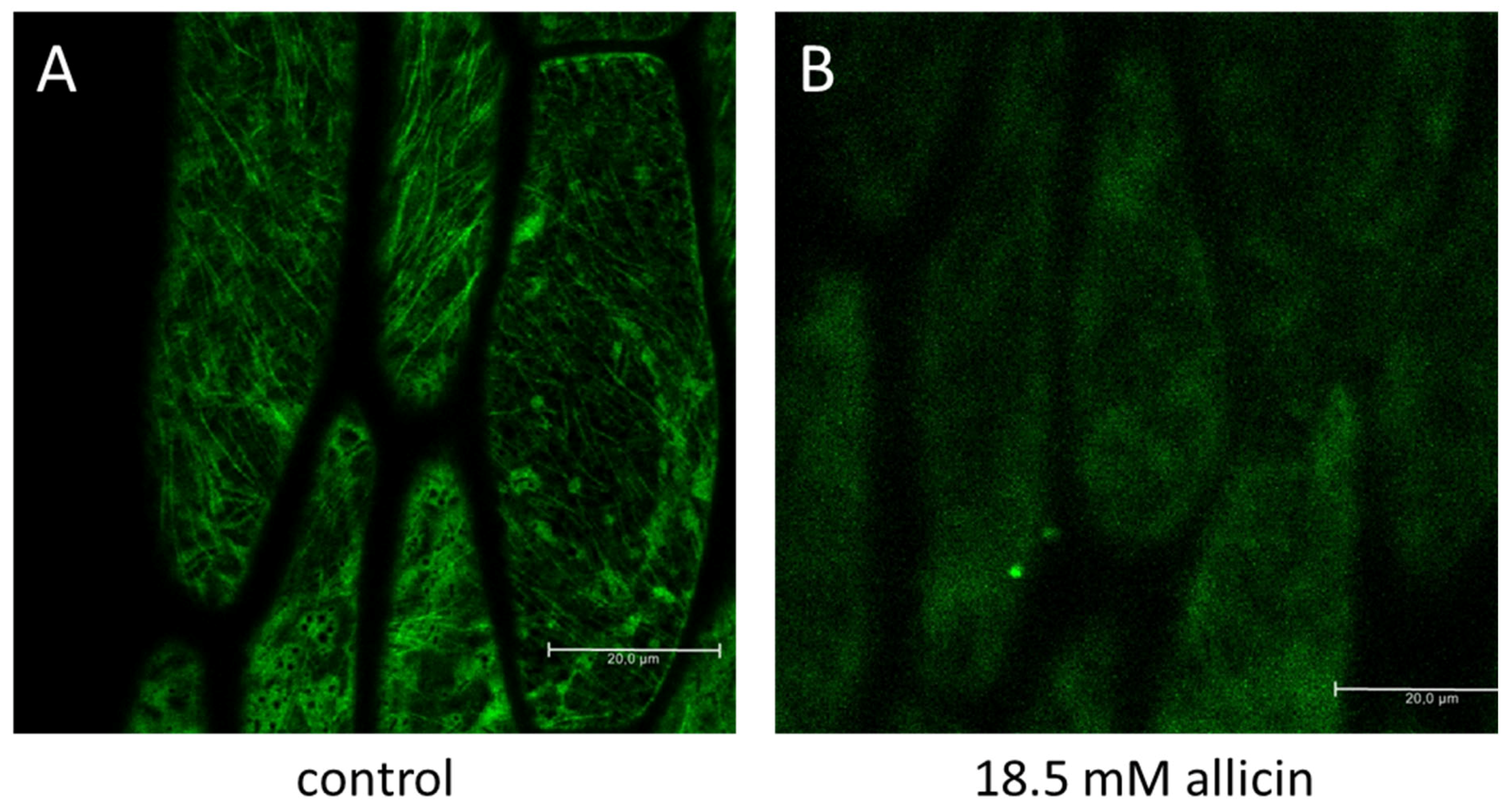
  75. Noll, U.; Schreiber, M.; Hermanns, M.; Mertes, C.A.; Slusarenko, A.J.; Gruhlke, M.C.H. Allicin from Garlic Disrupts the Cytoskeleton in Plants. bioRxiv 2021. [Google Scholar] [CrossRef]
  76. Leontiev, R.; Slusarenko, A.J. Finding the Starting Point for Mode-of-Action Studies of Novel Selenium Compounds: Yeast as a Genetic Toolkit. Curr. Org. Synth. 2017, 14, 1–7. [Google Scholar] [CrossRef]
  77. Borlinghaus, J.; Bolger, A.; Schier, C.; Vogel, A.; Usadel, B.; Gruhlke, M.C.H.; Slusarenko, A.J. Genetic and molecular characterization of multi-component resistance of Pseudomonas against allicin. Life Sci. Alliance 2020, 3, e202000670. [Google Scholar] [CrossRef] [PubMed] [Green Version]
  78. Wüllner, D.; Haupt, A.; Prochnow, P.; Leontiev, R.; Slusarenko, A.J.; Bandow, J.E. Interspecies Comparison of the Bacterial Response to Allicin Reveals Species-Specific Defense Strategies. Proteomics 2019, 19, 1900064. [Google Scholar] [CrossRef] [PubMed] [Green Version]
  79. Reiter, J.; Hübbers, A.M.; Albrecht, F.; Leichert, L.I.O.; Slusarenko, A.J. Allicin, a Natural Antimicrobial Defence Substance from Garlic, Inhibits DNA Gyrase Activity in Bacteria. Int. J. Med. Microbiol. 2020, 310, 151359. [Google Scholar] [CrossRef] [PubMed]
  80. Getti, G.T.M.; Poole, P.L. 2019 Allicin Causes Fragmentation of the Peptidoglycan Coat in Staphylococcus aureus by Effecting Synthesis and Aiding Hydrolysis: A Determination by MALDI-TOF Mass Spectrometry on Whole Cells. J. Med. Microbiol. 2019, 68, 667–677. [Google Scholar] [CrossRef]
  81. Slauch, J.M. How Does the Oxidative Burst of Macrophages Kill Bacteria? Still an Open Question. Mol. Microbiol. 2011, 80, 580–583. [Google Scholar] [CrossRef] [Green Version]
  82. Cutler, R.R.; Wilson, P. Antibacterial Activity of a New, Stable, Aqueous Extract of Allicin against Methicillin-Resistant Staphylococcus aureus. Brit. J. Biomed. Sci. 2004, 61, 71–74. [Google Scholar] [CrossRef] [PubMed]
  83. Gardan, L.; Bella, P.; Meyer, J.M.; Christen, R.; Rott, P.; Achouak, W.; Samson, R. Pseudomonas salomonii sp. nov., pathogenic on garlic, and Pseudomonas palleroniana sp. nov., isolated from rice. Int. J. Syst. Evol. Microbiol. 2002, 52, 2065–2074. [Google Scholar] [PubMed] [Green Version]
  84. Xin, X.F.; He, S.Y. Pseudomonas syringae pv. tomato DC3000: A Model Pathogen for Probing Disease Susceptibility and Hormone Signaling in Plants. Annu. Rev. Phytopathol. 2013, 51, 473–498. [Google Scholar]
  85. Stice, S.P.; Stumpf, S.D.; Gitaitis, R.D.; Kvitko, B.H.; Dutta, B. Pantoea ananatis Genetic Diversity Analysis Reveals Limited Genomic Diversity as Well as Accessory Genes Correlated with Onion Pathogenicity. Front. Microbiol. 2018, 9, 184. [Google Scholar] [CrossRef] [Green Version]
  86. Stice, S.P.; Thao, K.K.; Khang, C.H.; Baltrus, D.A.; Dutta, B.; Kvitko, B.H. Thiosulfinate Tolerance Is a Virulence Strategy of an Atypical Bacterial Pathogen of Onion. Curr. Biol. 2020, 30, 3130–3140.e6. [Google Scholar] [CrossRef]
  87. Loi, V.V.; Busche, T.; Tedin, K.; Bernhardt, J.; Wollenhaupt, J.; Huyen, N.T.T.; Weise, C.; Kalinowski, J.; Wahl, M.C.; Fulde, M.; et al. Redox-Sensing Under Hypochlorite Stress and Infection Conditions by the Rrf2-Family Repressor HypR in Staphylococcus aureus. Antioxid. Redox Signal. 2018, 29, 615–636. [Google Scholar] [CrossRef] [Green Version]
  88. Reiter, J.; Levina, N.; van der Linden, M.; Gruhlke, M.C.H.; Slusarenko, A.J. Diallylthiosulfinate (Allicin), a volatile antimicrobial from garlic (Allium sativum), kills human lung pathogenic bacteria, including MDR strains, as a vapor. Molecules 2017, 22, 1711. [Google Scholar] [CrossRef] [PubMed] [Green Version]
  89. Abraham, J.R.; Weiss, A. Jurkat T cells and development of the T-cell receptor signalling paradigm. Nat. Rev. Immunol. 2004, 4, 301–308. [Google Scholar] [CrossRef]
  90. Gruhlke, M.C.H.; Antelmann, H.; Bernhardt, J.; Kloubert, V.; Rink, L.; Slusarenko, A.J. The human allicin-proteome: S-thioallylation of proteins by the garlic defence substance allicin and its biological effects. Free Radic. Biol. Med. 2019, 131, 144–153. [Google Scholar] [CrossRef]
  91. Gruhlke, M.C.; Nicco, C.; Batteux, F.; Slusarenko, A.J. The Effects of Allicin, a Reactive Sulfur Species from Garlic, on a Selection of Mammalian Cell Lines. Antioxidants 2016, 6, 1. [Google Scholar] [CrossRef] [Green Version]
  92. Haase, H.; Hieke, N.; Plum, L.M.; Gruhlke, M.C.H.; Slusarenko, A.J.; Rink, L. Impact of allicin on macrophage activity. Food Chem. 2012, 134, 141–148. [Google Scholar] [CrossRef]
  93. Lawson, L.D.; Hunsaker, S.M. Allicin Bioavailability and Bioequivalence from Garlic Supplements and Garlic Foods. Nutrients 2018, 10, 812. [Google Scholar] [CrossRef] [PubMed] [Green Version]
  94. Scheffler, L.; Sauermann, Y.; Heinlein, A.; Sharapa, C.; Buettner, A. Detection of Volatile Metabolites Derived from Garlic (Allium sativum) in Human Urine. Metabolites 2016, 6, 43. [Google Scholar] [CrossRef] [Green Version]
  95. Scheffler, L.; Sharapa, C.; Amar, T.; Buettner, A. Identification and Quantification of Volatile Ramson-Derived Metabolites in Humans. Front. Chem. 2018, 6, 410. [Google Scholar] [CrossRef] [PubMed] [Green Version]
  96. Cai, X.J.; Block, E.; Uden, P.C.; Quimby, B.D.; Sullivan, J.J. Allium Chemistry: Identification of Natural Abundance Organoselenium Compounds in Human Breath after Ingestion of Garlic Using Gas Chromatography with Atomic Emission Detection. J. Agric. Food Chem. 1995, 43, 1751–1753. [Google Scholar] [CrossRef]
  97. Minchin, W.D. A Study in Tubercule Virus Polymorphism, and the Treatment of Tuberculosis and Lupus with Oleum allii; Bailliere, Tindall and Cox: London, UK, 1927. [Google Scholar]
  98. Reiter, J.; Borlinghaus, J.; Dörner, P.; Schröder, W.; Gruhlke, M.C.; Klaas, M.; Slusarenko, A.J. Investigation of the deposition behaviour and antibacterial effectivity of allicin aerosols and vapour using a lung model. Exp. Ther. Med. 2020, 19, 1541–1549. [Google Scholar] [CrossRef] [Green Version]
  99. Dörner, P.; Müller, P.M.; Reiter, J.; Gruhlke, M.C.; Slusarenko, A.J.; Schröder, W.; Klaas, M. Feasibility study of a surface-coated lung model to quantify active agent deposition for preclinical studies. Clin. Biomech. 2020, 76, 10529. [Google Scholar] [CrossRef] [PubMed]
  100. Shekh, S.; Reddy, K.K.A.; Gowd, K.H. In silico allicin induced S-thioallylation of SARS-CoV-2 main protease. J. Sulf. Chem. 2021, 42, 109–120. [Google Scholar] [CrossRef]
  101. Weßels, I.; Rolles, B.; Slusarenko, A.J.; Rink, L. Zinc deficiency as a possible risk factor for increased susceptibility and severe progression of COVID-19. Br. J. Nutr. 2021, 1–42. [Google Scholar] [CrossRef]
  102. Cha, J.H.; Choi, Y.J.; Cha, S.H.; Choi, C.H.; Cho, W.H. Allicin inhibits cell growth and induces apoptosis in U87MG human glioblastoma cells through an ERK-dependent pathway. Oncol. Rep. 2012, 28, 41–48. [Google Scholar] [CrossRef]
  103. Chen, H.; Zhu, B.; Zhao, L.; Liu, Y.; Zhao, F.; Feng, J.; Jin, Y.; Sun, J.; Geng, R.; Wei, Y. Allicin Inhibits Proliferation and Invasion in Vitro and in Vivo via SHP-1-Mediated STAT3 Signaling in Cholangiocarcinoma. Cell. Physiol. Biochem. 2018, 47, 641–653. [Google Scholar] [CrossRef] [PubMed]
  104. Li, X.; Ni, J.; Tang, Y.; Wang, X.; Tang, H.; Li, H.; Zhang, S.; Shen, X. Allicin inhibits mouse colorectal tumorigenesis through suppressing the activation of STAT3 signaling pathway. Nat. Prod. Res. 2019, 33, 2722–2725. [Google Scholar] [CrossRef] [PubMed]
  105. Zhuang, J.; Li, Y.; Chi, Y. Role of p38 MAPK activation and mitochondrial cytochrome-c release in allicin-induced apoptosis in SK-N-SH cells. Anticancer Drugs 2016, 27, 312–317. [Google Scholar] [CrossRef] [PubMed]
  106. Zou, X.; Liang, J.; Sun, J.; Hu, X.; Lei, L.; Wu, D.; Liu, L. Allicin sensitizes hepatocellular cancer cells to anti-tumor activity of 5-fluorouracil through ROS-mediated mitochondrial pathway. J. Pharmacol. Sci. 2016, 131, 233–240. [Google Scholar] [CrossRef] [Green Version]
  107. Gao, X.Y.; Geng, X.J.; Zhai, W.L.; Zhang, X.W.; Wei, Y.; Hou, G.J. Effect of combined treatment with cyclophosphamidum and allicin on neuroblastoma-bearing mice. Asian Pac. J. Trop. Med. 2015, 8, 137–141. [Google Scholar] [CrossRef] [Green Version]
  108. Jiang, W.; Huang, Y.; Wang, J.P.; Yu, X.Y.; Zhang, L.Y. The synergistic anticancer effect of artesunate combined with allicin in osteosarcoma cell line in vitro and in vivo. Asian Pac. J. Cancer Prev. 2013, 14, 4615–4619. [Google Scholar] [CrossRef] [PubMed] [Green Version]
  109. Jobani, B.M.; Najafzadeh, N.; Mazani, M.; Arzanlou, M.; Vardin, M.M. Molecular mechanism and cytotoxicity of allicin and all-trans retinoic acid against CD44(+) versus CD117(+) melanoma cells. Phytomedicine 2018, 48, 161–169. [Google Scholar] [CrossRef] [PubMed]
  110. Schultz, C.R.; Gruhlke, M.C.H.; Slusarenko, A.J.; Bachmann, A.S. Allicin, a Potent New Ornithine Decarboxylase Inhibitor in Neuroblastoma Cells. J. Nat. Prod. 2020, 83, 2518–2527. [Google Scholar] [CrossRef]
  111. Estrela, J.M.; Ortega, A.; Obrador, E. Glutathione in cancer biology and therapy. Crit. Rev. Clin. Lab. Sci. 2006, 43, 143–181. [Google Scholar] [CrossRef]
  112. Traverso, N.; Ricciarelli, R.; Nitti, M.; Marengo, B.; Furfaro, A.L.; Pronzato, M.A.; Marinari, U.M.; Domenicotti, C. Role of Glutathione in Cancer Progression and Chemoresistance. Oxid. Med. Cell. Longev. 2013, 2013, 972913. [Google Scholar] [CrossRef] [PubMed] [Green Version]
  113. O’Dwyer, P.J.; Hamilton, T.C.; LaCreta, F.P.; Gallo, J.M.; Kilpatrick, D.; Halbherr, T.; Brennan, J.; Bookman, M.A.; Hoffman, J.; Young, R.C.; et al. Phase I trial of buthionine sulfoximine in combination with melphalan in patients with cancer. J. Clin. Oncol. 1996, 14, 249–256. [Google Scholar] [CrossRef]
  114. Bailey, H.H.; Ripple, G.; Tutsch, K.D.; Arzoomanian, R.Z.; Alberti, D.; Feierabend, C.; Mahvi, D.; Schink, J.; Pomplun, M.; Mulcahy, R.T.; et al. Phase I study of continuous-infusion L-S,R-buthionine sulfoximine with intravenous melphalan. J. Nat. Cancer Inst. 1997, 89, 1789–1796. [Google Scholar] [CrossRef] [Green Version]
  115. Hernandez-Breijo, B.; Monserrat, J.; Ramirez-Rubio, S.; Cuevas, E.P.; Vara, D.; Díaz-Laviada, I.; Fernández-Moreno, M.D.; Román, I.D.; Gisbert, J.P.; Guijarro, L.G. Preclinical evaluation of azathioprine plus buthionine sulfoximine in the treatment of human hepatocarcinoma and colon carcinoma. World, J. Gastroenterol. 2011, 17, 3899–3911. [Google Scholar] [CrossRef]
Scheme 1. Enzyme-catalyzed biosynthesis of allicin. The internal dipole within the molecule is shown in red.
Molecules 26 01505 sch001
Scheme 2. Reaction of allicin with thiols. Two mol of reduced glutathione, other low molecular weight thiol, or accessible cysteine thiols in proteins (R—SH), can react with one mol of allicin to yield two mol of S-thioallylated product.
Molecules 26 01505 sch002
Figure 1. Sensing oxidative stress by specific thiol oxidation in yeast and mammalian cells. The sensing of low Mr electrophilic oxidants that cause oxidative stress is predominantly based on the oxidation of sensitive cysteine thiols, e.g., in Yap1 in yeast and in Keap1-Nrf2 in mammals. Allicin stimulates the expression of OSR genes in both yeast and mammals by a similar mechanism.
Molecules 26 01505 g001
Figure 2. The influence of allicin on the organization of tubulin. Three-day-old Arabidopsis seedlings expressing GFP-tagged tubulin were incubated for 10 min in 18.5 mM allicin and subsequently viewed with a confocal laser scanning microscope (Leica, Wetzlar, Germany). While the tubulin filament structure is clearly visible in the water control (A) it takes on a more amorphous appearance after treatment with allicin (B). The scale bar = 20 µm.
Molecules 26 01505 g002
Figure 3. Summary of mutants in GSH synthesis and metabolism shown to affect allicin resistance. The diagram shows (A) the interplay between the NADPH-generating steps of the Pentose Phosphate Pathway and (B) GSH synthesis and metabolism. The steps at which the respective deletion mutants (Δzwf1, Δgnd1, Δyap1, Δgsh1, Δgsh2, Δglr1) act in yeast cells are shown in red. Allicin reacts directly with reduced glutathione (GSH) to form S-allylmercaptoglutathione (GSSA). The Pentose Phosphate Pathway provides NADPH as an electron donor for the enzyme glutathione reductase (Glr), that reduces GSSA to GSH and allylmercaptan (ASH, prop-2-ene-1-thiol). Yap1 is the central regulator for the oxidative stress response in yeast and activates several genes important for the antioxidant response, including gsh1 and gsh2 whose protein products catalyze GSH biosynthesis, and glr1 which is necessary for the reduction of GSSG and GSSA to GSH by Glr1. Cysteine (Cys) and glutamine (Glu) are converted to γ-l-glutamyl-l-cysteine by Gsh1, and the addition of glycine (Gly) is catalyzed by Gsh2 to form GSH.
Molecules 26 01505 g003
Figure 4. Summary of some of the effects of allicin on bacterial cells. The bacteria are: Ec = E. coli K12 [31,78], Sa = S. aureus USA300 [59,80,87], Bs = Bacillus subtilis strain 168 [60,78], Pa = Pseudomonas aeruginosa PA01 [78], 01 = P. fluorescens 01 [88], AR1 = P. fluorescens AR1 [77]. Abbreviations are: Trx = thioredoxin; AhpD = alkylhydroperoxidase D; DsbA = disulfide bond protein A; GSH = glutathione; GSSG = glutathione disulfide; GSSA = S-allylmercaptoglutathione; BSH = bacillithiol; BSSB = bacillithiol disulfide; Gor = glutathione disulfide reductase; GyrA = DNA gyrase subunit A. The heat and oxidative stress regulons induced under allicin stress in different bacteria are indicated, that are either regulated by S-thioallylation of the redox-sensitive regulators (YodB, OhrR, HypR, CatR) or by other mechanisms, such as increased oxidatively damaged proteins (e.g., RpoH, HrcA, CtsR). Background information about the functions of the transcription factors and their regulons is given in Table S1.
Molecules 26 01505 g004
Figure 5. Summary of some of the effects of allicin on eukaryotic cells. Selected proteins are directly S-thioallylated and their biochemical function is thus altered (in most cases inhibition). This manifests itself in a general oxidative stress response, changes in metabolism and the cytoskeleton, an altered immune response and an influence on polyamine metabolism. A direct influence on cell membrane integrity can also be observed. At the same time, cells also respond to allicin exposure, for example through redox-sensitive transcription factors whose activity can be stimulated by allicin. Typical factors are the Nrf2-Keap1 system in mammals or the Yap1 transcription factor in yeast. These lead to the activation of protective mechanisms against oxidative stress. Various defense mechanisms are subsequently activated, with GSH synthesis being of particular importance. Associated with this is the Pentose Phosphate Pathway, in heterotrophic organisms the most important source of NADPH, the electron donor for glutathione reductase (Glr, GR). However, the expression of anti-apoptotic genes is also significant for protection against allicin, as is the phytohormone jasmonic acid in plants. Thioredoxins, especially Trx2, also seem to play a subordinate role, and their contribution is not as marked as that of the glutathione system. At = Arabidopsis thaliana, Cc = Chara corallina, Hs = Homo sapiens, Rd = Rhoeo discolor, Sc = Saccharomyces cerevisiae [24,49,53,63,75,88,90,92].
Molecules 26 01505 g005
Figure 6. Inhibition of bacteria by allicin vapor and solutions. (A) Bacteria-seeded agar in a Petri plate inverted over a drop of allicin solution in the lid prevents the growth of the bacteria and leads to a clear zone after incubation. (B) In this variation of the test the inoculum was spread over the surface of agar medium with a Drigalski spatula and allowed to grow overnight at 37 °C to make a lawn on the surface before inverting the plate over a droplet of allicin solution. A replica plate was then prepared from the lawn using a sterile velvet pad and this showed that the growth of bacteria was not only prevented by allicin vapor, but that bacteria in a pre-existing lawn can also be killed outright. (C) Agar diffusion test showing the inhibition of the growth of three strains of lung pathogenic Haemophilus influenzae (strains 2019, R2866, and 86-028 NP) spread over the surface of agar medium with a Drigalski spatula. Allicin solution (20 µL) at 5, 20 or 80 mM was pipetted into wells in the bacteria-seeded agar. (D) Agar plates of the 2019 and R2866 H. influenzae strains were inverted over a 20 µL droplet of allicin solution at 10, 20 or 80 mM (absolute amounts = 32.5, 65, 130, and 260 µg allicin, respectively). Plates were incubated for 17 h at 37 °C.
Molecules 26 01505 g006
Publisher’s Note: MDPI stays neutral with regard to jurisdictional claims in published maps and institutional affiliations.

