
Microwave-Assisted Synthesis of (Piperidin-1-yl)quinolin-3-yl)methylene)hydrazinecarbothioamides as Potent Inhibitors of Cholinesterases: A Biochemical and In Silico Approach

 ,
,
Abstract
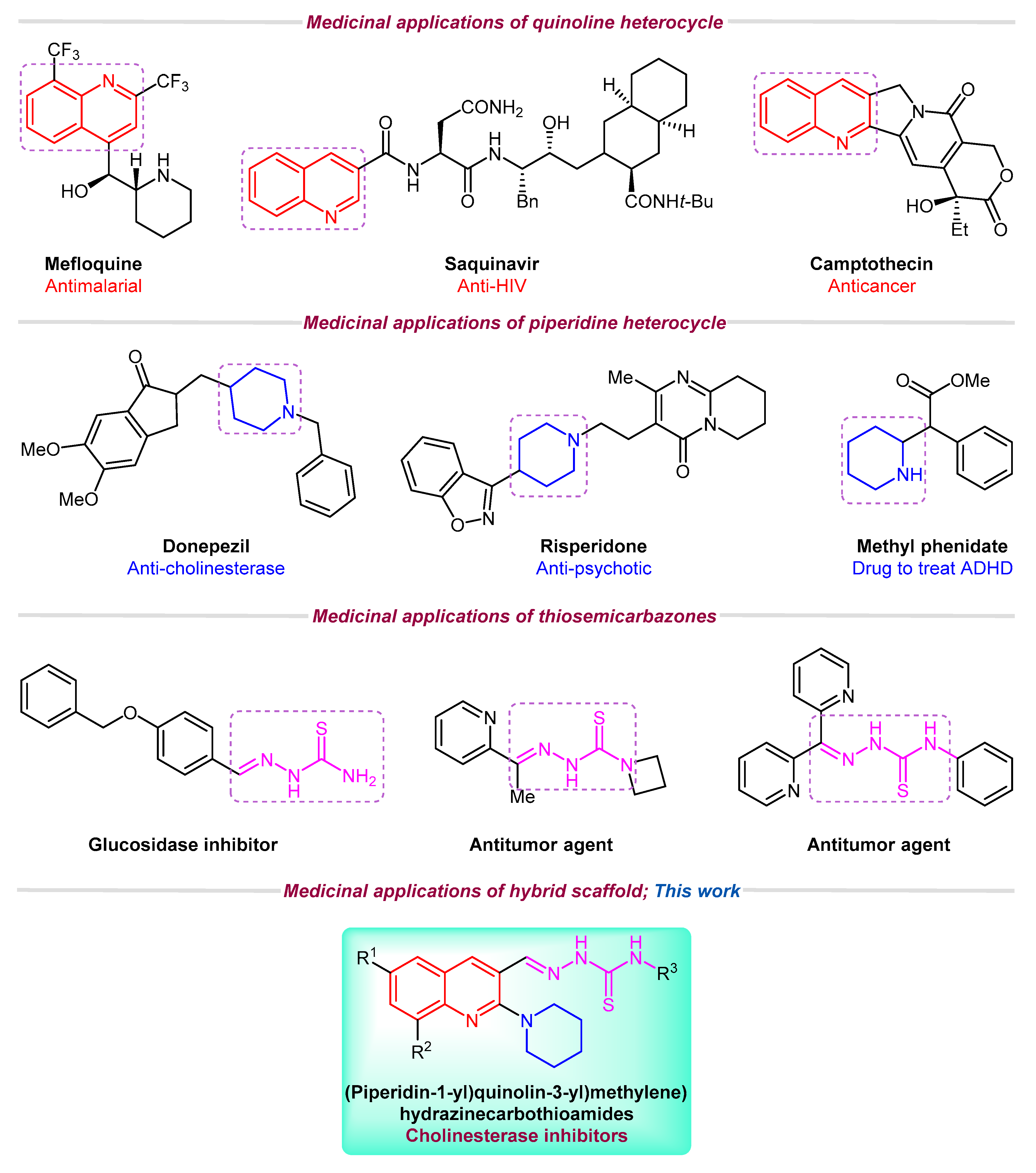
1. Introduction
2. Results and Discussion
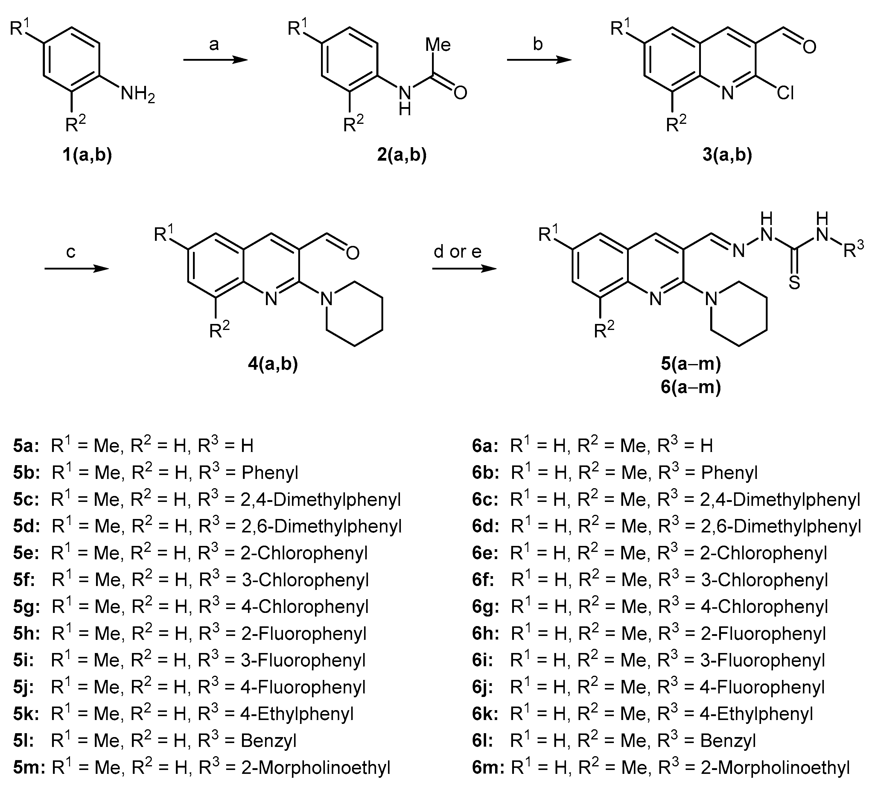
2.1. Synthetic Chemistry
2.2. Spectroscopic Characterization
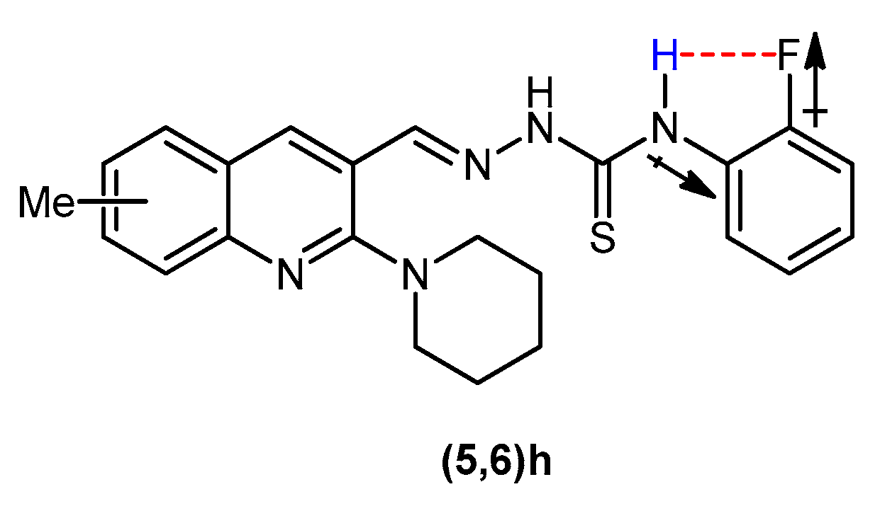
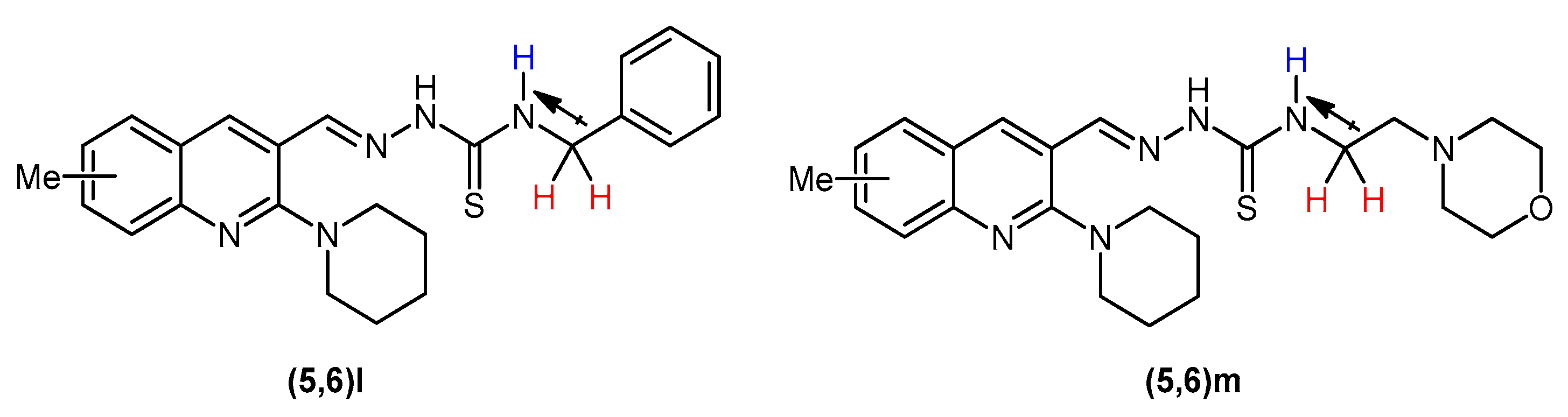
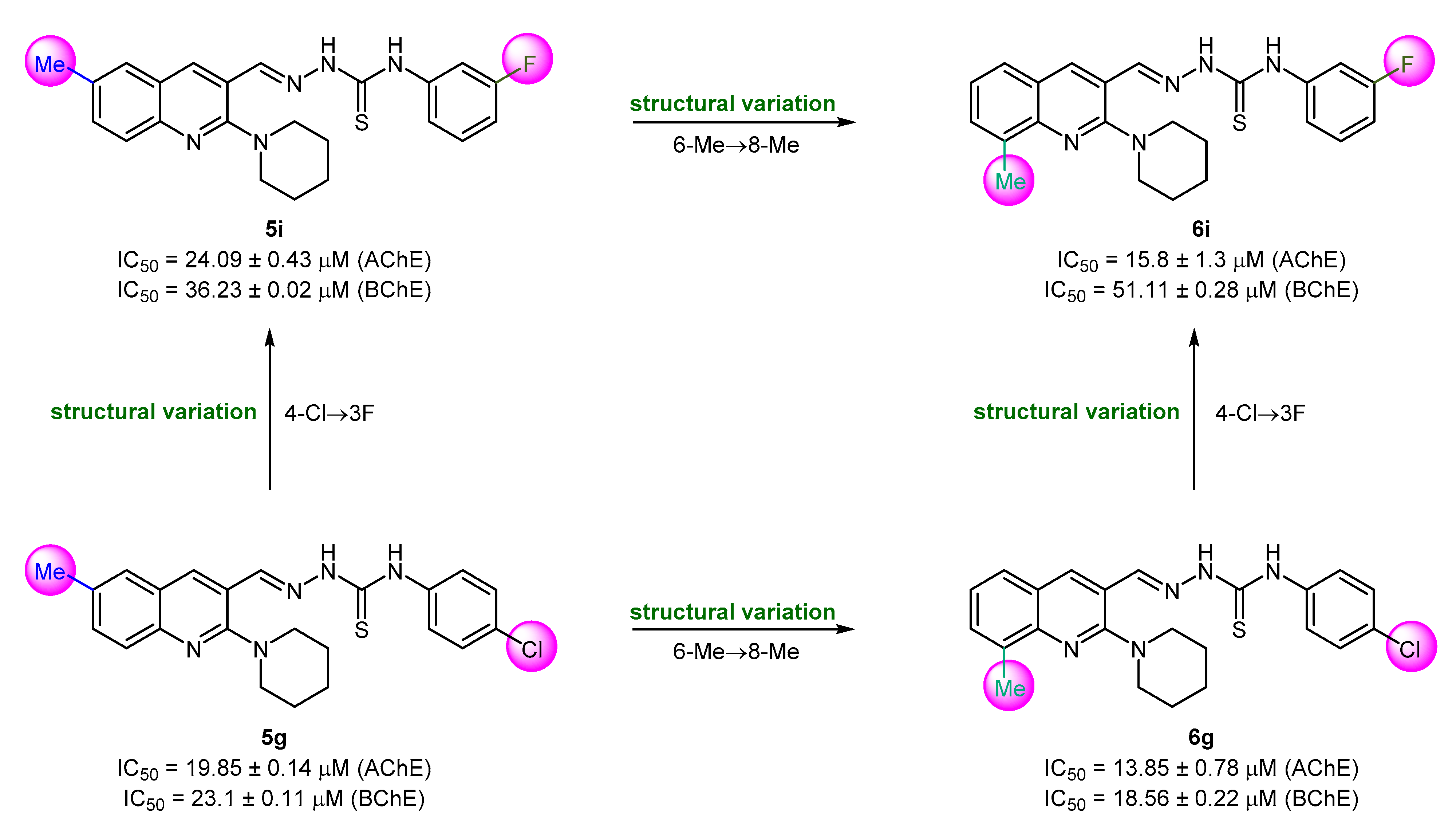
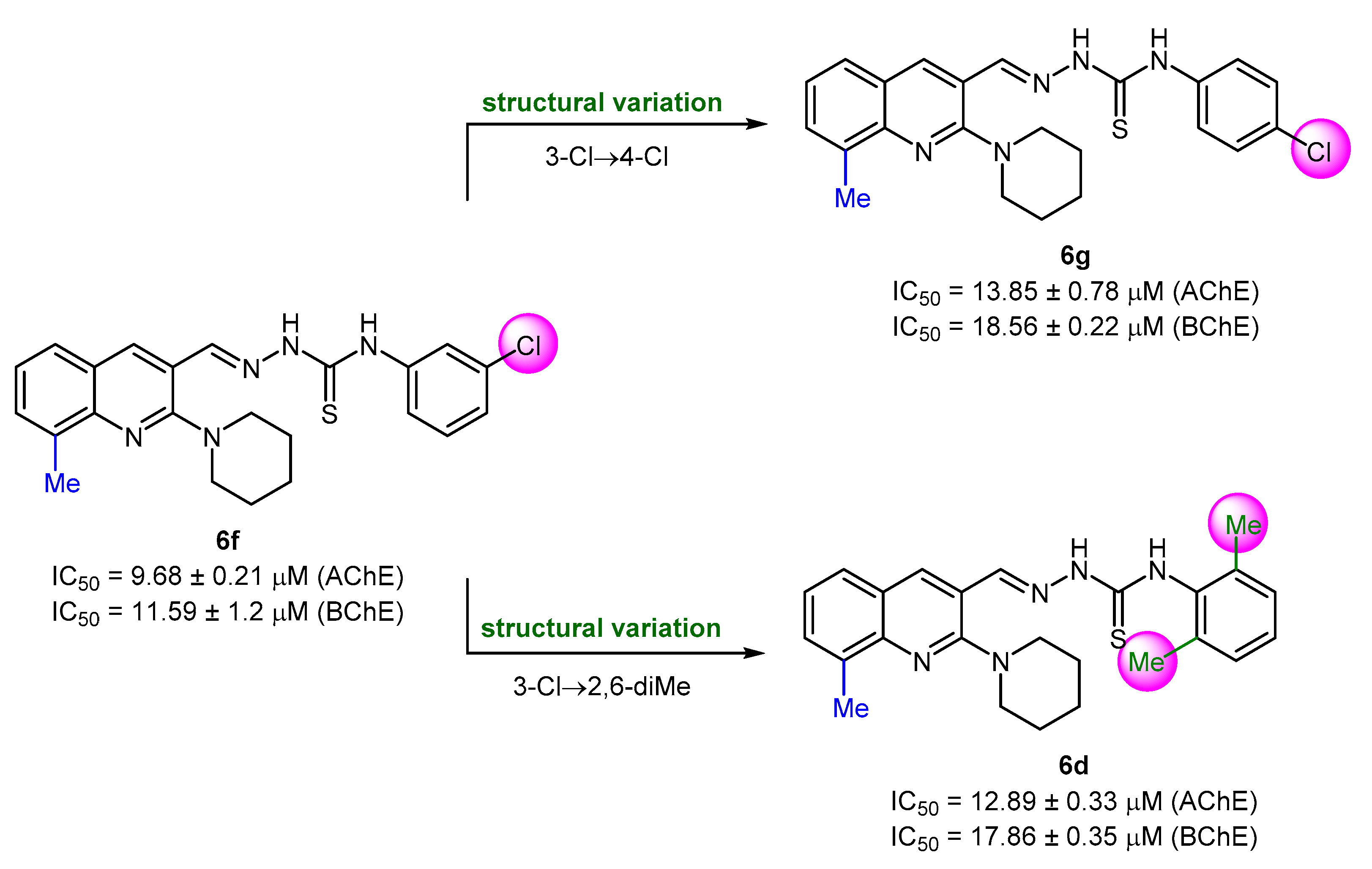
2.3. In Vitro Cholinesterase Inhibition and Structure–Activity Relationship Analyses
2.4. Molecular Docking Studies
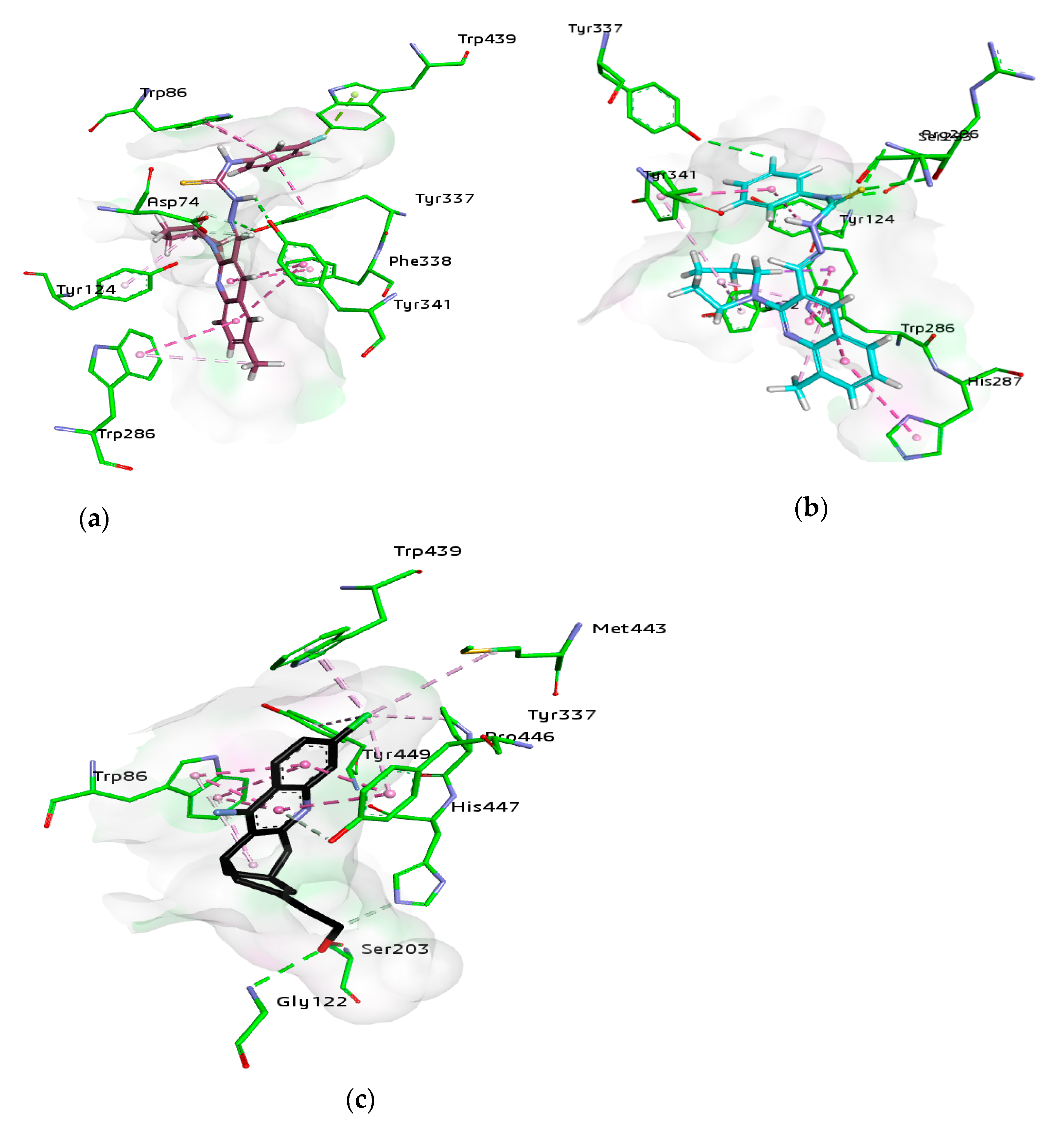
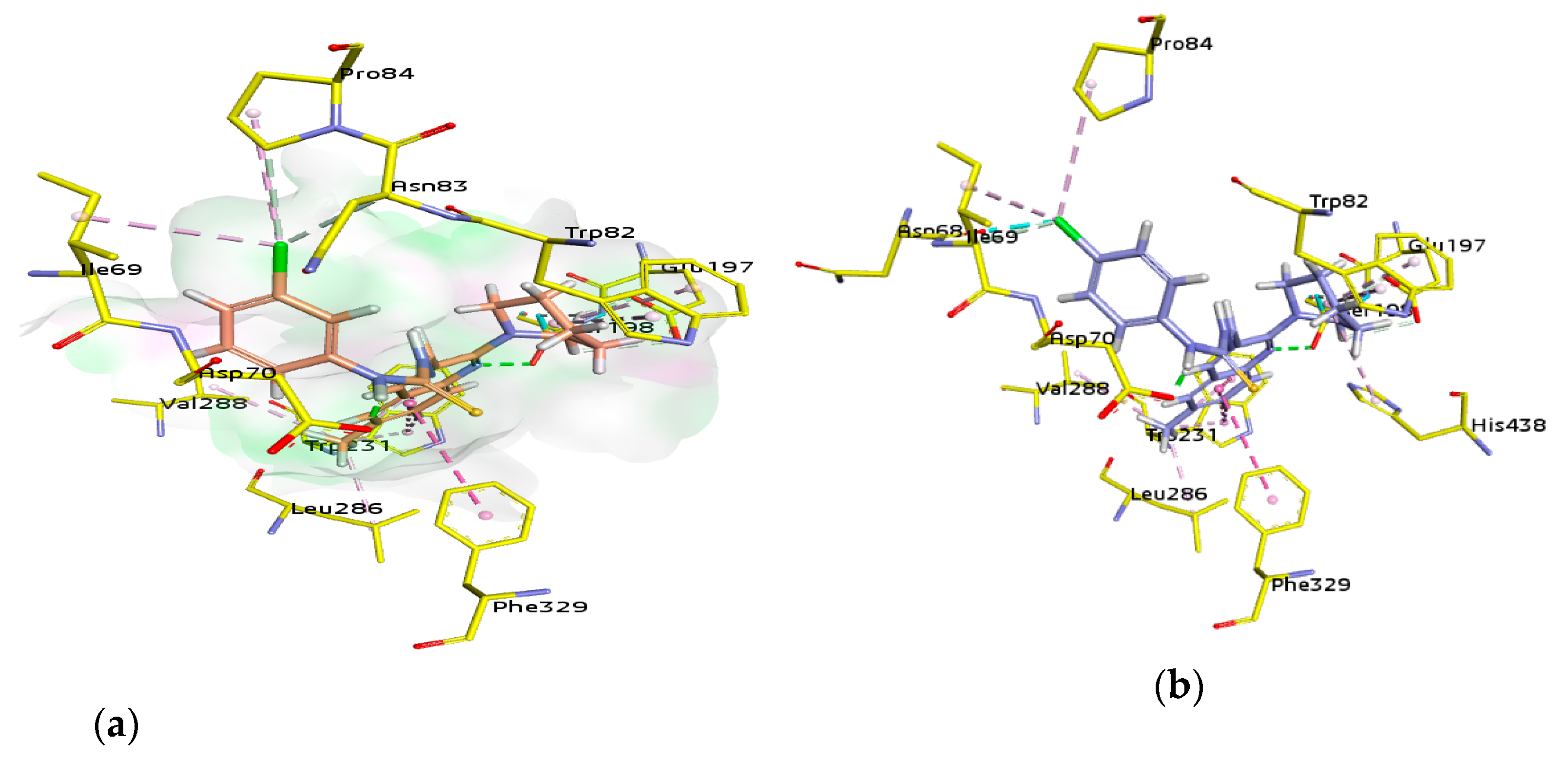
2.4.1. Molecular Docking Studies of Acetylcholinesterase (AChE)
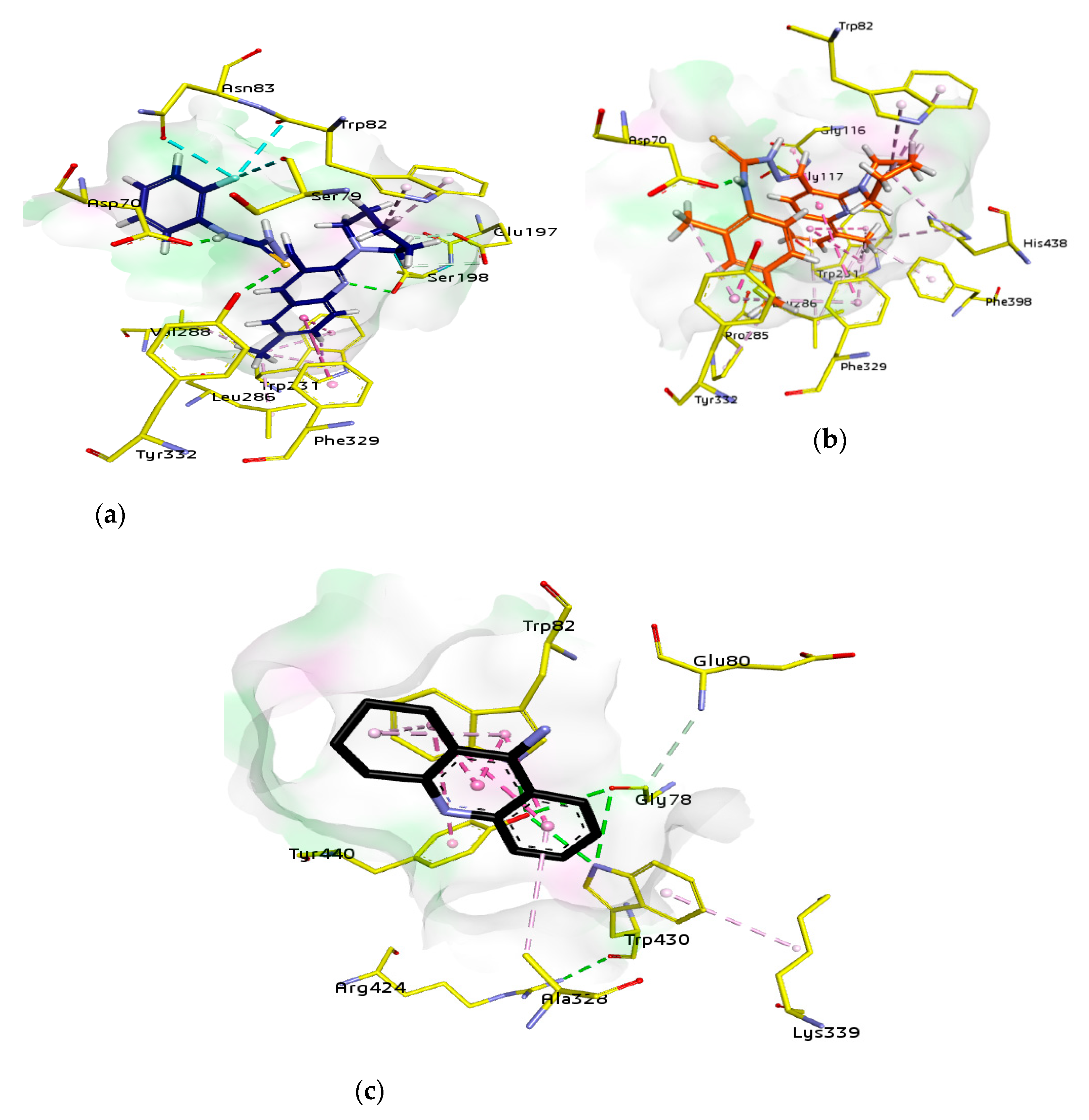
2.4.2. Molecular Docking Studies of Butyrylcholinesterase (BChE)
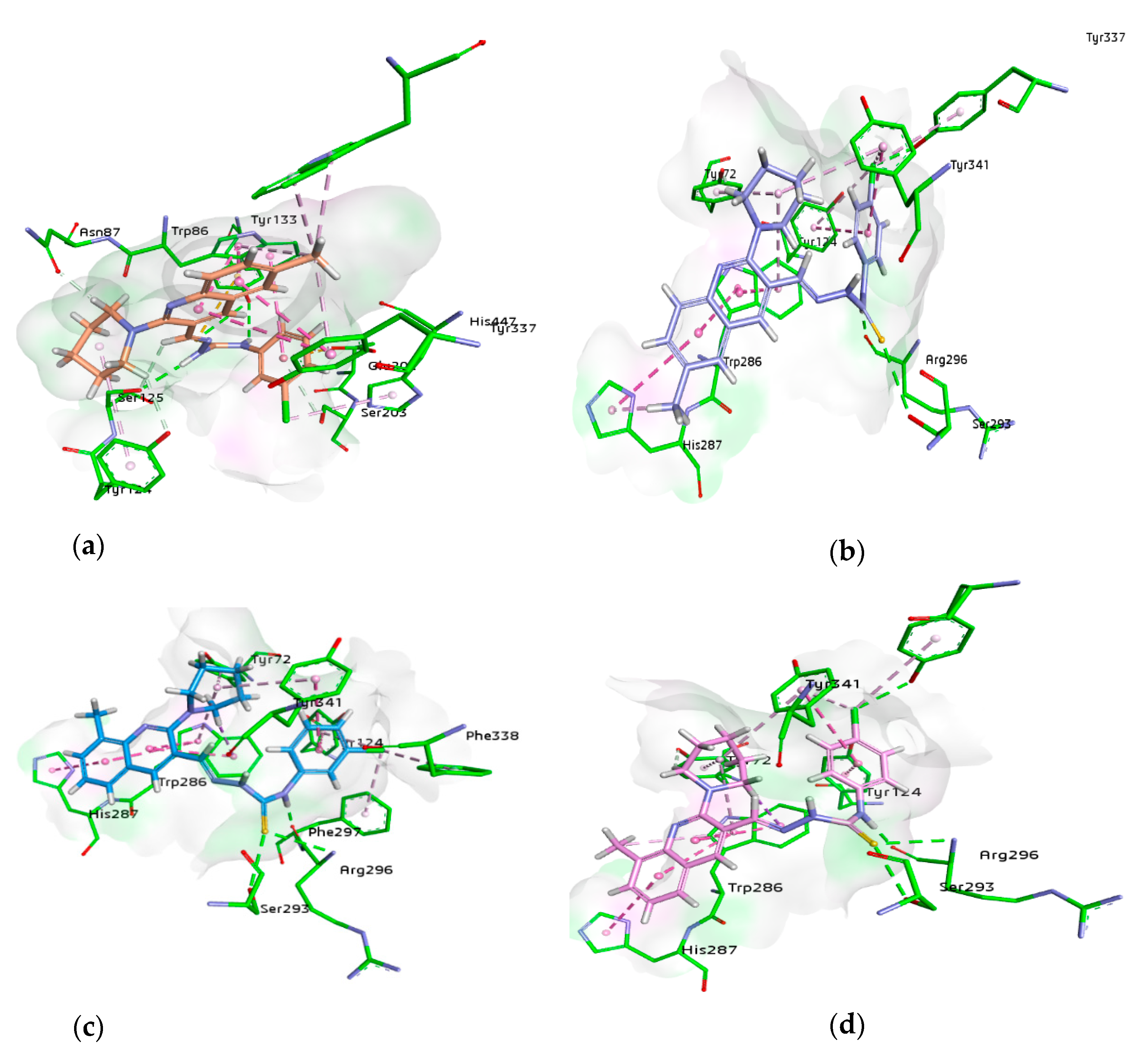
2.4.3. Dual Inhibitors of AChE and BChE
2.5. HYDE Assessment of Selective Compounds against Cholinesterases (AChE and BChE)
2.6. ADME Properties
2.7. In Vitro Cytotoxicity Testing
3. Materials and Methods
3.1. General
3.2. Preparation of Piperidinyl Quinoline-3-carbaldehydes (4a,b)
3.2.1. Preparation of 2-Chloroquinoline-3-carbaldehydes 3(a,b)
3.2.2. Preparation of Piperidinyl Quinoline-3-carbaldehydes (4a,b)
3.2.3. 6-Methyl-2-(piperidin-1-yl)quinoline-3-carbaldehyde (4a) Yield 98%
3.2.4. 8-Methyl-2-(piperidin-1-yl)quinoline-3-carbaldehyde (4b)
3.3. General Procedure for the Preparation of (Piperidin-1-yl)quinolin-3-yl)methylene)hydrazinecarbothioamides (5,6)
3.3.1. Method A: Conventional Synthesis
3.3.2. Method B: Microwave-Assisted Synthesis
3.4. In Vitro Cholinesterase Inhibition Assay
3.5. Molecular Docking Studies
3.6. Cytotoxicity
3.6.1. Sampling of Cell Lines
3.6.2. Culturing of Cell Lines
3.6.3. Cytotoxicity Calculation via MTT Assay
4. Conclusions
Supplementary Materials
Author Contributions
Funding
Institutional Review Board Statement
Informed Consent Statement
Data Availability Statement
Acknowledgments
Conflicts of Interest
Sample Availability
References
- Laferla, F.M.; Green, K.N.; Oddo, S. Intracellular amyloid-beta in Alzheimer’s disease. Nat. Rev. Neurosci. 2007, 8, 499–509. [Google Scholar] [CrossRef] [PubMed]
- Munoz-Torrero, D. Acetylcholinesterase inhibitors as disease-modifying therapies for Alzheimer’s disease. Curr. Med. Chem. 2008, 15, 2433–2455. [Google Scholar] [CrossRef] [PubMed]
- Belluti, F.; Bartolini, M.; Bottegoni, G.; Bisi, A.; Cavalli, A.; Andrisano, V.; Rampa, A. Benzophenone-based derivatives: A novel series of potent and selective dual inhibitors of acetylcholinesterase and acetylcholinesterase-induced beta-amyloid aggregation. Eur. J. Med. Chem. 2011, 46, 1682–1693. [Google Scholar] [CrossRef] [PubMed]
- Wang, L.; Kumar, R.; Pavlov, P.F.; Winblad, B. Small molecule therapeutics for tauopathy in Alzheimer’s disease: Walking on the path of most resistance. Eur. J. Med. Chem. 2021, 209, 112915. [Google Scholar] [CrossRef] [PubMed]
- Zhou, B.; Li, H.; Cui, Z.; Li, D.; Geng, H.; Gao, J.; Zhou, L. Simple analogues of natural product chelerythrine: Discovery of a novel anticholinesterase 2-phenylisoquinolin-2-ium scaffold with excellent potency against acetylcholinesterase. Eur. J. Med. Chem. 2020, 200, 112415. [Google Scholar] [CrossRef] [PubMed]
- Xu, M.; Peng, Y.; Zhu, L.; Wang, S.; Ji, J.; Rakesh, K.P. Triazole derivatives as inhibitors of Alzheimer’s disease: Current developments and structure-activity relationships. Eur. J. Med. Chem. 2019, 180, 656–672. [Google Scholar] [CrossRef] [PubMed]
- Li, Q.; He, S.; Chen, Y.; Feng, F.; Qu, W.; Sun, H. Donepezil-based multi-functional cholinesterase inhibitors for treatment of Alzheimer’s disease. Eur. J. Med. Chem. 2018, 158, 463–477. [Google Scholar] [CrossRef]
- Li, Q.; Yang, H.; Chen, Y.; Sun, H. Recent progress in the identification of selective butyrylcholinesterase inhibitors for Alzheimer’s disease. Eur. J. Med. Chem. 2017, 132, 294–309. [Google Scholar] [CrossRef]
- Dubois, B.; Feldman, H.H.; Jacova, C.; Cummings, J.L.; Dekosky, S.T.; Barberger-Gateau, P. Revising the definition of Alzheimer’s disease: A new lexicon. Lancet Neurol. 2010, 9, 1118–1127. [Google Scholar] [CrossRef]
- Aisen, P.S.; Andrieu, S.; Sampaio, C.; Carrillo, M.; Khachaturian, Z.S.; Dubois, B. Report of the task force on designing clinical trials in early (predementia) AD. Neurology 2011, 76, 280–286. [Google Scholar] [CrossRef]
- Dubois, B.; Hampel, H.; Feldman, H.H.; Scheltens, P.; Aisen, P.; Andrieu, S. Preclinical Alzheimer’s disease: Definition, natural history, and diagnostic criteria. Alzheimer’s & dementia. J. Alzheimer’s Assoc. 2016, 12, 292–323. [Google Scholar] [CrossRef]
- Nainwal, L.M.; Tasneem, S.; Akhtar, W.; Verma, G.; Khan, M.F.; Parvez, S.; Shaquiquzzaman, M.; Akhter, M.; Alam, M.M. Green recipes to quinoline: A review. Eur. J. Med. Chem. 2019, 164, 121–170. [Google Scholar] [CrossRef] [PubMed]
- Chu, X.M.; Wang, C.; Liu, W.; Liang, L.L.; Gong, K.K.; Zhao, C.Y.; Sun, K.L. Quinoline and quinolone dimers and their biological activities: An overview. Eur. J. Med. Chem. 2019, 161, 101–117. [Google Scholar] [CrossRef] [PubMed]
- Martin, R.E.; Butterworth, A.S.; Gardiner, D.L.; Kirk, K.; McCarthy, J.S.; Skinner-Adams, T.S. Saquinavir inhibits the malaria parasite’s chloroquine resistance transporter. Antimicrob. Agents Chemother. 2012, 56, 2283–2289. [Google Scholar] [CrossRef] [PubMed]
- Kuang, Y.H.; Patel, J.P.; Sodani, K.; Wu, C.P.; Liao, L.Q.; Patel, A.; Tiwari, A.K.; Dai, C.L.; Chen, X.; Fu, L.W.; et al. OSI-930 analogues as novel reversal agents for ABCG2-mediated multidrug resistance. Biochem. Pharmacol. 2012, 84, 766–774. [Google Scholar] [CrossRef]
- Rajanarendar, E.; Reddy, M.N.; Krishna, S.R.; Murthy, K.R.; Reddy, Y.N.; Rajam, M.V. Design, synthesis, antimicrobial, anti-inflammatory and analgesic activity of novel isoxazolyl pyrimido [4,5-b] quinolines and isoxazolyl chromeno [2,3-d] pyrimidin-4-ones. Eur. J. Med. Chem. 2012, 55, 273–283. [Google Scholar] [CrossRef]
- Musiol, R.; Serda, M.; Hensel-Bielowka, S.; Polanski, J. Quinoline-based antifungals. Curr. Med. Chem. 2010, 17, 1960–1973. [Google Scholar] [CrossRef]
- Hazra, A.; Mondal, S.; Maity, A.; Naskar, S.; Saha, P.; Paira, R.; Sahu, K.B.; Paira, P.; Ghosh, S.; Sinha, C.; et al. Amberlite-IRA-402 (OH) ion exchange resin mediated synthesis of indolizines, pyrrolo [1,2-a] quinolines and isoquinolines: Antibacterial and antifungal evaluation of the products. Eur. J. Med. Chem. 2011, 46, 2132–2140. [Google Scholar] [CrossRef]
- Zhang, Y.; Fang, Y.; Liang, H.; Wang, H.; Hu, K.; Liu, X.; Yi, X.; Peng, Y. Synthesis and antioxidant activities of 2-oxo-quinoline-3-carbaldehyde Schiff-base derivatives. Bioorg. Med. Chem. Lett. 2013, 23, 107–111. [Google Scholar] [CrossRef]
- Solomon, V.R.; Lee, H. Quinoline as a privileged scaffold in cancer drug discovery. Curr. Med. Chem. 2011, 18, 1488–1508. [Google Scholar] [CrossRef]
- Rizvi, S.U.F.; Siddiqui, H.L.; Nisar, M.; Khan, N.; Khan, I. Discovery and molecular docking of quinolyl-thienyl chalcones as anti-angiogenic agents targeting VEGFR-2 tyrosine kinase. Bioorg. Med. Chem. Lett. 2012, 22, 942–944. [Google Scholar] [CrossRef] [PubMed]
- Ratheesh, M.; Sindhu, G.; Helen, A. Anti-inflammatory effect of quinoline alkaloid skimmianine isolated from Ruta graveolens L. J. Inflamm. Res. 2013, 62, 367–376. [Google Scholar] [CrossRef] [PubMed]
- Das, P.; Deng, X.; Zhang, L.; Roth, M.G.; Fontoura, B.M.; Phillips, M.A.; De Brabander, J.K. SAR-based optimization of a 4-quinoline carboxylic acid analogue with potent antiviral activity. ACS Med. Chem. Lett. 2013, 4, 517–521. [Google Scholar] [CrossRef] [PubMed]
- Rizvi, S.U.F.; Ahmad, M.; Bukhari, M.H.; Montero, C.; Chatterjee, P.; Detorio, M.; Schinazi, R.F. Anti-HIV-1 screening of (2E)-3-(2-chloro-6-methyl/methoxyquinolin-3-yl)-1-(aryl) prop-2-en-1-ones. Med. Chem. Res. 2014, 23, 402–407. [Google Scholar] [CrossRef]
- Zaib, S.; Rizvi, S.U.F.; Aslam, S.; Ahmad, M.; Abid, S.M.A.; al-Rashida, M.; Iqbal, J. Quinolinyl-thienyl chalcones as monoamine oxidase inhibitors and their in silico modeling studies. Med. Chem. Res. 2015, 11, 580–589. [Google Scholar] [CrossRef]
- Massoud, M.A.; El-Sayed, M.A.; Bayoumi, W.A.; Mansour, B. Cytotoxicity and molecular targeting study of novel 2-chloro-3-substituted quinoline derivatives as antitumor agents. Lett. Drug Des. Discov. 2019, 16, 273–283. [Google Scholar] [CrossRef]
- Tejería, A.; Pérez-Pertejo, Y.; Reguera, R.M.; Carbajo-Andrés, R.; Balaña-Fouce, R.; Alonso, C.; Martin-Encinas, E.; Selas, A.; Rubiales, G.; Palacios, F. Antileishmanial activity of new hybrid tetrahydroquinoline and quinoline derivatives with phosphorus substituents. Eur. J. Med. Chem. 2019, 162, 18–31. [Google Scholar] [CrossRef]
- Chanquia, S.N.; Larregui, F.; Puente, V.; Labriola, C.; Lombardo, E.; Liñares, G.G. Synthesis and biological evaluation of new quinoline derivatives as antileishmanial and antitrypanosomal agents. Bioorg. Chem. 2019, 83, 526–534. [Google Scholar] [CrossRef]
- Muruganantham, N.; Sivakumar, R.; Anbalagan, N.; Gunasekaran, V.; Leonard, J.T. Synthesis, anticonvulsant and antihypertensive activities of 8-substituted quinoline derivatives. Biol. Pharm. Bull. 2004, 27, 1683–1687. [Google Scholar] [CrossRef]
- Kumar, H.; Devaraji, V.; Joshi, R.; Jadhao, M.; Ahirkar, P.; Prasath, R.; Bhavana, P.; Ghosh, S.K. Antihypertensive activity of a quinoline appended chalcone derivative and its site specific binding interaction with a relevant target carrier protein. RSC Adv. 2015, 5, 65496–65513. [Google Scholar] [CrossRef]
- De Kimpe, N.; Boelens, M.; Contreras, J. Rearrangement of 5-(bromomethyl)-1-pyrrolinium salts into functionalized piperidines. Tetrahedron Lett. 1996, 37, 3171–3174. [Google Scholar] [CrossRef]
- Taylor, R.D.; MacCoss, M.; Lawson, A.D. Rings in drugs: Miniperspective. J. Med. Chem. 2014, 57, 5845–5859. [Google Scholar] [CrossRef] [PubMed]
- Padmanilayam, M.; Scorneaux, B.; Dong, Y.; Chollet, J.; Matile, H.; Charman, S.A.; Creek, D.J.; Charman, W.N.; Tomas, J.S.; Scheurer, C.; et al. Antimalarial activity of N-alkyl amine, carboxamide, sulfonamide, and urea derivatives of a dispiro-1,2,4-trioxolane piperidine. Bioorg. Med. Chem. Lett. 2006, 16, 5542–5545. [Google Scholar] [CrossRef] [PubMed]
- Kamiński, K.; Wiklik, B.; Obniska, J. Synthesis, anticonvulsant properties, and SAR analysis of differently substituted pyrrolidine-2, 5-diones and piperidine-2,6-diones. Arch. Pharm. Chem. Life Sci. 2014, 347, 840–852. [Google Scholar] [CrossRef] [PubMed]
- Henderson, N.D.; Plumb, J.A.; Robins, D.J.; Workman, P. Synthesis and anti-cancer activity of 2,6-disubstituted N-methylpiperidine derivatives and their N-oxides. Anti-Cancer Drug Des. 1996, 11, 421–438. [Google Scholar] [PubMed]
- Balsamo, A.; Giorgi, I.; Lapucci, A.; Lucacchini, A.; Macchia, B.; Macchia, F.; Martini, C.; Rossi, A. 3-[(2-Ethoxyphenoxy) methyl] piperidine derivatives. Synthesis and antidepressant activity. J. Med. Chem. 1987, 30, 222–225. [Google Scholar] [CrossRef]
- Shaw, A.T.; Kim, D.W.; Nakagawa, K.; Seto, T.; Crinó, L.; Ahn, M.J.; De Pas, T.; Besse, B.; Solomon, B.J.; Blackhall, F.; et al. Crizotinib versus chemotherapy in advanced ALK-positive lung cancer. N. Engl. J. Med. 2013, 368, 2385–2394. [Google Scholar] [CrossRef]
- Ruela, A.L.M.; Carvalho, F.C.; Pereira, G.R. Exploring the phase behavior of monoolein/oleic acid/water systems for enhanced donezepil administration for Alzheimer disease treatment. J. Pharm. Sci. 2016, 105, 71–77. [Google Scholar] [CrossRef]
- Weiser, M.; Levi, L.; Levine, S.Z.; Bialer, M.; Shekh-Ahmad, T.; Matei, V.; Tiugan, A.; Cirjaliu, D.; Sava, C.; Sinita, E.; et al. A randomized, double-blind, placebo-and risperidone-controlled study on valnoctamide for acute mania. Bipolar Disord. 2017, 19, 285–294. [Google Scholar] [CrossRef]
- Luethi, D.; Kaeser, P.J.; Brandt, S.D.; Krähenbühl, S.; Hoener, M.C.; Liechti, M.E. Pharmacological profile of methylphenidate-based designer drugs. Neuropharmacology 2018, 134, 133–140. [Google Scholar] [CrossRef]
- Arce, E.R.; Putzu, E.; Lapier, M.; Maya, J.D.; Azar, C.O.; Echeverría, G.A.; Piro, O.E.; Medeiros, A.; Sardi, F.; Comini, M.; et al. New heterobimetallic ferrocenyl derivatives are promising antitrypanosomal agents. Dalton Trans. 2019, 48, 7644–7658. [Google Scholar] [CrossRef] [PubMed]
- Divar, M.; Khalafi-Nezhad, A.; Zomorodian, K.; Sabet, R.; Faghih, Z.; Jamali, M.; Pournaghz, H.; Khabnadideh, S. Synthesis of some novel semicarbazone and thiosemicarbazone derivatives of isatin as possible biologically active agents. J. Pharm. Res. Int. 2017, 1–13. [Google Scholar] [CrossRef]
- Dawood, D.H.; Batran, R.Z.; Farghaly, T.A.; Khedr, M.A.; Abdulla, M.M. New coumarin derivatives as potent selective COX-2 inhibitors: Synthesis, anti-Inflammatory, QSAR, and molecular modeling studies. Arch. Pharm. Chem. Life Sci. 2015, 348, 875–888. [Google Scholar] [CrossRef] [PubMed]
- Haribabu, J.; Subhashree, G.R.; Saranya, S.; Gomathi, K.; Karvembu, R.; Gayathri, D. Isatin based thiosemicarbazone derivatives as potential bioactive agents: Antioxidant and molecular docking studies. J. Mol. Struct. 2016, 1110, 185–195. [Google Scholar] [CrossRef]
- Bakherad, Z.; Safavi, M.; Fassihi, A.; Sadeghi-Aliabadi, H.; Bakherad, M.; Rastegar, H.; Ghasemi, J.B.; Sepehri, S.; Saghaie, L.; Mahdavi, M. Anti-cancer, anti-oxidant and molecular docking studies of thiosemicarbazone indole-based derivatives. Res. Chem. Intermed. 2019, 45, 2827–2854. [Google Scholar] [CrossRef]
- Elsayed, H.E.; Ebrahim, H.Y.; Haggag, E.G.; Kamal, A.M.; El Sayed, K.A. Rationally designed hecogenin thiosemicarbazone analogs as novel MEK inhibitors for the control of breast malignancies. Bioorg. Med. Chem. 2017, 25, 6297–6312. [Google Scholar] [CrossRef]
- Hussein, M.A.; Iqbal, M.A.; Asif, M.; Haque, R.A.; Ahamed, M.B.K.; Majid, A.M.A.; Guan, T.S. Synthesis, crystal structures and in vitro anticancer studies of new thiosemicarbazone derivatives. Phosphorus Sulfur Silicon Relat. Elem. 2015, 190, 1498–1508. [Google Scholar] [CrossRef]
- Pingaew, R.; Prachayasittikul, S.; Ruchirawat, S.; Prachayasittikul, V. Synthesis and cytotoxicity of novel N-sulfonyl-1,2,3,4-tetrahydroisoquinoline thiosemicarbazone derivatives. Med. Chem. Res. 2013, 22, 267–277. [Google Scholar] [CrossRef]
- Palanimuthu, D.; Poon, R.; Sahni, S.; Anjum, R.; Hibbs, D.; Lin, H.Y.; Bernhardt, P.V.; Kalinowski, D.S.; Richardson, D.R. A novel class of thiosemicarbazones show multi-functional activity for the treatment of Alzheimer’s disease. Eur. J. Med. Chem. 2017, 139, 612–632. [Google Scholar] [CrossRef]
- Ali, M.; Khan, K.M.; Salar, U.; Ashraf, M.; Taha, M.; Wadood, A.; Hamid, S.; Riaz, M.; Ali, B.; Shamim, S.; et al. Synthesis, in vitro alpha-glucosidase inhibitory activity, and in silico study of (E)-thiosemicarbazones and (E)-2-(2-(arylmethylene) hydrazinyl)-4-arylthiazole derivatives. Mol. Divers. 2018, 22, 841–861. [Google Scholar] [CrossRef]
- Khan, I.; Hanif, M.; Hussain, M.T.; Khan, A.A.; Aslam, M.A.S.; Rama, N.H.; Iqbal, J. Synthesis, acetylcholinesterase and alkaline phosphatase inhibition of some new 1,2,4-triazole and 1,3,4-thiadiazole derivatives. Aust. J. Chem. 2012, 65, 1413–1419. [Google Scholar] [CrossRef]
- Khan, I.; Ibrar, A.; Zaib, S.; Ahmad, S.; Furtmann, N.; Hameed, S.; Simpson, J.; Bajorath, J.; Iqbal, J. Active compounds from a diverse library of triazolothiadiazole and triazolothiadiazine scaffolds: Synthesis, crystal structure determination, cytotoxicity, cholinesterase inhibitory activity, and binding mode analysis. Bioorg. Med. Chem. 2014, 22, 6163–6173. [Google Scholar] [CrossRef] [PubMed]
- Khan, I.; Zaib, S.; Ibrar, A.; Rama, N.H.; Simpson, J.; Iqbal, J. Synthesis, crystal structure and biological evaluation of some novel 1,2,4-triazolo[3,4-b]-1,3,4-thiadiazoles and 1,2,4-triazolo[3,4-b]-1,3,4-thiadiazines. Eur. J. Med. Chem. 2014, 78, 167–177. [Google Scholar] [CrossRef] [PubMed]
- Khan, I.; Bakht, S.M.; Ibrar, A.; Abbas, S.; Hameed, S.; White, J.M.; Rana, U.A.; Zaib, S.; Shahid, M.; Iqbal, J. Exploration of a library of triazolothiadiazole and triazolothiadiazine compounds as a highly potent and selective family of cholinesterase and monoamine oxidase inhibitors: Design, synthesis, X-ray diffraction analysis and molecular docking studies. RSC Adv. 2015, 5, 21249–21267. [Google Scholar] [CrossRef]
- Ibrar, A.; Khan, A.; Ali, M.; Sarwar, R.; Mehsud, S.; Farooq, U.; Halimi, S.M.A.; Khan, I.; Al-Harrasi, A. Combined in vitro and in silico studies for the anticholinesterase activity and pharmacokinetics of coumarinyl thiazoles and oxadiazoles. Front. Chem. 2018, 6, 61. [Google Scholar] [CrossRef]
- Larik, F.A.; Saeed, A.; Faisal, M.; Hamdani, S.; Jabeen, F.; Channar, P.A.; Mumtaz, A.; Khan, I.; Kazi, M.A.; Abbas, Q.; et al. Synthesis, inhibition studies against AChE and BChE, drug-like profiling, kinetic analysis and molecular docking studies of N-(4-phenyl-3-aroyl-2(3H)-ylidene) substituted acetamides. J. Mol. Struct. 2020, 1203, 127459. [Google Scholar] [CrossRef]
- Ghobadian, R.; Mahdavi, M.; Nadri, M.; Moradi, A.; Edraki, N.; Akbarzadeh, T.; Sharifzadeh, M.; Bukhari, S.N.A.; Amini, M. Novel tetrahydrocarbazole benzyl pyridine hybrids as potent and selective butryl cholinesterase inhibitors with neuroprotective and β-secretase inhibition activities. Eur. J. Med. Chem. 2018, 155, 49–60. [Google Scholar] [CrossRef]
- Pérez-Areales, F.J.; Turcu, A.L.; Barniol-Xicota, M.; Pont, C.; Pivetta, D.; Espargaró, A.; Bartolini, M.; De Simone, A.; Andrisano, V.; Pérez, B.; et al. A novel class of multitarget anti-Alzheimer benzohomoadamantane-chlorotacrine hybrids modulating cholinesterases and glutamate NMDA receptors. Eur. J. Med. Chem. 2019, 180, 613–626. [Google Scholar] [CrossRef]
- Najafi, Z.; Mahdavi, M.; Saeedi, M.; Karimpour-Razkenari, E.; Asatouri, R.; Vafadarnejad, F.; Moghadam, F.H.; Khanavi, M.; Sharifzadeh, M.; Akbarzadeh, T. Novel tacrine-1,2,3-triazole hybrids: In vitro, in vivo biological evaluation and docking study of cholinesterase inhibitors. Eur. J. Med. Chem. 2017, 125, 1200–1212. [Google Scholar] [CrossRef]
- Meth-Cohn, O.; Narine, B.; Tarnowski, B. A versatile new synthesis of quinolines and related fused pyridines, Part 5. The synthesis of 2-chloroquinoline-3-carbaldehydes. J. Chem. Soc. Perkin Trans. 1981, 1, 1520–1530. [Google Scholar] [CrossRef]
- Munir, R.; Athar, M.M.; Rehman, M.Z.; Javid, N. Synthesis of 6/8-Methyl-2-(Piperidin-1-yl)Quinoline-3-Carbaldehydes; A Facile CTAB Catalyzed Protocol. Chiang Mai J. Sci. 2020, 47, 175–180. Available online: http://epg.science.cmu.ac.th/ejournal/ (accessed on 15 January 2021).
- Ebrahimi, H.P.; Hadi, J.S.; Alsalim, T.A.; Ghali, T.S.; Bolandnazar, Z. A novel series of thiosemicarbazone drugs: From synthesis to structure. Spectrochim. Acta A Biomol. Spectrosc. 2015, 137, 1067–1077. [Google Scholar] [CrossRef] [PubMed]
- Ferrari, M.B.; Pelizzi, C.; Pelosi, G.; Rodríguez-Argüelles, M.C. Preparation, characterization and X-ray structures of 1-methylisatin 3-thiosemicarbazone copper, nickel and cobalt complexes. Polyhedron 2002, 21, 2593–2599. [Google Scholar] [CrossRef]
- De Silva, N.N.; Albu, T.V. A theoretical investigation on the isomerism and the NMR properties of thiosemicarbazones. Cent. Eur. J. Chem. 2007, 5, 396–419. [Google Scholar] [CrossRef]
- Iftikhar, K.; Murtaza, S.; Kousar, N.; Abbas, A.; Tahir, M.N. Aminobenzoic acid derivatives as antioxidants and cholinesterase inhibitors: Synthesis, biological evaluation and molecular docking studies. Acta Polon. Pharma-Drug Res. 2018, 75, 385–396. [Google Scholar] [CrossRef]
- Nachon, F.; Carletti, E.; Ronco, C.; Trovaslet, M.; Nicolet, Y.; Jean, L.; Renard, P.Y. Crystal structures of human cholinesterases in complex with huprine W and tacrine: Elements of specificity for anti-Alzheimer’s drugs targeting acetyl- and butyryl-cholinesterase. Biochem. J. 2013, 453, 393–399. [Google Scholar] [CrossRef]
- LeadIT Version 2.3.2; BioSolveIT GmbH: Sankt Augustin, Germany, 2017; Available online: www.biosolveit.de/LeadIT (accessed on 20 September 2020).
- Daina, A.; Michielin, O.; Zoete, V. SwissADME: A free web tool to evaluate pharmacokinetics, drug-likeness and medicinal chemistry friendliness of small molecules. Sci. Rep. 2017, 7, 42717. [Google Scholar] [CrossRef]
- Daina, A.; Michielin, O.; Zoete, V. iLOGP: A simple, robust, and efficient description of n-octanol/water partition coefficient for drug design using the GB/SA approach. J. Chem. Inf. Model. 2014, 54, 3284–3301. [Google Scholar] [CrossRef]
- Daina, A.; Zoete, V. A BOILED-Egg to predict gastrointestinal absorption and brain penetration of small molecules. ChemMedChem 2016, 11, 1117–1121. [Google Scholar] [CrossRef]
- Ellman, G.L.; Courtney, K.D.; Andres, V.; Featherstone, R.M. A new and rapid colorimetric determination of acetylcholinesterase activity. Biochem. Pharmacol. 1961, 7, 88–95. [Google Scholar] [CrossRef]
- Labute, P. Protonate 3D, Chemical Computing Group. 2007. Available online: http://www.chemcomp.com/journal/proton.htm (accessed on 20 September 2020).
- Chemical Computing Group’s Molecular Operating Environment (MOE) MOE 2019. 0201. Available online: http://www.chemcomp.com/MOEMolecular_Operating_Environment.htm (accessed on 20 September 2020).
- BIOVIA Discovery Studio Client v19.1.0.18287. Accelrys Discovery Studio; Accelrys Software Inc.: San Diego, CA, USA, 2019.
- Schneider, N.; Lange, G.; Hindle, S.; Klein, R.; Rarey, M. A consistent description of hydrogen bond and dehydration energies in protein-ligand complexes: Methods behind the HYDE scoring function. J. Comput.-Aided Mol. Des. 2013, 27, 15–29. [Google Scholar] [CrossRef] [PubMed]
- Maqbool, T.; Awan, S.J.; Malik, S.; Hadi, F.; Shehzadi, S.; Tariq, K. In vitro anti-proliferative, apoptotic and antioxidative activities of medicinal herb kalonji (nigella sativa). Curr. Pharm. Biotechnol. 2019, 20, 1288–1308. [Google Scholar] [CrossRef] [PubMed]


| Compound | Conventional Approach | Microwave-Assisted Approach | Acetylcholinesterase (AChE) | Butyrylcholinesterse (BChE) |
|---|---|---|---|---|
| Yield (%) | IC50 ± SEM (μM) | |||
| 5a | 80 | 91 | 27.6 ± 0.08 | 35.3 ± 0.01 |
| 5b | 86 | 92 | 25.1 ± 0.25 | 33.1 ± 0.09 |
| 5c | 85 | 92 | 42.3 ± 0.99 | 37.2 ± 0.83 |
| 5d | 90 | 95 | 25.9 ± 0.96 | 29.8 ± 1.2 |
| 5e | 85 | 91 | 35.2 ± 0.54 | 40.26 ± 0.17 |
| 5f | 94 | 97 | 23.9 ± 0.25 | 24.6 ± 0.57 |
| 5g | 93 | 97 | 19.85 ± 0.14 | 23.1 ± 0.11 |
| 5h | 80 | 90 | 31.28 ± 0.37 | 25.12 ± 0.99 |
| 5i | 90 | 96 | 24.09 ± 0.43 | 36.23 ± 0.02 |
| 5j | 83 | 90 | 28.21 ± 0.94 | 42.36 ± 0.44 |
| 5k | 94 | 97 | 30.65 ± 0.56 | 32.01 ± 0.87 |
| 5l | 91 | 95 | 35.09 ± 1.2 | 45.02 ± 0.38 |
| 5m | 78 | 89 | 62.3 ± 0.68 | 59.35 ± 0.13 |
| 6a | 81 | 90 | 36.25 ± 0.36 | 60.02 ± 0.04 |
| 6b | 89 | 93 | 25.89 ± 0.45 | 28.77 ± 0.63 |
| 6c | 85 | 91 | 39.12 ± 0.27 | 24.59 ± 0.09 |
| 6d | 92 | 98 | 12.89 ± 0.33 | 17.86 ± 0.35 |
| 6e | 89 | 93 | 32.11 ± 0.15 | 37.06 ± 0.59 |
| 6f | 88 | 93 | 9.68 ± 0.21 | 11.59 ± 1.2 |
| 6g | 92 | 95 | 13.85 ± 0.78 | 18.56 ± 0.22 |
| 6h | 92 | 96 | 40.23 ± 0.25 | 45.12 ± 0.19 |
| 6i | 87 | 90 | 15.8 ± 1.3 | 51.11 ± 0.28 |
| 6j | 83 | 91 | 56.66 ± 0.41 | 51.03 ± 0.52 |
| 6k | 90 | 95 | 39.91 ± 0.85 | 32.01 ± 0.31 |
| 6l | 82 | 91 | 57.25 ± 0.03 | 41.02 ± 0.89 |
| 6m | 82 | 90 | 21.01 ± 2.3 | 35.14 ± 0.77 |
| Donepezil | ― | ― | 2.98 ± 0.62 | 7.21 ± 0.39 |
| Compounds | Acetylcholinesterase | Butyrylcholinesterase | ||
|---|---|---|---|---|
| Docking Score by FlexX for Top Pose (kcal mol−1) | Free Energy of Binding ΔG (kJ mol−1) | Docking Score by FlexX for Top Pose (kcal mol−1) | Free Energy of Binding ΔG (kJ mol−1) | |
| 5a | −23.1892 | −15 | −27.4250 | −13 |
| 5b | −27.3759 | −18 | −33.8532 | −18 |
| 5c | −15.1888 | −15 | −38.2179 | −19 |
| 5d | −34.0828 | −17 | −36.4835 | −20 |
| 5e | −21.2287 | −19 | −35.5422 | −17 |
| 5f | −30.9532 | −23 | −32.4958 | −21 |
| 5g | −24.9478 | −22 | −31.3943 | −20 |
| 5h | −25.3819 | −14 | −33.4593 | −22 |
| 5i | −26.3914 | −25 | −31.8917 | −18 |
| 5j | −27.7381 | −13 | −33.3263 | −19 |
| 5k | −18.8869 | −10 | −31.3938 | −17 |
| 5l | −23.4789 | −11 | −30.7439 | −14 |
| 5m | −25.4734 | −17 | −29.6099 | −13 |
| 6a | −25.2746 | −14 | −28.9743 | −17 |
| 6b | −24.0306 | −13 | −32.3915 | −14 |
| 6c | −14.5216 | −10 | −34.1889 | −22 |
| 6d | −21.9230 | −12 | −34.6624 | −20 |
| 6e | −26.5830 | −15 | −32.5630 | −19 |
| 6f | −25.5518 | −24 | −31.0974 | −20 |
| 6g | −25.8729 | −24 | −28.6461 | −19 |
| 6h | −24.8538 | −15 | −28.7235 | −14 |
| 6i | −26.5146 | −26 | −31.4126 | −18 |
| 6j | −30.6048 | −14 | −29.3584 | −17 |
| 6k | −19.5944 | −16 | −27.7883 | −11 |
| 6l | −22.5508 | −11 | −27.0438 | −16 |
| 6m | −28.8842 | −17 | −28.7614 | −15 |
| Huprine W | −16.29 | −23 | ― | ― |
| Tacrine | ― | ― | −17.70 | −18 |
| Properties | Compounds | |||||||
|---|---|---|---|---|---|---|---|---|
| 5f | 5g | 5h | 5i | 6c | 6f | 6g | 6i | |
| Physicochemical properties | ||||||||
| Mol. Wt. (g/mol) | 437.99 | 437.99 | 421.53 | 421.53 | 431.60 | 437.99 | 437.99 | 421.53 |
| No. of atoms | 30 | 30 | 30 | 30 | 31 | 0 | 30 | 30 |
| No. of aromatic atoms | 16 | 16 | 16 | 16 | 16 | 16 | 16 | 16 |
| Fraction Csp3 | 0.26 | 0.26 | 0.26 | 0.26 | 0.32 | 0.26 | 0.26 | 0.26 |
| No. of rotatable bonds | 6 | 6 | 6 | 6 | 6 | 6 | 6 | 6 |
| No. of H-bond acceptors | 2 | 2 | 3 | 3 | 2 | 2 | 2 | 3 |
| No. of H-bond donors | 2 | 2 | 2 | 2 | 2 | 2 | 2 | 2 |
| Molar refractivity | 134.05 | 134.05 | 128.99 | 128.99 | 138.97 | 134.05 | 134.05 | 128.99 |
| TPSA | 84.64 | 84.64 | 84.64 | 84.64 | 84.64 | 84.64 | 84.64 | 84.64 |
| Lipophilicity | ||||||||
| Log Po/w (iLOGP) | 4.45 | 4.45 | 4.28 | 4.25 | 4.43 | 3.96 | 4.38 | 4.00 |
| Log Po/w (XLOGP3) | 5.72 | 5.72 | 5.19 | 5.19 | 5.82 | 5.72 | 5.72 | 5.19 |
| Log Po/w (WLOGP) | 4.94 | 4.94 | 4.84 | 4.84 | 4.90 | 4.94 | 4.94 | 4.84 |
| Log Po/w (MLOGP) | 4.13 | 4.13 | 4.02 | 4.02 | 4.07 | 4.13 | 4.13 | 4.02 |
| Log Po/w (SILICOS-IT) | 5.84 | 5.84 | 5.62 | 5.62 | 6.26 | 5.84 | 5.84 | 5.62 |
| Consensus Log Po/w | 5.02 | 5.02 | 4.79 | 4.78 | 5.10 | 4.92 | 5.00 | 4.74 |
| Water solubility | ||||||||
| Log S (ESOL) | −6.16 | −6.16 | −5.72 | −5.72 | −6.17 | −6.16 | −6.16 | −5.72 |
| Solubility (mg/mL; mol/L) | 3.05 × 10⁻⁴; 6.95 × 10⁻⁷ | 3.05 × 10⁻⁴; 6.95 × 10⁻⁷ | 8.00 × 10⁻⁴; 1.90 × 10⁻⁶ | 8.00 × 10⁻⁴; 1.90 × 10⁻⁶ | 2.93 × 10⁻⁴; 6.79 × 10⁻⁷ | 3.05 × 10⁻⁴; 6.95 × 10⁻⁷ | 3.05 × 10⁻⁴; 6.95 × 10⁻⁷ | 8.00 × 10⁻⁴; 1.90 × 10⁻⁶ |
| Class | Poorly soluble | Poorly soluble | Moderately soluble | Moderately soluble | Poorly soluble | Poorly soluble | Poorly soluble | Moderately soluble |
| Log S (ALi) | −7.26 | −7.26 | −6.71 | −6.71 | −7.37 | −7.26 | −7.26 | −6.71 |
| Solubility (mg/mL; mol/L) | 2.38 × 10⁻⁵; 5.44 × 10⁻⁸ | 2.38 × 10⁻⁵; 5.44 × 10⁻⁸ | 8.14 × 10⁻⁵; 1.93 × 10⁻⁷ | 8.14 × 10⁻⁵; 1.93 × 10⁻⁷ | 1.85 × 10⁻⁵; 4.28 × 10⁻⁸ | 2.38 × 10⁻⁵; 5.44 × 10⁻⁸ | 2.38 × 10⁻⁵; 5.44 × 10⁻⁸ | 8.14 × 10⁻⁵; 1.93 × 10⁻⁷ |
| Class | Poorly soluble | Poorly soluble | Poorly soluble | Poorly soluble | Poorly soluble | Poorly soluble | Poorly soluble | Poorly soluble |
| Log S (SILICOS-IT) | −8.18 | −8.18 | −7.86 | −7.86 | −8.35 | −8.18 | −8.18 | −7.86 |
| Solubility (mg/mL; mol/L) | 2.89 × 10⁻⁶; 6.59 × 10⁻⁹ | 2.89 × 10⁻⁶; 6.59 × 10⁻⁹ | 5.82 × 10⁻⁶; 1.38 × 10⁻⁸ | 5.82 × 10⁻⁶; 1.38 × 10⁻⁸ | 1.95 × 10⁻⁶; 4.51 × 10⁻⁹ | 2.89 × 10⁻⁶; 6.59 × 10⁻⁹ | 2.89 × 10⁻⁶; 6.59 × 10⁻⁹ | 5.82 × 10⁻⁶; 1.38 × 10⁻⁸ |
| Class | Poorly soluble | Poorly soluble | Poorly soluble | Poorly soluble | Poorly soluble | Poorly soluble | Poorly soluble | Poorly soluble |
| Pharmacokinetics | ||||||||
| GI absorption | High | High | High | High | High | High | High | High |
| BBB permeant | No | No | No | No | No | No | No | No |
| P-gp substrate | No | No | No | No | Yes | No | No | No |
| CYP1A2 inhibitor | No | No | No | No | No | No | No | No |
| CYP2C19 inhibitor | Yes | Yes | Yes | Yes | Yes | Yes | Yes | Yes |
| CYP2C9 inhibitor | Yes | Yes | Yes | Yes | Yes | Yes | Yes | Yes |
| CYP2D6 inhibitor | No | No | No | No | No | No | No | No |
| CYP3A4 inhibitor | Yes | Yes | Yes | Yes | Yes | Yes | Yes | Yes |
| Log Kp (skin permeation) (cm/s) | −4.91 | −4.91 | −5.19 | −5.19 | −4.80 | −4.91 | −4.91 | −5.19 |
| Drug-likeness | ||||||||
| Lipinski | Yes; 0 violation | Yes; 0 violation | Yes; 0 violation | Yes; 0 violation | Yes; 0 violation | Yes; 1 violation: MLOGP > 4.15 | Yes; 0 violation | Yes; 1 violation: MLOGP > 4.15 |
| Ghose | No; 1 violation: MR > 130 | No; 1 violation: MR > 130 | Yes; 0 violation | Yes; 0 violation | No; 1 violation: MR > 130 | No; 1 violation: MR > 130 | No; 1 violation: MR > 130 | Yes; 0 violation |
| Veber | Yes; 0 violation | Yes; 0 violation | Yes; 0 violation | Yes; 0 violation | Yes; 0 violation | Yes; 0 violation | Yes; 0 violation | Yes; 0 violation |
| Egan | Yes; 0 violation | Yes; 0 violation | Yes; 0 violation | Yes; 0 violation | Yes; 0 violation | Yes; 0 violation | Yes; 0 violation | Yes; 0 violation |
| Muegge | No; 1 violation: XLOGP3 > 5 | No; 1 violation: XLOGP3 > 5 | No; 1 violation: XLOGP3 > 5 | No; 1 violation: XLOGP3 > 5 | No; 1 violation: XLOGP3 > 5 | No; 1 violation: XLOGP3 > 5 | No; 1 violation: XLOGP3 > 5 | No; 1 violation: XLOGP3 > 5 |
| Bioavailability score | 0.55 | 0.55 | 0.55 | 0.55 | 0.55 | 0.55 | 0.55 | 0.55 |
| Medicinal chemistry | ||||||||
| PAINS | 0 alert | 0 alert | 0 alert | 0 alert | 0 alert | 0 alert | 0 alert | 0 alert |
| Brenk | 2 alerts: imine_1, thiocarbonyl_group | 2 alerts: imine_1, thiocarbonyl_group | 2 alerts: imine_1, thiocarbonyl_group | 2 alerts: imine_1, thiocarbonyl_group | 2 alerts: imine_1, thiocarbonyl_group | 2 alerts: imine_1, thiocarbonyl_group | 2 alerts: imine_1, thiocarbonyl_group | 2 alerts: imine_1, thiocarbonyl_group |
| Lead-likeness | No; 1 Violation; XLOGP3 > 3.5 | No; 1 Violation; XLOGP3 > 3.5 | No; 1 Violation; XLOGP3 > 3.5 | No; 1 Violation; XLOGP3 > 3.5 | No; 1 Violation; XLOGP3 > 3.5 | No; 1 Violation; XLOGP3 > 3.5 | No; 1 Violation; XLOGP3 > 3.5 | No; 2 Violations; XLOGP3 > 3.5 |
| Synthetic accessibility | 3.36 | 3.35 | 3.47 | 3.40 | 3.62 | 3.39 | 3.38 | 3.43 |
| Cell Viability at Concentrations (μg/mL) | |||||||
|---|---|---|---|---|---|---|---|
| Compound | Control | 50 μg/mL | 100 μg/mL | 200 μg/mL | 500 μg/mL | 1000 μg/mL | Status |
| 5a | 100 | 95.4 | 88.9 | 92.8 | 95.8 | 102 | Non-cytotoxic |
| 5b | 100 | 106 | 107 | 139 | 169 | 227 | Proliferative |
| 5c | 100 | 105 | 109 | 122 | 188 | 262 | Proliferative |
| 5d | 100 | 105 | 108 | 110 | 134 | 131 | Proliferative |
| 5e | 100 | 94.2 | 97.2 | 158 | 196 | 76.3 | Cytotoxic |
| 5f | 100 | 84.8 | 94.9 | 93.9 | 93.2 | 56.9 | Cytotoxic |
| 5g | 100 | 84.3 | 96.4 | 94.1 | 122 | 135 | Proliferative |
| 5h | 100 | 113 | 121 | 83.8 | 151 | 209 | Proliferative |
| 5i | 100 | 118 | 99.4 | 97.2 | 99.4 | 149 | Proliferative |
| 5j | 100 | 93.4 | 98.1 | 107 | 138 | 166 | Proliferative |
| 5k | 100 | 111 | 110 | 99.6 | 106 | 132 | Proliferative |
| 5l | 100 | 101 | 101 | 107 | 109 | 133 | Proliferative |
| 5m | 100 | 94.0 | 102 | 109 | 107 | 115 | Non-cytotoxic |
| 6a | 100 | 82.5 | 86.3 | 80.6 | 83.3 | 77.9 | Cytotoxic |
| 6b | 100 | 89.91 | 95.72 | 92.82 | 112.3 | 69.21 | Cytotoxic |
| 6c | 100 | 87.46 | 115.7 | 153.7 | 186.7 | 233.1 | Proliferative |
| 6d | 100 | 75.00 | 93.87 | 91.26 | 128.7 | 173.4 | Proliferative |
| 6e | 100 | 102.2 | 112.5 | 167.4 | 179.7 | 212.1 | Proliferative |
| 6f | 100 | 109.7 | 110.1 | 108.8 | 139.0 | 188.7 | Proliferative |
| 6g | 100 | 113.1 | 106.3 | 110.7 | 149.5 | 214.1 | Proliferative |
| 6h | 100 | 100.4 | 110.0 | 125.6 | 151.1 | 201.9 | Proliferative |
| 6i | 100 | 95.55 | 102.5 | 131.4 | 112.2 | 186.2 | Proliferative |
| 6j | 100 | 102.9 | 93.71 | 94.81 | 107.9 | 114.4 | Non-cytotoxic |
| 6k | 100 | 103.2 | 104.6 | 107.3 | 137.7 | 156.6 | Proliferative |
| 6l | 100 | 103.2 | 104.6 | 107.3 | 137.7 | 156.6 | Proliferative |
| 6m | 100 | 97.87 | 96.75 | 95.29 | 97.41 | 87.58 | Cytotoxic |
Publisher’s Note: MDPI stays neutral with regard to jurisdictional claims in published maps and institutional affiliations. |
© 2021 by the authors. Licensee MDPI, Basel, Switzerland. This article is an open access article distributed under the terms and conditions of the Creative Commons Attribution (CC BY) license (http://creativecommons.org/licenses/by/4.0/).
Share and Cite
Munir, R.; Zia-ur-Rehman, M.; Murtaza, S.; Zaib, S.; Javid, N.; Awan, S.J.; Iftikhar, K.; Athar, M.M.; Khan, I. Microwave-Assisted Synthesis of (Piperidin-1-yl)quinolin-3-yl)methylene)hydrazinecarbothioamides as Potent Inhibitors of Cholinesterases: A Biochemical and In Silico Approach. Molecules 2021, 26, 656. https://doi.org/10.3390/molecules26030656
Munir R, Zia-ur-Rehman M, Murtaza S, Zaib S, Javid N, Awan SJ, Iftikhar K, Athar MM, Khan I. Microwave-Assisted Synthesis of (Piperidin-1-yl)quinolin-3-yl)methylene)hydrazinecarbothioamides as Potent Inhibitors of Cholinesterases: A Biochemical and In Silico Approach. Molecules. 2021; 26(3):656. https://doi.org/10.3390/molecules26030656
Chicago/Turabian StyleMunir, Rubina, Muhammad Zia-ur-Rehman, Shahzad Murtaza, Sumera Zaib, Noman Javid, Sana Javaid Awan, Kiran Iftikhar, Muhammad Makshoof Athar, and Imtiaz Khan. 2021. "Microwave-Assisted Synthesis of (Piperidin-1-yl)quinolin-3-yl)methylene)hydrazinecarbothioamides as Potent Inhibitors of Cholinesterases: A Biochemical and In Silico Approach" Molecules 26, no. 3: 656. https://doi.org/10.3390/molecules26030656
APA StyleMunir, R., Zia-ur-Rehman, M., Murtaza, S., Zaib, S., Javid, N., Awan, S. J., Iftikhar, K., Athar, M. M., & Khan, I. (2021). Microwave-Assisted Synthesis of (Piperidin-1-yl)quinolin-3-yl)methylene)hydrazinecarbothioamides as Potent Inhibitors of Cholinesterases: A Biochemical and In Silico Approach. Molecules, 26(3), 656. https://doi.org/10.3390/molecules26030656