
Chemical and Bioactive Characterization of the Essential Oils Obtained from Three Mediterranean Plants

 ,
,  ,
,  ,
,  ,
,  ,
,  ,
,  , ,
, ,  and
and 
Abstract
: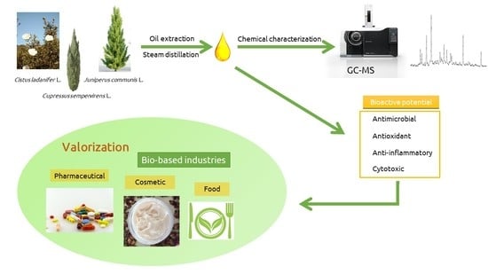
1. Introduction
2. Results and Discussion
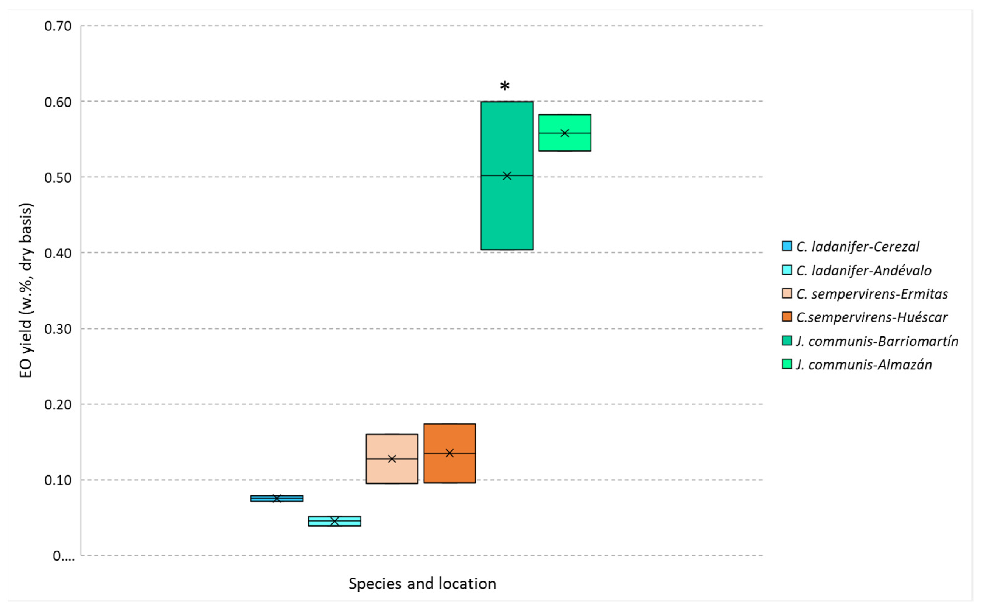
2.1. Essential Oil Yields
2.2. Chemical Composition
2.3. Bioactive Evaluation
2.3.1. Antibacterial Activity
2.3.2. Antioxidant Activity
2.3.3. Cytotoxic and Anti-Inflammatory Activity
3. Materials and Methods
3.1. Plant Material Collection and Conditioning
3.2. Essential Oils Extraction
3.3. Gas Chromatography/Mass Spectrometry (GC-MS) Analyses
3.4. Bioactive Evaluation
3.4.1. Antibacterial Activity
3.4.2. Antioxidant Activity
3.4.3. Cytotoxicity
3.4.4. Anti-Inflammatory Activity
3.4.5. Statistical Analysis
4. Conclusions
Author Contributions
Funding
Institutional Review Board Statement
Informed Consent Statement
Data Availability Statement
Conflicts of Interest
Sample Availability
References
- Serna-Loaiza, S.; Miltner, A.; Miltner, M.; Friedl, A. A Review on the Feed-Stocks for the Sustainable Production of Bioactive Compounds in Biorefineries. Sustainability 2019, 11, 6765. [Google Scholar] [CrossRef] [Green Version]
- Leipold, S.; Petit-Boix, A. The Circular Economy and the Bio-Based Sector—Perspectives of European and German Stakeholders. J. Clean. Prod. 2018, 201, 1125–1137. [Google Scholar] [CrossRef]
- De Corato, U.; De Bari, I.; Viola, E.; Pugliese, M. Assessing the Main Opportunities of Integrated Biorefining from Agro-Bioenergy Co/By-Products and Agroindustrial Residues into High-Value Added Products Associated to Some Emerging Markets: A Review. Renew. Sustain. Energy Rev. 2018, 88, 326–346. [Google Scholar] [CrossRef]
- Ferreira-Santos, P.; Zanuso, E.; Genisheva, Z.; Rocha, C.M.R.; Teixeira, J.A. Green and Sustainable Valorization of Bioactive Phenolic Compounds from Pinus By-Products. Molecules 2020, 25, 2931. [Google Scholar] [CrossRef]
- Nazzaro, F.; De Martino, L.; Fratianni, F.; De Feo, V. Essential Oils from Mediterranean Aromatic Plants. In The Mediterranean Diet, 2nd ed.; Preedy, V.R., Watson, R.R., Eds.; Academic Press: Cambridge, MA, USA, 2020; Chapter 49; pp. 555–564. [Google Scholar]
- Raina, R.; Verma, P.K.; Peshin, R.; Kour, H. Potential of Juniperus communis L. as a Nutraceutical in Human and Veterinary Medicine. Heliyon 2019, 5, e02376. [Google Scholar] [CrossRef] [Green Version]
- Orav, A.; Koel, M.; Kailas, T.; Müürisepp, M. Comparative Analysis of the Composition of Essential Oils and Supercritical Carbon Dioxide Extracts from the Berries and Needles of Estonian Juniper (Juniperus communis L.). Proced. Chem. 2010, 2, 161–167. [Google Scholar] [CrossRef] [Green Version]
- Gumral, N.; Kumbul, D.D.; Aylak, F.; Saygin, M.; Savik, E. Juniperus communis Linn Oil Decreases Oxidative Stress and Increases Antioxidant Enzymes in the Heart of Rats Administered a Diet Rich in Cholesterol. Toxicol. Ind. Health 2015, 31, 85–91. [Google Scholar] [CrossRef]
- Mediavilla, I.; Guillamón, E.; Ruiz, A.; Esteban, L.S. Essential Oils from Residual Foliage of Forest Tree and Shrub Species: Yield and Antioxidant Capacity. Molecules 2021, 26, 3257. [Google Scholar] [CrossRef] [PubMed]
- Ložienė, K.; Venskutonis, P.R. Juniper (Juniperus communis L.) Oils. In Essential Oils in Food Preservation, Flavor and Safety; Preedy, V.R., Ed.; Academic Press: San Diego, CA, USA, 2016; Chapter 56; pp. 495–500. [Google Scholar]
- Pandey, A.K.; Kumar, P.; Saxena, M.J.; Maurya, P. Distribution of Aromatic Plants in the World and Their Properties. In Feed Additives; Florou-Paneri, P., Christaki, E., Giannenas, I., Eds.; Academic Press: Cambridge, MA, USA, 2020; Chapter 6; pp. 89–114. [Google Scholar]
- Guimarães, R.; Barros, L.; Carvalho, A.M.; Sousa, M.J.; Morais, J.S.; Ferreira, I.C.F.R. Aromatic Plants as a Source of Important Phytochemicals: Vitamins, Sugars and Fatty Acids in Cistus ladanifer, Cupressus lusitanica and Eucalyptus gunnii Leaves. Ind. Crops Prod. 2009, 30, 427–430. [Google Scholar] [CrossRef]
- Cox, R.E.; Yamamoto, S.; Otto, A.; Simoneit, B.R.T. Oxygenated Di- and Tricyclic Diterpenoids of Southern Hemisphere Conifers. Biochem. Syst. Ecol. 2007, 35, 342–362. [Google Scholar] [CrossRef]
- Gomes, P.B.; Mata, V.G.; Rodrigues, A.E. Characterization of the Portuguese-Grown Cistus ladanifer Essential Oil. J. Essent. Oil Res. 2005, 17, 160–165. [Google Scholar] [CrossRef]
- Upadhyay, N.; Singh, V.K.; Dwivedy, A.K.; Das, S.; Chaudhari, A.K.; Dubey, N.K. Cistus ladanifer L. Essential Oil as a Plant Based Preservative against Molds Infesting Oil Seeds, Aflatoxin B1 Secretion, Oxidative Deterioration and Methylglyoxal Biosynthesis. LWT 2018, 92, 395–403. [Google Scholar] [CrossRef]
- Höferl, M.; Stoilova, I.; Schmidt, E.; Wanner, J.; Jirovetz, L.; Trifonova, D.; Krastev, L.; Krastanov, A. Chemical Composition and Antioxidant Properties of Juniper Berry (Juniperus communis L.) Essential Oil. Action of the Essential Oil on the Antioxidant Protection of Saccharomyces cerevisiae Model Organism. Antioxidants 2014, 3, 81–98. [Google Scholar] [CrossRef] [Green Version]
- Rguez, S.; Djébali, N.; Ben Slimene, I.; Abid, G.; Hammemi, M.; Chenenaoui, S.; Bachkouel, S.; Daami-Remadi, M.; Ksouri, R.; Hamrouni-Sellami, I. Cupressus sempervirens Essential Oils and Their Major Compounds Successfully Control Postharvest Grey Mould Disease of Tomato. Ind. Crops Prod. 2018, 123, 135–141. [Google Scholar] [CrossRef]
- Stratakos, A.C.; Koidis, A. Methods for Extracting Essential Oils. In Essential Oils in Food Preservation, Flavor and Safety; Preedy, V.R., Ed.; Academic Press: San Diego, CA, USA, 2016; Chapter 4; pp. 31–38. [Google Scholar]
- Tongnuanchan, P.; Benjakul, S. Essential Oils: Extraction, Bioactivities, and Their Uses for Food Preservation. J. Food Sci. 2014, 79, R1231–R1249. [Google Scholar] [CrossRef]
- Zidane, H.; Elmiz, M.; Aouinti, F.; Tahani, A.; Wathelet, J.; Sindic, M.; Elbachiri, A. Chemical Composition and Antioxidant Activity of Essential Oil, Various Organic Extracts of Cistus ladanifer and Cistus libanotis Growing in Eastern Morocco. Afr. J. Biotechnol. 2013, 12, 5314–5320. [Google Scholar]
- Mohammed, B.; Said, C.; Fouzia, F.; Kawtar, F.; Zoubida, H.; Abdelilah, O.; Elhourri, M.; Ghizlane, E. Chemical Composition and Antimicrobial Activity of the Essential Oil of Cistus ladanifer var. maculatus Dun. J. Microbiol. Biotechnol. Food Sci. 2018, 8, 925–930. [Google Scholar]
- Tavares, C.S.; Martins, A.; Faleiro, M.L.; Miguel, M.G.; Duarte, L.C.; Gameiro, J.A.; Roseiro, L.B.; Figueiredo, A.C. Bioproducts from Forest Biomass: Essential Oils and Hydrolates from Wastes of Cupressus lusitanica Mill. and Cistus ladanifer L. Ind. Crops Prod. 2020, 144, 112034. [Google Scholar] [CrossRef]
- Mediavilla, I.; Blázquez, M.A.; Ruiz, A.; Esteban, L.S. Influence of the Storage of Cistus ladanifer L. Bales from Mechanised Harvesting on the Essential Oil Yield and Qualitative Composition. Molecules 2021, 26, 2379. [Google Scholar] [CrossRef]
- Selim, S.A.; Adam, M.E.; Hassan, S.M.; Albalawi, A.R. Chemical Composition, Antimicrobial and Antibiofilm Activity of the Essential Oil and Methanol Extract of the Mediterranean Cypress (Cupressus sempervirens L.). BMC Complement. Altern. Med. 2014, 14, 179. [Google Scholar] [CrossRef]
- Ben Nouri, A.; Dhifi, W.; Bellili, S.; Ghazghazi, H.; Aouadhi, C.; Chérif, A.; Hammami, M.; Mnif, W. Chemical Composition, Antioxidant Potential, and Antibacterial Activity of Essential Oil Cones of Tunisian Cupressus sempervirens. J. Chem. 2015, 2015, 538929. [Google Scholar] [CrossRef] [Green Version]
- Sayed, A.F. Chemical Composition, Antioxidant, Anticancer Properties and Toxicity Evaluation of Leaf Essential Oil of Cupressus sempervirens. Not. Bot. Horti Agrobot. 2015, 43, 320–326. [Google Scholar]
- Angioni, A.; Barra, A.; Russo, M.T.; Coroneo, V.; Dessí, S.; Cabras, P. Chemical Composition of the Essential Oils of Juniperus from Ripe and Unripe Berries and Leaves and Their Antimicrobial Activity. J. Agric. Food Chem. 2003, 51, 3073–3078. [Google Scholar] [CrossRef] [PubMed]
- Kumar, A.; Yadav, L.B.S.; Ahmad, J.; Dubey, N.; Puri, S. Chemical Composition of Commercial Juniperus communis L. Leaf Oil. J. Essent. Oil Bear. Plant. 2007, 10, 310–313. [Google Scholar] [CrossRef]
- Bais, S.; Gill, N.S.; Rana, N.; Shandil, S. A Phytopharmacological Review on a Medicinal Plant: Juniperus communis. Int. Sch. Res. Not. 2014, 2014, 634723. [Google Scholar] [CrossRef] [Green Version]
- Yaglioglu, A.S.; Eser, F.; Yaglioglu, M.S.; Demirtas, I. The Antiproliferative and Antioxidant Activities of the Essential Oils of Juniperus Species from Turkey. Flavour Fragr. J. 2020, 35, 511–523. [Google Scholar] [CrossRef]
- Verdeguer, M.; Blázquez, M.A.; Boira, H. Chemical Composition and Herbicidal Activity of the Essential Oil from a Cistus ladanifer L. Population from Spain. Nat. Prod. Res. 2012, 26, 1602–1609. [Google Scholar] [CrossRef]
- Greche, H.; Mrabet, N.; Zrira, S.; Ismaïli-Alaoui, M.; Benjilali, B.; Boukir, A. The Volatiles of the Leaf Oil of Cistus ladanifer L. var. albiflorus and Labdanum Extracts of Moroccan Origin and Their Antimicrobial Activities. J. Essent. Oil Res. 2009, 21, 166–173. [Google Scholar] [CrossRef]
- ISO. Essential oil of cypress (Cupressus sempervirens L.). In ISO 20809; ISO Copyright Office: Geneva, Switzerland, 2017. [Google Scholar]
- Chatzopoulou, P.S.; Katsiotis, S.T. Chemical Investigation of the Leaf Oil of Juniperus communis L. J. Essent. Oil Res. 1993, 5, 603–607. [Google Scholar] [CrossRef]
- Cabral, C.; Francisco, V.; Cavaleiro, C.; Gonçalves, M.J.; Cruz, M.T.; Sales, F.; Batista, M.T.; Salgueiro, L. Essential Oil of Juniperus communis subsp. alpina (Suter) Čelak Needles: Chemical Composition, Antifungal Activity and Cytotoxicity. Phytother. Res. PTR 2012, 26, 1352–1357. [Google Scholar] [CrossRef]
- Caramiello, R.; Bocco, A.; Buffa, G.; Maffei, M. Chemotaxonomy of Juniperus communis, J. sibirica and J. intermedia. J. Essent. Oil Res. 1995, 7, 133–145. [Google Scholar] [CrossRef]
- Koukos, P.K.; Papadopoulou, K.I. Essential Oil of Juniperus communis L. Grown in Northern Greece: Variation of Fruit Oil Yield and Composition. J. Essent. Oil Res. 1997, 9, 35–39. [Google Scholar] [CrossRef]
- Pepeljnjak, S.; Kosalec, I.; Kalodera, Z.; Blazević, N. Antimicrobial Activity of Juniper Berry Essential Oil (Juniperus communis L., Cupressaceae). Acta Pharm. 2005, 55, 417–422. [Google Scholar]
- Mazari, K.; Bendimerad, N.; Bekhechi, C.; Fernandez, X. Chemical Composition and Antimicrobial Activity of Essential Oils Isolated from Algerian Juniperus phoenicea L. and Cupressus sempervirens L. J. Med. Plant. Res. 2010, 4, 959–964. [Google Scholar]
- Benali, T.; Bouyahya, A.; Habbadi, K.; Zengin, G.; Khabbach, A.; Achbani, E.H.; Hammani, K. Chemical Composition and Antibacterial Activity of the Essential Oil and Extracts of Cistus ladaniferus subsp. ladanifer and Mentha suaveolens against Phytopathogenic Bacteria and Their Ecofriendly Management of Phytopathogenic bacteria. Biocatal. Agric. Biotechnol. 2020, 28, 101696. [Google Scholar] [CrossRef]
- Benayad, N.; Mennane, Z.; Charof, R.; Hakiki, A.; Mosaddak, M. Antibacterial Activity of Essential Oil and Some Extracts of Cistus ladaniferus from Oulmes in Morocco. J. Mater. Environ. Sci. 2013, 4, 1066–1071. [Google Scholar]
- Murbach Teles Andrade, B.F.; Nunes Barbosa, L.; da Silva Probst, I.; Fernandes Júnior, A. Antimicrobial Activity of Essential Oils. J. Essent. Oil Res. 2014, 26, 34–40. [Google Scholar] [CrossRef]
- Hammer, K.A.; Carson, C.F.; Riley, T.V. Antimicrobial Activity of Essential Oils and Other Plant Extracts. J. Appl. Microbiol. 1999, 86, 985–990. [Google Scholar] [CrossRef] [PubMed] [Green Version]
- Boukhris, M.; Regane, G.; Yangui, T.; Sayadi, S.; Bouaziz, M. Chemical Composition and Biological Potential of Essential Oil from Tunisian Cupressus sempervirens L. J. Arid Land Stud. 2012, 22, 329–332. [Google Scholar]
- Falcão, S.; Bacém, I.; Igrejas, G.; Rodrigues, P.J.; Vilas-Boas, M.; Amaral, J.S. Chemical Composition and Antimicrobial Activity of Hydrodistilled Oil from Juniper Berries. Ind. Crops Prod. 2018, 124, 878–884. [Google Scholar] [CrossRef] [Green Version]
- Najar, B.; Shortrede, J.E.; Pistelli, L.; Buhagiar, J. Chemical Composition and In Vitro Cytotoxic Screening of Sixteen Commercial Essential Oils on Five Cancer Cell Lines. Chem. Biodivers. 2020, 17, e1900478. [Google Scholar] [CrossRef] [PubMed] [Green Version]
- Pires, T.; Dias, M.I.; Barros, L.; Alves, M.J.; Oliveira, M.; Santos-Buelga, C.; Ferreira, I. Antioxidant and Antimicrobial Properties of Dried Portuguese Apple Variety (Malus domestica Borkh. cv Bravo de Esmolfe). Food Chem. 2018, 240, 701–706. [Google Scholar] [CrossRef] [PubMed] [Green Version]
- Kostić, M.; Smiljković, M.; Petrović, J.; Glamočlija, J.; Barros, L.; Ferreira, I.; Ćirić, A.; Soković, M. Chemical, Nutritive Composition and a Wide Range of Bioactive Properties of Honey Mushroom Armillaria mellea (Vahl: Fr.) Kummer. Food Funct. 2017, 8, 3239–3249. [Google Scholar] [CrossRef] [Green Version]
- Wolfe, K.L.; Kang, X.; He, X.; Dong, M.; Zhang, Q.; Liu, R.H. Cellular Antioxidant Activity of Common Fruits. J. Agric. Food Chem. 2008, 56, 8418–8426. [Google Scholar] [CrossRef]
- Wolfe, K.L.; Liu, R.H. Structure-Activity Relationships of Flavonoids in the Cellular Antioxidant Activity Assay. J. Agric. Food Chem. 2008, 56, 8404–8411. [Google Scholar] [CrossRef]
- Barros, L.; Pereira, E.; Calhelha, R.C.; Dueñas, M.; Carvalho, A.M.; Santos-Buelga, C.; Ferreira, I.C.F.R. Bioactivity and Chemical Characterization in Hydrophilic and Lipophilic Compounds of Chenopodium ambrosioides L. J. Funct. Foods 2013, 5, 1732–1740. [Google Scholar] [CrossRef]
- Petrova, K.T.; Potewar, T.M.; Correia-da-Silva, P.; Barros, M.T.; Calhelha, R.C.; Ćiric, A.; Soković, M.; Ferreira, I.C. Antimicrobial and Cytotoxic Activities of 1,2,3-Triazole-Sucrose Derivatives. Carbohydr. Res. 2015, 417, 66–71. [Google Scholar] [CrossRef]
- Mandim, F.; Barros, L.; Calhelha, R.C.; Abreu, R.M.V.; Pinela, J.; Alves, M.J.; Heleno, S.; Santos, P.F.; Ferreira, I.C.F.R. Calluna vulgaris (L.) Hull: Chemical Characterization, Evaluation of Its Bioactive Properties and Effect on the Vaginal Microbiota. Food Funct. 2019, 10, 78–89. [Google Scholar] [CrossRef] [Green Version]


| N° | RT (min) | Compound | LRI a | LRI b | Relative % c | |||||
|---|---|---|---|---|---|---|---|---|---|---|
| C. ladanifer | C. sempervirens | J. communis | ||||||||
| Andévalo | Cerezal | Ermitas | Huéscar | Almazán | Barriomartín | |||||
| 1 | 8.07 | tricyclene | 917 | 921 | 0.30 ± 0.02 | 1.08 ± 0.05 # | - | - | - | - |
| 2 | 8.34 | α-thujene | 923 | 924 | - | - | 0.14 ± 0.01 | 0.49 ± 0.01 # | 0.92 ± 0.01 | 0.66 ± 0.06 |
| 3 | 8.81 | α-pinene | 933 | 932 | 42.50 ± 0.96 # | 19.27 ± 0.26 | 55.95 ± 0.46 | 52.32 ± 3.48 | 23.96 ± 0.41 | 35.05 ± 0.02 # |
| 4 | 9.37 | α-fenchene | 945 | 945 | - | - | 0.74 ± 0.03 | 0.64 ± 0.04 | - | - |
| 5 | 9.42 | camphene | 946 | 946 | 2.15 ± 0.07 | 6.66 ± 0.01 # | - | - | - | - |
| 6 | 10.61 | sabinene | 974 | 969 | - | - | 1.07 ± 0.01 | 2.46 ± 0.20 # | 7.86 ± 0.01 | 6.72 ± 0.40 |
| 7 | 10.70 | β-pinene | 979 | 974 | - | - | 1.25 ± 0.02 | 1.42 ± 0.13 | 1.43 ± 0.07 | 1.63 ± 0.01 |
| 8 | 11.49 | β-myrcene | 991 | 988 | - | - | 2.15 ± 0.07 | 2.63 ± 0.19 | 2.60 ± 0.03 | 3.24 ± 0.06 |
| 9 | 12.08 | α-phellandrene | 1003 | 1002 | - | - | - | - | 1.68 ± 0.06 | 1.25 ± 0.02 |
| 10 | 12.41 | 3-carene | 1010 | 1008 | - | - | 13.09 ± 2.70 | 16.18 ± 1.12 | - | - |
| 11 | 12.96 | p-cymene | 1021 | 1020 | 2.04 ± 0.01 | 1.10 ± 0.05 | - | - | - | - |
| 12 | 13.29 | limonene | 1030 | 1024 | 2.07 ± 0.02 | 1.15 ± 0.09 | 2.66 ± 0.01 | 2.74 ± 0.23 | 21.30 ± 0.03 # | 15.01 ± 0.26 |
| 13 | 13.53 | trimethylcyclohexanone | 1033 | 1027 * | 0.53 ± 0.01 | 2.57. ± 0.09 # | - | - | - | - |
| 14 | 14.62 | γ-terpinene | 1054 | 1059 | - | - | - | - | 0.69 ± 0.02 | 0.50 ± 0.14 |
| 15 | 16.14 | terpinolene | 1084 | 1086 | - | - | 2.60 ± 0.02 | 3.58 ± 0.34 # | 0.94 ± 0.01 | 0.80 ± 0.03 |
| 16 | 18.64 | trans-pinocarveol | 1138 | 1139 | 1.45 ± 0.05 | 1.37 ± 0.09 | - | - | - | - |
| 17 | 19.61 | pinocarvone | 1158 | 1160 | 1.01 ± 0.01 | 1.12 ± 0.02 | - | - | - | - |
| 18 | 20.18 | borneol | 1170 | 1165 | 1.22 ± 0.03 | 1.05 ± 0.03 | - | - | - | - |
| 19 | 20.57 | terpinen-4-ol | 1177 | 1174 | - | - | 0.12 ± 0.02 | 0.17 ± 0.03 | 0.77 ± 0.00 | 0.47 ± 0.01 |
| 20 | 25.40 | bornyl acetate | 1282 | 1283 | 4.16 ± 0.10 | 5.01 ± 0.02 | - | - | - | - |
| 21 | 27.99 | α-cubebene | 1345 | 1349 * | - | - | - | - | 0.51 ± 0.01 | 0.26 ± 0.01 |
| 22 | 28.15 | α-terpinyl acetate | 1345 | 1346 | - | - | 1.31 ± 0.01 | 1.33 ± 0.09 | - | - |
| 23 | 28.80 | cyclosativene | 1360 | 1369 | 1.05 ± 0.01 | 1.24 ± 0.02 | - | - | - | - |
| 24 | 29.15 | α-copaene | 1368 | 1374 | 0.73 ± 0.01 | 1.15 ± 0.09 | - | - | - | - |
| 25 | 29.85 | β-elemene | 1384 | 1389 | - | - | - | - | 1.48 ± 0.01 | 1.44 ± 0.01 |
| 26 | 30.98 | β-caryophyllene | 1411 | 1408 | - | - | - | - | 2.14 ± 0.08 | 3.51 ± 0.14 |
| 27 | 31.65 | cis-thujopsene | 1427 | 1429 | - | - | - | - | 8.19 ± 0.14 | 8.04 ± 0.07 |
| 28 | 31.70 | (E)-thujopsene | 1428 | 1431 * | - | - | - | - | 2.63 ± 0.05 | 1.10 ± 0.01 |
| 29 | 32.47 | humulene | 1447 | 1452 | - | - | - | - | 1.41 ± 0.10 # | 0.15 ± 0.04 |
| 30 | 32.65 | alloaromadendrene | 1451 | 1455 * | 1.54 ± 0.03 | 2.92 ± 0.05 # | - | - | - | - |
| 31 | 33.61 | germacrene D | 1474 | 1480 | - | - | 1.38 ± 0.01 | 3.13 ± 0.19 # | 2.69 ± 0.03 # | 0.14 ± 0.01 |
| 32 | 33.94 | viridiflorene | 1482 | 1496 | 1.55 ± 0.01 | 1.50 ± 0.04 | - | - | - | - |
| 33 | 34.62 | cuparene | 1499 | 1504 | - | - | - | - | 0.21 ± 0.01 | 0.26 ± 0.01 |
| 34 | 34.92 | γ-cadinene | 1506 | 1513 | - | - | - | - | 2.19 ± 0.08 # | 0.41 ± 0.01 |
| 35 | 35.13 | δ-cadinene | 1512 | 1522 | 1.25 ± 0.06 | 1.65 ± 0.03 | - | - | 1.41 ± 0.01 | 1.82 ± 0.01 |
| 36 | 36.53 | germacrene B | 1548 | 1559 | - | - | - | - | 0.79 ± 0.04 | 1.45 ± 0.08 # |
| 37 | 37.04 | palustrol | 1561 | 1567 | 0.63 ± 0.02 | 1.06 ± 0.05 | - | - | - | - |
| 38 | 37.44 | spathulenol | 1571 | 1577 | 0.81 ± 0.05 | 1.69 ± 0.02 | - | - | - | - |
| 39 | 38.39 | viridiflorol | 1595 | 1592 | 13.36 ± 1.14 | 24.13 ± 0.74 # | - | - | - | - |
| 40 | 38.58 | ledol | 1598 | 1600 | 4.06 ± 0.17 | 6.94 ± 0.36 | - | - | - | - |
| 41 | 38.61 | cedrol | 1601 | 1602 | - | - | 2.88 ± 0.11 | 4.63 ± 0.25 # | 1.20 ± 0.12 | 1.05 ± 0.03 |
| 42 | 38.74 | copaborneol | 1604 | 1592 * | 1.89 ± 0.03 | 1.76 ± 0.03 | - | - | - | - |
| 43 | 39.94 | τ-muurolol | 1647 | 1640 | - | - | - | - | 0.25 ± 0.03 | 0.50 ± 0.15 |
| Total Identified | 84.32 ± 0.18 | 84.48 ± 0.62 | 85.16 ± 2.17 | 91.76 ± 0.61 | 91.91 ± 0.21 | 88.40 ± 0.31 | ||||
| Monoterpene Hydrocarbons | 49.06 ± 1.04 # | 29.27 ± 0.27 | 79.66 ± 2.29 | 82.48 ± 1.20 | 61.41 ± 0.24 | 64.85 ± 0.33 | ||||
| Oxygen-Containing Monoterpenes | 7.83 ± 0.09 | 8.55 ± 0.11 | 1.43 ± 0.02 | 1.50 ± 0.12 | 0.77 ± 0.01 | 0.47 ± 0.01 | ||||
| Sesquiterpene Hydrocarbons | 6.10 ± 0.06 | 8.47 ± 0.10 | 1.38 ± 0.01 | 3.13 ± 0.19 # | 26.19 ± 0.38 # | 19.30 ± 0.07 | ||||
| Oxygen-Containing Sesquiterpenes | 20.78 ± 1.38 | 35.59 ± 1.00 # | 2.88 ± 0.11 | 4.63 ± 0.25 # | 3.53 ± 0.06 | 3.98 ± 0.04 | ||||
| Others | 0.53 ± 0.01 | 2.57 ± 0.09 # | - | - | - | - | ||||
| Controls | ||||||||||||||||||
|---|---|---|---|---|---|---|---|---|---|---|---|---|---|---|---|---|---|---|
| C. ladanifer | C. sempervirens | J. communis | Ampicilin | Imipenem | Vancomycin | |||||||||||||
| Andévalo | Cerezal | Ermitas | Huéscar | Almazán | Barriomartín | 20 mg/mL | 1 mg/mL | 1 mg/mL | ||||||||||
| Antimicrobial activity | MIC | MBC | MIC | MBC | MIC | MBC | MIC | MBC | MIC | MBC | MIC | MBC | MIC | MBC | MIC | MBC | MIC | MBC |
| Gram-negative bacteria | ||||||||||||||||||
| Escherichia coli | 0.6 | 0.6 | 0.6 | 0.6 | 2.5 | 2.5 | 2.5 | 2.5 | 1.25 | 1.25 | 2.5 | 2.5 | <0.15 | <0.15 | <0.0078 | <0.0078 | n.t. | n.t. |
| Klebsiella pneumoniae | >2.5 | >2.5 | >2.5 | >2.5 | >2.5 | >2.5 | >2.5 | >2.5 | >2.5 | >2.5 | >2.5 | >2.5 | 10 | 20 | <0.0078 | <0.0078 | n.t. | n.t. |
| Morganella morganii | 2.5 | 2.5 | 0.6 | 0.6 | >2.5 | >2.5 | >2.5 | >2.5 | >2.5 | >2.5 | >2.5 | >2.5 | 20 | >20 | <0.0078 | <0.0078 | n.t. | n.t. |
| Proteus mirabilis | >2.5 | >2.5 | >2.5 | >2.5 | >2.5 | >2.5 | >2.5 | >2.5 | >2.5 | >2.5 | >2.5 | >2.5 | <0.15 | <0.15 | <0.0078 | <0.0078 | n.t. | n.t. |
| Pseudomonas aeruginosa | 2.5 | >2.5 | 2.5 | >2.5 | >2.5 | >2.5 | >2.5 | >2.5 | >2.5 | >2.5 | >2.5 | >2.5 | >20 | >20 | 0.5 | 1 | n.t. | n.t. |
| Gram-positive bacteria | ||||||||||||||||||
| Enterococcus faecalis | 1.25 | 1.25 | 0.6 | 0.6 | 2.5 | 2.5 | 2.5 | 2.5 | 2.5 | 2.5 | 2.5 | 2.5 | <0.15 | <0.15 | n.t. | n.t. | <0.0078 | <0.0078 |
| Listeria monocytogenes | 0.6 | 0.6 | 0.3 | 0.3 | 2.5 | 2.5 | 2.5 | 2.5 | 2.5 | 2.5 | >2.5 | >2.5 | <0.15 | <0.15 | <0.0078 | <0.0078 | n.t. | n.t. |
| MRSA | 0.3 | 0.3 | 0.07 | 0.07 | 2.5 | >2.5 | 2.5 | >2.5 | 2.5 | 2.5 | 2.5 | 2.5 | <0.15 | <0.15 | n.t. | n.t. | 0.25 | 0.25 |
| C. ladanifer | C. sempervirens | J. communis | |||||
|---|---|---|---|---|---|---|---|
| Andévalo | Cerezal | Ermitas | Huéscar | Almazán | Barriomartín | Control | |
| Antioxidant Activity | |||||||
| Reducing Power Assay (mg/mL) | Trolox (mg/mL) | ||||||
| EC50 | 1.64 ± 0.34 b | 1.30 ± 0.07 c | 1.52 ± 0.04 b | 1.56 ± 0.01 b | 0.97 ± 0.01 d | 1.35 ± 0.20 b,c | 0.04 ± 0.01 |
| Cellular Antioxidant Activity (µg/mL) | Quercetin | ||||||
| % oxidation inhibition * | 83.24 ± 2.08 a,b | 81.13 ± 3.82 a,b | 73.09 ± 2.24 c,d | 65.91 ± 1.87 e | 78.31 ± 3.08 a,b,c | 68.79 ± 3.34 d,e | 95.30 ± 4.60 ** |
| GI50 | 336.18 ± 8.09 f | 895.45 ± 26.19 d | 1196.09 ± 39.41 c | 1218.65 ± 18.33 c | 324.76 ± 8.13 f | 1563.29 ± 58.02 a | 0.08 ± 0.01 |
| Citotoxicity GI50 (µg/mL) | Ellipticine(µg/mL) | ||||||
| AGS | 78.41 ± 7.11 c | 46.59 ± 4.37 d | 260.53 ± 9.25 a,b | 289.26 ± 19.61 a | 132.68 ± 4.37 b | 302.86 ± 7.92 a | 1.23 ± 0.03 |
| CaCo2 | 75.31 ± 0.99 c | 48.78 ± 0.09 d | 214.28 ± 16.56 a,b | 185.98 ± 6.01 b | 230.79 ± 5.32 a | 107.65 ± 5.15 c | 1.21 ± 0.02 |
| MCF-7 | 27.80 ± 1.28 c | 58.45 ± 1.39 b | 61.97 ± 5.56 b | 165.22 ± 6.95 a | 30.88 ± 1.85 c | 163.39 ± 5.04 a | 1.02 ± 0.02 |
| NCI-H460 | 14.27 ± 1.31 e | 53.80 ± 1.94 b | 19.85 ± 1.69 d,e | 281.64 ± 16.44 a | 44.87 ± 3.42 c | 41.99 ± 3.60 c | 1.01 ± 0.01 |
| PLP2 | 207.64 ± 6.44 a,b | 142.08 ± 1.60 b | 215.18 ± 19.64 a | 237.60 ± 25.84 a | 241.58 ± 9.52 a | 212.03 ± 23.26 a | 2.30 ± 0.10 |
| Vero | 70.77 ± 4.61 c | 46.03 ± 3.60 d | 190.95 ± 17.79 b | 233.69 ± 21.70 a | 240.73 ± 21.32 a | >400 | 1.10 ± 0.10 |
| Anti-inflammatory activity IC50 (µg/mL) | Dexamethasone (µg/mL) | ||||||
| IC50 | 19.27 ± 0.37 b | 21.00 ± 1.70 b | 11.34 ± 1.01 c | 14.41 ± 1.27 c | 84.80 ± 1.43 a | 23.98 ± 0.92 b | 6.30 ± 0.40 |
| Species | Harvesting Place (Province) | Date of Harvesting (DD/MM/YYYY) | Elevation (m) | Coordinates (UTM) |
|---|---|---|---|---|
| C. ladanifer | Andévalo (Huelva) | 5 October 2020 | 199 | 29T 670374; 4174215 |
| Cerezal (Zamora) | 8 September 2020 | 828 | 29T 744316; 4609402 | |
| C. sempervirens | Ermitas (Córdoba) | 17 December 2020 | 470 | 30S 339704; 4198106 |
| Huéscar (Granada) | 17 December 2020 | 988 | 30S 540991; 4187425 | |
| J. communis | Almazán (Soria) | 14 October 2020 | 1079 | 30T 537629; 4601484 |
| Barriomartín (Soria) | 14 September 2020 | 1402 | 30T 545081; 4649553 |
Publisher’s Note: MDPI stays neutral with regard to jurisdictional claims in published maps and institutional affiliations. |
© 2021 by the authors. Licensee MDPI, Basel, Switzerland. This article is an open access article distributed under the terms and conditions of the Creative Commons Attribution (CC BY) license (https://creativecommons.org/licenses/by/4.0/).
Share and Cite
Xavier, V.; Finimundy, T.C.; Heleno, S.A.; Amaral, J.S.; Calhelha, R.C.; Vaz, J.; Pires, T.C.S.P.; Mediavilla, I.; Esteban, L.S.; Ferreira, I.C.F.R.; et al. Chemical and Bioactive Characterization of the Essential Oils Obtained from Three Mediterranean Plants. Molecules 2021, 26, 7472. https://doi.org/10.3390/molecules26247472
Xavier V, Finimundy TC, Heleno SA, Amaral JS, Calhelha RC, Vaz J, Pires TCSP, Mediavilla I, Esteban LS, Ferreira ICFR, et al. Chemical and Bioactive Characterization of the Essential Oils Obtained from Three Mediterranean Plants. Molecules. 2021; 26(24):7472. https://doi.org/10.3390/molecules26247472
Chicago/Turabian StyleXavier, Virginie, Tiane C. Finimundy, Sandrina A. Heleno, Joana S. Amaral, Ricardo C. Calhelha, Josiana Vaz, Tânia C. S. P. Pires, Irene Mediavilla, Luis Saúl Esteban, Isabel C. F. R. Ferreira, and et al. 2021. "Chemical and Bioactive Characterization of the Essential Oils Obtained from Three Mediterranean Plants" Molecules 26, no. 24: 7472. https://doi.org/10.3390/molecules26247472
APA StyleXavier, V., Finimundy, T. C., Heleno, S. A., Amaral, J. S., Calhelha, R. C., Vaz, J., Pires, T. C. S. P., Mediavilla, I., Esteban, L. S., Ferreira, I. C. F. R., & Barros, L. (2021). Chemical and Bioactive Characterization of the Essential Oils Obtained from Three Mediterranean Plants. Molecules, 26(24), 7472. https://doi.org/10.3390/molecules26247472