
Anti-Fibrotic and Anti-Angiogenic Activities of Osbeckia octandra Leaf Extracts in Thioacetamide-Induced Experimental Liver Cirrhosis


 ,
,  ,
,  and
and 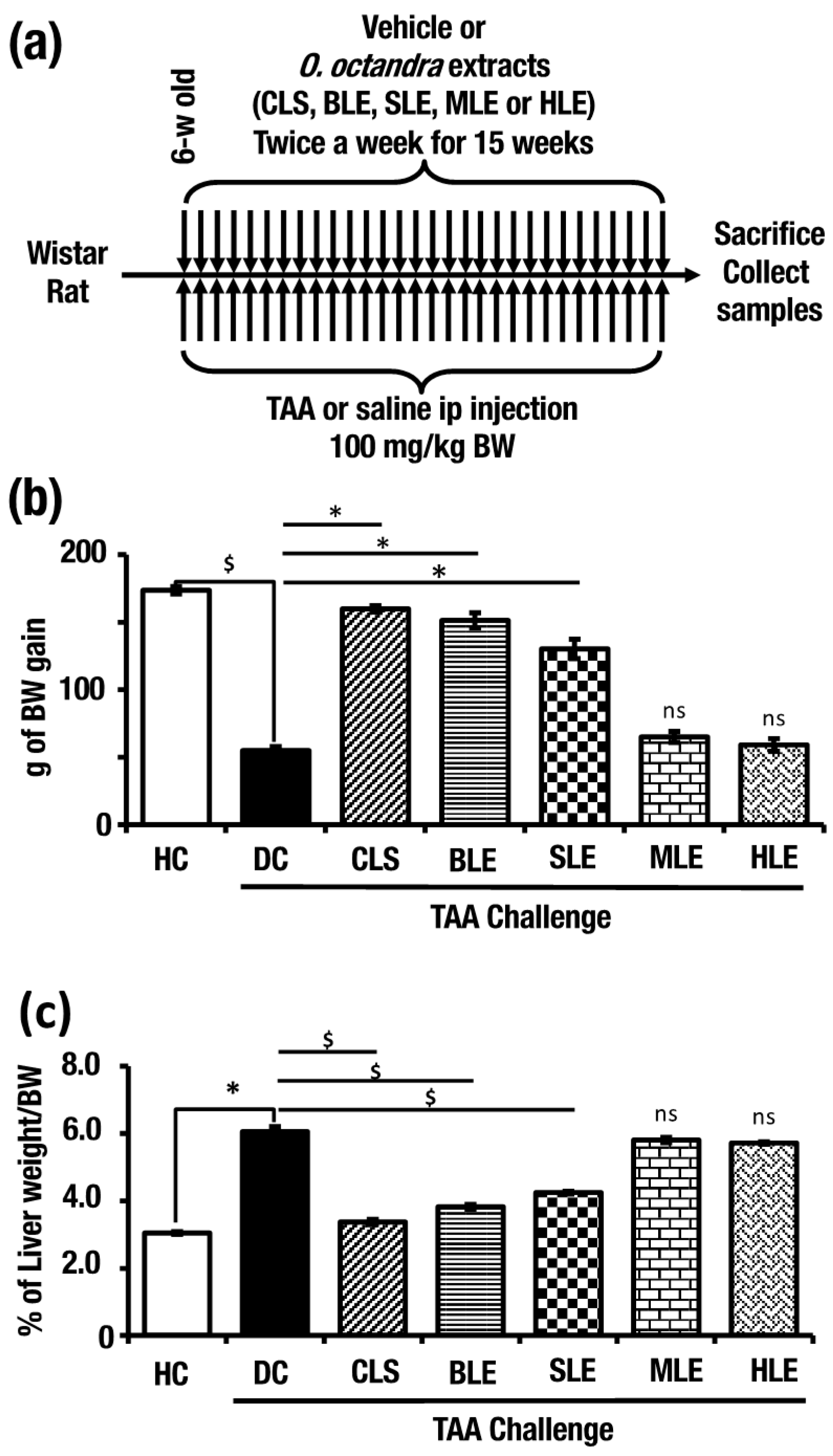
Abstract
1. Introduction
2. Results
2.1. O. octandra Extracts Prevented Body Weight Loss and Normalized Liver Weight
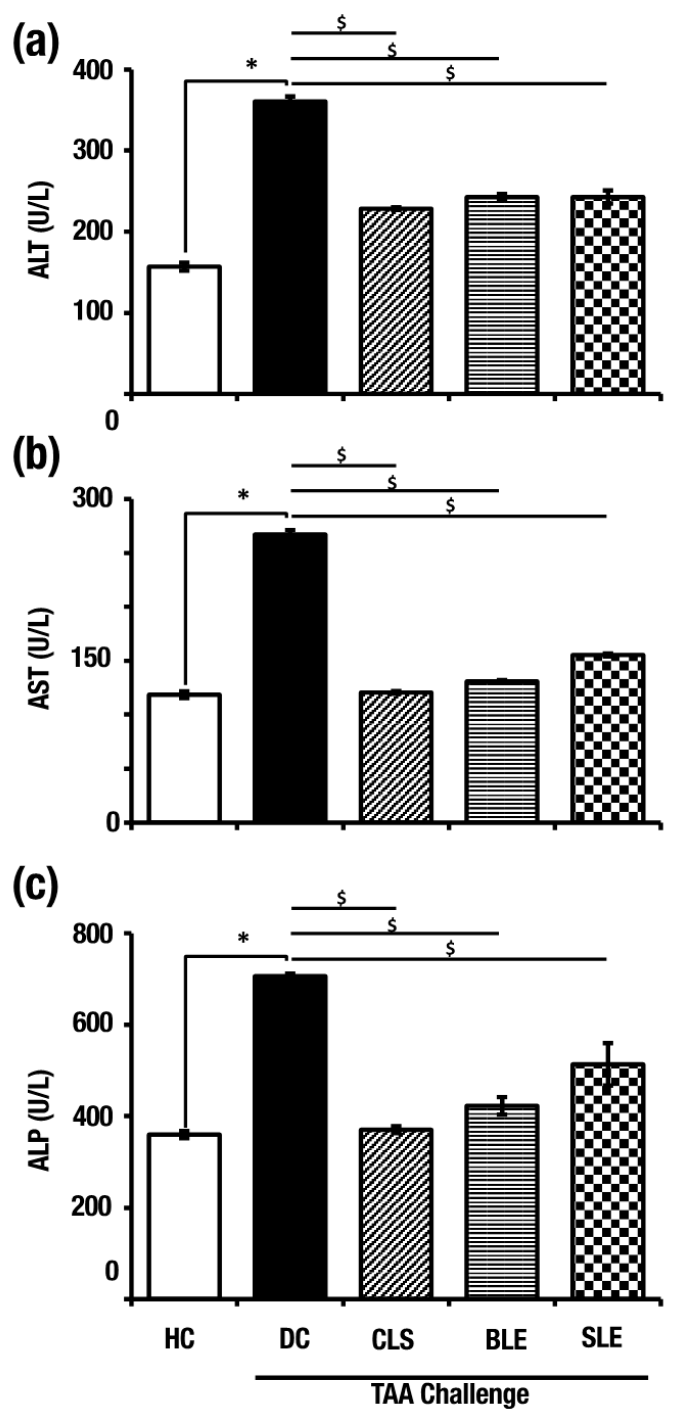
2.2. O. octandra Extracts Restored Serum Concentrations of Liver Enzymes
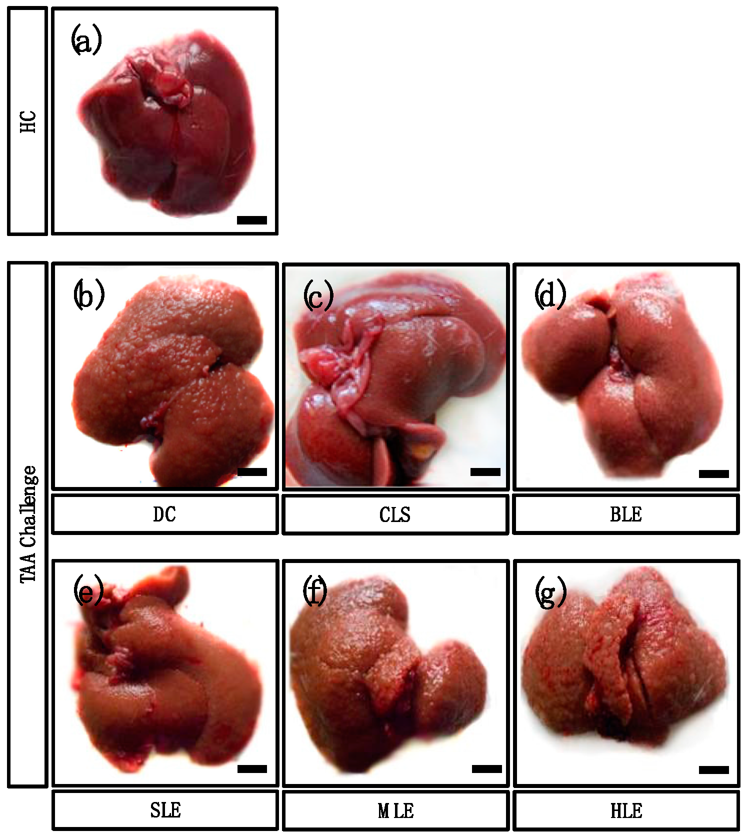
2.3. O. octandra Extracts Restored Gross Liver Appearance
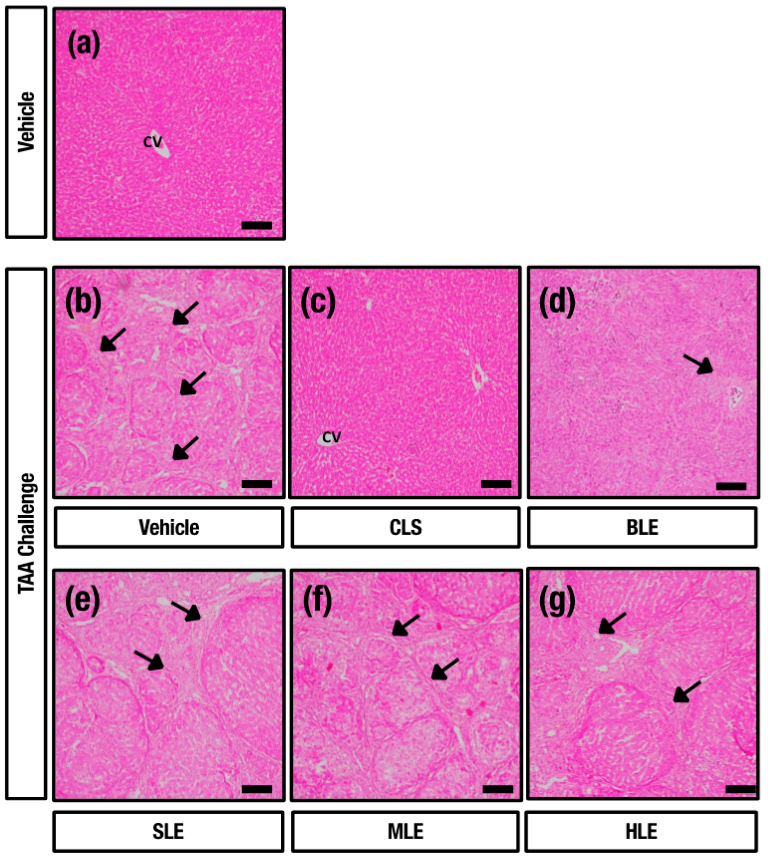
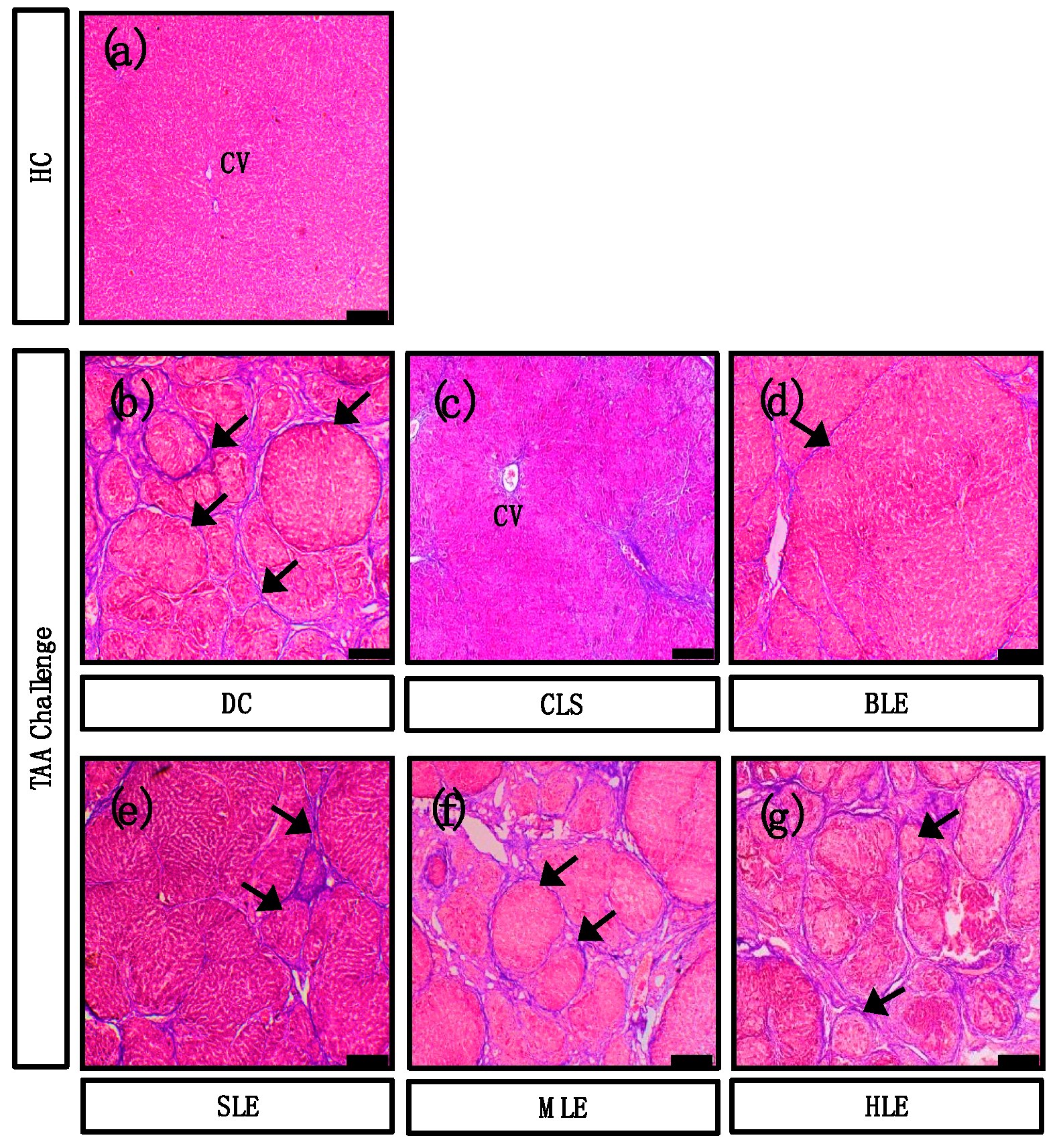
2.4. O. octandra Extracts Restore Liver Architecture
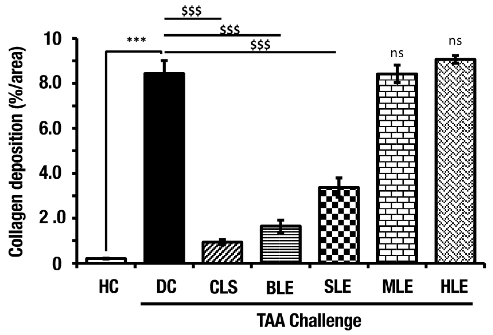
2.5. O. octandra Extracts Ameliorate Liver Fibrosis
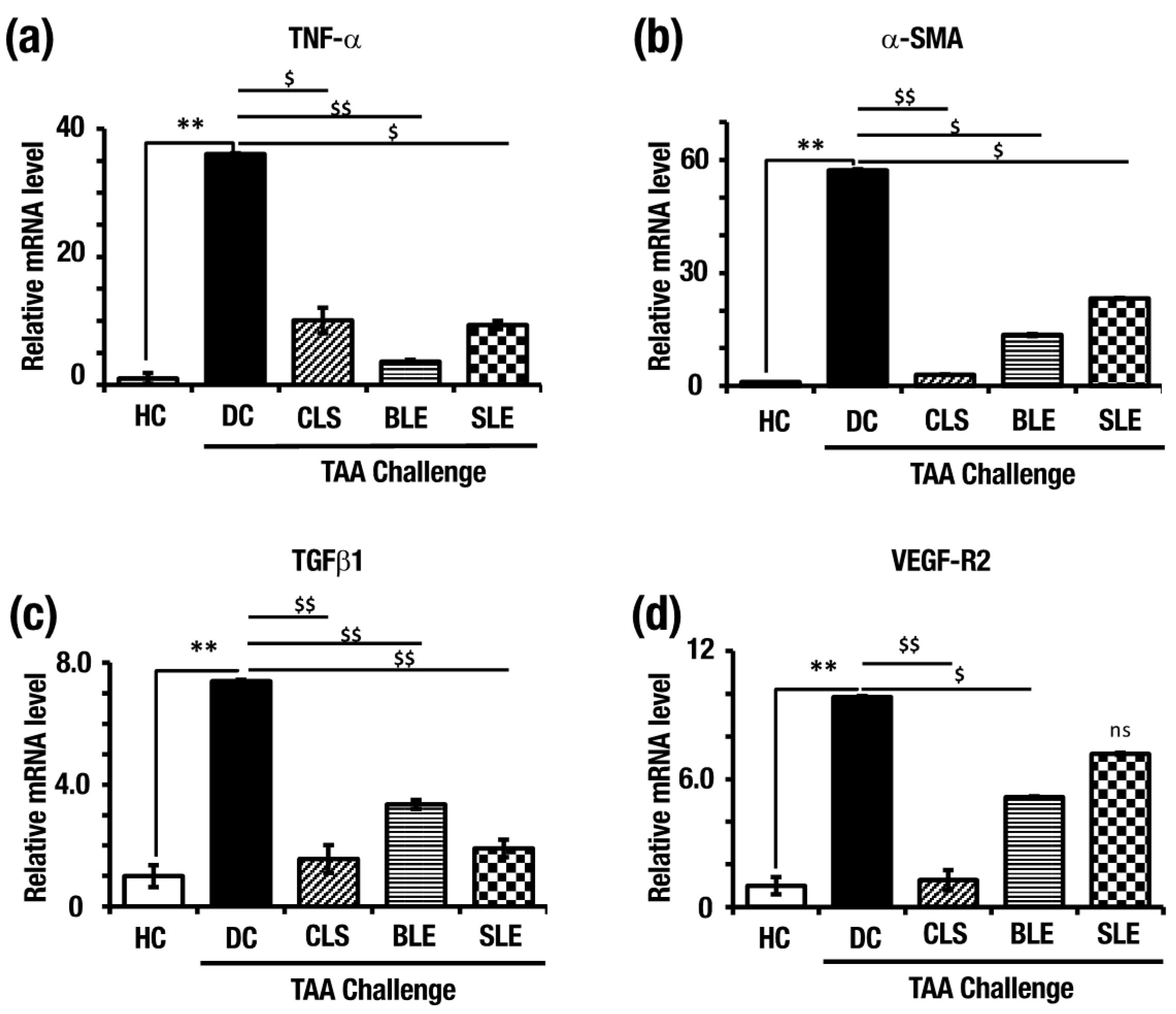
2.6. O. octandra Extracts Prevent Up-Regulation of Pro-Inflammatory and Profibrotic Cytokine mRNA
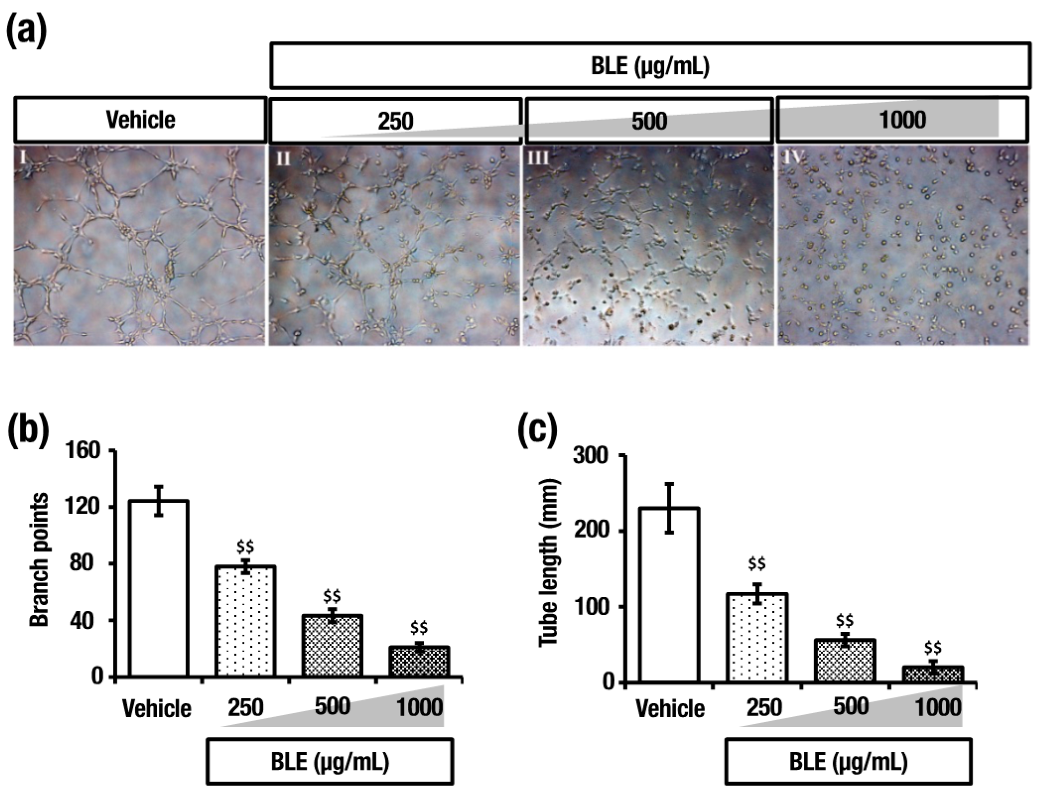
2.7. O. octandra Extract Prevents Angiogenesis
3. Discussion
4. Materials and Methods
4.1. Plant Materials
4.2. Preparation of Leaf Extracts
4.3. Experimental Animals
4.4. Body Weight Assessment and Liver Weight Assessment
4.5. Serum Collection and Blood Chemistry
4.6. Tissue Preparation and Histopathology
4.7. RNA Extraction and Real-Time Quantitative Polymerase Chain Reaction (qPCR)
4.8. Assessment of the Effects of Leaf Extract on Angiogenesis In Vitro
4.9. Statistical Analysis
5. Conclusions
Supplementary Materials
Author Contributions
Funding
Institutional Review Board Statement
Informed Consent Statement
Data Availability Statement
Acknowledgments
Conflicts of Interest
Sample Availability
References
- Mehta, G.A. A textbook for the liver decade. Lancet Gastroenterol. Hepatol. 2017, 2, 706. [Google Scholar] [CrossRef]
- Essawy, A.E.; Abdel-Moneim, A.M.; Khayyat, L.I.; Elzergy, A.A. Nigella sativa seeds protect against hepatotoxicity and dyslipidemia induced by carbon tetrachloride in mice. J. Appl. Pharm. Sci. 2012, 2, 21–25. [Google Scholar] [CrossRef]
- Mak, K.Y.; Chin, R.; Cunningham, S.C.; Habib, M.R.; Torresi, J.; Sharland, A.; Alexander, I.E.; Angus, P.W.; Herath, C.B. ACE2 Therapy Using Adeno-associated Viral Vector Inhibits Liver Fibrosis in Mice. Mol. Ther. 2015, 23, 1434–1443. [Google Scholar] [CrossRef]
- Mokdad, A.A.; Lopez, A.D.; Shahraz, S.; Lozano, R.; Mokdad, A.H.; Stanaway, J.; Murray, C.J.L.; Naghavi, M. Liver cirrhosis mortality in 187 countries between 1980 and 2010: A systematic analysis. BMC Med. 2014, 12, 1–24. [Google Scholar] [CrossRef]
- Özbek, H.; Uğraş, S.; Dülger, H.; Bayram, İ.; Tuncer, İ.; Öztürk, G.; Öztürk, A. Hepatoprotective effect of Foeniculum vulgare essential oil. Fitoterapia 2003, 74, 317–319. [Google Scholar] [CrossRef]
- Thabrew, M.I.; Hughes, R.D.; Gove, C.D.; Portmann, B.; Williams, R.; McFarlane, I.G. Protective effects of Osbeckia octandra against paracetamol-induced liver injury. Xenobiotica 1995, 25, 1009–1017. [Google Scholar] [CrossRef] [PubMed]
- Thabrew, M.I.; Jayatilaka, K.A.P.W. A comparative study of the beneficial effects of Osbeckia octandra and Osbeckia aspera in liver dysfunction in rats. Ceylon J. Med. Sci 1999, 42, 1–6. [Google Scholar] [CrossRef]
- Madrigal-Santillán, E.; Madrigal-Bujaidar, E.; Álvarez-González, I.; Sumaya-Martínez, M.T.; Gutiérrez-Salinas, J.; Bautista, M.; Morales-González, Á.; García-Luna y González-Rubio, M.; Aguilar-Faisal, J.L.; Morales-González, J.A. Review of natural products with hepatoprotective effects. World J. Gastroenterol. 2014, 20, 14787–14804. [Google Scholar] [CrossRef] [PubMed]
- Thabrew, M.I.; Gove, C.D.; Hughes, R.D.; McFarlane, I.G.; Williams, R. Protective effects of Osbeckia octandra against galactosamine and tert-butyl hydroperoxide induced hepatocyte damage. J. Ethnopharmacol. 1995, 49, 69–76. [Google Scholar] [CrossRef]
- Al-Attar, A.M.; Al-Rethea, H.A. Chemoprotective effect of omega-3 fatty acids on thioacetamide induced hepatic fibrosis in male rats. Saudi J. Biol. Sci. 2017, 2, 956–965. [Google Scholar] [CrossRef]
- Wiart, C. Plants classified in the family Polygonaceae. In Medicinal Plants of Asia and the Pacific; CRC Press: Boca Raton, FL, USA, 2006; pp. 47–51. [Google Scholar]
- Andrade, E.L.; Bento, A.F.; Cavalli, J.; Oliveira, S.K.; Schwanke, R.C.; Siqueira, J.M.; Freitas, C.S.; Marcon, R.; Calixto, J.B. Non-clinical studies in the process of new drug development–Part II: Good laboratory practice, metabolism, pharmacokinetics, safety and dose translation to clinical studies. Braz. J. Med. Biol. Res. 2016, 49, e5649. [Google Scholar] [CrossRef]
- Nicholl, D.S.; Daniels, H.M.; Ira Thabrew, M.; Grayer, R.J.; Simmonds, M.S.J.; Hughes, R.D. In vitro studies on the immunomodulatory effects of extracts of Osbeckia aspera. J. Ethnopharmacol. 2001, 78, 39–44. [Google Scholar] [CrossRef]
- Rhiouani, H.; El-Hilaly, J.; Israili, Z.H.; Lyoussi, B. Acute and sub-chronic toxicity of an aqueous extract of the leaves of Herniaria glabra in rodents. J. Ethnopharmacol. 2008, 118, 378–386. [Google Scholar] [CrossRef]
- Hung, Y.; Lee, C. Higher Anti-Liver Fibrosis Effect of Cordyceps militaris-fermented product cultured with deep ocean water via inhibiting proinflammatory factors and fibrosis-related factors expressions. Mar. Drugs 2017, 15, 168. [Google Scholar] [CrossRef]
- Grayer, J.; Thabrew, M.I.; Hughes, R.D.; Bretherton, S.; Lever, A.; Veitch, N.C.; Kite, G.C.; Lelli, R.; Simmonds, M.S.J. Phenolic and terpenoid constituents from the Sri Lankan medicinal plant Osbeckia aspera. Pharm. Biol. 2008, 46, 154–161. [Google Scholar] [CrossRef][Green Version]
- Jain, A.; Soni, M.; Deb, L.; Jain, A.; Rout, S.P.; Gupta, V.B.; Krishna, K.L. Antioxidant and hepatoprotective activity of ethanolic and aqueous extracts of Momordica dioica Roxb. leaves. J. Ethnopharmacol. 2008, 115, 61–66. [Google Scholar] [CrossRef] [PubMed]
- Rajapaksha, I.G.; Gunarathne, L.S.; Asadi, K.; Cunningham, S.C.; Sharland, A.; Alexander, I.E.; Angus, P.W.; Herath, C.B. Liver-Targeted Angiotensin Converting Enzyme 2 Therapy Inhibits Chronic Biliary Fibrosis in Multiple Drug-Resistant Gene 2-Knockout Mice. Hepatol. Commun. 2019, 3, 1656–1673. [Google Scholar] [CrossRef] [PubMed]
- Berardis, S.; Sattwika, P.D.; Najimi, M.; Sokal, E.M. Use of mesenchymal stem cells to treat liver fibrosis: Current situation and future prospects. World J. Gastroenterol. 2015, 21, 742–758. [Google Scholar] [CrossRef]
- Sanal, M.G. Cell therapy from bench to bedside: Hepatocytes from fibroblasts—The truth and myth of transdifferentiation. World J. Gastroenterol. 2015, 21, 6427–6433. [Google Scholar] [CrossRef]
- El-Boshy, M.E.; Abdelhamidb, F.; Richab, E.; Ashshia, A.; Gaitha, M.; Qustya, N. Attenuation of CCl4 Induced oxidative stress, immunosuppressive, hepatorenal damage by fucoidan in rats. J. Clin. Toxicol. 2017, 7, 1–6. [Google Scholar] [CrossRef]
- Zhang, F.; Hao, M.; Jin, H.; Yao, Z.; Lian, N.; Wu, L.; Shao, J.; Chen, A.; Zheng, S. Canonical hedgehog signalling regulates hepatic stellate cell-mediated angiogenesis in liver fibrosis. Br. J. Pharmacol. 2017, 174, 409–423. [Google Scholar] [CrossRef]
- Chan, Y.C.; Khanna, S.; Roy, S.; Sen, C.K. miR-200b Targets Ets-1 and Is Down-regulated by hypoxia to induce angiogenic response of endothelial cells. J. Biol. Chem. 2011, 286, 2047–2056. [Google Scholar] [CrossRef]
- Perera, P.; Ekanayaka, S.; Ranaweera, K. In vitro study on antiglycation activity, antioxidant activity and phenolic content of Osbeckia octandra L. leaf decoction. J. Pharmacogn. Phytochem. 2013, 2, 198–201. [Google Scholar]
- Wijesundera, K.K.; Izawa, T.; Tennakoon, A.H.; Golbar, H.M.; Tanaka, M.; Kuwamura, M.; Yamate, J. M1-/M2-macrophages contribute to the development of GST-P-positive preneoplastic lesions in chemically-induced rat cirrhosis. Exp. Toxicol. Pathol. 2015, 67, 467–475. [Google Scholar] [CrossRef]
- Smith, J.C.; Bolon, B. Isoflurane leakage from non-rebreathing rodent anaesthesia circuits: Comparison of emissions from conventional and modified ports. Lab. Anim. 2006, 40, 200–209. [Google Scholar] [CrossRef] [PubMed]
- Bonder, C.S.; Ajuebor, M.N.; Zbytnuik, L.D.; Kubes, P.; Swain, M.G. Essential role for neutrophil recruitment to the liver in concanavalin A-induced hepatitis. J. Immunol. 2004, 172, 45–53. [Google Scholar] [CrossRef] [PubMed]
- Arnaoutova, I.; Kleinman, H.K. In vitro angiogenesis: Endothelial cell tube formation on gelled basement membrane extract. Nat. Protoc. 2010, 5, 628–635. [Google Scholar] [CrossRef] [PubMed]


Publisher’s Note: MDPI stays neutral with regard to jurisdictional claims in published maps and institutional affiliations. |
© 2021 by the authors. Licensee MDPI, Basel, Switzerland. This article is an open access article distributed under the terms and conditions of the Creative Commons Attribution (CC BY) license (https://creativecommons.org/licenses/by/4.0/).
Share and Cite
Bogahawaththa, S.; Kodithuwakku, S.P.; Wijesundera, K.K.; Siriweera, E.H.; Jayasinghe, L.; Dissanayaka, W.L.; Rajapakse, J.; Herath, C.B.; Tsujita, T.; Wijayagunawardane, M.P.B. Anti-Fibrotic and Anti-Angiogenic Activities of Osbeckia octandra Leaf Extracts in Thioacetamide-Induced Experimental Liver Cirrhosis. Molecules 2021, 26, 4836. https://doi.org/10.3390/molecules26164836
Bogahawaththa S, Kodithuwakku SP, Wijesundera KK, Siriweera EH, Jayasinghe L, Dissanayaka WL, Rajapakse J, Herath CB, Tsujita T, Wijayagunawardane MPB. Anti-Fibrotic and Anti-Angiogenic Activities of Osbeckia octandra Leaf Extracts in Thioacetamide-Induced Experimental Liver Cirrhosis. Molecules. 2021; 26(16):4836. https://doi.org/10.3390/molecules26164836
Chicago/Turabian StyleBogahawaththa, Sudarma, Suranga P. Kodithuwakku, Kavindra K. Wijesundera, Eranga H. Siriweera, Lalith Jayasinghe, Waruna L. Dissanayaka, Jayanthe Rajapakse, Chandana B. Herath, Tadayuki Tsujita, and Missaka P. B. Wijayagunawardane. 2021. "Anti-Fibrotic and Anti-Angiogenic Activities of Osbeckia octandra Leaf Extracts in Thioacetamide-Induced Experimental Liver Cirrhosis" Molecules 26, no. 16: 4836. https://doi.org/10.3390/molecules26164836
APA StyleBogahawaththa, S., Kodithuwakku, S. P., Wijesundera, K. K., Siriweera, E. H., Jayasinghe, L., Dissanayaka, W. L., Rajapakse, J., Herath, C. B., Tsujita, T., & Wijayagunawardane, M. P. B. (2021). Anti-Fibrotic and Anti-Angiogenic Activities of Osbeckia octandra Leaf Extracts in Thioacetamide-Induced Experimental Liver Cirrhosis. Molecules, 26(16), 4836. https://doi.org/10.3390/molecules26164836