Article

Effects of Ulva sp. Extracts on the Growth, Biofilm Production, and Virulence of Skin Bacteria Microbiota: Staphylococcus aureus, Staphylococcus epidermidis, and Cutibacterium acnes Strains

by
Mathilde Fournière
1,2,*,
Gilles Bedoux
1,
Djouhar Souak
3,
Nathalie Bourgougnon
1,
Marc G. J. Feuilloley
3 and
Thomas Latire
1,2
1
Laboratoire de Biotechnologie et Chimie Marines LBCM EA 3884, IUEM, Université Bretagne Sud, 56000 Vannes, France
2
Université Catholique de l’Ouest Bretagne Nord, 22200 Guingamp, France
3
Laboratoire de Microbiologie Signaux et Microenvironnement LMSM EA4312, Université de Rouen Normandie, 27000 Évreux, France
*
Author to whom correspondence should be addressed.
Molecules 2021, 26(16), 4763; https://doi.org/10.3390/molecules26164763
Submission received: 5 July 2021 / Revised: 30 July 2021 / Accepted: 3 August 2021 / Published: 6 August 2021
(This article belongs to the Special Issue Marine Polysaccharides 2022)

Abstract

:
Ulva sp. is known to be a source of bioactive compounds such as ulvans, but to date, their biological activity on skin commensal and/or opportunistic pathogen bacteria has not been reported. In this study, the effects of poly- and oligosaccharide fractions produced by enzyme-assisted extraction and depolymerization were investigated, for the first time in vitro, on cutaneous bacteria: Staphylococcus aureus, Staphylococcus epidermidis, and Cutibacterium acnes. At 1000 μg/mL, poly- and oligosaccharide fractions did not affect the growth of the bacteria regarding their generation time. Polysaccharide Ulva sp. fractions at 1000 μg/mL did not alter the bacterial biofilm formation, while oligosaccharide fractions modified S. epidermidis and C. acnes biofilm structures. None of the fractions at 1000 μg/mL significantly modified the cytotoxic potential of S. epidermidis and S. aureus towards keratinocytes. However, poly- and oligosaccharide fractions at 1000 μg/mL induced a decrease in the inflammatory potential of both acneic and non-acneic C. acnes strains on keratinocytes of up to 39.8%; the strongest and most significant effect occurred when the bacteria were grown in the presence of polysaccharide fractions. Our research shows that poly- and oligosaccharide Ulva sp. fractions present notable biological activities on cutaneous bacteria, especially towards C. acnes acneic and non-acneic strains, which supports their potential use for dermo-cosmetic applications.

1. Introduction

The seaweed Ulva sp. (Chlorophyta, Ulvales, Ulvaceae) is the source of large-scale green tides every year on the Brittany coastline (France) due to its proliferation as a result of marine eutrophication [1,2,3,4]. These green algal blooms have a profound detrimental ecological impact, causing an alteration of the ecosystem structure and a decrease in indigenous biodiversity [5,6], but they also have a considerable economic impact [1]. The valorization of Ulva sp. for human healthcare is currently minimal. This seaweed from stranding events is principally used as a soil amendment or animal feed; it is also not valorized and simply degraded by decay or combustion [1]. Ulva sp. is an important feedstock for structural polysaccharides known as ulvans, which can represent up to 36% of its dry weight [7]. Ulvans are sulfated cell-wall matrix polysaccharides, mainly composed of rhamnose, uronic acids (glucuronic and iduronic), and xylose. The constitutive repeating disaccharide units of the ulvans are mostly aldobiuronic acids (ulvanobiuronic acids) and minority aldobioses (ulvanobioses). Aldobiuronic acids are divided into type A3S (β-d-glucuronic acid (1,4)-linked to α-l-rhamnose 3-sulfate) and type B3S (α-l-iduronic acid (1,4)-linked to α-l-rhamnose 3-sulfate). Ulvanobioses consist of sulfated or non-sulfated β-d-xylose (1,4)-linked to α-l-rhamnose 3-sulfate [8]. Ulvans are traditionally extracted by hot-water extraction at high temperatures (80–90 °C), known as a maceration procedure, followed by an ethanolic precipitation [7]. Novel technology called enzyme-assisted extraction (EAE) can be employed to extract ulvans, as it shows various advantages such as a high extraction yield, mild reaction conditions, the preservation of biological activities, and reduced energy consumption [9,10,11,12,13].
The skin is the largest organ of the human body; it is made up of overlapping layers organized in three major components: epidermis, dermis, and hypodermis. Keratinocytes are the main cells of the epidermis, and fibroblasts are the main cells of the dermis [14]. Ulvans have demonstrated in vitro biological activities towards skin components such as human skin fibroblasts [13,15,16,17,18], which can be related to the structural features of the skin (degree of sulfation, sulfation pattern, monosaccharide composition, molecular weight, and glycosidic linkage) [15,16].
Over the last 10 years, the skin microbiota has been integrated as an essential constitutive element of the cutaneous system. This microbiota is defined as the microorganisms living in or on the skin ecosystem [19] (e.g., 1 million/cm2), and it comprises bacteria, fungi [20,21], yeasts, viruses [22], archaea [23], and mites [24]. It mainly contributes to the maintenance of a healthy skin, and its dysbiosis is a source of inflammatory skin diseases [25,26,27,28]. Within the skin microbiota, it appears that some bacterial species such as Gram-positive bacteria Staphylococcus epidermidis, Staphylococcus aureus (Firmicutes phylum), and Cutibacterium acnes (formerly Propionibacterium acnes [29], Actinobacteria phylum) prevail at skin level, especially in moist and sebaceous areas [25,30,31,32]. S. aureus and S. epidermidis are aerobic bacteria mostly found in external layers of the stratum corneum [33], whereas anaerobic C. acnes is mostly found in more internal layers or skin appendages such as hair follicles and sebaceous glands [34]. C. acnes is highly heterogeneous, with six main phylotypes (IA1, IA2, IB, IC, II, and III) [35,36] and 10 principal ribotypes (RT1 to RT10) [37]. Fitz-Gibbon et al. (2013) showed that some C. acnes ribotypes are associated with acne, such as the RT4 strain, whereas the RT6 strain colonizes non-acneic skin [37]. Within its ecological niche, C. acnes is associated with S. aureus, and both contribute to a healthy microbiota [38]. S. aureus, S. epidermidis, and C. acnes play a fundamental role in the skin microbiota due to their commensal character and their contribution to skin homeostasis. However, these bacteria can turn to opportunistic pathogens depending upon the skin microenvironment [30,39,40,41]. In case of pathogenicity, these bacterial strains can be found in a biofilm form that offers strong resistance to antimicrobial compounds and to the action of an immune skin system [42,43,44].
Both research progress and European Union regulations have motivated the examination of the potential impact of dermo-cosmetic active ingredients on the cutaneous microbiota. The maintenance, protection, and restoration of the skin microbiota’s diversity and equilibrium, and the prevention of skin dysbiosis, constitute one of the research tracks in the cosmetic industry [30,45,46,47]. Active ingredients can modulate bacterial microflora by inducing or repressing bacterial metabolic pathways and can thus influence the skin homeostasis [30,48]. The main activities of ingredients targeted towards the skin bacteria are (1) the promotion or respect of commensal metabolism and/or bacterial diversity in order to limit invasion by pathogens, (2) the reduction of pathogen bacteria growth, virulence, and biofilm formation, and (3) the modulation of the skin microenvironment and immune system responses in case of skin dysbiosis [30].
Marine-derived poly- and oligosaccharides are often considered as potential prebiotics, corresponding to nutrients beneficial to health, for skin commensal bacteria [49]. Thus, seaweeds rich in poly-/oligosaccharides are viewed as potential prebiotics for the skin microbiota [50]. Recent studies have shown that cosmetic ingredients composed of seaweed extracts were able to restore a healthy microbiome and reinforce microbial diversity following stress-induced dysbiosis [51], or increase bacterial diversity in reactive and sensitive skins [52]. To date, however, no study has been conducted on the effects of ulvans or their derivatives on the skin microbiota.
Hence, this study investigates for the first time, in vitro, the effects of poly- and oligosaccharide fractions from Ulva sp. obtained using the novel green technology EAE on four cutaneous bacteria: Staphylococcus aureus MFP03 and Staphylococcus epidermidis MFP04, collected on normal human skin [53], and Cutibacterium acnes RT4 (HL045PA1/HM-516, “acneic” strain) and RT6 (HL110PA3/HM-554, “non-acneic” strain), initially isolated by Fitz-Gibbon et al. (2013) [37]. In this study, the effects of Ulva poly- and oligosaccharides on their growth, biofilm production, cytotoxicity, and/or inflammatory potential on keratinocytes are presented.

2. Results

2.1. Poly- and Oligosaccharide Ulva sp. Fractions: Biochemical Composition and Molecular Weight Distribution

The detailed biochemical composition analysis (carbohydrates, uronic acids, sulfate groups, and proteins), monosaccharide determination (rhamnose, glucuronic acid, and xylose), and molecular weight distribution of Ulva sp. fractions derived from EAE have already been reported [13,16].
The relevant data for the present study are then summarized. Polysaccharide fractions of crude ulvans (UE) and dialyzed ulvans (DS-UE) are composed of high-molecular-weight ulvans (Mw > 670 kDa). Their composition is characterized by 23.5–37.4% carbohydrates (rhamnose 50.2–50.4%, glucuronic acid 10.6–11.9%, and xylose 2.8–3.2%), 18.5–37.0% uronic acids, and 29.9–49.1% sulfate groups. The dialyzed fraction (DS-UE) is significantly richer in ulvans than the crude fraction (UE). Oligosaccharide fractions, depolymerized ulvans from radical hydrolysis by H2O2 (DEP-HD PP-UE), and depolymerized ulvans from acid hydrolysis by Amberlite resin (DEP-AD PP-UE) are constituted by low-molecular-weight ulvans (8 kDa for DEP-HD PP-UE and 1.5 kDa for DEP-AD PP-UE) with 24.4–30.4% carbohydrates (rhamnose 44.9–55.4% and glucuronic acid 7.5–11.0%) and 21.6–30.8% uronic acids. It is noteworthy that the depolymerization process conducted led to a partial loss of sulfates (not detected in DEP-AD PP-UE by Azure A method but identified in MALDI-TOF and only 6.6% in DEP-HD PP-UE).

2.2. Effects of Poly- and Oligosaccharide Ulva sp. Fractions on the Bacterial Growth Kinetic

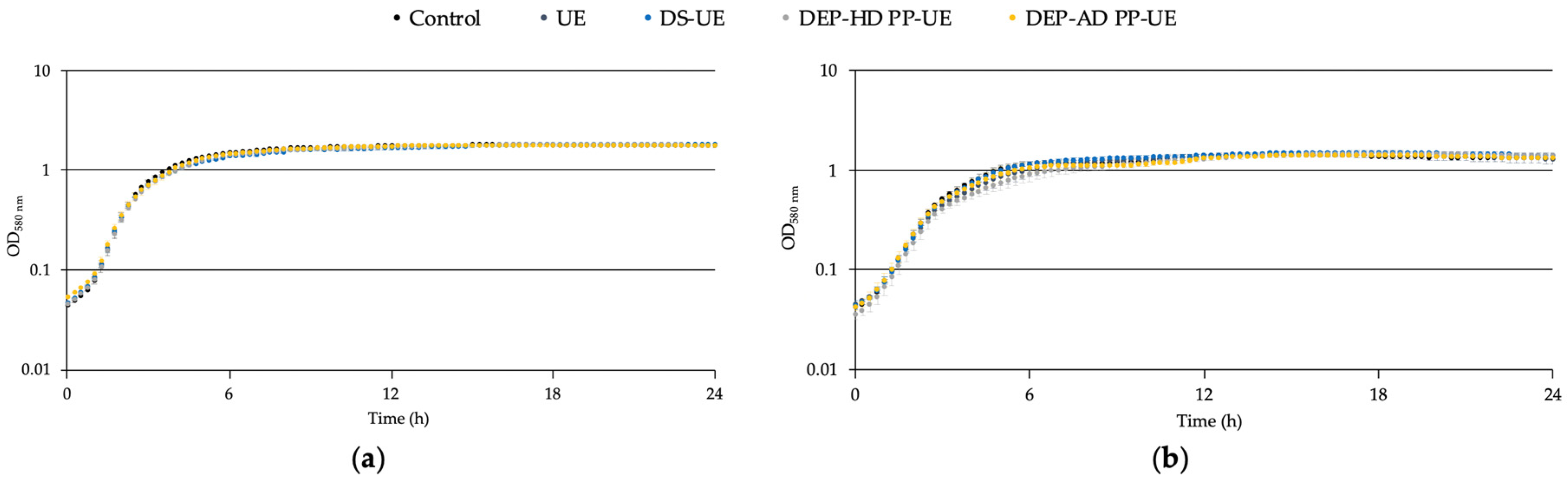
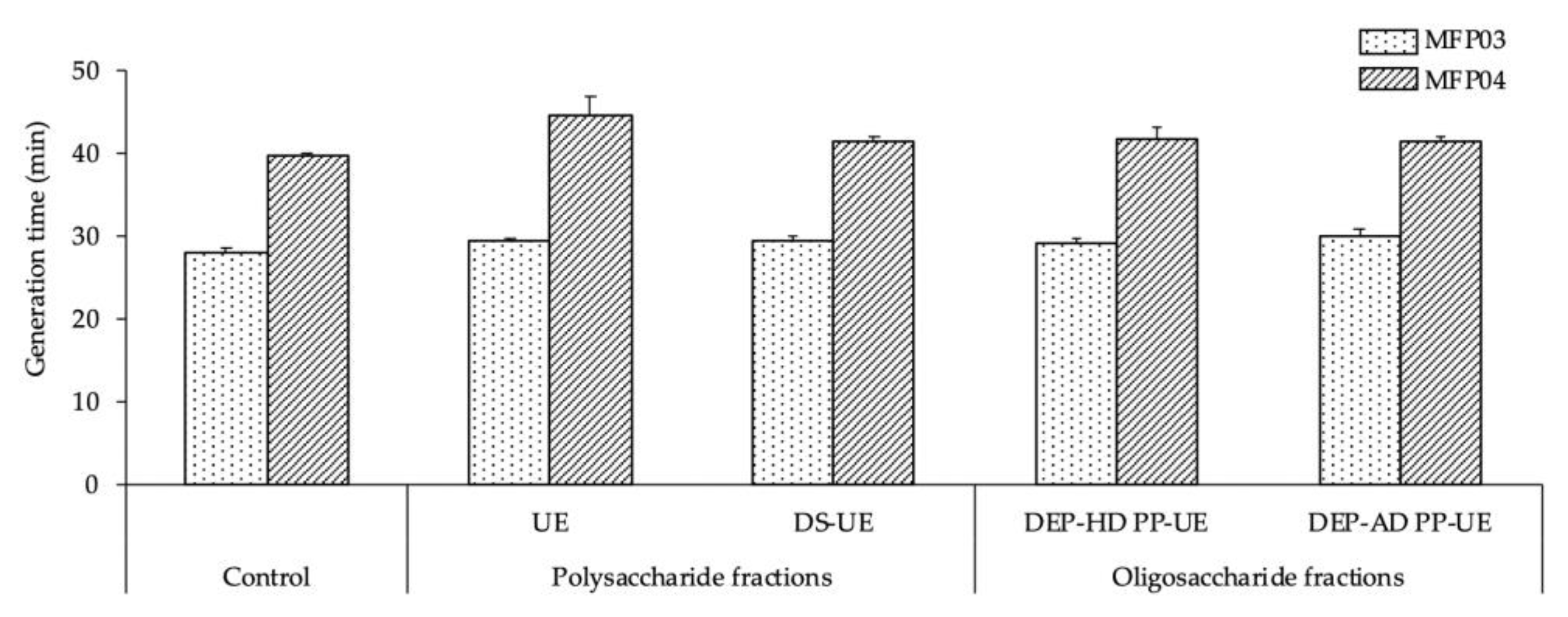
2.2.1. Effects on the Growth of S. aureus MFP03 and S. epidermidis MFP04

The effects of poly- and oligosaccharide Ulva sp. fractions at 1000 μg/mL on the growth of S. aureus MFP03 (Figure 1a) and S. epidermidis MFP04 (Figure 1b) in a tryptic soy broth (TSB) culture medium were evaluated by monitoring over 24 h.
Poly- and oligosaccharide fractions had no effect on the growth of S. aureus MFP03 (Figure 1a) and S. epidermidis MFP04 (Figure 1b).
In addition, the generation times of the bacteria (Figure 2) in a TSB culture medium, which were 27.8 ± 0.6 and 40 ± 0.3 min for S. aureus MFP03 and S. epidermidis MFP04, respectively, were statistically unchanged when the bacteria were exposed to poly- and oligosaccharide Ulva sp. fractions in comparison with the control.

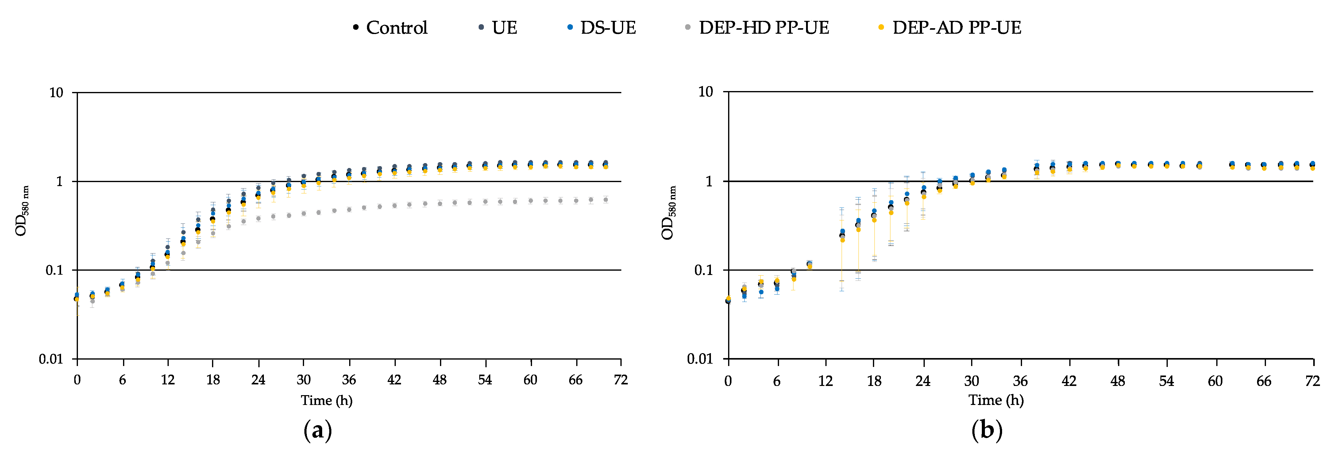
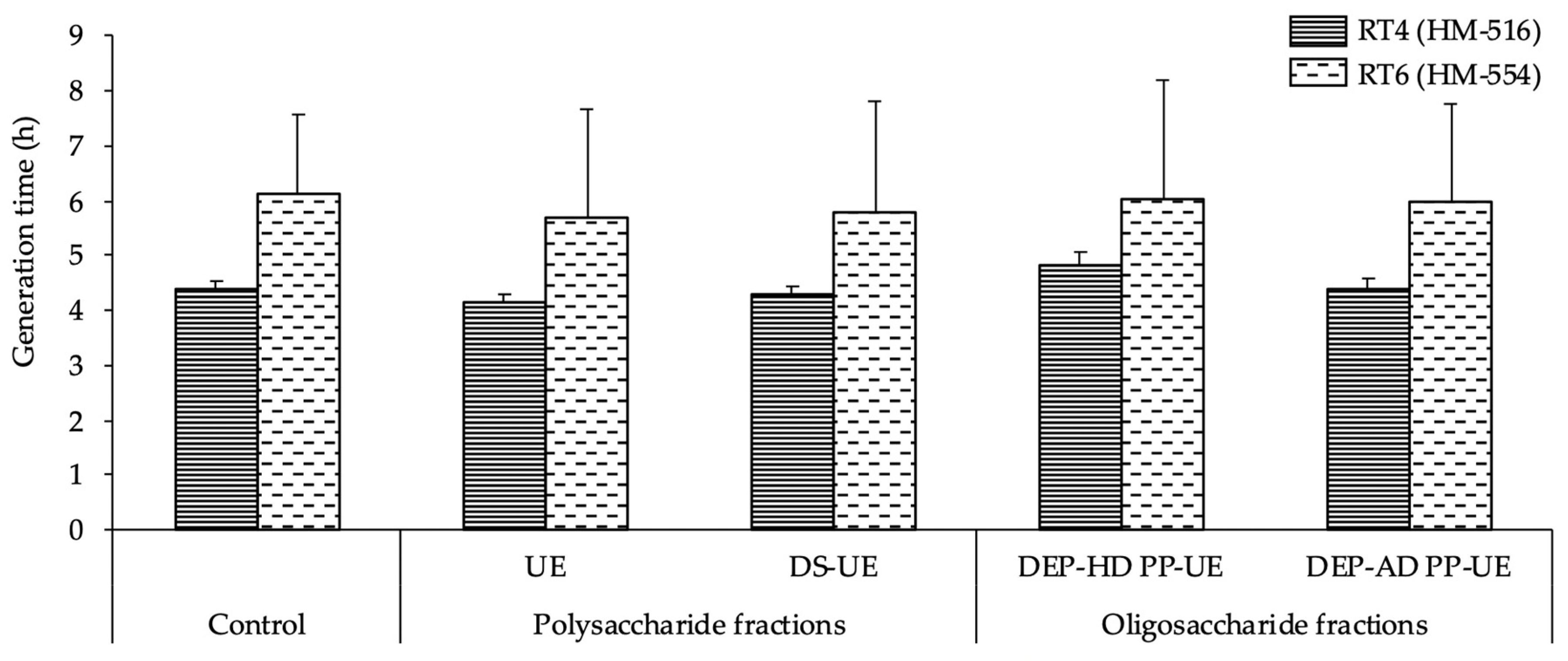
2.2.2. Effects on the Growth of Acneic and Non-Acneic C. acnes Strains

The effects of poly- and oligosaccharide Ulva sp. fractions at 1000 μg/mL on the growth of the acneic C. acnes RT4 strain (Figure 3a) in a reinforced clostridial medium (RCM) pastone +2% glucose and non-acneic C. acnes RT6 strain (Figure 3b) in RCM polypeptone +2% glucose were evaluated by monitoring over 72 h.
The growth of C. acnes RT4 was unchanged when exposed to polysaccharide fractions (UE and DS-UE) and oligosaccharide fraction DEP-AD PP-UE regarding both the exponential phase (generation time) and the stationary phase (Figure 3a). However, the oligosaccharide fraction DEP-HD PP-UE induced a decrease in the stationary phase of the RT4 strain, causing a 50% reduction in the final biomass (Figure 3a). The growth of C. acnes RT6 did not show a noticeable difference in the presence of poly- and oligosaccharide fractions when considering both exponential and stationary phases (Figure 3b). However, important variations were noted during the growth phase, probably because of the limited number of replicates and the harsh growth conditions of this strain, which is strongly anaerobic and hydrophobic [54].
In addition, the generation times of the bacteria (Figure 4), which were 4.4 ± 0.1 h for C. acnes RT4 and 6.1 ± 1.5 h for C. acnes RT6, were unchanged when the bacteria were exposed to poly- and oligosaccharide Ulva sp. fractions.

2.3. Effects of Poly- and Oligosaccharide Ulva sp. Fractions on Biofilm Production

Biofilms visualized by confocal laser scanning microscopy were submitted to image analysis in order to determine the biovolume (μm3/μm2), which provides an estimation of the biomass and mean thickness (μm). Results are expressed as a percentage of variation in relation to the control (i.e., bacteria grown in the absence of Ulva extracts).

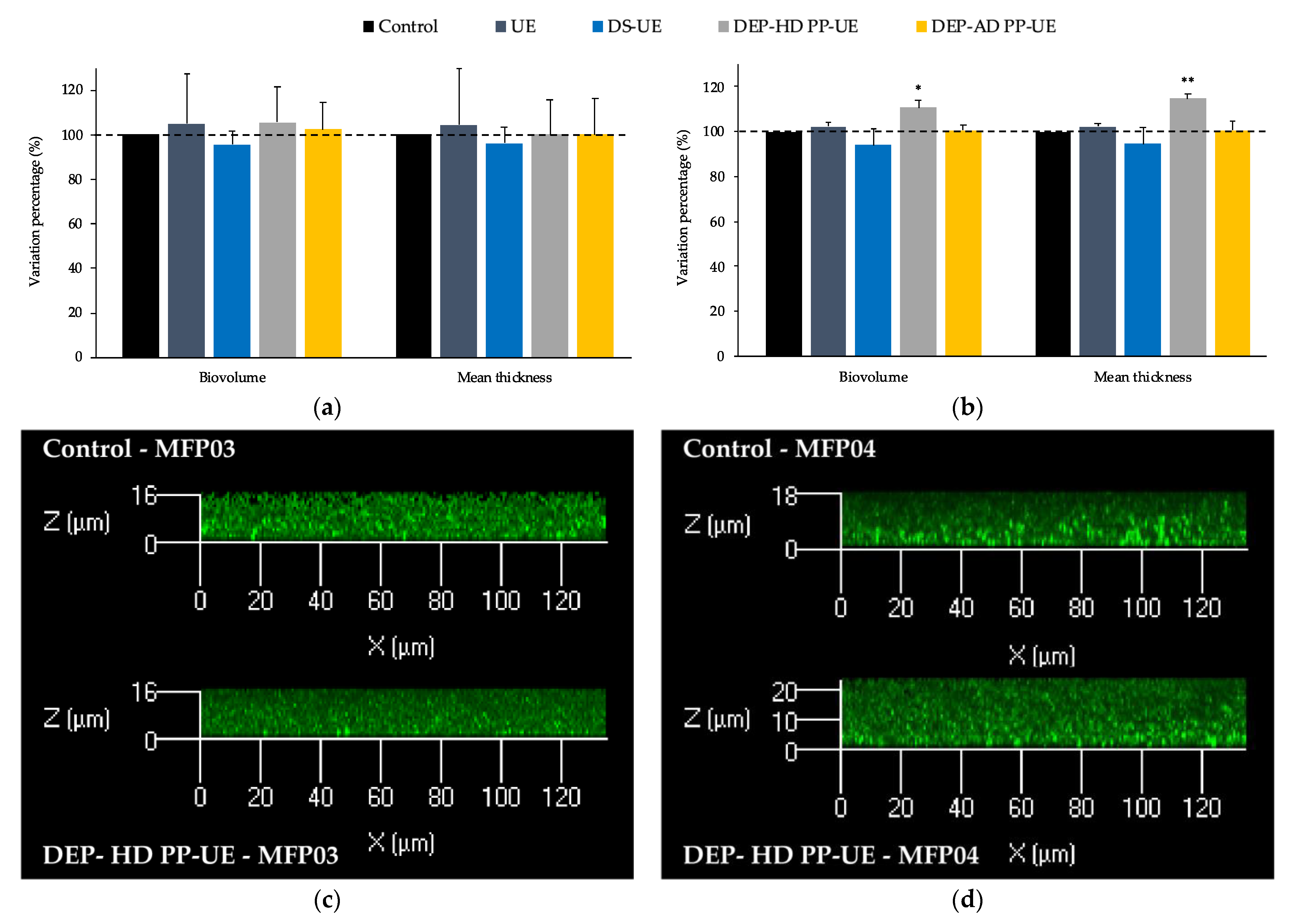
2.3.1. Effects on the Biofilm Production of S. aureus MFP03 and S. epidermidis MFP04

S. aureus MFP03 (Figure 5a) and S. epidermidis MFP04 (Figure 5b) preliminarily cultured in TSB in the absence (control) or presence of poly- and oligosaccharide Ulva sp. fractions (1000 μg/mL) were allowed to form mature biofilms over 48 h in static aerobic conditions.
S. aureus MFP03 biofilms formed by control and treated bacteria had identical mean thickness (4.5 ± 0.5 μm) and biovolume (3.4 ± 0.5 μm3/μm2) values (Figure 5a). In addition, biofilms showed similar 3D organization and density (Figure 5c). No significant modification of the biofilm structure of S. aureus MFP03 was induced by poly- and oligosaccharide Ulva sp. fractions (Figure 5a).
S. epidermidis MFP04 biofilms generated in the same conditions were observed by confocal laser scanning microscopy. In the control conditions, the mean thickness of the biofilms reached 7.1 ± 0.6 μm and the biovolume 4.6 ± 0.4 μm3/μm2 (Figure 5d). No significant modification of the biofilms formed by S. epidermidis MFP04 was induced by the polysaccharide fractions (UE and DS-UE) and oligosaccharide fraction DEP-AD PP-UE (Figure 5b). However, a significant increase in the biovolume (+10.6%; p < 0.05) and mean thickness (+14.7%; p < 0.01) was noted when the bacteria were exposed to the oligosaccharide fraction DEP-HD PP-UE (Figure 5b,d).

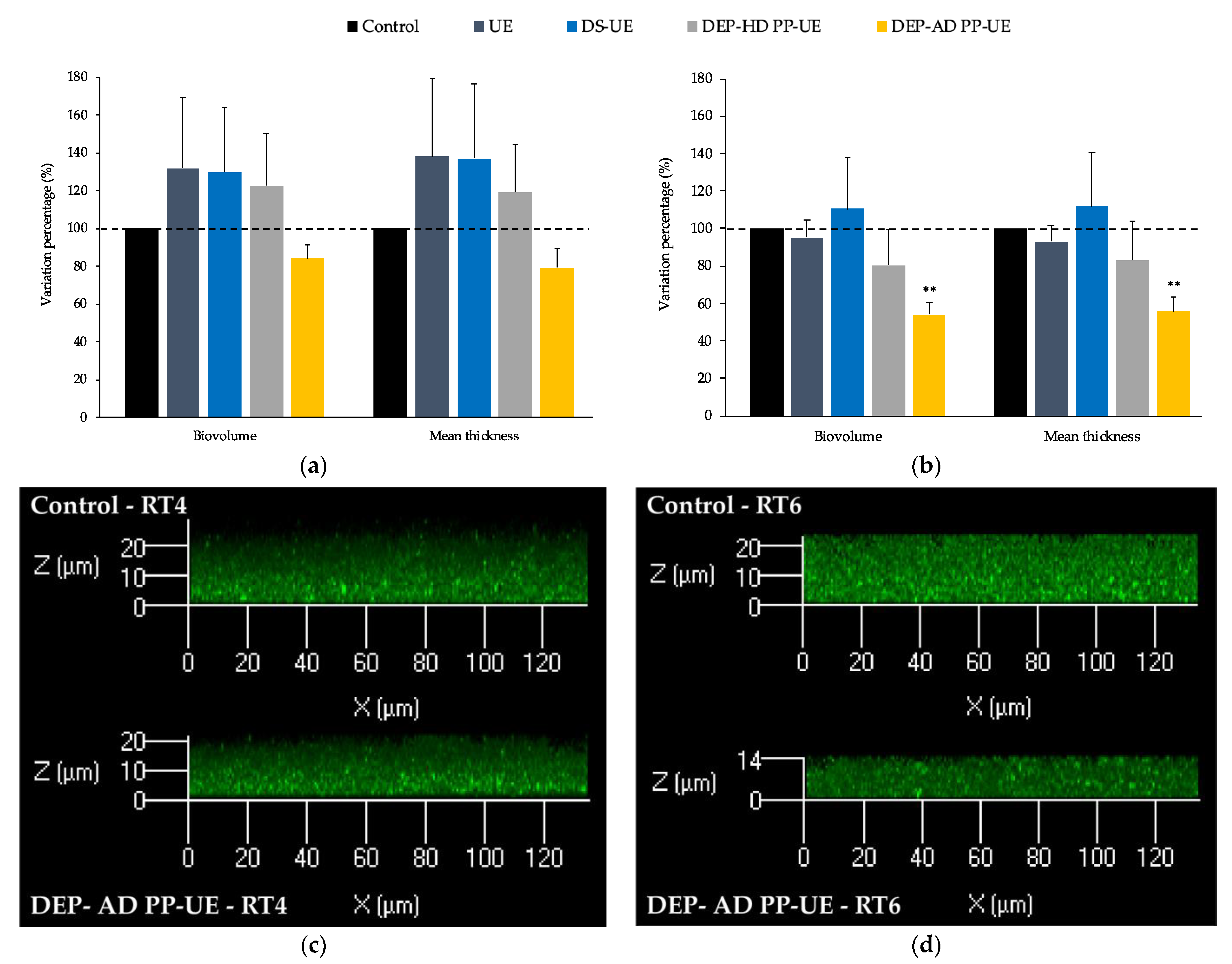
2.3.2. Effects on the Biofilm Production by Acneic and Non-Acneic C. acnes Strains

Acneic C. acnes RT4 (Figure 6a) and non-acneic C. acnes RT6 (Figure 6b) preliminarily cultured in the absence (control) or presence of poly- and oligosaccharide Ulva sp. fractions (1000 μg/mL) were allowed to form mature biofilms over 72 h in static anaerobic conditions.
C. acnes RT4 biofilms formed in control conditions reached a mean thickness of 9.5 ± 1.8 μm and a biovolume of 5.9 ± 1.8 μm3/μm2 (Figure 6c). No significant modification of the biofilm structure was induced by poly- and oligosaccharide Ulva sp. fractions, although decreases in biovolume (−16.0%) and mean thickness (−20.9%) were measured after exposure to DEP-AD PP-UE (Figure 6a).
Control C. acnes RT6 (Figure 6d) biofilms observed in the same conditions had a mean thickness of 5.2 ± 1.3 μm and a biovolume of 3.4 ± 0.9 μm3/μm2. No significant modification of the biofilm structure was induced by the polysaccharide (UE and DS-UE) and oligosaccharide fractions DEP-HD PP-UE (Figure 6b). However, a marked decrease (p < 0.01) in biovolume (−45.7%) and mean thickness (−44.1%) was induced by exposure of the bacteria to the oligosaccharide fraction DEP-AD PP-UE (Figure 6b,d).

2.4. Effects of Poly- and Oligosaccharide Ulva sp. Fractions on the Cytotoxic Activity of S. aureus MFP03 and S. epidermidis MFP04 towards HaCaT Keratinocytes

The bacteria S. aureus MFP03 and S. epidermidis MFP04 were pretreated with poly- and oligosaccharide Ulva sp. fractions at 1000 μg/mL before infecting HaCaT keratinocytes. The effects of the fractions on the cytotoxic activity of S. aureus MFP03 and S. epidermidis MFP04 towards HaCaT keratinocytes were assessed by measuring the LDH (lactate dehydrogenase) release using a PierceTM LDH Cytotoxicity kit assay (Figure 7). The results were expressed as a percentage of variation in comparison to the control (bacteria grown in TSB alone without exposure to Ulva extracts).
The cytotoxicity assay (Figure 7) showed that none of the poly- or oligosaccharide fractions significantly modified the cytotoxic potential of either strains (S. aureus MFP03 or S. epidermidis MFP04) towards HaCaT keratinocytes.

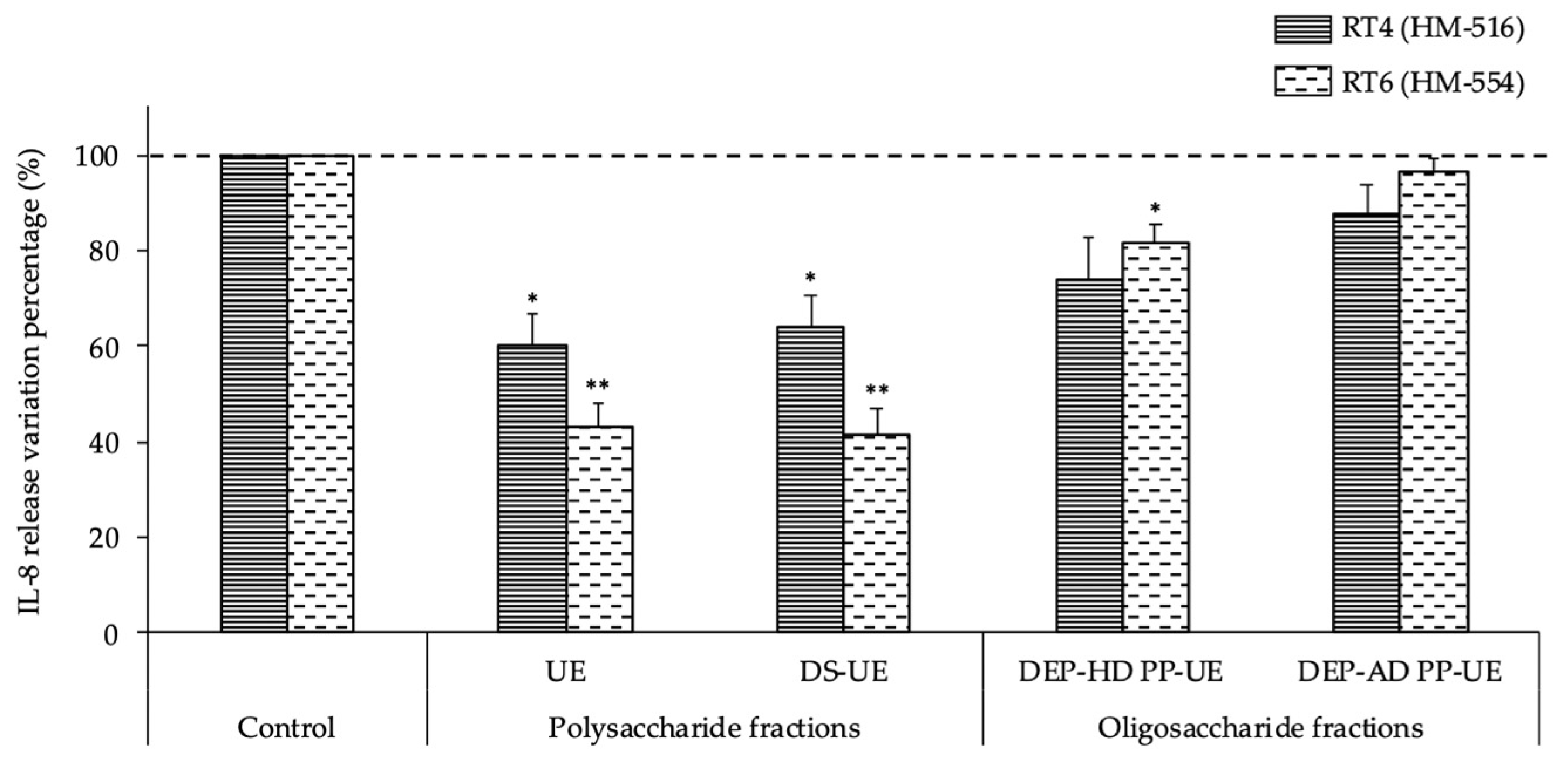
2.5. Effects of Poly- and Oligosaccharide Ulva sp. Fractions on the Inflammatory Potential of Acneic and Non-Acneic C. acnes Strains towards HaCaT Keratinocytes

The intrinsic cytotoxic activity of C. acnes RT4 and RT6 on keratinocytes is negligible [54]. Hence, the effects of Ulva extracts centered on the inflammatory activity of these bacteria. The induction of inflammation was evaluated by an assay of interleukin-8 (IL-8) released by HaCaT keratinocytes after exposure to the bacteria (Figure 8). Results were normalized as a percentage of variation of IL-8 concentration measured in the cell culture medium after incubation in the presence of control (untreated) RT4 or RT6 C. acnes.
The basal release of IL-8 by HaCaT keratinocytes was low (1.8 ± 0.3 pg/mL). Both C. acnes strains had a stimulatory effect on the IL-8 release by keratinocytes, and the acneic strain RT4 showed higher inflammatory potential; the IL-8 concentration in the culture medium reached 1051.2 ± 283.5 pg/mL at the end of the experiment and 801.6 ± 203.3 pg/mL after interaction with the non-acneic strain RT6 (data not shown).
The release of IL-8 by keratinocytes induced by the acneic C. acnes RT4 strain was significantly decreased (p < 0.05) when the strain was cultured in the presence of polysaccharide Ulva sp. fractions (−39.9% for UE and −36.1% for DS-UE) (Figure 8). The oligosaccharide Ulva sp. fractions also induced a decrease in IL-8 release by keratinocytes after infection with RT4, but the difference from the control was not statistically significant (−25.9% for DEP-HD PP-UE and −12.1% for DEP-AD PP-UE). The release of IL-8 by keratinocytes induced by the non-acneic C. acnes RT6 strain was significantly decreased after the strain was cultured in the presence of polysaccharide Ulva sp. fractions (−57.0% for UE and −58.3% for DS-UE; p < 0.01). In addition, the oligosaccharide Ulva sp. fraction DEP-HD PP-UE also induced a significant decrease (−18.3%; p < 0.05) in IL-8 release by keratinocytes after infection with RT6, while DEP-AD PP-UE induced a minor non-significant decrease.

3. Discussion

Our main goal was to evaluate for the first time the potential biological activities on model cutaneous bacteria (i.e., S. epidermidis, S. aureus, and C. acnes) of poly- and oligosaccharide fractions from Ulva sp. obtained after EAE, which is known to improve extraction yield and ulvan enrichment in comparison with maceration [13,55].
The effects of Ulva fractions on the bacteria were investigated by measuring growth, biofilm production, and cytotoxic or inflammatory potentials towards HaCaT keratinocytes. Because of the high content of poly- and oligosaccharide Ulva fractions in carbohydrates (50% rhamnose) and uronic acids (10% glucuronic acid), we postulate that the biological activities detected are mainly related to ulvan composition [7,16].

3.1. Effects of Poly- and Oligosaccharide Ulva sp.-Derived EAE Fractions on Commensal Staphylococcus Species and Acneic or Non-Acneic Cutibacterium acnes

Poly- and oligosaccharide Ulva sp. fractions at 1000 μg/mL had no effect on the growth of S. aureus MFP03 and S. epidermidis MFP04. Similarly, recent studies have demonstrated that polysaccharides from other seaweeds (Gracilaria sp., Sargassum vachellianum, and Fucus spiralis), eventually sulfated, and ulvans from Ulva reticulata do not exert an inhibitory effect on the growth of reference strains of S. aureus or S. epidermidis [56,57,58]. Because of its richness in carbohydrates, the composition of Ulva sp. poly- and oligosaccharides is close to that of PS291® (Téflose®, SOLABIA GROUP), a rhamnose-rich polysaccharide (60%) that has previously been examined on the same strains of S. aureus and S. epidermidis (MFP03 and MFP04) and also had no effect on their growth [59,60].
Thus, our results regarding the growth of S. aureus and S. epidermidis in the presence of poly- and oligosaccharide Ulva sp. fractions are congruent with the literature data on the effects of seaweed polysaccharides and polysaccharides rich in rhamnose. Although the skin bacterial microbiota is dominated by Staphylococcus species such as S. aureus and S. epidermidis, Cutibacterium acnes is also significantly present [32]. Our study demonstrates that acneic strain RT4 and non-acneic strain RT6 do not have their generation time modified in the presence of poly- and oligosaccharide Ulva sp. fractions at 1000 μg/mL. This is consistent with Gannesen et al. (2018) who showed that the rhamnose-rich polysaccharide PS291® also had minor effects on the growth kinetic of C. acnes RT4 [59].
In normal skin, S. epidermidis, S. aureus, and C. acnes usually behave as commensal strains in symbiosis with the cutaneous system [30,31], but in response to variations of the skin microenvironment [40,61,62,63,64], these bacteria can shift to a pathogen phenotype showing variations of biofilm formation and virulence expression [37,65,66,67,68].
As assessed by confocal laser scanning microscopy, the biofilm formation by S. epidermidis MFP04 was increased when the bacteria were cultured in the presence of DEP-HD PP-UE, while other fractions did not show any effect. In addition, no significant effect was observed on biofilm formation by S. aureus MFP03 cultured in the presence of poly- and oligosaccharide Ulva sp. fractions. To our knowledge, this is the first study investigating the effects of seaweed polysaccharides on the biofilm formation of S. aureus and S. epidermidis strains isolated from a healthy skin. Using the crystal violet staining technique, Hillion (2013) showed that the rhamnose-rich polysaccharide PS291® was capable of decreasing biofilm formation by S. epidermidis MFP04. Using confocal microscopy, Gannesen et al. (2018) demonstrated that this ingredient also decreased biofilm formation activity by S. aureus MFP03 [59,60]. However, Hillion (2013) also reported that another polysaccharide of high-molecular-weight (Mw = 2000 kDa) rich in galacturonic acid, L-fucose, and glucose had no effect on S. epidermidis MFP04 biofilm formation [60]. Thus, the effects of polysaccharides on biofilm formation by S. epidermidis MFP04 could be related to their monosaccharide composition. Moreover, Hillion (2013) also demonstrated that a low-molecular-weight oligorhamnose, obtained by hydrolysis of Rhamnosoft® (a cosmetic polysaccharide ingredient of 60 kDa, rich in rhamnose, galactose, and glucuronic acid) had a positive effect on S. epidermidis MFP04 biofilm formation [60]. Our results with DEP-HD PP-UE of low Mw (8 kDa) and monosaccharide composition similar to Rhamnosoft® are thus congruent with previous results.
The data on C. acnes biofilm production are relatively limited, and to date, no study has investigated the effects of seaweed polysaccharides on the biofilm formation by acneic and non-acneic forms of C. acnes. Our study demonstrates for the first time that Ulva sp.-derived polysaccharide fractions (UE and DS-UE) and oligosaccharide fraction DEP-HD PP-UE have no effect on biofilm formation by acneic and non-acneic strains C. acnes. Conversely, the oligosaccharide fraction DEP-AD PP-UE induced a marked decrease in biofilm formation by the non-acneic strain C. acnes RT6 (p < 0.01). A lower production of biofilm was also observed in the acneic strain C. acnes RT4, but the difference was not significant in relation to the control.
In previous studies, Enault et al. (2014) worked on a non-ribotyped strain of C. acnes, and Gannesen et al. (2018) worked on the acneic C. acnes RT4 strain. The authors showed an inhibitory effect of PS291® on biofilm formation that was explained by its overly strong anti-adhesive properties that could limit the early steps of biofilm formation [59,69]. As in our study, this ingredient led to a modification of the C. acnes RT4 biofilm architecture and to a reduction of the mean thickness [59]. Nevertheless, our results are only partially congruent with the literature, since one of the Ulva oligosaccharide fractions showed antibiofilm activity, which suggests a certain specificity in the structure-activity relationships of the polymer.
We also show that poly- and oligosaccharide Ulva sp. fractions did not induce any modification of the cytotoxic potential of S. aureus MFP03 and S. epidermidis MFP04 towards HaCaT keratinocytes. To our knowledge, no previous study has investigated the modulation of the cytotoxic potential of these staphylococci after culture in the presence of seaweed polysaccharides. However, our results are congruent with Hillion (2013) who showed that PS291® did not modify the cytotoxic potential of S. aureus MFP03 and S. epidermidis MFP04 towards HaCaT cells [60]. Because of their very limited cytotoxicity [54], this parameter was not investigated on C. acnes strains, and it was preferred to test the effects of Ulva extracts on the inflammatory potential of these bacteria [70,71]. IL-8 was used as a marker of C. acnes-induced inflammation in HaCaT keratinocytes. We observed that, as previously described [54], HaCaT keratinocytes are able to respond to both C. acnes strains (RT4 and RT6) with a marked increase in IL-8 release, and the RT4 acneic strain has a greater effect. Interestingly, Ulva sp. poly- and oligosaccharide fractions showed a marked anti-inflammatory potential by reducing IL-8 release by keratinocytes after exposure to RT4 (−39.9% maximal value) and RT6 (−58.3% maximum value) C. acnes. This anti-inflammatory activity may result from the binding of ulvan rhamnose residues to bacterial lectins and the inhibition of the bacterial adhesion to keratinocytes [72].

3.2. Potential Implications of the Sulfate Composition and Molecular Weight of Ulvan Fractions and Strain Specificity in Skin Bacteria Modulation

Our study demonstrates that Ulva sp. poly- and oligosaccharides have multiple effects on S. aureus, S. epidermidis, and C. acnes that are mostly strain-specific. Polysaccharide fractions with high-molecular-weight sulfated ulvans (UE and DS-UE) did not induce biofilm formation by S. epidermidis MFP04 and S. aureus MFP03, whereas oligosaccharide fraction DEP-HD PP-UE, with lower molecular weight (8 kDa) and partially desulfated ulvans, promoted biofilm formation by S. epidermidis MFP04. Similarly, only the oligosaccharide fraction DEP-AD PP-UE, which is composed of partially desulfated ulvans of the lowest Mw (1.5 kDa), showed an antibiofilm activity against C. acnes RT4. These results could be coincidental, but they could also be related to the higher efficiency of the desulfated forms of polysaccharides. Gadenne et al. (2015) showed that the best antifouling surface against an S. aureus strain was functionalized with a desulfated ulvan [73]. In addition to considering the inflammatory potential of C. acnes RT4 and RT6 towards HaCaT keratinocytes, we demonstrate that ulvan depolymerization, which leads to a decrease in molecular weight and sulfation degree, is associated with the weakest anti-inflammatory activity for oligosaccharides in comparison to polysaccharides. Thus, various parameters, including molecular weight and the poly- and oligosaccharide sulfation degree, influence the biological activities of Ulva sp. extracts on skin bacteria. On the other hand, we cannot neglect the presence of proteins in the poly- and oligosaccharide Ulva sp. fractions (10.5–16.8%), which suggests a potential influence of the protein/peptide content, amino acid composition, and even partial protein hydrolysis due to depolymerization treatments for Ulva sp. fractions production in modulation of the biological activities, as previously observed in fibroblasts [74].

4. Materials and Methods

4.1. Seaweed Material

The green seaweed Ulva sp. (Chlorophyta, Ulvales, Ulvaceae) was collected from the intertidal beach Landrézac (47°30′17.9″ N 2°42′37.1″ O) in Sarzeau (Brittany, France) on 28 May 2018, during the afternoon, at low tide, before stranding. The seaweed material was washed with tap water, ground to 3 mm in diameter, frozen at −25 °C, freeze-dried (Alpha 1–4 LSC, Martin Christ Gefriertrocknungsanlagen GmbH, Osterode am Harz, Germany), and stored at room temperature in the dark until use.

4.2. Poly- and Oligosaccharide Ulva sp. Fractions Production and Characterization

The detailed procedures of poly- and oligosaccharide Ulva sp. fractions production from EAE were fully described in Fournière et al. (2019) (see Figure 9 and Table 1) [13].
To summarize, enzymatic hydrolysis was performed at 50 °C for 3 h by using endo-protease Protamex® at 6% (w/dw) in distilled water on freeze-dried Ulva sp. material; next, the enzyme was inactivated at 90 °C for 15 min. The hydrolysate was treated by ethanolic precipitation (absolute ethanol, 1:5, v/v) at 4 °C for 24 h in order to produce a fraction rich in crude ulvans (UE).
A purification step was then performed by dialysis of the fraction rich in crude ulvans (UE, 10 mg/mL) at 4 °C for 7 days (cut-off 12–14 kDa, Spectra/Por®4 Dialysis Membrane, Spectrum Laboratories, Fisher Scientific, Illkirch, France) in order to produce the fraction of dialyzed ulvans (DS-UE).
Depolymerization procedures, radical hydrolysis by hydrogen peroxide (H2O2), and acid hydrolysis by ion-exchange resin were applied on an ethanolic precipitate solution (25 mg/mL) obtained from UE. For radical depolymerization, H2O2 (8%, v/v, Fisher Scientific, Illkirch, France) was mixed with the solution at 50 °C for 24 h. For acid depolymerization, ion-exchange resin Amberlite® FPC23 H (10 mL equivalent, Sigma-Aldrich, Saint Quentin Fallavier, France) was mixed with the solution at 80 °C for 24 h according to the protocol of Adrien et al. (2017), and the mixture after filtration was neutralized with NaOH (0.1 and 1 M) [15]. Next, both solutions from radical and acid depolymerization underwent dialysis at 4 °C for 48 h (cut-off of 500–1000 Da, Biotech CE Tubing, Spectra/Por®, Fisher Scientific, Illkirch, France).
These two depolymerization procedures led to the production of oligosaccharide fractions named DEP-HD PP-UE and DEP-AD PP-UE for radical hydrolysis by H2O2 and acid hydrolysis by ion-exchange resin procedure, respectively.
All produced fractions (crude, dialyzed, and depolymerized) were freeze-dried (Alpha 1–4 LSC, Martin Christ Gefriertrocknungsanlagen GmbH, Osterode am Harz, Germany) and stored at 4 °C until use.
The molecular weight distribution was analyzed by high-performance size-exclusion chromatography (HPSEC, UHPLC Ultimate 3000, Thermo Fisher Scientific, Waltham, MA USA). The biochemical composition was assessed by colorimetric methods. The monosaccharide composition was determined by high-performance anion-exchange chromatography with pulsed amperometric detection (HPAEC-PAD, DionexTM ICS-5000+ DC, Thermo Fisher Scientific, Illkirch, France), and completed with matrix-assisted laser desorption/ionization time-of-flight mass spectrometry (MALDI-TOF, Microflex ToF machine, Bruker Daltonik GmbH, Leipzig, Germany) [13].

4.3. Bacterial Strains and Culture Conditions

4.3.1. Staphylococcus aureus and Staphylococcus epidermidis Strains

Staphylococcus aureus MFP03 and Staphylococcus epidermidis MFP04 were initially isolated from the skin of healthy volunteers [53]. The bacteria, stored at −140 °C in cryobead form, were initially plated on tryptic soy agar (TSA, Sigma-Aldrich, Saint-Quentin Fallavier, France). After overnight (O/N) incubation in static aerobic conditions at 37 °C, the Petri plate could be stored at 4 °C before use within one month. Colonies were transferred in a 10 mL tryptic soy broth (TSB) medium into a 100 mL sterile Erlenmeyer flask (1:10 ratio for aerobic conditions). These pre-cultures were incubated O/N under agitation at 180 rpm (rotations per minute) and 37 °C.

4.3.2. Cutibacterium acnes Strains

The acneic strain RT4 HL045PA1/HM-516 and the non-acneic strain RT6 HL110PA3/HM-554 of Cutibacterium acnes, initially isolated by Fitz-Gibbon et al. (2013) [37], were diffused by BEI Resources American Type Culture Collection (Virginia, USA). These strains refer to phylotypes IA1 and II, respectively [35]. The bacteria, stored at −140°C in cryobead form, were initially plated on agar brain heart infusion (BHI) for RT6 and on agar reinforced clostridial medium (RCM, Sigma-Aldrich, Saint-Quentin Fallavier, France) pastone +2% glucose for RT4. As these strains grew under anaerobic conditions, the plates were incubated under anoxic static conditions at 37 °C for 72 h, using BD GasPackTM EZ pouch systems (BD).
Colonies were transferred into sterile conical 15 mL plastic tubes (Falcon) filled to maximal capacity (15 mL) with RCM polypeptone +2% glucose for RT6 and RCM pastone +2% glucose for RT4. These pre-cultures were incubated in anoxic static conditions at 37 °C for 72 h.

4.4. Bacterial Growth Kinetic

The effects of poly- and oligosaccharide fractions on bacterial growth were evaluated by considering generation time, which corresponds to the necessary time for the bacteria to double in number. The generation time (G) of bacterial strains was calculated with Equation (1) and expressed in minutes for S. aureus MFP03 and S. epidermidis MFP04 and in hours for C. acnes RT4 and RT6:
G   ( time   unit ) = ln 2 Slope   of   the   growth   curve   in   exponential   phase   × 100 .

4.4.1. Staphylococcus aureus and Staphylococcus epidermidis Strains

After pre-culture, S. aureus MFP03 and S. epidermidis MFP04 were collected (stationary phase) and cultured at an initial OD580nm (optical density at 580 nm) = 0.08 in TSB, supplemented, or not (control condition), with poly- and oligosaccharide Ulva sp. fractions at 1000 μg/mL, in a Bioscreen HoneyComb plate (Thermo Fisher Scientific, Waltham, MA, USA). OD measurement was performed automatically in a microplate reader (Bioscreen C, Oy Growth Curves Ab Ltd., Helsinki, Finland) at 580 nm every 15 min for 24 h with continuous shaking.

4.4.2. Cutibacterium acnes Strains

After pre-culture, C. acnes strains were collected (stationary phase) and cultured at an initial OD580nm = 0.08 in RCM pastone +2% glucose (RT4) or RCM polypeptone +2% glucose (RT6) supplemented, or not (control condition), with poly- and oligosaccharide Ulva sp. fractions at 1000 μg/mL, in a 96-well flat-bottomed polystyrene plate (NUNC) under anoxic static conditions. For C. acnes RT4, the microplate was sealed with silicone and parafilm before incubation, and OD was monitored in the microplate reader (TECAN Spark 10M, Männedorf, Switzerland) automatically every 2 h at 580 nm for 72 h. In the case of C. acnes RT6, the plate was incubated in a Whitley A85 Workstation for 72 h and OD measurement was performed manually every 2 h in the microplate reader (TECAN Spark 10M, Männedorf, Switzerland) at 580 nm.

4.5. Biofilm Formation Activity: Confocal Laser Scanning Microscopy

The bacteria were pre-cultured as previously described (Section 4.3.1 for S. aureus MFP03 and S. epidermidis MFP04, and Section 4.3.2 for C. acnes RT4 and RT6). Then, the bacteria at an initial OD580nm = 0.08 in a specific medium (TSB for S. aureus MFP03 and S. epidermidis MFP04, RCM pastone +2% glucose for C. acnes RT4, and RCM polypeptone +2% glucose for C. acnes RT6), supplemented, or not (control condition), with poly- and oligosaccharide Ulva sp. fractions at 1000 μg/mL, were grown under aerobic conditions for 16 h for S. aureus MFP03 and S. epidermidis MFP04 (O/N under agitation at 180 rpm), and under anoxic static conditions for 72 h for C. acnes RT4 and RT6.
After incubation, the bacteria in stationary phase were collected by centrifugation (8000× g for 5 min at room temperature). The supernatant was removed, and the bacterial pellet was rinsed with sterile physiological water NaCl 0.9% (PS) in order to remove all traces of the culture medium and poly- or oligosaccharide fractions. The biomass was again centrifuged (8000× g for 5 min at room temperature) before being resuspended in PS (bacterial suspension) at OD580nm = 0.1 for S. aureus MFP03 and S. epidermidis MFP04, and OD580nm = 1 for C. acnes RT4 and RT6.
Next, aliquots of 200 μL of bacterial suspension of S. aureus MFP03 or S. epidermidis MFP04 and 1 mL of bacterial suspension of C. acnes RT4 or RT6, were transferred to the wells of 24-well-plates with flat glass bottoms (SensoPlate, Greiner Bio-One, Frickenhausen, Germany). Each bacterial suspension was tested in triplicate for each condition under investigation, using at least four independent experiments for each strain.
After distribution of bacterial aliquots, the plates were incubated at 37 °C for 2 h to allow primary adhesion, under static and aerobic conditions for S. aureus MFP03 and S. epidermidis MFP04, and under static and anoxic conditions for C. acnes RT4 and RT6 (Whitley A85 Workstation, Don Whitley Scientific, Bingley, United Kingdom). Next, the non-adhered bacteria were carefully removed, and a fresh culture medium was added: 200 μL/well of TSB +2% glucose for S. aureus MFP03 and S. epidermidis MFP04, 1 mL/well of RCM pastone +2% glucose for C. acnes RT4, and 1 mL/well of RCM polypeptone +2% glucose for C. acnes RT6. The plates were incubated at 37 °C for 46 h under static and aerobic conditions for S. aureus MFP03 and S. epidermidis MFP04, and at 37 °C for 70 h under static and anoxic conditions for C. acnes RT4 and RT6 (Whitley A85 Workstation, Don Whitley Scientific, Bingley, UK).
At the end of the incubation time, the culture medium was carefully removed from each well, and the bacteria were stained with SYTO9 Green Fluorescent Nucleic Acid Stain (Thermo Fisher Scientific, Waltham, MA, USA) diluted at ratio 1:5000 in PS (300 μL/well). The plates were incubated in the dark at room temperature for 15 min. The SYTO9 solution was removed from each well, and PS was added (300 μL/well), followed by the addition of 1 drop/well of ProLongTM Diamond Antifade Mountant (Fisher Scientific, Illkirch, France). After 24 h of incubation at room temperature in the dark, the plates were stored at 4 °C for prior analysis within one month.
The biofilms were observed using an LSM 710 inverted confocal laser scanning microscope (Carl Zeiss® Microscopy, Oberkochen, Germany) with an oil immersion objective (×63). Three-dimensional (3D) images and orthocuts were obtained using the ZEN® 2009 software package. The images are representative of the biofilm structure observed in a mean of four different fields in each well over the triplicate wells and four independent studies. The ImageJ software package with COMSTAT 2.1 plugin was used for image analysis [75], and the average biofilm thickness (μm) and biovolume (μm3/μm2) were determined.

4.6. HaCaT Keratinocytes Cell Culture and Infection

The HaCaT keratinocytes cell line (CLS, Cell Lines Services, 300493, Eppelheim, Germany) was obtained from the skin of a 62-year-old Caucasian male.
The keratinocytes were cultured in Dulbecco’s modified eagle medium (DMEM-F12, Lonza, Basel, Switzerland) supplemented with 10% (v/v) fetal bovine serum (FBS) and 1% antibiotic solution (v/v) of penicillin (100 IU/mL)/streptomycin (100 μg/mL), in a 25 cm2 flask, in a temperature-controlled incubator with 5% CO2 at 37 °C. The cells were sub-cultured by trypsinization (0.05%) and EDTA (ethylenediaminetetraacetic acid) solution after reaching 80% confluence. The cells were used between passages 30 and 45.
The cells were seeded onto 24-well plates at a final density of 2.5 × 10⁵ cells per well and grown until they reached 80% confluence. Twenty-four hours before infection with the bacteria, the cell medium was replaced by DMEM-F12 alone (an antibiotic and serum-free cell culture medium).
The bacteria grown in the presence or absence (control) of poly- and oligosaccharide Ulva sp.-derived EAE fractions at 1000 μg/mL were collected in the stationary phase and centrifuged (8000× g at room temperature for 5 min). The supernatants were removed, and the bacterial pellets were resuspended in PS. Another similar centrifugation was performed in order to remove any trace of the culture bacteria medium. The bacterial pellets were resuspended in PS, and OD580nm was measured in order to infect HaCaT cells at an MOI (Multiplicity Of Infection, bacteria-to-cell ratio) of 10:1 for S. epidermidis MFP04 and S. aureus MFP03 for 16 h, and of 50:1 for C. acnes RT4 and RT6 for 18 h.

4.7. LDH Cytotoxicity Studies

The lactate dehydrogenase (LDH) assay is based on the measurement of LDH, a stable cytosolic enzyme, which is released upon cell lysis. LDH was measured in culture supernatants using the conversion of tetrazolium salt into a red formazan product. The amount of red formazan measured by spectrophotometry is proportional to the number of lysed cells.
The amount of LDH released by HaCaT keratinocytes was determined after 16 h of infection with S. epidermidis MFP04 or S. aureus MFP03 using the PierceTM LDH Cytotoxicity kit (88954, Thermo Fisher Scientific, Waltham, MA, USA). The LDH assay was performed following the supplier’s instructions. LDH maximum release control was performed by adding 10 μg/well of lysis buffer (Triton X100 0.1% in PS) 45 min before adding the reagent. At the end of the infection period (16 h, cells exposed to bacteria), 50 μL of the cell culture supernatant were transferred onto a new 96-well microplate (non-sterile). Next, 50 μL of reaction mixture were added per well, and solutions were mixed by gentle tapping. The microplate was incubated at room temperature in the dark for 30 min. The reaction was stopped by adding 50 μL/well of stop solution, and the solutions were mixed by gentle tapping. LDH release was measured with a microplate reader (TECAN Spark 10M, Männedorf, Switzerland) by subtracting the 680 nm absorbance value (background signal from instrument) from the absorbance value at 490 nm. Experiments were carried out on three biological replicates.

4.8. Interleukin-8 Secretion Study

The inflammatory response of HaCaT keratinocytes to acneic (RT4) and non-acneic (RT6) C. acnes strains grown in RCM (pastone and polypeptone, respectively) +2% glucose supplemented, or not supplemented, with poly- or oligosaccharide Ulva sp. fractions at 1000 μg/mL, was evaluated by assaying interleukin-8 (IL-8) secretion into the cell culture medium.
The amount of IL-8 released by HaCaT keratinocytes was determined using the Human IL-8 ELISA kit (KHC0081, Thermo Fisher Scientific, Waltham, MA, USA). After 18 h of infection (i.e., cells exposed to bacteria), the cell culture supernatants were collected and stored at −80 °C prior to analysis. The samples to analyze were diluted at 1:10 ratio in a sample buffer supplied by the manufacturer beforehand. The dosage was performed following the manufacturer’s instructions. Thus, 50 μL of sample, control, or Human IL-8 Standard (0 to 1000 pg/mL) were added per well. Then, 50 μL of Human IL-8 Biotin Conjugate were added into each well. After mixing by gentle tapping, the plate was incubated at room temperature for 1 h and 30 min. Next, the solution was thoroughly aspirated, and the wells were washed four times with 1X wash buffer. Next, 100 μL of 1X streptavidin-HRP solution were added into each well, and the plate was incubated at room temperature for 30 min. Again, the solution was thoroughly aspirated, and the wells were washed four times with 1X wash buffer. Next, 100 μL of stabilized chromogen were added into each well, and the plate was incubated at room temperature in the dark for 30 min. Finally, 100 μL of stop solution were added into each well, the plate was taped on the side to mix, and the absorbance was read with a microplate reader at 450 nm (Varioskan Lux, Thermo Fisher Scientific, Vantaa, Finland).

4.9. Statistical Analysis

All values are expressed as means of n experiments ± standard error (SE). Statistical analyses were performed using Addinsoft (2020). Shapiro–Wilk’s test was used to evaluate the normal distribution of the data. Significant differences between control and experimental samples were analyzed by Student’s t-test (independent, two-sided) when a normal distribution occurred and by Mann–Whitney’s test (non-parametric test) when the data were not normally distributed. Statistically significant differences compared with control are indicated by asterisks and related to the alpha value: * p < 0.05 and ** p < 0.01. Differences compared with control that are not statistically significant are characterized by the absence of asterisks on the figures and named “NS” in the main text.

5. Conclusions

This study demonstrates for the first time that the growth of representative skin bacteria S. epidermidis, S. aureus, and C. acnes is not modified in the presence of poly- and oligosaccharide Ulva sp.-derived EAE fractions at 1000 μg/mL. In addition, the fractions did not induce a modification of the cytotoxic potential of staphylococci towards keratinocytes. The authors suggest that the modulation of the biofilm formation activity of S. epidermidis MFP04 and C. acnes RT4 and RT6 by oligosaccharide Ulva sp. fractions could provide an advantage in cutaneous dysbiosis conditions such as acne. The effects of these oligosaccharide fractions should be investigated at the initial adhesion step of the biofilm formation process. The study also reveals that poly- and oligosaccharide fractions have an anti-inflammatory potential towards C. acnes strains infecting keratinocytes. Hence, these biological activities could be used to promote the restoration of skin homeostasis in the case of inflammatory processes in skin pathologies. The authors suggest that these sulfated rhamnose-rich seaweed molecules could be used for dermo-cosmetic or dermo-therapeutic applications. Further investigations are required, such as ex vivo and in vivo studies using clinical tests, to complete the demonstration and document the safety of the potential uses.

Author Contributions

Conceptualization, M.F., T.L., N.B. and G.B.; methodology, M.F., T.L. and G.B.; software, M.F. and D.S.; formal analysis, M.F.; investigation, M.F., D.S. and T.L.; resources, M.F., D.S., M.G.J.F., T.L. and G.B.; data curation, M.F.; writing—original draft preparation, M.F.; writing—review and editing, M.F., T.L., G.B., D.S., M.G.J.F. and N.B.; visualization, M.F.; supervision, T.L., G.B. and N.B.; project administration, T.L., G.B. and N.B.; funding acquisition, T.L. and G.B. All authors have read and agreed to the published version of the manuscript.

Funding

This research was funded by Conseil Départemental des Côtes d’Armor (CD22) [INR00089] and Guingamp Paimpol Agglomération (GPA).

Institutional Review Board Statement

Not applicable.

Informed Consent Statement

Not applicable.

Data Availability Statement

The data presented in this study are available on request from the corresponding author without restriction.

Acknowledgments

We are grateful to the Conseil Départemental des Côtes d’Armor and Guingamp Paimpol Agglomération (GPA) for contributing to this research by providing some of the PhD funding. We would like to thank Pr. Marc Feuilloley from LMSM (EA4312) for providing all the necessary equipment and bacterial strains for this study, and we are thankful for the collaboration with the LMSM team, especially Magalie Barreau and Djouhar Souak.

Conflicts of Interest

The authors have no conflict of interest.

Sample Availability

Samples of the compounds (poly- and oligosaccharide Ulva sp. fractions) are available from the authors.

References

  1. Charlier, R.; Morand, P.; Finkl, C.; Thys, A. Green Tides on the Brittany Coasts. Environ. Res. Eng. Manag. 2007, 3, 52–59. [Google Scholar]
  2. Briand, X. Seaweed harvesting in Europe. In Seaweed Resources in Europe: Uses and Potential; Guiry, M.D., Blunden, G., Eds.; John Wiley & Sons: Chichester, UK, 1991; pp. 259–308. ISBN 0-471-92947-6. [Google Scholar]
  3. CEVA. Bilan 2019 Des Proliférations D’algues Vertes Sur Les Principales Baies Bretonnes. 2019, pp. 1–7. Available online: https://www.ceva-algues.com/document/etude-et-suivi-des-marees-d-algues-vertes-en-bretagne/ (accessed on 21 September 2020).
  4. Gao, G.; Clare, A.S.; Rose, C.; Caldwell, G.S. Eutrophication and warming-driven green tides (Ulva rigida) are predicted to increase under future climate change scenarios. Mar. Pollut. Bull. 2017, 114, 439–447. [Google Scholar] [CrossRef] [PubMed] [Green Version]
  5. Van Alstyne, K.L.; Nelson, T.A.; Ridgway, R.L. Environmental chemistry and chemical ecology of “green tide” Seaweed blooms. Integr. Comp. Biol. 2015, 55, 518–532. [Google Scholar] [CrossRef]
  6. Ye, N.H.; Zhang, X.W.; Mao, Y.Z.; Liang, C.W.; Xu, D.; Zou, J.; Zhuang, Z.M.; Wang, Q.Y. “Green tides” are overwhelming the coastline of our blue planet: Taking the world’s largest example. Ecol. Res. 2011, 26, 477–485. [Google Scholar] [CrossRef]
  7. Kidgell, J.T.; Magnusson, M.; de Nys, R.; Glasson, C.R.K. Ulvan: A systematic review of extraction, composition and function. Algal Res. 2019, 39, 101422. [Google Scholar] [CrossRef]
  8. Lahaye, M.; Robic, A. Structure and functional properties of Ulvan, a polysaccharide from green seaweeds. Biomacromolecules 2007, 8, 1765–1774. [Google Scholar] [CrossRef]
  9. Garcia-Vaquero, M.; Rajauria, G.; O’Doherty, J.V.; Sweeney, T. Polysaccharides from macroalgae: Recent advances, innovative technologies and challenges in extraction and purification. Food Res. Int. 2017, 99, 1011–1020. [Google Scholar] [CrossRef] [PubMed] [Green Version]
  10. Hardouin, K.; Bedoux, G.; Burlot, A.-S.; Nyvall-Collén, P.; Bourgougnon, N. Enzymatic Recovery of Metabolites from Seaweeds: Potential Applications. Adv. Bot. Res. 2014, 71, 279–320. [Google Scholar]
  11. Pliego-Cortés, H.; Wijesekara, I.; Lang, M.; Bourgougnon, N.; Bedoux, G. Current knowledge and challenges in extraction, characterization and bioactivity of seaweed protein and seaweed-derived proteins. Adv. Bot. Res. 2020, 95, 289–326. [Google Scholar]
  12. Terme, N.; Hardouin, K.; Cortès, H.P.; Peñuela, A.; Freile-Pelegrín, Y.; Robledo, D.; Bedoux, G.; Bourgougnon, N. Emerging seaweed extraction techniques: Enzyme-assisted extraction a key step of seaweed biorefinery? In Sustainable Seaweed Technologies; Torres, M.D., Kraan, S., Dominguez, H., Eds.; Elsevier: Amsterdam, The Netherlands, 2020; pp. 225–256. ISBN 9780128179437. [Google Scholar]
  13. Fournière, M.; Latire, T.; Lang, M.; Terme, N.; Bourgougnon, N.; Bedoux, G. Production of Active Poly- and Oligosaccharidic Fractions from Ulva sp. by Combining Enzyme-Assisted Extraction (EAE) and Depolymerization. Metabolites 2019, 9, 182. [Google Scholar] [CrossRef] [Green Version]
  14. Lai-Cheong, J.E.; McGrath, J.A. Structure and function of skin, hair and nails. Medicine 2017, 45, 347–351. [Google Scholar] [CrossRef]
  15. Adrien, A.; Bonnet, A.; Dufour, D.; Baudouin, S.; Maugard, T.; Bridiau, N. Pilot production of ulvans from Ulva sp. and their effects on hyaluronan and collagen production in cultured dermal fibroblasts. Carbohydr. Polym. 2017, 157, 1306–1314. [Google Scholar] [CrossRef] [PubMed]
  16. Fournière, M.; Bedoux, G.; Lebonvallet, N.; Leschiera, R.; Le Goff-Pain, C.; Bourgougnon, N.; Latire, T. Poly- and Oligosaccharide Ulva sp. Fractions from Enzyme-Assisted Extraction Modulate the Metabolism of Extracellular Matrix in Human Skin Fibroblasts: Potential in Anti-Aging Dermo-Cosmetic Applications. Mar. Drugs 2021, 19, 156. [Google Scholar] [CrossRef]
  17. Ko, H.J.; Kim, G.B.; Lee, D.H.; Lee, G.S.; Pyo, H.B. The effect of hydrolyzed Jeju Ulva pertusa on the proliferation and type I collagen synthesis in replicative senescent fibroblasts. J. Soc. Cosmet. Sci. Korea 2013, 39, 177–186. [Google Scholar]
  18. Cai, C.; Guo, Z.; Yang, Y.; Geng, Z.; Tang, L.; Zhao, M.; Qiu, Y.; Chen, Y.; He, P. Inhibition of hydrogen peroxide induced injuring on human skin fibroblast by Ulva prolifera polysaccharide. Int. J. Biol. Macromol. 2016, 91, 241–247. [Google Scholar] [CrossRef]
  19. Grice, E.A.; Segre, J.A. The skin microbiome. Nat. Rev. Microbiol. 2011, 9, 244–253. [Google Scholar] [CrossRef] [PubMed]
  20. Findley, K.; Oh, J.; Yang, J.; Conlan, S.; Deming, C.; Meyer, J.A.; Schoenfeld, D.; Nomicos, E.; Park, M.; Kong, H.H.; et al. Topographic diversity of fungal and bacterial communities in human skin. Nature 2013, 498, 367–370. [Google Scholar] [CrossRef] [PubMed]
  21. Gao, Z.; Perez-Perez, G.I.; Chen, Y.; Blaser, M.J. Quantitation of major human cutaneous bacterial and fungal populations. J. Clin. Microbiol. 2010, 48, 3575–3581. [Google Scholar] [CrossRef] [Green Version]
  22. Foulongne, V.; Sauvage, V.; Hebert, C.; Dereure, O.; Cheval, J.; Gouilh, M.A.; Pariente, K.; Segondy, M.; Burguière, A.; Manuguerra, J.-C.; et al. Human Skin Microbiota: High Diversity of DNA Viruses Identified on the Human Skin by High Throughput Sequencing. PLoS ONE 2012, 7, e38499. [Google Scholar] [CrossRef] [Green Version]
  23. Probst, A.J.; Auerbach, A.K.; Moissl-Eichinger, C. Archaea on Human Skin. PLoS ONE 2013, 8, e65388. [Google Scholar] [CrossRef] [PubMed] [Green Version]
  24. Lacey, N.; Kavanagh, K.; Tseng, S.C.G. Under the lash: Demodex mites in human diseases. Biochem 2009, 31, 20–24. [Google Scholar] [CrossRef] [Green Version]
  25. Grice, E.A.; Kong, H.H.; Conlan, S.; Deming, C.B.; Davis, J.; Young, A.C.; Bouffard, G.G.; Blakesley, R.W.; Murray, P.R.; Green, E.D.; et al. Topographical and Temporal Diversity of the Human Skin Microbiome. Science 2009, 324, 1190–1192. [Google Scholar] [CrossRef] [PubMed] [Green Version]
  26. Chen, P.; He, G.; Qian, J.; Zhan, Y.; Xiao, R. Potential role of the skin microbiota in Inflammatory skin diseases. J. Cosmet. Dermatol. 2021, 20, 400–409. [Google Scholar] [CrossRef] [PubMed]
  27. Rosenthal, M.; Goldberg, D.; Aiello, A.; Larson, E.; Foxman, B. Skin microbiota: Microbial community structure and its potential association with health and disease. Infect. Genet. Evol. 2011, 11, 839–848. [Google Scholar] [CrossRef] [PubMed] [Green Version]
  28. Cogen, A.L.; Nizet, V.; Gallo, R.L. Skin microbiota: A source of disease or defence? Br. J. Dermatol. 2008, 158, 442–455. [Google Scholar] [CrossRef] [Green Version]
  29. Scholz, C.F.P.; Kilian, M. The natural history of cutaneous propionibacteria, and reclassification of selected species within the genus Propionibacterium to the proposed novel genera Acidipropionibacterium gen. nov., Cutibacterium gen. nov. and Pseudopropionibacterium. Int. J. Syst. Evol. Microbiol. 2016, 66, 4422–4432. [Google Scholar] [CrossRef] [PubMed]
  30. Fournière, M.; Latire, T.; Souak, D.; Feuilloley, M.G.J.; Bedoux, G. Staphylococcus epidermidis and Cutibacterium acnes: Two Major Sentinels of Skin Microbiota and the Influence of Cosmetics. Microorganisms 2020, 8, 1752. [Google Scholar] [CrossRef] [PubMed]
  31. Yang, J.-J.; Chang, T.-W.; Jiang, Y.; Kao, H.-J.; Chiou, B.-H.; Kao, M.-S.; Huang, C.-M. Commensal Staphylococcus aureus Provokes Immunity to Protect against Skin Infection of Methicillin-Resistant Staphylococcus aureus. Int. J. Mol. Sci. 2018, 19, 1290. [Google Scholar] [CrossRef] [Green Version]
  32. Christensen, G.J.M.; Brüggemann, H. Bacterial skin commensals and their role as host guardians. Benef. Microbes 2014, 5, 201–215. [Google Scholar] [CrossRef]
  33. Byrd, A.L.; Belkaid, Y.; Segre, J.A. The human skin microbiome. Nat. Rev. Microbiol. 2018, 16, 143–155. [Google Scholar] [CrossRef]
  34. Kong, H.H.; Segre, J.A. Skin Microbiome: Looking Back to Move Forward. J. Investig. Dermatol. 2012, 132, 933–939. [Google Scholar] [CrossRef] [Green Version]
  35. McDowell, A. Over a Decade of recA and tly Gene Sequence Typing of the Skin Bacterium Propionibacterium acnes: What Have We Learnt? Microorganisms 2017, 6, 1. [Google Scholar] [CrossRef] [PubMed] [Green Version]
  36. Dagnelie, M.-A.; Khammari, A.; Dréno, B.; Corvec, S. Cutibacterium acnes molecular typing: Time to standardize the method. Clin. Microbiol. Infect. 2018, 24, 1149–1155. [Google Scholar] [CrossRef] [PubMed]
  37. Fitz-Gibbon, S.; Tomida, S.; Chiu, B.-H.; Nguyen, L.; Du, C.; Liu, M.; Elashoff, D.; Erfe, M.C.; Loncaric, A.; Kim, J.; et al. Propionibacterium acnes strain populations in the human skin microbiome associated with acne. J. Investig. Dermatol. 2013, 133, 2152–2160. [Google Scholar] [CrossRef] [Green Version]
  38. Frenard, C.; Dagnelie, M.A.; Khammari, A.; Saint-Jean, M.; Boisrobert, A.; Corvec, S.; Dreno, B. Do Cutibacterium acnes and Staphylococcus aureus define two different types of folliculitis? Bacteriological study of scalp folliculitis. J. Eur. Acad. Dermatol. Venereol. 2018, 32, e266–e268. [Google Scholar] [CrossRef]
  39. Tyner, H.; Patel, R. Propionibacterium acnes biofilm—A sanctuary for Staphylococcus aureus? Anaerobe 2016, 40, 63–67. [Google Scholar] [CrossRef]
  40. Racine, P.; Janvier, X.; Clabaut, M.; Catovic, C.; Souak, D.; Boukerb, A.M.; Groboillot, A.; Konto-Ghiorghi, Y.; Duclairoir-Poc, C.; Lesouhaitier, O.; et al. Dialog between skin and its microbiota: Emergence of “Cutaneous Bacterial Endocrinology. Exp. Dermatol. 2020, 29, 790–800. [Google Scholar] [CrossRef]
  41. Liu, Q.; Mazhar, M.; Miller, L.S. Immune and Inflammatory Reponses to Staphylococcus aureus Skin Infections. Curr. Dermatol. Rep. 2018, 7, 338–349. [Google Scholar] [CrossRef]
  42. Coenye, T.; Peeters, E.; Nelis, H.J. Biofilm formation by Propionibacterium acnes is associated with increased resistance to antimicrobial agents and increased production of putative virulence factors. Res. Microbiol. 2007, 158, 386–392. [Google Scholar] [CrossRef] [Green Version]
  43. Achermann, Y.; Goldstein, E.J.C.; Coenye, T.; Shirtliffa, M.E. Propionibacterium acnes: From Commensal to Opportunistic Biofilm-Associated Implant Pathogen. Clin. Microbiol. Rev. 2014, 27, 419–440. [Google Scholar] [CrossRef] [PubMed] [Green Version]
  44. Di Domenico, E.G.; Cavallo, I.; Capitanio, B.; Ascenzioni, F.; Pimpinelli, F.; Morrone, A.; Ensoli, F. Staphylococcus aureus and the Cutaneous Microbiota Biofilms in the Pathogenesis of Atopic Dermatitis. Microorganisms 2019, 7, 301. [Google Scholar] [CrossRef] [Green Version]
  45. Wallen-Russell, C.; Wallen-Russell, S. Meta Analysis of Skin Microbiome: New Link between Skin Microbiota Diversity and Skin Health with Proposal to Use This as a Future Mechanism to Determine Whether Cosmetic Products Damage the Skin. Cosmetics 2017, 4, 14. [Google Scholar] [CrossRef] [Green Version]
  46. Sfriso, R.; Egert, M.; Gempeler, M.; Voegeli, R.; Campiche, R. Revealing the secret life of skin - with the microbiome you never walk alone. Int. J. Cosmet. Sci. 2019, 42, 116–126. [Google Scholar] [CrossRef] [PubMed]
  47. Souak, D.; Barreau, M.; Courtois, A.; André, V.; Duclairoir-Poc, C.; Feuilloley, M.G.J.; Gault, M. Challenging Cosmetic Innovation: The Skin Microbiota and Probiotics Protect the Skin from UV-Induced Damage. Microorganisms 2021, 9, 936. [Google Scholar] [CrossRef]
  48. Holland, K.T.; Bojar, R.A. Cosmetics: What is their influence on the skin microflora? Am. J. Clin. Dermatol. 2002, 3, 445–449. [Google Scholar] [CrossRef]
  49. Sardari, R.R.R.; Nordberg Karlsson, E. Marine Poly- and Oligosaccharides as Prebiotics. J. Agric. Food Chem. 2018, 66, 11544–11549. [Google Scholar] [CrossRef]
  50. De Jesus Raposo, M.F.; De Morais, A.M.M.B.; De Morais, R.M.S.C. Emergent sources of prebiotics: Seaweeds and microalgae. Mar. Drugs 2016, 14, 27. [Google Scholar] [CrossRef] [PubMed]
  51. Morvan, P.-Y.; Vallee, R. Evaluation of the Effects of Stressful Life on Human Skin Microbiota. Appl. Microbiol. 2018, 4, 1000140. [Google Scholar]
  52. Filaire, E.; Vialleix, C.; Cadoret, J.-P.; Guénard, S.; Muller, C.; Dreux-Zigha, A.; Berthon, J.-Y. Characterization of Reactive and Sensitive Skin Microbiota: Effect of Halymenia durvillei (HD) Extract Treatment. Cosmetics 2019, 6, 69. [Google Scholar] [CrossRef] [Green Version]
  53. Hillion, M.; Mijouin, L.; Jaouen, T.; Barreau, M.; Meunier, P.; Lefeuvre, L.; Lati, E.; Chevalier, S.; Feuilloley, M.G.J. Comparative study of normal and sensitive skin aerobic bacterial populations. Microbiologyopen 2013, 2, 953–961. [Google Scholar] [CrossRef]
  54. Borrel, V.; Gannesen, A.V.; Barreau, M.; Gaviard, C.; Duclairoir-Poc, C.; Hardouin, J.; Konto-Ghiorghi, Y.; Lefeuvre, L.; Feuilloley, M.G.J. Adaptation of acneic and non acneic strains of Cutibacterium acnes to sebum-like environment. Microbiologyopen 2019, 8, e00841. [Google Scholar] [CrossRef] [PubMed] [Green Version]
  55. Hardouin, K.; Bedoux, G.; Burlot, A.-S.; Donnay-Moreno, C.; Bergé, J.-P.; Nyvall-Collén, P.; Bourgougnon, N. Enzyme-assisted extraction (EAE) for the production of antiviral and antioxidant extracts from the green seaweed Ulva armoricana (Ulvales, Ulvophyceae). Algal Res. 2016, 16, 233–239. [Google Scholar] [CrossRef] [Green Version]
  56. Assaw, S.; Rosli, N.L.; Azmi, N.A.M.; Mazlan, N.W.; Ismail, N. Antioxidant and antibacterial activities of polysaccharides and methanolic crude extracts of local edible red seaweed Gracilaria sp. Malays. Appl. Biol. 2018, 47, 135–144. [Google Scholar]
  57. Jesumani, V.; Du, H.; Pei, P.; Aslam, M.; Huang, N. Comparative study on skin protection activity of polyphenol-rich extract and polysaccharide-rich extract from Sargassum vachellianum. PLoS ONE 2020, 15, e0227308. [Google Scholar] [CrossRef] [Green Version]
  58. Van Tran, T.T.; Truong, H.B.; Tran, N.H.V.; Quach, T.M.T.; Nguyen, T.N.; Bui, M.L.; Yuguchi, Y.; Thanh, T.T.T. Structure, conformation in aqueous solution and antimicrobial activity of ulvan extracted from green seaweed Ulva reticulata. Nat. Prod. Res. 2018, 32, 2291–2296. [Google Scholar] [CrossRef]
  59. Gannesen, A.V.; Borrel, V.; Lefeuvre, L.; Netrusov, A.I.; Plakunov, V.K.; Feuilloley, M.G.J. Effect of two cosmetic compounds on the growth, biofilm formation activity, and surface properties of acneic strains of Cutibacterium acnes and Staphylococcus aureus. Microbiologyopen 2018, 8, e00659. [Google Scholar] [CrossRef]
  60. Hillion, M. Interactions Peau/Microbiote Cutané: Etude du Microbiote Cutané Cultivable et Influence de Produits Cosmétiques sur la Virulence Bactérienne. Apports de la Technique de Spectrométrie de Masse MALDI-TOF. Ph.D. Thesis, Université de Rouen, Évreux, France, January 2013. [Google Scholar]
  61. Lesouhaitier, O.; Clamens, T.; Rosay, T.; Desriac, F.; Louis, M.; Rodrigues, S.; Gannesen, A.V.; Plakunov, V.K.; Bouffartigues, E.; Tahrioui, A.; et al. Host Peptidic Hormones Affecting Bacterial Biofilm Formation and Virulence. J. Innate Immun. 2019, 11, 227–241. [Google Scholar] [CrossRef]
  62. N’Diaye, A.; Mijouin, L.; Hillion, M.; Diaz, S.; Konto-Ghiorghi, Y.; Percoco, G.; Chevalier, S.; Lefeuvre, L.; Harmer, N.J.; Lesouhaitier, O.; et al. Effect of Substance P in Staphylococcus aureus and Staphylococcus epidermidis virulence: Implication for skin homeostasis. Front. Microbiol. 2016, 7, 506. [Google Scholar] [CrossRef]
  63. N’Diaye, A.; Gannesen, A.V.; Borrel, V.; Maillot, O.; Enault, J.; Racine, P.J.; Plakunov, V.; Chevalier, S.; Lesouhaitier, O.; Feuilloley, M.G.J. Substance P and calcitonin gene-related peptide: Key regulators of cutaneous microbiota homeostasis. Front. Endocrinol. 2017, 8, 15. [Google Scholar] [CrossRef] [Green Version]
  64. Mijouin, L.; Hillion, M.; Ramdani, Y.; Jaouen, T.; Duclairoir-Poc, C.; Follet-Gueye, M.-L.; Lati, E.; Yvergnaux, F.; Driouich, A.; Lefeuvre, L.; et al. Effects of a Skin Neuropeptide (Substance P) on Cutaneous Microflora. PLoS ONE 2013, 8, e78773. [Google Scholar] [CrossRef]
  65. Brandwein, M.; Steinberg, D.; Meshner, S. Microbial biofilms and the human skin microbiome. Biofilms Microbiomes 2016, 2, 3. [Google Scholar] [CrossRef] [Green Version]
  66. Holmberg, A.; Lood, R.; Mörgelin, M.; Söderquist, B.; Holst, E.; Collin, M.; Christensson, B.; Rasmussen, M. Biofilm formation by Propionibacterium acnes is a characteristic of invasive isolates. Clin. Microbiol. Infect. 2009, 15, 787–795. [Google Scholar] [CrossRef] [Green Version]
  67. Mekni, M.A.; Bouchami, O.; Achour, W.; Ben Hassen, A. Strong biofilm production but not adhesion virulence factors can discriminate between invasive and commensal Staphylococcus epidermidis strains. APMIS 2012, 120, 605–611. [Google Scholar] [CrossRef]
  68. Otto, M. Staphylococcus epidermidis—The “accidental” pathogen. Nat. Rev. Microbiol. 2009, 7, 555–567. [Google Scholar] [CrossRef] [Green Version]
  69. Enault, J.; Saguet, T.; Yvergnaux, F.; Feuilloley, M.G.J. PS291®, a rhamnose-rich polysaccharide obtained by fermentation, is reducing Propionibacterium acnes adhesion and biofilm formation activity. In Proceedings of the International Federation of Societies Cosmetic Chemists, Paris, France, 27–30 October 2014; pp. 2801–2810. [Google Scholar]
  70. Holland, C.; Mak, T.N.; Zimny-Arndt, U.; Schmid, M.; Meyer, T.F.; Jungblut, P.R.; Brüggemann, H. Proteomic identification of secreted proteins of Propionibacterium acnes. BMC Microbiol. 2010, 10, 230. [Google Scholar] [CrossRef] [Green Version]
  71. Beylot, C.; Auffret, N.; Poli, F.; Claudel, J.P.; Leccia, M.T.; Del Giudice, P.; Dréno, B. Propionibacterium acnes: An update on its role in the pathogenesis of acne. J. Eur. Acad. Dermatol. Venereol. 2014, 28, 271–278. [Google Scholar] [CrossRef]
  72. Lloyd, D.H.; Viac, J.; Werling, D.; Rème, C.A.; Gatto, H. Role of sugars in surface microbe-host interactions and immune reaction modulation. Vet. Dermatol. 2007, 18, 197–204. [Google Scholar] [CrossRef]
  73. Gadenne, V.; Lebrun, L.; Jouenne, T.; Thebault, P. Role of molecular properties of ulvans on their ability to elaborate antiadhesive surfaces. J. Biomed. Mater. Res. 2015, 103, 1021–1028. [Google Scholar] [CrossRef] [PubMed]
  74. Bodin, J.; Adrien, A.; Bodet, P.-E.; Dufour, D.; Baudouin, S.; Maugard, T.; Bridiau, N. Ulva intestinalis Protein Extracts Promote In Vitro Collagen and Hyaluronic Acid Production by Human Dermal Fibroblasts. Molecules 2020, 25, 2091. [Google Scholar] [CrossRef] [PubMed]
  75. Heydorn, A.; Nielsen, A.T.; Hentzer, M.; Sternberg, C.; Givskov, M.; Ersboll, B.K.; Molin, S. Quantification of biofilm structures by the novel computer program COMSTAT. Microbiology 2000, 146, 2395–2407. [Google Scholar] [CrossRef] [Green Version]
Figure 1. Effects of poly- and oligosaccharide Ulva sp. fractions at 1000 μg/mL on the growth kinetic of (a) S. aureus MFP03 (n = 3) and (b) S. epidermidis MFP04 (n = 3). Representation of the growth kinetic curve and error standard of each measurement are in log10 scale.
Molecules 26 04763 g001
Figure 2. Effects of poly- and oligosaccharide Ulva sp. fractions at 1000 μg/mL on the generation time of S. aureus MFP03 (n = 3) and S. epidermidis MFP04 (n = 3). No statistically significant differences were observed in comparison with the control.
Molecules 26 04763 g002
Figure 3. Effects of poly- and oligosaccharide Ulva sp. fractions on the growth kinetic of (a) C. acnes RT4 and (b) C. acnes RT6. The representation of the growth kinetic curve and error standard of each measurement are in log10 scale.
Molecules 26 04763 g003
Figure 4. Effects of poly- and oligosaccharide Ulva sp. fractions at 1000 μg/mL on the generation time of C. acnes RT4 and C. acnes RT6. No differences were observed between control and treated bacteria.
Molecules 26 04763 g004
Figure 5. Effects of poly- and oligosaccharide Ulva sp. fractions at 1000 μg/mL on the biofilm production of (a) S. aureus MFP03 (n = 4) and (b) S. epidermidis MFP04 (n = 4) evaluated by confocal laser scanning microscopy. Significant differences between control (bacteria preliminarily cultured in TSB alone) and treated bacteria preliminarily cultured in TSB in the presence of the fractions are indicated by * p < 0.05 and ** p < 0.01. (c) Representative views of X/Z sections of the biofilms formed in control conditions (MFP03 preliminarily cultured in TSB alone) and when MFP03 were preliminarily cultured in TSB in the presence of DEP-HD PP-UE at 1000 μg/mL. (d) Representative views of X/Z sections of the biofilms formed in control conditions (MFP04 preliminarily cultured in TSB alone) and when MFP04 were preliminarily cultured in TSB in the presence of DEP-HD PP-UE at 1000 μg/mL.
Molecules 26 04763 g005
Figure 6. Effects of poly- and oligosaccharide Ulva sp. fractions at 1000 μg/mL on the biofilm production of (a) C. acnes RT4 (n = 4) and (b) C. acnes RT6 (n= 4) evaluated by confocal laser scanning microscopy. Significant differences between the control (bacteria preliminarily cultured in medium alone) and after the bacteria were preliminarily cultured in a cultured medium in presence of the fractions are indicated by ** p < 0.01. (c) Representative views of X/Z sections of the biofilms formed in control conditions (RT4 preliminarily cultured in RCM pastone +2% glucose alone) and when RT4 were preliminarily cultured in RCM pastone +2% glucose in the presence of DEP-AD PP-UE at 1000 μg/mL. (d) Representative views of X/Z sections of the biofilms formed in control conditions (RT6 preliminarily cultured in RCM polypeptone +2% glucose alone) and when RT6 were preliminarily cultured in RCM polypeptone +2% glucose in the presence of DEP-AD PP-UE at 1000 μg/mL.
Molecules 26 04763 g006
Figure 7. Effects of poly- and oligosaccharide Ulva sp. fractions at 1000 μg/mL on the cytotoxic potential of S. aureus MFP03 (n = 3) and S. epidermidis MFP04 (n = 3) towards HaCaT keratinocytes evaluated by LDH (lactate dehydrogenase assay). No statistically significant differences were observed in comparison to the control.
Molecules 26 04763 g007
Figure 8. Effects of poly- and oligosaccharide Ulva sp. fractions at 1000 μg/mL on the inflammatory potential of C. acnes RT4 (n = 3) and C. acnes RT6 (n = 3) towards HaCaT keratinocytes evaluated by IL-8 ELISA assay. Significant differences between the control (bacteria preliminarily cultured in medium alone) and after the bacteria were preliminarily cultured with fractions are indicated by * p < 0.05 and ** p < 0.01.
Molecules 26 04763 g008
Figure 9. Detailed procedures for poly- and oligosaccharide Ulva sp.-derived EAE fractions [16].
Molecules 26 04763 g009
Table 1. Description of poly- and oligosaccharide Ulva sp.-derived EAE fractions and their obtention process [16].
Fraction NameDescription
Polysaccharide fractionsUEFraction rich in crude ulvans
DS-UEDialyzed fraction from crude ulvans
Oligosaccharide fractionsDEP-HD PP-UEDepolymerized fraction rich in ulvans obtained by radical H2O2 hydrolysis
DEP-AD PP-UEDepolymerized fraction rich in ulvans obtained by acid resin hydrolysis
Publisher’s Note: MDPI stays neutral with regard to jurisdictional claims in published maps and institutional affiliations.

