
Step-by-Step Design of New Theranostic Nanoformulations: Multifunctional Nanovectors for Radio-Chemo-Hyperthermic Therapy under Physical Targeting

 , ,
, ,  ,
,  ,
,  and
and
Abstract
:1. Introduction
1.1. Loading and Delivery of Oxygen to Hypoxic Tissues
1.2. Loading and Delivery of Anticancer Drugs
1.3. Magnetic Physical Targeting and Hyperthermia
2. Results
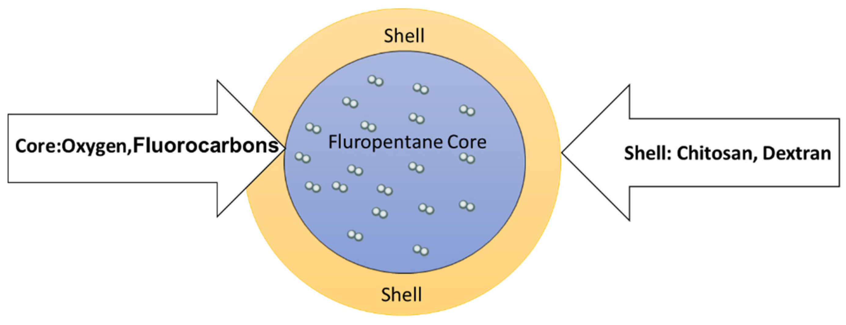
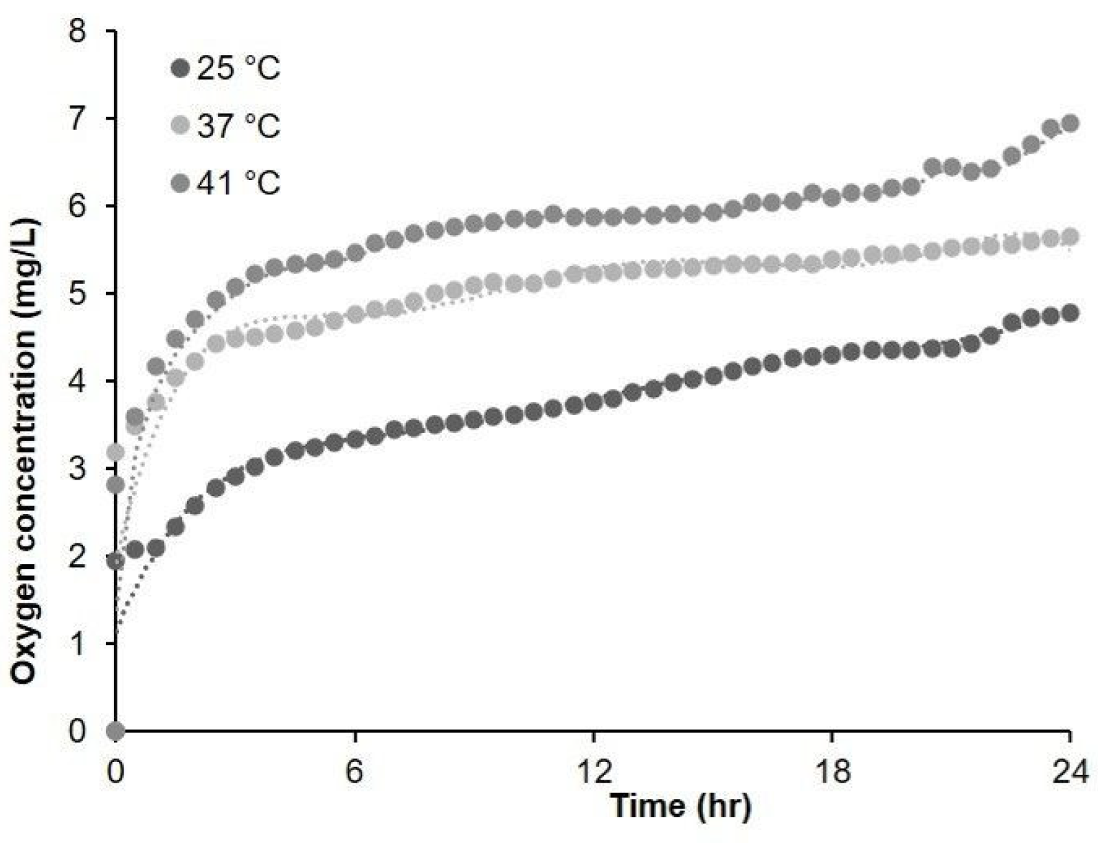
2.1. Oxygen-Loaded Nanosystems: Loading and Delivery of Oxygen to Hypoxic Tissues
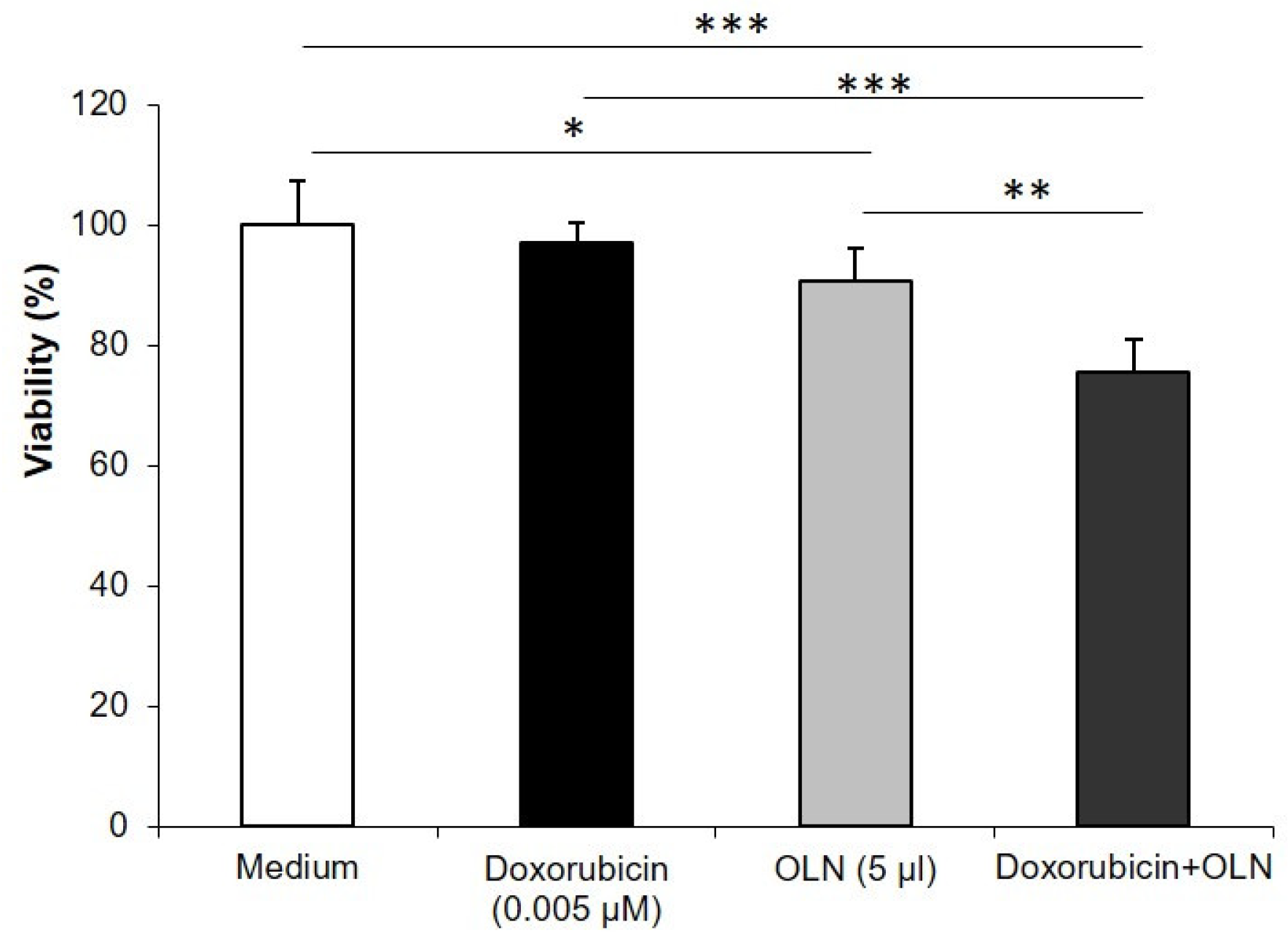
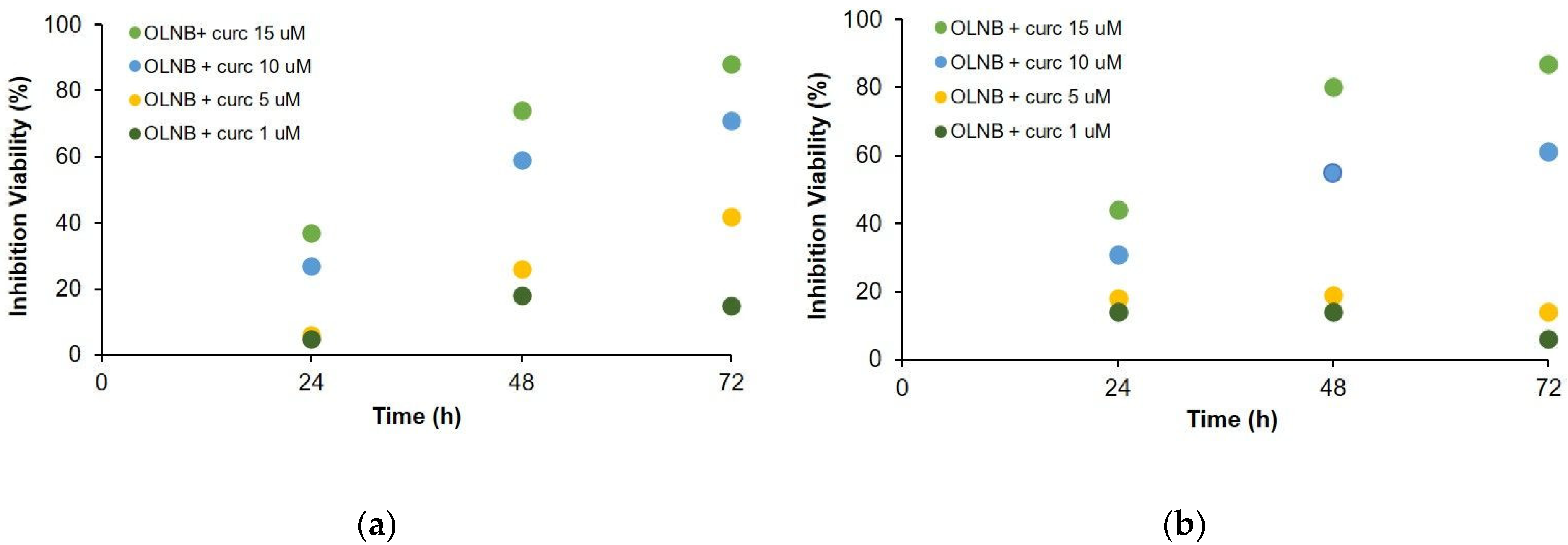
2.2. Drug-Loaded Oxygen-Loaded Nanosystems: Loading and Delivery of Anticancer Drugs
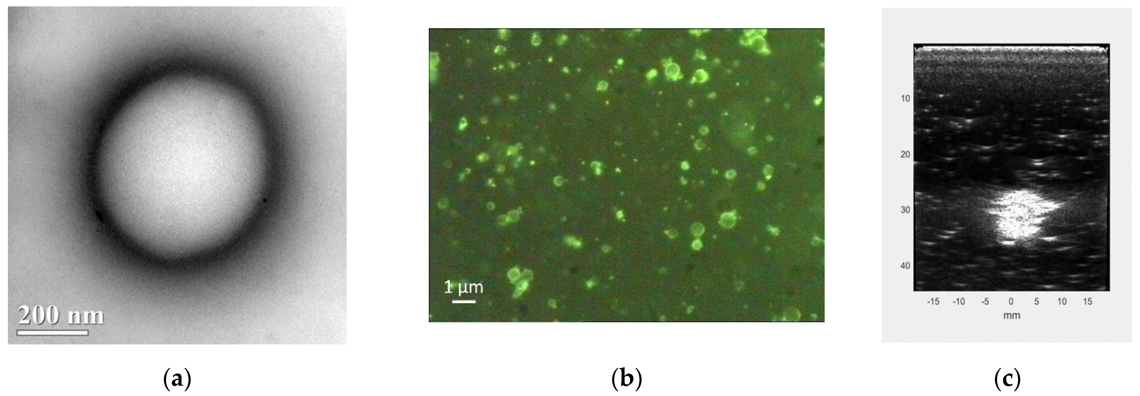
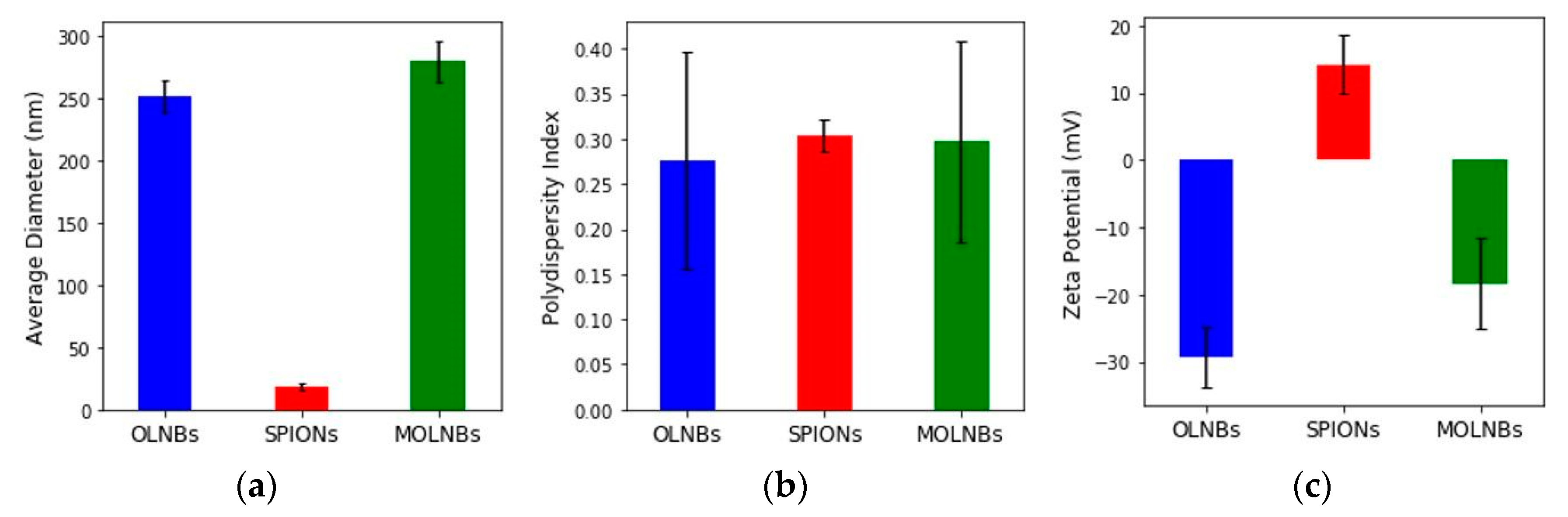
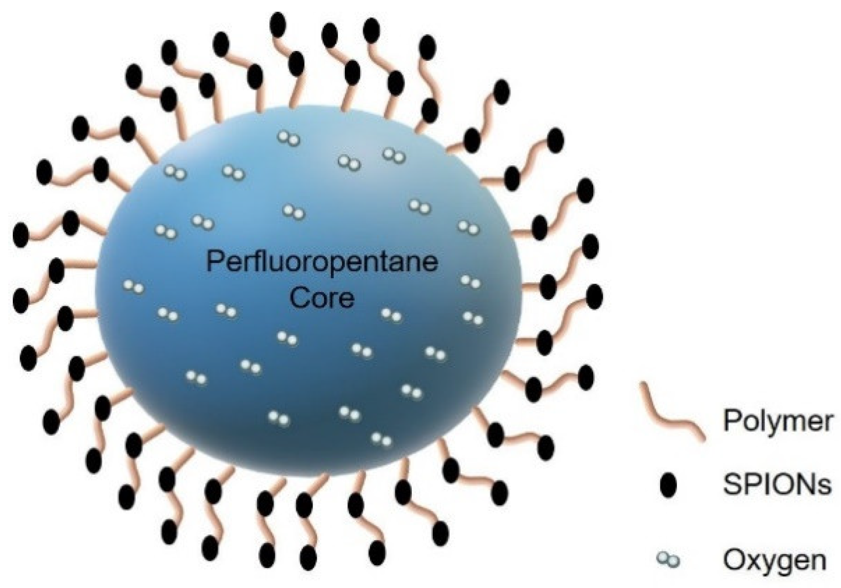
2.3. SPION-Decorated OLNBs: Manufacturing and Physicochemical Characterization of MOLNBs
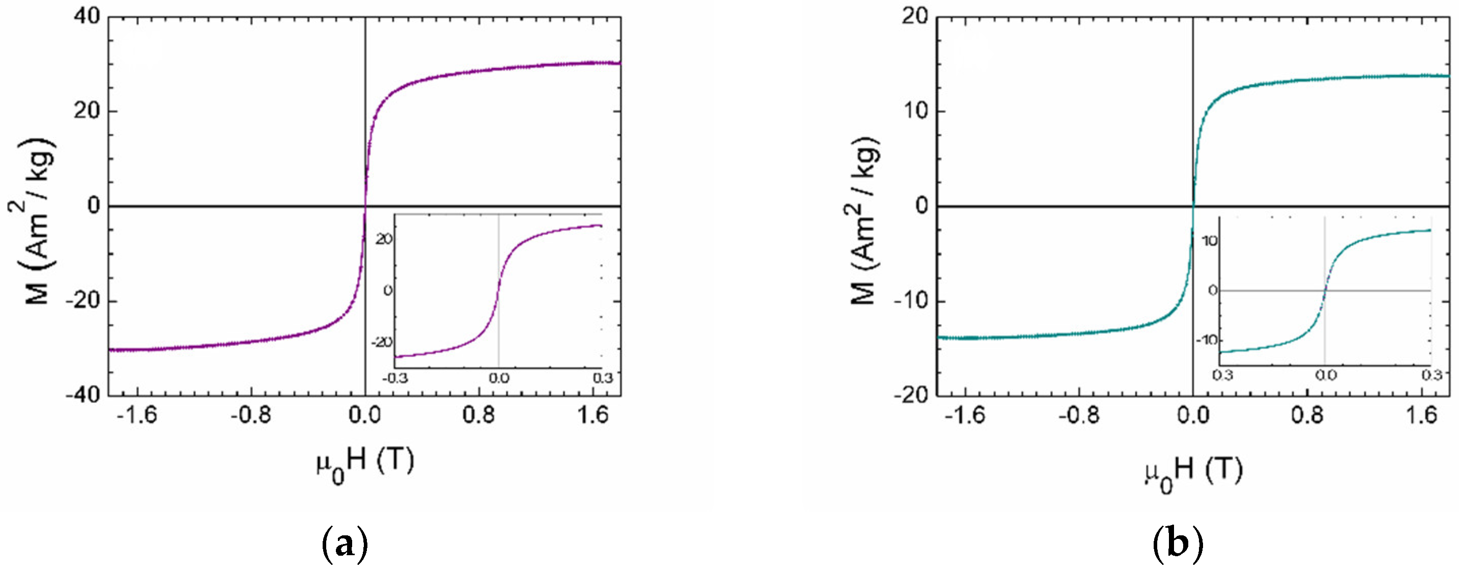
2.4. Magnetic Characterization of MOLNBs
In Vitro MRI Test
2.5. Magnetic and Hyperthermic Properties of MOLNBs
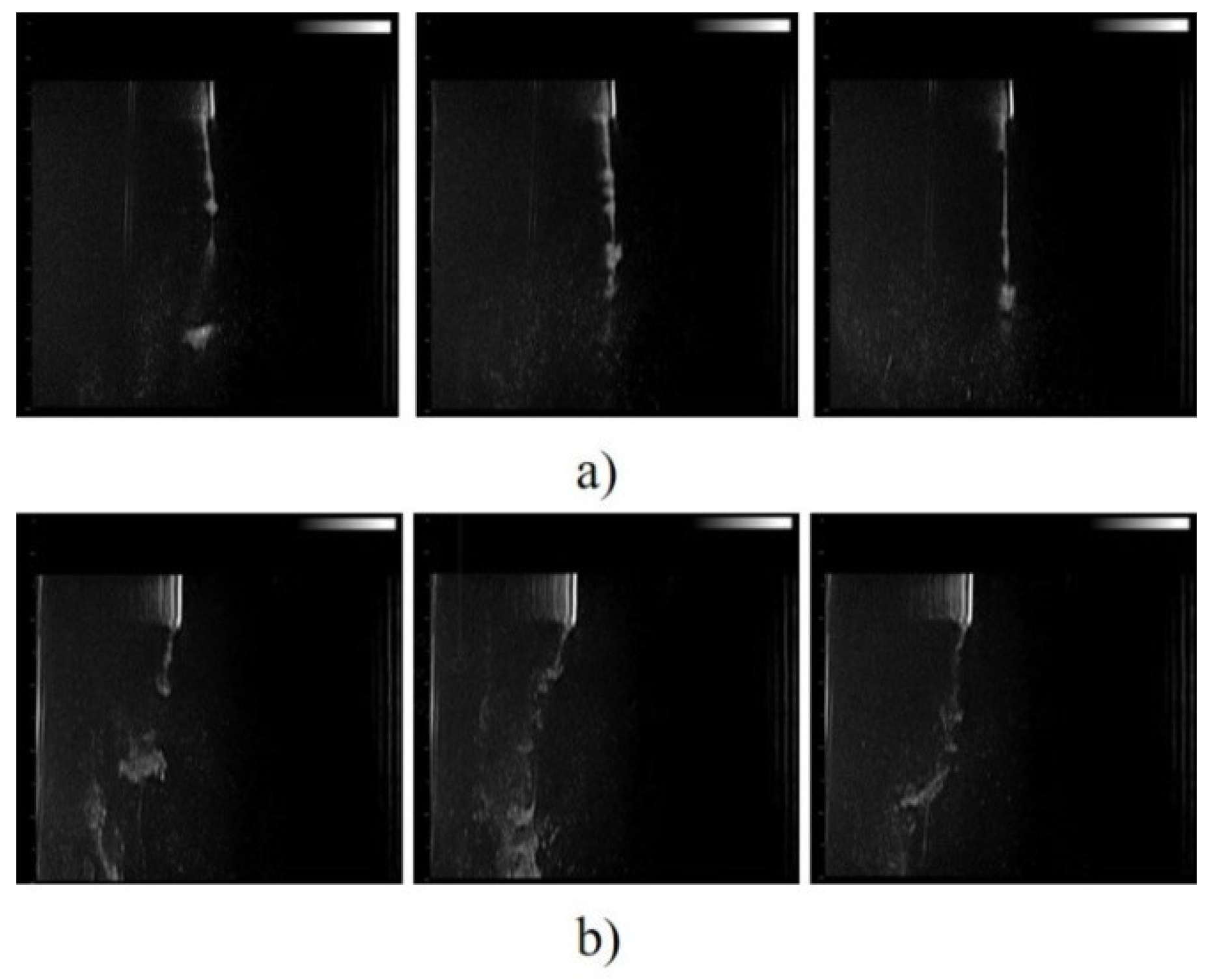
2.6. Drivability by the Application of Weak Static Magnetic Fields of MOLNBs
3. Discussion
4. Materials and Methods
4.1. Preparation of Chitosan Oxygen-Carrying Nanobubbles
4.2. Dextran and Dextran Sulfate Oxygen-Loaded Nanodroplets
4.3. Synthesis of SPIONs
4.4. Preparation of MOLNB Formulations
4.5. In Vitro Oxygen Release Study
4.6. Physicochemical Characterization of OLNBs, OLNDs and MOLNBs
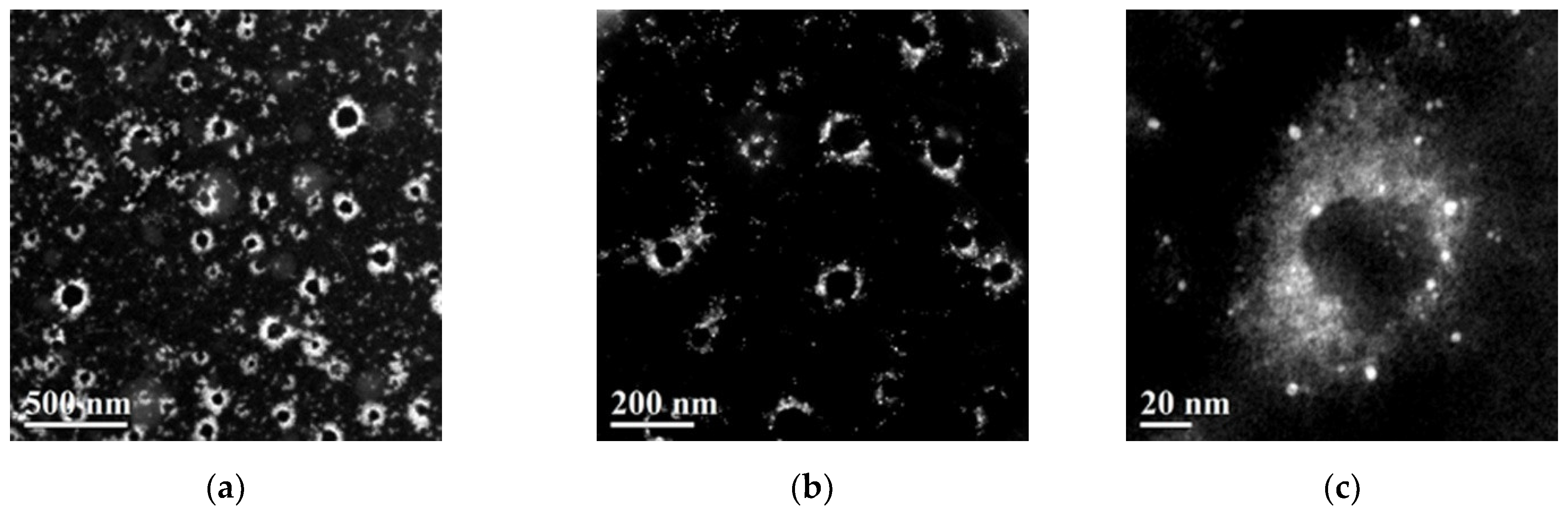
4.7. Morphological Evaluation
4.8. Magnetic Measurements and Hyperthermic Properties
4.9. Evaluation of Antitumor Effect of Doxorubicin and Curcumin-Loaded OLNDs
4.10. Curcumin Release during HT
4.11. MRI Testing
4.12. Magnetic Field and US Imaging Monitoring
5. Patents
Supplementary Materials
Author Contributions
Funding
Institutional Review Board Statement
Informed Consent Statement
Data Availability Statement
Acknowledgments
Conflicts of Interest
References
- Datta, N.R.; Rogers, S.; Ordóñez, S.G.; Puric, E.; Bodis, S. Hyperthermia and Radiotherapy in the Management of Head and Neck Cancers: A Systematic Review and Meta-Analysis. Int. J. Hyperth. 2016, 32, 31–40. [Google Scholar] [CrossRef]
- Gao, S.; Zheng, M.; Ren, X.; Tang, Y.; Liang, X. Local Hyperthermia in Head and Neck Cancer: Mechanism, Application and Advance. Oncotarget 2016, 7, 57367–57378. [Google Scholar] [CrossRef] [Green Version]
- Lambert, A.; Schwarz, L.; Borbath, I.; Henry, A.; Van Laethem, J.-L.; Malka, D.; Ducreux, M.; Conroy, T. An Update on Treatment Options for Pancreatic Adenocarcinoma. Adv. Med. Oncol. 2019, 11, 1758835919875568. [Google Scholar] [CrossRef] [PubMed] [Green Version]
- Ferraris, C.; Cavalli, R.; Panciani, P.P.; Battaglia, L. Overcoming the Blood–Brain Barrier: Successes and Challenges in Developing Nanoparticle-Mediated Drug Delivery Systems for the Treatment of Brain Tumours. Int. J. Nanomed. 2020, 15, 2999–3022. [Google Scholar] [CrossRef]
- Cerna, T.; Stiborova, M.; Adam, V.; Kizek, R.; Eckschlager, T. Nanocarrier Drugs in the Treatment of Brain Tumors. J. Cancer Metastasis Treat. 2016, 2, 407–416. [Google Scholar] [CrossRef]
- Guiot, C.; Zullino, S.; Priano, L.; Cavalli, R. The Physics of Drug-Delivery across the Blood-Brain Barrier. Ther. Deliv. 2016, 7, 153–156. [Google Scholar] [CrossRef] [PubMed] [Green Version]
- Van Liew, H.D.; Burkard, M.E. Relationship of Oxygen Content to PO2 for Stabilized Bubbles in the Circulation: Theory. J. Appl. Physiol. 1996, 81, 500–508. [Google Scholar] [CrossRef] [PubMed]
- Burkard, M.E.; Van Liew, H.D. Oxygen Transport to Tissue by Persistent Bubbles: Theory and Simulations. J. Appl. Physiol. 1994, 77, 2874–2878. [Google Scholar] [CrossRef] [PubMed]
- Zhao, J.; Liu, C.-S.; Yuan, Y.; Tao, X.-Y.; Shan, X.-Q.; Sheng, Y.; Wu, F. Preparation of Hemoglobin-Loaded Nano-Sized Particles with Porous Structure as Oxygen Carriers. Biomaterials 2007, 28, 1414–1422. [Google Scholar] [CrossRef]
- Porter, T.; Kricsfeld, D.; Cheatham, S.; Li, S. Effect of Blood and Microbubble Oxygen and Nitrogen Content on Perfluorocarbon-Filled Dextrose Albumin Microbubble Size and Efficacy: In Vitro and In Vivo Studies. J. Am. Soc. Echocardiogr. 1998, 11, 421–425. [Google Scholar] [CrossRef]
- Unger, E.C.; Porter, T.; Culp, W.; Labell, R.; Matsunaga, T.; Zutshi, R. Therapeutic Applications of Lipid-Coated Microbubbles. Adv. Drug Deliv. Rev. 2004, 56, 1291–1314. [Google Scholar] [CrossRef] [PubMed]
- Unger, E.C.; Matsunaga, T.O.; McCreery, T.; Schumann, P.; Sweitzer, R.; Quigley, R. Therapeutic Applications of Microbubbles. Eur. J. Radiol. 2002, 42, 160–168. [Google Scholar] [CrossRef]
- Wolfson, M.R.; Shaffer, T.H. Pulmonary Applications of Perfluorochemical Liquids: Ventilation and Beyond. Paediatr. Respir. Rev. 2005, 6, 117–127. [Google Scholar] [CrossRef] [PubMed]
- Kong, S.D.; Lee, J.; Ramachandran, S.; Eliceiri, B.P.; Shubayev, V.I.; Lal, R.; Jin, S. Magnetic Targeting of Nanoparticles across the Intact Blood–Brain Barrier. J. Control. Release 2012, 164, 49–57. [Google Scholar] [CrossRef] [Green Version]
- Cedervall, T.; Lynch, I.; Lindman, S.; Berggård, T.; Thulin, E.; Nilsson, H.; Dawson, K.A.; Linse, S. Understanding the Nanoparticle–Protein Corona Using Methods to Quantify Exchange Rates and Affinities of Proteins for Nanoparticles. Proc. Natl. Acad. Sci. USA 2007, 104, 2050–2055. [Google Scholar] [CrossRef] [Green Version]
- Pedziwiatr-Werbicka, E.; Horodecka, K.; Shcharbin, D.; Bryszewska, M. Nanoparticles in Combating Cancer: Opportunities and Limitations. A Brief Review. Curr. Med. Chem. 2021, 28, 346–359. [Google Scholar] [CrossRef]
- Zhang, T.-T.; Li, W.; Meng, G.; Wang, P.; Liao, W. Strategies for Transporting Nanoparticles across the Blood–Brain Barrier. Biomater. Sci. 2016, 4, 219–229. [Google Scholar] [CrossRef] [PubMed]
- Home-Focused Ultrasound Foundation. Available online: https://www.fusfoundation.org/ (accessed on 28 December 2020).
- Yang, C.; Xiao, H.; Sun, Y.; Zhu, L.; Gao, Y.; Kwok, S.; Wang, Z.; Tang, Y. Lipid Microbubbles as Ultrasound-Stimulated Oxygen Carriers for Controllable Oxygen Release for Tumor Reoxygenation. Ultrasound Med. Biol. 2018, 44, 416–425. [Google Scholar] [CrossRef] [PubMed]
- Estelrich, J.; Escribano, E.; Queralt, J.; Busquets, M.A. Iron Oxide Nanoparticles for Magnetically-Guided and Magnetically-Responsive Drug Delivery. Int. J. Mol. Sci. 2015, 16, 8070–8101. [Google Scholar] [CrossRef] [Green Version]
- Feng, Q.; Liu, Y.; Huang, J.; Chen, K.; Huang, J.; Xiao, K. Uptake, Distribution, Clearance, and Toxicity of Iron Oxide Nanoparticles with Different Sizes and Coatings. Sci. Rep. 2018, 8, 2082. [Google Scholar] [CrossRef] [PubMed]
- D’Agata, F.; Ruffinatti, F.A.; Boschi, S.; Stura, I.; Rainero, I.; Abollino, O.; Cavalli, R.; Guiot, C. Magnetic Nanoparticles in the Central Nervous System: Targeting Principles, Applications and Safety Issues. Molecules 2017, 23, 9. [Google Scholar] [CrossRef] [PubMed] [Green Version]
- Novoselova, M.V.; German, S.V.; Sindeeva, O.A.; Kulikov, O.A.; Minaeva, O.V.; Brodovskaya, E.P.; Ageev, V.P.; Zharkov, M.N.; Pyataev, N.A.; Sukhorukov, G.B.; et al. Submicron-Sized Nanocomposite Magnetic-Sensitive Carriers: Controllable Organ Distribution and Biological Effects. Polymers 2019, 11, 1082. [Google Scholar] [CrossRef] [PubMed] [Green Version]
- Owens, D.E.; Peppas, N.A. Opsonization, Biodistribution, and Pharmacokinetics of Polymeric Nanoparticles. Int. J. Pharm. 2006, 307, 93–102. [Google Scholar] [CrossRef] [PubMed]
- Ficiarà, E.; Ansari, S.A.; Argenziano, M.; Cangemi, L.; Monge, C.; Cavalli, R.; D’Agata, F. Beyond Oncological Hyperthermia: Physically Drivable Magnetic Nanobubbles as Novel Multipurpose Theranostic Carriers in the Central Nervous System. Molecules 2020, 25, 2104. [Google Scholar] [CrossRef] [PubMed]
- Chang, D.; Lim, M.; Goos, J.A.C.M.; Qiao, R.; Ng, Y.Y.; Mansfeld, F.M.; Jackson, M.; Davis, T.P.; Kavallaris, M. Biologically Targeted Magnetic Hyperthermia: Potential and Limitations. Front. Pharmacol. 2018, 9, 831. [Google Scholar] [CrossRef] [PubMed] [Green Version]
- Liu, X.; Zhang, Y.; Wang, Y.; Zhu, W.; Li, G.; Ma, X.; Zhang, Y.; Chen, S.; Tiwari, S.; Shi, K.; et al. Comprehensive Understanding of Magnetic Hyperthermia for Improving Antitumor Therapeutic Efficacy. Theranostics 2020, 10, 3793–3815. [Google Scholar] [CrossRef] [PubMed]
- Cavalli, R.; Bisazza, A.; Giustetto, P.; Civra, A.; Lembo, D.; Trotta, G.; Guiot, C.; Trotta, M. Preparation and Characterization of Dextran Nanobubbles for Oxygen Delivery. Int. J. Pharm. 2009, 381, 160–165. [Google Scholar] [CrossRef] [PubMed] [Green Version]
- Cavalli, R.; Bisazza, A.; Rolfo, A.; Balbis, S.; Madonnaripa, D.; Caniggia, I.; Guiot, C. Ultrasound-Mediated Oxygen Delivery from Chitosan Nanobubbles. Int. J. Pharm. 2009, 378, 215–217. [Google Scholar] [CrossRef]
- Prato, M.; Magnetto, C.; Jose, J.; Khadjavi, A.; Cavallo, F.; Quaglino, E.; Panariti, A.; Rivolta, I.; Benintende, E.; Varetto, G.; et al. 2H,3H-Decafluoropentane-Based Nanodroplets: New Perspectives for Oxygen Delivery to Hypoxic Cutaneous Tissues. PLoS ONE 2015, 10, e0119769. [Google Scholar] [CrossRef]
- Khadjavi, A.; Stura, I.; Prato, M.; Minero, V.G.; Panariti, A.; Rivolta, I.; Gulino, G.R.; Bessone, F.; Giribaldi, G.; Quaglino, E.; et al. “In Vitro”, “In Vivo” and “In Silico” Investigation of the Anticancer Effectiveness of Oxygen-Loaded Chitosan-Shelled Nanodroplets as Potential Drug Vector. Pharm. Res. 2018, 35, 75. [Google Scholar] [CrossRef] [Green Version]
- Bessone, F.; Argenziano, M.; Grillo, G.; Ferrara, B.; Pizzimenti, S.; Barrera, G.; Cravotto, G.; Guiot, C.; Stura, I.; Cavalli, R.; et al. Low-Dose Curcuminoid-Loaded in Dextran Nanobubbles Can Prevent Metastatic Spreading in Prostate Cancer Cells. Nanotechnology 2019, 30, 214004. [Google Scholar] [CrossRef]
- Zullino, S.; Argenziano, M.; Ansari, S.; Ciprian, R.; Nasi, L.; Albertini, F.; Cavalli, R.; Guiot, C. Superparamagnetic Oxygen-Loaded Nanobubbles to Enhance Tumor Oxygenation During Hyperthermia. Front. Pharmacol. 2019, 10. [Google Scholar] [CrossRef]
- Khan, M.S.; Hwang, J.; Lee, K.; Choi, Y.; Kim, K.; Koo, H.-J.; Hong, J.W.; Choi, J. Oxygen-Carrying Micro/Nanobubbles: Composition, Synthesis Techniques and Potential Prospects in Photo-Triggered Theranostics. Molecules 2018, 23, 2210. [Google Scholar] [CrossRef] [PubMed] [Green Version]
- Ramaswamy, B.; Kulkarni, S.D.; Villar, P.S.; Smith, R.S.; Eberly, C.; Araneda, R.C.; Depireux, D.A.; Shapiro, B. Movement of Magnetic Nanoparticles in Brain Tissue: Mechanisms and Safety. Nanomedicine 2015, 11, 1821–1829. [Google Scholar] [CrossRef] [PubMed] [Green Version]
- Shamloo, A.; Pedram, M.Z.; Heidari, H.; Alasty, A. Computing the Blood Brain Barrier (BBB) Diffusion Coefficient: A Molecular Dynamics Approach. J. Magn. Magn. Mater. 2016, 410, 187–197. [Google Scholar] [CrossRef]
- Householder, K.T.; Dharmaraj, S.; Sandberg, D.I.; Wechsler-Reya, R.J.; Sirianni, R.W. Fate of Nanoparticles in the Central Nervous System after Intrathecal Injection in Healthy Mice. Sci. Rep. 2019, 9, 1–11. [Google Scholar] [CrossRef] [PubMed] [Green Version]
- Sandberg, D.I.; Rytting, M.; Zaky, W.; Kerr, M.; Ketonen, L.; Kundu, U.; Moore, B.D.; Yang, G.; Hou, P.; Sitton, C.; et al. Methotrexate Administration Directly into the Fourth Ventricle in Children with Malignant Fourth Ventricular Brain Tumors: A Pilot Clinical Trial. J. Neurooncol. 2015, 125, 133–141. [Google Scholar] [CrossRef] [Green Version]
- Ortner, M.; Coliado Bandeira, L.G. Magpylib: A Free Python Package for Magnetic Field Computation. SoftwareX 2020, 11, 100466. [Google Scholar] [CrossRef]


| Shell Polymer | Perfluoropentane C5F12 (PFP) Core | Decafluoropentane C5H2F10 (DFP) Core |
|---|---|---|
| Chitosan/Dextran/Dextran sulfate | OLNB | OLND |
| Concentration (mg/mL) | Blank | 0.5 | 1 | 2 | 2.5 |
|---|---|---|---|---|---|
| T2 (ms) | 2000 | 58.82 | 55.55 | 30.30 | 29.41 |
Publisher’s Note: MDPI stays neutral with regard to jurisdictional claims in published maps and institutional affiliations. |
© 2021 by the authors. Licensee MDPI, Basel, Switzerland. This article is an open access article distributed under the terms and conditions of the Creative Commons Attribution (CC BY) license (https://creativecommons.org/licenses/by/4.0/).
Share and Cite
Ansari, S.A.; Ficiarà, E.; D’Agata, F.; Cavalli, R.; Nasi, L.; Casoli, F.; Albertini, F.; Guiot, C. Step-by-Step Design of New Theranostic Nanoformulations: Multifunctional Nanovectors for Radio-Chemo-Hyperthermic Therapy under Physical Targeting. Molecules 2021, 26, 4591. https://doi.org/10.3390/molecules26154591
Ansari SA, Ficiarà E, D’Agata F, Cavalli R, Nasi L, Casoli F, Albertini F, Guiot C. Step-by-Step Design of New Theranostic Nanoformulations: Multifunctional Nanovectors for Radio-Chemo-Hyperthermic Therapy under Physical Targeting. Molecules. 2021; 26(15):4591. https://doi.org/10.3390/molecules26154591
Chicago/Turabian StyleAnsari, Shoeb Anwar, Eleonora Ficiarà, Federico D’Agata, Roberta Cavalli, Lucia Nasi, Francesca Casoli, Franca Albertini, and Caterina Guiot. 2021. "Step-by-Step Design of New Theranostic Nanoformulations: Multifunctional Nanovectors for Radio-Chemo-Hyperthermic Therapy under Physical Targeting" Molecules 26, no. 15: 4591. https://doi.org/10.3390/molecules26154591
APA StyleAnsari, S. A., Ficiarà, E., D’Agata, F., Cavalli, R., Nasi, L., Casoli, F., Albertini, F., & Guiot, C. (2021). Step-by-Step Design of New Theranostic Nanoformulations: Multifunctional Nanovectors for Radio-Chemo-Hyperthermic Therapy under Physical Targeting. Molecules, 26(15), 4591. https://doi.org/10.3390/molecules26154591