Article

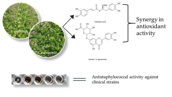
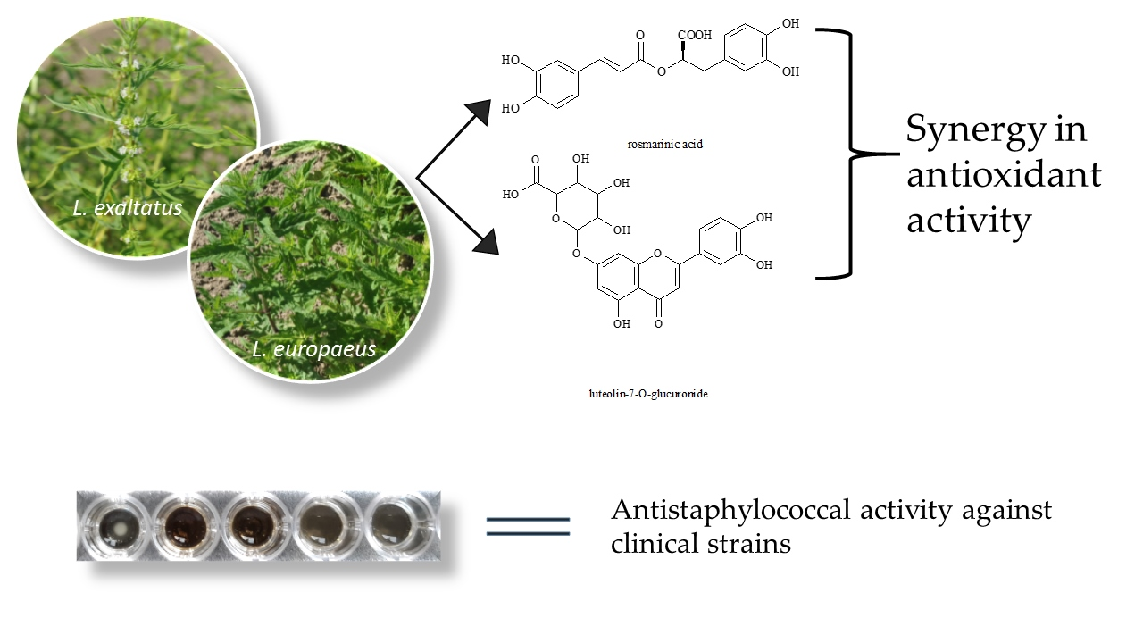
Antimicrobial and Antioxidant Properties of Four Lycopus Taxa and an Interaction Study of Their Major Compounds

1
Department of Pharmacognosy and Botany, Faculty of Pharmacy, Comenius University in Bratislava, Odbojárov 10, 832 32 Bratislava, Slovakia
2
Institute of Microbiology, Faculty of Medicine and the University Hospital in Bratislava, Comenius University in Bratislava, Sasinkova 4, 811 08 Bratislava, Slovakia
3
Department of Microbiology and Virology, Faculty of Natural Sciences, Comenius University in Bratislava, Mlynská dolina, Ilkovičova 6, 842 15 Bratislava, Slovakia
4
Department of Molecular Biology, Faculty of Natural Sciences, Comenius University in Bratislava, Mlynská dolina, Ilkovičova 6, 842 15 Bratislava, Slovakia
5
Department of Pharmaceutical Analysis and Nuclear Pharmacy, Faculty of Pharmacy, Comenius University in Bratislava, Odbojárov 10, 832 32 Bratislava, Slovakia
6
National Agricultural and Food Centre, Research Institute of Plant Production, 92168 Piešťany, Slovakia
*
Author to whom correspondence should be addressed.
Molecules 2020, 25(6), 1422; https://doi.org/10.3390/molecules25061422
Submission received: 18 February 2020 / Revised: 13 March 2020 / Accepted: 17 March 2020 / Published: 20 March 2020
(This article belongs to the Special Issue Bioactive Natural Compounds and Their Mechanisms of Action)

Abstract

:
The compositions of leaf infusions of three genotypes of Lycopus europaeus L. with origins in central Europe, namely L. europaeus A (LeuA), L. europaeus B (LeuB), and L. europaeus C (LeuC), and one genotype of L. exaltatus (Lex), were examined by LC-MS-DAD (Liquid Chromatography Mass Spectrometry and Diode Array Detection) analysis. This revealed the presence of thirteen compounds belonging to the groups of phenolic acids and flavonoids, with a predominance of rosmarinic acid (RA) and luteolin-7-O-glucuronide (LGlr). The antimicrobial activity of leaf infusions was tested on the collection strains of Gram-positive and Gram-negative bacteria, and on the clinical Staphylococcus aureus strains. We detected higher activity against Gram-positive bacteria, of which the most susceptible strains were those of Staphylococcus aureus, including methicillin-resistant and poly-resistant strains. Furthermore, we examined the antioxidant activity of leaf infusions using 2,2-diphenyl-1-picrylhydrazyl (DPPH) and 2,2′-azino-bis(3-ethylbenzothiazoline-6-sulphonic acid) (ABTS) methods, and on NIH/3T3 cell lines using dichlorodihydrofluorescein diacetate (DCFH-DA). We also studied the mutual interactions between selected infusions, namely RA and/or LGlr. In the mixtures of leaf infusion and RA or LGlr, we observed slight synergism and a high dose reduction index in most cases. This leads to the beneficial dose reduction at a given antioxidant effect level in mixtures compared to the doses of the parts used alone. Therefore, our study draws attention to further applications of the Lycopus leaves as a valuable alternative source of natural antioxidants and as a promising topical antibacterial agent for medicinal use.

Graphical Abstract

1. Introduction

Medicinal plants and herbal preparations have been traditionally used as mixtures of natural substances in phytomedicine for centuries [1]. There exist many good reasons for this practice, such as synergism among constituents, mutual protection of active substances due to their instability, or in some cases, the active substances are still unknown [2]. Synergy or antagonism is present when two or more ingredients of the complex mutually increase or decrease each other’s effect. The final response is higher or lower than a simple summation of single effects (additivity) [3]. This can lead to enhancement in the efficacy of mixtures, reduction of their dosage, and decline of their adverse or toxic effects in clinical practice [4]. The synergistic combinations of agents can be more effective than single-drug treatment thanks to the decrease or even elimination of resistance development, especially in resistant bacteria or viruses [5]. Medicinal plants contain plenty of heterogeneous substances with a positive interaction potential, which makes them a promising area for synergy research.
Lycopus L. is a genus of approximately 16 species of flowering plants in the family Lamiaceae, subfamily Nepetoidae [6]. All are herbaceous and native to Europe, East Asia, and North America. The species are often grown in wetlands, damp meadows, stream banks, and other aquatic environments [7]. This genus is characterized by dentate or pinnatifid opposite leaves; flowers in compact, sessile verticillasters in the leaf axils; 4–5 lobed tubulate or campanulate calyces; and a dry, tetrahedral, one-seeded nutlet with corky crests [6,8]. The most important constituents of this genus are essential oil, terpenoids (mostly diterpenes and triterpenes), flavonoids, and phenolic and hydroxycinnamic acids [7,9].
Two species are commonly found in Europe, Lycopus europaeus L. and L. exaltatus L.f. The first one is a perennial plant native to Europe and Asia and naturalized in the U.S. The plant is odorless and has a bitter taste. Its herbal juice yields a black dye, supposedly used by gypsies to tan their skin, hence the common name of gypsywort. It is also known as the bugleweed, Wolfstrappkraut, bitter bugle, or water horehound [10,11,12].
The biological effects of this plant are attributed to phenolic compounds, mainly to derivatives of hydroxycinnamic acid and flavonoids. According to Fialová et al. [13], the major compounds in leaf infusion are rosmarinic acid and luteolin-7-O-glucuronide. Other extracts of the aerial parts revealed a high amount of isopimarane-type diterpenoids and alicyclic diterpenes [9,14,15]. L. europaeus is traditionally suggested for treating mild hyperthyroidism with vegetative nervous disturbances. It shows a clear reduction of hyperthyroid symptoms, particularly of cardiac symptoms, and increased body temperature. Extracts of the plant are traditionally also used in tenseness and pain of the mammary gland [15,16,17,18,19]. The methanol extract of the plant showed central and peripheral analgesic and anti-inflammatory effects, which could be associated with the presence of terpenoids, flavonoids, and tannins [11]. The same extract demonstrated significant antitussive activity by inhibition of cough reflex [10]. L. europaeus also possessed antiparasitic activity against T. gallinae [20], and antioxidant and antimicrobial activity against various bacterial species [12,13,14,15].
L. exaltatus is also native to Europe and Asia. Unlike the L. europaeus, the leaves of which are only coarsely serrate or sinuate, the leaves of L. exaltatus are deeply pinnatifid [21]. The phenolic fingerprints of L. exaltatus and L. europaeus are similar, as proven by Bucar and Kartnig [22].
Considering the antioxidant, anti-inflammatory, analgesic, and antimicrobial properties of L. europaeus, its extracts or isolated compounds might be helpful in the future as potential remedies for the treatment of infections in human or veterinary medicine. Therefore, the aim of our study was a detailed evaluation of the antimicrobial and antioxidant activity of three L. europaeus genotypes and one L. exaltatus genotype with origins in Central Europe, and to provide phytochemical analysis and fingerprinting of polar phenolic compounds in the leaf infusions. To the best of our knowledge, this is the first study on the biological activities of L. exaltatus.

2. Results

2.1. Identification and Quantification

Secondary metabolites of four Lycopus leaf infusions were identified using Liquid Chromatography-Tandem Mass Spectrometry (LC-MS/MS). Acquired MS spectra were compared with authentic standards or with database searches. The maximum allowed mass deviation was 10 ppm. Eleven compounds, including phenolic acids and flavonoids, were found in almost all samples. Identified secondary metabolites are presented in Table 1, along with their retention times (TR), observed mass in negative ionization mode, MS/MS fragment ions of each secondary metabolite, as well as their quantity in each infusion. The compounds detected in the analyzed infusions were characterized using MS data, together with the interpretation of the observed MS/MS spectra in comparison with standards and data listed in the available literature [13,22].
Peak 1, which presented a pseudomolecular [M − H] ion at m/z 191, with all typical product ions presented at m/z 111, 87, and 85, was identified as citric acid. Danshensu (trihydroxyphenyl propanoic acid) was identified as the second compound with the precursor ion [M − H] at m/z 197 and product ions at m/z 179 and 135. Caffeic acid was identified at a retention time of 21.59 min, with a molecular [M − H] ion identified at m/z 179 and product ion at m/z 135 (which is attributed to the loss of CO2), and was compared with an authentic standard. The small peak at 24.58 min is most probably flavonoid quercetin glucuronide with precursor ion [M − H] at m/z 477 and with aglycone ion at m/z 301. Peak 5 showed a precursor ion at m/z 463 [M − H] and its MS/MS spectrum presented a product ion at m/z 301 belonging to quercetin glucoside. Lithospermic acid was detected in 26 min, with a molecular ion [M − H] at m/z 537 and typical fragments at m/z 359, 197, 179, 161, and 135. A small peak (peak 7) presented a pseudomolecular [M − H] ion at m/z 521, with fragmentation products at m/z 359 [M−H−162], 179, and 161. This peak was identified according to literature as rosmarinic acid hexoside [23]. One of two dominant peaks was recorded in 28.57 min and was identified and compared with authentic standard luteolin-7-O-glucuronide (LGlr), with a typical MS [M − H] ion at m/z 461 and product ion at m/z 285. Peak 9, sagerinic acid, a caffeic acid derivative, was identified based on molecular ion [M − H] at m/z 719, yielding fragments at m/z 539, 359, and 161. Peak 10, sulfated rosmarinic acid, was identified according to the literature [23]. It showed the molecular ion [M − H] at m/z 439 and a product ion at m/z 359 due to the loss of 80 Da (sulfated moiety). A small peak at 34.12 min belonged to apigenin-glucuronide, with molecular ion [M − H] at m/z 445 and with aglycone ion at m/z 269. The most dominant peak at 34.48 min revealed the expected presence of the main compound rosmarinic acid (RA), with [M − H] at m/z 359. Peak 13 was identified as a caffeic acid derivative, with [M − H] at m/z 549 and fragments at m/z 359 and 161.
The phenolic compounds present in the samples were characterized according to their UV, mass spectra, and retention times, and by comparison with authentic standards when available. For the quantitative analysis, external standards were used. We used LGlr (at λ = 360 nm) for the determination of flavonoids and their derivatives, and RA (at λ = 320 nm) for the determination of phenolic acids and their derivatives. Their calibration curves were obtained by injection of known concentrations (5–100 ppm). Both standards showed good linearity. The following r2 values were obtained: LGlr r2 = 0.9976, regression curve y = 237.26x − 450.72 and RA r2 = 0.999, regression curve y = 340.5x + 297.09. The limit of detection (LOD) of LGlr was 1.89 µg/mL and the limit of quantification (LOQ) was 6.31 µg/mL. The LOD of RA was 1.33 µg/mL and the LOQ was 4.45 µg/mL. The results were expressed in μg per mL of leaf infusion. The examinations of secondary metabolites in Lycopus leaf infusions were performed in triplicate. The quantitative results were calculated from calibration curves, expressed as mean values and standard deviation (SD).

2.2. Evaluation of Antimicrobial Activity

The antimicrobial activity of the Lycopus infusions was tested using microorganisms most frequently isolated from local infections in humans. Collection strains of Staphylococcus aureus, Enterococcus faecalis, Pseudomonas aeruginosa, Escherichia coli, Klebsiella pneumoniae, and Proteus mirabilis, as well as 28 clinical S. aureus strains and one collection strain of a yeast Candida albicans, were included. For the first screening, 7 well-characterized collection strains were used (for details see the “Material and methods”). The results of antibacterial activities of the four tested Lycopus infusions on bacterial collection strains are shown in Table 2.
Staphylococci were the most susceptible; the minimal inhibitory concentrations (MIC) and minimal bactericidal concentrations (MBC) of Lycopus infusions were between 2.5 and 5 mg/mL in both methicillin-resistant S. aureus (CCM 4750) and methicillin-susceptible S. aureus (CCM4223) S. aureus strains. The most susceptible Gram-negative bacterial species was Proteus mirabilis, with MICs and MBCs between 2.5 and 10 mg/mL. Concerning the yeast strain of Candida albicans CCM 90028, it was found to be resistant against any of the samples in the tested concentration range. When comparing the examined plant infusions, the L. exaltatus (Lex) infusion was slightly less active than those of L. europaeus.
Based on the results with collection strains, the most effective sample—L. europaeus B (LeuB)—was chosen for further testing, while clinical strains of S. aureus were used as microbial targets. These strains were isolated from infected skin, soft tissue, and wounds (19 strains), and from bloodstream infections (9 strains). Out of 28 strains, 14 were resistant to methicillin or oxacillin (MRSA) and were mecA gene-positive. To ensure the appropriate variability of the tested strains, they had various antimicrobial susceptibility patterns and came from different Spa-types. MIC and MBC values of LeuB infusion for clinical S. aureus strains are shown in Table 3.
As shown in Table 3, the antibacterial activity of LeuB infusion on clinical S. aureus strains was comparable to results obtained with the collection S. aureus strains (MICs were found in the interval from 1.25 to 2.5 mg/mL; MBCs reached values from 1.25 to 5 mg/mL). There was no correlation between the MIC or MBC values and the susceptibility of clinical strains to methicillin or to the other antibiotics.

2.3. Antioxidant Activity by ABTS (2,2′-azino-bis(3-ethylbenzothiazoline-6-sulphonic acid), DPPH (2,2-diphenyl-1-picrylhydrazyl), and DCFH-DA (dichlorodihydrofluorescein diacetate) Assays

All infusions showed antioxidant activity at the concentration range between 1.25 and 40 µg/mL. The reference compounds, RA and LGlr, possessed antioxidant effects at the concentration range between 0.31 to 20 µg/mL. All tested infusions showed concentration-dependent antioxidant activity. The concentration of a sample necessary to decrease the oxidative stress by 50% (IC50) under the experimental conditions was calculated and presented in Table 4 using CompuSyn software. Therefore, a lower IC50 value indicates a higher antioxidant activity.

2.4. Interaction Analysis by ABTS, DPPH, and DCFH-DA Assays

As we discovered, the most abundant compounds of Lycopus leaf infusions were RA and LGlr. Therefore, we choose both substances for interaction analysis. Moreover, we also choose the most effective infusion for each method of antioxidant activity testing (LeuB for ABTS method and Lex for DCFH-DA and DPPH assays, respectively). Equivalent amounts of RA, LGlr, and selected infusions were tested for mutual interactions in mixtures using methods of antioxidant activity testing (ABTS, DPPH, and DCFH-DA). As can be seen in Table 5, all tested combinations showed the synergistic or nearly additive effect. The combination of RA and LGlr showed moderate synergism in all assays. We also provided the interaction analysis between different infusions, which was observed for additive effect only; therefore, we did not present those results here.
DRI values for all tested combinations were higher than 1. This means that the concentration of the samples can be reduced when using the weight equivalent mixtures to achieve the same effect compared to the single-drug treatment. For example, the dose of LGlr can be reduced 5.38 times and RA can be reduced 1.61 times, respectively, when tested in their mixture in the ABTS assay to achieve the same effect as if each compound was used separately. In the latter case, we would need 7.37 µg/mL of LGlr or 2.20 µg/mL of RA; however, when combined together, we need only 1.37 µg/mL each to achieve the effect of a single agent. A significant dose decrease of Lycopus leaf infusion is also present in all assays when combined with RA and LGlr. In the ABTS assay, we can reduce the dose of LeuB, RA, and LGlr 7.74 times, 1.95 times, and 6.52 times, respectively. It is more effective to use this triple combination, because to achieve the same effect we needed only 1.13 µg/mL of LeuB, RA, and LGlr; on the other hand, when used alone we need 8.75 µg/mL of LeuB, 2.20 µg/mL of RA, and 7.37 µg/mL of LGlr. Similar dose reduction also occurs in DCFH-DA and DPPH assays. The DCFH-DA assay showed the biggest dose reduction. We needed only 0.19 µg/mL of Lex, RA, and LGlr, as opposed to 2.17 µg/mL of Lex, 0.94 µg/mL of LGlr, and 0.63 µg/mL of RA to achieve the same effect.

3. Discussion

Until today, a few studies were published about biological activities of species from the genus Lycopus. This was one of the reasons why we have chosen to examine different taxa of Lycopus with origins in Central Europe. We analyzed and tested infusions (water extract), the most common form of usage of medicinal plants as remedies in traditional medicine.
Using the LC-MS-DAD (Liquid Chromatography Mass Spectrometry and Diode Array Detection) we revealed the presence of thirteen phenolic compounds and quantified them. There were two compounds with dominant amounts in all infusions: rosmarinic acid (202–237 µg/mL) and luteolin-7-O-glucuronide (279–449 µg/mL). Many of the identified compounds were also detected in L. europaeus in the past [13]. Next, we looked for the disparities between three genotypes of L. europaeus and between different species. We did not detect a high variability in the composition of different genotypes of Lycopus between either species, namely L. europaeus and L. exaltatus, which can also be seen in our results of bioactivities.
The emergence and spread of resistance to antibiotics, as well as the evolution of new strains of disease-causing agents, represent the main factors justifying the need to find and develop new antimicrobial agents. New sources, especially plant sources, are, thus, being investigated. Secondary metabolites form the defense system in plants, which fights against predators such as herbivores, insects, and microorganisms. These substances may act through different mechanisms than conventional antibiotics, and thus they can be utilized in the treatment of infections caused by resistant bacteria. These mechanisms include inhibition of synthesis of nucleic acids, direct interaction with the cell membrane structures of bacteria, which lead to their damage, inhibitory effects on electron transport in the bacterial respiratory chain, and quorum sensing inhibition [25,26]. Several studies on the antimicrobial activity of L. europaeus extracts or isolated compounds of several microbial species have been published in the past. They examined both Gram-positive and Gram-negative bacteria as target microorganisms [12,13,14,15]. Gibbons et al. [14] studied an extract and isolated isopimarane diterpenes from L. europaeus. None of the tested diterpenes displayed any antibacterial activity, but in combination with tetracycline and erythromycin, a two-fold reduction of the minimal inhibitory concentrations of these antibiotics against two strains of methicillin-resistant S. aureus, possessing multidrug resistance efflux pumps, was observed. Radulović et al. [15] isolated a new abietane-type diterpenoid named euroabienol from L. europaeus fruits and tested its antimicrobial activity against fifteen strains of bacteria. Euroabienol was most active against S. aureus and least active against E. coli. On the other hand, L. europaeus essential oil, studied by Radulović et al. [12], possessed selectivity towards two Gram-negative strains—E. coli and K. pneumoniae. Fialová et al. [13] tested the antibacterial activity of L. europaeus leaf infusion on Staphylococcus aureus clinical strains from catheter-related and skin infections. The infusion showed bactericidal activity at concentrations ranging from 2.5 to 5 mg/mL, without respect to the antimicrobial susceptibility patterns of the tested strains. However, the antimicrobial activity of L. exaltatus has not been tested yet. In our study, the tested Lycopus infusions were the most active against staphylococci, independently of the antimicrobial susceptibility or the clonal characteristics of the strains. The most susceptible Gram-negative bacterial species was Proteus mirabilis. Against the other tested Gram-negative collection strains, as well as the strain of Enterococcus faecalis and Candida albicans, the examined Lycopus infusions were active only at concentrations >10 mg/mL (or were ineffective in the used concentration range). Differences in the susceptibility of various bacterial species were probably based on the well-known differences in their cell walls, such as different permeability due to the different structures of Gram-positive and Gram-negative bacterial cell walls, the charge of the lipopolysaccharide molecules in the Gram-negative cell walls (e.g., Proteus spp. versus the majority of the other Gram-negative bacteria), or an insufficient redox potential on the cytoplasmic membrane in Enterococcus spp. The reason for the resistance of the tested Candida albicans strains might be their similar origin (impermeable cell wall). However, we have not evaluated the interaction of the individual secondary metabolites contained in the Lycopus infusions with the cell walls of the mentioned microbes. Concerning the antibacterial activity of the examined Lycopus infusions, we suggest that it could be bound to the presence of RA, which even in solo has excellent antimicrobial activity [27,28,29,30]. To equalize the activity of tested infusions and solo RA, we had to triple or even quadruple the doses of infusions [29]. Taking into account the MIC and MBC values of tested Lycopus infusions for the bacterial strains used in the study, the infusions might be effective in the treatment of local infections. Tt is known that the application of topical remedies enables an immediate contact of active compounds at rather high concentrations with the microorganisms present in the infectious focus.
Antioxidants have proven beneficial effects on health. There are many studies confirming their ability to prevent diseases, including cancer, neurodegenerative, autoimmune, or cardiovascular diseases [31,32,33,34]. Among different secondary plant metabolites, phenolics and flavonoids are the main compounds with considerable antioxidant activity [35,36,37]. Lycopus spp. contains a high amount of phenolics and flavonoids; therefore, it is a perspective genus possessing antioxidant properties. Antioxidant activities of different Lycopus spp., including L. europaeus, were previously published [13,38,39], but the activity of L. exaltatus leaf infusion was explored for the first time. For antioxidant activity evaluation, we used DPPH and ABTS in vitro scavenging assays. Both 2,2-diphenyl-1-picrylhydrazyl (DPPH) and 2,2′-azino-bis(3-ethylbenzothiazoline-6-sulfonic acid) diammonium salt (ABTS) are well-known radicals and radical precursors. They are active in assays based on electron transfer in the protic solvent [40]. We used DCFH-DA on NIH-3T3 cell line for the evaluation of intracellular oxidative stress inhibition by plant infusions. Some external antioxidants could help intracellular antioxidant systems to reduce the damage due to free radicals and reactive oxygen species (ROS). In the chosen method, we used the fluorescence-based indicator DCFH-DA [41,42]. As a generator of oxidative stress, we used H2O2 and we studied the inhibition of artificially created oxidative stress in the presence of plant samples.
The free radical scavenging activity of Lycopus infusions might be related to the content of phenolics, where RA and LGlr were detected as the most abundant ones. Phenolic compounds can easily donate electrons to reactive radicals as DPPH or ABTS•+. RA is a well-known antioxidant and there are many studies that confirm its effects [43,44,45,46]. Luteolin is also known for its antioxidant activity [47]. According to literature, glycosylation of flavonoids reduces their antioxidant activity when compared to the corresponding aglycones [48]. One study compared the antioxidant activity of RA and LGlr using DPPH assay. The antioxidant activity of RA was a few times higher than the effect of LGlr [49], which corresponds to our results. Our results showed that RA is more than three times more effective than LGlr (IC50 2.20 µg/mL and 7.37 µg/mL, respectively) using ABTS assay. LeuB has the highest amount of RA (237.5 ± 0.8 µg/mL) and is also the most effective sample in this method (IC50 8.75 µg/mL). In DCFH-DA and DPPH assays (IC50 0.63 µg/mL and 1.81 µg/mL, respectively), RA is only 1.5 and 1.8 times more effective than LGlr (IC50 0.94 µg/mL and 3.29 µg/mL, respectively). Therefore, we suppose that samples with a noticeably higher amount of LGlr probably possess higher antioxidant activity than other samples. Only one sample (Lex) had a comparable amount of RA as the other samples (216.2 ± 0.1 µg/mL), but 1.6 times more LGlr (449.8 ± 0.4 µg/mL) than LeuB, which was the most effective sample in the ABTS assay. This could be the reason why Lex was the most effective sample in DCFH-DA and DPPH assays (IC50 values of 2.17 µg/mL and 8.30 µg/mL, respectively). The second highest amount of LGlr was found in L. europaeus (LeuA), which was the second most effective sample in DCFH-DA and DPPH assays (IC50 values of 2.64 µg/mL and 8.42 µg/mL, respectively).
Diverse secondary metabolites are present in plants. This heterogeneity assumes a high likelihood of interactions among molecules. Because the mode of action of complex mixtures (including the derivatives from plants) cannot be attributed to a single compound in most cases, interaction analysis, which evaluates the character of the mutual reaction, is necessary. Synergy or antagonism is present if the effect of a sample combination is greater or lesser than from the dose–effect relationships of single agents (i.e., additivity). Because of its complexity, the synergy study has become a key area in phytomedicine research in recent years [40]. For the interaction evaluation, the combination index derived by Chou was used [24]. We applied an interaction analysis between RA and LGlr using ABTS, DPPH, and DCFH-DA assays for the first time. RA and luteolin acted synergistically when testing anti-inflammatory activity in lipopolysaccharide-stimulated RAW 264.7 macrophage cells [50], so there was a chance they might also act synergistically in our models. In our study, the combination of RA and LGlr showed moderate synergism in all assays. In all cases (tested combinations) we detected a synergistic or nearly additive effect, which may support preferable usage of mixtures and combinations for the future.

4. Materials and Methods

4.1. Plant Material

Aerial parts of the three genotypes of L. europaeus, namely L. europaeus A (LeuA), L. europaeus B (LeuB), and L. europaeus C (LeuC), and one genotype of L. exaltatus (Lex), were collected in July during sunny weather. The plants were picked up from different places, as shown in Table 6. The harvested plants (about 20–30 individuals) were 1 year old and all of them were collected during their flowering time. The plants were dried at room temperature (25 °C) in shade. The leaves were separated from the stems and flowers.

4.2. The Preparation of Infusions

The infusions were prepared according to the article ‘‘Decocta Infusa’’ in the Czech–Slovak Pharmacopoeia, 4th edition [51], as follows: 15 g of leaves were macerated in 150 mL of boiling deionized water for 5 min and then cooled at room temperature for 45 min. The obtained infusion was filtered, frozen, and lyophilized. The yields of infusions were 21.33% for LeuA, 19.77% for LeuB, 17.29% for LeuC, and 20.34% for Lex.

4.3. LC-MS-DAD Analyses: Identification and Quantification of the Constituents

The LC-MS-DAD analyses of four samples of Lycopus leaf infusions were performed on an Agilent 1260 Infinity LC System (Agilent Technologies, Santa Clara, CA, USA), equipped with a binary pump, an autosampler, a column thermostat, and a diode array detector (DAD), coupled to a quadrupole–time-of-flight (6520 Accurate-Mass QTOF) instrument equipped with an orthogonal electrospray ionisation source (ESI) (Agilent Technologies, Santa Clara, CA, USA). The separation of Lycopus infusions was carried out on a Kromasil C18 column (150 mm × 4.6 mm, 5 µm, Sigma-Aldrich, Munich, Germany) at 35 °C and a flow rate of 0.4 mL/min. Deionized water (adjusted to pH 3.1 with HCOOH/NH4HCO3) and acetonitrile were used as mobile phases A and B, respectively. The following gradient program was used: 5% B (0 min), 20% B (20 min), 20% B (25 min), 40% B (40 min), 50% B (60 min), 65% B (65 min), 100% B (90 min), 100% (95 min), and 5% (96 min). The ESI ion source parameters were as follows: capillary voltage: 3.5 kV; nebulizer: 40 psi (N2); dry gas flow: 10 L/min (N2); and dry temperature: 300 °C. The mass spectrometer was operated in an auto MS2 mode, where each negative ion MS scan (m/z 100–3000, average of four spectra) was followed by MS2 scans (m/z 100–3000, average of four spectra, isolation window of 4 amu, collision energy 20 eV) of the two most intense precursor ions. Ions were excluded from analyses for 0.5 min after two MS2 spectra had been acquired. Nitrogen was used as the collision gas. Phenolic compounds were identified by comparing their UV and mass spectra with literature and authentic standards when available and by measuring accurate m/z values.
The quantitative determination of phenolic compounds in Lycopus leaf infusions was performed by the method of external standards. The HPLC-DAD (High-Performance Liquid Chromatography with a Diode-Array Detector) chromatograms were acquired at two wavelengths of 320 nm and 360 nm, respectively. The standard of RA (purity 96%, Sigma-Aldrich, St. Louis, MO, USA) was used for quantification of phenolic acids and their derivatives (λ = 320 nm) and LGlr (primary RS, purity > 95%, Sigma Aldrich St. Louis, MO, USA) was used as a standard for quantification of flavonoids and their glycosides (λ = 360 nm) (see Table 1). The examinations of secondary metabolites in Lycopus leaf infusions were performed in triplicate. The quantitative results were calculated from calibration curves, expressed as mean values and standard deviation (SD).

4.4. Antimicrobial Activity Testing

Seven bacterial collection strains (Staphylococcus aureus CCM 4223, Staphylococcus aureus CCM 4750, Enterococcus faecalis CCM 4224, Pseudomonas aeruginosa CCM 3955, Escherichia coli CCM 3954, Klebsiella pneumoniae CCM 4415, Proteus mirabilis CCM 7188), one collection strain of Candida albicans (CCM 90028), and 28 clinical S. aureus strains were used in the study. The collections strains were purchased from the Czech Collection of Microorganisms, Brno, Czech Republic (for more detailed characteristics, see Table 7); the clinical strains were isolated from infections of patients hospitalized at the University Hospital in Bratislava, Slovakia. Nineteen strains originated from infections associated with skin, soft tissue, or wounds, and 9 strains came from bloodstream infections (Table 8).
The antimicrobial susceptibility of clinical S. aureus strains was detected in oxacillin, erythromycin, clindamycin, tetracycline, cotrimoxazole, ciprofloxacin, and mupirocin, using the disk diffusion method according to the EUCAST recommendations [52]. For the testing, Mueller–Hinton agar (OXOID, Great Britain) and commercial antibiotic disks (OXOID, Great Britain) were used. Resistance to methicillin or oxacillin was confirmed by penicillin binding protein 2a latex agglutination test (MRSA-screen, Denka Seiken Co, Japan) and by PCR (Polymerase Chain Reaction) for mecA gene detection [53]. Spa typing was performed according to Ridom Bioinformatics [54]. PCR products were purified by illustra TM ExoProStar TM (GE Healthcare, Chicago, IL, United States). The PCR amplicons were sequenced by BigDye Terminator v3.1 Cycle Sequencing Kit (Applied Biosystems, Foster City, CA, USA) and sequencing was carried out on ABI PRISM 3130xl Genetic Analyser (Applied Biosystems, Foster City, CA, USA). DNA sequences were analyzed with BioNumerics software (Applied Maths, Sint-Martens-Latem, Belgium).
The minimal inhibitory (MIC) and minimal bactericidal concentrations (MBC) of plant material infusions were tested by broth microdilution assay according to the EUCAST recommendations [52]. For antimicrobial activity testing, the lyophilized plant infusions were dissolved in sterile Aqua Pro Injection and filtration-sterilized (Merck Millipore Ltd., Ireland). The testing was performed in U-shaped, sterile 96-well microtiter plates (Roll s.a.s., Piove di Sacco, Italy). The samples in the working concentration were added to the starting wells with a double concentrated antibiotic susceptibility testing medium (Mueller-Hinton Broth, OXOID Ltd., Basingstoke, UK). Afterward, serial geometric dilutions from 0.3125 to 5 mg/mL were prepared in the final volume of 100 µL. Bacterial inoculum was prepared from a culture grown overnight on blood agar. Well-isolated colonies were suspended in sterile physiologic solution and the suspension was adjusted to contain 1 × 10⁶ CFU/mL. The standardized suspensions of microorganisms were added to each well in 10 µL aliquots (except for the sterility control wells, i.e., negative controls) and incubated for 24 h at 35 °C. Wells with microorganisms in the medium free of the tested agent were used as growth control (positive controls). MIC was determined as the lowest concentration of antimicrobial agent that completely inhibited the growth of the tested microorganisms, detected by the unaided eye. MBCs were determined by sub-culturing the samples from wells yielding a negative microbial growth. After their overnight incubation at 35 °C on the agar medium free of antimicrobial agents, the MBCs were determined as the lowest concentration at which 99.9% of the tested microbial inoculum was killed.
The activity of the plant infusions on yeast Candida albicans was tested by a modified broth microdilution test according to the Clinical and Laboratory Standards Institute document M27-A1 [55]. Then, 100 µL aliquots of yeast cell suspension in physiologic saline, prepared from 24 h culture grown aerobically on Sabouraud agar (BD, Sparks, MD, USA), were added to the samples of 2-fold-concentrated tested plant infusions in 100 µL volume of Sabouraud-broth-modified antibiotic medium 13 (BD, Sparks, MD, USA) in the U-shaped microtiter plate wells. The final concentration of the yeast cells in each well was 1 × 10⁴ yeasts/mL. Microtiter plates were incubated at 35 °C for 48 h aerobically. Samples without yeasts and without tested plant infusions were run in each test plate. The MICs and MBCs were determined using the method described in susceptibility testing of bacterial strains.
All MIC and MBC tests were performed in three independent runs.

4.5. Evaluation of Antioxidant Activity

4.5.1. DPPH Radical Scavenging Assay

Total free radical scavenging capacity of the infusions from different plant samples was estimated spectrophotometrically using the stable 2,2-diphenyl-1-picrylhydrazyl radical (DPPH, Sigma-Aldrich, St. Louis, MO, USA), which has an absorption maximum at 515 nm [56]. A solution of the radical was prepared by dissolving 4.4 mg DPPH in 200 mL distilled methanol. Lyophilized plants were dissolved in deionized water in various concentrations, while RA and LGlr were dissolved in distilled ethanol. Here, 25 μL of the samples and 225 μL of DPPH solution were added to 96-well Greiner UV star microplates (Greiner-Bio One GmbH, Frickenhausen, Germany) and mixed. Absorbance was measured after 30 min at the wavelength of 515 nm using an Infinite M200 microplate reader (Tecan AG, Grӧdig, Austria) and compared to a blank. The capability to scavenge the DPPH radical was calculated using Equation (1):
DPPH   scavenging   effect   ( % ) = ( A B A A A B ) ×   100
where AB is the absorbance of blank and AA is the absorbance of the sample. The antioxidant activity of plant infusions was tested in quadruplicate and compared with RA, which was used as positive control.

4.5.2. ABTS Radical Scavenging Assay

The free radical scavenging activity of plant samples was determined spectrophotometrically by the ABTS decolorization assay [57]. ABTS radical cations were produced via the reaction of 7 mM water solution of ABTS (purity ≥ 98%, Sigma-Aldrich, St. Louis, MO, USA) with 2.45 mM water solution of potassium persulfate (purity ≥ 99.0, Sigma-Aldrich, St. Louis, MO, USA) (1:1, V/V), then stored in the dark at room temperature for 16–24 h before use. Then, 1.1 mL of the ABTS radical cation solution was then diluted with distilled ethanol to obtain the final volume of 50 mL. Plant lyophilisates were dissolved in deionized water, while RA and LGlr were dissolved in distilled ethanol in various concentrations. Here, 2.5 μL of samples and 247.5 μL of diluted ABTS radical cation solution were added to 96-well Greiner UV star microplates (Greiner-Bio One GmbH, Frickenhausen, Germany) and mixed. Absorbance was measured after 6 min at a 734 nm wavelength using an Infinite M200 microplate reader (Tecan AG, Grӧdig, Austria) and compared to a blank. Percent inhibition of absorbance at 734 nm was calculated using Equation (2):
ABTS   scavenging   effect   ( % ) = ( A B A A A B ) ×   100
where AB is the absorbance of the ABTS radical cation + ethanol, while AA is the absorbance of the ABTS radical cation + sample. The antioxidant activity measurements of plant infusions were performed in quadruplicate and compared with RA, which was used as positive control.

4.5.3. Detection of Intracellular Oxidative Stress

NIH-3T3 cells (mouse embryonic fibroblasts) were obtained from the Department of Pharmacology and Toxicology, Faculty of Pharmacy, Comenius University in Bratislava, Slovakia. The cells were grown in DMEM (Sigma-Aldrich, St. Louis, MO, USA) supplemented with 10% FBS (Sigma-Aldrich, St. Louis, MO, USA), 100 µg/mL streptomycin (Sigma-Aldrich, St. Louis, MO, USA), and 100 IU/mL penicillin (Sigma-Aldrich, St. Louis, MO, USA) at 37 °C in a humidified atmosphere with 5% CO2. The cells were sub-cultured twice a week. Detection of oxidative stress with DCFH-DA was determined according to Miranda-Rottmann et al. [58] with slight modifications. The ROS generation was measured using the fluorescence-based indicator, dichlorodihydrofluorescein diacetate (DCFH-DA, purity ≥ 97%, Sigma-Aldrich, St. Louis, MO, USA). The non-ionic DCFH-DA crosses the cell membrane and is hydrolyzed to nonfluorescent DCFH by intracellular esterases. In the presence of ROS, such as hydrogen peroxide, DCFH is rapidly oxidized to fluorescent dichlorofluorescein (DCF), which can be detected using a sensitive fluorescence spectrophotometer. The fluorescence intensity reflects the amount of intracellular ROS formed [59]. The 3T3 cells were seeded in the black 96-well plates (Sarstedt, Nümbrecht, Germany) at 15,000 cells/100 uL/well. After 24 h incubation, the medium was removed and replaced by the serum-free medium (Sigma-Aldrich, St. Louis, MO, USA). After 1 h incubation, 5 µl of the tested infusions (RA and LGlr) in various concentrations were added. The tested samples were dissolved in the serum-free medium. After 1 h, DCFH-DA was added (10 µM in final concentration). After 15 min, H2O2 (Sigma-Aldrich, St. Louis, MO, USA) was added (100 µM in final concentration). The intracellular fluorescence of DCF was measured by excitation and emission at 480 and 530 nm, and compared to a blank after 15 min in an Infinite M200 microplate reader (Tecan AG, Grӧdig, Austria). All measurements were performed in quadruplicate and compared with RA.

4.6. Interaction Analysis of Antioxidant Activities

For quantitative determination of drug interactions, combination index (CI) derived by Chou was used, where CI < 1, = 1, and > 1 indicate synergism, additive effect, and antagonism, respectively [24]. Equation (3) shows the general equation for an n-drug combination at x% inhibition:
( C I ) x n = j = 1 n   ( D ) j /   ( D x ) j
where n(CI)x is the combination index for n drugs that exerts x% inhibition, (Dx) is a dose “alone” that exerts x% inhibition, and D is a dose “in combination” that inhibits a system by x%.
Using the CI grading system, synergism and antagonism were subdivided by Chou [24] into 11 ranges, as seen in Table 10.
One of the major objectives of using synergistic drug combinations is the reduction of the dose of the drugs needed to achieve the same effect. The dose reduction index (DRI) shows how much the dose of each drug in a synergistic combination can be reduced at a given effect level compared with the doses of each drug alone. The DRI is important in clinical situations, in which dose reduction leads to reduced toxicity, while the effect remains the same [24]. The calculation of DRI is shown in Equation (4):
( D R I ) 1 = ( D x ) 1 ( D ) 1 ;   ( D R I ) 2 = ( D x ) 2 ( D ) 2 e t c .
The value of DRI > 1 is beneficial because it indicates a dose reduction but does not necessarily always indicate synergism. The greater the DRI value, the higher the dose reduction for a given therapeutic effect. Both CI and DRI, as well as concentration leading to 50% inhibition (IC50), were calculated using a median effect analysis by CompuSyn software (version 1.0.1, ComboSyn Inc., Paramus, NJ, USA). For the analysis, we chose the most effective sample in each assay (ABTS, DPPH, and DCFH-DA) and combined it with RA or LGlr (1:1 µg). We also tested a weight equivalent combination of RA and LGlr and their triple combination with the Lycopus leaf infusions. We did not test equimolar concentrations of RA and LGlr because we did not know the molar weights of lyophilisates. We used the same solutions in all experiments, so we chose the weight equivalent concentration.

5. Conclusions

Plants from the genus Lycopus may be considered as valuable antioxidant and antimicrobial agents. This study shows that the antioxidant activity of Lycopus infusions expressed as IC50 is lower than 10 µg/mL, which means a high antioxidant efficacy, especially for L. exaltatus, the antioxidant activity of which was studied for the first time. Furthermore, we detected a moderate synergistic effect between the most abundant compounds of infusion, namely rosmarinic acid and luteolin-7-O-glucuronide. A synergistic effect also occurs in the combination of infusion and rosmarinic acid or luteolin-7-O-glucuronide, respectively, and their triple combination. This interaction study, performed with Lycopus for the first time, reveals a high dose reduction index, which may lead to the possible use of lower doses of substances in combination in comparison to the doses of single substances. Lycopus infusions were also effective against diverse clinical Staphylococcus aureus strains with varying antimicrobial susceptibility, including methicillin-resistant ones. All these results suggest that the plants of the genus Lycopus will play an important role as promising antioxidants and antimicrobial agents in the future, especially in the treatment of local staphylococcal infections.

Author Contributions

Conceptualization, E.T., S.B.F., and L.S.; plant material selection, I.Č.; experiments, methodology, and data curation, E.T., S.B.F., L.S., S.D., M.S., and A.L., funding acquisition, S.B.F., P.M., and M.N.; project administration, S.B.F., E.T., and L.S.; writing—original draft, E.T., S.B.F., L.S., and E.K.; supervision, S.B.F., P.M., M.N., and L.S. All authors have read and agreed to the published version of the manuscript.

Funding

This work was supported by the Grant Agency of Ministry of Education, Science, Research, and Sport of Slovakia (grant no. VEGA-1/0284/20) and by the Slovak Research and Development Agency under contract numbers APVV-15-0123 and APVV-17-0281.

Acknowledgments

The authors would like to thank to Diana Vavrincová (Department of Pharmacology and Toxicology, Faculty of Pharmacy, Comenius University in Bratislava, Slovakia) for NIH/3T3 cells (mouse embryonic fibroblasts).

Conflicts of Interest

The authors declare no conflict of interest.

References

  1. Wagner, H.; Ulrich-Merzenich, G. Synergy research: Approaching a new generation of phytopharmaceuticals. Phytomedicine 2009, 16, 97–110. [Google Scholar] [CrossRef] [PubMed]
  2. Williamson, E. Synergy and other interactions in phytomedicines. Phytomedicine 2001, 8, 401–409. [Google Scholar] [CrossRef] [PubMed]
  3. Ma, X.H.; Zheng, C.J.; Han, L.Y.; Xie, B.; Jia, J.; Cao, Z.W.; Li, Y.X.; Chen, Y.Z. Synergistic therapeutic actions of herbal ingredients and their mechanisms from molecular interaction and network perspectives. Drug Discov. Today 2009, 14, 579–588. [Google Scholar] [CrossRef] [PubMed]
  4. Kurin, E.; Nagy, M. Theoretical Models for Analysis of Syergy and Antagonism of Drugs (Teoretické modely analýzy synergie a antagonizmu liečiv). Chem. Listy 2012, 106, 653–659. [Google Scholar]
  5. Lehár, J.; Krueger, A.; Zimmermann, G.; Borisy, A. High-order combination effects and biological robustness. Mol. Syst. Biol. 2008, 4, 215. [Google Scholar] [CrossRef] [PubMed]
  6. Moon, H.-K.; Hong, S.-P. Nutlet morphology and anatomy of the genus Lycopus (Lamiaceae: Mentheae). J. Plant. Res. 2006, 119, 633–644. [Google Scholar] [CrossRef] [PubMed]
  7. Peng, T.; Yin, Z.; Yang, B.; Kang, W. Research Progress on the Chemical Constituents of Lycopus Genus. Curr. Opin. Complement. Alternat. Med. 2014, 1, 28–33. [Google Scholar]
  8. Moon, H.-K.; Hong, S.-P. Pollen morphology of the genus Lycopus (Lamiaceae). Ann. Bot. Fenn. 2003, 40, 191–198. [Google Scholar]
  9. Hussein, A.A.; Rodríguez, B.; de la Paz Martí-nez-Alcázar, M.; Cano, F.H. Diterpenoids from Lycopus europaeus and Nepeta septemcrenata: Revised Structures and New Isopimarane Derivatives. Tetrahedron 1999, 55, 7375–7388. [Google Scholar] [CrossRef]
  10. Aziz, A.; Khan, I.A.; Perveen, A.; Agha, S.; Munawar, S.H.; Manzoor, Z. Evaluation of antitussive activity of Lycopus europaeus on cough reflex induced by different cough induced models in mice. Int. J. Pharma. Sci. 2013, 3, 381–385. [Google Scholar]
  11. Aziz, A.; Khan, I.A.; Hussain, M.; Afzal, A. Antinociceptive and anti-inflammatory activity of the extract of Lycopus europaes on laboratory animals. Int. J. Res. Dev. Pharm. Life Sci. 2014, 3, 896–904. [Google Scholar]
  12. Radulović, N.; Denić, M.; Stojanović-Radić, Z.; Skropeta, D. Fatty and Volatile Oils of the Gypsywort Lycopus europaeus L. and the Gaussian-Like Distribution of its Wax Alkanes. J. Am. Oil Chem. Soc. 2012, 89, 2165–2185. [Google Scholar] [CrossRef]
  13. Fialová, S.; Slobodníková, L.; Veizerová, L.; Grančai, D. Lycopus europaeus: Phenolic fingerprint, antioxidant activity and antimicrobial effect on clinical Staphylococcus aureus strains. Nat. Prod. Res. 2015, 29, 2271–2274. [Google Scholar] [CrossRef]
  14. Gibbons, S.; Oluwatuyi, M.; Veitch, N.C.; Gray, A.I. Bacterial resistance modifying agents from Lycopus europaeus. Phytochemistry 2003, 62, 83–87. [Google Scholar] [CrossRef]
  15. Radulović, N.; Denić, M.; Stojanović-Radić, Z. Antimicrobial phenolic abietane diterpene from Lycopus europaeus L. (Lamiaceae). Bioorg. Med. Chem. Lett. 2010, 20, 4988–4991. [Google Scholar] [CrossRef] [PubMed]
  16. Bär, B.; Roesel, R.; Bollbach, D.; Hagels, H.; Langer, E. Wolfstrapp. DAZ 2000, 140, 67–76. [Google Scholar]
  17. Beer, A.-M.; Wiebelitz, K.R.; Schmidt-Gayk, H. Lycopus europaeus (Gypsywort): Effects on the thyroidal parameters and symptoms associated with thyroid function. Phytomedicine 2008, 15, 16–22. [Google Scholar] [CrossRef] [PubMed]
  18. Eiling, R.; Wieland, V.; Niestroj, M. Besserung der Symptome einer leichten Schilddrüsenüberfunktion mit einem Trockenextrakt aus Wolfstrappkraut (Thyreogutt® mono). Wien. Med. Wochenschr. 2013, 163, 95–101. [Google Scholar] [CrossRef]
  19. Vonhoff, C.; Baumgartner, A.; Hegger, M.; Korte, B.; Biller, A.; Winterhoff, H. Extract of Lycopus europaeus L. reduces cardiac signs of hyperthyroidism in rats. Life Sci. 2006, 78, 1063–1070. [Google Scholar] [CrossRef]
  20. Nikpay, A.; Soltani, M. In vitro anti-parasitic activities of Pulicaria dysenterica and Lycopus europaeus methanolic extracts against Trichomonas gallinae. J. Herbmed. Pharmacol. 2018, 7, 112–118. [Google Scholar] [CrossRef]
  21. Henderson, N.C. A Taxonomic Revision of the Genus Lycopus (Labiatae). Am. Midl. Nat. 1962, 68, 95. [Google Scholar] [CrossRef]
  22. Bucar, F.; Kartnig, T. Flavone Glucuronides of Lycopus virginicus. Planta Med. 1995, 61, 378–380. [Google Scholar] [CrossRef] [PubMed]
  23. Miron, T.L.; Herrero, M.; Ibáñez, E. Enrichment of antioxidant compounds from lemon balm (Melissa officinalis) by pressurized liquid extraction and enzyme-assisted extraction. J. Chromatogr. A. 2013, 1288, 1–9. [Google Scholar] [CrossRef] [PubMed] [Green Version]
  24. Chou, T.-C. Theoretical Basis, Experimental Design, and Computerized Simulation of Synergism and Antagonism in Drug Combination Studies. Pharmacol. Rev. 2006, 58, 621–681. [Google Scholar] [CrossRef]
  25. Fialova, S.; Rendekova, K.; Mucaji, P.; Slobodnikova, L. Plant Natural Agents: Polyphenols, Alkaloids and Essential Oils as Perspective Solution of Microbial Resistance. Curr. Org. Chem. 2017, 21. [Google Scholar] [CrossRef]
  26. Lu, L.; Hu, W.; Tian, Z.; Yuan, D.; Yi, G.; Zhou, Y.; Cheng, Q.; Zhu, J.; Li, M. Developing natural products as potential anti-biofilm agents. Chin. Med. 2019, 14, 11. [Google Scholar] [CrossRef] [PubMed] [Green Version]
  27. Ekambaram, S.; Perumal, S.; Balakrishnan, A.; Marappan, N.; Gajendran, S.; Viswanathan, V. Antibacterial synergy between Rosmarinic acid and antibiotics against Methicillin resistant Staphylococcus aureus. J. Intercult. Ethnopharmacol. 2016, 5, 358. [Google Scholar] [CrossRef]
  28. Klančnik, A.; Guzej, B.; Kolar, M.H.; Abramovič, H.; Možina, S.S. In Vitro Antimicrobial and Antioxidant Activity of Commercial Rosemary Extract Formulations. J. Food Prot. 2009, 72, 1744–1752. [Google Scholar] [CrossRef]
  29. Slobodníková, L.; Fialová, S.; Hupková, H.; Grančai, D. Rosmarinic Acid Interaction with Planktonic and Biofilm Staphylococcus aureus. Nat. Prod. Commun. 2013, 8. [Google Scholar] [CrossRef] [Green Version]
  30. Bittner Fialová, S.; Kello, M.; Čoma, M.; Slobodníková, L.; Drobná, E.; Holková, I.; Garajová, M.; Mrva, M.; Zachar, V.; Lukáč, M. Derivatization of Rosmarinic Acid Enhances its in vitro Antitumor, Antimicrobial and Antiprotozoal Properties. Molecules 2019, 24, 1078. [Google Scholar] [CrossRef] [Green Version]
  31. Goszcz, K.; Deakin, S.J.; Duthie, G.G.; Stewart, D.; Leslie, S.J.; Megson, I.L. Antioxidants in Cardiovascular Therapy: Panacea or False Hope? Front. Cardiovasc. Med. 2015, 2, 29. [Google Scholar] [CrossRef]
  32. Khan, M.F.; Wang, G. Environmental agents, oxidative stress and autoimmunity. Curr.Opin. Toxicol. 2018, 7, 22–27. [Google Scholar] [CrossRef] [PubMed]
  33. Leopold, J.A. Antioxidants and coronary artery disease: From pathophysiology to preventive therapy. Coron. Artery Dis. 2015, 26, 176–183. [Google Scholar] [CrossRef] [PubMed] [Green Version]
  34. Singh, N.; Ghosh, K.K. Recent Advances in the Antioxidant Therapies for Alzheimer’s Disease: Emphasis on Natural Antioxidants. In Pathology, Prevention and Therapeutics of Neurodegenerative Disease; Singh, S., Joshi, N., Eds.; Springer: Singapore, 2019; pp. 253–263. [Google Scholar]
  35. Croft, K.D. Dietary polyphenols: Antioxidants or not? Arch. Biochem. Biophys. 2016, 595, 120–124. [Google Scholar] [CrossRef] [PubMed] [Green Version]
  36. Pandey, K.B.; Rizvi, S.I. Plant Polyphenols as Dietary Antioxidants in Human Health and Disease. Oxid. Med. Cell Longev. 2009, 2, 270–278. [Google Scholar] [CrossRef] [Green Version]
  37. Papuc, C.; Goran, G.V.; Predescu, C.N.; Nicorescu, V.; Stefan, G. Plant Polyphenols as Antioxidant and Antibacterial Agents for Shelf-Life Extension of Meat and Meat Products: Classification, Structures, Sources, and Action Mechanisms. Compr. Rev. Food Sci. F. 2017, 16, 1243–1268. [Google Scholar] [CrossRef] [Green Version]
  38. Lu, Y.; Huang, J.; Li, Y.; Ma, T.; Sang, P.; Wang, W.; Gao, C. Variation in nutritional compositions, antioxidant activity and microstructure of Lycopus lucidus Turcz. root at different harvest times. Food Chem. 2015, 183, 91–100. [Google Scholar] [CrossRef]
  39. Ślusarczyk, S.; Hajnos, M.; Skalicka-Woźniak, K.; Matkowski, A. Antioxidant activity of polyphenols from Lycopus lucidus Turcz. Food Chem. 2009, 113, 134–138. [Google Scholar] [CrossRef]
  40. Kurin, E.; Mučaji, P.; Nagy, M. In Vitro Antioxidant Activities of Three Red Wine Polyphenols and Their Mixtures: An Interaction Study. Molecules 2012, 17, 14336–14348. [Google Scholar] [CrossRef] [Green Version]
  41. Miláčková, I.; Meščanová, M.; Ševčíková, V.; Mučaji, P. Water leaves extracts of Cornus mas and Cornus kousa as aldose reductase inhibitors: The potential therapeutic agents. Chem. Pap. 2017, 71, 2335–2341. [Google Scholar] [CrossRef]
  42. Bittner Fialová, S.; Kurin, E.; Trajčíková, E.; Jánošová, L.; Šušaníková, I.; Tekeľová, D.; Nagy, M.; Mučaji, P. Mentha Rhizomes as an Alternative Source of Natural Antioxidants. Molecules 2020, 25, 200. [Google Scholar] [CrossRef] [PubMed] [Green Version]
  43. Adomako-Bonsu, A.G.; Chan, S.L.; Pratten, M.; Fry, J.R. Antioxidant activity of rosmarinic acid and its principal metabolites in chemical and cellular systems: Importance of physico-chemical characteristics. Toxicol. in Vitro 2017, 40, 248–255. [Google Scholar] [CrossRef] [PubMed]
  44. Alagawany, M.; Abd El-Hack, M.E.; Farag, M.R.; Gopi, M.; Karthik, K.; Malik, Y.S.; Dhama, K. Rosmarinic acid: Modes of action, medicinal values and health benefits. Anim. Health. Res. Rev. 2017, 18, 167–176. [Google Scholar] [CrossRef]
  45. Erkan, N.; Ayranci, G.; Ayranci, E. Antioxidant activities of rosemary (Rosmarinus officinalis L.) extract, blackseed (Nigella sativa L.) essential oil, carnosic acid, rosmarinic acid and sesamol. Food Chem. 2008, 110, 76–82. [Google Scholar] [CrossRef]
  46. Nadeem, M.; Imran, M.; Aslam Gondal, T.; Imran, A.; Shahbaz, M.; Muhammad Amir, R.; Wasim Sajid, M.; Batool Qaisrani, T.; Atif, M.; Hussain, G.; et al. Therapeutic Potential of Rosmarinic Acid: A Comprehensive Review. Appl. Sci. 2019, 9, 3139. [Google Scholar] [CrossRef] [Green Version]
  47. De Martino, L.; Mencherini, T.; Mancini, E.; Aquino, R.P.; De Almeida, L.F.R.; De Feo, V. In Vitro Phytotoxicity and Antioxidant Activity of Selected Flavonoids. Int. J. Mol. Sci. 2012, 13, 5406–5419. [Google Scholar] [CrossRef] [PubMed]
  48. Rice-Evans, C.A.; Miller, N.J.; Paganga, G. Structure-antioxidant activity relationships of flavonoids and phenolic acids. Free Radic. Biol. Med. 1996, 20, 933–956. [Google Scholar] [CrossRef]
  49. Özgen, U.; Mavi, A.; Terzi, Z.; Kazaz, C.; Aşçı, A.; Kaya, Y.; Seçen, H. Relationship Between Chemical Structure and Antioxidant Activity of Luteolin and Its Glycosides Isolated from Thymus sipyleus subsp. sipyleus var. sipyleus. Rec. Nat. Pod. 2011, 5, 12–21. [Google Scholar] [CrossRef] [Green Version]
  50. Cho, B.O.; Yin, H.H.; Fang, C.Z.; Ha, H.O.; Kim, S.J.; Jeong, S.I.; Jang, S.I. Synergistic Anti-inflammatory Effect of Rosmarinic Acid and Luteolin in Lipopolysaccharide-Stimulated RAW264.7 Macrophage Cells. Korean J. Food Sci. Technol. 2015, 47, 119–125. [Google Scholar] [CrossRef]
  51. PhBs, I.V. Pharmacopoea Bohemoslovaca; Avicenum: Praha, Czech Republic, 1987; pp. 43–44. [Google Scholar]
  52. EUCAST Documents. Available online: http://www.eucast.org/ast_of_bacteria/ (accessed on 20 April 2019).
  53. Martineau, F.; Picard, F.J.; Ke, D.; Paradis, S.; Roy, P.H.; Ouellette, M.; Bergeron, M.G. Development of a PCR Assay for Identification of Staphylococci at Genus and Species Levels. J. Clin. Microbiol. 2001, 39, 2541–2547. [Google Scholar] [CrossRef] [Green Version]
  54. DNA Sequencing of the spa Gene; Document Version 1.1, June 2004. Ridom Bioinformatics. Available online: http://www.ridom.de/doc/Ridom_spa_sequencing.pdf (accessed on 20 November 2019).
  55. CLSI. Reference Method for Broth Dilution Antifungal Susceptibility Testing of Yeasts; Approved Standard-Third Edition. In CLSI Document M27-A3; Clinical and Laboratory Standards Institute: Wayne, PA, USA, 2008; Available online: https://clsi.org/media/1461/m27a3_sample.pdf (accessed on 20 November 2019).
  56. Blois, M.S. Antioxidant Determinations by the Use of a Stable Free Radical. Nature 1958, 181, 1199–1200. [Google Scholar] [CrossRef]
  57. Re, R.; Pellegrini, N.; Proteggente, A.; Pannala, A.; Yang, M.; Rice-Evans, C. Antioxidant activity applying an improved ABTS radical cation decolorization assay. Free Radic. Biol. Med. 1999, 26, 1231–1237. [Google Scholar] [CrossRef]
  58. Miranda-Rottmann, S.; Aspillaga, A.A.; Pérez, D.D.; Vasquez, L.; Martinez, A.L.F.; Leighton, F. Juice and Phenolic Fractions of the Berry Aristotelia chilensis Inhibit LDL Oxidation in Vitro and Protect Human Endothelial Cells against Oxidative Stress. J. Agric. Food Chem. 2002, 50, 7542–7547. [Google Scholar] [CrossRef] [PubMed]
  59. Hadrich, F.; Garcia, M.; Maalej, A.; Moldes, M.; Isoda, H.; Feve, B.; Sayadi, S. Oleuropein activated AMPK and induced insulin sensitivity in C2C12 muscle cells. Life Sci. 2016, 151, 167–173. [Google Scholar] [CrossRef] [PubMed] [Green Version]
Sample Availability: The tested lyophilisates are available from the authors.
Table 1. Content compounds of Lycopus leaf infusions, their corresponding retention times (TR), molecular ions [M − H], and MS2 fragments in LC-MS/MS analysis, in addition to quantitative abundance of polar phenolic compounds (µg/mL) in four Lycopus infusions.
CompoundTR (min)[[M − H]MS2
(20 eV) (m/z)
Mass Concentration (µg/mL)* ± SD
LeuALeuBLeuCLex
1Citric acid4.76191.0191111.0084
87.0087
85.037
NDLODLODLOD
2Danshensu (trihydroxyphenyl propanoic acid)11.64197.0449179.0343
135.0443
LODLODLODLOD
3Caffeic acid21.59179.0342135.044515.7 ± 1.716.8 ± 0.216.9 ± 0.123.1 ± 0.1
4Quercetin glucuronide24.58477.0656301.033238.1 ± 1.421.5 ± 0.3ND84.3 ± 0.3
5Quercetin glucoside25.91463.0857301.032434.0 ± 7.7LODLODLOD
6Lithospermic acid26.00537.1034359.0739
197.0456
179.0340
161.0235
135.0431
LODLODLOD13.9 ± 0.1
7Rosmarinic acid hexoside28.42521.1301359.0767
179.0376
161.0223
LOD17.4 ± 0.616.3 ± 0.5LOD
8Luteolin-7-O-glucuronide28.57461.0731285.0393315.1 ± 3.6279.6 ± 1.4275.1 ± 0.9449.8 ± 0.4
9Sagerinic acid29.96439.0318359.0751
197.8040
161.0232
12.0 ± 2.415.4 ± 0.312.4 ± 0.211.1 ± 4.2
10Sulphated rosmarinic acid30.37719.1620539.1182
359.0774
161.0450
11.8 ± 0.618.7 ± 0.116.6 ± 0.413.0 ± 0.1
11Apigenin-7-O-glucuronide34.12445.0753269.044427.6 ± 0.123.0 ± 2.736.5 ± 1.227.2 ± 0.1
12Rosmarinic acid34.48359.0751197.0483
161.0234
213.7 ± 0.3237.5 ± 0.8202.3 ± 0.1216.2 ± 0.1
13Caffeic acid derivative36.52549.2332359.0767
161.0295
19.6 ± 0.3LOD7.6 ± 0.2168.0 ± 0.3
*Values (μg/mL infusion) are presented as means ± standard deviation (n = 3); external standards: luteolin-7-O-glucuronide (used for flavonoid determination), rosmarinic acid (used for phenolic acid determination). ND—not detected; LOD—limit of detection, TR – retention time, LeuA—L. europaeus A; LeuB—L. europaeus B; LeuC—L. europaeus C; Lex—L. exaltatus;
Table 2. Activity of Lycopus infusions on the bacterial collection strains.
StrainLeuALeuBLeuCLex
MIC
[mg/mL]
MBC
[mg/mL]
MIC
[mg/mL]
MBC
[mg/mL]
MIC
[mg/mL]
MBC
[mg/mL]
MIC
[mg/mL]
MBC
[mg/mL]
S. aureus
CCM 4223
2.52.52.52.52.52.555
S. aureus
CCM 4750
2.52.52.52.52.52.52.52.5
E. faecalis
CCM 4224
>40>4040>4040>404040
P. aeruginosa
CCM 3955
204040>4020404040
E. coli
CCM 3954
404040>40>40>4040>40
K. pneumoniae
CCM 4415
>40>40>40>40>40>4040>40
P. mirabilis
CCM 7188
552.52.52.55510
Note: LeuA—L. europaeus A; LeuB—L. europaeus B; LeuC—L. europaeus C; Lex—L. exaltatus; MIC—minimal inhibitory concentration; MBC—minimal bactericidal concentration, CCM - Czech Collection of Microorganisms.
Table 3. Antimicrobial activity of L. europaeus B infusion against clinical Staphylococcus aureus strains, expressed as minimal inhibitory concentration and minimal bactericidal concentration versus number of inhibited and killed strains, respectively.
MIC
[mg/mL]
Number of Strains (%)MRSA
(n)
MSSA
(n)
MBC [mg/mL]Number of Strains (%)MRSA
(n)
MSSA
(n)
1.2510 (36%)821.259 (32%)72
2.518 (64%)6122.518 (64%)711
5---51 (4%)-1
MIC—minimal inhibitory concentration; MBC—minimal bactericidal concentration; n—number of strains; MRSA—methicillin-resistant S. aureus; MSSA—methicillin-susceptible S. aureus.
Table 4. The concentration of a sample necessary to decrease the oxidative stress by 50% (IC50) of plants infusions, rosmarinic acid, and luteolin-7-O-glucuronide by ABTS, DPPH, and DCFH-DA assay.
SampleABTSDPPHDCFH-DA
IC50 (μg/mL)r aIC50 (μg/mL)r aIC50 (μg/mL)r a
LeuA9.350.998.420.992.640.98
LeuB8.750.989.610.992.890.98
LeuC9.610.969.020.982.920.99
Lex10.410.978.300.982.170.97
LGlr7.370.963.290.990.940.98
RA2.200.971.810.990.630.99
a r, linear correlation coefficient of the median-effect plot; LeuA—L. europaeus A; LeuB—L. europaeus B; LeuC—L.europaeus C; Lex—L. exaltatus; LGlr—luteolin-7-O-glucuronide; RA—rosmarinic acid; ABTS (2,2′-azino-bis(3-ethylbenzothiazoline-6-sulphonic acid), DPPH (2,2-diphenyl-1-picrylhydrazyl), DCFH-DA (dichlorodihydrofluorescein diacetate).
Table 5. Interaction analysis of Lycopus spp. infusions, luteolin-7-O-glucuronide, and rosmarinic acid.
MethodSample CombinationIC50 (μg/mL) ar bCI cSDA dCombined EffectDRI e
ABTSLeuB:RA2.320.970.66±0.01Synergism7.19:1.25
LeuB:LGlr6.640.980.83±0.01Moderate synergism2.64:2.22
LGlr:RA2.740.980.81±0.01Moderate synergism5.38:1.61
LeuB:RA:LGlr3.390.990.80±0.01Moderate synergism7.74:1.95:6.52
DPPHLex:RA2.750.960.93±0.01Nearly additive effect6.03:1.31
Lex:LGlr2.960.970.63±0.01Synergism5.60:2.22
LGlr:RA1.900.960.81±0.03Moderate synergism3.46:1.90
Lex:RA:LGlr2.570.990.84±0.01Moderate synergism9.70:2.12:3.85
DCFH-DALex:RA0.730.980.74±0.01Moderate synergism6.00:1.74
Lex:LGlr0.980.980.75±0.02Moderate synergism4.44:1.91
LGlr:RA0.540.960.72±0.02Moderate synergism3.45:2.32
Lex:RA:LGlr0.580.990.61±0.01Synergism11.16:3.23:4.80
a IC50 (μg/mL) of the equal mass concentration infusion combinations; b r—linear correlation coefficient of the median effect plot; c CI—combination index, a quantitative determination of drug interactions based on the mass action law, where CI < 1, = 1, and > 1 indicate synergism, additive effect, and antagonism, respectively (the combined effect is evaluated according to Chou [24]); d SDA—sequential deletion analysis, iterative sequential deletion of one dose (or concentration) of a drug at a time for repetitive CI calculations; e DRI—dose reduction index shows the extent that the dose of each drug in a synergistic combination can be reduced at a given effect level compared with the doses of each sample alone; LeuA—L. europaeus A; LeuB—L. europaeus B; LeuC—L. europaeus C; Lex—L. exaltatus; LGlr—luteolin-7-O-glucuronide; RA—rosmarinic acid.
Table 6. The harvest locations of plants.
LatitudeLongitudeElevation (m)
LeuA50°52′30,8″14°28′18,1″380
LeuB49°04′39.5″14°20′45.9″342
LeuC46°07′51.42″17°56′12.3″174
Lex47°54′27.00″20°19′19.44″324
LeuA—L. europaeus A; LeuB—L. europaeus B; LeuC—L. europaeus C; Lex—L. exaltatus.
Table 7. The tested bacterial collection strains.
Bacterial SpeciesThe CCM / ATCC No of StrainNote
Staphylococcus aureusCCM 4223 / ATCC 29213MSSA; ATM susceptibility QC strain
Staphylococcus aureusCCM 4750 / ATCC 43300MRSA; methicillin susceptibility reference strain
Enterococcus faecalisCCM 4224 / ATCC 29212 ATM susceptibility QC strain
Pseudomonas aeruginosaCCM 3955 / ATCC 27853 ATM susceptibility QC strain
Escherichia coliCCM 3954 / ATCC 25922 ATM susceptibility QC strain
Klebsiella pneumoniaeCCM 4415 / ATCC 10031 ATM susceptibility QC strain
Proteus mirabilisCCM 7188 / ATCC 29906 Assays of ATM preservative QC
Note: ATM—antimicrobial; QC—quality control; CCM—Czech Collection of Microorganisms; ATCC—American Type Culture Collection.
Table 8. The tested clinical Staphylococcus aureus strains.
StrainOriginAntimicrobial SusceptibilitymecA GeneSpa-Type
OXAERYCLITETCOTCIPMUP
1Skin, soft tissue, or wound infection SRRSSSSt571
2SRRSSSSt024
3SRRSSSSt084
4SRRSSRRt2716
5SRRRSSSt160
6SSSSSSSt1309
7SRRRSRRt2448
8SSSSSSSt1491
9SSSSSSSt065
10SRRSSRSt4559
11RRRRSRS+t032
12RSSSSRS+t032
13RRRSSRS+t189
14RRRSSRS+t003
15RRSSSRS+t008
16RRRSSRS+t718
17RRRSSRS+t003
18RRRSSRS+t1148
19RRRSSRS+*
20CVCRSSSSRS+t032
21HC SRSRSSSt024
22SRSSSSSt036
23SRRSSSSt1451
24SRRSSRSt3723
25RRRISRS+t003
26RRRSSRS+t014
27RSSSSRS+t032
28RRRSSRS+t4559
Note: CVC—central venous catheter; HC—hemoculture; OXA—oxacillin; ERY—erythromycin; CLI—clindamycin; TET—tetracycline; COT—cotrimoxazole; CIP—ciprofloxacin; MUP—mupirocin; mecA gene coding for PBP2a protein, responsible for methicillin resistance in staphylococci; +—positive; * new spa type, uploaded to the international database and waiting for t-number assignment; S—susceptible, standard dosing regimen; R—resistant; I—susceptible, increased exposure; the corresponding MIC values are included in Table 9.
Table 9. The MIC values corresponding to the susceptibility categories (S and R) of the tested antibiotics, based on the EUCAST (The European Committee on Antimicrobial Susceptibility Testing) clinical break-points [52].
AntibioticSusceptible [mg/L]Resistant [mg/L]
Oxacillin<2>2
Erythromycin≤1>2
Clindamycin≤0.25>0.5
Tetracycline≤1>2
Cotrimoxazole≤2>4
Ciprofloxacin≤0.001>1
Mupirocin≤1NA
NA—not applicable; only screening.
Table 10. The scales of combined effects using the CI grading.
Range of Combination IndexDescription
<0.1Very strong synergism
0.1–0.3Strong synergism
0.3–0.7Synergism
0.7–0.85Moderate synergism
0.85–0.90Slight synergism
0.90–1.10Nearly additive
1.10–1.20Slight antagonism
1.20–1.45Moderate antagonism
1.45–3.3Antagonism
3.3–10Strong antagonism
>10Very strong antagonism

