Rapid Characterizaiton of Chemical Constituents of the Tubers of Gymnadenia conopsea by UPLC–Orbitrap–MS/MS Analysis

 ,
,
Abstract
1. Introduction
2. Results and Discussion
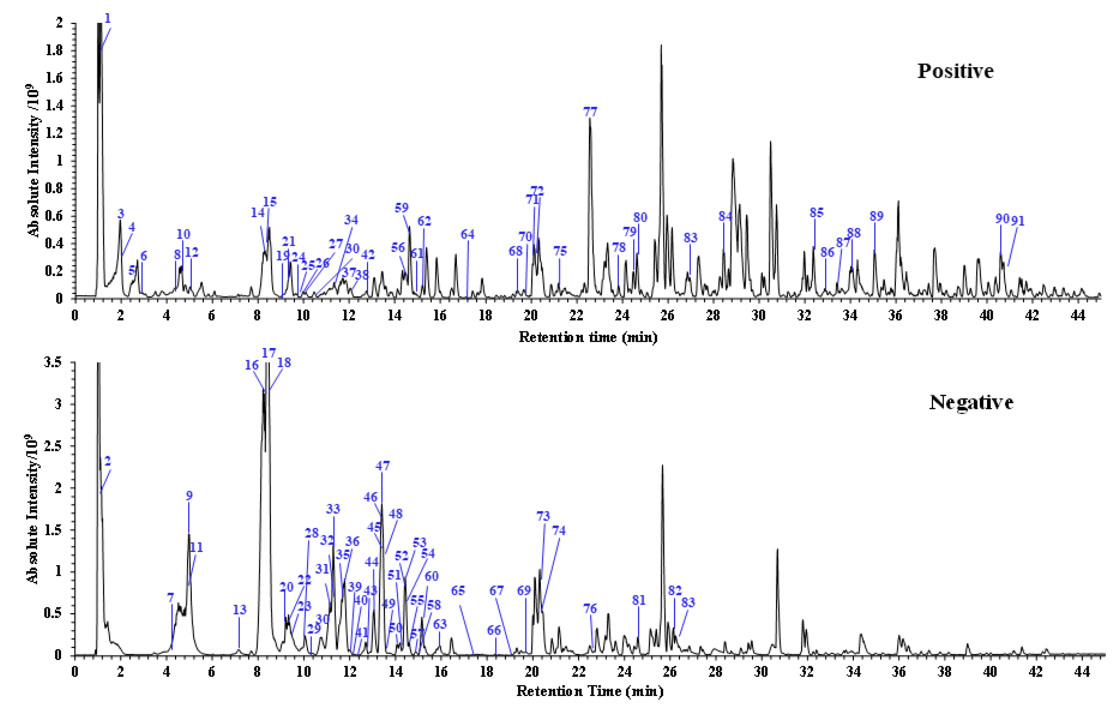
2.1. Optimization of Ultra-High Performance Liquid Chromatography (UPLC) and Mass Spectrometry (MS) Conditions
2.2. Identification of Main Constituents in G. conopsea Extract
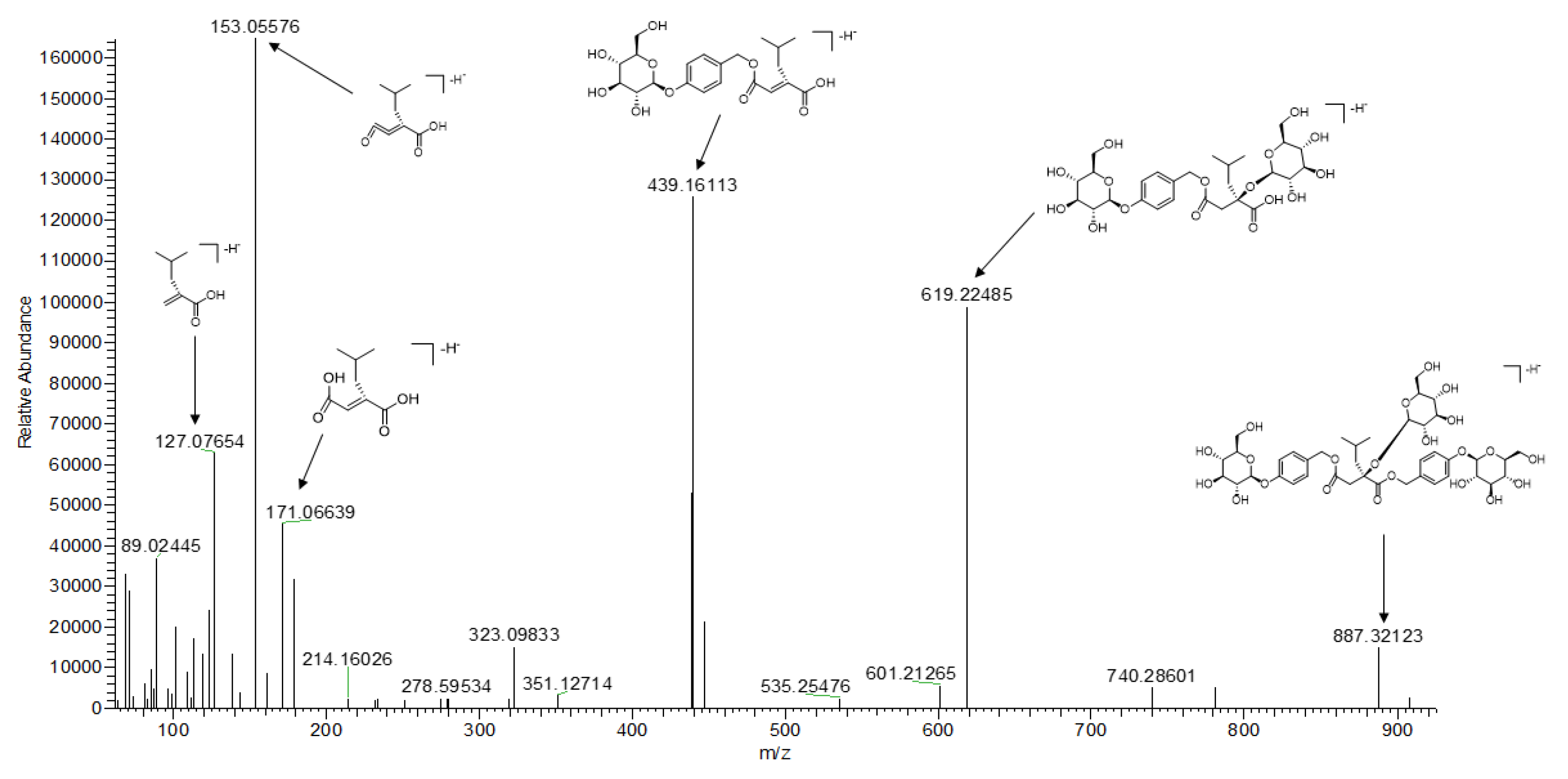
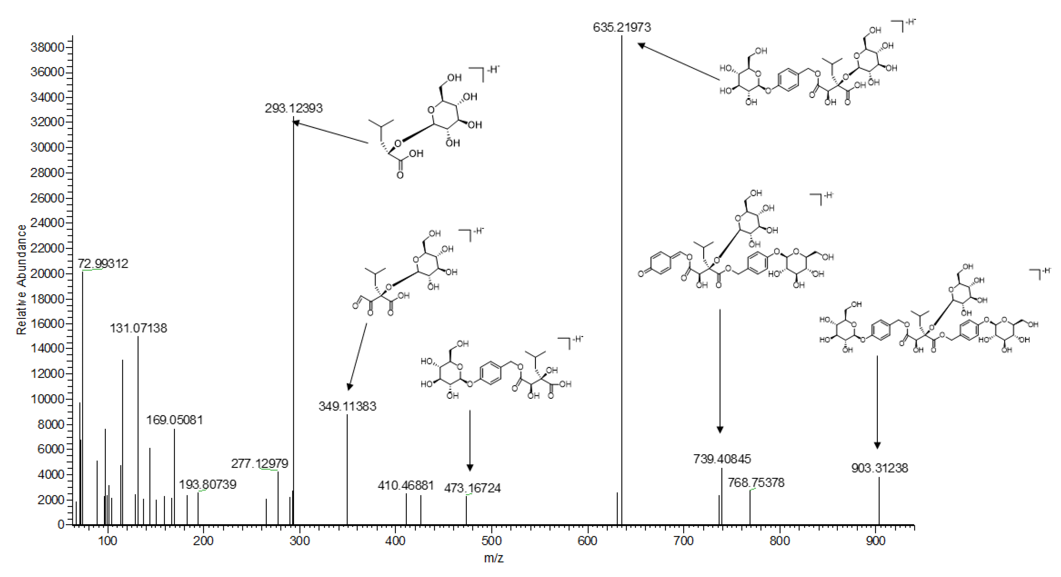
2.2.1. Succinic Acid Ester Glycosides
2.2.2. Stilbenes
2.2.3. Phenanthrenes
2.2.4. Phenolic Acid derivatives
2.2.5. Alkaloid
2.2.6. Terpenoid and Steroid
2.2.7. Others
3. Materials and Methods
3.1. Chemicals and Reagents
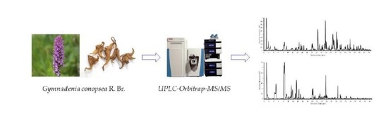
3.2. Materials and Sample Preparation
3.3. UPLC–Orbitrap–MS/MS
4. Conclusions
Supplementary Materials
Author Contributions
Funding
Conflicts of Interest
References
- Flora of China; Science Press: Beijing, China, 1999; Volume 17, pp. 388–394.
- Jiangsu New Medical College. Dictionary of Traditional Chinese Medicine; Shanghai Science and Technology Publishing House: Shanghai, China, 1977; Volume 1, pp. 436–437. [Google Scholar]
- Matsuda, H.; Morikawa, T.; Xie, H.H.; Yoshikawa, M. Antiallergic phenanthrenes and stilbenes from the tubers of Gymnadenia conopsea. Planta Med. 2004, 70, 847–855. [Google Scholar] [CrossRef] [PubMed]
- Zi, J.C.; Li, S.; Liu, M.T.; Gan, M.L.; Lin, S.; Song, W.X.; Zhang, Y.L.; Fan, X.N.; Yang, Y.; Zhang, J.J.; et al. Glycosidic constituents of the tubers of Gymnadenia conopsea. J. Nat. Prod. 2008, 71, 799–805. [Google Scholar] [CrossRef] [PubMed]
- Morikawa, T.; Xie, H.H.; Matsuda, H.; Yoshikawa, M. Glucosyloxybenzyl 2–isobutylmalates from the tubers of Gymnadenia conopsea. J. Nat. Prod. 2006, 69, 881–886. [Google Scholar] [CrossRef] [PubMed]
- Zi, J.C.; Lin, S.; Zhu, C.G.; Yanga, Y.C.; Shi, J.G. Minor constituents from the tubers of Gymnadenia conopsea. J. Asian Nat. Prod. Res. 2010, 12, 477–484. [Google Scholar] [CrossRef] [PubMed]
- Zhang, J.J.; Shi, J.G.; Wang, Y.F.; Zhang, D.; Gao, M.; Yang, Y.C.; Huang, S.Y. Using of Succinate Derivatives in the Treatment of Dementia. CN Patent 1511520A, 14 June 2004. [Google Scholar]
- Shang, X.F.; Guo, X.; Liu, Y.; Pan, H.; Miao, X.; Zhang, J. Gymnadenia conopsea (L.) R. Br.: A systemic review of the ethnobotany, phytochemistry, and pharmacology of an important Asian folk medicine. Front. Pharmacol. 2017, 8, 24. [Google Scholar] [CrossRef] [PubMed]
- Wang, Z.; Qu, Y.; Wang, L.; Zhang, X.; Xiao, H. Ultra–high performance liquid chromatography with linear ion trap–Orbitrap hybrid mass spectrometry combined with a systematic strategy based on fragment ions forthe rapid separation and characterization of components in Stellera chamaejasme extract. J. Sep. Sci. 2016, 39, 1379–1388. [Google Scholar] [CrossRef]
- Wang, Z.X.; Liu, J.Y.; Zhong, X.J.; Li, J.J.; Wang, X.; Ji, L.L.; Shang, X.Y. Rapid characterization of chemical components in edible mushroom Sparassis crispa by UPLC–orbitrap MS analysis and potential inhibitory effects on allergic rhinitis. Molecules 2019, 24, 3014. [Google Scholar] [CrossRef]
- Zhang, G.; Chen, S.S.; Zhou, W.; Meng, J.; Deng, K.; Zhou, H.N.; Hu, N.; Suo, Y.R. Rapid qualitative and quantitative analyses of eighteen phenolic compounds from Lycium ruthenicum Murray by UPLC–Q–Orbitrap MS and their antioxidant activity. Food Chem. 2018, 269, 150–156. [Google Scholar] [CrossRef]
- Gao, Y.; Sun, L.P.; Zhuang, Y.L. UPLC–Q–Orbitrap–MS(2) analysis of Moringa oleifera leaf extract and its antioxidant, antibacterial and anti–inflammatory activities. Nat. Prod Res. 2019, 1–5. [Google Scholar] [CrossRef]
- Cai, M.; Zhou, Y.; Gesang, S.; Bianba, C.; Ding, L.S. Chemical fingerprint analysis of rhizomes of Gymnadenia conopsea by HPLC-DAD-MSn. J. Chromatogr. B Analyt Technol. Biomed Life Sci. 2006, 844, 301–307. [Google Scholar] [CrossRef]
- Huang, S.Y.; Li, G.Q.; Shi, J.G.; Mo, S.Y.; Wang, S.J.; Yang, Y.C. Chemical constituents of the rhizomes of Coeloglossum viride var. bracteatum. J. Asian Nat. Prod. Res. 2004, 6, 49–61. [Google Scholar] [CrossRef] [PubMed]
- Li, Z.H.; Guo, X.M.; Cao, Z.L.; Liu, X.J.; Liao, X.N.; Huang, C.; Xu, W.Q.; Liu, L.; Yang, P. New MS network analysis pattern for the rapid identification of constituents from traditional Chinese medicine prescription Lishukang capsules in vitro and in vivo based on UHPLC/Q–TOF–MS. Talanta 2018, 189, 606–621. [Google Scholar] [CrossRef] [PubMed]
- Sahakitpichan, P.; Mahidol, C.; Disadee, W.; Chimnoi, N.; Ruchirawat, S.; Kanchanapoom, T. Glucopyranosyloxybenzyl derivatives of (R)–2–benzylmalic acid and (R)–eucomic acid, and an aromatic glucoside from the pseudobulbs of Grammatophyllum speciosum. Tetrahedron 2013, 69, 1031–1037. [Google Scholar] [CrossRef]
- Li, M.; Guo, S.X.; Wang, C.L.; Xiao, P.G. Quantitative determination of five glucosyloxybenzyl 2–isobutylmalates in the tubers of Gymnadenia conopsea and Coeloglossum Viride var. bracteatum by HPLC. J. Chromatogr. Sci. 2009, 47, 709–713. [Google Scholar] [CrossRef]
- Dai, Y.; Yeo, S.C.M.; Barnes, P.J.; Donnelly, L.E.; Loo, L.C.; Lin, H.S. Pre–clinical pharmacokinetic and metabolomic analyses of isorhapontigenin, a dietary resveratroll derivative. Front. Pharmacol. 2018, 9, 1–14. [Google Scholar] [CrossRef]
- Zhou, W.J.; Song, J.Z.; Fu, W.W.; Tan, H.S.; Bian, Z.X.; Xu, H.X. Chemical comparison of two dosage forms of Hemp Seed Pills by UHPLC–Q–ToF–MS/MS and multivariate statistical techniques. J. Pharm. Biomed. Anal. 2013, 84, 59–68. [Google Scholar] [CrossRef]
- Ehrhardt, C.; Arapitsas, P.; Stefanini, M.; Flick, G.; Mattivi, F. Analysis of the phenolic composition of fungus–resistant grape varieties cultivated in Italy and Germany using UHPLC–MS/MS. J. Mass Spectrom. 2014, 49, 860–869. [Google Scholar] [CrossRef]
- Rodriguez–Cabo, T.; Lopez, P.; Ramil, M.; Cela, R.; Rodriguez, I. Investigation of liquid chromatography quadrupole time–of–flight mass spectrometry performance for identification and determination of hydroxylated stilbene antioxidants in wine. J. Chromatogr. A. 2014, 1337, 162–170. [Google Scholar] [CrossRef]
- Singh, D.; Kumar, S.; Pandey, R.; Hasanain, M.; Sarkar, J.; Kumar, B. Bioguided chemical characterization of the antiproliferative fraction of edible pseudo bulbs of Malaxis acuminata D. Don by HPLC–ESI–QTOF–MS. Med. Chem. Res. 2017, 26, 3307–3314. [Google Scholar] [CrossRef]
- Wang, Y.; Guan, S.H.; Meng, Y.H.; Zhang, Y.B.; Cheng, C.R.; Shi, Y.Y.; Feng, R.H.; Zeng, F.; Wu, Z.Y.; Zhang, J.X.; et al. Phenanthrenes, 9,10–dihydrophenanthrenes, bibenzyls with their derivatives, and malate or tartrate benzyl ester glucosides from tubers of Cremastra appendiculata. Phytochemistry 2013, 94, 268–276. [Google Scholar] [CrossRef]
- Bai, L.; Masukawa, N.; Yamaki, M.; Takagi, S. Four stilbenoids from Pleione bulbocodioides. Phytochemistry 1998, 48, 327–331. [Google Scholar] [CrossRef]
- Shiao, Y.J.; Chen, W.P.; Lin, Y.L. New polyphenols and triterpene from the pseudobulbs of Pleione formosana. J. Chin. Chem. Soc. 2009, 56, 828–833. [Google Scholar] [CrossRef]
- Morikawa, T.; Xie, H.H.; Matsuda, H.; Wang, T.; Yoshikawa, M. Bioactive constituents from chinese natural medicines. XVII. Constituents with radical scavenging effect and new glucosyloxybenzyl 2–isobutylmalates from Gymnadenia conopsea. Chem. Pharm. Bull. 2006, 54, 506–513. [Google Scholar] [CrossRef] [PubMed]
- Liu, C.S.; Liang, X.; Wei, X.H.; Chen, F.L.; Tang, Q.F.; Tan, X.M. Comparative pharmacokinetics of major bioactive components from Puerariae Radix–Gastrodiae Rhizome extract and their intestinal absorption in rats. J. Chromatogr. B Anal. Technol. Biomed. Life Sci. 2019, 1105, 38–46. [Google Scholar] [CrossRef] [PubMed]
- Melzer, N.; Wittenburg, D.; Hartwig, S.; Jakubowski, S.; Kesting, U.; Willmitzer, L.; Lisec, J.; Reinsch, N.; Repsilber, D. Investigating associations between milk metabolite profiles and milk traits of Holstein cows. J. Dairy Sci. 2013, 96, 1521–1534. [Google Scholar] [CrossRef]
- Simerska, P.; Monti, D.; Cechova, I.; Pelantova, H.; Mackova, M.; Bezouska, K.; Riva, S.; Kren, V. Induction and characterization of an unusual α–D–galactosidase from Talaromyces flavus. J. Biotechnol. 2007, 128, 61–71. [Google Scholar] [CrossRef]
- Shen, S.; Wang, J.B.; Chen, X.; Liu, T.T.; Zhuo, Q.; Zhang, S.Q. Evaluation of cellular antioxidant components of honeys using UPLC–MS/MS and HPLC–FLD based on the quantitative composition–activity relationship. Food Chem. 2019, 293, 169–177. [Google Scholar] [CrossRef]
- Tachai, S.; Nuntawong, N. Uncommon secondary metabolites from Etlingera pavieana rhizomes. Nat. Prod. Res. 2016, 30, 2215–2219. [Google Scholar] [CrossRef]
- Kammerer, B.; Kahlich, R.; Biegert, C.; Gleiter, C.H.; Heide, L. HPLC–MS/MS analysis of willow bark extract contained in pharmaceutical preparations. Phytochem. Anal. 2005, 16, 470–478. [Google Scholar] [CrossRef]
- Zhang, K.X.; Yan, M.L.; Han, S.; Cong, L.F.; Wang, L.Y.; Zhang, L.; Sun, L.L.; Bai, H.Y.; Wei, G.H.; Du, H.; et al. Identification of chemical markers for the discrimination of radix Angelica sinensis grown in geoherb and non–geoherb regions using UHPLC–QTOF–MS/MS based metabolomics. Molecules 2019, 24, 3536. [Google Scholar] [CrossRef]
- Casado, N.; Morante–Zarcero, S.; Perez–Quintanilla, D.; Camara, J.S.; Sierra, I. Dispersive solid–phase extraction of polyphenols from juice and smoothie samples using hybrid mesostructured silica followed by ultra–high–performance liquid chromatography–ion–trap tandem mass spectrometry. J. Agric. Food Chem. 2019, 67, 955–967. [Google Scholar] [CrossRef] [PubMed]
- Liang, L.; Xu, J.; Zhou, W.W.; Brand, E.; Chen, H.B.; Zhao, Z.Z. Integrating targeted and untargeted metabolomics to investigate the processing chemistry of polygoni multiflori radix. Front. Pharmacol. 2018, 9, 934/1–934/19. [Google Scholar] [CrossRef] [PubMed]
- Liu, J.; Zou, S.H.; Liu, W.; Li, J.; Wang, H.; Hao, J.; He, J.; Gao, X.M.; Liu, E.; Chang, Y.X. An established HPLC–MS/MS method for evaluation of the influence of salt processing on pharmacokinetics of six compounds in cuscutae semen. Molecule 2019, 24, 2502. [Google Scholar] [CrossRef] [PubMed]
- Bergantin, C.; Maietti, A.; Cavazzini, A.; Pasti, L.; Tedeschi, P.; Brandolini, V.; Marchetti, N. Bioaccessibility and HPLC–MS/MS chemical characterization of phenolic antioxidants in Red Chicory (Cichorium intybus). J. Funct. Foods. 2017, 33, 94–102. [Google Scholar] [CrossRef]
- Dresel, M.; Dunkel, A.; Hofmann, T. Sensomics analysis of key bitter compounds in the hard resin of hops (Humulus lupulus L.) and their contribution to the bitter profile of pilsner–type beer. J. Agric. Food Chem. 2015, 63, 3402–3418. [Google Scholar] [CrossRef]
- Piccolella, S.; Crescente, G.; Volpe, M.G.; Paolucci, M.; Pacifico, S. UHPLC–HR–MS/MS–guided recovery of bioactive flavonol compounds from Greco di Tufo vine leaves. Molecules 2019, 24, 3630. [Google Scholar] [CrossRef]
- Di Paola Naranjo, R.D.; Otaiza, S.; Saragusti, A.C.; Baroni, V.; Carranza, A.d.V.; Peralta, I.E.; Valle, E.M.; Carrari, F.; Asis, R. Hydrophilic antioxidants from Andean tomato landraces assessed by their bioactivities in vitro and in vivo. Food Chem. 2016, 206, 146–155. [Google Scholar] [CrossRef]
- Raju, K.S.R.; Rashid, M.; Gundeti, M.; Taneja, I.; Malik, M.Y.; Singh, S.K.; Chaturvedi, S.; Challagundla, M.; Singh, S.P.; Gayen, J.R.; et al. LC–ESI–MS/MS method for the simultaneous determination of isoformononetin, daidzein, and equol in rat plasma: Application to a preclinical pharmacokinetic study. J. Chromatogr. B Anal. Technol. Biomed. Life Sci. 2019, 1129, 12176, Ahead of Print. [Google Scholar] [CrossRef]
- Tenfen, A.; Vechi, G.; Cechinel–Zanchett, C.C.; Lorenzett, T.S.; Reginato–Couto, C.E.; Siebert, D.A.; Vitali, L.; Micke, G.; Klein–Junior, L.C.; Cechinel Filho, V. Phenolic profile by HPLC–ESI–MS/MS of six Brazilian Eugenia species and their potential as cholinesterase inhibitors. Nat. Prod. Res. 2019. Ahead of Print. [Google Scholar] [CrossRef]
- Cao, Y.T.; Wu, J.H.; Pan, H.Y.; Wang, L.H. Chemical profile and multicomponent quantitative analysis for the quality evaluation of toad venom from different origins. Molecules 2019, 24, 3595. [Google Scholar] [CrossRef]
- Slyundina, M.S.; Borisov, R.S.; Zaikin, V.G. Novel Reactive Matrices for the Analysis of Alcohols by Matrix–Assisted Laser Desorption/Ioization Mass Spectrometry. J. Anal. Chem. 2018, 73, 1347–1352. [Google Scholar] [CrossRef]
- Wang, T.; Zhang, F.J.; Zhuang, W.B.; Shu, X.C.; Wang, Z. Metabolic variations of flavonoids in leaves of T. media and T. mairei obtained by UPLC–ESI–MS/MS. Molecules 2019, 24, 3323. [Google Scholar] [CrossRef] [PubMed]
- Wang, X.B.; Han, C.; Wu, K.Q.; Luo, L.; Wang, Y.; Du, X.Z.; He, Q.; Ye, F.Q. Design, synthesis and ability of non–gold complexed substituted purine derivatives to inhibit LPS–induced inflammatory response. Eur. J. Med. Chem. 2018, 149, 10–21. [Google Scholar] [CrossRef] [PubMed]
- Marklova, E.; Hais, L.M. Chromatographic behavior of some indole acids on a Sephadex column in water and in the presence of salts. J. Chromatogr. 1977, 131, 205–213. [Google Scholar] [CrossRef]
- Deda, O.; Virgiliou, C.; Orfanidis, A.; Gika, H.G. Study of fecal and urinary metabolite perturbations induced by chronic ethanol treatment in mice by UHPLC–MS/MS targeted profiling. Metabolites 2019, 9, 232. [Google Scholar] [CrossRef] [PubMed]
- Zhang, Z.H.; Li, W.J.; Lin, R.C.; Dai, Z.; Li, X.F. Isolation and structure elucidation of alkaloids from Pinellia ternata. Heterocycles 2013, 87, 637–643. [Google Scholar]
- Lin, P.C.; Yao, J.; Wu, J.; Tian, J.; Bao, Y.; Lin, S. A new ureido–substituted amino acid from the tubers of Gymnadenia conopsea. Chin. Chem. Lett. 2017, 28, 257–259. [Google Scholar] [CrossRef]
- Gonzalez, O.; Alonso, R.M.; Ferreiros, N.; Weinmann, W.; Zimmermann, R.; Dresen, S. Development of an LC–MS/MS method for the quantitation of 55 compounds prescribed in combined cardiovascular therapy. J. Chromatogr. B Anal. Technol. Biomed. Life Sci. 2011, 879, 243–252. [Google Scholar] [CrossRef]
- Zaher, A.M.; Moharram, A.M.; Davis, R.; Panizzi, P.; Makboul, M.A.; Calderon, A.I. Characterisation of the metabolites of an antibacterial endophyte Botryodiplodia theobromae Pat. of Dracaena draco L. by LC–MS/MS. Nat. Prod. Res. 2015, 29, 2275–2281. [Google Scholar] [CrossRef] [PubMed]
- Li, Z.F.; Wang, Y.W.; Ouyang, H.; Lu, Y.; Qiu, Y.; Feng, Y.L.; Jiang, H.L.; Zhou, X.; Yang, S.L. A novel dereplication strategy for the identification of two new trace compounds in the extract of Gastrodia elata using UHPLC/Q–TOF–MS/MS. J. Chromatogr. B Anal. Technol. Biomed. Life Sci. 2015, 988, 45–52. [Google Scholar] [CrossRef] [PubMed]
- Hua, Y.S.; Dennis, J.K. Increasing the sensitivity of an LC–MS method for screening material extract for organic extractables via mobile phase optimization. J. Chromatogr. Sci. 2012, 50, 213–227. [Google Scholar] [CrossRef] [PubMed]
- Wu, C.T.; Wang, X.; Liu, Y.P.; Di, X.; Xu, M. Intracellular Accumulation as an Indicator of Cytotoxicity to screen Hepatotoxic Components of Chelidonium majus L. by LC–MS/MS. Molecules 2019, 24, 2410. [Google Scholar] [CrossRef] [PubMed]
- Li, Z.F.; Zhao, C.X.; Zhao, X.J.; Xia, Y.Y.; Sun, X.S.; Xie, W.Y.; Ye, Y.R.; Lu, X.; Xu, G.W. Deep anotation of hdroxycinnamic aid aides in pants bsed on Ultra–High–Performance Liquid Chromatography–High–Resolution Mass Spectrometry and its in Silico database. Anal. Chem. 2018, 90, 14321–14330. [Google Scholar] [CrossRef] [PubMed]
- Lin, P.; Qin, Z.F.; Yao, Z.H.; Wang, L.; Zhang, W.Y.; Yu, Y.; Dai, Y.; Zhou, H.; Yao, X.S. Metabolites profile of Gualou Xiebai Baijiu decoction (a classical traditional Chinese medicine prescription) in rats by ultra–performance liquid chromatography coupled with quadrupole time–of–flight tandem mass spectrometry. J. Chromatogr. B Anal. Technol. Biomed. Life Sci. 2018, 1085, 72–88. [Google Scholar] [CrossRef] [PubMed]
- Marek, E.M.; Koslitz, S.; Weiss, T.; Fartasch, M.; Schluter, G.; Kafferlein, H.U.; Bruning, T. Quantification of N–phenyl–2–naphthylamine by gas chromatography and isotope–dilution mass spectrometry and its percutaneous absorption ex vivo under workplace conditions. Arch Toxicol. 2017, 91, 3587–3596. [Google Scholar] [CrossRef]
- Panusa, A.; Petrucci, R.; Lavecchia, R.; Zuorro, A. UHPLC–PDA–ESI–TOF/MS metabolic profiling and antioxidant capacity of arabica and robusta coffee silverskin: Antioxidants vs. phytotoxins. Food Res. Int. 2017, 99, 155–165. [Google Scholar] [CrossRef]
- Oldoni, T.L.C.; Merlin, N.; Karling, M.; Carpes, S.T.; Matias de Alencar, S.; Morales, R.G.F.; Aparecido da Silva, E.; Pilau, E.J. Bioguided extraction of phenolic compounds and UHPLC–ESI–Q–TOF–MS/MS characterization of extract of Moringa oleifera leaves collected in Brazil. Food Res. Int. 2019, 125, 108647, Ahead of Print. [Google Scholar] [CrossRef]
- Zhang, Y.B.; Yang, W.Z.; Yao, C.L.; Feng, R.H.; Yang, M.; Guo, D.A.; Wu, W.Y. New triterpenic acids from Uncaria rhynchophylla: Chemistry, NO–inhibitory activity, and tandem mass spectrometric analysis. Fitoterapia 2014, 96, 39–47. [Google Scholar] [CrossRef]
- Gray, B.P.; Teale, P. The use of a simple backflush technology to improve sample throughput and system robustness in routine gas chromatography tandem mass spectrometry analysis of doping control samples. J. Chromatogr. A. 2010, 1217, 4749–4752. [Google Scholar] [CrossRef]
- Singer, J.W.; Samuels, A.I.; Adamson, J.W. Steroids and hematopoiesis. 1. The effect of steroids on in vitro erythroid colony growth: Structure/activity relationships. J. Cell. Physiol. 1976, 88, 127–133. [Google Scholar] [CrossRef]
- Zhang, W.D.; Jin, M.M.; Jiang, H.H.; Yang, J.X.; Wang, Q.; Du, Y.F.; Cao, L.; Xu, H.J. Study on the metabolites of betulinic acid in vivo and in vitro by ultra high performance liquid chromatography with time–of–flight mass spectrometry. J. Sep. Sci. 2019, 42, 628–635. [Google Scholar] [CrossRef]
- Wang, C.Z.; Zhang, N.Q.; Wang, Z.Z.; Qi, Z.; Zheng, B.B.; Li, P.Y.; Liu, J.P. Rapid characterization of chemical constituents of Platycodon grandiflorum and its adulterant Adenophora stricta by UPLC–QTOF–MS/MS. J. Mass Spectrom. 2017, 52, 643–656. [Google Scholar] [CrossRef] [PubMed]
- Gauvin, A.; Smadja, J.; Aknin, M.; Gaydou, E.M. Sterol composition and chemotaxonomic considerations in relation to sponges of the genus Xestospongia. Biochem. Syst. Ecol. 2004, 32, 469–476. [Google Scholar] [CrossRef]
- Lee, S.J.; Jeong, E.M.; Ki, A.Y.; Oh, K.S.; Kwon, J.; Jeong, J.H.; Chung, N.J. Oxidative defense metabolites induced by salinity stress in roots of Salicornia herbacea. J. Plant Physiol. 2016, 206, 133–142. [Google Scholar] [CrossRef] [PubMed]
- Khatal, L.; More, H. Development and validation of a liquid chromatography–tandem mass spectrometry method for quantification of Lupeol in plasma and its application to pharmacokinetic study in rats. J. Chromatogr. B Anal. Technol. Biomed. Life Sci. 2019, 1121, 58–65. [Google Scholar] [CrossRef]
- Prinsen, P.; Gutierrez, A.; Faulds, C.B.; del Rio, J.C. Comprehensive Study of Valuable Lipophilic Phytochemicals in Wheat Bran. J. Agric. Food Chem. 2014, 62, 1664–1673. [Google Scholar] [CrossRef]
- Gao, X.X.; Wang, N.; Jia, J.P.; Wang, P.Y.; Zhang, A.R.; Qin, X.M. Chemical profiling of Dingkun Dan by ultra High performance liquid chromatography Q exactive orbitrap high resolution mass spectrometry. J. Pharm. Biomed. Anal. 2020, 177, 112732. [Google Scholar] [CrossRef]
- Han, S.W.; Wang, C.; Cui, B.S.; Sun, H.; Zhang, J.J.; Li, S. Hepatoprotective activity of glucosyloxybenzyl succinate derivatives from the pseudobulbs of Pleione bulbocodioides. Phytochemistry 2019, 157, 71–81. [Google Scholar] [CrossRef]
- Jia, J.; Liu, M.; Wen, Q.; He, M.Z.; Ouyang, H.; Chen, L.Y.; Li, J.M.; Feng, Y.L.; Zhong, G.Y.; Yang, S.L. Screening of anti–complement active ingredients from Eucommia ulmoides Oliv. branches and their metabolism in vivo based on UHPLC–Q–TOF/MS/MS. J. Chromatogr. B Anal. Technol. Biomed. Life Sci. 2019, 1124, 26–36. [Google Scholar] [CrossRef]
- Lobine, D.; Cummins, I.; Govinden–Soulange, J.; Ranghoo–Sanmukhiya, M.; Lindsey, K.; Chazot, P.L.; Ambler, C.A.; Grellscheid, S.; Sharples, G.; Lall, N.; et al. Medicinal Mascarene Aloes: An audit of their phytotherapeutic potential. Fitoterapia 2018, 124, 120–126. [Google Scholar] [CrossRef]
- Rosenthal, I.; Wolfram, E.; Peter, S.; Meier, B. Validated method for the analysis of frangulins A and B and glucofrangulins A and B using HPLC and UHPLC. J. Nat. Prod. 2014, 77, 489–496. [Google Scholar] [CrossRef] [PubMed]
- Xu, Y.Y.; Cai, H.; Cao, G.; Duan, Y.; Pei, K.; Tu, S.C.; Zhou, J.; Xie, L.; Sun, D.D.; Zhao, J.Y.; et al. Profiling and analysis of multiple constituents in Baizhu Shaoyao San before and after processing by stir–frying using UHPLC/Q–TOF–MS/MS coupled with multivariate statistical analysis. J. Chromatogr. B Anal. Technol. Biomed. Life Sci. 2018, 1083, 110–123. [Google Scholar] [CrossRef] [PubMed]
Sample Availability: Samples of the compounds are not available from the authors. |


| No | R.T. (min) | Compound Name | Formula | Exact Mass | Error (ppm) | Adduct Ion (m/z) | MS2 Fragment (m/z) | Ref. |
|---|---|---|---|---|---|---|---|---|
| Succinic Acid Ester Glycosides | ||||||||
| 9 | 4.605 | coelovirins E | C14H24O11 | 368.13181 | −0.14 | 367.12473 [M − H]− | 293.12454, 187.06120, 143.07137 a, 99.08157 | [14] |
| 16 | 8.430 | dactylorhin C | C14H23O10 | 352.13690 | 0.09 | 351.12982 [M − H]− | 179.05595, 171.06635,127.07648 a | [15] |
| 28 | 10.072 | coelovirins D | C27H40O17 | 636.22664 | 0.15 | 635.21948 [M − H]− | 349.11404 a, 293.12415, 277.12915,143.07129 | [14] |
| 29 | 10.308 | grammatophylloside C | C24H28O12 | 508.28186 | 2.09 | 507.14993 [M − H]− | 221.04546,203.03497 a, 177.05568, 149.06070, 107.05019 | [16] |
| 31 | 10.748 | Coelovirin B | C21H30O12 | 474.17371 | 0.63 | 473.16614 [M − H]− | 367.12451, 293.10284, 187.06094, 159.06616,143.0729, 115.07640, 99.08151 a | [14] |
| 32 | 11.08 | (−)-(2R,3S)-1-(4-β-d-glucopyranosyloxybenzyl)-2-O-β-d-glucopyranosyl-4-{4-[α-d-glucopyranosyl-(1-4)-β-d-glucopyranosyloxy]-benzyl}-2-isobutyltartrate | C46H66O28 | 1066.37406 | −0.06 | 1065.37610 [M − H]− | 797.27228 a, 635.21936, 455.17773, 293.12411 | [4] |
| 33 | 11.291 | dactylorhin B | C40H56O23 | 904.32147 | 1.42 | 903.31238 [M + H] + | 739.40845, 635.21973 a, 473.16724, 349.11383, 293.12393 | [15] |
| 35 | 11.678 | loroglossin | C34H46O18 | 742.26858 | 0.04 | 741.26056 [M − H]− | 455.15555, 285.09799, 349.11484, 277.12958 a, 187.09761, 123.04520 | [17] |
| 36 | 11.756 | dactylorhin E | C27 H40 O16 | 620.23185 | −0.34 | 619.22369 [M − H]− | 439.16074, 285.09821, 179.05609,153.05569 a | [15] |
| 44 | 13.063 | coelovirins A | C21H30O11 | 458.17903 | 0.49 | 457.17169 [M − H]− | 285.09793, 189.07683, 171.06650,153.05566, 127.07648 a 123.04527 | [14] |
| 46 | 13.420 | (−)-(2R,3S)-1-(4-β-d-glucopyranosyloxybenzyl)-4-methyl 2-isobutyltartrate | C22H32O12 | 488.18950 | 0.25 | 487.18188 [M − H]− | 189.07649, 171.06628, 153.05579, 129.09218 a, 99.08157 | [4] |
| 47 | 13.420 | dactylorhin A | C40H56O22 | 888.32675 | 1.49 | 887.32123 [M − H]− | 619.22485,439.16113, 323.09833, 153.05572 a, 171.06639, 127.07654 | [15] |
| 48 | 13.425 | gymnoside II | C21H30O11 | 458.17897 | 0.35 | 457.17175 [M − H]− | 285.09827,171.06633, 153.05576, 127.07654,123.04524, 99.08158 | [15] |
| 52 | 14.412 | gymnoside III | C42H58O23 | 930.33937 | −1.11 | 929.33154 [M − H]− | 661.23553, 619.22565 481.17163, 439.16144, 153.05579 a | [5] |
| 53 | 14.431 | gymnosides VII | C50H62O24 | 1046.36365 | 1.21 | 1045.35632 [M − H]− | 741.26141, 635.21967, 455.15485, 349.11420, 293.12424 a | [5] |
| 54 | 14.436 | gymnoside I | C21H30O11 | 458.17897 | 0.35 | 457.17169 [M − H]− | 351.12991 171.06636, 127.07649 a, 123.04526, 99.08160 | [15] |
| 55 | 14.440 | militarine | C34H46O17 | 726.27387 | 0.51 | 725.26599 [M − H]− | 457.17157 a, 285.09799, 153.05573, 127.07654, 123.04519 | [17] |
| Stilbenes | ||||||||
| 38 | 11.995 | isorhapontigenin | C15H14O4 | 258.08932 | −0.42 | 259.09647 [M + H]+ | 227.07019,199.07533 a, 135.04410, 107.04953 | [18] |
| 39 | 12.018 | rhaponticin | C21H24O9 | 420.14210 | −0.16 | 419.13513 [M − H]− | 256.07437, 241.05089 a, 213.05588 | [19] |
| 40 | 12.116 | piceatannol | C14H12O4 | 244.07371 | −0.57 | 243.06630 [M − H]− | 149.02441 a, 121.02955, 93.03458 | [20] |
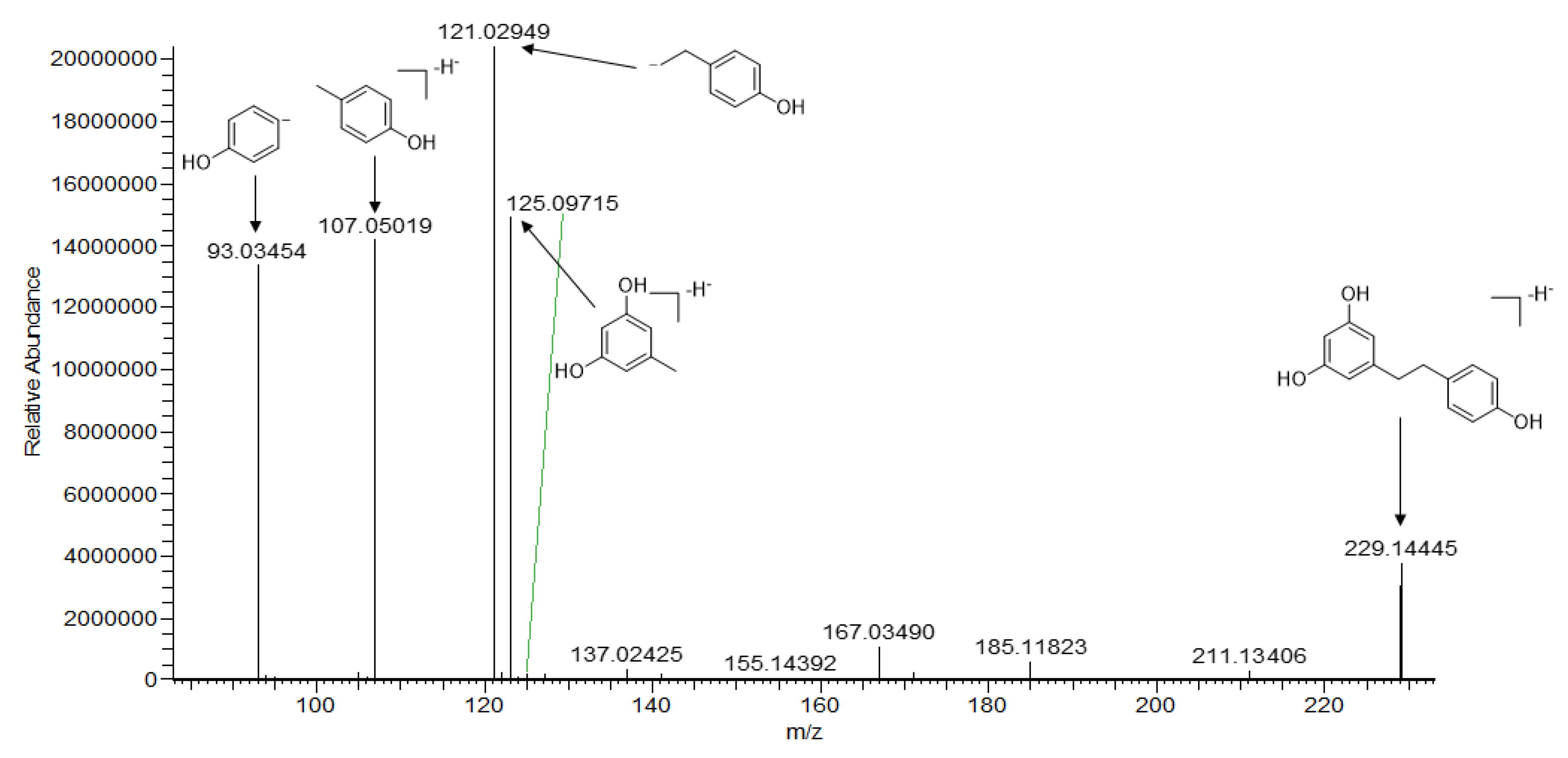
| 57 | 14.568 | dihydro-resveratroll | C14H14O3 | 230.09433 | −0.05 | 229.14445 [M − H]− | 123.04518, 121.02949 a 107.05019, 93.03454 | [21] |
| 64 | 17.405 | batatasin III | C15 H16O3 | 244.11001 | 0.23 | 245.11731 [M − H]− | 227.10683, 151.07535, 137.05969, 121.06501 a | [22] |
| 69 | 19.445 | 3,3′-dihydroxy-4-(4-hydroxybenzyl)-5-methoxybibenzyl | C22H22O4 | 350.15206 | 0.71 | 349.14474 [M − H]− | 255.10283, 243.10271 a, 227.07153, 106.04240, 93.03458 | [23] |
| 72 | 19.998 | bulbocodin C | C29H28O5 | 456.19405 | 0.83 | 455.18674 [M − H]− | 361.14493 a, 331.09796, 304.11102, 255.10280, 93.03461 | [24] |
| 73 | 20.542 | bulbocodin D | C29H28O5 | 456.19372 | 0.88 | 455.18680 [M − H]− | 440.09048, 361.1088 a, 349.10840, 255.06645, 93.03416 | [24] |
| 76 | 22.298 | 3,3′-dihydroxy-2,6-bis(4-hydroxybenzyl)-5-methoxybibenzyl | C29H28O4 | 440.19894 | 0.42 | 439.19168 [M − H]− | 424.16870, 345.14984 a, 333.11353, 93.03459 | [25] |
| Phenanthrenes | ||||||||
| 71 | 19.863 | 1-((4-hydroxyphenyl)methyl)-4-methoxy-2,7-phenanthrenediol | C22H18O4 | 346.12087 | 1.03 | 347.12778 [M + H]+ | 253.08589 a, 235.07544, 207.08047, 107.04955, | [26] |
| 74 | 21.160 | gymconopin A | C22H20O4 | 348.13616 | 0.02 | 347.12888 [M − H]− | 332.10544 a, 239.07147, 226.06348, 93.03457 | [26] |
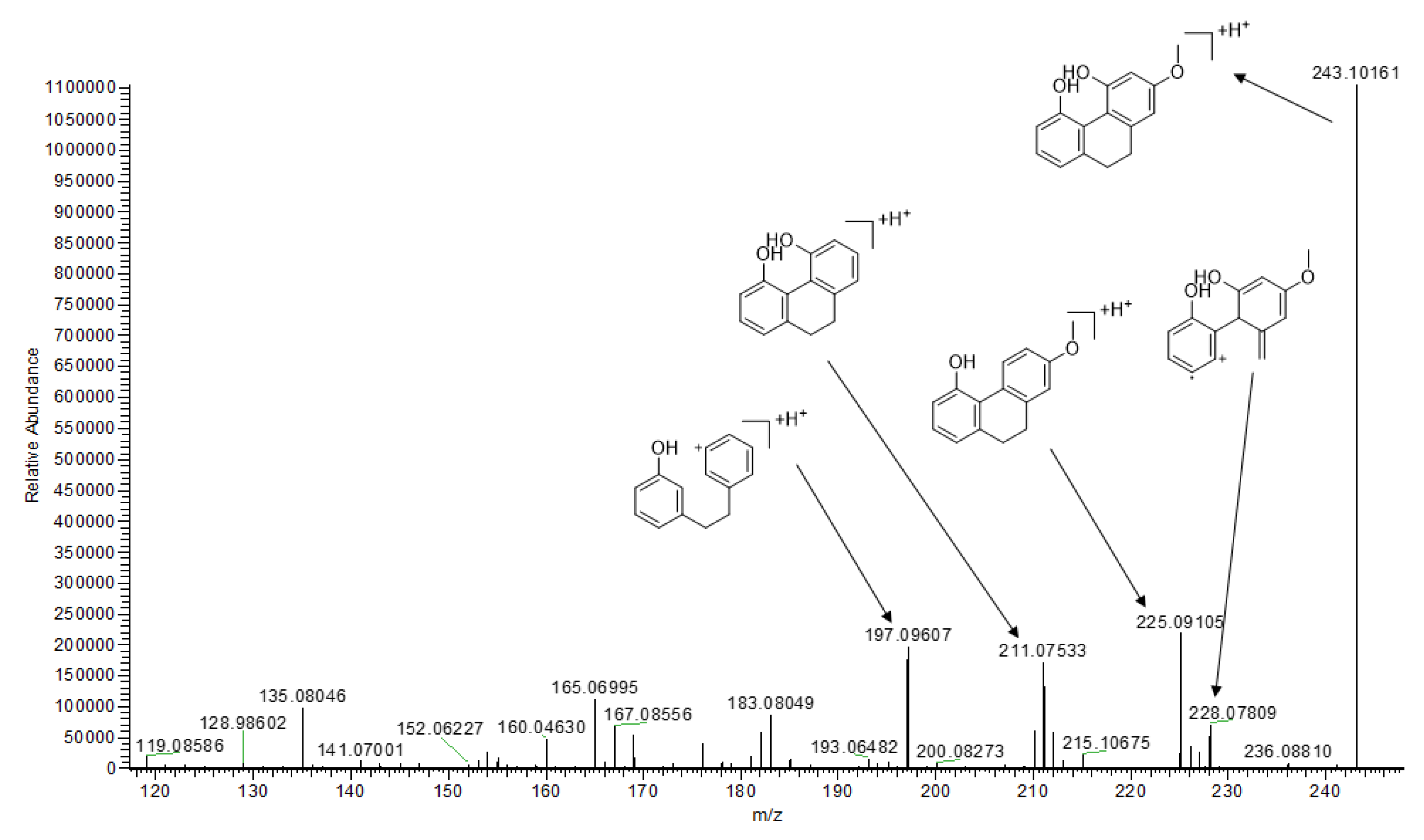
| 75 | 21.191 | 9,10-dihydro-2-methoxy-4,5-phenanthrenediol | C15H14O3 | 242.09439 | 0.25 | 243.10161 [M + H]+ | 228.07809, 225.09105 a, 211.07533 197.09607 | [26] |
| 82 | 26.152 | blestriarene A | C30H26O6 | 482.17309 | 0.03 | 481.16586 [M − H]− | 466.14246, 241.05086 a, 210.06853 | [26] |
| 83 | 26.438 | gymconopin | C30H26O6 | 482.17308 | 0.27 | 481.16583 [M − H]− | 241.05081,225.09227, 210.06870 a | [26] |
| 84 | 27.870 | blestriarene B | C30H24O6 | 480.15759 | 0.63 | 481.16461 [M + H]+ | 257.08075 a, 225.05467, 211.07530, 207.04405 | [26] |
| Phenolic Acid Derivatives | ||||||||
| 7 | 4.203 | (−)-4-[β-d-glucopyranosyl-(1-4)-β-d-glucopyranosyloxy]benzyl alcohol] | C19H28O12 | 448.15814 | 0.15 | 447.15176 [M − H]− | 341.10901 a,179.05614, 161.04562, 119.03497, 89.02443 | [5] |
| 11 | 4.877 | (+)-4-[α-d-glucopyranosyl-(1-4)-β-d-glucopyranosyloxy]benzyl alcohol | C19H28O12 | 448.15811 | 0.12 | 447.15079 [M − H]− | 341.10901 a,179.05614, 161.04575, 89.02444, 71.01380 | [5] |
| 13 | 7.711 | 4-methoxyphenyl β-d-glucopyranoside | C13H18O7 | 286.10521 | −0.16 | 285.09793 [M − H]− | 179.11877, 161.04642, 123.04515 a | [27] |
| 17 | 8.943 | dactylose B | C12H16O6 | 256.09481 | 0.49 | 255.08772 [M − H]− | 237.11345,237.07713, 165.05467, 123.04523 a | [28] |
| 18 | 9.049 | phenyl-3-deoxyheopyranoside | C12H16O5 | 240.09993 | −0.63 | 239.09271 [M − H]− | 179.07149 a, 162.06873, 121.02957 | [29] |
| 21 | 9.267 | isoferulic acid | C10H10O4 | 194.05803 | 0.64 | 195.06535 [M + H]+ | 177.05464 a, 149.05975, 145.02840, 117.03376 | [30] |
| 22 | 9.549 | ferulic acid | C10H10O4 | 194.05808 | −0.88 | 195.06541 [M − H]− | 177.05453, 149.05968, 145.02832 a, 117.03370 | [31] |
| 23 | 9.562 | p-doumaric acid | C9H8O3 | 164.04738 | −0.23 | 163.04010 [M − H]− | 119.05019 a, 93.03452 | [30] |
| 25 | 9.621 | (E)-4-methoxycinnamic acid | C10H10O3 | 178.06311 | −0.69 | 179.07040 [M + H]+ | 147.04402 a, 137.05974, 119.04941, 91.05477 | [31] |
| 34 | 11.595 | tremuloidin | C20H22O8 | 390.13185 | −0.97 | 389.12460 [M + H]+ | 341.10324, 193.05069 a, 150.03229, 134.03743 | [32] |
| 43 | 12.631 | chlorogenic acid | C16H18O9 | 354.09569 | 1.67 | 353.08841 [M − H]− | 179.03511 a,135.04527, 177.01929, 109.02952 | [33] |
| 45 | 13.353 | quercetin-3β-D-glucoside | C21H20O12 | 464.09555 | −0.15 | 463.08832 [M − H]− | 300.02747 a, 271.02481, 255.02997 | [34] |
| 49 | 13.665 | cirsimarin | C23H24O11 | 476.13197 | −0.22 | 475.12469 [M − H]− | 307.08240 a, 167.03502, 152.01154 | [35] |
| 50 | 14.041 | astragalin | C21H20O11 | 448.10073 | −0.39 | 447.09341 [M − H]− | 284.03262, 255.03510 a, 227.03510 | [36] |
| 56 | 14.470 | kaempferol-7-O-glucoside | C21H20O11 | 448.10072 | −0.36 | 449.10794 [M + H]+ | 287.05487 a, 258.05228, 145.04948 | [37] |
| 59 | 14.609 | desmethylxanthohumol | C18H22O5 | 340.13105 | 0.07 | 341.13831 [M + H]+ | 323.12762, 217.08611, 153.05446, 137.05969 a, 187.07526 | [38] |
| 61 | 14.917 | isorhamnetin | C16H12O7 | 316.05854 | −0.74 | 317.06573 [M + H]+ | 302.04196 a, 274.04684, 273.03922, 153.01820 | [39] |
| 63 | 16.015 | naringenin chalcone | C15H12O5 | 272.06856 | −0.33 | 271.06131 [M − H]− | 177.01930, 151.00363 a, 145.02951, 119.05019 | [40] |
| 65 | 17.450 | equol | C15H14O3 | 242.09429 | −0.72 | 243.10172 [M − H]− | 228.07822, 211.07527, 149.05972, 135.04405, 123.04429,107.04951 a | [41] |
| 82 | 24.670 | galangin | C15H10O5 | 270.05291 | −0.31 | 269.04562 [M − H]− | 241.05077, 225.05580 a | [42] |
| Alkaloids | ||||||||
| 1 | 1.112 | dl-arginine | C6H14N4O2 | 174.11176 | −0.48 | 175.11899 [M + H]+ | 158.09248,130.09763,116.07089, 112.08723, 70.06586 a | [43] |
| 3 | 1.946 | Adenosine | C10H13N5O4 | 267.09653 | 0.84 | 268.10388 [M + H]+ | 136.06180a, 119.03542, | [43] |
| 4 | 1.961 | 6-quinolinecarboxylic acid | C10 H7NO2 | 173.04785 | 0.03 | 174.05510 [M + H]+ | 156.04442, 146.06017 a, 130.06531,128.04971 | [44] |
| 5 | 2.479 | l-Phenylalanine | C9H11NO2 | 165.07921 | −1.40 | 166.08640 [M + H]+ | 149.05977, 131.04926, 120.08099 a,103.05462 | [45] |
| 6 | 3.100 | N-(4-methyoxyphenyl)-1H-pyrazolo [3,4-d]pyrimidin | C12H11N5O | 241.09636 | −0.14 | 242.10341 [M + H]+ | 136.06171, 107.04944 a | [46] |
| 8 | 4.329 | trans-indole-3-acrylic acid | C11H9NO2 | 187.06348 | −0.29 | 188.07060 [M + H]+ | 170.06012, 146.06004 a, 144.08080, 118.06541 | [47] |
| 10 | 4.856 | Guanine | C5H5N5O | 151.04946 | −0.34 | 152.05661 [M + H]+ | 135.03011 a, 110.03517 | [48] |
| 12 | 5.444 | 5′-S-Methyl-5′-thioadenosine | C11H15N5O3S | 297.08965 | −0.29 | 298.09668 [M + H]+ | 136.06178 a, 163.04239, 145.03169 | [49] |
| 14 | 8.361 | conopsamide A | C14H21N3O4 | 295.15315 | 1.05 | 294.14621 [M − H]− | 188.10416, 131.08266 a, | [50] |
| 15 | 8.420 | befunolol | C16H21NO4 | 291.14681 | 0.90 | 292.25405 [M + H]+ | 277.13074, 151.03897, 124.11227 a, | [51] |
| 19 | 9.067 | cyclo(tyrosy-tyrosyl) | C18H18N2O4 | 326.12667 | −0.05 | 327.13342 [M + H]+ | 221.09201, 203.08133, 175.08655,158.06003, 107.04946 a | [6] |
| 24 | 9.596 | cyclo(leucylprolyl) | C11H18N2O2 | 210.13695 | 0.58 | 211.14403 [M + H]+ | 193.08359, 183.14925, 138.12781, 127.08688, 114.09170, 70.06586 a | [52] |
| 26 | 9.758 | N-(4-hydroxybenzy) adenine riboside | C17H19N5O5 | 373.13861 | −0.05 | 374.14581 [M + H]+ | 242.10358, 148.06180, 136.06180 a, 107.04951 | [53] |
| 27 | 9.827 | dibenzylamine | C14H15N | 197.12062 | −0.89 | 198.12784 [M + H]+ | 181.10126, 106.06558,91.05482 a | [54] |
| 30 | 10.699 | (+)-chelidonine | C20H19NO5 | 353.12643 | −0.30 | 354.13321 [M + H]+ | 336.12274,293.08057, 188.07043 a, 206.08098, 149.05965 | [55] |
| 37 | 11.822 | (2E)-3-(4-hydroxy-phenyl)-N-[2-(4-hydroxy-phenyl)-ethyl]-acrylamide | C17H17NO3 | 283.12083 | 0.06 | 284.12769 [M + H]+ | 147.04390 a, 164.07062, 121.06493, 119.04931 | [56] |
| 42 | 12.834 | 2,3,4,9-tetrahydro-1H-β-carboline-3-carboxylic acid | C12H12N2O2 | 216.09012 | −1.13 | 217.09723 [M + H]+ | 144.08080 a, 156.08093, 118.06545 | [57] |
| 58 | 14.582 | dl-tryptophan | C11H12N2O2 | 204.08987 | 0.03 | 203.08272 [M − H]− | 159.09279, 142.06619, 116.05058 a, 74.24770 | [48] |
| 78 | 23.937 | N-phenyl-2-naphthylamine | C16H13N | 219.10478 | 0.08 | 220.11194 [M + H]+ | 143.07289 a, 128.06215 | [58] |
| Terpenoids and Steroids | ||||||||
| 41 | 12.664 | mascaroside | C26H36O11 | 524.22615 | −0.73 | 523.21875 [M − H]− | 361.6602 a, 179.07140, 165.05576, 101.02450 | [59] |
| 51 | 14.349 | (±)-abscisic acid | C15H20O4 | 264.13613 | 0.12 | 263.12869 [M − H]− | 219.13905 a,204.11546, 201.12842, 151.07640 | [60] |
| 77 | 23.323 | (3β,5α,9α)-3,6,19-trihydroxyurs-12-en-28-oic acid | C30H48O5 | 488.35032 | −0.29 | 489.35718 [M + H]+ | 471.34665 a,453.33636, 435.32520, 265.21689 | [61] |
| 80 | 24.638 | (3β,17β)-estr-5(10)-ene-3,17-diol | C18H28O2 | 276.20882 | 0.12 | 277.21600 [M + H]+ | 259.20557, 235.16937, 221.15327, 149.13251, 121.10139, 107.08587, 93.07037 a, | [62] |
| 85 | 28.595 | 17α-methyl-5α-androstane-3β,11β,17β-triol | C20H34O3 | 322.25091 | 0.37 | 323.25797 [M + H]+ | 305.24716, 277.21613 a, 259.20554, 179.14297, 151.11176, 135.11687, 107.08589 | [63] |
| 86 | 32.654 | lup-20(29)-en-28-al | C30H48O2 | 440.36543 | −0.04 | 441.37292 [M + H]+ | 423.36244 a, 405.35190, 191.14313, 151.11177, 109.10156, 123.08073 | [64] |
| 87 | 33.514 | lupenone | C30H48O | 424.37052 | −0.02 | 425.37735 [M + H]+ | 407.36710 a, 231.21080, 191.17928, 177.16399, 109.10153 | [65] |
| 88 | 34.104 | poriferasterol | C29H48O | 412.37052 | −0.07 | 413.37762 [M + H]+ | 395.36703 a,353.33051, 255.21051, 213.16359, 159.11682, 105.07026 | [66] |
| 89 | 35.684 | 4,4-dimethyl-5α-cholesta-8,14,24-trien-3β-ol | C29H46O | 410.35496 | −0.12 | 411.36194 [M + H]+ | 393.35141, 353.32016, 253.19467, 175.11179 a, 147.11678 | [67] |
| 90 | 40.568 | lupeol | C30H50O | 426.38611 | 0.13 | 427.39322 [M + H]+ | 409.38208, 191.17934, 121.10136, 109.10149, 95.08600 a | [68] |
| 91 | 41.305 | (22E)-stigmasta-3,5,22-triene | C29H46 | 394.35992 | 0.06 | 395.36719 [M + H]+ | 297.25775, 241.19502, 173.13257, 159.11693, 145.10123 a | [69] |
| Others | ||||||||
| 2 | 1.354 | citric acid | C6H8O7 | 192.02699 | 0.05 | 191.01979 [M − H]− | 173.00919, 129.01920, 111.00877 a, 87.00876, | [70] |
| 20 | 9.247 | butanedioic acid | C8H14O5 | 190.08414 | 0.15 | 189.07680 [M − H]− | 171.06630, 129.05573 a, 143.07171, 127.07654, 99.08161 | [71] |
| 60 | 14.911 | pinoresinol | C20H22O6 | 358.1417 | 0.75 | 359.14969 [M − H]− | 163.03735, 137.05968 a, 131.04922 | [72] |
| 62 | 15.501 | benzyl-[(6-oxo-7,8,9,10-tetrahydro-6H-benzo[c]chromen-3yl)oxy]-acetate | C22H20O5 | 364.13133 | −0.72 | 365.13849 [M + H]+ | 271.09637, 239.07021, 147.04408, 107.04951 a | [72] |
| 66 | 18.242 | aloeresin A | C28H28O11 | 540.16377 | −1.15 | 539.15643 [M − H]− | 377.10330 a, 283.06125, 163.00378 | [73] |
| 67 | 19.175 | frangulin B | C20H18O9 | 402.09545 | −0.9 | 401.08740 [M − H]− | 357.06149, 313.07181, 121.02949 a | [74] |
| 68 | 19.422 | cleomiscosin A | C20H18O8 | 386.10051 | −0.91 | 387.10724 [M + H]+ | 357.06030 a, 329.06540, 301.07065, 245.04463, 149.05989 | [75] |
| 70 | 19.772 | bis-(methylbenzylidene)-sorbitol | C22H26O6 | 386.17321 | −0.69 | 387.18051 [M + H]+ | 105.07003 a, 119.04945, 103.05464 | [75] |
| 80 | 24.129 | umbelliferone | C9H6O3 | 162.03168 | 0.09 | 163.03894 [M + H]+ | 135.04408 a,133.02847, 107.04951, 105.04509 | [33] |
© 2020 by the authors. Licensee MDPI, Basel, Switzerland. This article is an open access article distributed under the terms and conditions of the Creative Commons Attribution (CC BY) license (http://creativecommons.org/licenses/by/4.0/).
Share and Cite
Wang, X.; Zhong, X.-J.; Zhou, N.; Cai, N.; Xu, J.-H.; Wang, Q.-B.; Li, J.-J.; Liu, Q.; Lin, P.-C.; Shang, X.-Y. Rapid Characterizaiton of Chemical Constituents of the Tubers of Gymnadenia conopsea by UPLC–Orbitrap–MS/MS Analysis. Molecules 2020, 25, 898. https://doi.org/10.3390/molecules25040898
Wang X, Zhong X-J, Zhou N, Cai N, Xu J-H, Wang Q-B, Li J-J, Liu Q, Lin P-C, Shang X-Y. Rapid Characterizaiton of Chemical Constituents of the Tubers of Gymnadenia conopsea by UPLC–Orbitrap–MS/MS Analysis. Molecules. 2020; 25(4):898. https://doi.org/10.3390/molecules25040898
Chicago/Turabian StyleWang, Xin, Xiang-Jian Zhong, Na Zhou, Ning Cai, Jia-Hui Xu, Qing-Bo Wang, Jin-Jie Li, Qian Liu, Peng-Cheng Lin, and Xiao-Ya Shang. 2020. "Rapid Characterizaiton of Chemical Constituents of the Tubers of Gymnadenia conopsea by UPLC–Orbitrap–MS/MS Analysis" Molecules 25, no. 4: 898. https://doi.org/10.3390/molecules25040898
APA StyleWang, X., Zhong, X.-J., Zhou, N., Cai, N., Xu, J.-H., Wang, Q.-B., Li, J.-J., Liu, Q., Lin, P.-C., & Shang, X.-Y. (2020). Rapid Characterizaiton of Chemical Constituents of the Tubers of Gymnadenia conopsea by UPLC–Orbitrap–MS/MS Analysis. Molecules, 25(4), 898. https://doi.org/10.3390/molecules25040898