Article

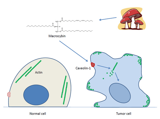
Macrocybin, a Natural Mushroom Triglyceride, Reduces Tumor Growth In Vitro and In Vivo through Caveolin-Mediated Interference with the Actin Cytoskeleton

by
Marcos Vilariño
1,
Josune García-Sanmartín
1,
Laura Ochoa-Callejero
1,
Alberto López-Rodríguez
2,
Jaime Blanco-Urgoiti
2 and
Alfredo Martínez
1,*
1
Oncology Area, Center for Biomedical Research of La Rioja (CIBIR), 26006 Logroño, Spain
2
CsFlowchem, Campus Universidad San Pablo CEU, Boadilla del Monte, 28668 Madrid, Spain
*
Author to whom correspondence should be addressed.
Molecules 2020, 25(24), 6010; https://doi.org/10.3390/molecules25246010
Submission received: 6 November 2020 / Revised: 3 December 2020 / Accepted: 17 December 2020 / Published: 18 December 2020
(This article belongs to the Special Issue Anticancer Properties of Natural and Derivative Products)

Abstract

:
Mushrooms have been used for millennia as cancer remedies. Our goal was to screen several mushroom species from the rainforests of Costa Rica, looking for new antitumor molecules. Mushroom extracts were screened using two human cell lines: A549 (lung adenocarcinoma) and NL20 (immortalized normal lung epithelium). Extracts able to kill tumor cells while preserving non-tumor cells were considered “anticancer”. The mushroom with better properties was Macrocybe titans. Positive extracts were fractionated further and tested for biological activity on the cell lines. The chemical structure of the active compound was partially elucidated through nuclear magnetic resonance, mass spectrometry, and other ancillary techniques. Chemical analysis showed that the active molecule was a triglyceride containing oleic acid, palmitic acid, and a more complex fatty acid with two double bonds. The synthesis of all possible triglycerides and biological testing identified the natural compound, which was named Macrocybin. A xenograft study showed that Macrocybin significantly reduces A549 tumor growth. In addition, Macrocybin treatment resulted in the upregulation of Caveolin-1 expression and the disassembly of the actin cytoskeleton in tumor cells (but not in normal cells). In conclusion, we have shown that Macrocybin constitutes a new biologically active compound that may be taken into consideration for cancer treatment.

Graphical Abstract

1. Introduction

The discovery of new anticancer drugs can be accomplished through different approaches, including the screening of natural products, and the testing of synthetic compound libraries, computer-assisted design, and machine learning, among others [1]. Living organisms constitute an almost unlimited source of compounds with potential biological activity and, so far, have provided the majority of currently approved therapies for the treatment of cancer [2,3]. A few examples include taxol, which was obtained from the bark of the Pacific Yew tree [4,5], vinblastine and related alkaloids from Vinca [6], camptothecin from the bark of a Chinese tree [7], or trabectedin and other drugs which were purified from marine organisms [8]. In addition, many natural products are the basis for further chemical development and the rational design of synthetic drugs [9].
The mechanisms of action by which natural products reduce cancer cell growth are diverse. In the case of taxanes, direct interaction with tubulin results in cytoskeleton hyperpolymerization and mitosis arrest [10]; vinblastine has the reverse effect, depolymerizing the cytoskeleton, but achieves the same final outcome, stopping cell division [10]; others, such as some mushroom extracts, induce immunomodulation [11,12], or affect other cancer hallmarks [13].
Mushrooms have been used for millennia in Eastern traditional medicine as cancer remedies [13,14] and recent scientific publications have corroborated the presence of anticancer molecules in mushroom extracts. For instance, some polysaccharides from Ganoderma possess anticancer activity through immunomodulatory, anti-proliferative, pro-apoptotic, anti-metastatic, and anti-angiogenic effects [15]; lentinan, a β-glucan from Lentinus is licensed as a drug for treating gastric cancer where it activates the complement system [16]; other β-glucans from Ganoderma and Grifola [17] also show great potential as modulators of the immune system; lectins from Clitocybe provide distinct carbohydrate-binding specificities, showing immunostimulatory and adhesion-/phagocytosis-promoting properties [18]; furthermore, triterpenoids from Inonotus have been shown to induce apoptosis in lung cancer cell lines [19]. In addition, the immense number of mushroom species around the world [20], and their sessile nature, suggests that many fungal natural products may still be waiting for discovery.
Our goal in this study was to screen a number of scarcely known mushroom species collected in the rain forest of Costa Rica looking for new antitumor molecules. The most potent extract was found in specimens of Macrocybe titans, a local mushroom of very large proportions which usually grows in the anthills of leaf cutter ants [21]. The molecule responsible for the anticancer activity was isolated, characterized, and synthesized, and its “anticancer” properties were demonstrated both in vitro and in vivo.

2. Results

Several mushroom species were collected in the rainforests of Costa Rica, classified by expert botanists, and brought to the laboratory for extract preparation. Each specimen was subjected to several extraction procedures, including water- and ethanol-based techniques. All extracts were freeze-dried and sent to the Center for Biomedical Research of La Rioja (CIBIR) for anticancer activity testing in cell lines. Several extracts from different species showed some “anticancer” properties, defined as the ability to destroy cancer cells while being innocuous to nontumoral cells (Figure 1).
The most promising sample was the precipitated phase of a 95% ethanol extract from the mushroom Macrocybe titans. Specifically, we began with 3.5 Kg of fresh mushroom specimens. These were dried to obtain 367.2 g of dry mushroom powder, which was extracted with two washes, 30 min each, in 500 mL 95% ethanol at room temperature, with ultrasounds. The ethanol extracts were kept at −20 °C for 24 h and, then, the soluble phase was separated from the precipitate. This species was chosen for further analysis.
The initial extract was fractionated following different techniques and the resulting fractions were tested again in the cell lines to follow the anticancer activity. The finally successful strategy consisted of an initial alkaloidal separation, followed by column chromatography with polymeric resin (HP20-SS) using different mobile phases. The best ratio of selectivity was obtained with an IPA:CH2Cl2 8:2 mobile phase at pH 12.0 (Figure 2).
Once the molecule responsible for the differential growth inhibitory activity was pure enough, the final fraction was subjected to nuclear magnetic resonance (NMR), mass spectrometry, and 2D-COSY (Supplementary Materials, Natural extract characterization) to determine its chemical structure.
Data analysis identified our molecule as a triglyceride containing palmitic acid, oleic acid, and a more complex fatty acid of 18–20 carbons containing two double bonds. Since the final structure was impossible to determine, we decided to synthesize all compatible triglycerides (Table 1) and test them for biological activity with the cell lines. The presence of the two double bonds provides the molecule with optical activity. Some molecules were synthesized as racemic mixtures. For other molecules, both enantiomers were synthesized and tested. From the 10 triglycerides that were synthesized (Table 1) only one exerted differential growth inhibitory activity, specifically the R enantiomer of TG (C18:2, 9z,12z; C16:0; C18:1, 9z) (Supplementary Materials, Growth modulatory activity of synthetic triglycerides). A search of the Chemical Abstract database came out negative, indicating that this is a new structure. This triglyceride was named Macrocybin because of its origin from the Macrocybe titans mushroom.
To determine the similarities between the synthetic molecule and the mushroom fraction, their NMR spectra were compared (Figure 3) and they were very similar.
Toxicity assays for TG10 demonstrated a wide ratio of selectivity between A549 and NL20, with IC50 values of 13.4 and 50.1 µg/mL, respectively (Figure 4), thus confirming the in vitro anticancer activity of the synthetic triglyceride, Macrocybin.
To determine whether this new molecule had antitumor properties in vivo, a xenograft study was performed, using A549 as the tumor-initiating cell. Tumors of mice receiving vehicle injections grew progressively until they reached the maximum volume allowed for humane reasons and the mice had to be sacrificed. On the other hand, tumors injected with Macrocybin grew somewhat more slowly (p < 0.05 after 40 days of treatment), indicating a therapeutic function for the triglyceride (Figure 5).
To investigate the potential mechanism of action driving this antitumor activity, A549 and NL20 cells were stained with cytoskeleton-labeling moieties after exposure to Macrocybin. No differences were found in the tubulin cytoskeleton (results not shown) but the actin cytoskeleton, as stained with phallacidin, was dismantled in A549 tumor cells by the triglyceride, whereas it was unaffected in NL20 cells. The actin molecules of A549 translocated to the cell membrane forming filopodia (Figure 6).
Macrocybin is a complex lipid and, as such, could be internalized into the cell through a number of lipid transport mechanisms [22]. To investigate whether a preferential transport into tumoral cells takes place, we analyzed Macrocybin-induced changes in lipid transport molecules (Table 2) through qRT-PCR. A549 and NL20 cells were seeded in 6-well plates and Macrocybin (or PBS as the vehicle) was added at 37 µg/mL for 6 h. The only significant differences were found for Caveolin-1 whose expression increased significantly (p < 0.05) in Macrocybin-treated A549 cells over the untreated controls. In contrast, Macrocybin did not affect Caveolin-1 expression in NL20 cells (Figure 7).

3. Discussion

We have shown that the Costa Rican mushroom Macrocybe titans has “anticancer” properties. The molecule responsible for this activity was isolated to purity and identified as a complex triglyceride with formula 2R-TG (C18:2, 9z,12z; C16:0; C18:1, 9z). This molecule was named Macrocybin and was synthesized in the laboratory. The synthetic molecule retained the “anticancer” properties both in vitro and in a xenograft model in vivo. The mechanism of action of the new molecule involves Caveolin-1 overexpression and actin cytoskeleton disorganization in the cancer cells.
The mushroom M. titans is a species that inhabits the tropical and subtropical regions of America and has been found between Florida and Argentina [23]. Other species of the same genus are found in tropical regions around the world, and include M. gigantea (India), M. crassa (Sri Lanka), M. lobayensis (Ghana), and M. spectabilis (Mauritius), among others [21]. Macrocybe titans arguably produces the largest carpophores in the world of fungi, with some specimens reaching 100 cm in diameter and weighing up to 20 Kg [21]. Interestingly, the specimens found in Costa Rica appear predominantly in the vicinity of the nests of gardening ants (Atta cephalotes) suggesting a potential symbiotic relationship between these two species [21]. All members of the Macrocybe genus are considered edible [24]. Although these mushrooms have not been reported as medicinal remedies in traditional pharmacopeia, recent studies have found that some polysaccharides of M. titans inhibit melanoma cell migration [25] and that the fruiting body of M. gigantea contains antimicrobial compounds [26]. These discoveries underscore the need for preserving biodiversity as a source for novel drugs and drug precursors [27].
The idea of a triglyceride acting as a therapy against cancer may seem rather counterintuitive. High levels of triglycerides in the blood constitute a clear risk for cancer initiation and progression since they provide a rich source of energy for developing tumors [28] and may increase metastatic potential [29]. Nevertheless, there are some specific types of cancer, such as breast [30] or prostate [28], where high levels of circulating triglycerides correlate with a better prognosis. In addition, a few studies have identified specific lipids with therapeutic functions. For instance, conjugated linoleic acid has been described as a natural anticarcinogenic compound [31].
Our studies testing the anticancer efficacy of different synthetic candidates indicate a high level of specificity in the anticancer actions of Macrocybin. Even the S enantiomer of Macrocybin was devoid of physiological activity. This indicates that the anticancer activity is dependent on a very specific interaction of Macrocybin with biological components (receptors and/or membrane transporters) of the tumor cells, rather than a bulk role, such as energy provider (a role that generic blood triglycerides may play).
Macrocybin was able to reduce tumor growth in a xenograft model of human lung cancer. As a triglyceride, Macrocybin has a modest solubility in water-based vehicles. Perhaps future formulations using micelles [32] or nanoparticles [33] may increase Macrocybin biodisponibility and antitumor efficacy. Furthermore, combinations of Macrocybin with other chemotherapeutic or immunotherapeutic drugs may represent promising avenues to reduce tumor burden [34].
Our results show that Macrocybin differentially affects the actin cytoskeleton of tumor vs. nontumor cells and that this mechanism is mediated by changes in the expression of Caveolin-1. Caveolin-1 has been shown as a lipid transporter in a variety of cell types [35,36,37]. Changes in the expression of this protein have been related to lung cancer prognosis [38], although some studies suggest that this relationship may be context-dependent [39]. This protein may constitute the entry point for Macrocybin into the cell and the fact that its expression is affected in tumor cells but not in nontumor cells may partially explain the preferential toxicity of the triglyceride for cancer cells.
Disruption of the actin cytoskeleton is a common mechanism of action for many natural products with antitumor properties. For instance, proteoglucans extracted from Grifola mushrooms affect actin cytoskeleton rearrangements, thus decreasing breast cancer cell motility [40]. Cucurbitacin I disrupts actin filaments in A549 cells, resulting in a reduction in lung cancer cell growth [41]. Enterolactone, a flaxseed-derived lignan, alters Focal Adhesion Kinase-Sarcoma Protein (FAK-Src) signaling and disorganizes the actin cytoskeleton, suppressing the migration and invasion of lung cancer cells [42]. Another example is narciclasine, an isocarbostyril alkaloid isolated from Amaryllidaceae plants, which impairs actin cytoskeleton organization and induces apoptosis in brain cancers [43]. All these studies suggest that interfering with the actin cytoskeleton of tumor cells provides a useful approach to induce tumor cell death and to prevent metastasis by reducing tumor cell motility.
Given the ubiquity among cancer cells of Caveolin-1 and the actin cytoskeleton, Macrocybin may constitute a common therapeutic agent for a variety of cancers. Many further investigations will be necessary to evaluate, in more detail, the usefulness of Macrocybin as an antitumoral drug or lead structure. These studies must include, among others, studies on the systemic applicability of Macrocybin. Future studies would also have to investigate whether other tumors are also susceptible to this new antitumor compound.

4. Materials and Methods

4.1. Mushroom Collection and Extract Preparation

Several mushroom specimens belonging to different species were collected in the rainforests of Costa Rica by expert personnel of the Instituto Nacional de Biodiversidad de Costa Rica (INBio, Santo Domingo, Costa Rica), under a specific permit issued by the Costa Rican Government (Oficina Técnica de la Comisión Nacional de la Gestión de la Biodiversidad, CONAGEBIO, San José, Costa Rica). Mushroom fragments were subjected to different extraction procedures, including crude extracts, extraction in hot water (80 °C) for several 30 min incubations, or in 95% ethanol at 50 °C for several 30 min incubations, with different exposures to ultrasound treatments. Final fractions, including the pellet and supernatant of each extraction, were freeze-dried and sent to CIBIR for screening. Voucher samples (FRS 01) of the fungal collection are kept at INBio (Santo Domingo, Costa Rica).

4.2. Anticancer Screening Strategy (Toxicity Assays)

Two human cell lines were used to screen the “anticancer” properties of the mushroom extracts: lung adenocarcinoma A549, and non-tumoral immortalized bronchiolar epithelial cell line NL20 (ATCC). Both cell lines were exposed in parallel to different concentrations of particular extracts for 5 days, and cytotoxicity was measured by the MTS method (Cell Titer, Promega, Madison, WI, USA), as reported in [44]. To identify a sample as having “anticancer” properties, it had to destroy cancer cells while preserving non-tumoral cells (or at least showing a wide therapeutic window between both lines). This strategy was used iteratively to guide extract purification until the final molecule could be identified.
Following ATCC’s protocols, A549 cells were maintained in Ham’s F12 medium containing 10% fetal bovine serum (FBS) and NL20 cells in complete medium (Ham’s F12 with 2 mM L-glutamine, 1.5 g/L sodium bicarbonate, 2.7 g/L D-glucose, 0.1 mM NEAA, 0.005 mg/mL insulin, 10 ng/mL EGF, 0.001 mg/mL transferrin, 500 ng/mL hydrocortisone, and 4% FBS).
Before starting the project, optimization of the toxicity assay was performed, testing different numbers of cells per well, days of incubation with the extracts, and FBS contents. We wanted all cells to receive the same number of extracts in the same medium. We, therefore, chose the NL20 medium, which is the most complex and restrictive. FBS concentration was reduced to 1% to allow for a 5-day incubation period. Specifically, A549 and NL20 were seeded in 96-well plates at different densities (2000 and 10,000 cells/well, respectively) in a complete NL20 medium containing 1% FBS, in a final volume of 50 µL/well, and incubated at 37 °C in a humidified atmosphere, containing 5% CO2. The next day, extracts were added at the indicated concentrations, in another 50 µL/well, in the same medium, and incubated for 5 days. At the end of this period, 15 µL of Cell Titer were added per well and, after an additional incubation of 4 h, color intensity was assessed in a plate reader (POLARstar Omega, BMG Labtech, Ortenberg, Germany) at 490 nm.
To determine IC50 values, treatments were diluted at 0.6 mg/mL in test medium and 8 serial double dilutions were prepared. These solutions were added to cells and incubated as above. For each concentration point, 8 independent repeats were performed. Graphs and IC50 values were obtained with GraphPad Prism 8.3.0 software.
Cell lines were authenticated by STR profiling (IDEXX BioAnalytics, Westbroock, ME, USA).

4.3. Fractionation Strategies

The mushroom with the highest anticancer activity was chosen for further analysis. The initial extract was subjected to different fractionation protocols, which included different chromatographic techniques. Each time new fractions were separated, they were analyzed for their “anticancer” properties through cell line screening (as above) and the positive fractions were subjected to further fractionation until a pure enough compound, that could be identified through analytical chemistry methods, was obtained.
Many combinations were tested, but the one that resulted in final successful purification included alkaloidal separation, followed by column chromatography with polymeric resin HP20-SS, using different mobile phases.

4.4. Elucidation of the Compound’s Chemical Structure

The final extract was subjected to analytical techniques, including 1H-nuclear magnetic resonance (NMR), 13C-NMR, DEPT-135-NMR, mass spectroscopy (Q-TOF), and bidimensional experiments heteronuclear multiplke-quantum correlation (HMQC) and correlation spectroscopy (COSY). Interpretation of the data indicated that the new compound was a triglyceride and the component fatty acids were identified to a certain degree of confidence, but the exact location of the fatty acids in the triglyceride, and the position of the two double bonds in the more complex fatty acid, could not be completely determined. Therefore, we synthesized the most probable molecules (Table 1) and tested them with our cell line screening strategy. Given the presence of two double bonds in one of the fatty acids, the triglyceride demonstrated optical activity. Therefore, both enantiomers were synthesized (Table 1) and tested.

4.5. Triglyceride Synthesis

The molecules in Table 1 were synthesized following standard techniques. The details are shown in the Supplementary Materials (Triglyceride synthesis).

4.6. Anticancer Activity in a Xenograft Model

Once the active synthetic molecule (Macrocybin) was available, it was tested in vivo following standard protocols [45]. Briefly, 10 × 106 A549 cells were injected in the flank of twenty 8-week-old non-obese diabetic (NOD) scid gamma mice (NSG, Stock No. 005557, The Jackson Laboratory). Animals were randomly divided into 2 experimental groups and when tumors became palpable, they were injected intratumorally with 100 µL of the vehicle (PBS) in the control group (n = 10), or with 0.1 mg/mL Macrocybin in 100 µL of PBS in the treatment group (n = 10), three times a week. Just before injection, tumor volume was estimated with a caliper by measuring the maximum length and width of the tumor and by applying the following formula: Volume = (width)2 × length/2 [46]. When tumor volume reached 2000 mm3, mice were sacrificed for ethical consideration.
All procedures involving animals were carried out in accordance with the European Communities Council Directive (2010/63/UE), and Spanish legislation (RD53/2013) on animal experiments, and with approval from CIBIR’s committee on the ethical use of animals (Órgano Encargado del Bienestar Animal del Centro de Investigación Biomédica de La Rioja, OEBA-CIBIR).

4.7. Cytoskeleton Staining and Confocal Microscopy

To test whether Macrocybin, like other natural products, acts through cytoskeleton interactions, A549 and NL20 cells were seeded in 8-well chamber slides (Lab-Tek II), treated with different concentrations of Macrocybin (or the vehicle) for 24 h, washed, fixed with 10% formalin, and permeabilized for 10 min with 0.1% Triton X-100. For cytoskeleton imaging, cells were exposed to 1:1000 mouse anti-tubulin antibody (T6074, Sigma-Aldrich, Sant Louis, MI, USA) overnight at 4 °C, washed with PBS, incubated with a mixture of 1:200 Bodipy-phallacidin (Molecular Probes) and 1:400 goat-anti mouse IgG labeled with Alexa Fluor 633 (A-21052, Invitrogen, Waltham, MA, USA), and washed again. Nuclear staining was achieved with DAPI (ProLong Gold Antifade Mountant, Invitrogen). Slides were observed with a confocal microscope (TCS SP5, Leica, Badalona, Spain).

4.8. Gene Expression

To identify the cellular pathways potentially involved in Macrocybin action, mRNA was extracted from treated and untreated A549 and NL20 cells using the RNeasy MiniKit (Qiagen, Hilden, Germany). Total RNA (1 µg) was reverse transcribed using the SuperScriptR III Reverse Transcriptase Kit (Thermo Fisher Scientific, Waltham, MA, USA), and quantitative real-time PCR was performed as described [47]. Specific primers are shown in Table 2. GAPDH was used as a housekeeping gene.

4.9. Statistical Analysis

All datasets were tested for normalcy and homoscedasticity. Normally distributed data were evaluated by Student’s t test or by ANOVA followed by the Dunnet’s post hoc test while data not following a normal distribution were analyzed with the Kruskal–Wallis test followed by the Mann–Whitney U test. All data were analyzed with GraphPad Prism 8.3.0 software and were considered statistically significant when p < 0.05.

5. Conclusions

In conclusion, we have shown that the natural mushroom product, Macrocybin, is a new, biologically active compound that reduces tumor growth by disassembling the actin cytoskeleton, providing a potential new strategy to fight cancer.

Supplementary Materials

The following are available online: Natural extract characterization, triglyceride synthesis, growth modulatory activity of synthetic triglycerides, spectral comparison of aliphatic components, and actual values for xenograft and qRT-PCR studies.

Author Contributions

Conceptualization, M.V., and A.M.; methodology, J.G.-S., L.O.-C., A.L.-R., and J.B.-U.; formal analysis, J.G.-S., L.O.-C., A.L.-R., and J.B.-U.; investigation, M.V., J.G.-S., L.O.-C., A.L.-R., and J.B.-U.; data curation, J.G.-S., L.O.-C., J.B.-U., and A.M.; writing—original draft preparation, A.M.; writing—review and editing, A.M.; supervision, A.M. All authors have read and agreed to the published version of the manuscript.

Funding

This research was funded by Fundación Rioja Salud (FRS), la Agencia de Desarrollo Económico de La Rioja (ADER), project 2017-I-IDD-00067, and Fondos FEDER.

Acknowledgments

The authors gratefully acknowledge the work of Kattia Rosales and her colleagues collecting specimens at the Instituto Nacional de Biodiversidad de Costa Rica (INBio).

Conflicts of Interest

The authors declare no conflict of interest. The funders had no role in the design of the study; in the collection, analyses, or interpretation of data; in the writing of the manuscript, or in the decision to publish the results.

References

  1. Hu, Y.; Zhao, T.; Zhang, N.; Zhang, Y.; Cheng, L. A Review of Recent Advances and Research on Drug Target Identification Methods. Curr. Drug Metab. 2019, 20, 209–216. [Google Scholar] [CrossRef] [PubMed]
  2. Rates, S. Plants as source of drugs. Toxicon 2001, 39, 603–613. [Google Scholar] [CrossRef]
  3. Newman, D.J.; Cragg, G.M. Natural Products as Sources of New Drugs over the Last 25 Years. J. Nat. Prod. 2007, 70, 461–477. [Google Scholar] [CrossRef] [PubMed] [Green Version]
  4. Guéritte-Voegelein, F.; Guenard, D.; Potier, P. Taxol and Derivatives: A Biogenetic Hypothesis. J. Nat. Prod. 1987, 50, 9–18. [Google Scholar] [CrossRef]
  5. Barbuti, A.M.; Chen, Z.-S. Paclitaxel Through the Ages of Anticancer Therapy: Exploring Its Role in Chemoresistance and Radiation Therapy. Cancers 2015, 7, 2360–2371. [Google Scholar] [CrossRef]
  6. Martino, E.; Casamassima, G.; Castiglione, S.; Cellupica, E.; Pantalone, S.; Papagni, F.; Rui, M.; Siciliano, A.M.; Collina, S. Vinca alkaloids and analogues as anti-cancer agents: Looking back, peering ahead. Bioorg. Med. Chem. Lett. 2018, 28, 2816–2826. [Google Scholar] [CrossRef]
  7. Wall, M.E.; Wani, M.C.; Cook, C.E.; Palmer, K.H.; McPhail, A.T.; Sim, G.A. Plant Antitumor Agents. I. The Isolation and Structure of Camptothecin, a Novel Alkaloidal Leukemia and Tumor Inhibitor from Camptotheca acuminata1,2. J. Am. Chem. Soc. 2005, 88, 3888–3890. [Google Scholar] [CrossRef]
  8. Jimenez, P.; Wilke, D.V.; Branco, P.C.; Bauermeister, A.; Rezende-Teixeira, P.; Susana, G.; Costa-Lotufo, L.V. Enriching cancer pharmacology with drugs of marine origin. Br. J. Pharmacol. 2020, 177, 3–27. [Google Scholar] [CrossRef] [Green Version]
  9. Hamburger, M.; Hostettmann, K. 7. Bioactivity in plants: The link between phytochemistry and medicine. Phytochemistry 1991, 30, 3864–3874. [Google Scholar] [CrossRef]
  10. Matson, D.R.; Stukenberg, P.T. Spindle Poisons and Cell Fate: A Tale of Two Pathways. Mol. Interv. 2011, 11, 141–150. [Google Scholar] [CrossRef]
  11. Guggenheim, A.G.; Wright, K.M.; Zwickey, H.L. Immune Modulation From Five Major Mushrooms: Application to Integrative Oncology. Integr. Med. 2014, 13, 32–44. [Google Scholar]
  12. Benson, K.F.; Stamets, P.; Davis, R.; Nally, R.; Taylor, A.; Slater, S.; Jensen, G.S. The mycelium of the Trametes versicolor (Turkey tail) mushroom and its fermented substrate each show potent and complementary immune activating properties in vitro. BMC Complement. Altern. Med. 2019, 19, 1–14. [Google Scholar] [CrossRef] [PubMed] [Green Version]
  13. Blagodatski, A.; Yatsunskaya, M.; Mikhailova, V.; Tiasto, V.; Kagansky, A.; Katanaev, V.L. Medicinal mushrooms as an attractive new source of natural compounds for future cancer therapy. Oncotarget 2018, 9, 29259–29274. [Google Scholar] [CrossRef] [PubMed] [Green Version]
  14. Sullivan, R.; Smith, J.E.; Rowan, N.J. Medicinal Mushrooms and Cancer Therapy: Translating a traditional practice into Western medicine. Perspect. Biol. Med. 2006, 49, 159–170. [Google Scholar] [CrossRef] [PubMed]
  15. Sohretoglu, D. Ganoderma lucidum Polysaccharides as An Anti-cancer Agent. Anti-Cancer Agents Med. Chem. 2018, 18, 667–674. [Google Scholar] [CrossRef] [PubMed]
  16. Ina, K.; Kataoka, T.; Ando, T. The Use of Lentinan for Treating Gastric Cancer. Anti-Cancer Agents Med. Chem. 2013, 13, 681–688. [Google Scholar] [CrossRef] [Green Version]
  17. Rossi, P.; Difrancia, R.; Quagliariello, V.; Savino, E.; Tralongo, P.; Randazzo, C.L.; Berretta, M. B-glucans from Grifola frondosa and Ganoderma lucidum in breast cancer: An example of complementary and integrative medicine. Oncotarget 2018, 9, 24837–24856. [Google Scholar] [CrossRef] [Green Version]
  18. Pohleven, J.; Kos, J.; Sabotic, J. Medicinal Properties of the Genus Clitocybe and of Lectins from the Clouded Funnel Cap Mushroom, C. nebularis (Agaricomycetes): A Review. Int. J. Med. Mushrooms 2016, 18, 965–975. [Google Scholar] [CrossRef]
  19. Baek, J.; Roh, H.-S.; Baek, K.; Lee, S.; Lee, S.; Song, S.-S.; Kim, K.H. Bioactivity-based analysis and chemical characterization of cytotoxic constituents from Chaga mushroom (Inonotus obliquus) that induce apoptosis in human lung adenocarcinoma cells. J. Ethnopharmacol. 2018, 224, 63–75. [Google Scholar] [CrossRef]
  20. Mueller, G.M.; Schmit, J.P. Fungal biodiversity: What do we know? What can we predict? Biodivers. Conserv. 2007, 16, 1–5. [Google Scholar] [CrossRef]
  21. Pegler, D.N.; Lodge, D.J.; Nakasone, K.K. The Pantropical Genus Macrocybe Gen. nov. Mycologia 1998, 90, 494. [Google Scholar] [CrossRef]
  22. Lev, S. Non-vesicular lipid transport by lipid-transfer proteins and beyond. Nat. Rev. Mol. Cell Biol. 2010, 11, 739–750. [Google Scholar] [CrossRef] [PubMed]
  23. Ramirez, N.A.; Niveiro, N.; Michlig, A.; Popoff, O. First record of Macrocybe titans (Tricholomataceae, Basidiomycota) in Argentina. Check List. 2017, 13, 153–158. [Google Scholar] [CrossRef] [Green Version]
  24. Razaq, A.; Nawaz, R.; Khalid, A.N. An Asian edible mushroom, Macrocybe gigantea: Its distribution and ITS-rDNA based phylogeny. Mycosphere 2016, 7, 525–530. [Google Scholar] [CrossRef]
  25. Milhorini, S.D.S.; Smiderle, F.R.; Biscaia, S.M.P.; Rosado, F.R.; Trindade, E.S.; Iacomini, M. Fucogalactan from the giant mushroom Macrocybe titans inhibits melanoma cells migration. Carbohydr. Polym. 2018, 190, 50–56. [Google Scholar] [CrossRef]
  26. Gaur, T.; Rao, P.B. Analysis of Antibacterial Activity and Bioactive Compounds of the Giant Mushroom, Macrocybe gigantea (Agaricomycetes), from India. Int. J. Med. Mushrooms 2017, 19, 1083–1092. [Google Scholar] [CrossRef]
  27. Sen, T.; Samanta, S.K. Medicinal plants, human health and biodiversity: A broad review. Adv. Biochem. Eng. Biotechnol. 2015, 147, 59–110. [Google Scholar]
  28. Ulmer, H.; Borena, W.; Rapp, K.; Klenk, J.; Strasak, A.; Diem, G.; Concin, H.; Nagel, G. Serum triglyceride concentrations and cancer risk in a large cohort study in Austria. Br. J. Cancer 2009, 101, 1202–1206. [Google Scholar] [CrossRef] [Green Version]
  29. Shen, Y.; Wang, C.; Ren, Y.; Ye, J. A comprehensive look at the role of hyperlipidemia in promoting colorectal cancer liver metastasis. J. Cancer 2018, 9, 2981–2986. [Google Scholar] [CrossRef]
  30. Ni, H.; Liu, H.; Gao, R. Serum Lipids and Breast Cancer Risk: A Meta-Analysis of Prospective Cohort Studies. PLoS ONE 2015, 10, e0142669. [Google Scholar] [CrossRef]
  31. Cannella, C.; Giusti, A. Conjugated linoleic acid: A natural anticarcinogenic substance from animal food. Ital. J. Food Sci. 2000, 12, 123–127. [Google Scholar]
  32. Hanafy, N.A.N.; El-Kemary, M.; Leporatti, S. Micelles structure development as a strategy to improve smart cancer therapy. Cancers 2018, 10, 238. [Google Scholar] [CrossRef] [PubMed] [Green Version]
  33. Nicolas, J.; Couvreur, P. Polymer nanoparticles for the delivery of anticancer drug. Med. Sci. 2017, 33, 11–17. [Google Scholar]
  34. Joshi, S.; Durden, D.L. Combinatorial Approach to Improve Cancer Immunotherapy: Rational Drug Design Strategy to Simultaneously Hit Multiple Targets to Kill Tumor Cells and to Activate the Immune System. J. Oncol. 2019, 2019, 1–18. [Google Scholar] [CrossRef] [PubMed] [Green Version]
  35. Meshulam, T.; Simard, J.R.; Wharton, J.; Hamilton, J.A.; Pilch, P.F. Role of Caveolin-1 and Cholesterol in Transmembrane Fatty Acid Movement. Biochemistry 2006, 45, 2882–2893. [Google Scholar] [CrossRef] [PubMed]
  36. Siddiqi, S.; Sheth, A.; Patel, F.; Barnes, M.; Mansbach, C.M. Intestinal caveolin-1 is important for dietary fatty acid absorption. Biochim. Biophys. Acta 2013, 1831, 1311–1321. [Google Scholar] [CrossRef] [PubMed] [Green Version]
  37. Gerbod-Giannone, M.-C.; Dallet, L.; Naudin, G.; Sahin, A.; Decossas, M.; Poussard, S.; Lambert, O. Involvement of caveolin-1 and CD36 in native LDL endocytosis by endothelial cells. Biochim. Biophys. Acta 2019, 1863, 830–838. [Google Scholar] [CrossRef]
  38. Chen, D.; Shen, C.; Du, H.; Zhou, Y.; Che, G.-W. Duplex value of caveolin-1 in non-small cell lung cancer: A meta analysis. Fam. Cancer 2014, 13, 449–457. [Google Scholar] [CrossRef]
  39. Shi, Y.-B.; Li, J.; Lai, X.-N.; Jiang, R.; Zhao, R.-C.; Xiong, L.-X. Multifaceted Roles of Caveolin-1 in Lung Cancer: A New Investigation Focused on Tumor Occurrence, Development and Therapy. Cancers 2020, 12, 291. [Google Scholar] [CrossRef] [Green Version]
  40. Alonso, E.N.; Ferronato, M.J.; Fermento, M.E.; Gandini, N.A.; Romero, A.L.; Guevara, J.A.; Facchinetti, M.M.; Curino, A.C. Antitumoral and antimetastatic activity of Maitake D-Fraction in triple-negative breast cancer cells. Oncotarget 2018, 9, 23396–23412. [Google Scholar] [CrossRef] [Green Version]
  41. Guo, H.; Kuang, S.; Song, Q.-L.; Liu, M.; Sun, X.-X.; Yu, Q. Cucurbitacin I inhibits STAT3, but enhances STAT1 signaling in human cancer cells in vitro through disrupting actin filaments. Acta Pharmacol. Sin. 2018, 39, 425–437. [Google Scholar] [CrossRef] [PubMed] [Green Version]
  42. Chikara, S.; Lindsey, K.; Borowicz, P.; Christofidou-Solomidou, M.; Reindl, K. Enterolactone alters FAK-Src signaling and suppresses migration and invasion of lung cancer cell lines. BMC Complement. Altern. Med. 2017, 17, 1–12. [Google Scholar] [CrossRef] [PubMed] [Green Version]
  43. Van Goietsenoven, G.; Mathieu, V.; Lefranc, F.; Kornienko, A.; Evidente, A.; Kiss, R. Narciclasine as well as other Amaryllidaceae Isocarbostyrils are Promising GTP-ase Targeting Agents against Brain Cancers. Med. Res. Rev. 2013, 33, 439–455. [Google Scholar] [CrossRef] [PubMed]
  44. Iwai, N.; Martinez, A.; Miller, M.-J.; Vos, M.; Mulshine, J.L.; Treston, A.M. Autocrine growth loops dependent on peptidyl α-amidating enzyme as targets for novel tumor cell growth inhibitors. Lung Cancer 1999, 23, 209–222. [Google Scholar] [CrossRef]
  45. Zitvogel, L.; Pitt, J.M.; Daillère, L.Z.J.M.P.R.; Smyth, M.J.; Kroemer, G. Mouse models in oncoimmunology. Nat. Rev. Cancer 2016, 16, 759–773. [Google Scholar] [CrossRef]
  46. Monga, S.P.S.; Wadleigh, R.; Sharma, A.; Adib, H.; Strader, D.; Singh, G.; Harmon, J.W.; Berlin, M.; Monga, D.K.; Mishra, L. Intratumoral Therapy of Cisplatin/Epinephrine Injectable Gel for Palliation in Patients With Obstructive Esophageal Cancer. Am. J. Clin. Oncol. 2000, 23, 386–392. [Google Scholar] [CrossRef]
  47. Ochoa-Callejero, L.; Garcia-Sanmartin, J.; Martínez-Herrero, S.; Rubio-Mediavilla, S.; Narro-Íñiguez, J.; Martinez, A. Small molecules related to adrenomedullin reduce tumor burden in a mouse model of colitis-associated colon cancer. Sci. Rep. 2017, 7, 17488. [Google Scholar] [CrossRef] [Green Version]
Figure 1. Toxicity of different mushroom extracts on cell lines A549 (A) and NL20 (B). All extracts were added at 0.3 mg/mL in a growth medium containing 1% FBS and incubated for 5 days. Comparing extract effects in both cell lines, three behaviors can be identified: (i) no effect on either cell line (extract 4.1, for example), (ii) toxic for both cell lines (extract 4.3), and (iii) “antitumoral” effect (toxic for A549 and nontoxic for NL20, extracts 4.2 and 6.3). Bars represent the mean ± SD of 8 independent measures. ***: p < 0.001 vs. control.
Molecules 25 06010 g001
Figure 2. Toxicity of several fractions obtained by chromatography of the initial M. titans extract on cell lines A549 (blue bars) and NL20 (red bars). All extracts were added at 0.3 mg/mL in a growth medium containing 1% FBS and incubated for 5 days. Fraction 21 (F21) presents the desired characteristics of destroying the cancer cells while preserving the nontumoral cells. Bars represent the mean ± SD of 8 independent measures. ***: p < 0.001 between cell lines.
Molecules 25 06010 g002
Figure 3. Comparison of 13C-NMR spectra of olefinic carbons of the synthetic triglyceride TG10 (red) and the natural extract (green). Spectra comparing the aliphatic carbons are shown in Supplementary Materials.
Molecules 25 06010 g003
Figure 4. Concentration-dependent toxicity of synthetic Macrocybin on cell lines A549 (blue triangles) and NL20 (red circles). The synthetic triglyceride is more toxic for tumor cells than for nontumoral cells. Error bars represent the SD of 8 independent measures.
Molecules 25 06010 g004
Figure 5. Xenograft experiment. Evolution of tumor volume in control (blue diamonds) and Macrocybin treated (red squares) A549 tumors grown in the flank of experimental mice. Cell injection was performed on day 0, and intratumoral injection began on day 15. There was a significant difference between treatments (ANOVA, p < 0.05). Each point represents the mean ± SD for 10 mice. *: p < 0.05 between treatments. Raw data values are shown in the Supplementary Materials.
Molecules 25 06010 g005
Figure 6. Representative confocal microscopy images of A549 and NL20 cells treated, or not (control), with 37 µg/mL Macrocybin for 24 h. The actin cytoskeleton was stained with Bodipy-phallacidin (green) and the nuclei with DAPI (blue). Following treatment, A549 cells lose their stress fibers and actin migrates to the cell membrane to produce small filopodia. Scale bar = 5 µm.
Molecules 25 06010 g006
Figure 7. Gene expression for A549 and NL20 cells that were treated with 37 µg/mL Macrocybin (or control) for 6 h. Several lipid membrane transport proteins were analyzed by qRT-PCR. The only changes were observed in the expression of Caveolin-1 (Cav1) on treated A549 cells. Caveolin expression values were relativized to the housekeeping gene GAPDH. *: p < 0.05 vs. control (CTL). This is a representative example of 3 independent experiments. Raw data values are provided in the Supplementary Materials.
Molecules 25 06010 g007
Table 1. Triglycerides synthesized for this study and the chemical structure of Macrocybin. The structure of the other triglycerides is shown in Supplementary Materials (Triglyceride synthesis).
Molecules 25 06010 i001
Internal CodeChemical Formula
TG1TG (C16:0; C18:1, 9z; C20:2, 11z,14z)
TG2TG (C16:0; C20:2, 11z,14z; C18:1, 9z)
TG3TG (C16:0; C18:1, 9z; C18:1, 9z)
TG4TG (C18:0; C18:1, 9z; C18:2, 9z,12z)
TG52S-TG (C20:2, 11z,14z; C18:1, 9z; C16:0)
TG62R-TG (C20:2, 11z,14z; C18:1, 9z; C16:0)
TG72S-TG (C20:2, 11z,14z; C16:0; C18:1, 9z)
TG82R-TG (C20:2, 11z,14z; C16:0; C18:1, 9z)
TG92S-TG (C18:2, 9z,12z; C16:0; C18:1, 9z)
TG10 (Macrocybin)2R-TG (C18:2, 9z,12z; C16:0; C18:1, 9z)
Table 2. Primers used for qRT-PCR in this study. Annealing temperature was 60 °C for all primers. GAPDH was used as a housekeeping gene.
Target GeneForward PrimerReverse PrimerAmplicon Size
CAV1GCGACCCTAAACACCTCAACCAGCAAGCGGTAAAACCAGT149
hGOT2_F (=FABPpm)TGGTGCCTACCGGGATGATAGGCAGAAAGACATCTCGGCT153
hSLC27A1_F (=FATP1)GCCAAATCGGGGAGTTCTACTTGAAACCACAGGAGCCGA85
hSLC27A4_F (=FATP4)CAAGACCATCAGGCGCGATACCGAACGGTAGAGGCAAACA118
GAPDHAAATCCCATCACCATCTTCCGACTCCACGACGTACTCAGC81
Sample Availability: Samples of Macrocybin are available from the authors, while supplies last.
Publisher’s Note: MDPI stays neutral with regard to jurisdictional claims in published maps and institutional affiliations.

