Article

p-Aminophenylalanine Involved in the Biosynthesis of Antitumor Dnacin B1 for Quinone Moiety Formation

1
State Key Laboratory of Microbial Metabolism, Joint International Research Laboratory of Metabolic & Developmental Sciences, and School of Life Sciences & Biotechnology, Shanghai Jiao Tong University, Shanghai 200240, China
2
Zhuhai Precision Medical Center, Zhuhai People’s Hospital, Zhuhai Hospital Affiliated with Jinan University, Zhuhai 519000, China
*
Author to whom correspondence should be addressed.
Molecules 2020, 25(18), 4186; https://doi.org/10.3390/molecules25184186
Submission received: 25 August 2020 / Revised: 8 September 2020 / Accepted: 10 September 2020 / Published: 12 September 2020

Abstract

:
Actinosynnema species produce diverse natural products with important biological activities, which represent an important resource of antibiotic discovery. Advances in genome sequencing and bioinformatics tools have accelerated the exploration of the biosynthetic gene clusters (BGCs) encoding natural products. Herein, the completed BGCs of dnacin B1 were first discovered in two Actinosynnema pretiosum subsp. auranticum strains DSM 44131T (hereafter abbreviated as strain DSM 44131T) and X47 by comparative genome mining strategy. The BGC for dnacin B1 contains 41 ORFs and spans a 66.9 kb DNA region in strain DSM 44131T. Its involvement in dnacin B1 biosynthesis was identified through the deletion of a 9.7 kb region. Based on the functional gene analysis, we proposed the biosynthetic pathway for dnacin B1. Moreover, p-amino-phenylalanine (PAPA) unit was found to be the dnacin B1 precursor for the quinone moiety formation, and this was confirmed by heterologous expression of dinV, dinE and dinF in Escherichia coli. Furthermore, nine potential PAPA aminotransferases (APAT) from the genome of strain DSM 44131T were explored and expressed. Biochemical evaluation of their amino group transformation ability was carried out with p-amino-phenylpyruvic acid (PAPP) or PAPA as the substrate for the final product formation. Two of those, APAT4 and APAT9, displayed intriguing aminotransferase ability for the formation of PAPA. The proposed dnacin B1 biosynthetic machinery and PAPA biosynthetic investigations not only enriched the knowledge of tetrahydroisoquinoline (THIQ) biosynthesis, but also provided PAPA building blocks to generate their structurally unique homologues.

Graphical Abstract

1. Introduction

The genus Actinosynnema belongs to rare actinomycete and is closely related to Nocardia in phylogeny [1,2]. To date, two species including Actinosynnema mirum [1] and Actinosynnema pretiosum [3] with two subspecies were discovered in this genus. Although species in this genus are limited, Actinosynnema-origined natural products exhibit a number of biological and pharmacological activities, such as antitumor (ansamitocin [4,5,6,7], dnacin B1 [8] and actinosynneptides [9]), antibacterial (nocardicins [10]), anti-fungi (validoxylamine A [11]), metalloprotease inhibitor (propioxatin [12]), and siderophore (mirubactin [13]) (Figure 1A). Its promising application in pharmacy promoted the sequencing of the completed genome of four Actinosynnema strains (including two type strains) [14,15,16,17], which not only boosted the titer optimization of bioactive products by systematic metabolic engineering, but also facilitated the discovery of the Actinosynnema-derived antibiotics and dissection of their biosynthetic machineries.
Strain DSM 44131T (= A. pretiosum subsp. auranticum ATCC 31309) is an aerobic actinobacterium and its mycelium displays the typical feature of the genus Actinosynnema [3]. The strain is of interest due to its production of potent anti-tumor agents ansamitocin and dnacin B1. The chemical structure of dnacin B1 contains an intricate polycyclic architecture, which features the characteristic of naphthyridinomycin (NDM) class among the tetrahydroisoquinoline (THIQ) alkaloids [18]. The THIQ alkaloids display strong antitumor and a broad spectrum of antimicrobial activities [19], which are attributed to their ability to alkylate the DNA in the minor groove. The remarkable efficacy of anticancer application has been proved by the marketing of ecteinascidin 743 (ET-743) [20]. The THIQ alkaloids fascinate both biological and chemical studies by virtue of their complex chemical structures, complicated biosynthetic and biochemical pathways, and their pharmacological potential.
The biosynthesis of THIQ antibiotics has been intensively investigated by classical precursor feeding experiments, as well as genetic and biochemical approaches. The biosynthetic pathway of natural THIQ consists of the unified iterative non-ribosomal peptide synthase (NRPS) for Pictet-Spengler (PS) reaction, non-proteinogenic amino acid building blocks, a cryptic fatty acyl chain or a leader peptide for NRPS, a hydroxyethyl moiety from ketose, and a powerful terminal reductase domain embedded in the C-terminal of last NRPS module [21]. In addition, the biosynthetic investigations on THIQ alkaloids had demonstrated that the quinone units of ET-743 [22], saframycin A (SFM-A) [23], safracin B (SAC-B) [24], NDM [25] and quinocarcin (QNC) [26] were originated from tyrosine or phenylalanine. To date, dnacin B1 represented a unique structure within THIQ alkaloids for its amino-substituted quinone moiety. However, the BGC of dnacin B1 and its biosynthetic mechanism are still elusive.
In this research, we present the results of the comparative genome mining within four high-homology Actinosynnema strains, leading to the discovery of dnacin B1 BGC from strain DSM 44131T and strain X47. Targeted disruption of a 9.7 kb NRPS encoding DNA region in the chromosome of strain DSM 44131T confirmed that BGC was responsible for dnacin B1 biosynthesis. In addition, we focus on the analysis of the genetic basis for PAPA, which was the precursor of dnacin B1 for the quinone moiety, and the heterologous expression of PAPA was achieved in E. coli. Moreover, the aminotransferases responsible for the amino group transformation in the mature PAPA were systematically explored and evaluated. This research presented the understanding of the biosynthesis mechanism of dnacin B1 and enriched the building blocks of THIQ antibiotics for creating new bioactive compounds.

2. Results

2.1. Genome Sequencing Revealed a Circular Chromosome of Strain DSM 44131T

The rapid development in genome sequence technology has enhanced the blooming of genome sequencing for various microorganisms and promoted the development of a number of bioinformatics tools for natural product exploration [27,28]. In order to deeply explore the secondary metabolite potential of A. pretiosum subsp. auranticum, the high-quality draft genome of strain DSM 44131T was sequenced through Illumina Miseq technology, and the acquired reads were assembled by A5-miseq. The genome sequence revealed a circular chromosome of 8,105,537 bp, a G+C content of 73.95%, and 6,663 protein coding genes. Total 7 rRNA and 57 tRNA genes were predicted as well (Table S3). The information of each predicted gene, such as gene product annotation and KEGG orthology, were available on the GeneBank database. The function analysis of the predicted proteins was based on COGs categories using the COGs database. The categories assigned a part of genes to transcription (9.37%), carbohydrate transport and metabolism (5.63%), amino acid transport and metabolism (4.91%), signal transduction mechanisms (4.55%), inorganic ion transport and metabolism (4.05%), energy production and conversion (3.94%), and secondary metabolites biosynthesis, transport and catabolism (3.03%). While a large number of genes were assigned to unknown function category (31.51%) (Figure S1). The genome sequence data have been deposited in the GenBank database and the accession number is JABBHD000000000.1; the BioProject number is PRJNA541432.

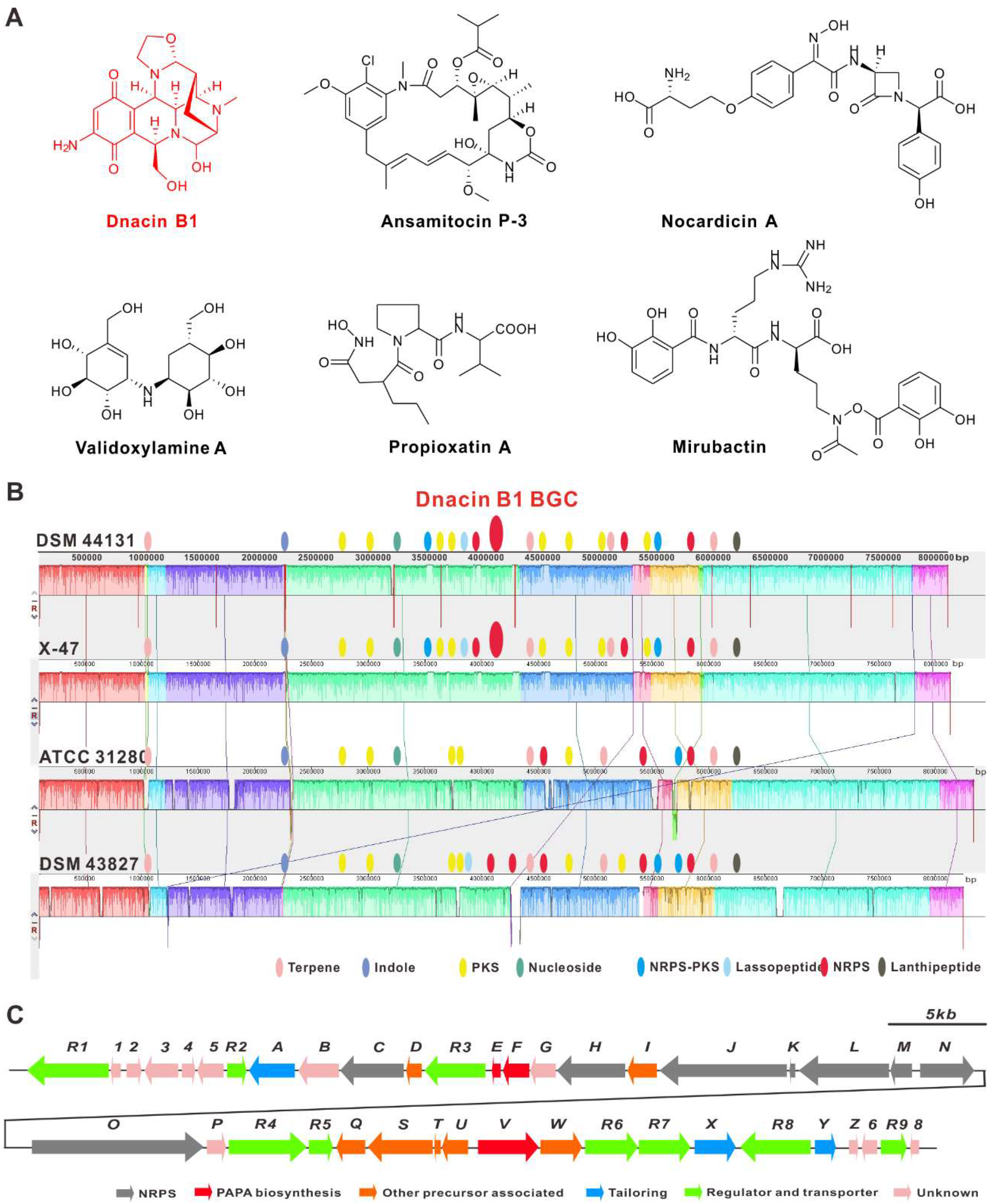
2.2. Comparative Genomic Analysis within Actinosynnema Resulted in the Discovery of Dnacin B1 BGC

Comparative genomics has not only been used in the evaluation of the evolutionary relationship between different strains, but also in the prediction of the putative functional genes in many strains [29,30]. The online antiSMASH analysis of the harbored BGCs within chromosomes of these four strains indicated that strain DSM 44131T was nearly identical to strain X47 (CP023445.1), and different from strain DSM 43827T (CP001630.1) and strain ATCC 31280 (CP029607.1) (Tables S4 and S5), in which strain DSM 44131T contained 28 potential BGCs, which was the average number of typical streptomycete strains (Table S4). In addition, the 66.9 kb DNA fragment garnered our significant interest, given that it was located in both strain DSM 44131T and strain X47, close to a NRPS BGC, and exhibiting 64% similarity with the biosynthetic gene cluster of the THIQ alkaloid, naphthyridinomycin (NDM) (Figure 1B). This discovery showed us that this region should be related to the biosynthesis of two THIQ alkaloids, dnacin A1 and dnacin B1, which were isolated from strain DSM 44131T (identified as Nocardia sp. No. C-14482 before) [8].
Subsequently, the crude extract of strain DSM 44131T was prepared, as well as the initial HPLC-MS analysis, however, it failed to detect the production of dnacin. To facilitate the following study, we tried to improve dnacin yield through media optimization and supplement with resin HP-20, and the titer was evaluated by the transcription levels of relevant genes with qPCR analysis and the inhibition activity against S. aureus ATCC 25923. A set of optimizations of the fermentation conditions in four different media (including H, K, 17 and J medium) were carried out. Fortunately, J medium showed a preferred expression level of the targeted functional genes, which might be due to a better growth level or some other unknown reasons caused by resin HP-20 absorption (Figure S2). The HPLC-MS analysis displayed that dnacin B1 was clearly produced in strain DSM 44131T, fermented in J medium with resin HP-20 for three days (Figure 2D,E).

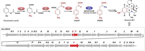
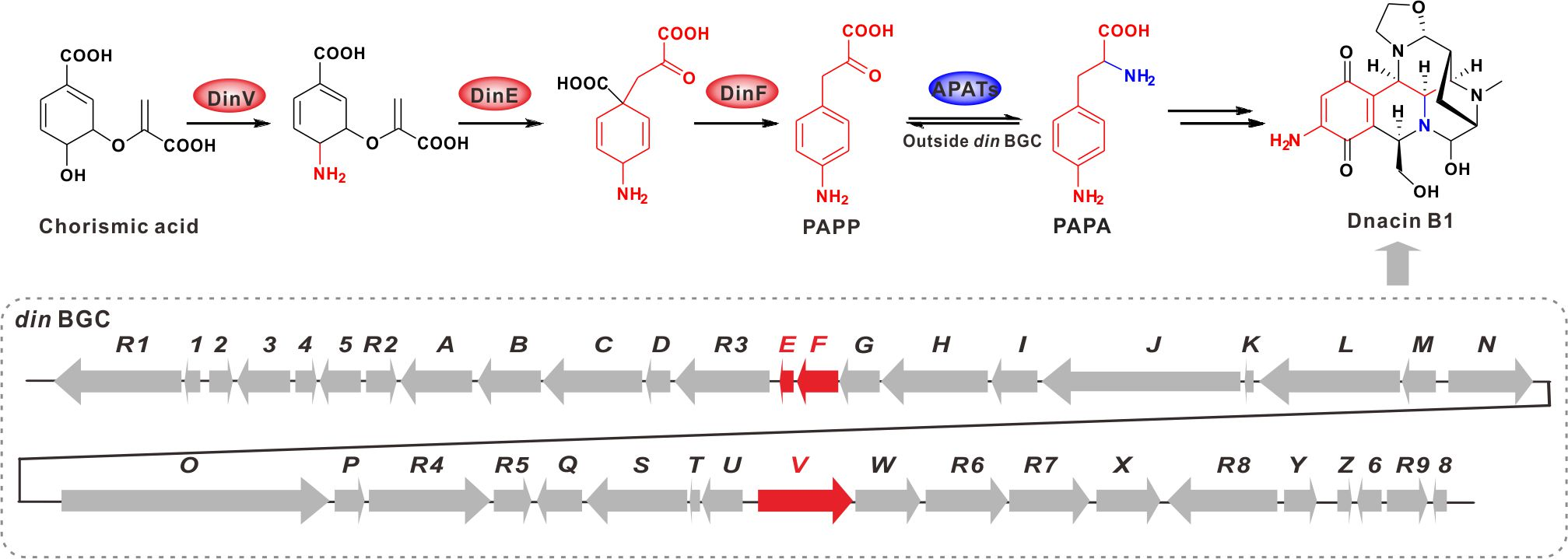
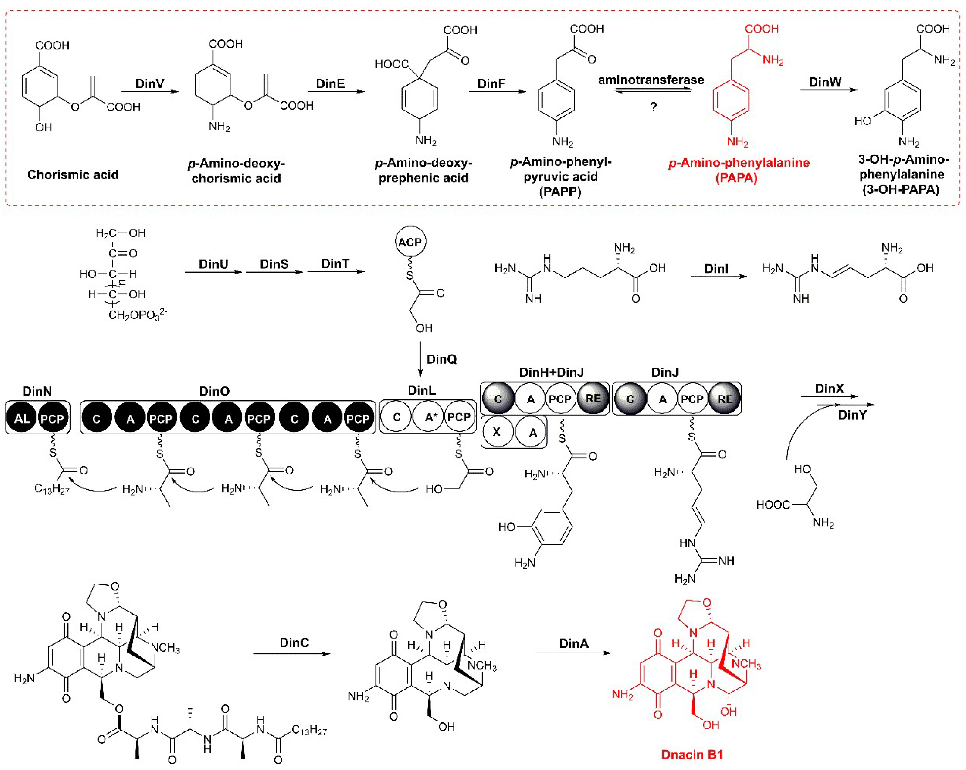
2.3. Functional Genes Involved in the Assembly of Dnacin B1 Skeleton

The detailed bioinformatics analysis of the dnacin B1 BGC from strain DSM 44131T revealed that it contained 41 open reading frames encoding for NRPS, PAPA NRPS extender unit, Pictet–Spengler (PS) reaction, tailoring modification, resistance and regulations (Figure 1C) [21,31,32]. The putative function of the related genes was listed in Table 1. To verify the function of four modular NRPS genes (dinL, dinM, dinN and dinO) in dnacin B1 biosynthesis, a dnacin B1 BGC inactivation (Δdin) mutant HXJA01 was constructed by deleting these four genes and replaced with an apramycin resistance gene. HPLC-MS analysis displayed that no dnacin B1 was produced in mutant HXJA01 compared with the wild type strain, indicating the BGC was responsible for the biosynthesis of dnacin B1 in strain DSM 44131T (Figure 2).
In dnacin B1 BGC, the modular NRPS genes dinL, dinJ, dinN and dinO displayed about 52% sequence similarity with those located in NDM BGC. The enzymes encoded by these four genes were responsible for a typical tetrahydroisoquinoline NRPS biosynthesis system (Figure 3) [21]. DinN was a didomain enzyme with acyl-CoA ligase (AL) and peptidyl carrier protein (PCP) activities, responsible for the cryptic fatty acyl chain loading and initiation of the dnacin B1 skeleton assembly. DinO was similar to the typical NRPS proteins composed of three C-A-PCP modules, and all of the A domains recognized alanine as the extension substrate.
The two-component transketolases encoded by dinS and dinU were homologous to napB and napD in the NDM BGC, supporting their functions related to the generation of hydroxyethyl unit from d-xylutose-5-phosphate and transferred to DinT [33]. DinQ resembled a family of KASIII proteins that served as acyl unit-loading activity, which was probably involved in tethering glyceryl group to the 4′-phosphopantetheinyl arm of the ACP of DinT. The A domain of DinL (46% identity with A domain in NapL) was inactive, whereas the C domain was in charge of the condensation of the hydroxyethyl group and the nascent N-fatty acid substituted peptidyl chain, providing the tethered PCP with a linear thioester intermediate.
DinJ played an important role as NRPS in THIQ biosynthesis. It comprised a tetradomain including a reductase (RE) domain embedded at the terminal of the C-A-PCP, which catalyzed PS- and Mannich-type reactions, conferring the core scaffolds in dnacin B1 biosynthesis [26]. The 3-OH-PAPA precursor was transferred to the PCP domain in DinJ after the adenylation by DinH. The A domain of DinJ (69% indentity with A domain of NapJ) recognized and activated L-(E)-4,5-dehydroarginine, which was modified by DinI (non-heme iron, α-ketoglutarate-dependent oxygenase). Subsequently, DinJ-RE domain reduced the assembling hybrid-peptidyl chain anchored at the PCP domain at DinL to release the aldehyde intermediate. Then, the DinJ-C domain catalyzed the aldehyde intermediate and the PCP linked aromatic acid through PS reaction. The DinJ-RE domain further mediated the reduction in the DinJ-PCP-linked product to deliver an aldehyde intermediate, preparing for the intramolecular Mannich reaction with another DinJ-PCP-tethered L-(E)-4,5-dehydroarginine substrate. The resultant DinJ-PCP attached intermediately, then underwent reduction and cyclization to form the final aldehyde product.
In addition to the NRPS skeleton assembly and tetrahydroisoquinoline scaffold formation, a set of post-tailoring enzymes encoded by dinX, dinY, dinC and dinA were present in dnacin B1 BGC. Two tailoring enzymes DinX and DinY were predicted to proceed with the hydroxylation and N-methylation of dnacin B1 skeleton. The gene product of dinX showed 67% sequence similarity with the FAD-dependent monooxygenase NapA, which might be responsible for the hydroxylation in naphthyridinomycin biosynthetic pathway. DinY showed 65% sequence similarity with the methyltransferase NapV, which might be in charge of the N-methylation in dnacin B1 biosynthetic pathway. The peptidase DinC showed 56% similarity with NapG, which might be the membrane-bound peptidase participating in the cleavage of leader peptide and then exporting the precursor from the cell. DinA, an FAD-linked oxidase with 75% similarity with NapU, might have the same function as NapU, which was confirmed to be in charge of oxidative activation and over the oxidative inactivation of a matured prodrug [31]. The dnacin B1 BGC contained three regulator genes, separately belonging to the TetR family (dinR2), SARP family (dinR3) and LysR family (dinR9). DinR8, annotated as a DNA excision repair enzyme UvrA, showed 84% similarity with NapR1, and was deduced as the resistance gene in dnacin B1 biosynthesis [34]. In addition, five ABC transporter genes, dinR1 and dinR4-7 were also found in dnacin B1 BGC (Table 1).

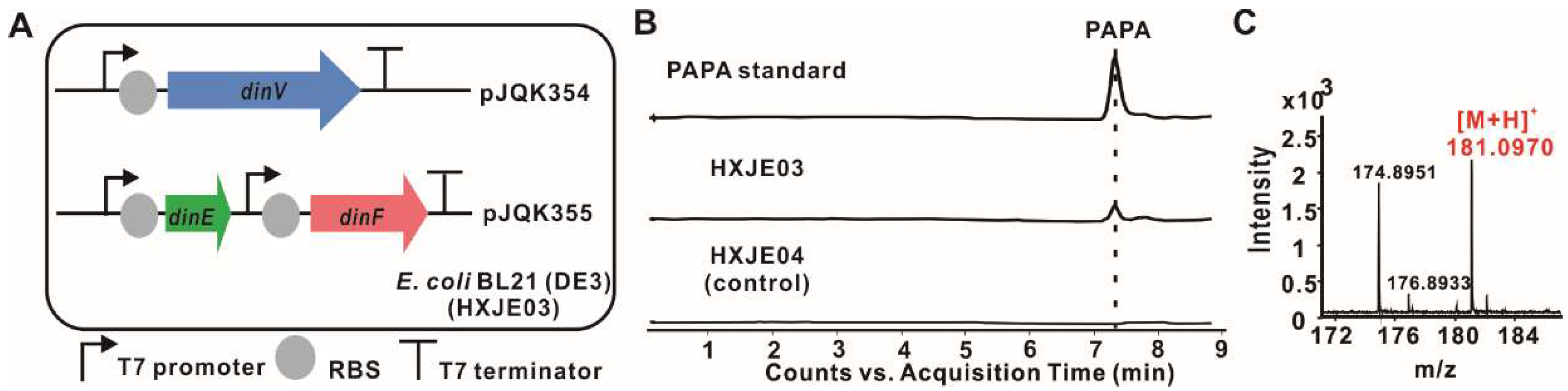
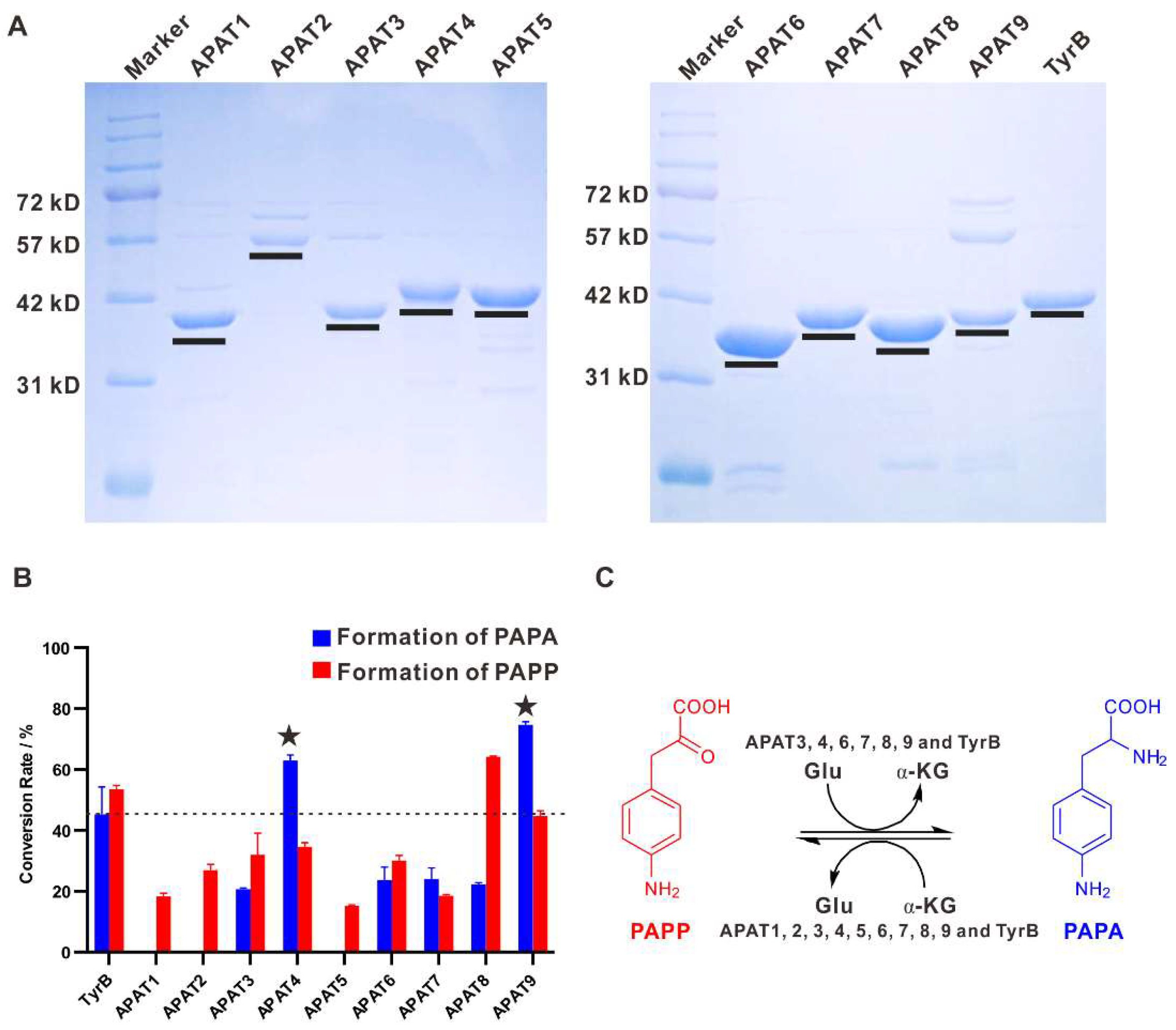
2.4. Reconstitution of PAPA Pathway Revealed TyrB Participating in the Mature of PAPA

In the dinacin B1 biosynthetic machinery, DinV resembled the proteins from aminodeoxychorismate synthase (ADCS) family that are widely found in Actinobacteria, and was involved in the dehydration of C-5 hydroxyl group and transferring an amino group into chorismic acid at C-3 to generate of p-amino-deoxychorismate [35]. This was further proved by a phylogeny comprising ADCS, anthranilate synthase (AS), isochorismate synthase (IS) and salicylate synthase (SS) (Figure S3). Genes dinE and dinF shared a homology with 4-amino-4-deoxychorismate mutase papB and dehydrogenase papC from Streptomyces venezuelae, respectively, and might be candidates for the transformation of enol pyruvic acid and decarboxylation to generate p-amino-phenylpyruvic acid [36]. Finally, an aminotransferase of the TyrB family, whose encoding gene was located apart from the BGC of dnacin B1, mediated an amino group transformation at the keto position to produce PAPA. The PAPA biosynthetic machinery in strain DSM 44131T was also considered to be a pathway parallel to that from chorismic acid to tyrosine (Figure S4).
In order to reconstitute the originated PAPA pathway in dnacin B1 BGC, dinV was cloned from gDNA of strain DSM 44131T and inserted into the vector pETDuet to obtain pJQK354, dinE and dinF were amplified and inserted into vector pCDFDuet to afford pJQK355. Each gene was under the control of its own T7 promoter and transcription ended by the terminator. These two recombinant plasmids were together transferred into E. coli BL21 (DE3), conferring HXJE03. Meanwhile, both pETDuet and pCDFDuet were transferred into E. coli BL21 (DE3) generating HXJE04 as the control strain. Then, the fermentation was carried out in modified M9 medium with corresponding antibiotics and induced by IPTG at 25 °C and 220 rpm. After the fermentation was completed, HPLC-MS analysis of both fermentation broth showed that PAPA was clearly produced in HXJE03, while there was no PAPA detected in strain HXJE04 (Figure 4). In the absence of aminotransferase that catalyzed the last mature step of PAPA, the achievement of the PAPA production in strain HXJE03 suggested that the endogenous aminotransferases might have participated in the last amino transformation, especially for the aminotransferase involved in the aromatic amino acid biosynthesis. This deduction was reasonable because PAPA has a similar chemical structure to tyrosine, and as we know, the aminotransferase TyrB catalyzed the last step of amino group transformation in the mature tyrosine. In order to evaluate the enzymatic property of aminotransferase in PAPA biosynthesis, gene tryB, responsible for the amino transformation in tyrosine biosynthesis, was cloned from the gDNA of E. coli MG1655 and heterologous expressed in E. coli BL21 (DE3). As was expected, incubation of the recombinant N-His6-TyrB with PAPP and glutamate (Glu) as the substrates generated PAPA, whereas PAPP also could be produced in the presence of PAPA and α-ketoglutarate (α-KG) as substrates (Figure 5). The chemical structures of PAPA from the TyrB-catalyzed reactions were determined by NMR and HR-ESI-MS analysis (Figures S5–S8). This result indicated that the TyrB involved in the tyrosine biosynthesis displayed a flexible substrate and transferred an amino group in a reversible reaction with PAPP or PAPA and related auxiliary substrates.

2.5. Aminotransferases Responsible for the PAPA Maturation Dispersed within the Genome

Aiming at exploring the possible aminotransferase participating in PAPA biosynthesis during dnacin B1 assembly, nine candidate aminotransferase (AT) genes apat1-9 were screened from the annotation results of the whole genome of strain DSM 44131T with the reference sequence of TyrB (Table S6). To date, the aminotransferases have been divided into five classes, among which tyrosine aminotransferase (TyrB) belongs to class I, together with aspartate aminotransferase, alanine aminotransferase and other aromatic aminotransferase [37]. Moreover, class I is further divided into Ia, Ib and Ic subgroups [38,39]. To better confirm the function of the nine aminotransferases APAT1-9, the phylogenetic relationship was analyzed among these candidates and the other related class I aminotransferases (Figure S10). Consequently, TyrB was located in the Ia subgroup, while all of the nine candidates, together with some extremophile-originated aminotransferases, gathered into the Ib subgroup. This result implied that the unusual function or property might be involved in the strain DSM 44131T-originated aminotransferases.
To further correlate the function of the nine aminotransferases APAT1-9 in vitro in the biosynthesis of PAPA from PAPP or the reversed reaction, the encoded genes were amplified from the gDNA of strain DSM 44131T and cloned into pET28a, respectively. The obtained plasmids were individually transferred into E. coli BL21 (DE3), and all of the N-terminally His6-tagged fusion proteins were obtained after purification by Ni2+ affinity chromatography. For systematic comparison of their catalytic abilities, equal amounts of APAT1-9 and TyrB were used to convert PAPP to PAPA using Glu as amino donors, or convert PAPA to PAPP with α-KG as an amino acceptor, individually at optimal reaction conditions. The reactions were subjected to HPLC for the yield analysis of the final products with authentic standards. Remarkably, APAT4 and APAT9 showed a closer relationship in the phylogenetic tree and displayed the preferable conversion rate of 63% and 74% for PAPA formation using PAPP as the amino group acceptor, which were superior to TyrB catalyzed ability. APAT3, APAT6, APAT7 and APAT8 showed the moderated conversion rates (less than 30%) during PAPA maturation with PAPP as the substrate. However, APAT1, APAT2 and APAT5 only exhibited catalytic capacity for the PAPP formation with PAPA as the substrate (Figure 5). Our systematic investigation of the aminotransferases involved in PAPA biosynthesis from the genome of strain DSM 44131T present the preferred aminotransferase candidates for further heterologous expression of PAPA in other hosts.

3. Discussion

Nowadays, great efforts have illuminated the mechanisms underpinning the biosynthesis of a diverse array of natural products from various bacteria. This research makes it possible to link the known natural products to their biosynthetic gene clusters. Moreover, the development of genome sequencing technology and many computational tools for natural product discovery have promised a remarkable increase in mining bacterial cryptic BGCs [40]. The available information on the genomic sequences of Actinosynnema revealed their circular chromosome structures with a relatively high GC content (>73%) among Actinobacteria. The initial comparative genome analysis of four sequenced Actinosynnema strains exhibited extremely high homology with the almost identical multilocus sequences by phylogenetic marker analysis. However, detailed analysis by Antismash revealed that chromosome harbored diversified secondary metabolite BGCs. Based on the comparative information of the BGCs of Actinosynnema species, we found the gene cluster responsible for dnacin B1 biosynthesis in strain DSM 44131T, which was not reported before.
The diversity of THIQ structures resulted mainly from different kinds of amino acid precursors for the recognition of NRPS proteins and diverse post-modifications. Previous incorporation experiments with labeled precursor and biosynthetic investigations demonstrated that the non-proteinogenic aromatic amino acid was responsible for the quinone moiety of THIQ and originated from tyrosine in ET-743, SFMs, SAC-B, NDM and phenylalanine in QNC. The bioinformatics analysis showed that DinV, DinE and DinF individually encoded 4-amino-4-deoxychorismate synthase, 4-amino-4-deoxychorismate mutase and 4-amino-4-deoxyprephenate dehydrogenase in dnacin B1 BGC, and displayed 48%, 34% and 40% similarities with PapA, PapB and PapC in S. venezuelae, which were responsible for the biosynthesis of PAPA in chloramphenicol. Thus, it was deduced that, similarly to the chloramphenicol biosynthesis, the quinone moiety of dnacin B1 arose from PAPA rather than tyrosine. In addition, the biosynthetic pathway reconstitution of PAPA in E. coli suggested that TyrB was responsible for the transformation of amino group into PAPP, although the detailed biochemical analysis of TyrB in PAPA assembly was unclear. Our systematic screening of the aminotransferases involved in PAPA biosynthesis from the genome of strain DSM 44131T resulted in the discovery of APAT4 and APAT9, displaying intriguing amino transformation ability, which might be a preferred aminotransferase candidate for further heterologous expression of PAPA in other hosts. All the screened APAT genes, including apat4 and apat9, were found in the other three Actinosynnema strains regardless of the production of dnacin B1 or PAPA (Table S6). This appearance revealed that the biosynthesis of PAPA does share the same aminotransferases as other aromatic amino acids in basic metabolism.
The biosynthetic pathway of PAPA can be verified by the co-occurrence of DinV, DinE and DinF in a heterologous host E. coli BL21 (DE3), which makes it possible to explore more PAPA pathways containing BGCs by MultiGeneBlast. Our genome mining results displayed that homologs of PAPA biosynthetic gene cassettes were discovered in several Streptomyces species (Figure S9), as well as in Saccharopolyspora species (Figure S9). Not all the aminotransferase genes involved in PAPA biosynthesis were anchored in these BGCs. Remarkably, detailed analysis of these explored BGCs revealed that the encoding metabolites were distributed into non-ribosomal peptides and other hybrid types. This finding reveals that PAPA biosynthesis genes would be the useful baits for discovering novel antibiotics using bioinformatics analysis.

4. Materials and Methods

4.1. Bacterial Strains, Plasmids and Reagents

All the bacterial strains and plasmids used in this research were summarized in Table S1. All the chemical, biochemical, enzymes and other molecular biological reagents were purchased from standard commercial sources. All the primers used in this research are listed in Table S2.

4.2. General Experimental Procedures

NMR spectra of PAPA were acquired in D2O using a Bruker AVANCE III-600 spectrometer (Bruker Corp., Germany) with tetramethylsilane (TMS) as an internal standard. HR-ESI-MS data were recorded using an Agilent 1290 Infinity II/6545 QTOF LC/MS instrument (Agilent Technologies, Santa Clara, CA, USA), with an Agilent TC C18 column (4.6 × 150 mm, 5 μm). The purification of PAPA was carried on Agilent 1290 Infinity II/1292 Prep FC instrument with an Agilent Eclipse XDB-C18 column (9.4 × 250 mm, 5 μm). Column chromatography separation for dnacin B1 detection was carried on MCI gel CHP20/P120 (Mitsubishi, Tokyo, Japan).

4.3. DNA Sequencing for Strain DSM 44131T

The DNA sequencing of strain DSM 44131T was performed at Shanghai Personal Biotechnology Co., Ltd. The gene sequence assembly was using A5-miseq v20150522. Genes in the assembled sequence were predicted by GeneMarks (http://exon.gatech.edu/GeneMark/, version 4.32 April 2015) [41]. tRNA, rRNA and ncRNA were identified using tRNAscan-SE (version 1.3.1), Barrnap (0.9-dev) (http://github.com/tseemann/barrnap) and Rfam database, respectively. Protein-coding sequences were annotated based on BLASTP searches against NCBI NR, COG and KEGG databases with an E-value cut-off of 1e-6.

4.4. Bioinformatics Analysis

Three Actinosynnema genome sequences (A. pretiosum subsp. pretiosum ATCC 31280, A. mirum DSM 43827T, A. pretiosum subsp. aurantium X47) were downloaded from the NCBI GenBank database for the genomic comparative analysis along with strain DSM 44131T. The pair-wise alignment was done by MAUVE [42]. The secondary metabolites’ gene clusters contained in all the genomes were predicted using antiSMASH (https://antismash.secondarymetabolites.org/#!/start) [43]. The orfs in dnacin B1 biosynthetic gene cluster (dnacin B1 BGC) were analyzed using FramePlot 4.0 beta program (http://biosyn.nih.go.jp/2ndfind/) [44]. The corresponding proteins were compared with other known proteins using BLAST (https://blast.ncbi.nlm.nih.gov/Blast.cgi). The prediction of amino acid specificities of A domains was performed on the PKS/NRPS analysis website (http://nrps.igs.umaryland.edu/) [45].
To mine the potential PAPA specific aminotransferase, the whole genome sequence of strain DSM 44131T was analyzed by KEGG (https://www.genome.jp/kegg/). Seven tyrosine aminotransferase homologies were found in the KEGG annotation. Another two genes were found through local blast analysis of strain DSM 44131T genome with the sequence of an aminotransferase gene tyrB originated from E. coli as a reference.

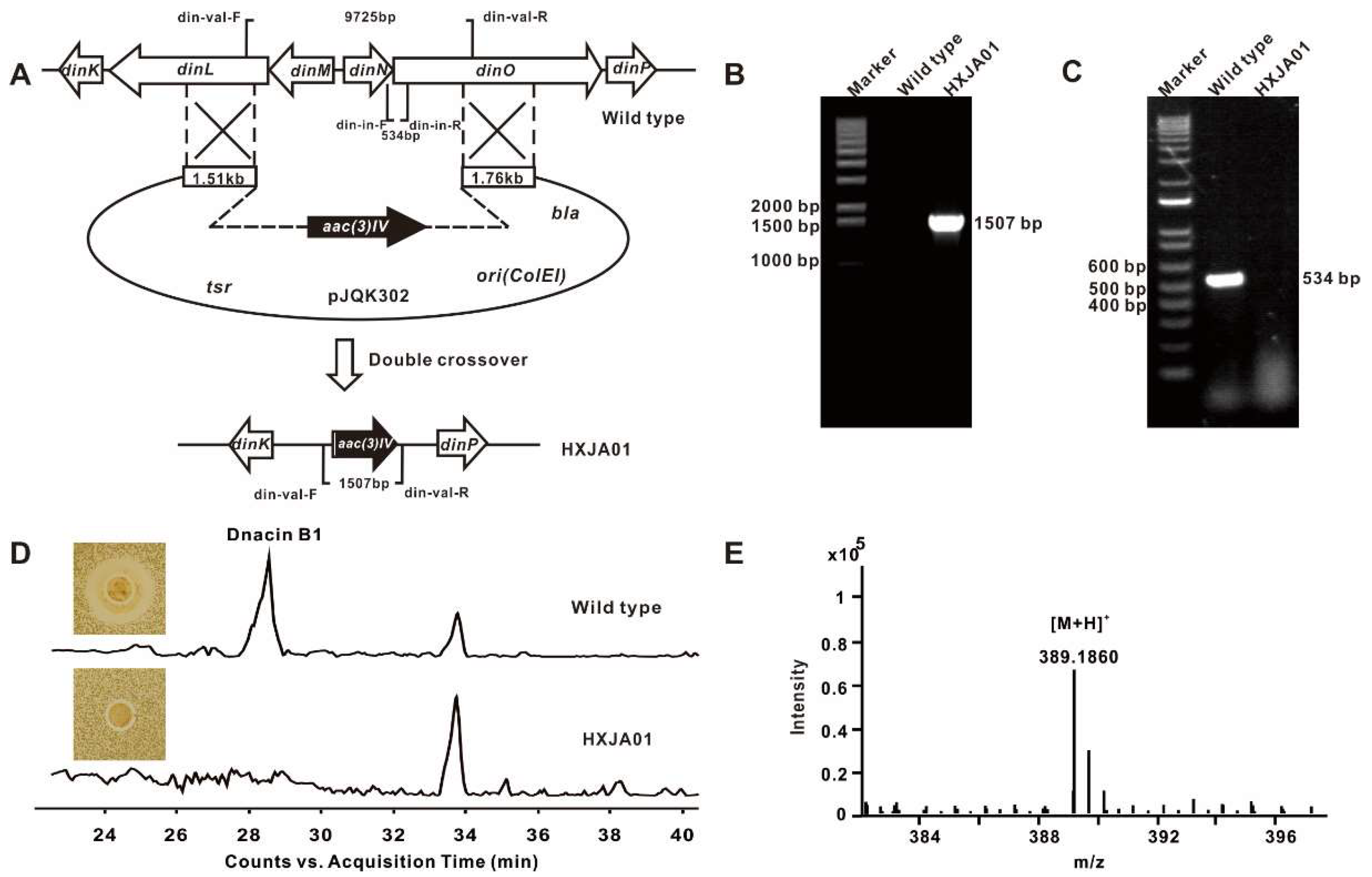
4.5. Construction of the Dnacin B1 BGC Inactivation Mutant

Homologous recombination was used to construct the Δdin mutant strain in this research. As for the Δdin mutant strain, two pairs of primers din-del-L-F/R and din-del-R-F/R were used to amplify the left and right 1.7 kb/1.5 kb homologous arms with the gDNA of strain DSM 44131T as template. The sequenced homologous arms were digested by EcoRI/HindIII and HindIII/KpnI, following purified by gel purification kit, respectively. The two arms were inserted into EcoRI/KpnI double-digested pJTU1278 to produce pJQK301. Then, an apramycin resistance gene cassette was inserted into the HindIII digested pJQK301 at the middle of the two arms to yield the pJQK302. The plasmid pJQK302 was transferred into E. coli ET12567/pUZ8002, and cultured in LB broth containing 50 μg/mL kanamycin, 25 μg/mL chloramphenicol and 50 μg/mL apramycin for conjugation. The E. coli-Actinosynnema bi-parental conjugation was performed for transformation of the pJQK302 into strain DSM 44131T [46]. The double-crossover exconjugants named HXJA01 were screened by resistance and confirmed by PCR with primers din-val-F/R and din-in-F/R.

4.6. Optimization of Fermentation Medium

Four different fermentation mediums (H, K, 17 and J) were chosen to compare the production of dnacin B1. All ingredients were shown as the following. H medium: yeast extract 10 g/L, corn starch 20 g/L, glucose 5 g/L, glycerol 40 g/L, K2HPO3 0.5 g/L, FeSO4·7H2O 0.2 g/L, CaCO3 5 g/L, pH 7.4. K medium: yeast extract 8 g/L, malt extract 10 g/L, sucrose 15 g/L, soluble starch 25 g/L, MgCl2 0.2 g/L, pH 7.4. 17 medium: glucose 10 g/L, soybean flour 30 g/L, corn steep liquor 10 g/L, glycerol 5 g/L, yeast extract 5 g/L, sucrose 10 g/L, NaCl 5 g/L, CaCO3 2 g/L, MgCl2 0.2 g/L, pH 7.4. J medium: dextrin 50 g/L, corn steep liquor 30 g/L, (NH4)2SO4 1 g/L, CaCl2 10 g/L, CaCO3 5 g/L, pH 7.4.
The wild type strain DSM 44131T was grown on a YMG (yeast extract 4 g/L, malt extract 10 g/L, glucose 4 g/L, pH 7.2) agar plate at 28 °C for activation. The mycelium was inoculated into 50 mL TSBY (tryptone soybean broth 30 g/L, yeast extract 10 g/L, sucrose 103 g/L, pH 7.2) in 250 mL shake-flasks as seed cultures at 28 °C and 220 rpm for 24 h on a rotary shaker. For fermentation, 5 mL of the seed culture was inoculated into 100 mL fermentation broth in 500 mL shake-flasks at 28 °C and 220 rpm. After being cultured for 48 h, each medium was taken in half bottles to add HP20 resin of 6% (w/v) of fermentation volume.

4.7. Total RNA Extraction and q-PCR Analysis

Cells were harvested at 96 h of fermentation to extract total RNA according to the manufacturer’s protocol using Redzol reagent and SiMax membrane spincolumns (SBS Genetech; Shanghai, China). The gDNA was removed using DNaseI (Fermentas, Vilnius, Lithuania) for 4 h at 37 °C before reverse transcription. The first strand of cDNA was synthesized using the RevertAid H Minus First Strand cDNA Synthesis Kit (ThermoFisher, Waltham, MA, USA). All the experiments were performed in triplicate.
The expression levels of the dnacin B1 BGC in various fermentation broth were estimated by quantitative real-time PCR. Five genes (dinI, dinP, dinQ, dinX and dinY) located in different operons of the dnacin B1 BGC were chosen, along with the housekeeper gene hrdB as the control. The gene-specific primers were list in Table S2. The quantitative real-time PCR reaction was performed on Thermo QuantStudio 3 Real-Time PCR System with Maxima™ SYBR Green/ROX qPCR Master Mix (ThermoFisher, USA) as follows: initial denaturation at 94 °C for 5 min followed by 32 cycles of denaturation at 94 °C for 30 s, annealing at 58 °C for 30 s, and extension at 72 °C for 30 s, with a final extension at 72 °C for 10 min. Finally, 2 μL products were subjected to run electrophoresis on 1.5% (w/v) agarose gel.

4.8. Production and Detection of Dnacin B1 in Strain DSM 44131T

For dnacin B1 detection, 2 L fermentation broth was harvested and centrifuged at 5000 rpm for 15 min. After being filtered, the supernatant was combined and fractionated by MCI gel eluted with gradients of 10% to 100% methanol. Each fraction was evaporated under reduced pressure at 40 °C. The fraction was dissolved in 1 mL methanol, and tested by the bioassay experiments using Staphylococcus aureus ATCC 25923 as the indicator strain for screening of the dnacin B1-containing candidates. The samples with a larger inhibition zone against S. aureus ATCC 25923 were combined and filtered for HPLC-MS analysis with an Agilent TC C18 column. The column was equilibrated with 95% solvent A (H2O, 0.1% formic acid) and 5% solvent B (methanol), and an analytic method was developed with the following program at a flow rate of 0.4 mL/min and UV detection at 280 nm: 0–25 min, a linear gradient from 5% B to 60% B; 25–45 min, a linear gradient from 60% B to 100% B; 45–53 min, constant 100% B; 53–54 min, a linear gradient from 100% B to 5% B; 54–60 min, constant 5% B. The compound with a retention time at 28 min showed an [M + H]+ ion at m/z = 389.1860 on a mass spectrometer, which was consisted with the molecular weight of dnacin B1 (C19H24N4O5, Exact Mass: 388.1747).

4.9. Reconstruction of PAPA Biosynthesis Pathway in E. coli

Three genes dinV, dinE and dinF were amplified by PCR using gDNA of strain DSM 44131T as a template, with primers dinV-F/R, dinE-F/R and dinF-F/R, respectively. Gene dinV was inserted in BamHI and HindIII sites of the pETDuet vector to construct pJQK354, while dinE and dinF was inserted, respectively, in BamHI/HindIII and NdeI/XhoI sites of pCDFDuet vector successively to construct pJQK355. The two recombinant plasmids were co-introduced into E. coli BL21 (DE3) and screened by resistance and confirmed by PCR. Finally, the dinV, dinE and dinF heterologous expression strain HXJE03 was obtained.
Then, strain HXJE03 was cultured in LB broth containing 50 μg/mL streptomycin, 100 μg/mL ampicillin at 37 °C and 220 rpm overnight, and inoculated at a ratio of 2% (v/v) in 500 mL flasks containing 100 mL fresh modified M9 medium at 37 °C and 220 rpm [47]. When the optical density reached about 0.6 at 600 nm, 0.5 mM IPTG was supplemented into the broth and the cells were grown at 25 °C and 220 rpm for another 48 h. An equal volume of methanol was added into the fermentation broth to break the cells. After being centrifuged at 12,000 rpm at 4 °C, the supernatant was filtered and analyzed by HPLC-MS on an Agilent TC C18 column. The column was equilibrated with 98% solvent A (H2O, 0.1% formic acid) and 2% solvent B (methanol), and an analytic method was developed with the following program at a flow rate of 0.4 mL/min and UV detection at 245 nm: 0–10 min, constant 2% B; 10–12 min, a linear gradient from 2% B to 50% B; 12–17 min, constant 50% B. The compound with a retention time at 7.2 min showed a [M + H]+ ion at m/z = 181.0970 on a mass spectrometer, which consisted of the molecular weight of PAPA (C9H12N2O2, Exact Mass: 180.0899).

4.10. Protein Expression and Purification

All nine PAPA aminotransferase genes from strain DSM 44131T along with tyrB from E. coli were cloned with corresponding primers (Table S2), and inserted in the NdeI and EcoRI sites of the pET28a vector. The recombinant plasmids were then transferred into E. coli BL21 (DE3). The final strains were individually inoculated into 5 mL LB medium containing 50 μg/mL kanamycin and incubated at 220 rpm, at 37 °C overnight. Then, 1 mL seed cultures were transformed into 2 L flasks containing 500 mL fresh LB medium incubated at 37 °C, until OD600 was reached at about 0.6, then 0.5 mM IPTG was supplied, and incubated another 20 h at 16 °C.
To purify the recombinant proteins, cells were harvested by centrifugation at 5000 rpm, 4 °C for 25 min, then the cells were resuspended in a 50 mL binding buffer (500 mM NaCl, 20 mM Tris, 5 mM imidazole, pH 7.9). After being sonicated on ice, the supernatants were collected by centrifugation at 10,000 rpm and at 4 °C for 40 min. Before loading onto Ni-NTA column (GE Healthcare, Ni Sepharose 6 Fast Flow), the supernatant was filtered through 0.45 μm filters. After being equilibrated with 20 mL binding buffer, the samples were eluted with washing buffer (binding buffer containing 100 mM imidazole) to obtain relatively pure N-His6-tagged recombinant proteins. Then, the purified proteins were concentrated and desalted by centrifugation at 3000 rpm at 4 °C using Amicon® Ultra-15 centrifugal filter (Millipore) with TGE buffer (50 mM Tris, 0.5 mM EDTA, 50 mM NaCl, 5% glycerol, pH 7.9). Finally, all proteins were analyzed by SDS-PAGE and quantified by nano-drop (Figure 5A).

4.11. Enzyme Activity Analysis

The enzymatic properties of recombinant-purified TyrB and APATs were performed according to previous research [48]. In this study, both PAPA and PAPP were used as the substrates for the reactions and the reverse to test the reversible activity of corresponding enzymes.
For the overall comparison of the aminotransferase activities of nine APATs and TyrB in vitro, equal amounts of enzymes were used in the 50 μL reaction mixture, containing 50 mM Tris-HCl (pH 7.0), 100 μM PLP, 0.5 mM PAPP as an amino acceptor and 5 mM Glu as an amino donor. For the reverse reaction, 0.5 mM PAPA was used as an amino donor and 5 mM α-KG as an amino acceptor. The reaction was initiated by adding 70 μM enzyme, and incubated for 40 min at 37 °C. Then, an equal volume acetonitrile was added to abort the reaction. After being centrifuged at 12,000 rpm, 4 °C for 15 min, the supernatants were detected by HPLC. All reactions were performed in triplicate. For the purification of PAPA via enzyme reaction by TyrB, an amplified reaction system of 50 mL volume was processed. The final enzyme reaction mixture was separated by preparative HPLC with a 2:98 ration of methanol and 0.1% formic acid H2O at 1.8 mL/min to yield PAPA (tR = 8 min, 2.2 mg).

5. Conclusions

In summary, on the basis of genome sequence of strain DSM 44131T, comparative genomic analysis plus with online AntiSmash analysis was carried out among the species in Actinobacteria. Detailed analysis of the diversified BGCs located in the genome of Actinobacteria resulted in the discovery of dnacin B1 BGCs in the genomes of strain DSM 44131T and strain X47. Bioinformatics analysis indicated that forty-one genes were composed in the 66.9 kb cluster, including three genes for PAPA synthesis, four NRPS genes, and genes putatively involved in extension unit biosynthesis, modification, regulation, transport and resistance. The proposed biosynthetic pathway of dnacin B1 revealed PAPA as the aromatic precursor for quinone moiety formation. The biosynthetic cassette of PAPA containing of dinV, dinE and dinF was constructed, and there was successful heterologous expression in E. coli. In order to explore the PAPA aminotransferase involved in PAPA biosynthesis in the strain DSM 44131T, nine candidates of APAT1-9 were cloned, and soluble proteins were obtained. The biochemical identification revealed that APAT4 and APAT9 exhibited fascinating amino transformation ability compared to TyrB, which originated from E. coli. This research not only enriched the repertoire of THIQ biosynthetic investigations but also provides the superior gene elements for the construction of the PAPA biosynthetic pathway. More attractively, target-directed genome mining using a PAPA biosynthetic cassette as a probe promises a renaissance of the accelerated discovery of novel PAPA-derived natural antibiotics.

Supplementary Materials

The following are available online, Table S1: Strains and plasmids used in this study; Table S2: Primers used in this study; Table S3: General genomic features of Actinosynnema pretiosum subsp. auranticum DSM 44131T; Table S4: AntiSMASH analysis of secondary metabolite BGCs of strains DSM 44131T, X47, DSM 43827T and ATCC 31280 genome sequences; Table S5: Similarity analysis of the predictable BGCs among strains DSM 44131T, X47, DSM 43827T and ATCC 31280 genomes with known clusters; Table S6: The sequence analysis of nine APAT genes in strain DSM 44131T; Figure S1: COG function classification of the annotated genes in strain DSM 44131T genome; Figure S2: Transcriptional analysis of functional genes within dnacin B1 BGC in strain DSM 44131T with different fermentation media; Figure S3: Biochemical and phylogenetic analysis of DinV; Figure S4: p-Amino-phenylalanine (PAPA) biosynthetic pathway is parallel to that of tyrosine (TYR); Figure S5: HR-ESI-MS analysis of PAPA; Figure S6: 1H-NMR spectrum of PAPA in D2O (600 MHz); Figure S7: 13C-NMR spectrum of PAPA in D2O (150 MHz). Figure S8: HR-ESI-MS analysis of PAPP; Figure S9: Discovery of bioactive natural products by mining PAPA biosynthetic gene cassette; Figure S10: Phylogenetic analysis of APATs in strain DSM 44131T.

Author Contributions

Conceptualization, Q.K.; methodology, X.H., X.L. (Xiaobin Li); validation, X.H.; formal analysis, Z.D. and L.B.; investigation, X.H.; data curation, X.L. (Xing Li), Y.S., H.W., Y.O. and Q.K.; writing—original draft preparation, X.H.; writing—review and editing, Q.K., Y.O.; supervision, Z.D., L.B. and Q.K.; project administration, Q.K.; funding acquisition, Q.K., Y.O. All authors have read and agreed to the final version of the manuscript.

Funding

This research was funded by the National Key Research and Development Program of China (No. 2019YFA0905400), the National Natural Science Foundation of China (No. 31770034, 21661140002 and 31700027), the Shanghai Municipal Council of Science and Technology (19ZR1475600) and the Startup Fund for Youngman Research at SJTU (17 × 100040064).

Conflicts of Interest

The authors declare no conflict of interest.

References

  1. Hasegawa, T.; Lechevalier, M.P.; Lechevalier, H.A. New genus of the Actinomycetales: Actinosynnema gen. nov. Int. J. Syst. Evol. Microbiol. 1978, 28, 304–310. [Google Scholar] [CrossRef]
  2. Stackebrandt, E.; Rainey, F.A.; Ward-Rainey, N.L. Proposal for a new hierarchic classification system, Actinobacteria classis nov. Int. J. Syst. Evol. Microbiol. 1997, 47, 479–491. [Google Scholar] [CrossRef] [Green Version]
  3. Hasegawa, T.; Tanida, S.; Hatano, K.; Higashide, E.; Yoneda, M. Motile Actinomycetes: Actinosynnema pretiosum subsp. pretiosum sp. nov., subsp. nov., and Actinosynnema pretiosum subsp. auranticum subsp. nov. Int. J. Syst. Evol. Microbiol. 1983, 33, 314–320. [Google Scholar] [CrossRef] [Green Version]
  4. Higashide, E.; Asai, M.; Ootsu, K.; Tanida, S.; Kozai, Y.; Hasegawa, T.; Kishi, T.; Sugino, Y.; Yoneda, M. Ansamitocin, a group of novel maytansinoid antibiotics with antitumour properties from Nocardia. Nature 1977, 270, 721–722. [Google Scholar] [CrossRef]
  5. Kang, Q.; Shen, Y.; Bai, L. Biosynthesis of 3,5-AHBA-derived natural products. Nat. Prod. Rep. 2012, 29, 243–263. [Google Scholar] [CrossRef] [PubMed]
  6. Siyu, M.; Hong, C.; Li, C.; Chuanxi, W.; Wei, J.; Xiaoming, C.; Huangjian, Y.; Wei, H.; Wei, Z. Two novel ansamitocin analogs from Actinosynnema pretiosum. Nat. Prod. Res. 2013, 27, 1532–1536. [Google Scholar] [CrossRef]
  7. Wei, G.Z.; Bai, L.Q.; Yang, T.; Ma, J.; Zeng, Y.; Shen, Y.M.; Zhao, P.J. A new antitumour ansamitocin from Actinosynnema pretiosum. Nat. Prod. Res. 2010, 24, 1146–1150. [Google Scholar] [CrossRef]
  8. Tanida, S.; Hasegawa, T.; Muroi, M.; Higashide, E. Dnacins, new antibiotics I. Producing organism, fermentation, and antimicrobial activities. J. Antibiot. 1980, 33, 1443–1448. [Google Scholar] [CrossRef]
  9. Lu, C.; Xie, F.; Shan, C.; Shen, Y. Two novel cyclic hexapeptides from the genetically engineered Actinosynnema pretiosum. Appl. Microbiol. Biotechnol. 2017, 101, 2273–2279. [Google Scholar] [CrossRef]
  10. Watanabe, K.; Okuda, T.; Yokose, K.; Furumai, T.; Maruyama, H.B. Actinosynnema mirum, a new producer of nocardicin antibiotics. J. Antibiot. 1983, 36, 321–324. [Google Scholar] [CrossRef]
  11. Asamizu, S.; Abugreen, M.; Mahmud, T. Comparative metabolomic analysis of an alternative biosynthetic pathway to Pseudosugars in Actinosynnema mirum DSM 43827. ChemBioChem 2013, 14, 1548–1551. [Google Scholar] [CrossRef] [PubMed] [Green Version]
  12. Murao, S.; Imafuku, S.; Oyama, H. Isolation of propioxatin A from Actinosynnema sp. SI-23 during a screening for serratia piscatorum metalloproteinase inhibitors. Biosci. Biotechnol. Biochem. 1997, 61, 561–562. [Google Scholar] [CrossRef] [PubMed] [Green Version]
  13. Giessen, T.W.; Franke, K.B.; Knappe, T.A.; Kraas, F.I.; Bosello, M.; Xie, X.; Linne, U.; Marahiel, M.A. Isolation, structure elucidation, and biosynthesis of an unusual hydroxamic acid ester-containing siderophore from Actinosynnema mirum. J. Nat. Prod. 2012, 75, 905–914. [Google Scholar] [CrossRef] [PubMed]
  14. Land, M.; Lapidus, A.; Mayilraj, S.; Chen, F.; Copeland, A.; Del Rio, T.G.; Nolan, M.; Lucas, S.; Tice, H.; Cheng, J.F.; et al. Complete genome sequence of Actinosynnema mirum type strain (101). Stand. Genomic. Sci. 2009, 1, 46–53. [Google Scholar] [CrossRef] [Green Version]
  15. Li, J.; Sun, R.; Ning, X.; Wang, X.; Wang, Z. Genome-scale metabolic model of Actinosynnema pretiosum ATCC 31280 and its application for ansamitocin P-3 production improvement. Genes 2018, 9, 364. [Google Scholar] [CrossRef] [Green Version]
  16. Wang, J.; Hu, X.; Sun, G.; Li, L.; Jiang, B.; Li, S.; Bai, L.; Liu, H.; Yu, L.; Wu, L. Genome-guided discovery of pretilactam from Actinosynnema pretiosum ATCC 31565. Molecules 2019, 24, 2281. [Google Scholar] [CrossRef] [Green Version]
  17. Zhong, C.; Zong, G.; Qian, S.; Liu, M.; Fu, J.; Zhang, P.; Li, J.; Cao, G. Complete genome sequence of Actinosynnema pretiosum X47, an industrial strain that produces the antibiotic ansamitocin AP-3. Curr. Microbiol. 2019, 76, 954–958. [Google Scholar] [CrossRef]
  18. Muroi, M.; Tanida, S.; Asai, M.; Kishi, T. Dnacins, new antibiotics II. Isolation and characterization. J. Antibiot. 1980, 33, 1449–1456. [Google Scholar] [CrossRef] [Green Version]
  19. Scott, J.D.; Williams, R.M. Chemistry and biology of the tetrahydroisoquinoline antitumor antibiotics. Chem. Rev. 2002, 102, 1669–1730. [Google Scholar] [CrossRef]
  20. Le, V.H.; Inai, M.; Williams, R.M.; Kan, T. Ecteinascidins. A review of the chemistry, biology and clinical utility of potent tetrahydroisoquinoline antitumor antibiotics. Nat. Prod. Rep. 2015, 32, 328–347. [Google Scholar] [CrossRef] [Green Version]
  21. Tang, G.; Tang, M.; Song, L.; Zhang, Y. Biosynthesis of tetrahydroisoquinoline antibiotics. Curr. Top. Med. Chem. 2016, 16, 1717–1726. [Google Scholar] [CrossRef] [PubMed]
  22. Jeedigunta, S.; Krenisky, J.; Kerr, R. Diketopiperazines as advanced intermediates in the biosynthesis of ecteinascidins. Tetrahedron 2000, 56, 3303–3307. [Google Scholar] [CrossRef]
  23. Mikami, Y.; Takahashi, K.; Yazawa, K.; Arai, T.; Namikoshi, M.; Iwasaki, S.; Okuda, S. Biosynthetic studies on saframycin A, a quinone antitumor antibiotic produced by Streptomyces lavendulae. J. Biol. Chem. 1985, 260, 344–348. [Google Scholar] [PubMed]
  24. Velasco, A.; Acebo, P.; Gomez, A.; Schleissner, C.; Rodriguez, P.; Aparicio, T.; Conde, S.; Munoz, R.; de la Calle, F.; Garcia, J.L.; et al. Molecular characterization of the safracin biosynthetic pathway from Pseudomonas fluorescens A2-2: Designing new cytotoxic compounds. Mol. Microbiol. 2005, 56, 144–154. [Google Scholar] [CrossRef] [PubMed]
  25. Zmijewski, M.J.; Mikolajczak, M.; Viswanatha, V.; Hruby, V.J. Biosynthesis of the antitumor antibiotic naphthyridinomycin. J. Am. Chem. Soc. 1982, 104, 4969–4971. [Google Scholar] [CrossRef]
  26. Hiratsuka, T.; Koketsu, K.; Minami, A.; Kaneko, S.; Yamazaki, C.; Watanabe, K.; Oguri, H.; Oikawa, H. Core assembly mechanism of quinocarcin/SF-1739: Bimodular complex nonribosomal peptide synthetases for sequential mannich-type reactions. Chem. Biol. 2013, 20, 1523–1535. [Google Scholar] [CrossRef] [PubMed] [Green Version]
  27. Ziemert, N.; Alanjary, M.; Weber, T. The evolution of genome mining in microbes—A review. Nat. Prod. Rep. 2016, 33, 988–1005. [Google Scholar] [CrossRef] [Green Version]
  28. Dittmann, E.; Gugger, M.; Sivonen, K.; Fewer, D.P. Natural product biosynthetic diversity and comparative genomics of the cyanobacteria. Trends. Microbiol. 2015, 23, 642–652. [Google Scholar] [CrossRef]
  29. Feyereisen, M.; Mahony, J.; Kelleher, P.; Roberts, R.J.; O’Sullivan, T.; Geertman, J.-M.A.; van Sinderen, D. Comparative genome analysis of the Lactobacillus brevis species. BMC Genom. 2019, 20, 416. [Google Scholar] [CrossRef] [Green Version]
  30. Yao, T.; Liu, J.; Liu, Z.; Li, T.; Li, H.; Che, Q.; Zhu, T.; Li, D.; Gu, Q.; Li, W. Genome mining of cyclodipeptide synthases unravels unusual tRNA-dependent diketopiperazine-terpene biosynthetic machinery. Nat. Commun. 2018, 9, 4091. [Google Scholar] [CrossRef]
  31. Zhang, Y.; Wen, W.; Pu, J.; Tang, M.; Zhang, L.; Peng, C.; Xu, Y.; Tang, G. Extracellularly oxidative activation and inactivation of matured prodrug for cryptic self-resistance in naphthyridinomycin biosynthesis. Proc. Natl. Acad. Sci. USA 2018, 115, 11232–11237. [Google Scholar] [CrossRef] [PubMed] [Green Version]
  32. Pu, J.; Peng, C.; Tang, M.; Zhang, Y.; Guo, J.; Song, L.; Hua, Q.; Tang, G. Naphthyridinomycin biosynthesis revealing the use of leader peptide to guide nonribosomal peptide assembly. Org. Lett. 2013, 15, 3674–3677. [Google Scholar] [CrossRef] [PubMed]
  33. Peng, C.; Pu, J.; Song, L.; Jian, X.; Tang, M.; Tang, G. Hijacking a hydroxyethyl unit from a central metabolic ketose into a nonribosomal peptide assembly line. Proc. Natl. Acad. Sci. USA 2012, 109, 8540–8545. [Google Scholar] [CrossRef] [PubMed] [Green Version]
  34. Prija, F.; Prasad, R. DrrC protein of Streptomyces peucetius removes daunorubicin from intercalated dnrI promoter. Microbiol. Res. 2017, 202, 30–35. [Google Scholar] [CrossRef]
  35. Chang, Z.; Sun, Y.; He, J.; Vining, L.C. p-Aminobenzoic acid and chloramphenicol biosynthesis in Streptomyces venezuelae: Gene sets for a key enzyme, 4-amino-4-deoxychorismate synthase. Microbiology 2001, 147, 2113–2126. [Google Scholar] [CrossRef] [Green Version]
  36. Yanai, K.; Sumida, N.; Okakura, K.; Moriya, T.; Watanabe, M.; Murakami, T. Para-position derivatives of fungal anthelmintic cyclodepsipeptides engineered with Streptomyces venezuelae antibiotic biosynthetic genes. Nat. Biotechnol. 2004, 22, 848–855. [Google Scholar] [CrossRef]
  37. Grishin, N.V.; Phillips, M.A.; Goldsmith, E.J. Modeling of the spatial structure of eukaryotic ornithine decarboxylases. Protein Sci. Publ. Protein Soc. 1995, 4, 1291–1304. [Google Scholar] [CrossRef] [Green Version]
  38. Okamoto, A.; Kato, R.; Masui, R.; Yamagishi, A.; Oshima, T.; Kuramitsu, S. An aspartate aminotransferase from an extremely thermophilic bacterium, Thermus thermophilus HB8. J. Biochem. 1996, 119, 135–144. [Google Scholar] [CrossRef]
  39. Son, H.F.; Kim, K.J. Structural insights into a novel class of aspartate aminotransferase from Corynebacterium glutamicum. PLoS ONE 2016, 11, e0158402. [Google Scholar] [CrossRef] [Green Version]
  40. Palazzotto, E.; Tong, Y.; Lee, S.Y.; Weber, T. Synthetic biology and metabolic engineering of actinomycetes for natural product discovery. Biotechnol. Adv. 2019, 37, 107366. [Google Scholar] [CrossRef]
  41. Coil, D.; Jospin, G.; Darling, A.E. A5-miseq: An updated pipeline to assemble microbial genomes from Illumina MiSeq data. Bioinformatics 2014, 31, 587–589. [Google Scholar] [CrossRef] [PubMed]
  42. Darling, A.C.E.; Mau, B.; Blattner, F.R.; Perna, N.T. Mauve: Multiple alignment of conserved genomic sequence with rearrangements. Genome Res. 2004, 14, 1394–1403. [Google Scholar] [CrossRef] [PubMed] [Green Version]
  43. Blin, K.; Shaw, S.; Steinke, K.; Villebro, R.; Ziemert, N.; Lee, S.Y.; Medema, M.H.; Weber, T. AntiSMASH 5.0: Updates to the secondary metabolite genome mining pipeline. Nucleic Acids. Res. 2019, 47, 81–87. [Google Scholar] [CrossRef] [PubMed] [Green Version]
  44. Ishikawa, J.; Hotta, K. FramePlot: A new implementation of the frame analysis for predicting protein-coding regions in bacterial DNA with a high G+C content. FEMS Microbiol. Lett. 1999, 174, 251–253. [Google Scholar] [CrossRef] [PubMed]
  45. Bachmann, B.O.; Ravel, J. Methods for In Silico prediction of microbial polyketide and nonribosomal peptide biosynthetic pathways from DNA sequence data. In Methods in Enzymology; Academic Press: San Diego, CA, USA, 2009; Volume 458, pp. 181–217. [Google Scholar]
  46. Ning, X.; Wang, X.; Wu, Y.; Kang, Q.; Bai, L. Identification and engineering of post-PKS modification bottlenecks for Ansamitocin P-3 titer improvement in Actinosynnema pretiosum subsp. pretiosum ATCC 31280. Biotechnol. J. 2017, 12, 1700484. [Google Scholar] [CrossRef]
  47. Masuo, S.; Zhou, S.; Kaneko, T.; Takaya, N. Bacterial fermentation platform for producing artificial aromatic amines. Sci. Rep. 2016, 6, 25764. [Google Scholar] [CrossRef] [Green Version]
  48. Wang, M.; Toda, K.; Maeda, H.A. Biochemical properties and subcellular localization of tyrosine aminotransferases in Arabidopsis thaliana. Phytochemistry 2016, 132, 16–25. [Google Scholar] [CrossRef] [Green Version]
Sample Availability: Samples of the compounds are not available from the authors.
Figure 1. The diversified biosynthetic gene clusters located in the chromosome of Actinosynnema strains (DSM 44131T, X47, ATCC 31280 and DSM 43827T). (A) The biological natural products discovered from the genus Actinosynnema. (B) Comparative genome sequence alignment among the Actinosynnema strains performed by MAUVE, indicating the variations in secondary metabolite-encoded biosynthetic gene clusters (BGCs) and discovering the BGC of dnacin B1 in the genome of strain DSM 44131T and strain X47. (C) Genetic organization of dnacin B1 BGC.
Molecules 25 04186 g001
Figure 2. Inactivation of dnacin B1 BGC. (A) Construction of the dnacin B1 BGC inactivation mutant by homologous recombination. (B) PCR validation of wild type and mutant HXJA01. When using din-val-F/R as primers, the product of mutant HXJA01 was 1507 bp, whereas there was no product in wild type. (C) PCR analysis of wild type and mutant HXJA01. When using din-in-F/R as primers, the product of wild type was 534 bp, whereas there was no product in mutant HXJA01. (D) HPLC-MS and bioassay analysis of wild type and mutant HXJA01. (E) HR-ESI-MS analysis result of the dnacin B1.
Molecules 25 04186 g002
Figure 3. Proposed biosynthetic pathway of dnacin B1. The biosynthetic pathway of assumed precursor 3-OH-PAPA was presented in red dotted box.
Molecules 25 04186 g003
Figure 4. Heterologous expression of PAPA in E. coli BL21 (DE3). (A) Schematic representation of PAPA synthesis in engineered E. coli strain HXJE03. Genes dinV, dinE and dinF were all expressed under the control of T7 promoter. (B) HPLC-MS analysis of PAPA standard and the products of engineered E. coli strains (HXJE03 and HXJE04). (C) HR-ESI-MS analysis result of PAPA.
Molecules 25 04186 g004
Figure 5. Expression and characterization of APATs and TyrB. (A) SDS-PAGE of nine purified APATs and TyrB expressed in E. coli BL21 (DE3). (B) Conversion rate analysis of recombinant nine APATs and TyrB in vitro. (C) The proposed reversible reactions of APATs and TyrB.
Molecules 25 04186 g005
Table 1. Deduced functions of ORFs in the dnacin B1 biosynthetic gene cluster.
ORFNumber of
Amino Acids
Proposed FunctionSequence Similarity (Protein, Origin)% Identity/
Similarity
Accession No.
DinR1951ABC transporterIQ63_41860, Streptomyces acidiscabies64/75KND24720.1
ORF1117unknownSD37_10880, Amycolatopsis orientalis55/75ANN21703.1
ORF2175protease inhibitorAC230_04780, Streptomyces caatingaensis38/54KNB54254.1
ORF3395histidine kinaseA9W97_15640, Mycobacterium gordonae51/60OBJ88517.1
ORF4159ATP-binding proteinRKT69099.1, Saccharothrix variisporea45/51DFJ66_2293
ORF5306epimeraseDI639_00155, Leifsonia xyli74/83PZO61411.1
DinR2228TetR family transcriptional regulatorSTRAU_2618, Streptomyces aurantiacus58/71EPH44178.1
DinA534flavin adenine dinucleotide (FAD)-linked oxidaseNapU, Streptomyces lusitanus64/75AGD80628.1
DinB471gamma-aminobutyraldehyde dehydrogenaseMCBG_02966, Micromonospora sp. M4253/65EWM65833.1
DinC741peptidaseNapG, Streptomyces lusitanus48/56AGD80614.1
DinD183flavin mononucleotide (FMN) reductaseSsuE, Streptomyces noursei58/67WP_067345371.1
DinR3707regulatory proteinNapR3, Streptomyces lusitanus55/68AGD80615.1
DinE1034-amino-4-deoxychorismate mutasePapB, Streptomyces venezuelae34/53BAD21142.1
DinF3074-amino-4-deoxyprephenate dehydrogenasePapC, Streptomyces venezuelae40/51BAD21141.1
DinG300unknownCLV43_102760, Umezawaea tangerina47/57PRY45195.1
DinH796adenylation domainNapH, Streptomyces lusitanus51/60AGD80616.1
DinI342non-heme iron hydroxylaseNapI, Streptomyces lusitanus63/75AGD80617.1
DinJ1482NRPSNapJ, Streptomyces lusitanus68/79AGD80618.1
Module 5C-A-PCP-RE
DinK66MbtH-like proteinNapK, Streptomyces lusitanus73/84AGD80619.1
DinL1125NRPSNapL, Streptomyces lusitanus54/64AGD80620.1
Module 4C-A-PCP
DinM252thioesterase IINapM, Streptomyces lusitanus51/64AGD80621.1
DinN632AMP-dependent synthetase and ligaseNapN, Streptomyces lusitanus64/74AGD80622.1
LoadingAL-PCP
DinO3049NRPSNapO, Streptomyces lusitanus52/63AGD80623.1
Module 1C-A-PCP
Module 2C-A-PCP
Module 3C-A-PCP
DinP223hypothetical proteinNapP, Streptomyces lusitanus45/59AGD80633.1
DinR4909ABC transporterB0I31_10111, Saccharothrix carnea56/67PSL57800.1
DinR5280ABC transporterCLV69_102810, Yuhushiella deserti53/69TDX97704.1
DinQ3363-oxoacyl-synthase III (KS)NapE, Streptomyces lusitanus72/80AGD80612.1
DinS754TransketolaseNapD, Streptomyces lusitanus59/68AGD80611.1
DinT76acyl carrier protein (ACP)NapC, Streptomyces lusitanus63/75AGD80633.1
DinU306transketolaseNapB, Streptomyces lusitanus68/77AGD80609.1
DinV7064-amino-4-deoxychorismate synthasePapA, Streptomyces venezuelae48/58BAD21140.1
DinW4864-hydroxyphenylacetate 3-monooxygenaseEWI31_24745, Streptomyces tsukubensis56/69TAI42134.1
DinR6613ABC transporterCLV43_102777, Umezawaea tangerina70/78PRY45212.1
DinR7601ABC transporterDIU55_13605, Firmicutes bacterium51/66PZN68908.1
DinX477monooxygenaseNapA, Streptomyces lusitanus69/76AGD80608.1
DinR8818UV-repair proteinNapR1, Streptomyces lusitanus75/84AGD80606.1
DinY244methyltransferaseQncJ, Streptomyces melanovinaceus47/65AGD95052.1
DinZ118long-chain-fatty-acid--CoA ligaseNCTC13184_06982, Nocardia africana60/81SUA48430.1
ORF6182sugar O-acetyltransferaseE1091_12580, Micromonospora fluostatini64/72TDB92781.1
DinR9304LysR family transcriptional regulatorAFR_16085, Actinoplanes friuliensis57/69AGZ41499.1
ORF7104transposaseCLV43_10550, Umezawaea tangerina56/61PRY41292.1

