Article

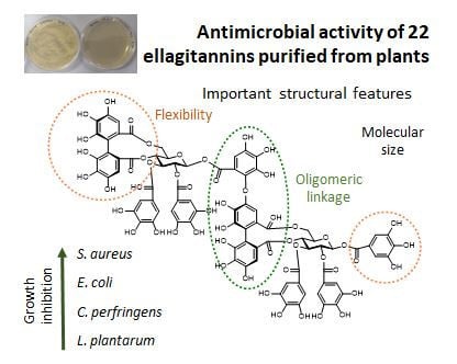
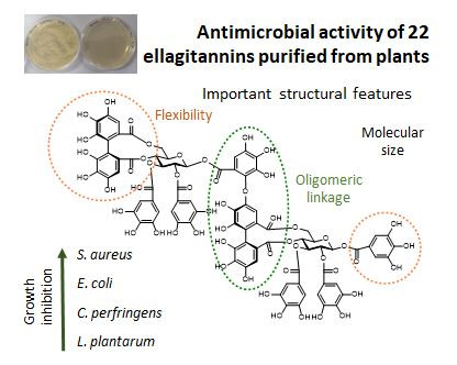
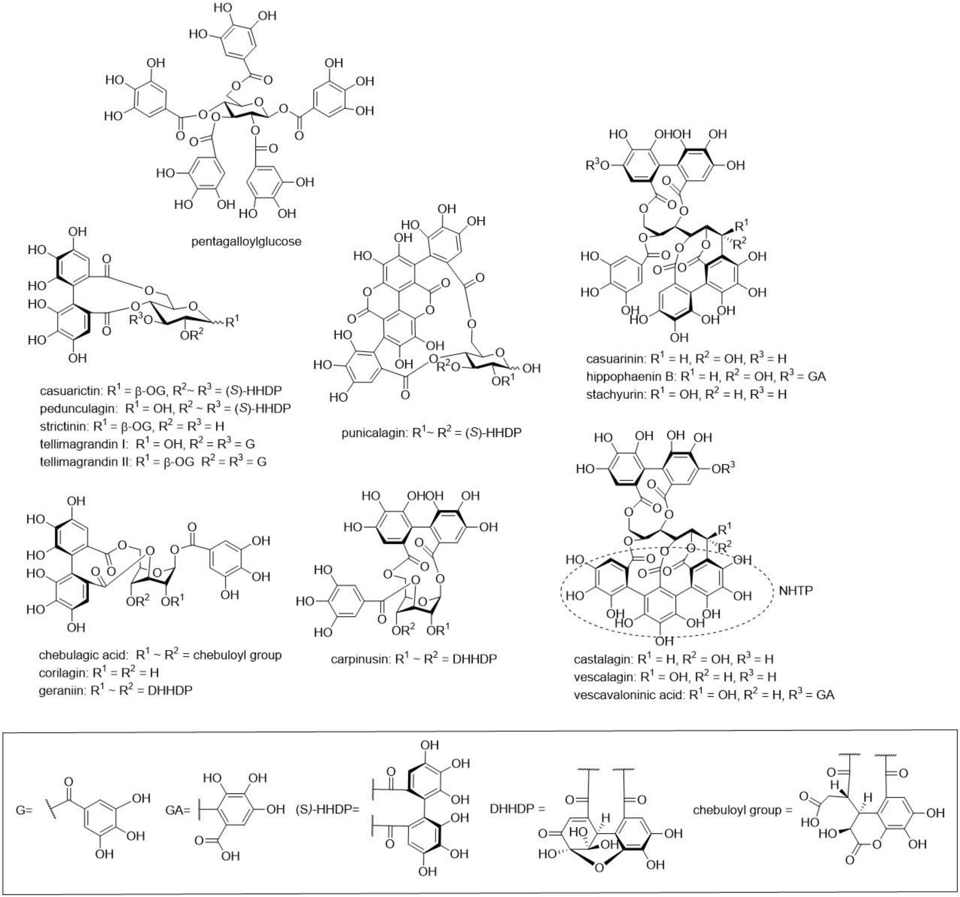
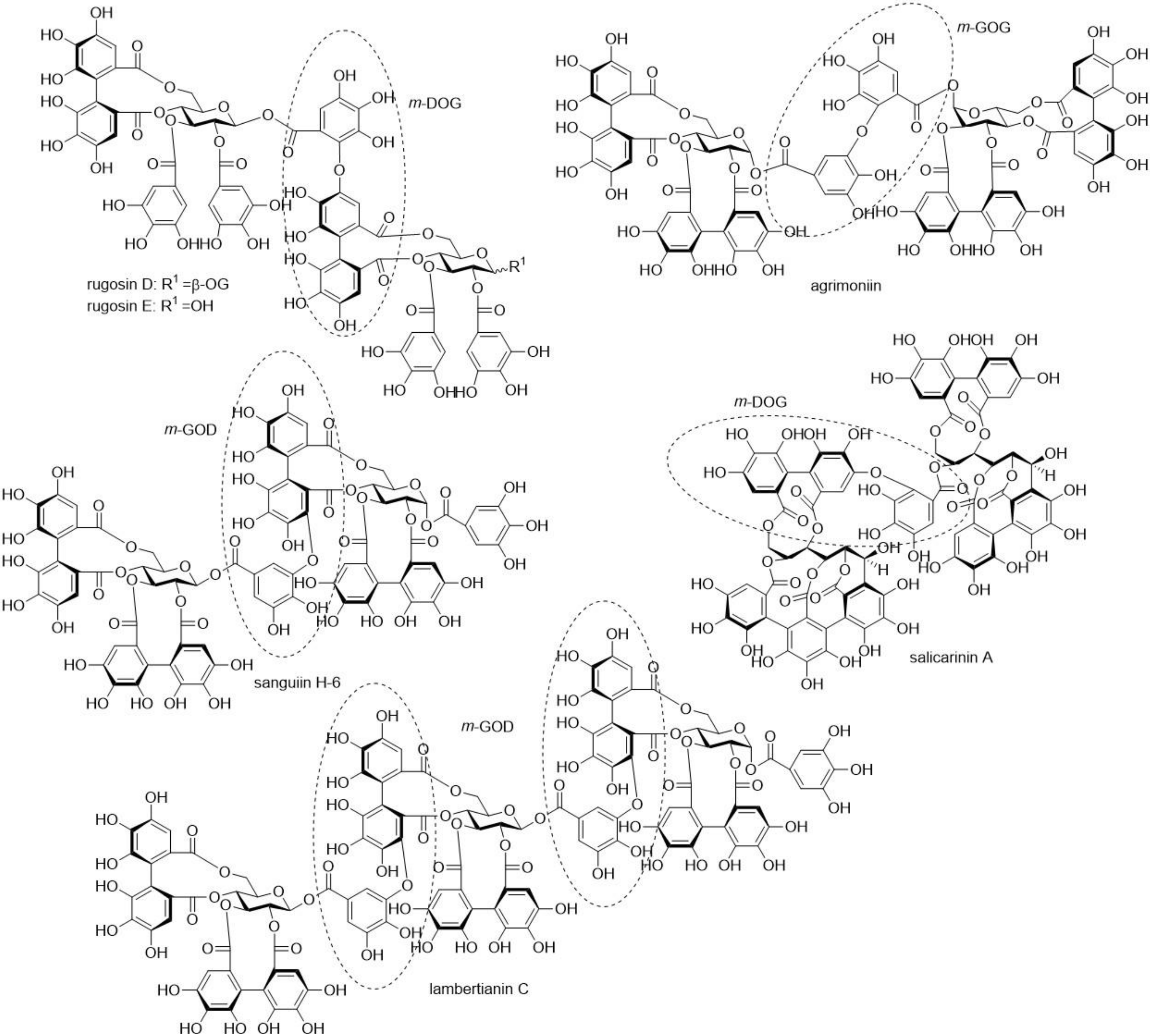
Antimicrobial Activities of Ellagitannins against Clostridiales perfringens, Escherichia coli, Lactobacillus plantarum and Staphylococcus aureus

1
Natural Chemistry Research Group, Department of Chemistry, University of Turku, FI-20014 Turku, Finland
2
Department of Food and Nutritional Studies, The University of Reading, Reading RG6 6AH, UK
3
Folium Science, Unit-DX, Bristol BS2 OXJ, UK
*
Author to whom correspondence should be addressed.
Molecules 2020, 25(16), 3714; https://doi.org/10.3390/molecules25163714
Submission received: 25 June 2020 / Revised: 6 August 2020 / Accepted: 13 August 2020 / Published: 14 August 2020

Abstract

:
In this study, we tested the growth inhibition effect of 22 individual ellagitannins and of pentagalloylglucose on four bacterial species, i.e., Clostridiales perfringens, Escherichia coli, Lactobacillus plantarum and Staphylococcus aureus. All tested compounds showed antimicrobial effects against S. aureus, and almost all against E. coli and C. perfringens. For L. plantarum, no or very weak growth inhibition was detected. The level of inhibition was the greatest for S. aureus and the weakest for C. perfringens. For S. aureus, the molecular size or flexibility of ellagitannins did not show a clear relationship with their antimicrobial activity, even though rugosins E and D and pentagalloylglucose with four or five free galloyl groups had a stronger growth inhibition effect than the other ellagitannins with glucopyranose cores but with less free galloyl groups. Additionally, our results with S. aureus showed that the oligomeric linkage of ellagitannin might have an effect on its antimicrobial activity. For E. coli, the molecular size, but not the molecular flexibility, of ellagitannins seemed to be an important factor. For C. perfringens, both the molecular size and the flexibility of ellagitannin were important factors. In previous studies, corilagin was used as a model for ellagitannins, but our results showed that other ellagitannins are much more efficacious; therefore, the antimicrobial effects of ellagitannins could be more significant than previously thought.

Graphical Abstract

1. Introduction

Dietary tannins can affect animal nutrition and health in several ways, for example, by improving the uptake of amino acids from feed proteins, by increasing anthelmintic effects against parasitic intestinal nematodes, and by lowering gaseous ammonia and methane emissions [1,2,3,4,5]. Tannins have also been shown to possess antimicrobial effects against several pathogens [6,7,8] and have been studied as an alternative for antibiotics or synthetic anthelmintics [9,10].
Tannins, i.e., condensed tannins (syn. proanthocyanidins), hydrolysable tannins including simple galloyl derivatives, gallotannins, ellagitannins (ETs), and phlorotannins are a unique group of plant specialized metabolites with diverse structures [11]. They have the well-known ability to form complexes with proteins and precipitate them [12]. In general, their molecular size, conformational mobility and flexibility in addition to the affinity of the polyphenol for water are important factors influencing the capacity of tannins to interact with and bind to proteins. Different tannins can have very different bioactivities depending on the exact tannin structure and, for example, ETs can establish simultaneously strong and weak interactions with proteins [13]. The anthelmintic activity of ETs against Haemonchus contortus and Trichostrongylus colubriformis third-stage infective larvae is increased as the molecular size of tannin increases [10], whereas the anthelmintic activity of hydrolysable tannins against egg hatching and motility of L1- and L2-stage H. contortus larvae indicate a clear relationship between the structure of hydrolysable tannins and the activity observed [14]. No clear relationship was observed between activity and molecular size, but the optimal structure was found for pentagalloylglucose. The anthelmintic properties of hydrolysable tannins vary depending, for example, on the structure of tannins, the parasite species and their growth stages [10,14,15,16]. Similarly, the antimicrobial effects of tannins seem to be species-specific and linked to tannin structure, not only to their ability to precipitate proteins [9].
Most of tannin-related antimicrobial studies have been conducted on plant extracts instead of pure compounds, making it difficult or impossible to compare the antimicrobial activity of individual compounds or to assess structure–activity relationships. Additionally, several mechanisms of action have been proposed through which tannins can induce their antimicrobial effects [9]. So far, only few studies have tested the antimicrobial activity of individual ETs [17,18,19,20]. Kolodziej et al. (1999) [18] tested the antimicrobial activities of tannin and related compounds and noticed that the ETs corilagin and phyllanthusiin C were weak inhibitors of Escherichia coli, Klebsiella pneumoniae, Bacillus subtilis, Pseudomonas aeruginosa, and Proteus mirabilis (with minimum inhibitory concentrations (MICs) of 1000 or 2000 µg/mL). However, corilagin showed moderate activity against Staphylococcus aureus (MIC 250 µg/mL). Corilagin inhibits the growth of E. coli by disrupting its membrane permeability and that of S. aureus by inhibiting protein expression [21]. In addition, Taguri et al. (2006) found that castalagin had a comparatively strong antibacterial activity against several bacteria species, and punicalagin had a moderate activity in comparison to other polyphenols [19]. Funatogawa et al. (2004) tested altogether 21 individual ETs together with pentagalloylglucose against Helicobacter pylori and E. coli (JC-2) and found that all tannins tested showed promising antibacterial activity against H. pylori and that monomeric ETs revealed stronger antibacterial activity than ET oligomers [20]. The tannins examined did not reveal any antibacterial activity against E. coli at the tested concentrations (maximum tannin concentration 100 µg/mL).
Our study analysed the antimicrobial activity of individual ETs against four different bacteria species: E. coli, S. aureus, C. perfringens and L. plantarum. E. coli is a Gram-negative bacterium, whereas S. aureus, C. perfringens and L. plantarum are Gram-positive. All four species were used previously in tannin-related antibacterial studies. Twenty-two ETs were selected, so that they comprised monomeric hexahydroxydiphenoyl (HHDP) esters, monomeric dehydroHHDP (DHHDP) esters and acyclic C-glucosidic ET monomers, including also nonahydroxytriphenoyl (NHTP) esters and ET dimers and trimers with varying types of bonds between the monomers and with acyclic or cyclic glucose cores (see structural examples in Figure 1 and Figure 2). The use of pure compounds was a necessity in order to reveal structure–activity relationships. We wanted to answer the following questions: (i) What are the antimicrobial effects of ETs? (ii) What are the structural features of ETs that affect their antimicrobial activity? (iii) Do ETs have unwanted antimicrobial effects against probiotics?

2. Results and Discussion

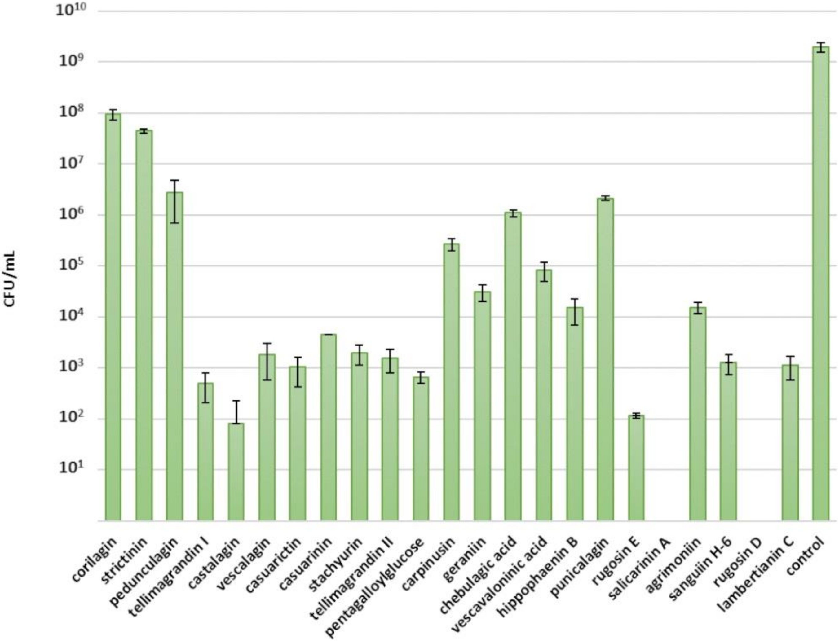
2.1. Inhibitory Effects of Ellagitannins against S. aureus

The tested compounds showed the greatest inhibition against S. aureus (Figure 3). Salicarinin A and rugosin D inhibited the growth of S. aureus completely at a 0.5 mM concentration. Casuarictin, tellimagrandins I and II, pentagalloylglucose, stachyurin, casuarinin, vescalagin, castalagin, rugosin E, sanguiin H-6 and lambertianin C showed efficient inhibition as well, as a tannin concentration of 0.5 mM slowed growth and total bacterial yield (Figure 3). Previously, Taguri et al. (2006) reported castalagin to be a stronger inhibitor (MIC 267 µg/mL) than other polyphenols against S. aureus, while punicalagin showed a moderate antibacterial activity (MIC 600 µg/mL) [19].
Rugosin D, which consists of two tellimagrandin II monomers connected by an m-DOG-linkage, was more efficient than the corresponding monomer and, similarly, rugosin E, which consists of tellimagrandin I and tellimagrandin II monomers, was more efficient than the corresponding monomers at restricting the growth of S. aureus. Correspondingly, salicarinin A, which is a dimer of vescalagin and stachyurin connected by an m-DOG-linkage, was more efficient than its monomers at restricting S. aureus growth. However, no trend was clearly discernible when comparing the antimicrobial activity with the molecular weight of the ETs.
We also looked at the effects of other structural features of ETs in detail. The monomeric HHDP esters tellimagrandin I and II and casuarictin had similar inhibitory effects against S. aureus as the C-glucosidic ET monomer vescalagin (Figure 3). Correspondingly, dimeric ETs with a glucopyranose cores, namely, rugosins E and D, were similar inhibitors as the C-glycosidic dimer salicarinin A. This suggests that the configuration of the central glucose core of ET does not affect its antimicrobial activity, whereas the type of the oligomeric linkage may play a role in the antimicrobial activity. The ET dimers with an m-DOG linkage, namely, salicarinin A and rugosins E and D, exhibited stronger inhibition than the ET oligomers with m-GOD linkages, namely, sanguiin H-6 and lambertianin C. The dimeric ET agrimoniin with an m-GOG-linkage was clearly a less effective inhibitor than the other dimeric ETs with m-DOG and m-GOD linkages.
The role of free galloyl groups in the ET structure was not so straightforward. The tannins with glucopyranose cores having four or five free galloyl groups, i.e., rugosins E and D and pentagalloylglucose, were more efficient inhibitors of S. aureus than the other cyclic ETs with less free galloyl groups. Tellimagrandin II, with an additional galloyl group, was a weaker inhibitor in comparison to tellimagrandin I, whereas rugosin D, with an additional galloyl group, was a stronger inhibitor in comparison to rugosin E. Interestingly, the acyclic ETs having no free galloyl groups, such as castalagin and salicarinin A, also showed efficient inhibition. Acyclic ETs have NHTP groups in their structures instead of free galloyl groups (Figure 1) and, therefore, are rather rigid. Previous studies have shown that molecular flexibility is an important factor for the bioactivity of ETs [10,13,14]. For example, our anthelmintic results clearly indicated that the intramolecular oxidative coupling that results in the formation of HHDP and NHTP groups decreased the inhibitory activity of hydrolysable tannins against egg hatching of H. contortus [14]. Based on our current observations, it seems that molecular flexibility does not have a significant role in the antimicrobial activity of ETs against S. aureus. Castalagin, with an NHTP group, was a stronger inhibitor than casuarinin, which has galloyl and HHDP groups, and, similarly, vescalagin, with an NHTP group, was a stronger inhibitor than stachyurin, with galloyl and HHDP groups.
The orientation of the hydroxyl group at the C-1 position in C-glycosidic ETs did not seem to have an effect on the antimicrobial activity against S. aureus. Castalagin, with an α-oriented hydroxyl group, was a stronger inhibitor than vescalagin, with a β-oriented hydroxyl group, whereas casuarinin, with an α-oriented hydroxyl group, was a weaker inhibitor than stachyurin, with a β-oriented hydroxyl group.
Monomeric DHHDP esters, i.e., carpinusin and geraniin, showed moderate inhibition of the growth of S. aureus: carpinusin was more efficient than geraniin. In general, monomeric ETs with the 4C1 conformation of central glucose core, namely, corilagin, geraniin, chebulagic acid and carpinusin, showed moderate inhibition. The weakest growth inhibitors of S. aureus were strictinin and corilagin. They were the only 2 of the tested 23 compounds that have two free OH-groups in their central glucose unit. Strictinin previously showed weaker anthelmintic activity in relation to other ellagitannins against T. colubriformis [10].

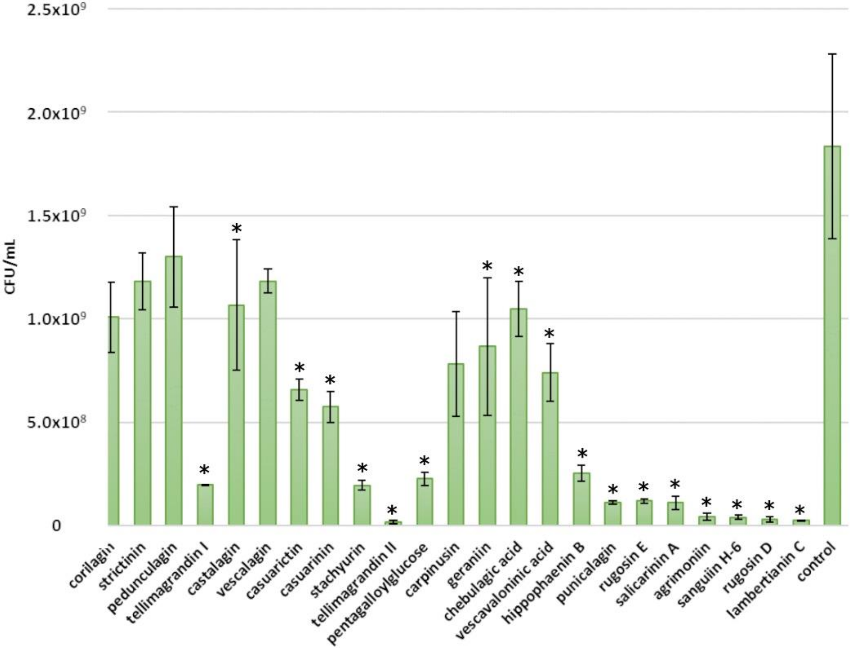
2.2. Inhibitory Effects of Ellagitannins against E. coli

Some growth inhibition was also observed in E. coli. Almost all tested ETs as well as pentagalloylglucose showed inhibition against E. coli (Figure 4). The most efficient compounds were tellimagrandin II, rugosin D, agrimoniin, sanguiin H-6 and lambertianin C. They were able to reduce the growth from 109 CFU/mL to 107 CFU/mL with a 0.5 mM tannin concentration. Previously, it was reported that punicalagin is a weaker inhibitor of E. coli (MIC 2133 µg/mL) than castalagin (MIC 533 µg/mL) [19]. Our results did not support this observation, as punicalagin appeared to be a greater inhibitor than castalagin. In the same study, the effects of punicalagin and castalagin were found to be stronger on S. aureus than on E. coli, similarly to our study. Another study tested pentagalloylglucose, strictinin, pedunculagin, tellimagrandin I, tellimagrandin II, casuarictin, corilagin, geraniin, casuriniin, agrimoniin and rugosin D against E. coli (JC-2) and found them not to be active [20].
There seems to be a medium correlation between the molecular size of ETs and the intensity of their antimicrobial activity against E. coli (Figure 5). Oligomeric ETs (molecular weight >1700 g/mol) inhibited over 90% of the E. coli growth, and their average growth inhibition was 97%. The average growth inhibition for ET monomers was 63%, and the sole monomers with over 90% growth inhibition were punicalagin and tellimagrandin II. Sanguiin H-6, which consists of two casuarictin monomers, and lambertianin C, which consists of two casuarictin and one potentillin monomers, were both more effective than casuarictin. Correspondingly, salicarinin A, which is a dimer of vescalagin and stachyurin, was more efficient than its monomers. However, tellimagrandin II seems to be an exception, as it was more efficient than rugosin D (which consists of two tellimagrandin II monomers) and rugosin E (which consists of tellimagrandin I and II monomers).
The number of free galloyl groups may be an important factor for the antimicrobial effects of ETs against E. coli. Additional free galloyl groups enhanced the inhibitory effects of ET: tellimagrandin II and rugosin D were more efficient than tellimagrandin I and rugosin E, respectively. However, the activity of pentagalloylglucose was lower than that of tellimagrandins I and II, indicating the importance of HHDP in the structure.
The effects of other structural ET features on the growth inhibition of E. coli were not so clear. The conformation of the central glucose core of ETs did not seem to affect the antimicrobial activity; for example, strictinin with a 4C1 glucopyranose core, chebulagic acid with a 1C4 glucopyranose core and castalagin with acyclic glucose had similar activities. The orientation of the hydroxyl group at the C-1 position of ETs did not seem to have an effect on the antimicrobial activity against E. coli. The α-anomer castalagin was more efficient than the corresponding β-anomer vescalagin, whereas the α-anomer casuarinin was less efficient than the corresponding β-anomer stachyurin. The formation of the HHDP group by oxidative coupling from two galloyl groups did not explain these differences. Casuarictin was more efficient than strictinin, whereas pedunculagin was less efficient than tellimagrandin I. Molecular flexibility did not seem to be important: pentagalloylglucose, with five free galloyl groups, was less efficient than tellimagrandin II, with one HHDP and three galloyl groups, and the acyclic ETs, with rigid NHTP groups, did not differ from cyclic ETs. The oligomeric linkages did not make a difference either, as all ET oligomers were effective against E. coli.

2.3. Inhibitory Effects of Ellagitannins against C. perfringens

Weak inhibition was observed for C. perfringens. However, almost all ETs tested showed at least some inhibitory effects. The best inhibitors, salicarinin A and agrimoniin, were able to lower C. perfringens growth only to 107 CFU/mL from 108 CFU/mL (Figure 6).
The molecular size of the ET seemed to be the main explanatory factor for the antimicrobial effects of ETs against C. perfringens (a small correlation between the molecular size of ETs and their antimicrobial strength is shown in Figure 7), similarly to E. coli. The average growth inhibition for oligomeric ETs was 77%, while for monomeric ETs it was 61%. As with E. coli, punicalagin and tellimagrandin II were the most effective monomers (growth inhibition 85%). Compared with other oligomeric ETs, rugosin E showed significantly lower growth inhibition, corresponding only to 43%. Again, it was noticed that salicarinin A, which is a dimer of vescalagin and stachyurin connected by an m-DOG-linkage, was more efficient than its monomers. Additionally, sanguiin H-6 (casuarictin dimer) and lambertianin C (casuarictin trimer) were both more effective than casuarictin. However, the same was not observed for dimeric rugosins E and D, as rugosin D was as effective as its monomer, tellimagrandin II, and rugosin E was a weaker inhibitor than its monomers.
The number of free galloyl groups may be an important factor for the antimicrobial effects of ETs against C. perfringens. The additional free galloyl group enhanced the inhibitory effects of the ET: tellimagrandin II and rugosin D were more efficient than tellimagrandin I and rugosin E, respectively. The other structural features of ETs did not show any overall clear effect. It was reported that, according to the MIC values, the antibacterial activities of punicalagin and castalagin are stronger against C. perfringens than against S. aureus [19]. Our results indicate the opposite; however, a comparison between our and this study is difficult, as different methods and strains were used.

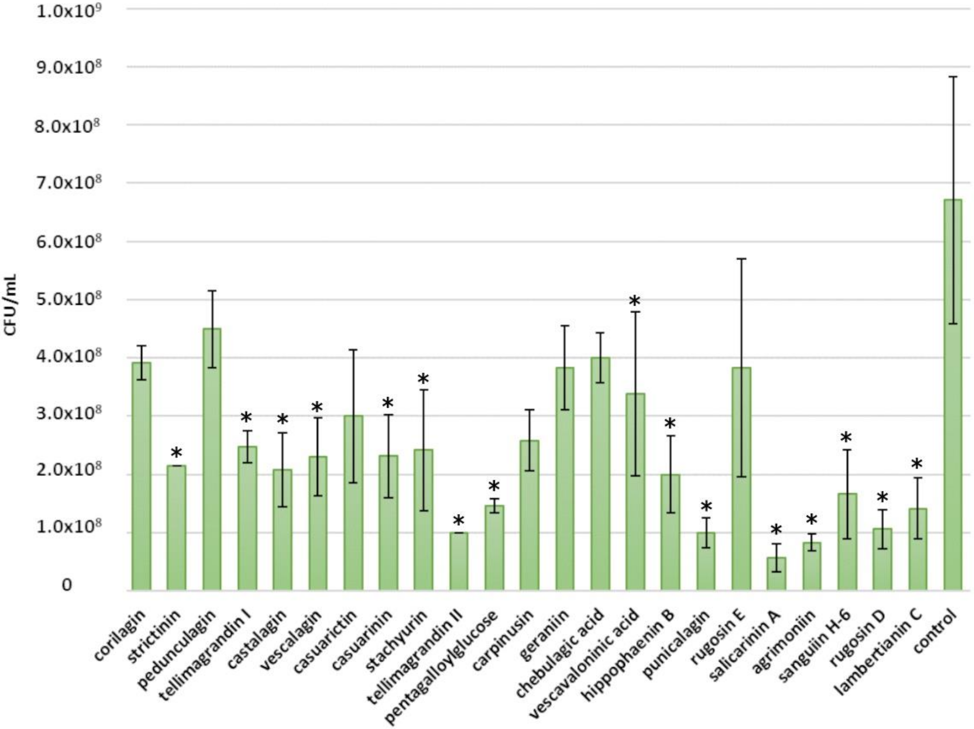
2.4. Inhibitory Effects of Ellagitannins against L. plantarum

No or very weak growth inhibition was detected for L. plantarum, as a growth corresponding to 108 CFU/mL was observed both in the positive control and after the addition of a 0.5 mM tannin solution (Figure 8). However, even though the growth in the control seemed always to be higher, based on a paired t-test, strictinin, castalagin, stachyurin, pentagalloylglucose, geraniin, punicalagin and lambertianin C showed a minor inhibition against L. plantarum. In this case, the lack of growth inhibition is considered an advantage, as Lactobacillus comprises many probiotic species. There are several supporting previous studies conducted with different plant, fruit and berry extracts that reported either no or low growth inhibition of Lactobacillus by these extracts [22,23] or that observed growth inhibition only with high extract concentrations [24]. Pomegranate extract and juice containing punicalagin have been shown to even promote the growth of Lactobacillus [25]. However, also contradictory results have been reported, as Choe et al. (2020) showed that a sanguiin H-6-rich blackberry seed flour extract inhibited the growth of Lactobacillus [26].

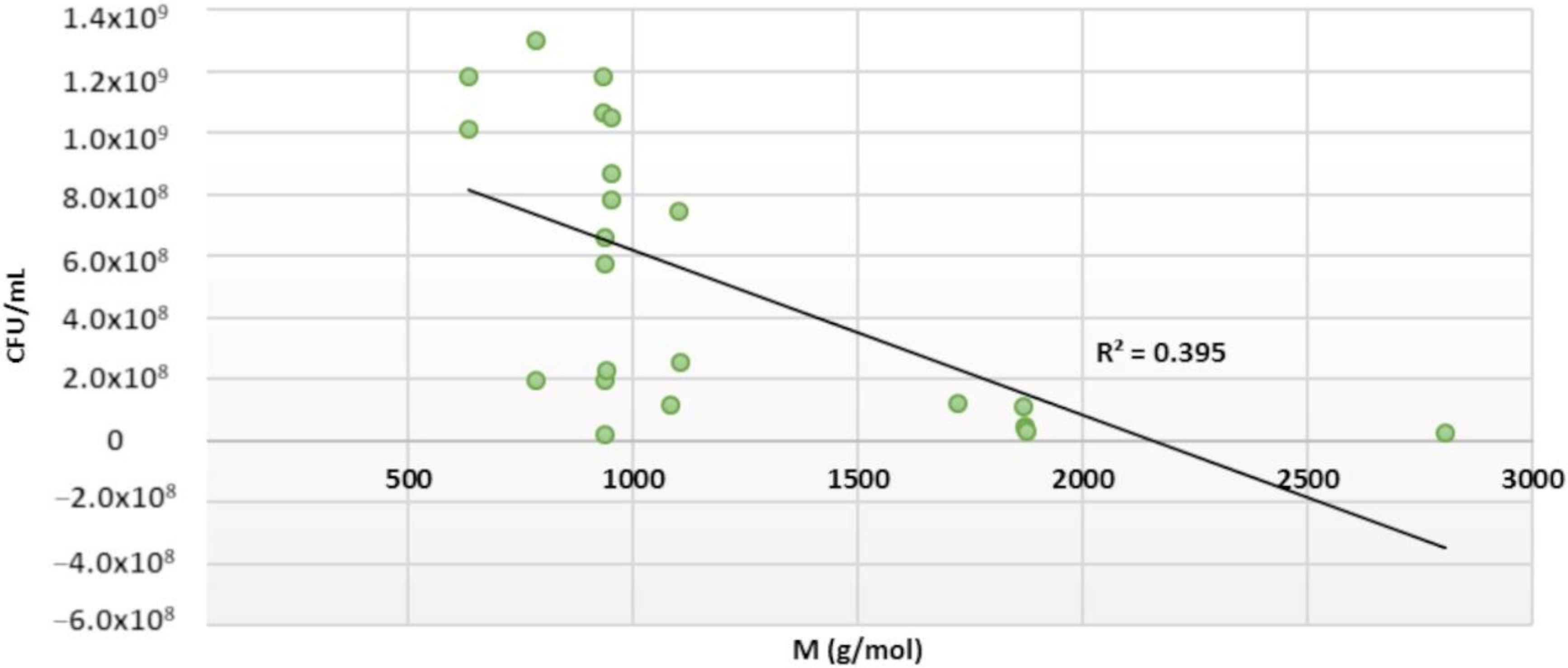
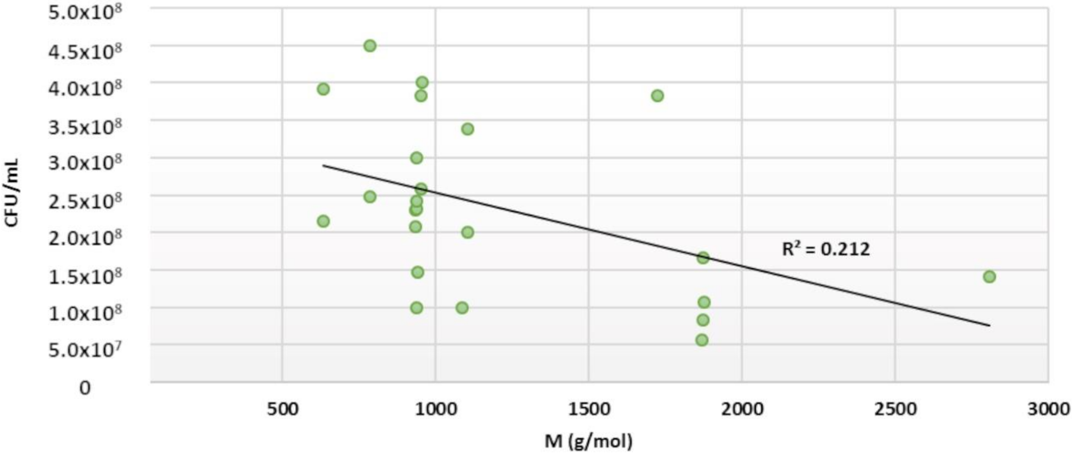
2.5. Structure–Activity Relationships

Out of the four tested bacteria, S. aureus was found to be the most sensitive species to tannins. This observation is supported by previous studies. For example, Puupponen-Pimiä et al. (2005) have shown that ellagitannin-rich berries and berry extracts were able to inhibit the growth of S. aureus [8]. They have also shown in an earlier study that phenolic berry extracts could inhibit the growth of E. coli but not that of Lactobacillus [24].
Taguri et al. (1999) reported that the pyrogallol group is an important structural feature for the antimicrobial activity of polyphenols but they could not see a clear relationship between the activity and the number of pyrogallol groups [19]. Instead of considering the pyrogallol group, we focused on the number of free galloyl groups. With S. aureus, we could not see a clear correlation between the number of free galloyl groups and antibacterial activity, although tannins with glucopyranose cores having four or five free galloyl groups (rugosins E and D and pentagalloylglucose) inhibited the growth of S. aureus more extensively than the other cyclic ETs with less free galloyl groups. However, the inhibitory effects of the ET against E. coli and C. perfringens seemed to be enhanced by the additional free galloyl group.
Between-study comparisons of tannin structure and antibacterial activity of tannins are difficult, as the methods and bacterial strains vary in different studies. Additionally, several different mechanisms of action have been proposed for the antibacterial activity of tannins, for example, inhibition of extracellular microbial enzymes and oxidative phosphorylation [27] and disruption of cellular membrane permeability [21,28]. The different methods of evaluation of antibacterial potency pose its own challenges. The potency is usually evaluated by MIC values that are generally reported as µg/mL. This, however, should be taken into consideration when monomeric and oligomeric ETs are compared, as their molecular masses are different. For example, Funatogava et al. (2004) reported that monomeric ETs showed stronger antimicrobial activity against H. pylori than ET oligomers, most of the ET monomers having a MIC value of 12.5 µg/mL and oligomers of 25 µg/mL [20]. However, depending on the exact molecular mass of ETs, their molar potency could actually be equal. According to our results, the molecular size of ET can enhance the antibacterial activity, as oligomeric ETs were more effective in inhibiting the growth of E. coli and C. perfringens than monomeric ETs. Additionally, our results with S. aureus showed that the oligomeric linkage might have an effect on the antimicrobial activity, as the ET dimers with an m-DOG linkage exhibited stronger inhibition than ET oligomers with m-GOD or m-GOG linkages. Furthermore, we found that the poor solubility of some tannins and their ability to bind proteins make it challenging or impossible to determine their MIC values in culture solution.
It has been stated that the antibacterial potencies of tannins are less pronounced than anticipated [17,18]. However, in previous studies, corilagin was used as a model for ETs [18]. Our results show that corilagin is one of the weakest inhibitors among ETs. Other ETs are much more efficient; therefore, we can conclude that the antimicrobial effects of ETs are more significant than thought. ETs appear to be potent antimicrobials.

3. Materials and Methods

3.1. Tannin Selection, Isolation and Characterization

The extraction and isolation of ETs and pentagalloylglucose (Figure 1 and Figure 2) followed mainly our previous methods consisting in extraction, column chromatography on Sephadex LH-20 and preparative and semipreparative HPLC [11,13,29,30,31,32,33]. Twenty-two structurally different ETs were purified from different plant species. ETs were selected so that they represented different branches of the ET biosynthetic pathway, and the hydrophobicities of ETs were also taken into account [34]. Acyclic monomeric ETs: castalagin and vescalagin were isolated from purple loosestrife flowers and leaves, and vescavaloninic acid from English oak acorns [35,36,37,38,39]; stachyurin, casuarinin and hippophaenin B (α anomer) from sea buckthorn leaves [31,37,40,41]. According to Matsuo et al. (2015) [42], the stereochemistry of the NHTP group of castalagin and vescalagin was corrected to be in (S,R) configuration. Monomeric HHDP esters with a glucopyranose core, tellimagrandin I and tellimagrandin II, were isolated from meadowsweet inflorescence [30,37,43], pedunculagin from raspberry leaves [30,31,40], corilagin from Terminalia chebula fruits [37] and casuarictin and strictinin from sea buckthorn leaves [31,37,40]. The monomeric DHHDP esters geraniin and carpinusin were from wood cranesbill leaves and flowers [44], and modified DHHDP ester chebulagic acid from T. chebula fruits [37]. Punicalagin, containing the gallagyl group in its structure, was isolated from T. chebula fruits [37]. Pentagalloylglucose was used as a model for a hydrophobic compound and purified from tannic acid purchased from J.T. Baker (Denventer, Holland) as described in Salminen et al. (2001) [30]. The m-GOD oligomers, namely, dimeric sanguiin H-6 and trimeric lambertianin C, were isolated from raspberry leaves [31,45]; the m-DOG dimers, rugosins D and E, were from meadowsweet inflorescence [46], the m-GOG dimer agrimoniin was from silverweed leaves [31,37,47], and the acyclic dimer, salicarinin A, from purple loosestrife flowers and leaves [30,40,48]. The structures were confirmed by UPLC–DAD–ESI-MS and NMR spectroscopy based on Karonen et al. [13,33]. 1H-NMR spectra of the ETs and pentagalloylglucose are presented in Supplementary Materials (Figures S1–S23).

3.2. Bacterial Culture Conditions

Four different organisms were used for the antimicrobial tests: E. coli (APEC46), C. perfringens (NCTC 8678 from human faeces) and L. plantarum (NCIMB 30187 from pickled cabbage); a methicillin-susceptible strain of S. aureus was isolated from ham (National Agricultural Research Foundation, Lycovrissi, Greece) [49]. MRS broth and plates were used for the cultivation of L. plantarum, and Luria–Bertani (LB) broth and plates were used for the cultivation of E. coli, S. aureus and C. perfringens. E. coli and S. aureus were cultivated aerobically at 37 °C, whereas C. perfringens and L. plantarum were cultivated anaerobically at 37 °C. Stock cultures were maintained at −80 °C. Before antimicrobial testing, the cultures were transferred from the stock culture on solid media and incubated for one day.

3.3. Antimicrobial Testing of ETs

An inoculum of bacterial culture was grown overnight on LB/MRS plates. A dilution of bacteria was made from the plate in LB/MRS broth, and the optical density of the dilution at 600 nm was adjusted to 0.010. The inhibitory assay was performed on a flat-bottomed 96-well plate. Then, 50 µL of LB/MRS broth, 100 µL of tannin solution (1 mM in LB/MRS broth with 5% ethanol) and 50 µL of the bacteria solution were added to each well. The total volume was 200 µL, and the final tannin concentration was 0.5 mM. In addition, 100 µL of LB/MRS broth with 5 % ethanol was added to the positive control instead of the tannin solution. Three replicates were used in the experiment. The tannin concentration of 0.5 mM was chosen to avoid tannin-induced precipitation of broth proteins.
The plate was incubated at 37 °C for 24 h without shaking. After 24 h, a 50 µL sample was taken, diluted in LB/MRS broth and plated on LB/MRS plates. The plates were incubated overnight at 37 °C, and colonies were counted approximately after 24 h, except for S. aureus, for which the colonies were counted after 48 h.

3.4. Statistical Analysis

A paired t-test was used for statistical analysis, and a p value <0.05 was considered statistically significant. To estimate if there is a correlation between the molecular size of ETs and their antimicrobial strength, the bacterial growth after ET treatment (CFU/mL) was plotted against the molecular weight of the ET and fitted with a linear regression model.

4. Conclusions

All ETs were shown to exhibit antimicrobial effects. The strength for the growth inhibition was in the following order: S. aureus > E. coli > C. perfringens. The structural features of ellagitannins that affected most clearly their antimicrobial activity were the molecular size for E. coli and C. perfringens, the molecular flexibility for C. perfringens and the type of oligomeric linkage for S. aureus. ETs did not exert unwanted antimicrobial effects against probiotics, as no or very weak inhibition was observed for L. plantarum.

Supplementary Materials

1H-NMR spectra of the ellagitannins and pentagalloylglucose tested for their antimicrobial activity (Figures S1–S23).

Author Contributions

Conceptualization, M.J.W. and M.K.; methodology, E.P., G.W., M.J.W. and M.K.; software, E.P.; validation, M.J.W. and M.K.; formal analysis, E.P.; investigation, E.P.; resources, G.W., M.J.W. and M.K.; data curation, E.P. and M.K.; writing—original draft preparation, E.P. and M.K.; writing—review and editing, E.P., G.W., M.J.W. and M.K.; visualization, E.P.; supervision, G.W., M.J.W. and M.K.; project administration, M.K.; funding acquisition, M.J.W. and M.K. All authors have read and agreed to the published version of the manuscript.

Funding

This research was funded by the Academy of Finland, grant number 310549. Analyses and characterization of ETs on the UPLC−ESI-MS/MS system were made possible by a Strategic Research Grant of University of Turku (Ecological Interactions).

Acknowledgments

The authors want to thank Susanna Räikkönen for her kind help in the isolation and purification of the ETs used and Valtteri Virtanen for measuring the 1H NMR spectrum for pentagalloylglucose. Optifeed (Academy of Finland project no 298177) is thanked for kindly supplying some of the purified ETs used in the study.

Conflicts of Interest

The authors declare no conflict of interest. The funders had no role in the design of the study; in the collection, analyses, or interpretation of data; in the writing of the manuscript, or in the decision to publish the results.

References

  1. Lowry, J.; McSweeney, C.; Palmer, B. Changing perceptions of the effect of plant phenolics on nutrient supply in the ruminant. Aust. J. Agric. Res. 1996, 47, 829–842. [Google Scholar] [CrossRef]
  2. Min, B.; Barry, T.; Attwood, G.; McNabb, W. The effect of condensed tannins on the nutrition and health of ruminants fed fresh temperate forages: A review. Anim. Feed Sci. Technol. 2003, 106, 3–19. [Google Scholar] [CrossRef]
  3. Waghorn, G.; McNabb, W. Consequences of plant phenolic compounds for productivity and health of ruminants. Proc. Nutr. Soc. 2003, 62, 383–392. [Google Scholar] [CrossRef] [PubMed]
  4. Mueller-Harvey, I.; Bee, G.; Dohme-Meier, F.; Hoste, H.; Karonen, M.; Kölliker, R.; Lüscher, A.; Niderkorn, V.; Pellikaan, W.F.; Salminen, J.-P.; et al. Benefits of condensed tannins in forage legumes fed to ruminants: Importance of structure, concentration, and diet composition. Crop. Sci. 2019, 59, 1–25. [Google Scholar] [CrossRef] [Green Version]
  5. Baert, N.; Pellikaan, W.F.; Karonen, M.; Salminen, J.-P. A study of the structure-activity relationship of oligomeric ellagitannins on ruminal fermentation in vitro. J. Dairy Sci. 2016, 99, 8041–8052. [Google Scholar] [CrossRef] [PubMed] [Green Version]
  6. Gontijo, D.C.; Gontijo, P.C.; Brandão, G.C.; Diaz, M.A.N.; de Oliveira, A.B.; Fietto, L.G.; Leite, J.P.V. Antioxidant study indicative of antibacterial and antimutagenic activities of an ellagitannin-rich aqueous extract from the leaves of Miconia latecrenata. J. Ethnopharmacol. 2019, 236, 114–123. [Google Scholar] [CrossRef] [PubMed]
  7. Reddy, M.; Gupta, S.; Jacob, M.; Khan, S.; Ferreira, D. Antioxidant, antimalarial and antimicrobial activities of tannin-rich fractions, ellagitannins and phenolic acids from Punica granatum L. Planta Med. 2007, 73, 461–467. [Google Scholar] [CrossRef]
  8. Puupponen-Pimiä, R.; Nohynek, L.; Hartmann-Schmidlin, S.; Kähkönen, M.; Heinonen, M.; Määttä-Riihinen, K.; Oksman-Caldentey, K.-M. Berry phenolics selectively inhibit the growth of intestinal pathogens. J. Appl. Microbiol. 2005, 98, 991–1000. [Google Scholar] [CrossRef]
  9. Huang, Q.; Liu, X.; Zhao, G.; Hu, T.; Wang, Y. Potential and challenges of tannins as an alternative to in-feed antibiotics for farm animal production. Anim. Nutr. 2018, 4, 137–150. [Google Scholar] [CrossRef]
  10. Karonen, M.; Ahern, J.R.; Legroux, L.; Suvanto, J.; Engström, M.; Sinkkonen, J.; Salminen, J.-P.; Hoste, H. Ellagitannins inhibit the exsheathment of H. contortus and T. colubriformis larvae: The efficiency increases as the molecular size increases. J. Agric. Food Chem. 2020, 68, 4176–4186. [Google Scholar]
  11. Salminen, J.-P.; Karonen, M. Chemical ecology of tannins and other phenolics: We need a change in approach. Funct. Ecol. 2011, 25, 325–338. [Google Scholar] [CrossRef]
  12. Hagerman, A.E. Fifty years of polyphenol–protein complexes. In Recent Advances in Polyphenol Research; Cheynier, V., Sarni-Manchado, P., Quideau, S., Eds.; Wiley-Blackwell: Hoboken, NJ, USA, 2012; Volume 3, pp. 71–97. [Google Scholar]
  13. Karonen, M.; Oraviita, M.; Mueller-Harvey, I.; Salminen, J.-P.; Green, R.J. Ellagitannins with glucopyranose cores have higher affinities to proteins than acyclic ellagitannins by isothermal titration calorimetry. J. Agric. Food Chem. 2019, 67, 12730–12740. [Google Scholar] [CrossRef] [PubMed]
  14. Engström, M.T.; Karonen, M.; Ahern, J.R.; Baert, N.; Payré, B.; Hoste, H.; Salminen, J.-P. Chemical structures of plant hydrolyzable tannins reveal their in vitro activity against egg hatching and motility of Haemonchus contortus Nematodes. J. Agric. Food Chem. 2016, 64, 840–851. [Google Scholar] [CrossRef]
  15. Hoste, H.; Jackson, F.; Athanasiadou, S.; Thamsborg, S.M.; Hoskin, S.O. The effects of tannin-rich plants on parasitic nematodes in ruminants. Trends Parasitol. 2006, 22, 253–261. [Google Scholar] [CrossRef]
  16. Hoste, H.; Martinez-Ortiz-De-montellano, C.; Manolaraki, F.; Brunet, S.; Ojeda-Robertos, N.; Fourquaux, I.; Torres-Acosta, J.F.J.; Sandoval-Castro, C.A. Direct and indirect effects of bioactive tannin-rich tropical and temperate legumes against nematode infections. Vet. Parasitol. 2012, 186, 18–27. [Google Scholar] [CrossRef] [PubMed]
  17. Yoshida, T.; Hatano, T.; Ito, H.; Okuda, T. Structural diversity and antimicrobial activities of ellagitannins. In Chemistry and Biology of Ellagitannins: An Underestimated Class of Bioactive Plant Polyphenolsiology Ellagitannins: An Underestimated Class of Bioactive Plant Polyphenols; Quideau, S., Ed.; World Scientific Publishing: Singapore, 2009; pp. 55–93. [Google Scholar]
  18. Kolodziej, H.; Kayser, O.; Latté, K.P.; Kiderlen, A. Enhancement of antimicrobial activity of tannins and related compounds by immune modulatory effects. In Plant Polyphenols 2: Chemistry, Biology, Ecology; Gross, G.G., Hemingway, R.W., Yoshida, T., Eds.; Kluwer Academic/Plenum Publishers: New York, NY, USA, 1999; pp. 575–594. ISBN 9781461541394. [Google Scholar]
  19. Taguri, T.; Tanaka, T.; Kouno, I. Antibacterial spectrum of plant polyphenols and extracts depending upon hydroxyphenyl structure. Biol. Pharm. Bull. 2006, 29, 2226–2235. [Google Scholar] [CrossRef] [PubMed] [Green Version]
  20. Funatogawa, K.; Hayashi, S.; Shimomura, H.; Yoshida, T.; Hatano, T.; Ito, H.; Hirai, Y. Antibacterial activity of hydrolyzable tannins derived from medicinal plants against Helicobacter pylori. Microbiol. Immunol. 2004, 48, 251–261. [Google Scholar] [CrossRef]
  21. Li, N.; Luo, M.; Fu, Y.; Zu, Y.; Wang, W.; Zhang, L.; Yao, L.; Zhao, C.; Sun, Y. Effect of corilagin on membrane permeability of Escherichia coli, Staphylococcus aureus and Candida albicans. Phyther. Res. 2013, 27, 1517–1523. [Google Scholar]
  22. Ranilla, L.G.; Apostolidis, E.; Shetty, K. Antimicrobial activity of an amazon medicinal plant (Chancapiedra) (Phyllanthus niruri L.) against Helicobacter pylori and lactic acid bacteria. Phyther. Res. 2012, 26, 791–799. [Google Scholar] [CrossRef]
  23. Bialonska, D.; Kasimsetty, S.G.; Schrader, K.K.; Ferreira, D. The effect of pomegranate (Punica granatum L.) byproducts and ellagitannins on the growth of human gut bacteria. J. Agric. Food Chem. 2009, 57, 8344–8349. [Google Scholar] [CrossRef]
  24. Puupponen-Pimiä, R.; Nohynek, L.; Meier, C.; Kähkönen, M.; Heinonen, M.; Hopia, A.; Oksman-Caldentey, K.-M. Antimicrobial properties of phenolic compounds from berries. J. Appl. Microbiol. 2001, 90, 494–507. [Google Scholar] [CrossRef] [PubMed]
  25. Li, Z.; Summanen, P.H.; Komoriya, T.; Henning, S.M.; Lee, R.P.; Carlson, E.; Heber, D.; Finegold, S.M. Pomegranate ellagitannins stimulate growth of gut bacteria in vitro: Implications for prebiotic and metabolic effects. Anaerobe 2015, 34, 164–168. [Google Scholar] [CrossRef]
  26. Choe, U.; Li, Y.; Yu, L.; Gao, B.; Wang, T.T.Y.; Sun, J.; Chen, P.; Yu, L. Chemical composition of cold-pressed blackberry seed flour extract and its potential health-beneficial properties. Food Sci. Nutr. 2020, 8, 1215–1225. [Google Scholar] [CrossRef] [PubMed]
  27. Scalbert, A. Antimicrobial properties of tannins. Phytochemistry 1991, 30, 3875–3883. [Google Scholar] [CrossRef]
  28. Liu, X.L.; Hao, Y.Q.; Jin, L.; Xu, Z.J.; McAllister, T.A.; Wang, Y. Anti-Escherichia coli O157:H7 properties of purple prairie clover and sainfoin condensed tannins. Molecules 2013, 18, 2183–2199. [Google Scholar] [CrossRef] [PubMed]
  29. Salminen, J.-P.; Ossipov, V.; Loponen, J.; Haukioja, E.; Pihlaja, K. Characterisation of hydrolysable tannins from leaves of Betula pubescens by high-performance liquid chromatography-mass spectrometry. J. Chromatogr. A 1999, 864, 283–291. [Google Scholar] [CrossRef]
  30. Salminen, J.-P.; Ossipov, V.; Haukioja, E.; Pihlaja, K. Seasonal variation in the content of hydrolysable tannins in leaves of Betula pubescens. Phytochemistry 2001, 57, 15–22. [Google Scholar] [CrossRef]
  31. Moilanen, J.; Salminen, J.-P. Ecologically neglected tannins and their biologically relevant activity: Chemical structures of plant ellagitannins reveal their in vitro oxidative activity at high pH. Chemoecology 2008, 18, 73–83. [Google Scholar] [CrossRef]
  32. Karonen, M.; Parker, J.; Agrawal, A.; Salminen, J.-P. First evidence of hexameric and heptameric ellagitannins in plants detected by liquid chromatography/electrospray ionisation mass spectrometry. Rapid Commun. Mass Spectrom. 2010, 24, 3151–3156. [Google Scholar] [CrossRef]
  33. Karonen, M.; Oraviita, M.; Mueller-Harvey, I.; Salminen, J.-P.; Green, R.J. Binding of an oligomeric ellagitannin series to bovine serum albumin (BSA): Analysis by isothermal titration calorimetry (ITC). J. Agric. Food Chem. 2015, 63, 10647–10654. [Google Scholar] [CrossRef]
  34. Virtanen, V.; Karonen, M. Partition coefficients (logP) of hydrolysable tannins. Molecules 2020, 25, 3691. [Google Scholar] [CrossRef]
  35. Rauha, J.-P.; Wolfender, J.-L.; Salminen, J.-P.; Pihlaja, K.; Hostettmann, K.; Vuorela, H. Characterization of the polyphenolic composition of purple loosestrife (Lythrum salicaria). Zeitschrift Naturforsch. Sect. C J. Biosci. 2001, 56, 13–20. [Google Scholar] [CrossRef] [PubMed]
  36. Salminen, J.-P.; Roslin, T.; Karonen, M.; Sinkkonen, J.; Pihlaja, K.; Pulkkinen, P. Seasonal variation in the content of hydrolyzable tannins, flavonoid glycosides, and proanthocyanidins in oak leaves. J. Chem. Ecol. 2004, 30, 1693–1711. [Google Scholar] [CrossRef] [PubMed]
  37. Moilanen, J.; Sinkkonen, J.; Salminen, J.-P. Characterization of bioactive plant ellagitannins by chromatographic, spectroscopic and mass spectrometric methods. Chemoecology 2013, 23, 165–179. [Google Scholar] [CrossRef]
  38. Yarnes, C.T.; Boecklen, W.J.; Tuominen, K.; Salminen, J.-P. Defining phytochemical phenotypes: Size and shape analysis of phenolic compounds in oaks (Fagaceae, Quercus) of the Chihuahuan Desert. Can. J. Bot. 2006, 84, 1233–1248. [Google Scholar] [CrossRef]
  39. Yarnes, C.T.; Boecklen, W.J.; Salminen, J.-P. No simple sum: Seasonal variation in tannin phenotypes and leaf-miners in hybrid oaks. Chemoecology 2008, 18, 39–51. [Google Scholar] [CrossRef]
  40. Yoshida, T.; Tanaka, K.; Chen, X.; Okuda, T. Tannins from Hippophae rhamnoides. Phytochemistry 1991, 30, 663–666. [Google Scholar] [CrossRef]
  41. Suvanto, J.; Tähtinen, P.; Valkamaa, S.; Engström, M.T.; Karonen, M.; Salminen, J.-P. Variability in foliar ellagitannins of Hippophaë rhamnoides L. and identification of a new ellagitannin, hippophaenin C. J. Agric. Food Chem. 2018, 66, 613–620. [Google Scholar] [CrossRef]
  42. Matsuo, Y.; Wakamatsu, H.; Omar, M.; Tanaka, T. Reinvestigation of the stereochemistry of the C-glycosidic ellagitannins, vescalagin and castalagin. Org. Lett. 2015, 17, 46–49. [Google Scholar] [CrossRef]
  43. Okuda, T.; Yoshida, T.; Hatano, T.; Yazaki, K.; Ashida, M. Ellagitannins of the casuarinaceae, stachyuraceae and myrtaceae. Phytochemistry 1980, 21, 2871–2874. [Google Scholar] [CrossRef]
  44. Tuominen, A.; Toivonen, E.; Mutikainen, P.; Salminen, J.-P. Defensive strategies in Geranium sylvaticum. Part 1: Organ-specific distribution of water-soluble tannins, flavonoids and phenolic acids. Phytochemistry 2013, 95, 394–407. [Google Scholar] [CrossRef] [PubMed]
  45. Mullen, W.; Yokota, T.; Lean, M.E.J.; Crozier, A. Analysis of ellagitannins and conjugates of ellagic acid and quercetin in raspberry fruits by LC-MSn. Phytochemistry 2003, 64, 617–624. [Google Scholar] [CrossRef]
  46. Moilanen, J.; Koskinen, P.; Salminen, J.-P. Distribution and content of ellagitannins in finnish plant species. Phytochemistry 2015, 116, 188–197. [Google Scholar] [CrossRef]
  47. Okuda, T.; Yoshida, T.; Hatano, T.; Iwasaki, M.; Kubo, M.; Orime, T.; Yoshizaki, M.; Naruhashi, N. Hydrolysable tannins as chemotaxonomic markers in the rosaceae. Phytochemistry 1992, 31, 3091–3096. [Google Scholar] [CrossRef]
  48. Piwowarski, J.P.; Kiss, A.K. C-glucosidic ellagitannins from lythri herba (European Pharmacopoeia): Chromatographic profile and structure determination. Phytochem. Anal. 2013, 24, 336–348. [Google Scholar] [CrossRef] [PubMed]
  49. Karatzas, K.A.G.; Zervos, A.; Tassou, C.C.; Mallidis, C.G.; Humphrey, T.J. Piezotolerant small-colony variants with increased thermotolerance, antibiotic susceptibility, and low invasiveness in a clonal Staphylococcus aureus population. Appl. Environ. Microbiol. 2007, 73, 1873–1881. [Google Scholar] [CrossRef] [Green Version]
Sample Availability: Samples of the compounds are not available from the authors.
Figure 1. Structures of monomeric ellagitannins and of pentagalloylglucose utilised in this study.
Molecules 25 03714 g001
Figure 2. Structures of oligomeric ellagitannins utilised in this study.
Molecules 25 03714 g002
Figure 3. Growth of Staphylococcus aureus following a 24 h treatment with 0.5 mM ellagitannin and pentagalloylglucose solutions. Growth is expressed as number of colony-forming units (CFU) per mL (CFU/mL, mean values and standard deviations, n = 3). All 23 tested tannins are in the order of increasing molecular weight (CFU/mL in base 10 logarithmic scale); p < 0.05 for all compounds.
Molecules 25 03714 g003
Figure 4. Growth of Escherichia coli after a 24 h treatment with 0.5 mM ellagitannin and pentagalloylglucose solutions. Growth is expressed as CFU/mL (mean values and standard deviations, n = 3). All 23 tested tannins are in the order of increasing molecular weight; * p < 0.05.
Molecules 25 03714 g004
Figure 5. The effect of the molecular size of ellagitannin on its antimicrobial strength against E. coli (24 h treatment with 0.5 mM tannin solution). A linear regression model was used.
Molecules 25 03714 g005
Figure 6. Growth of Clostridiales perfringens after a 24 h treatment with 0.5 mM ellagitannin and pentagalloylglucose solutions. Growth is expressed as CFU/mL (mean values and standard deviations, n = 3). All tested tannins are in the order of increasing molecular weight; * p < 0.05.
Molecules 25 03714 g006
Figure 7. The effect of ellagitannin molecular size on its antimicrobial strength against C. perfringens (24 h treatment with 0.5 mM tannin solution). A linear regression model was used.
Molecules 25 03714 g007
Figure 8. Growth of Lactobacillus plantarum after a 24 h treatment with 0.5 mM ellagitannin and pentagalloylglucose solutions. Growth is expressed as CFU/mL (mean values and standard deviations, n = 3); * p < 0.05.
Molecules 25 03714 g008

