Article

Evaluation of Antioxidant and Antibacterial Activities, Cytotoxicity of Acacia seyal Del Bark Extracts and Isolated Compounds

by
Abdirahman Elmi
1,2,
Rosella Spina
1,
Arnaud Risler
1,
Stéphanie Philippot
1,
Ali Mérito
2,
Raphaël E. Duval
1,3,
Fatouma Mohamed Abdoul-latif
2 and
Dominique Laurain-Mattar
1,*
1
Université de Lorraine, CNRS, L2CM, F-54000 Nancy, France
2
Medicinal Research Institute, Centre d’Etudes et de Recherche de Djibouti, IRM-CERD, Route de l’Aéroport, Haramous B.P. 486, Djibouti
3
ABC Platform®, Faculté de Pharmacie, F-54505 Vandoeuvre-lès-Nancy, France
*
Author to whom correspondence should be addressed.
Molecules 2020, 25(10), 2392; https://doi.org/10.3390/molecules25102392
Submission received: 27 April 2020 / Revised: 11 May 2020 / Accepted: 13 May 2020 / Published: 21 May 2020
(This article belongs to the Special Issue Biological Activities of Natural Products)

Abstract

:
Water extract of Acacia seyal bark is used traditionally by the population in Djibouti for its anti-infectious activity. The evaluation of in vitro antibacterial, antioxidant activities and cytotoxicity as well as chemical characterization of Acacia seyal bark water and methanolic extracts were presented. The water extract has a toxicity against the MRC-5 cells at 256 μg/mL while the methanolic extract has a weak toxicity at the same concentration. The methanolic extract has a strong antioxidant activity with half maximal inhibitory concentration (IC50) of 150 ± 2.2 μg/mL using 1-diphenyl-2-picrylhydrazyl (DPPH) and IC50 of 27 ± 1.3 μg/mL using 2,2′-azino-bis 3-ethylbenzthiazoline-6-sulphonic acid (ABTS) radical methods. For ferric reducing/antioxidant power (FRAP) assay, the result is 45.74 ± 5.96 μg Vitamin C Equivalent (VCE)/g of dry weight (DW). The precipitation of tannins from methanol crude extract decreases the MIC from 64 µg/mL to 32 µg/mL against Staphylococcus aureus and Corynebacterium urealyticum. However, the antioxidant activity is higher before tannins precipitation than after (IC50 = 150 µg/mL for methanolic crude extract and 250 µg/mL after tannins precipitation determined by DPPH method). By matrix-assisted laser desorption/ionization time-of-flight mass spectrometry (MALDI-TOF MS) analysis, the results showed that the condensed tannins consist of two types of catechin and gallocatechin-based oligomers. The fractionation led to the identification of three pure compounds: two flavanols catechin and epicatechin; one triterpene as lupeol; and a mixture of three steroids and one fatty acid: campesterol, stigmasterol, clionasterol, and oleamide.

1. Introduction

The genus Acacia, belonging to the Fabaceae family, is composed of more than 1350 species of trees and shrubs found mostly in semi-arid places in Australia and Africa. Its fruits and bark is commonly used to tan leather or as a dye [1]. Acacia is known because of its production of Arabic gum [2] such as Acacia nilotica. This plant is also popular for its medicinal use and for scientific interest to search bioactive constituents. The phytochemistry of the genus Acacia differs according to the species and is not known very precisely. Several families of active chemical compounds are found in the literature such as tannins, flavonoids, alkaloids, terpenes, fatty acids, polysaccharides, anthocyanins, and saponins from different parts of Acacia [3,4,5,6]. The bark of various species of Acacia is traditionally used to treat dysentery [7], diarrhea [8], chronic pain [9], dental infection [10], and the eye’s infections [11]. The most studied species of Acacia is Acacia nilotica. An ethnopharmacological screening realized in Randa (Djibouti) [7] showed that different plants are emerging for their antimicrobial interest. One of these plants is Acacia seyal. This tree grows almost exclusively in tropical Africa. The ethanolic extracts of the bark of A. seyal showed in vitro antimycobacterial and cyclooxygenase inhibition [12], antibacterial [13], antimalarial [14], and anticancer activities [15]. The analysis of specialized metabolites in leaves, seeds and flowers of A. seyal revealed rich contents of flavonoids and saponosides [5]. However, despite the interest in the activity of this plant, to our knowledge no study has identified the chemical constituents of the bark of A. seyal, a traditional medicinal plant from Djibouti (Figure S1). The objective of this study was to evaluate the possible cytotoxicity, antibacterial and antioxidant activities of water, and methanolic extracts of bark of A. seyal as well as their chemical composition.

2. Results and Discussion

Before the characterization of condensed tannins and the isolation and the identification of pure compounds from A. seyal bark, bioactivities of crude extracts (water and methanolic extracts) were determined using total polyphenol content (TPC), total flavonoid content (TFC), extractable tannins, antioxidant activity, cytotoxicity, and antibacterial activity. The extraction yields of the bark of A. seyal are 13.5% using the Soxhlet apparatus with methanol as a solvent and 2.4% with water maceration.

2.1. Determination of Phenolic, Flavonoid Contents and Extractable Tannins

Water and methanolic extracts of A. seyal were evaluated for their TPC, TFC and extractable tannins and results are presented in Table 1. The total polyphenolic contents were expressed as mg gallic acid equivalent (GAE)/g of dry materials and the total flavonoid contents were expressed as mg quercetin equivalent (QE)/g of dry materials.
The amounts of total polyphenols and total flavonoids varied according to the type of bark extract studied and ranged from 1820.5 to 1927.1 mg GAE/100 g and from 13.8 and 30.9 mg QE/100 g in water and methanolic extracts respectively. Methanolic extract showed the highest polyphenolic (1927.1 mg GAE/100 g) and flavonoid (30.9 mg QE/100 g) contents. It should be noted that the flavonoid content in the methanolic extract was approximately three times higher than in the water extract, respectively 30.9 and 13.8 mg QE/100 g. Comparing these results with previous studies, it was clear that the bark of A. seyal contained higher flavonoid contents (up to 30.9 mg QE/100 g) than the fruits of strawberry and loquat known for their high content in flavonoids (14.6 and 14.2 mg QE/100g, respectively) [16]. To determine the tannins content, the precipitation of tannins from methanolic and water extracts was performed following the procedure described in the materials and methods section. Water and methanolic extracts of A. seyal bark are rich in tannins with quite similar content in both extracts (Table 1). Comparing these total results with polyphenol content, it was clear that polyphenols are mainly tannins.
It is known that condensed tannins can represent in different plant organs more than 20% of the dry material [17]. Bark of A. confusa showed also high levels of phenolics [18], however, bark of A. seyal had higher levels of extractable tannins and might be a potential source of novel natural antioxidant compounds.

2.2. Antioxidant Activity

The antioxidant activity using DPPH (1-diphenyl-2-picrylhydrazyl) and ABTS (2,2′-azino-bis 3-ethylbenzthiazoline-6-sulphonic acid) scavenging activities and ferric reducing/antioxidant power (FRAP) assays was investigated for: methanolic crude extract, the corresponding filtrate devoid of tannins denoted as FM and the precipitate constituted by tannins denoted as PM; water extract, the corresponding filtrate devoid of tannins denoted as FW and the precipitate constituted by tannins denoted as PW (Table 2).
Both extracts revealed a reducing power, however, the precipitate of tannins from methanolic extract showed the highest scavenging activity to DPPH radical and to ABTS radical with an IC50 value of 95 μg/mL and 16 μg/mL respectively. The precipitate of tannins from water extract exhibited higher DPPH and ABTS radical scavenging activities with IC50 values of 136 and 21 μg/mL, respectively, than the water extract, which has IC50 values of 183 and 33 μg/mL respectively. Furthermore, the ABTS radical scavenging ability of the methanolic and water extracts (27 and 33 μg/mL respectively) was higher to those of Vitamin C used as control (70 μg/mL). A good antioxidant activity of A. confusa bark extract, measured with the DPPH method, was observed with an IC50 of 5 μg/mL [19]. An underestimation of the antioxidant capacity was noted by the DPPH method compared to the ABTS method. This observation was also reported by Arnao [20]. This decrease would be caused by interference induced at the reading wavelength of 517 nm. The latter shows the peak absorption of DPPH unlike ABTS, which has different absorption domains. Nevertheless, this method is widely used in the evaluation of antioxidant activities. In addition, a good correlation is obtained between these two tests with R2 = 0.998 for the methanolic extract (Figure S2a). The correlation is less good for the water extract with R2 = 0.89 (Figure S2b) because of the low solubility of DPPH in water.
Thus, the use of the DPPH method may be justified but must be accompanied by other methods to conclude on the antioxidant effect of a sample. A third method of evaluating antioxidant activity was used, the FRAP assay (Table 2). In this case, the activity is expressed in equivalent amount of a positive reference, Vitamin C. The precipitate of tannins PM from methanolic extract and the methanolic extract revealed the best FRAP capacity (54.4 and 45.7 µg VCE/g respectively), while water extract, precipitate of tannins PW and filtrate FW showed weak activity (5.7, 6.7, 3.7 µg VCE/g respectively). The strong antioxidant activity of the precipitate of tannins from methanolic and water extracts may be due to the presence of condensed tannins, which are polyphenols. Indeed, the link between polyphenols and antioxidant activity is widely demonstrated in the literature [21]. Methanolic extract from leaves and fruits of A. seyal showed also a strong antioxidant activity (DPPH method) with IC50 of 76 and 77 μg/mL, respectively [22]. In this later study, also the strong antioxidant activity is linked to a high phenolic content, 10.24 and 10.1 mg GAE/g of leaves and fruits extracts respectively. In the end, PM extract and methanolic crude extract of A. seyal bark showed the highest antioxidant activity using DPPH and ABTS radical scavenging activities (IC50 with low values) and also with FRAP (high value). The precipitate and the methanolic crude extract contained phenolic compounds.

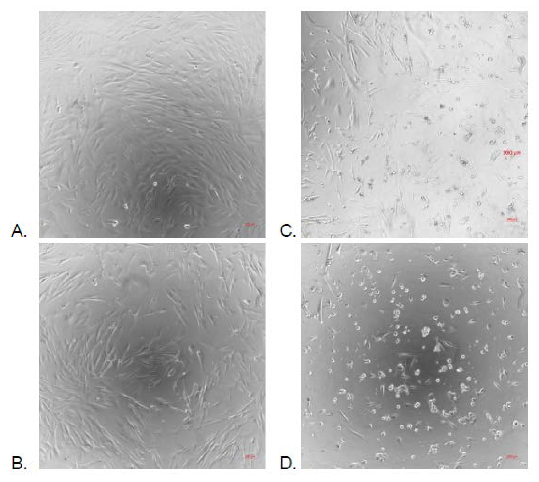
2.3. Cytotoxicity Activity

Methanolic crude extract and water extract were evaluated for their cytotoxicity at various concentrations (100 μg/mL, 128 μg/mL and 256 μg/mL) against embryonic lung MRC-5 cell line using MTT test. Methanolic extract at 100 μg/mL and 128 μg/mL did not show any cytotoxic activity against MRC-5 cells (100% and 98% of cell survival respectively), while they showed moderate toxicity at a concentration of 256 μg/mL (78.17% of cell survival). However, the water extract exhibited an increasing toxicity against MRC-5 cells depending on the increasing concentrations tested. This toxicity was shown by the decrease of the cell density and modification of the morphology with microscopic observations (Figure 1). The picture of culture in cell layers could correctly report both the decrease in the number of cells and the change in the morphology of the survivors. The viability could not have been measured since water extract alone was able to convert tetrazolium salt in formazan.
Additional technic of analysis with neutral red was also performed but failed to produce reliable measurement. For more investigations, the precipitate of tannins from water extract PW and the fraction devoid of tannin from water extract FW were evaluated for their toxicity and a concentration causing 50% of cell death is sought. Precipitate of tannins PW has an IC50 of 241.2 ± 16.8 μg/mL while the fraction devoid of tannin FW is not toxic because the cell viability at 1024 μg/mL that represented the highest concentration tested is 88.9 ± 5.2%. Tannins could therefore be responsible for the cellular toxicity of the water extract observed. However, methanolic extract containing the same percentage of tannins as water extract (82.8% and 80.7% respectively) showed no toxicity against MRC-5 cell line. It has been reported that phenolic compounds showed the growth inhibition in breast cancer cells [22] and they have been suggested as responsible for the cytotoxic effects of Baccharis species [23] whereas flavonoids induced cytotoxicity in various human cell lines [24]. Both flavonoid and total polyphenolic compound contents are higher in non-toxic methanolic extract than in toxic water extract of A. seyal. These results suggest that it could be quality other than quantity that matters on the potency of phenolic compounds. The pure compound ƴ-sitosterol isolated from Acacia nilotica leaves showed anticancer properties [25]. This compound was also isolated from the no toxic methanolic extract of A. seyal. This suggests that the cytotoxicity observed in water extract of A. seyal bark could be from other specialized metabolites apart from this compound and phenolics.

2.4. Antibacterial Activity

The crude water and methanolic extracts were evaluated for the antibacterial activities against nine human bacterial pathogen strains: Escherichia coli, Staphylococcus aureus, Pseudomonas aeruginosa, Corynebacterium urealyticum, Shigella sonnei, Salmonella enteric sv. typhi, Klebsiella pneumoniae, Staphylococcus epidermidis, and Streptococcus agalactiae. Water extract was devoid of any antibacterial activity against all bacterial strains tested except against Staphylococcus aureus with a MIC value of 512 µg/mL. The methanolic extract showed a significant activity against two Gram-positive bacteria: Staphylococcus aureus and Corynebacterium urealyticum with MIC values of 64 µg/mL and one Gram-negative bacteria: Pseudomonas aeruginosa with a MIC value of 512 µg/mL. The antibacterial activity of methanolic extracts devoid of tannins (FM) and free tannin precipitate from methanolic extracts (PM) was evaluated, and results of the MIC values are presented in Table 3.
The methanolic fraction devoid of tannins FM showed significant activity against S. aureus and C. urealyticum (MIC values of 32 µg/mL) and against P. aeruginosa (MIC value of 512 µg/mL) while the crude methanolic extract showed a lower activity against S. aureus and C. urealyticum (MIC values of 64 µg/mL). Surprisingly, the improvement in potency of the fraction FM devoid of tannins seemed to indicate that the tannins were antagonists to the antibacterial activity. Tannins are water-soluble and the results showed that the MIC values of both water extract and precipitate of tannins from water extract PM were similar against the tested strains. However, tannins are known to possess antibacterial activity by their capacity to form bonds with glycoproteins present on the external membrane of bacteria [26]. This leads to suggest that the antibacterial activity observed with the fraction devoid of tannin FM was due to other substances. Also extracts of Bolusanthus speciosus with small amount of phenolics and no tannin detected showed the highest antimicrobial effects [27]. The synergistic antibacterial effects of polyphenolic compounds have been previously reported in the literature [28]. The sensitivity of S. aureus, P. aeruginosa and C. urealyticum to the methanolic extract devoid of tannins FM and its 12 fractions (F1 to F12) as well as isolated compounds 1, 2 and 3 was carried out (Table 3). Reduction in potency of the methanolic extract devoid of tannins FM when fractionated was observed except for the fraction F12, which showed the same significant activity against S. aureus as FM (MIC value of 32 µg/mL) and the fractions F5, F8 and F11, which showed higher activity against S. aeruginosa (128 µg/mL) than the extract FM (>256). Fractions F2, F5, F7, and F8 possessed activity against S. aureus with an MIC value of 128 μg/mL. Against C. urealyticum, only the fractions F5 and F12 showed an activity (MIC value of 128 µg/mL) that was lower than the extract FM (MIC value of 32 µg/mL). The pure compounds 1, 2 and 3 did not show any antibacterial activity.

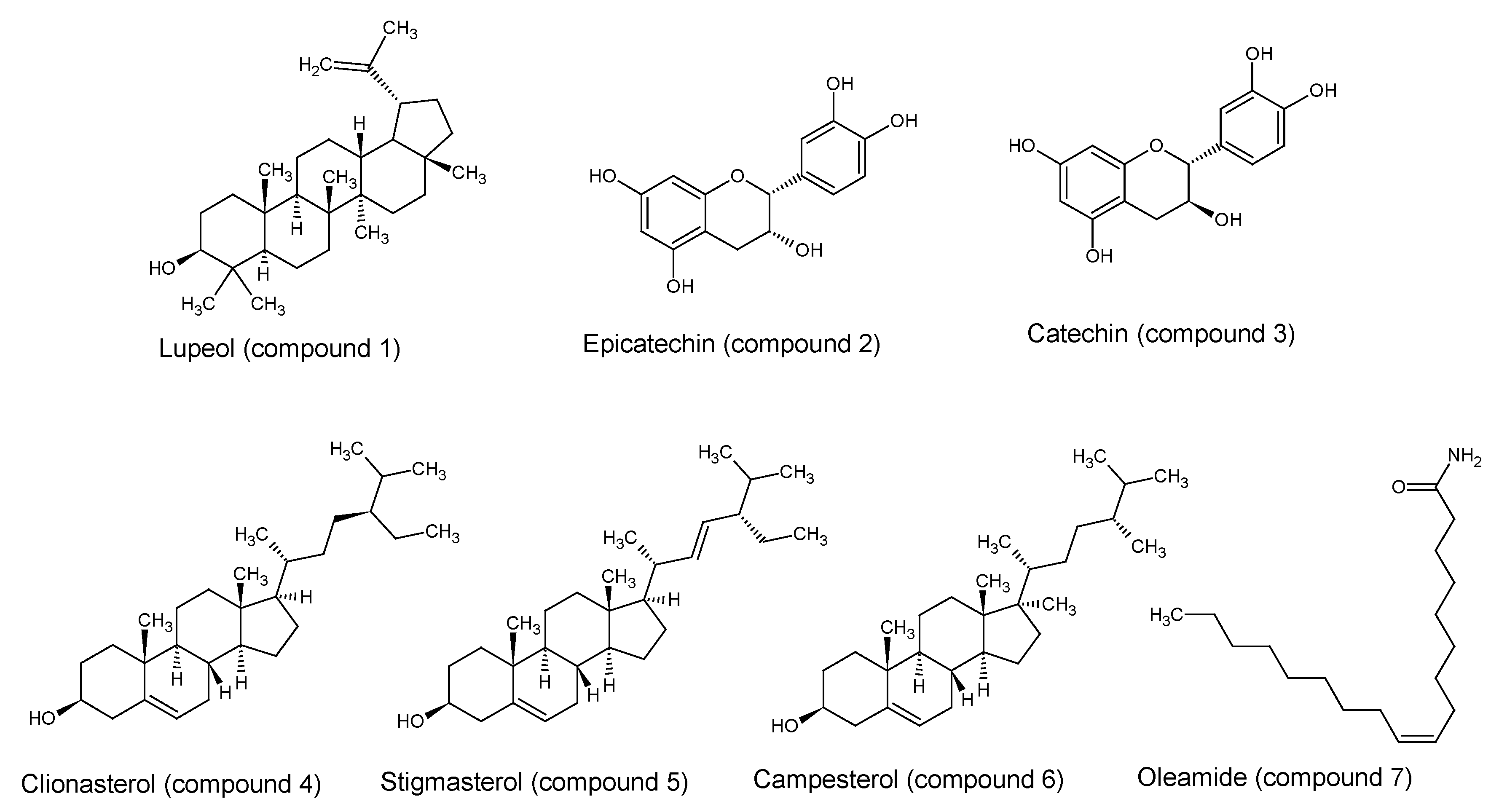
2.5. Chemical Constituents

The fractions F4, F5 and F12 were further investigated for their phytochemical characteristics. One triterpene was identified from F4 by GC-MS and 1H-NMR as lupeol (compound 1). Two flavanols were also isolated from F12 and their structure was determined by NMR and mass fragmentation. The structures are confirmed by comparison with the literature data [29] and authentic specimens as epicatechin (compound 2) and catechin (compound 3). The original spectra are shown in the Supplementary Material.
Four compounds were identified in the fraction F5 by GC-MS; three steroids as clionasterol (compound 4), stigmasterol (compound 5), and campesterol (compound 6); and one fatty acid as oleamide (compound 7) [30]. All structures are presented in Figure 2.
The description of the isolated compounds (1, 2, 3) and the compounds identified by GC-MS (4, 5, 6, 7) is: Compound 1, Lupeol: (C30H50O, RetIndex: 2848), GC-MS: m/z = 426 (38) [M+], 411 (7), 316 (11), 257 (10), 207 (71), 189 (82), 135 (66), 81 (100), 55 (61). 1H-NMR (400 MHz, CDCl3): δ (ppm) 4.62 (d, 1H, J = 2.45), 4.50 (dd, 1H, J = 2.45, 1.32), 3.11 (dd, 1H, J = 11, 5.3), 2.30 (td, 1H, J = 11, 5.8), 1.86–1.79 (m), 1.61 (s, CH3), 1.46–1.38 (m), 1.27–1.21 (m), 1.19 (s, CH3), 1.13–1.00 (m), 0.95 (s, CH3), 0.89 (s, CH3), 0.83–0.77 (m), 0.75 (s, CH3), 0.72 (s, CH3), 0.69 (s, CH3). Compound 2, Epicatechin was obtained as brown powder. 1H-NMR (400 MHz, CD3OD): δ (ppm) 6.98 (d, 1H, H-2′, J = 1.2), 6.8 (dd, 1H, H-6′, J = 8.2, 1.5), 6.76 (d, 1H, H-5′, J = 8.2), 5.96 ppm (d, 1H, H-6, J = 2.2), 5.94 ppm (d,1H, H-8, J = 2.3), 4.82 (sbr, 1H, H-2), 4.17 (m, 1H, H-3), 2.86 (d, 1H, H-4a, J = 16.6, 4.7), 2.74 (dd, 1H, H-4b, J = 16.5, 2.7). 13C-NMR (100 MHz, CD3OD): δ (ppm)158.1 (C-7), 157.7 (C-5), 157.4 (C-9), 146.0 (C-4′), 145.8 (C-3′), 132.4 (C-1′), 119.5 (C-2′), 116.0 (C-5′), 115.5 (C-6′), 100.2 (C-10), 96.5 (C-6), 96.0 (C-8), 80.0 (C-2), 67.6 (C-3), 29.3 (C-4). HRESIMS (negative mode) m/z 289.0726 [M-H] (calcd. for C15H13O6, 289.0712). Compound 3, Catechin was obtained as brown powder. 1H-NMR (400 MHz, CD3OD): δ (ppm); 6.85 (d, 1H, H-6′, J = 2,0), 6.76 (d, 1H, H-5′, J = 8.0), 6.72 (dd, 1H, H-2′, J = 8.2, 2.0), 5.93 (d, 1H, H-6, J = 2.2), 5.86 (d, H-8, J = 2.3), 4.57 (d, 1H, H-2, J = 7.6), 3.97 (td, 1H, H-3, J = 7.8, 5.6), 2.86 (dd, 1H, H-4a, J = 16.1, 5.4), 2.49 (dd, 1H, H-4b, J = 16.1, 8.1). 13C-NMR (100 MHz, CD3OD): δ (ppm) 157.9 (C-7), 157.7 (C-5), 157.0 (C-9), 146.31 (C-4′), 146.33 (C-3′), 132.4 (C-1′), 120.1 (C-2′), 116.2 (C-5′), 115.4 (C-6′), 100.9 (C-10), 96.4 (C-6), 95.6 (C-8), 82.9 (C-2), 68.9 (C-3), 28.3 (C-4). HRESIMS (negative mode) m/z 289.0719 [M-H] (calcd. for C15H13O6, 289.0712). Compound 4, Clionasterol (C29H50O, RetIndex: 2731), GC-MS: m/z = 414 (100) [M+], 354 (5), 329 (42), 255 (30), 213 (31), 145 (44), 81 (66), 43 (87). Compound 5, Stigmasterol (C29H48O, RetIndex: 2739), GC-MS: m/z = 412 (59) [M+], 351 (59), 300 (27), 255 (45), 213 (13), 145 (39), 83 (100), 55 (90), 43 (33). Compound 6, Campesterol (C28H48O, RetIndex: 2632), GC-MS: m/z = 400 (100) [M+], 367 (23), 315 (55), 255 (27), 213 (30), 159 (43), 145 (50), 81 (61), 43 (89). Compound 7, Oleamide (C18H35NO, RetIndex: 2228), GC-MS: m/z = 281 (7) [M+], 126 (8), 72 (86), 59 (100), 55 (36).

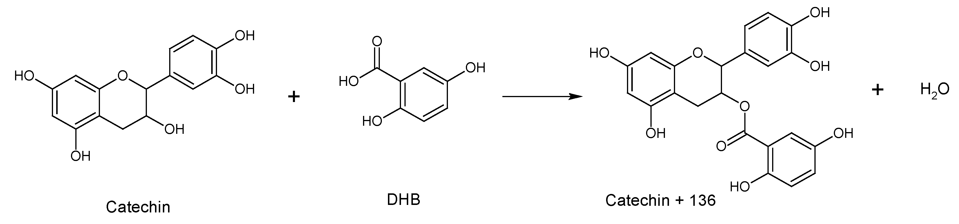
2.6. Characterization of Condensed Tannins by MALDI-TOF MS Analysis

The structure of tannin oligomers isolated from methanolic and water extracts of A. seyal bark as well as their degree of polymerization were investigated by matrix-assisted laser desorption/ ionization time-of-flight (MALDI-TOF) mass spectrometry. It is a suitable method for examining tannin oligomers [31]. Figure 3 shows the MALDI-TOF mass spectra of the polymeric tannin mixtures of both extracts, recorded as Na+ adducts in the positive ion reflectron mode.
The theoretical molecular weight of each peak is calculated, using equation (1), from combinations of its repeating units [32]:
M + Na = 23   ( Na ) + 2   ( endgroups ,   2 × H ) + 304 × N 1 + 288 × N 2 + 152 × N 3
The polymeric character is reflected by the periodic peak series representing different polymers. Condensed tannins isolated from the methanolic and water extracts are characterized by mass spectra with a series of peaks with various molecular weights from 737.3 to 2345.6 Da and from 617 to 2345.6 Da respectively. Two types of sequences were identified for the condensed tannins of the methanolic extract (S1 and S2) and of the aqueous extract (S3 and S4) (Table 4).
In the S1 series of the methanolic extract, the peaks correspond to combinations of these two monomers, epi/catechin (distance of 288 Da) and gallo-epi/catechin (distance of 304 Da), added by 136 Da that correspond to the product of an esterification between the catechin and the matrix, which is a carboxylic acid. This esterification could take place during the MALDI-TOF analysis, between the catechin and the DBH (2,5 dihydroxybenzoic) matrix. This produces a catechin monomer increased by 136 Da to the molar weight, a phenomenon already reported by Duval et al. [33] (Figure 4).

3. Materials and Methods

3.1. Chemicals

1,1-Diphenyl-2-picrylhydrazyl (DPPH), 2,2′-azinobis-3-ethylbenzothiazoline-6-sulfonic acid (ABTS), Vitamin C, Trolox, iron (II) sulfate (FeSO4), iron (III) chloride (FeCl3), Folin–Ciocalteu reagent (FC reagent), hydrochloric acid (HCl), sulphuric acid (H2SO4), organics solvants, trichloroacetic acid (TCA), dimethyl sulfoxide (DMSO), potassium persulfate, sodium dihydrogen phosphate, sodium hydroxide (NaOH), CD3OD and CDCl3 were purchased from Sigma Aldrich (St Quentin Fallavier, Franceor Acros organics, part of Thermo Fischer (Illkirch, France). The solvents used are HPLC profile grade.

3.2. Plant Materials

The bark of Acacia seyal Del. (Fabaceae) was collected in the locality of Day from the Tadjourah region, North of Djibouti in December 2015 (Latitude: 11°48’16.8660” N, Longitude: 42°37’46.1256” E). The identification was carried out by Prof. Maha Kordofani of the University of Khartoum of Sudan and its floor filed under No. 46/HND/2015 to the national herb of Djibouti.

3.3. Preparation of Plant Extracts

First the bark of A. seyal was crushed. Six-hundred-and-fifteen grams of the dry powder was extracted with 5 liters of methanol in Soxhlet apparatus for 7 hours and then evaporated under reduced pressure to dryness to obtain 82.3 g. Two-hundred grams of the dry powder was extracted with 600 mL of water using maceration during 30 h as indicated in the traditional method and then lyophilized leading to 4.9 g.

3.4. Precipitation of Tannins from Crude Extracts

Tannins present in methanolic and water extracts were precipitated according to the protocol indicated by Lhuillier [34] with some light modifications to replace chloroform by dichloromethane. To 3 g of both solid crude extracts were added 80 mL of methanol as solvent in two separated flasks. Then dichloromethane was added in a ratio of 8:92 (methanol: dichloromethane, v/v). Spontaneously, the tannins precipitated. The mixture thus obtained was left for 3 hours at 4 °C and then filtered. After evaporation, methanolic extract devoid of tannins was noted as FM and the precipitate of tannins from methanolic extract was noted as PM. From water extract, the filtrate noted as FW corresponds to the water extract devoid of tannins and PW to the tannins precipitate.

3.5. Determination of the Total Phenol Content (TPC) in the Extracts

The TPC of all extracts was evaluated using the Folin–Ciocalteu method [35] with some modification. Briefly, 100 mL of the extract solution (10 mgmL1 MeOH) was shaken for 3 min with 1 mL of diluted (1:10) Folin–Ciocalteu reagent. Then, 900 mL of 10% Na2CO3 was added and the final volume was made up to 5.0 mL with distilled water. After the mixture was allowed to stand for 2 h at room temperature, the absorbance at 725 nm was measured using a UV-vis spectrophotometer.
The results of TPC were estimated using a standard curve prepared using gallic acid and expressed as milligram GAEs per gram of extract on dry weight basis.

3.6. Determination of the Total Flavonoid Content (TFC) in the Extracts

The total flavonoid content was determined using [36] method as adapted by [37]. Briefly, sample solution (1 mg/mL; 1 mL) was mixed with the same volume of aluminium trichloride (2%) in methanol. Similarly, a blank was prepared by adding sample solution (1 mL) to methanol (1 mL) without AlCl3. The sample and control absorbance were read at 415 nm after a 10 min incubation at room temperature. The absorbance of the blank was subtracted from that of the sample. Quercetin was used as a reference standard and the total flavonoid content was expressed as milligrams of quercetin equivalents (mg QE/g extract).

3.7. Preparation of Antioxidant Activities Tests

3.7.1. DPPH Method

The free-radical-scavenging activity of the extracts of A. seyal was measured using an improved DPPH assay [38]. The extract solution with concentration of 0.3 mL was mixed with a solution of 0.2 mmol/L DPPH in methanol (2.7 mL). The mixture was mixed vigorously and then left to stand for 1 h at room temperature before measuring the absorbance value at 517 nm. The percent of inhibition rate of radical scavenging activity was calculated using the following Equation (2):
%   Inhibition   = [ ( As Ai ) / As ] × 100
where “As” is the absorbance of DPPH alone, and “Ai” is the absorbance of DPPH in the presence of various extracts. The concentrations of Trolox and Vitamin C (VC) identical to the experimental samples were used as reference.

3.7.2. ABTS Method

The ability of the extract to scavenge ABTS radical was determined according to a previously published method [39]. ABTS was dissolved in deionized water at 7 mmol/L concentration, and potassium persulfate with a concentration of 2.45 mmol/L was added afterward. The reaction mixture was kept in the dark at room temperature for 16 h. The mixture was then diluted with 80% ethanol to obtain an absorbance value of 0.700 ± 0.005 at 734 nm. Test substances (0.3 mL) at various concentrations were incubated with ABTS + solution (2.7 mL) in a 30 1C water bath for 30 min in the dark. The absorbance at 734 nm was immediately recorded. Samples of BHT and VC at the same concentrations were used as references. The level of radical scavenging was calculated using the aforementioned equation for DPPH.

3.7.3. FRAP Method

The FRAP assay was carried out according to [40] with slight modification. Briefly, the FRAP reagent was prepared from acetate buffer (pH 3.6), 10 mmol TPTZ solution in 40 mmol HCl and 20 mmol iron (III) chloride solution in proportions of 10:1:1 (v/v), respectively. The FRAP reagent was prepared fresh daily and was warmed to 37 C in a water bath prior to use. Fifty microliters of sample were added to 1.5 ml of the FRAP reagent. The absorbance of the reaction mixture was then recorded at 593 nm after 4 min. The standard curve was constructed using Vitamin C solution (0.065–33.3 µg/ml), and the results were expressed as µmol VCE/g dry weight of plant material. All the measurements were taken in triplicate and the mean values were calculated.

3.8. Cytotoxicity Assay

The toxicity of the extracts on cell viability was evaluated on MRC-5 cells, a fibroblast derived from normal lung tissue (ECACC, reference 05090501) using the MTT test, based on the reduction of MTT in formazan crystals by metabolically active cells [41]. MRC-5 cells is a line of normal cells (non-cancerous) which have the capacity to multiply under in vivo culture conditions. This cell line is commonly used as a model for the evaluation of toxicity. Cells were plated at 10000 cells per well in complete medium in 96-well tissue culture plates and grown for 48 hours at 37 °C. Then medium was discarded and replaced by fresh medium containing extracts previously dissolved in dimethyl sulfoxide. Three different controls were added: medium alone, cells in medium and extract in medium. After 24 h incubation, medium was replaced by 100 µl of 0.5 mg MTT solution in medium per well and plates were incubated for 4 hours at 37 °C. Then formazan crystals were dissolved by 100 µl of dimethyl sulfoxide. Absorbance was measured at 540 nm using a 96-well plate reader. Percentages of survival and half maximal inhibitory concentration (IC50) were calculated using MS Office Excel. The water extract was able to convert MTT in formazan. That is why a confirmation of its toxicity was carried out by microscopic observation.

3.9. Antibacterial Assay

For experiments, the following bacteria have been used:
Escherichia coli ABC5 (ATCC 25922), Shigella sonnei ABC16 (ATCC 9290), Salmonella enterica sv. typhi ABC17 (ATCC 13311), Staphylococcus aureus ABC1 (ATCC 29213), Pseudomonas aeruginosa ABC4 (ATCC 27853), Klebsiella pneumoniae ABC12 (ATCC 700603), Streptococcus agalactiae ABC6 (ATCC 27956), Staphylococcus epidermidis ABC91 (clinical origin), and Corynebacterium urealyticum ABC145 (clinical origin).
To evaluate the antibacterial activities of the compounds, the Minimal Inhibitory Concentration (MIC) was determined by broth microdilution method based on ISO 20776-1:2006 standard [42], in accordance with CLSI [43] and EUCAST [44] guidelines.
Bacterial suspensions were prepared by suspending three to five isolated colonies from Mueller-Hinton Agar, Cation-Adjusted (MHA-CA) in 5 mL of Mueller-Hinton Broth, Cation-Adjusted (MHB-CA).
After 24 h of growth at 35 °C, the medium was removed by centrifugation (5 min at 3000 g) and cells were resuspended in 0.85% NaCl to obtain a 0.5 McFarland suspension.
This suspension was then adjusted in MHB-CA at 1 × 10⁶ [0.4 × 10⁶ − 1.6 × 10⁶] CFU/mL.
Twofold serial dilutions of the compounds (prediluted in DMSO) were prepared in 0.05 mL MHB-CA in round-bottom, untreated 96-well plates. An equal volume (0.05 mL) of bacterial inoculum was added to each well. The final concentration of DMSO never exceeded 5% of the final volume. Each condition was repeated in the eight lines of the microplate.
Final bacterial concentration in the wells was 5 × 10⁵ [2 × 10⁵ − 8 × 10⁵] CFU/mL and checked as described in ISO 20776-1:2006 standard.
In each line, a positive growth control (compound diluent, without compound) and two negative controls (0.05 mL MHB-CA with or without compound at the highest assayed concentration, and without bacteria) were included.
After incubation for 18h at 35 °C, the MICs were visually determined as recommended by ISO standard. In all experiments, bacteria were tested against vancomycin (for Gram positive bacteria) and gentamicin (for Gram negative bacteria) by microdilution method according to ISO 20776-1:2006 as control. Expected results were obtained (MIC ≤ 1 mg/L).

3.10. Isolation and Characterization of Pure Compounds

3.10.1. Isolation of Pure Compounds

The methanolic extract devoid of tannins FM was subjected to flash chromatography eluted with toluene: ethyl acetate (75:15 v/v). A total of 12 fractions were obtained (F1–F12) on combining the eluates according to their similarity behaviour on TLC. Fraction F4 (45 mg) was submitted to preparative TLC using the previous mobile phase. One pure compound was obtained from F4 (2 mg, compound 1). Fraction F12 (2032 mg) was subjected to flash chromatography eluted with ethyl acetate–methanol–water (100:13.5:10 v/v) and then subjected to preparative HPLC using a gradient of methanol to water with 2% formic acid (1000 mL) as eluent to obtain two pure compounds: compound 2 (18 mg) and compound 3 (7 mg). Fraction F5 (52.3 mg) was analysed by GC-MS analysis to found 4 compounds (compounds 4, 5, 6, 7).

3.10.2. MALDI-TOF MS for Characterization of Condensed Tannins

The precipitated tannin sample is dissolved in methanol (10 mg/ml). The material used is 2,5 dihydroxybenzoic (DHB, 100 mg/ml in a 50/50 water/acetonitrile mixture). To improve the formation of ions, the water contains NaCl (1 mg/ml). A mixture of 10 μl of the tannin sample and 10 μl of the DHB matrix is prepared and then 1 μl of this mixture is deposited on a stainless steel plate and dried at room temperature. The recording is made with a MALDI-TOF mass spectrometry Ultraflex III (Bruker Daltonics, Bremen, Germany), equipped with a NdYAG laser, 266 nm at 200 Hz. The MALDI-TOF spectra is recorded in positive reflector mode using Na+ as adducts. Each spectrum is obtained after 1000 shots of laser.

3.10.3. GC-MS

GC-MS analysis was performed using QP2010-Shimadzu (Shimadzu, Kyoyo, Japan) equipment operating in the EI mode at 70 eV. An SLB5 columnDB-5 ms (30 m, 0.25 mm film thickness) was employed with a 36 min temperature program of 60–320 at 10 °C/min followed by a 10 min hold at 320 °C. The injector temperature was 250 °C, the flow rate of the carrier gas (helium) was 1 mL/min, and the split ratio was 1:50. The interval of the scan m/z was between 35 and 900 and the identification of the compounds is based on an individual spectrum comparison of each compound in the Shimadzu NIST08 data.

3.10.4. NMR

The NMR spectra (1H, 13C, COSY, HSQC, HMBC) were recorded on a Bruker Avance III 400 MHz spectrometer (Bruker biospin, Billerica, USA), Bruker BBFO Probe. The coupling constant (J) is expressed in Hz.

3.10.5. LC-HRESIMS

Liquid Chromatography Hight Resolution-Mass chromatography (LC HRESIMS) analysis was performed using a micrOTOF-QTM (Bruker Daltonics, Bruker, Bremen, Germany) apparatus operating in the ESI positive and negative mode, set capillary on 4500v, dry gas 10 ml/min, set dry heater 190 °C.

3.11. Statistical Analysis

The measurements are repeated three times and the data are expressed mean + standard error of the mean (SEM). Statistical significance was defined as p < 0.05.

4. Conclusions

The results obtained suggest that A. seyal bark could be a potential source of active natural compounds like flavonoids, steroids, triterpenes, and tannins as catechin and gallocatechin-based oligomers. This work also evaluated the antioxidant and antibacterial activities of A. seyal bark extracts. The bark is rich in tannins, which could be responsible for the observed antioxidant activity but not the antibacterial activity. However, campesterol combined with stigmasterol, clionasterol and oleamide was proven to show antibacterial activity against S. aureus. The cytotoxicity of A. seyal water bark extract revealed in this study against the MRC-5 cells indicated that the safety of the bark of this plant in the traditional medicine in Djibouti should be verified by other experiments including in vivo tests and clinical studies.

Supplementary Materials

The following are available online at https://www.mdpi.com/1420-3049/25/10/2392/s1. In the supplementary materials it is possible to find: in Figure S1 the picture of the Acacia seyal plant; in Figure S2 the linear correlation between ABTS and DPPH of IC50 of methanol and water Acacia seyal extracts; in Figure S3–S4 EIMS and 1H NMR spectra of lupeol; in Figure S5–S10 HRESIMS, 1H, 13C, COSY, HSQC, HMBC of Epicatechin; in Figure S11–S17 HRESIMS, 1H, 13C, COSY, HSQC, HMBC of Catechin; in Figure S18–S21 EIMS spectra of Clionasterol, Stigmasterol, Campesterol and Oleamide respectively.

Author Contributions

Conceptualization, A.E., F.M.A.-l.; A.M. and D.L.-M.; methodology, A.E., R.S., D.L.-M.; formal analysis, A.E., R.S., S.P., A.R.; resources, A.E., F.M.A.-l., A.M., R.S., D.L.-M.; data curation, A.E., F.M.A.-l., R.S., D.L.M., R.E.D.; writing—original draft preparation, A.E., R.S., D.L.-M.; writing—review and editing, R.S., D.L.-M., F.M.A.-l., A.M., S.P., A.R., R.E.D.; supervision, F.M.A.-l., R.S., D.L.-M.; funding acquisition, A.E., F.M.A.-l., R.S. and D.L.-M. All authors have read and agreed to the published version of the manuscript.

Funding

This research was funded by Research Center (CERD) in Djibouti, L2CM laboratory and Institut Jean Barriol in University of Lorraine. The APC was funded by the “Impact Biomolecules” project of the “Lorraine Université d’Excellence.

Acknowledgments

The authors acknowledge support of “Laboratoire Lorrain de Chimie Moléculaire L2CM UMR 7053” by the “Impact Biomolecules” project of the “Lorraine Université d’Excellence”(Investissements d’avenir – ANR), The authors acknowledge: Agence Universitaire de la Francophonie (AUF) for scholarship of A.E.; François Dupire from Platform of Mass Spectrometry, Pascal Lemiere from HPLC platform and ABC® platform in UMR 7053-L2CM team; Platform of Mass Spectrometry of LCP-A2MC laboratory, EA 4632.

Conflicts of Interest

The authors declare no conflict of interest.

Abbreviations

VCVitamin C
QEQuercetin Equivalent
VCEVitamin C Equivalent
GAEGallic acid Equivalent
TPCTotal polyphenols content
TFCTotal flavonoids content
FMfiltrate devoid of tannin from methanolic extract
PMprecipitate of tannins from methanolic extract
FWfiltrate devoid of tannin from water extract
PWprecipitate of tannins from water extract

References

  1. Nacro, M.; Millogo-Rasolodimbi, J. Plantes tinctoriales et plantes à tanins du Burkina Faso; ScientifikA: Amiens, France, 1993; ISBN 978-2-909894-09-6. [Google Scholar]
  2. Nie, S.-P.; Wang, C.; Cui, S.W.; Wang, Q.; Xie, M.-Y.; Phillips, G.O. The core carbohydrate structure of Acacia seyal var. seyal (Gum arabic). Food Hydrocoll. 2013, 32, 221–227. [Google Scholar] [CrossRef]
  3. Rather, L.J.; ul-Islam, S.; Mohammad, F. Acacia nilotica (L.): A review of its traditional uses, phytochemistry, and pharmacology. Sustain. Chem. Pharm. 2015, 2, 12–30. [Google Scholar] [CrossRef]
  4. Revathi, S.; Govindarajan, R.K.; Rameshkumar, N.; Hakkim, F.L.; Mohammed, A.-B.; Krishnan, M.; Kayalvizhi, N. Anti-cancer, anti-microbial and anti-oxidant properties of Acacia nilotica and their chemical profiling. Biocatal. Agric. Biotechnol. 2017, 11, 322–329. [Google Scholar] [CrossRef]
  5. Abdel-Farid, I.B.; Sheded, M.G.; Mohamed, E.A. Metabolomic profiling and antioxidant activity of some Acacia species. Saudi J. Biol. Sci. 2014, 21, 400–408. [Google Scholar] [CrossRef] [PubMed] [Green Version]
  6. Abdulrazak, S.A.; Fujihara, T.; Ondiek, J.K.; Ørskov, E.R. Nutritive evaluation of some Acacia tree leaves from Kenya. Anim. Feed Sci. Technol. 2000, 85, 89–98. [Google Scholar] [CrossRef]
  7. Abdoul-latif, F.M.; Osman, D.A.; Fourreh, A.E.; Abdallah, A.H.; Merito, A.; Hassan, S.; Asfaw, Z.; Kelbessa, E. Candidate medicinal plant species of djiboutian pharmacopeia for testing pharmacological activities on common microbial diseases. Int. J. Pharm. Pharm. Sci. 2016, 78–84. [Google Scholar] [CrossRef] [Green Version]
  8. Geissler, P.W.; Harris, S.A.; Prince, R.J.; Olsen, A.; Odhiambo, R.A.; Oketch-Rabah, H.; Madiega, P.A.; Andersen, A.; Mølgaard, P. Medicinal plants used by Luo mothers and children in Bondo district, Kenya. J. Ethnopharmacol. 2002, 83, 39–54. [Google Scholar] [CrossRef]
  9. Wambugu, S.N.; Mathiu, P.M.; Gakuya, D.W.; Kanui, T.I.; Kabasa, J.D.; Kiama, S.G. Medicinal plants used in the management of chronic joint pains in Machakos and Makueni counties, Kenya. J. Ethnopharmacol. 2011, 137, 945–955. [Google Scholar] [CrossRef]
  10. Hassan-Abdallah, A.; Merito, A.; Hassan, S.; Aboubaker, D.; Djama, M.; Asfaw, Z.; Kelbessa, E. Medicinal plants and their uses by the people in the Region of Randa, Djibouti. J. Ethnopharmacol. 2013, 148, 701–713. [Google Scholar] [CrossRef]
  11. Hammiche, V.; Maiza, K. Traditional medicine in Central Sahara: Pharmacopoeia of Tassili N’ajjer. J. Ethnopharmacol. 2006, 105, 358–367. [Google Scholar] [CrossRef]
  12. Eldeen, I.M.S.; Van Staden, J. Cyclooxygenase inhibition and antimycobacterial effects of extracts from Sudanese medicinal plants. South. Afr. J. Bot. 2008, 74, 225–229. [Google Scholar] [CrossRef] [Green Version]
  13. Eldeen, I.M.S.; Van Staden, J. In vitro pharmacological investigation of extracts from some trees used in Sudanese traditional medicine. South Afr. J. Bot. 2007, 73, 435–440. [Google Scholar] [CrossRef] [Green Version]
  14. Muthaura, C.N.; Keriko, J.M.; Mutai, C.; Yenesew, A.; Gathirwa, J.W.; Irungu, B.N.; Nyangacha, R.; Mungai, G.M.; Derese, S. Antiplasmodial potential of traditional antimalarial phytotherapy remedies used by the Kwale community of the Kenyan Coast. J. Ethnopharmacol. 2015, 170, 148–157. [Google Scholar] [CrossRef] [PubMed]
  15. Saeed, M.E.M.; Abdelgadir, H.; Sugimoto, Y.; Khalid, H.E.; Efferth, T. Cytotoxicity of 35 medicinal plants from Sudan towards sensitive and multidrug-resistant cancer cells. J. Ethnopharmacol. 2015, 174, 644–658. [Google Scholar] [CrossRef] [PubMed]
  16. Lin, J.-Y.; Tang, C.-Y. Determination of total phenolic and flavonoid contents in selected fruits and vegetables, as well as their stimulatory effects on mouse splenocyte proliferation. Food Chem. 2007, 101, 140–147. [Google Scholar] [CrossRef]
  17. Mugedo, J.Z.A.; Waterman, P.G. Sources of tannin: Alternatives to wattle (Acacia mearnsii) among indigenous Kenyan species. Econ. Bot. 1992, 46, 55–63. [Google Scholar] [CrossRef]
  18. Wei, S.-D.; Zhou, H.-C.; Lin, Y.-M.; Liao, M.-M.; Chai, W.-M. MALDI-TOF MS analysis of condensed tannins with potent antioxidant activity from the leaf, stem bark and root bark of Acacia confusa. Mol. Basel Switz. 2010, 15, 4369–4381. [Google Scholar] [CrossRef] [Green Version]
  19. Tung, Y.-T.; Wu, J.-H.; Huang, C.-Y.; Kuo, Y.-H.; Chang, S.-T. Antioxidant activities and phytochemical characteristics of extracts from Acacia confusa bark. Bioresour. Technol. 2009, 100, 509–514. [Google Scholar] [CrossRef]
  20. Arnao, M.B. Some methodological problems in the determination of antioxidant activity using chromogen radicals: A practical case. Trends Food Sci. Technol. 2000, 11, 419–421. [Google Scholar] [CrossRef]
  21. Halake, K.; Birajdar, M.; Lee, J. Structural implications of polyphenolic antioxidants. J. Ind. Eng. Chem. 2016, 35, 1–7. [Google Scholar] [CrossRef]
  22. Jaganathan, S.K.; Mandal, S.M.; Jana, S.K.; Das, S.; Mandal, M. Studies on the phenolic profiling, anti-oxidant and cytotoxic activity of Indian honey: In vitro evaluation. Nat. Prod. Res. 2010, 24, 1295–1306. [Google Scholar] [CrossRef] [PubMed]
  23. Jaramillo-García, V.; Trindade, C.; Lima, E.; Guecheva, T.N.; Villela, I.; Martinez-Lopez, W.; Corrêa, D.S.; de Ferraz, A.B.F.; Moura, S.; Sosa, M.Q.; et al. Chemical characterization and cytotoxic, genotoxic, and mutagenic properties of Baccharis trinervis (Lam, Persoon) from Colombia and Brazil. J. Ethnopharmacol. 2018, 213, 210–220. [Google Scholar] [CrossRef]
  24. Marzouk, M.M. Flavonoid constituents and cytotoxic activity of Erucaria hispanica (L.) Druce growing wild in Egypt. Arab. J. Chem. 2016, 9, 411–415. [Google Scholar] [CrossRef] [Green Version]
  25. Sundarraj, S.; Thangam, R.; Sreevani, V.; Kaveri, K.; Gunasekaran, P.; Achiraman, S.; Kannan, S. γ-Sitosterol from Acacia nilotica L. induces G2/M cell cycle arrest and apoptosis through c-Myc suppression in MCF-7 and A549 cells. J. Ethnopharmacol. 2012, 141, 803–809. [Google Scholar] [CrossRef] [PubMed]
  26. Tittikpina, N.K.; Nana, F.; Fontanay, S.; Philippot, S.; Batawila, K.; Akpagana, K.; Kirsch, G.; Chaimbault, P.; Jacob, C.; Duval, R.E. Antibacterial activity and cytotoxicity of Pterocarpus erinaceus Poir extracts, fractions and isolated compounds. J. Ethnopharmacol. 2018, 212, 200–207. [Google Scholar] [CrossRef] [PubMed]
  27. Mulaudzi, R.B.; Ndhlala, A.R.; Kulkarni, M.G.; Finnie, J.F.; Van Staden, J. Antimicrobial properties and phenolic contents of medicinal plants used by the Venda people for conditions related to venereal diseases. J. Ethnopharmacol. 2011, 135, 330–337. [Google Scholar] [CrossRef]
  28. Tafesh, A.; Najami, N.; Jadoun, J.; Halahlih, F.; Riepl, H.; Azaizeh, H. Synergistic Antibacterial Effects of Polyphenolic Compounds from Olive Mill Wastewater. Available online: https://www.hindawi.com/journals/ecam/2011/431021/ (accessed on 25 April 2020).
  29. Donovan, J.L.; Luthria, D.L.; Stremple, P.; Waterhouse, A.L. Analysis of (+)-catechin, (-)-epicatechin and their 3′- and 4′-O-methylated analogs. A comparison of sensitive methods. J. Chromatogr. B. Biomed. Sci. App. 1999, 726, 277–283. [Google Scholar] [CrossRef]
  30. NIST Standard Reference Databases: Analytical Chemistry. Available online: https://www.nist.gov/srd/nist-standard-reference-databases-analytical-chemistry (accessed on 25 April 2020).
  31. Zhang, L.L.; Lin, Y.M. HPLC, NMR and MALDI-TOF MS analysis of condensed tannins from Lithocarpus glaber leaves with potent free radical scavenging activity. Mol. Basel Switz. 2008, 13, 2986–2997. [Google Scholar] [CrossRef] [Green Version]
  32. Navarrete, P.; Pizzi, A.; Pasch, H.; Rode, K.; Delmotte, L. MALDI-TOF and 13C NMR characterization of maritime pine industrial tannin extract. Ind. Crop. Prod. 2010, 32, 105–110. [Google Scholar] [CrossRef]
  33. Duval, A.; Avérous, L. Characterization and physicochemical properties of condensed tannins from Acacia catechu. J. Agric. Food Chem. 2016, 64, 1751–1760. [Google Scholar] [CrossRef]
  34. Lhuillier, A. Contribution à l’étude Phytochimique de Quatre Plantes Malgaches: Agauria salicifolia Hook.f ex Oliver, Agauria polyphylla Baker (Ericaceae), Tambourissa trichophylla Baker (Monimiaceae) et Embelia concinna Baker (Myrsinaceae). Ph.D. Thesis, National Polytechnic Institute of Toulouse, Toulouse, France, 2007. [Google Scholar]
  35. Butsat, S.; Siriamornpun, S. Antioxidant capacities and phenolic compounds of the husk, bran and endosperm of Thai rice. Food Chem. 2010, 119, 606–613. [Google Scholar] [CrossRef]
  36. Dowd, L.E. Spectrophotometric determination of quercetin. Anal. Chem. 1959, 31, 1184–1187. [Google Scholar] [CrossRef]
  37. Arvouet-Grand, A.; Vennat, B.; Pourrat, A.; Legret, P. Standardization of propolis extract and identification of principal constituents. J. Pharm. Belg. 1994, 49, 462–468. [Google Scholar]
  38. Huang, B.; Ban, X.; He, J.; Tong, J.; Tian, J.; Wang, Y. Hepatoprotective and antioxidant activity of ethanolic extracts of edible lotus (Nelumbo nucifera Gaertn.) leaves. Food Chem. 2010, 120, 873–878. [Google Scholar] [CrossRef]
  39. Huang, B.; Ke, H.; He, J.; Ban, X.; Zeng, H.; Wang, Y. Extracts of Halenia elliptica exhibit antioxidant properties in vitro and in vivo. Food Chem. Toxicol. 2011, 49, 185–190. [Google Scholar] [CrossRef]
  40. Benzie, I.F.; Strain, J.J. The ferric reducing ability of plasma (FRAP) as a measure of “antioxidant power”: The FRAP assay. Anal. Biochem. 1996, 239, 70–76. [Google Scholar] [CrossRef] [Green Version]
  41. Mosmann, T. Rapid colorimetric assay for cellular growth and survival: Application to proliferation and cytotoxicity assays. J. Immunol. Methods 1983, 65, 55–63. [Google Scholar] [CrossRef]
  42. Clinical Laboratory Testing and in vitro Diagnostic Test Systems—Susceptibility Testing of Infectious Agents and Evaluation of Performance of Antimicrobial Susceptibility Test Devices—Part 1: Reference Method for Testing the in Yitro Activity of Antimicrobial Agents Against Rapidly Growing Aerobic Bacteria Involved in Infectious Diseases. Available online: https://www.iso.org/standard/41630.html (accessed on 25 April 2020).
  43. Cockerill, F. Performance Standards for Antimicrobial Susceptibility Testing: Twenty-Third Informational Supplement; Clinical and Laboratory Standards Institute: Wayne, PA, USA, 2013; ISBN 978-1-56238-865-2. [Google Scholar]
  44. EUCAST: MIC Determination. Available online: https://eucast.org/ast_of_bacteria/mic_determination/ (accessed on 25 April 2020).
Sample Availability: Samples of the crude extracts and compounds 1, 2 and 3 are available from the authors.
Figure 1. Microscopic observation of the effect of water Acacia seyal extract at different concentrations on MRC-5 cells at 24 hours. (A): DMSO control, (B): 64 μg/mL, (C): 128 μg/mL, (D): 256 μg/mL, observation under ×10 air objective.
Molecules 25 02392 g001
Figure 2. Identified compounds in A. seyal bark.
Molecules 25 02392 g002
Figure 3. MALDI-TOF positive reflectron mode mass spectra of the condensed tannins from methanolic (a) and water (b) extracts of Acacia seyal bark.
Molecules 25 02392 g003
Figure 4. Esterification of catechin with the matrix molecule (DHB: 2,5 dihydroxybenzoic) during the MALDI-TOF experiment.
Molecules 25 02392 g004
Table 1. Total polyphenols content (TPC) and total flavonoids content (TFC), extractable tannins of two extracts of A. seyal bark.
ExtractsTPC
(mg GAE/100 g)
TFC
(mg QE/100 g)
Extractable Tannins (%)
Methanolic extract 1927.1 ± 11.130.9 ± 1.582.8 ± 2.3%
Water extract1820.5 ± 13.613.8 ± 5.380.7 ± 1.1%
GAE: Gallic acid Equivalent. QE: Quercetin Equivalent. Values are representative of three independent determinations. p values ≤ 0.05.
Table 2. Antioxidant activity of bark extracts of A. seyal.
Extracts, Fractions and StandardsDPPH IC50
(µg/mL)
ABTS IC50
(µg/mL)
FRAP
(µg VE/g)
Methanolic crude extract150 ± 2.227 ± 1.345.7 ± 6.1
Filtrate FM250 ± 1250 ± 320.7 ± 2.3
Precipitate PM95 ± 316 ± 0.854.4 ± 9.3
Water crude extract 183 ± 5.333 ± 25.7 ± 1.0
Filtrate FW321 ± 1341 ± 13.7 ± 1.6
Precipitate PW136 ± 521 ± 16.7 ± 1.1
Vitamin C70 ± 770 ± 1.5ND
Trolox110 ± 2.518 ± 3.3ND
Values are representative of three independent determinations. p values ≤ 0.01. VCE: Vitamin C Equivalent; ND: not determined; FM: filtrate devoid of tannin from methanolic extract; PM: precipitate of tannins from methanolic extract; FW: filtrate devoid of tannin from water extract; PW: precipitate of tannins from water extract.
Table 3. Antibacterial activity of extracts and isolated compounds from Acacia seyal bark expressed as MICs (µg/mL).
Extracts, Fractions and Pure CompoundsStaphylococcus aureusPseudomonas aeruginosaCorynebacterium
urealyticum
Water extract512>1024>1024
Methanolic extract6451264
PM512>1024>1024
FM3251232
F1>256>256>256
F2128>256256
F3>256>256>256
F4>256>256>256
F5128128128
F6256256>256
F7128256>256
F8128128>256
F9256>256>256
F10>256>256>256
F11>256128256
F1232>1024128
Compound 1512>1024512
Compound 2>1024>1024>1024
Compound 3102410241024
Table 4. MALDI-TOF MS of condensed tannins from methanolic and water extracts of Acacia seyal bark.
FractionSeries[M+Na]
Experimental
[M+Na]
Calculated
N1N2N3N2+ DHBDPPolymer
Precipitate (PM) from Methanol Extract
PMS1737.3737.102012Dimer
PMS11025.31025.203013Trimer
PMS11329.41329.313014Tetramer
PMS11633.41633.323015Pentamer
PMS2889.3889.203003Trimer
PMS2905.2905.212003Trimer
PMS2921.2921.221003Trimer
PMS21193.01193.313004Tetramer
PMS21481.41481.314005Pentamer
PMS21769.51769.415006Hexamer
PMS22057.52057.416007Heptamer
PMS22345.62345.617008Octamer
Precipitate (PW) from Water Extract
PWS3617.2617.111002Dimer
PWS3905.2905.212003Trimer
PWS31193.31193.313004Tetramer
PWS31481.41481.314005Pentamer
PWS31769.41769.415006Hexamer
PWS32057.52057.416007Heptamer
PWS32345.62345.517008Octamer
PWS32633.62633.618009Nonamer
PWS4769.2769.111102Dimer
PWS41057.21057.212103Trimer
PWS41345.31345.313104Tetramer
PWS41633.41633.314105Pentamer
N1—Gallocatechin monomer with molecular weight 304.0583, N2—Catechine monomer with molecular weight 288.0634, N3—Gallic acid monomer with molecular weight 152.010956, DHB—(2,5dihydroxybenzoic) matrix with molecular weight 136.016, S1–S2—precipitate of tannins from water extract (PW), S3–S4—precipitate of tannins from methanol extract (PM), DP—Degree of polymerization.

