
SPME-GC/MS Analysis of Methanol in Biospecimen by Derivatization with Pyran Compound


Abstract
1. Introduction
2. Results and Discussion
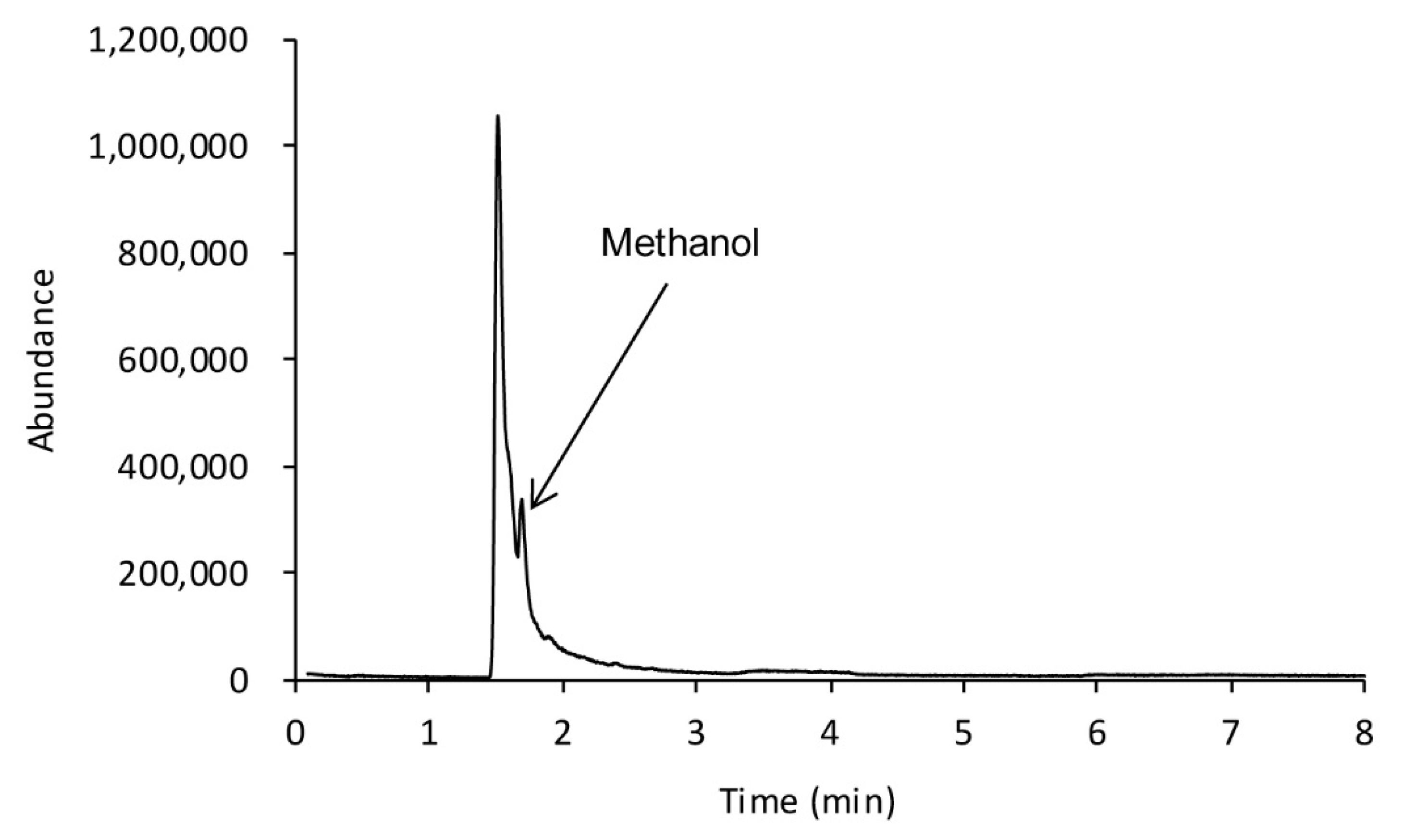
2.1. GC-FID Analysis
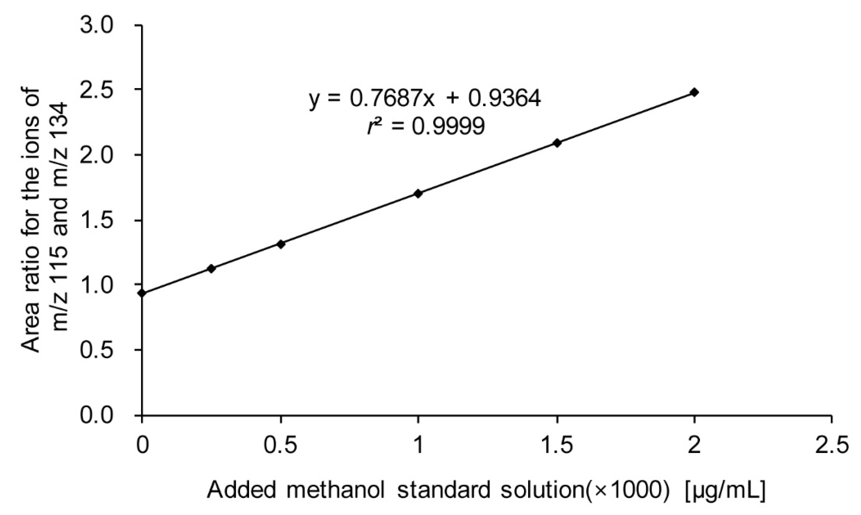
2.2. Derivatization and GC/MS Analysis
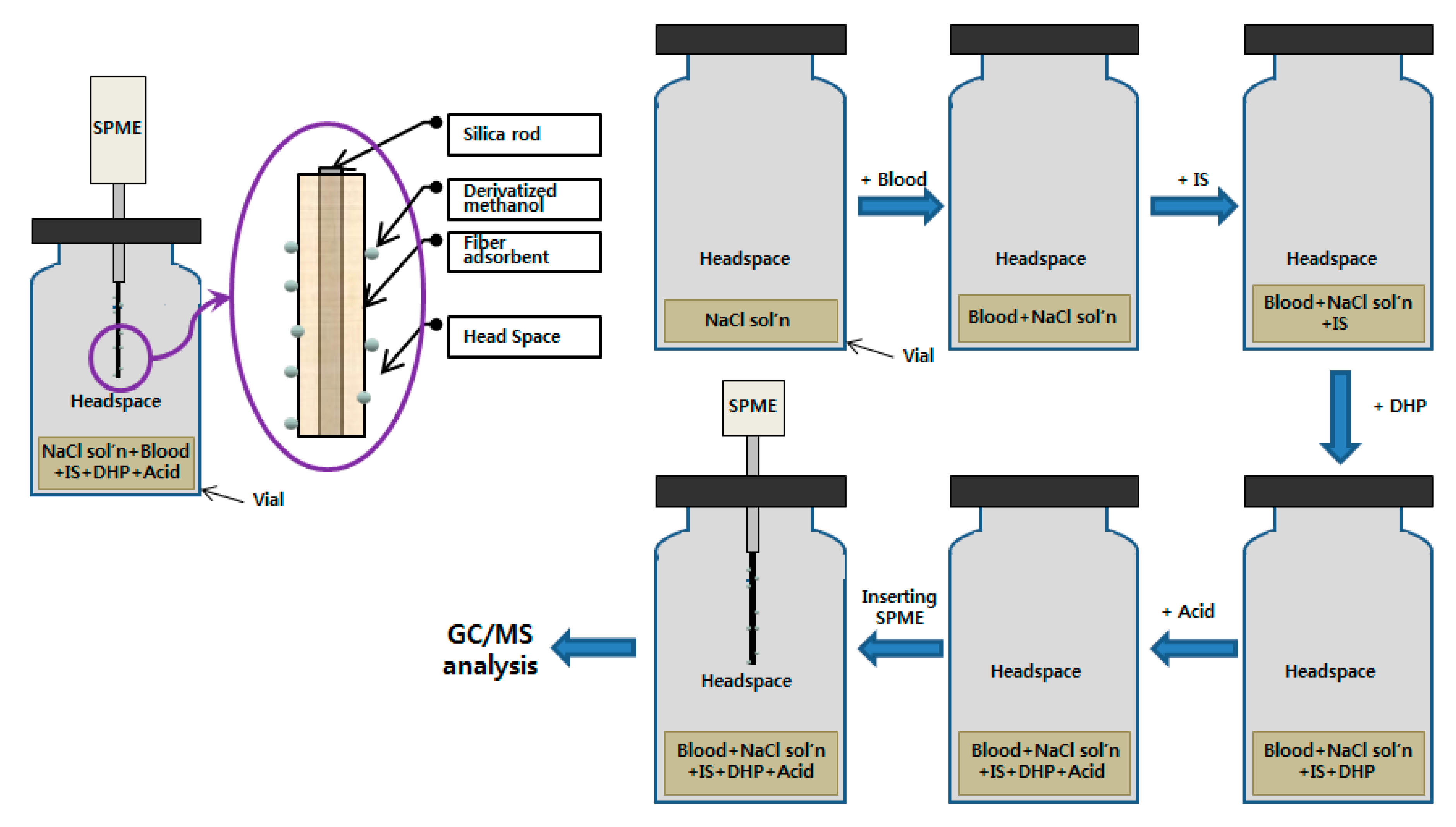
2.3. Extraction
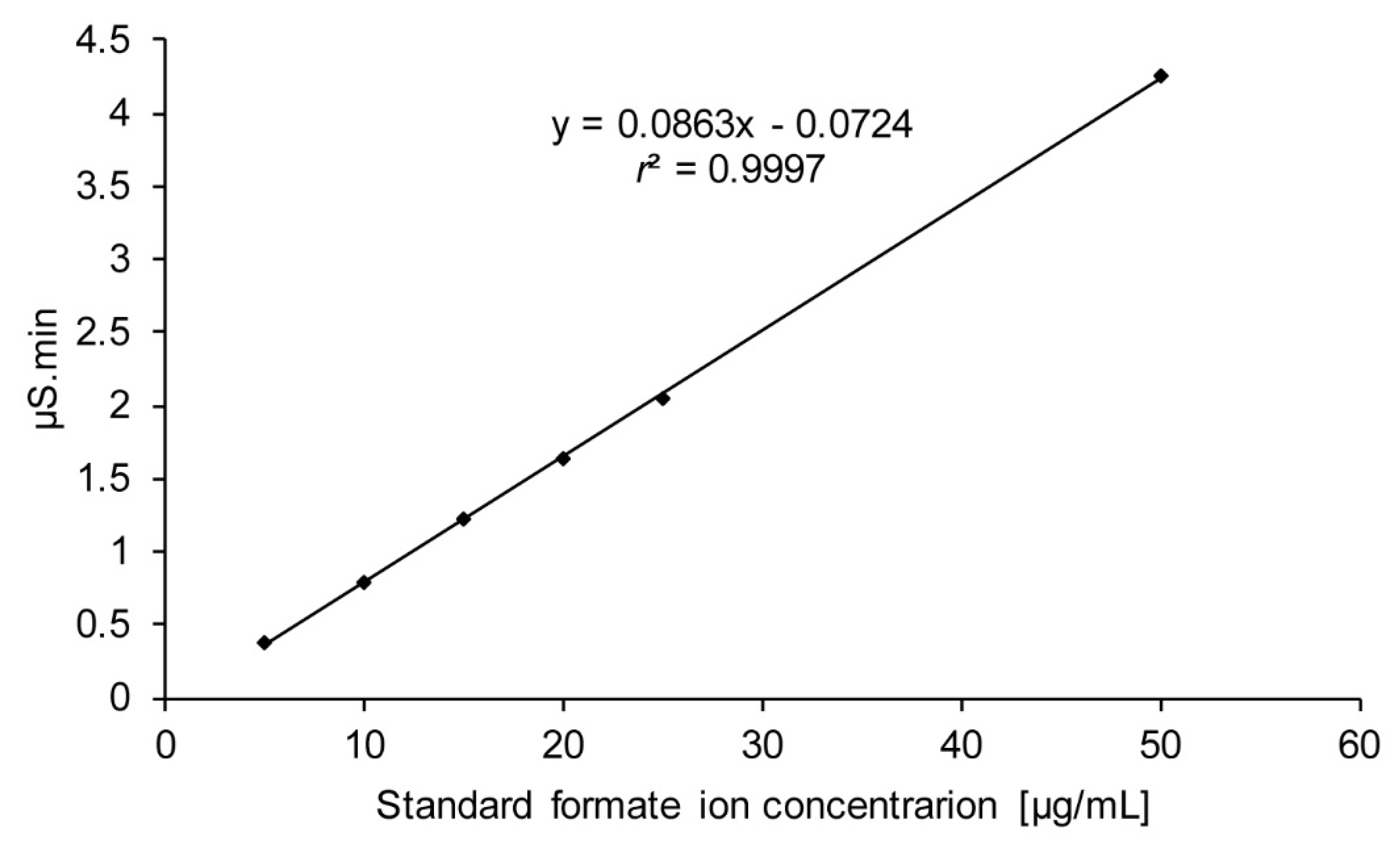
2.4. IC Analysis
3. Materials and Methods
3.1. Chemicals and Reagents
3.2. Instruments
3.3. Sample Preparation
4. Conclusions
Author Contributions
Funding
Conflicts of Interest
References
- Tephly, T.R. The toxicity of methanol. Life Sci. 1991, 48, 1031–1041. [Google Scholar] [CrossRef]
- McMartin, K.E.; Martin-Amat, G.; Makar, A.B.; Tephly, T.R. Methanol poisoning: Role of formate metabolism in the monkey. In Alcohol and Aldehyde Metabolizing Systems, 1st ed.; Thurman, R.G., Drott, H.R., Williamson, J.R., Chance, B., Eds.; Academic Press Inc.: Philadelphia, PA, USA, 1977; Volume 2, pp. 429–440. [Google Scholar]
- Srinivasan, S.; KumariDubey, k.; Singhal, R.S. Influence of food commodities on hangover based on alcohol dehydrogenase and aldehyde dehydrogenase activities. Curr. Res. Nutr. Food Sci. 2019, 1, 8–16. [Google Scholar] [CrossRef]
- Kapur, B.M.; Baber, M. FASD: Folic acid and formic acid - an unholy alliance in the alcohol abusing mother. Biochem. Cell Biol. 2018, 96, 189–197. [Google Scholar] [CrossRef] [PubMed]
- Baselt, R.C. Disposition of Toxic Drugs and Chemicals in Man, 11th ed.; Biomedical Publications: Seal Beach, CA, USA, 2017; pp. 1326–1327. [Google Scholar]
- Frenia, M.L.; Schauben, J.L. Methanol inhalation toxicity. Ann. Emerg. Med. 1993, 22, 1919–1923. [Google Scholar] [CrossRef]
- Karadeniz, H.; Birincioglu, I. Methyl alcohol poisoning in Trabzon (Turkey). J. Forensic Sci. 2011, 56, 822–824. [Google Scholar] [CrossRef] [PubMed]
- Jeffrey, B.; Kenneth, M.; Scott, P.; Cynthia, A.; Ken, K. Fomepizole for the Treatment of Methanol Poisoning. N. Engl. J. Med. 2001, 344, 424–429. [Google Scholar]
- Pappas, S.C.; Silverman, M. Treatment of methanol poisoning with ethanol and hemodialysis. Can. Med. Assoc. J. 1982, 126, 1391–1394. [Google Scholar] [PubMed]
- Smith, M.E. Interrelations in Ethanol and Methanol metabolism. J. Pharmacol. Exp. Ther. 1961, 134, 233–237. [Google Scholar] [PubMed]
- Bursová, M.; Hložek, T.; Čabala, R. Simultaneous Determination of Methanol, Ethanol and Formic Acid in Serum and Urine by Headspace GC-FID. J. Anal. Toxicol. 2015, 39, 741–745. [Google Scholar] [CrossRef] [PubMed]
- Kaya, S.; Mergen, G.; Dural, E.; Aliyev, V.; Yalçin, S.; Söylemezoglu, T.; Kayaalti, Z. Simultaneous Headspace-GC–FID Analysis for Methanol and Ethanol in Blood, Saliva and Urine: Validation of Method and Comparison of Specimens. LC GC Eur. 2011, 24, 292–298. [Google Scholar]
- Abirami, P.; Rajendran, A. GC-MS analysis of methanol extracts of Vernonia cinerea. Euro. J. Exp. Bio. 2012, 2, 9–12. [Google Scholar]
- Schauer, N.; Steinhauser, D.; Strelkov, S.; Schomburg, D.; Allison, G.; Moritz, T.; Lundgren, K.; Roessner-Tunali, U.; Forbes, M.G.; Willmitzer, L.; et al. GC–MS libraries for the rapid identification of metabolites in complex biological samples. FEBS Lett. 2005, 579, 1332–1337. [Google Scholar] [CrossRef] [PubMed]
- Visser, A.E.; Swatloski, R.P.; Griffin, S.T.; Hartman, D.H.; Rogers, R.D. Liquid/Liquid Extraction of Metal Ions in Room Temperature Ionic Liquids. Sep. Sci. Technol. 2001, 36, 785–804. [Google Scholar] [CrossRef]
- Berrueta, L.A.; Gallo, B.; Vicente, F. A review of solid phase extraction: Basic principles and new developments. Chromatographia 1995, 40, 474–483. [Google Scholar] [CrossRef]
- Schummer, C.; Delhomme, O.; Appenzeller, B.; Wennig, R.; Millet, M. Comparison of MTBSTFA and BSTFA in derivatization reactions of polar compounds prior to GC/MS analysis. Talanta 2009, 77, 1473–1482. [Google Scholar] [CrossRef] [PubMed]
- Ouyang, G.; Pawliszyn, J. Recent developments in SPME for on-site analysis and monitoring. TrAC Trend Anal. Chem. 2008, 25, 692–703. [Google Scholar] [CrossRef]
- Hakkarainen, M. Developments in multiple headspace extraction. J. Biochem. Biophys. Method. 2007, 70, 229–233. [Google Scholar] [CrossRef] [PubMed]
- Lin, D.L.; Chang, W.T.; Kuo, T.L.; Liu, R.H. Chemical Derivatization and the Selection of Deuterated Internal Standard for Quantitative Determination—Methamphetamine Example. J. Anal. Toxicol. 2000, 24, 275–280. [Google Scholar] [CrossRef] [PubMed]
- Sporkert, F.; Pragst, F. Use of headspace solid-phase microextraction (HS-SPME) in hair analysis for organic compounds. Forensic Sci. Int. 2000, 107, 129–148. [Google Scholar] [CrossRef]
- Popp, P.; Paschke, A. Solid phase microextraction of volatile organic compounds using carboxen-polydimethylsiloxane fibers. Chromatographia 1997, 46, 419–424. [Google Scholar] [CrossRef]


© 2019 by the authors. Licensee MDPI, Basel, Switzerland. This article is an open access article distributed under the terms and conditions of the Creative Commons Attribution (CC BY) license (http://creativecommons.org/licenses/by/4.0/).
Share and Cite
Lee, J.-B.; Jeong, Y.A.; Ahn, D.J.; Bang, I.S. SPME-GC/MS Analysis of Methanol in Biospecimen by Derivatization with Pyran Compound. Molecules 2020, 25, 41. https://doi.org/10.3390/molecules25010041
Lee J-B, Jeong YA, Ahn DJ, Bang IS. SPME-GC/MS Analysis of Methanol in Biospecimen by Derivatization with Pyran Compound. Molecules. 2020; 25(1):41. https://doi.org/10.3390/molecules25010041
Chicago/Turabian StyleLee, Joon-Bae, Yong Ae Jeong, Dae Jun Ahn, and Iel Soo Bang. 2020. "SPME-GC/MS Analysis of Methanol in Biospecimen by Derivatization with Pyran Compound" Molecules 25, no. 1: 41. https://doi.org/10.3390/molecules25010041
APA StyleLee, J.-B., Jeong, Y. A., Ahn, D. J., & Bang, I. S. (2020). SPME-GC/MS Analysis of Methanol in Biospecimen by Derivatization with Pyran Compound. Molecules, 25(1), 41. https://doi.org/10.3390/molecules25010041