Article

The Biochemical and Functional Characterization of M28 Aminopeptidase Protein Secreted by Acanthamoeba spp. on Host Cell Interaction

by
Jian-Ming Huang
1,†,
Yao-Tsung Chang
2,† and
Wei-Chen Lin
1,3,4,*
1
Institute of Basic Medical Sciences, College of Medicine, National Cheng Kung University, Tainan 701, Taiwan
2
Department of Biochemistry and Molecular Biology, College of Medicine, National Cheng Kung University, Tainan 701, Taiwan
3
Department of Parasitology, College of Medicine, National Cheng Kung University, Tainan 701, Taiwan
4
Department of Microbiology and Immunology, College of Medicine, National Cheng Kung University, Tainan 701, Taiwan
*
Author to whom correspondence should be addressed.
These authors contributed equally to this work.
Molecules 2019, 24(24), 4573; https://doi.org/10.3390/molecules24244573
Submission received: 30 October 2019 / Revised: 11 December 2019 / Accepted: 12 December 2019 / Published: 13 December 2019
(This article belongs to the Special Issue Design and Application of Metal-Binding Proteins)

Abstract

:
Acanthamoeba are a free-living protozoan whose pathogenic strain can cause severe human diseases, such as granulomatous encephalitis and keratitis. As such, the pathogenic mechanism between humans and Acanthamoeba is still unknown. In our previous study, we identified the secreted Acanthamoeba M28 aminopeptidase (M28AP) and then suggested that M28AP can degrade human C3b and iC3b for inhibiting the destruction of Acanthamoeba spp. with the human immune response. We constructed the produced the recombinant M28AP from a CHO cell, which is a mammalian expression system, to characterize the biochemical properties of Acanthamoeba M28AP. The recombinant M28AP more rapidly hydrolyzed Leu-AMC than Arg-AMC and could be inhibited by EDTA treatment. We show that recombinant M28AP can be delivered into the individual cell line and cause cell line apoptosis in a co-culture model. In conclusion, we successfully investigated the potential molecular characteristics of M28AP.

1. Introduction

Acanthamoeba spp. is a free-living pathogenic protozoan that causes severe sight-threatening infection, such as granulomatous amoebic encephalitis (GAE) and Acanthamoeba keratitis [1,2]. Acanthamoeba spp. is widely distributed in the environment, including in lakes, pools, soil, and dust [3]. The Acanthamoeba spp. life cycle includes both motile trophozoites and dormant cysts. The trophozoite feeds on a variety of organic materials, such as bacteria via phagocytosis. The dormant cyst stage has double walls to resist and survive in harsh environmental conditions [4]. Free-living amoebae, such as Naegleria spp., Sappinia spp., Acanthamoeba spp., and Balamuthia spp., are known to cause fatal human central nervous system (CNS) infections. Headache, fever, nausea, vomiting, stiff neck, photophobia, confusion, and death are included as symptoms [5]. Acanthamoeba spp. infecting the human CNS may result in GAE through the blood–brain barrier [6]. Acanthamoeba spp. causes the amoebae invasion of the alveolar blood vessels through the respiratory tract. Patients undergoing immunosuppressive therapy or excessively use steroids are at risk of GAE. A previous a case report showed that GAE was caused by Acanthamoeba spp. in a patient with kidney transplant [7].
Acanthamoeba keratitis (AK) is a rare and severe parasitic infection ocular infection that results from free-living pathogenic Acanthamoeba spp. [1,2]. However, AK has increased with the widespread use of contact lenses over the past two decades [3,8,9,10]. Acanthamoeba spp. infects patients wearing contact lenses over a long period of time through corneal injury [11]. Patients suffering from AK may experience lid edema, photophobia, epithelial defect, and ring-like stromal infiltrate [12]. AK has serious consequences, such as loss of vision if not treated adequately and immediately, due to difficulty in early diagnosis and poor response to most antibiotics [5,13,14,15,16]. Therefore, the mechanism through which the virulence factors of Acanthamoeba spp. cause tissue invasion or infection in these patients must be elucidated.
Previous studies reported that aminopeptidase could play important roles in infections. Secreted matrix metalloproteases are synthesized as proenzymes into the extracellular space and they can be activated by many mechanisms, including normal physiology and pathology during development, wound healing, and cancer cell metastasis [17,18]. In parasites, an aspartyl aminopeptidase-like gene has been identified in the Toxoplasma gondii genome that affects the growth of T. gondii via parasite replication and growth [19]. GP63, which is a parasite metalloprotease of Leishmania species, is known in the cleavage and degradation of the activity of the complement system to avoid the human immune system [20]. An M17 leucine aminopeptidase of Acanthamoeba castellanii plays an important role during the encystation of Acanthamoeba spp. [21]. Acanthamoeba spp. produces cysteine, serine, and metalloproteases, which are increased in pathogenic Acanthamoeba strains [22,23].
We previously showed that the M20/M25/M40 superfamily aminopeptidase protein, which is an aminopeptidase found in Acanthamoeba-secreted proteins (Asp), causes cell damage and disruption [24]. We also identified a novel protein that was secreted by Acanthamoeba spp., an M28 aminopeptidase (M28AP), as a target of the human innate immune defense [25,26]. However, little is known regarding the biochemical and functional characterization of M28AP. Aminopeptidases, which are also called metalloproteases, are a highly diverse set of proteolytic enzymes that catalyze the cleavage of amino acids. Aminopeptidases distribute across homologous protease families M1 through M91 [27]. Metalloprotease has a conserved HEXXH active site motif that is mediated by one or two divalent ions, often zinc. The conserved HEXXH active site motif activates the nucleophilic attack of water molecules on substrate peptide bonds [28]. A previous study showed that families of metalloproteases could be divided into five groups. The first set has a glutamic acid residue at the metal-binding site (HEXXH+E), such as M1, M2, M4, M5, and M13. The second set has a third histidine residue at the metal-binding site (HEXXH+H), such as M7, M10, M11, and M12. The third set has an additional metal-binding site (HEXXH) that is yet unidentified, such as M3, M6, M8, M9, M26, M27, M30, M31, and M12. The fourth group is bound at motifs other than HEXXH, for example, M14, M15, M16, M17, and M24. The final group of families is until unknown in the metal ligands, which includes M18, M19, M20, M22, M23, M25, M28, M29, and M33 [29]. In the present study, we found that recombinant M28AP that was purified from CHO cells had aminopeptidase activity to hydrolyze a highly sensitive substrate. As metalloprotease, the recombinant M28AP was also inhibited by EDTA treatment. In a co-culture model, the recombinant M28AP can be delivered into individual cell line and cause cell line apoptosis in vitro.

2. Results

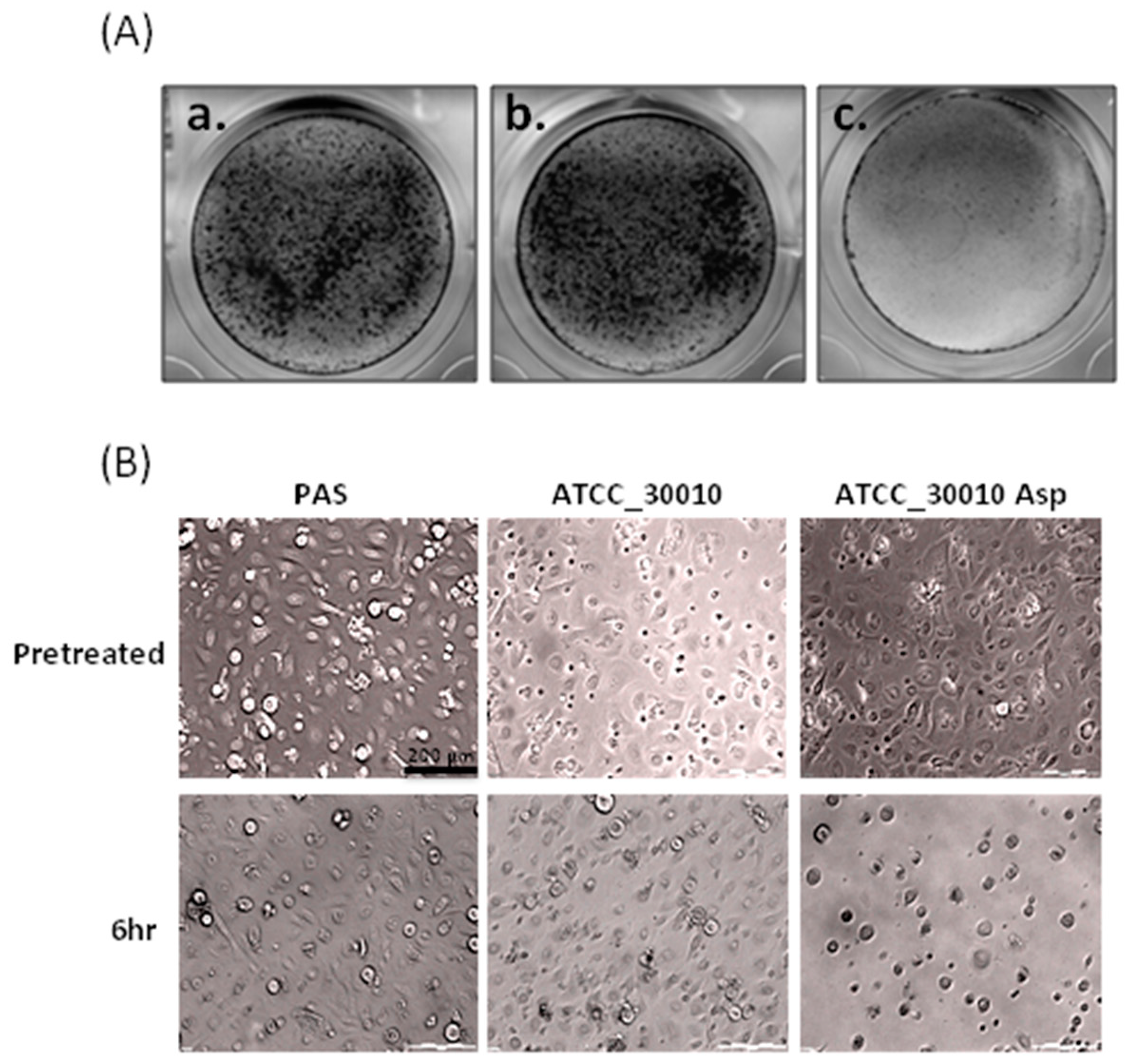
2.1. The Virulence of Asp Induces Cell Damage in Human Primary Corneal Epithelial Cells

We chose a suitable strain and corrected the damage-induced duration for observation while using CPE assays to clarify the pathogenesis mechanisms of Acanthamoeba spp. The results show that no significant change occurred in the cell coverage area between the corneal epithelial cells that were co-incubated six hours with the Acanthamoeba spp. and control (mock) (Figure 1A_a,b). We isolated the Asp of Acanthamoeba spp. to treat the corneal epithelial cells while using a CPE assay to investigative whether Asp are involved the cell disruption process of Acanthamoeba spp. The results of the CPE assay showed that Asp of Acanthamoeba spp. could disrupt the co-incubated corneal epithelial cells (Figure 1A_c). We observed that Asp of Acanthamoeba spp. induced the loss of cell adhesion ability in a time-dependent manner via microscopy (Figure 1B). We suggested that Asp of Acanthamoeba spp. might have the important virulence factors to affect the cell line.

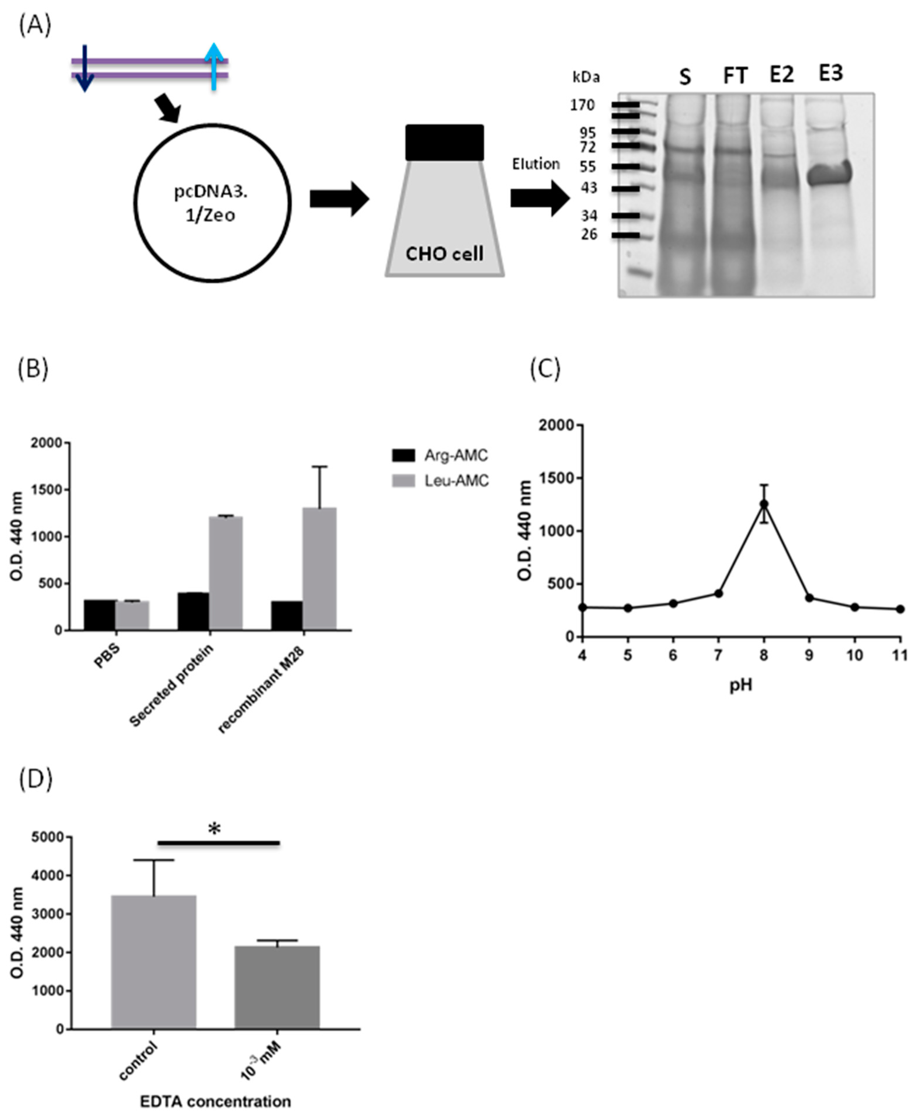
2.2. Biochemical Characterization of M28AP

In a previous study, we established the extracellular secreted proteomic database of Acanthamoeba spp. [25]. We found a novel protein secreted by Acanthamoeba spp., M28AP, from the extracellular secreted proteomic database as a target of the human innate immune defense system [26]. We purified the recombinant M28AP from CHO cells to characterize the biochemical properties of M28AP (Figure 2A). We used the recombinant M28AP and collected Asp of Acanthamoeba spp. that contained extracellular vesicles and soluble proteins to treat Leu-AMC and Arg-AMC to detect whether the recombinant M28AP from CHO cells is active. The results showed that Asp and the recombinant M28AP more rapidly hydrolyzed Leu-AMC than Arg-AMC, which a highly sensitive substrate for aminopeptidase (Figure 2B). The recombinant M28AP showed aminopeptidase activity in the approximately optimal pH range (pH 7.0–9.0), with a maximum at pH 8.0 (Figure 2C). Metal chelators and EDTA were used for the inhibition of M28AP enzymatic activity and stability, since M28AP is a metalloenzyme. The result showed that recombinant M28AP treated with EDTA at concentrations of 10−3 mM inhibited approximately 40% of the enzymatic activity when compared with the control (Figure 2D).

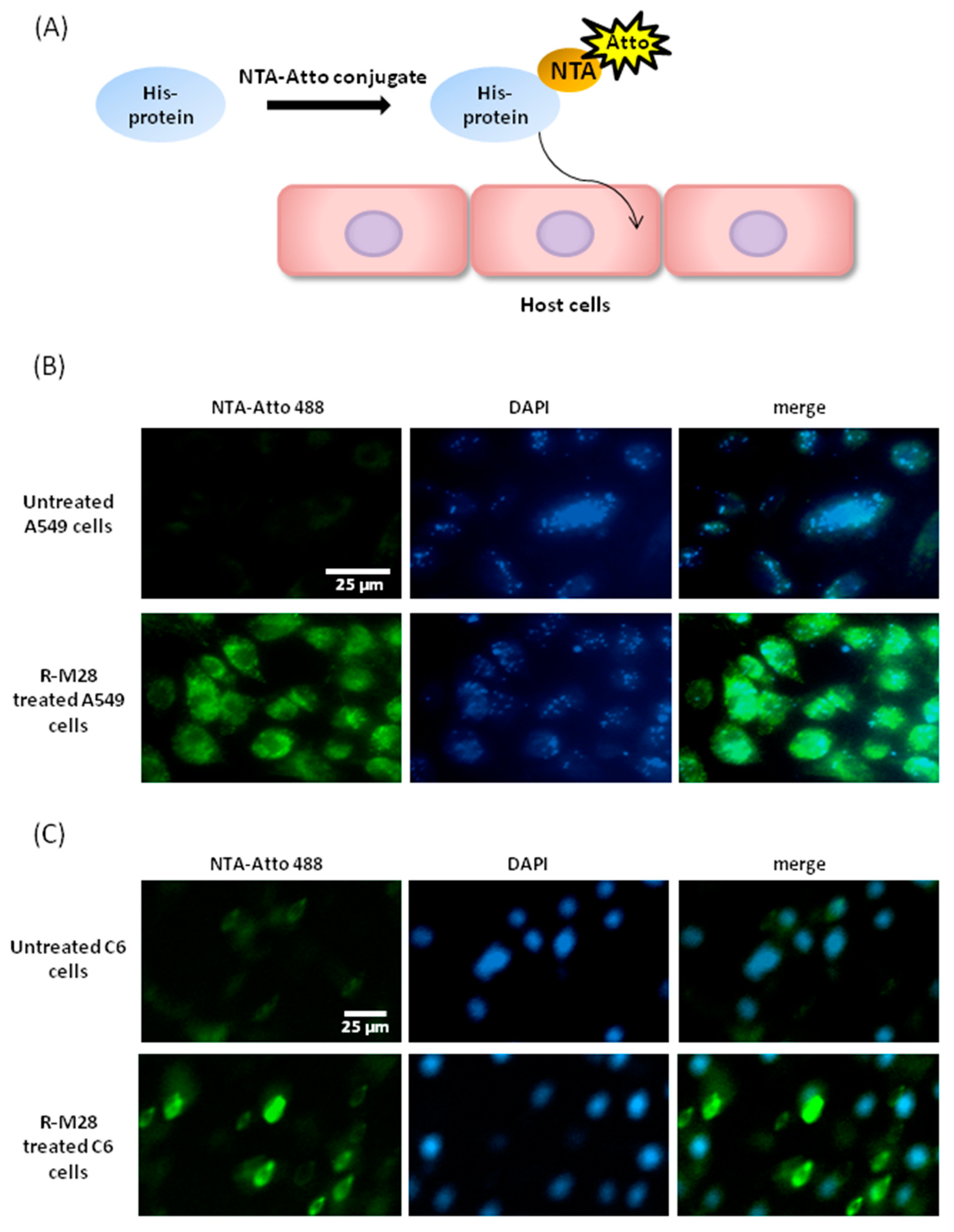
2.3. The Role of M28Ap as Virulence Factor and Host Cell Damage

We used NTA-Atto conjugated with his-tag protein, the recombinant M28AP, to observe M28AP host–cell interaction to determine whether the secreted protein, M28AP, was delivered into individual cell line (Figure 3A). In this study, we substituted the C6 and A549 cell lines for human primary corneal epithelial cells, because primary cells have a limited lifespan and are harder to culture than these cell lines. The results showed that the recombinant M28AP was delivered into the individual cell line and not only the C6 and A549 cells after four hours of incubation (Figure 3B,C). We suggest that the M28AP of Acanthamoeba spp. is secreted and delivered into cell line to cause cell damage.
An Annexin V assay kit was used to detect apoptotic cells to investigative whether the M28AP of Acanthamoeba spp. causes cell line damage. The result showed that C6 cells that were treated with recombinant M28AP were both Annexin V and PI positive (Figure 4). Hence, we suggest that M28AP of Acanthamoeba spp. may act as a virulence factor to cause late cell apoptosis and cell death. However, from the results, we observed that M28AP caused minor apoptosis of C6 cells, although the apoptotic cells were detected while using the Annexin V assay. Therefore, we suggest that Asp contain many different proteases and M28AP might be a minor virulence factor that causes cell line damage via apoptotic pathways.

3. Discussion

3.1. Biochemical Characterization of M28AP

Aminopeptidases, which are also called metalloproteases, are a highly diverse set of proteolytic enzymes that catalyze the cleavage of amino acids and are distributed across homologous protease families M1 through M91 [27]. Metalloproteases cleave a peptide bond with a hydrophobic side chain, such as Leu, Ile, Met, Phe, or Tyr. These hydrophobic residues fit into the specificity pocket of the different aminopeptidase [30]. The matrix metalloproteases 2 and 7 of Bacillus cereus were cleaved with high efficiency at the Leu–Gly or Leu–Ala bond by the residue in the P1′ position [31]. A previous study showed that the M20/M25/M40 superfamily aminopeptidase protein, an aminopeptidase found in Asp can be hydrolyzed Leu-AMC more rapidly than Arg-AMC [24]. In this study, the results showed that Asp more rapidly hydrolyzed Leu-AMC than Arg-AMC, as well as the recombinant M28AP (Figure 2B). These results aligned with the secreted proteomics analysis, which proved that Asp contain the M28AP, because the recombinant M28AP hydrolyzed Leu-AMC, which matches the Asp hydrolyzed Leu-AMC subtracts [25].
The normal pH range of human tears is 6.5 to 7.6; the mean value is 7.0 [32]. The normal human blood plasma pH value is 7.4 [33]. The adaptation to pH allows for human pathogens to invade the tissues and bloodstream to cause severe infections [34]. AK and GAE are severe sight-threatening infections that result from the free-living pathogenic protozoans Acanthamoeba spp. [1,2,5]. Recombinant M28AP showed aminopeptidase activity in the approximately optimal pH range (pH 7.0–9.0), with maximum activity at pH 8.0 (Figure 2C). The human blood plasma and tears may act as suitable buffer solutions for the Acanthamoeba spp. to secrete the active M28AP. Hence, we suggested that the M28AP has high virulence to cause host different damage, such as complement degradation in these buffer solutions, not only human tear, but blood plasma.

3.2. Cell Line Apoptosis by M28AP

In this study, the CPE assay results showed that Asp of Acanthamoeba spp. could disrupt the co-incubated corneal epithelial cells (Figure 1A_c). A previous study showed that the M20/M25/M40 superfamily aminopeptidase protein, an aminopeptidase that is found in Asp, causes cell line disruption after Acanthamoeba spp. and C6 cell co-culturing [24]. In general, the cell line causes organelle stress and induces apoptosis pathways due to parasite infection [35]. Another study showed that the two main apoptotic pathways are the extrinsic and intrinsic pathways [36]. These pathways are activated by the cleavage of caspase-3, causing cell DNA fragmentation, the formation of apoptotic bodies, and the degradation of cytoskeletal and nuclear proteins [37]. Mannose-induced protein 133 (MIP 133), proteases secreted by Acanthamoeba spp., causes contact-independent cytolysis of corneal epithelial cells, and is able to penetrate corneal tissue [38]. Soluble metabolites that are released by Acanthamoeba spp. lead to human monocyte cell death through apoptosis [23]. In general, phosphatidylserine is hidden within the plasma membrane on the cytoplasmic face of the membrane. However, during cell apoptosis, phosphatidylserine is translocated to the cell surface. Annexin V is a Ca2+-dependent protein that has high affinity for phosphatidylserine, so it plays a role as a probe for detecting apoptosis. Propidium iodide (PI) is a fluorescent dye that binds the DNA through the membrane of late apoptotic cells and dead cells. Our results showed that C6 cells are both Annexin V- and PI-positive, because the recombinant M28AP was delivered into the individual cell line (Figure 4). The cells that were treated with the recombinant M28AP were in late apoptosis or already dead. Hence, these results showed that M28AP that was secreted by Acanthamoeba spp. induced the apoptosis of cell line.

4. Materials and Methods

4.1. Culture of Acanthamoeba Strains

ATCC_30010, non-pathogenic Acanthamoeba strain, were axenically cultured in protease peptone-yeast extract-glucose (PYG) medium (pH 6.5) at 28 °C in cell culture T75 flasks [39]. Trophozoites were harvested in the logarithmic growth phase after cultivation for 3–5 days to 80%. The Acanthamoeba cell passaging was washed three times and then resuspended in Page’s modified Neff’s amoeba saline (PAS) buffer [40].

4.2. Culture of C6 Glioma Rat Cell Line, A549 Human Cell Line and Human Primary Corneal Epithelial Cells

C6 glioma rat cell line and A549 human cell line were axenically cultured in Dulbecco’s Modified Eagle Medium (DMEM) High Glucose medium (GibcoTM, Thermo, Waltham, MA, USA) at 37 °C with 5% CO2 in cell culture flasks. Cell passaging was washed in phosphate buffered saline (PBS). Human primary corneal epithelial cells were axenically cultured in the EpiGRO™ Human Epidermal Keratinocyte Complete Culture Media Kit (Merck Millipore, Darmstadt, Germany).

4.3. Isolation of Secreted Proteins

Acanthamoeba spp., at an approximate density of 1.0 × 10⁶ parasites/mL, were washed three times and resuspended in PAS buffer for 4 h, after which the parasites were removed by centrifugation. The cell-free media containing secreted proteins was filtered through an Amicon® Ultra-4 3K Centrifugal Filter Unit (Merck Millipore, Darmstadt, Germany) and then collected by centrifugation at 959× g for 75 min [24]. The protein concentration was measured with an ND-1000 (NanoDrop, Thermo, Waltham, MA, USA) and a protein assay (Bio-Rad, Hercules, CA, USA).

4.4. Cytopathic Effect Assay (CPE) and Giemsa Stain

Human primary corneal epithelial cells were axenically cultured in an EpiGRO™ Human Epidermal Keratinocyte Complete Culture Media Kit (Merck Millipore, Darmstadt, Germany). The human primary corneal epithelial cells were cultured to 3 × 10⁵ cells/mL in 24-well plate cell culture dishes. After 24 h of growth, Acanthamoeba live cells at a concentration of 3 × 10⁵ were co-cultured with human primary corneal epithelial cells. Acanthamoeba spp., at a density of 1.0 × 10⁶ parasites/mL in PBS for 4 h, as well as their secreted proteins, were collected. The Acanthamoeba-secreted proteins (150 μg) were co-incubated with human primary corneal epithelial cells for 2 h, followed by microscopy observations and CPE assays. The cells were fixed in wells with methanol with acetic acid at 3:1 for 15 min. After air drying, we added 1 mL Giemsa Buffer:Giemsas Azur-Eosin-Methylenblaulosung stain buffer (9:1) in a fixed well for 15 min. The wells were rinsed three times with ddH2O and then left to air dry.

4.5. Total RNA Isolation and cDNA Synthesis

We used a Total RNA Extraction Miniprep System (VIOGENE, Taiwan) to isolate RNA. High Capacity cDNA Reverse Transcription Kits (Thermo, Waltham, MA, USA) were used in this study. The kit components were allowed to thaw on ice. The reverse transcription conditions were set at the following times and temperatures: 25 °C for 10 min, 37 °C for 120 min, and 85 °C for 5 min; finally, the cDNA was stored at 4 °C. The reaction volume was 20 μL.

4.6. DNA Construction, Cell Culture and Transient Transfection

The M28AP sequence was amplified while using PCR with Acanthamoeba spp. cellular cDNA as the template. Forward M28AP_clon_BsrGI_F (5′-GGA TGT ACA GCA TGG TCG CTC ACG GAA GG-3′) and reverse M28AP_clon_EcoRI_stop_His_R (5′-CCG GAA TTC TCA ATG GTG ATG GTG ATG GTG CAG AGG ATC GGC GAA-3′) primers were used to amplify the cDNA encoding M28AP. The cleavage sites for the BsrGI and EcoRI restriction enzymes were incorporated at the 5′ ends of the primers for cloning into the pcDNA3.1/Zeo expression plasmid (Thermo, Waltham, MA, USA). A ratio of 1.0 μg plasmid DNA/mL of transfection culture volume was used for ExpiCHO transfection and transfection was performed at the 100 mL scale in 500 mL non-baffled flasks. The cultures were maintained at the indicated rpm on a shaker with a 19 mm throw. Cell culture and transient transfection were accomplished by following previously described protocols with minor modifications [41]. The ExpiCHO-S cells were cultured in ExpiCHO Expression medium (Thermo, Waltham, MA, USA) in a humidified 8% CO2 incubator at 36.5 °C and 125 rpm. Cells were split to 1~2 × 10⁶ cells/mL in each well the day prior to transfection. After 48 h, cells were >10 × 106 cells/mL and they were diluted to 6.5 × 10⁶ cells/mL with the addition of fresh ExpiCHO expression medium. ExpiCHO transfection was performed using the ExpiCHO Expression System Kit (Thermo, Waltham, MA, USA), according to the manufacturer’s protocol. Briefly, ExpiFectamine transfection reagent and M28AP plasmid were separately diluted in OptiPRO SFM (Thermo, Waltham, MA, USA). ExpiFectamine and DNA mixtures were immediately combined and we waited for up to 5 min. The ExpiFectamine/plasmid complexes were then added to the cells. For transfection completed with the Max Titer Protocol, enhancer and 8% v/v feed were added 22~24 h post-transfections; the cultures were then temperature shifted to 32 °C and an additional 8% v/v feeds were added on days 3, 5, and 7 post-transfection. ExpiCHO Max Titer Protocol transfection was harvested on day 11 post-transfection. The cell cultures were centrifuged at 5000 rpm for 15 min at 4 °C and the supernatants were collected for purification.

4.7. SDS-PAGE and Coomassie Blue Staining

All of the protein samples were separated with 12% SDS-PAGE (T-Pro, Taiwan). SDS-PAGE running buffer consists of 3.0 g of Tris base, 14.4 g of glycine, and 1.0 g of SDS in 1000 mL of H2O. Following electrophoresis, the gels were placed in a solution of 50% methanol and 30% acetic acid containing distilled water for 2 h and were then placed in a solution of 30% methanol containing distilled water for 30 min to remove the acetic acid. Coomassie Blue stain was added to the gel until the protein band was detected while using a scanner (Epson, Taiwan).

4.8. Biochemical Properties of Recombinant Protein M28AP

The biochemical properties of recombinant protein M28AP, while using PBS buffer as the control and the activity of Asp, was assayed by the hydrolysis of L-leucine-7-amido-4-methylcoumarin hydrochloride (Leu-AMC) and l-arginine-AMC (Arg-AMC, Sigma-Aldrich, st. Louis, MO, USA). We added approximately 1 μg/mL the recombinant protein M28AP solution to 195 μL of assay buffer (50 mM Tris-HCl, pH 8.0) containing 10 μM Leu-AMC or Arg-AMC up to 200 μL, which was then incubated for 30 min at 37 °C. The release of fluorescence was measured at an excitation wavelength of 370 nm and an emission wavelength of 440 nm while using a FlexStation 3 Multi-Mode microplate reader (Molecular Devices, Sunnyvale, CA, USA) [21].

4.9. Ni-NTA-Atto Conjugates for Recombinant Protein M28AP- Cell Line Interaction

Ni-NTA-Atto conjugates provide specific and highly sensitive detection of His-tagged fusion proteins. The Ni-NTA-Atto conjugates were incubated with recombinant protein M28AP (50 μg) at 37 °C for 30 min. After incubation, the Ni-NTA-Atto-M28AP complex was added to C6 and A549 cells for 4 h. After washing three times with PBS, a CellR microscope (Olympus CellR, Tokyo, Japan) was used for light and fluorescence microscopy. Image analysis was performed with CellR software (Olympus CellR, Tokyo, Japan).

4.10. Annexin V Apoptosis Assay

C6 cells, with an approximate density of 1.0 × 10⁵ cells/mL, were seeded overnight. Recombinant protein M28AP (14 μg) was added to C6 cells at 37 °C for 24 h. Annexin V-FAM + PI Apoptosis Detection Reagent (Leadgen, Taiwan) were used. We mixed 5 μL of Annexin V- FAM and 5 μL of PI with 500 μL binding buffer to C6 cells, which were then gently mixed and incubated for 15~20 min. at 37 °C in the dark. After washing three times with PBS, a CellR microscope (Olympus CellR, Tokyo, Japan) was used for light and fluorescence microscopy. CellR software was used to analyze the images.

Author Contributions

W.-C.L. conceived and designed the experiments; J.-M.H. and Y.-T.C. performed the experiments; W.-C.L. and J.-M.H. analyzed the data; and J.-M.H. and W.-C.L. wrote the paper.

Funding

This research was supported by the Ministry of Science and Technology (MOST) to WCL (grant MOST 106-2320-B-006-070-MY3).

Acknowledgments

We acknowledge Professor Woei-Jer Chuang and Jyh-Wei Shin for providing reagents/materials/analysis tools.

Conflicts of Interest

All authors declare no conflicts of interest.

References

  1. McCulley, J.P.; Alizadeh, H.; Niederkorn, J.Y. Acanthamoeba Keratitis. Eye. Contact. Lens. 1995, 21, 73–76. [Google Scholar]
  2. Visvesvara, G.S.; Moura, H.; Schuster, F.L. Pathogenic and Opportunistic Free-Living Amoebae: Acanthamoeba spp., Balamuthia Mandrillaris, Naegleria Fowleri, and Sappinia Diploidea. FEMS Immunol. Med. Mic. 2007, 50, 1–26. [Google Scholar] [CrossRef] [PubMed] [Green Version]
  3. Brown, T.; Cursons, R.; Keys, E. Amoebae from Antarctic Soil and Water. Appl. Environ. Microbiol. 1982, 44, 491–493. [Google Scholar]
  4. PAGE, F.C. Re-Definition of the Genus Acanthamoeba with Descriptions of Three Species. J. Protozool. 1967, 14, 709–724. [Google Scholar] [CrossRef]
  5. Marciano-Cabral, F.; Cabral, G. Acanthamoeba spp. as Agents of Disease in Humans. Clin. Microbiol. Rev. 2003, 16, 273–307. [Google Scholar] [CrossRef] [Green Version]
  6. Martinez, A.J.; Visvesvara, G.S. Free-living, Amphizoic and Opportunistic Amebas. Brain. Pathol. 1997, 7, 583–598. [Google Scholar] [CrossRef]
  7. Salameh, A.; Bello, N.; Becker, J.; Zangeneh, T. Fatal granulomatous amoebic encephalitis caused by Acanthamoeba in a patient with kidney transplant: A case report. Open Forum Infect. Dis. 2015, 2, ofv104. [Google Scholar] [CrossRef]
  8. Broxton, P.; Woodcock, P.; Gilbert, P. A study of the Antibacterial Activity of Some Polyhexamethylene Biguanides towards Escherichia Coli ATCC 8739. J. Appl. Bacteriol. 1983, 54, 345–353. [Google Scholar] [CrossRef]
  9. Auran, J.D.; Starr, M.B.; Jakobiec, F.A. Acanthamoeba Keratitis. Cornea 1987, 6, 2–26. [Google Scholar] [CrossRef]
  10. Allen, M.J.; Morby, A.P.; White, G.F. Cooperativity in the Binding of the Cationic Biocide Polyhexamethylene Biguanide to Nucleic Acids. Biochem. Biophys. Res. Commun. 2004, 318, 397–404. [Google Scholar] [CrossRef]
  11. Li, M.-L.; Shih, M.-H.; Huang, F.-C.; Tseng, S.-H.; Chen, C.-C. Treatment of early Acanthamoeba Keratitis with Alcohol-Assisted Epithelial Debridement. Cornea 2012, 31, 442–446. [Google Scholar] [CrossRef] [PubMed]
  12. Lorenzo-Morales, J.; Khan, N.A.; Walochnik, J. An Update on Acanthamoeba Keratitis: Diagnosis, Pathogenesis and Treatment. Parasite 2015, 22, 10. [Google Scholar] [CrossRef] [PubMed] [Green Version]
  13. Clarke, D.W.; Niederkorn, J.Y. The Pathophysiology of Acanthamoeba Keratitis. Trends. Parasitol. 2006, 22, 175–180. [Google Scholar] [CrossRef] [PubMed]
  14. Siddiqui, R.; Khan, N.A. Biology and Pathogenesis of Acanthamoeba. Parasites Vectors 2012, 5, 6. [Google Scholar] [CrossRef] [Green Version]
  15. Khan, N.A. Acanthamoeba: Biology and Increasing Importance in Human Health. FEMS Microbiol. Rev. 2006, 30, 564–595. [Google Scholar] [CrossRef] [Green Version]
  16. Wynter-Allison, Z.; Lorenzo Morales, J.; Calder, D.; Radlein, K.; Ortega-Rivas, A.; Lindo, J.F. Acanthamoeba Infection as a Cause of Severe Keratitis in a Soft Contact Lens Wearer in Jamaica. Am. J. Trop. Med. Hyg. 2005, 73, 92–94. [Google Scholar] [CrossRef]
  17. Klein, T.; Bischoff, R. Physiology and Pathophysiology of Matrix Metalloproteases. Amino Acids 2011, 41, 271–290. [Google Scholar] [CrossRef] [Green Version]
  18. Sbardella, D.; Fasciglione, G.F.; Gioia, M.; Ciaccio, C.; Tundo, G.R.; Marini, S.; Coletta, M. Human Matrix Metalloproteinases: An Ubiquitarian Class of Enzymes involved in Several Pathological Processes. Mol. Aspects. Med. 2012, 33, 119–208. [Google Scholar] [CrossRef] [Green Version]
  19. Zheng, J.; Cheng, Z.; Jia, H.; Zheng, Y. Characterization of Aspartyl Aminopeptidase from Toxoplasma Gondii. Sci. Rep. 2016, 6, 34448. [Google Scholar] [CrossRef]
  20. Murase, L.S.; de Souza, J.V.P.; de Lima Neto, Q.A.; de Mello, T.F.P.; Cardoso, B.M.; Lera-Nonose, D.S.S.L.; Teixeira, J.J.V.; Lonardoni, M.V.C.; Demarchi, I.G. The Role of Metalloproteases in Leishmania Species Infection in the New World: A Systematic Review. Parasitology 2018, 145, 1499–1509. [Google Scholar] [CrossRef]
  21. Lee, Y.-R.; Na, B.-K.; Moon, E.-K.; Song, S.-M.; Joo, S.-Y.; Kong, H.-H.; Goo, Y.-K.; Chung, D.-I.; Hong, Y. Essential Role for an M17 Leucine Aminopeptidase in Encystation of Acanthamoeba Castellanii. PLoS ONE 2015, 10, e0129884. [Google Scholar] [CrossRef] [PubMed]
  22. Lorenzo-Morales, J.; Ortega-Rivas, A.; Foronda, P.; Abreu-Acosta, N.; Ballart, D.; Martinez, E.; Valladares, B. RNA Interference (RNAi) for the Silencing of Extracellular Serine Proteases Genes in Acanthamoeba: Molecular Analysis and Effect on Pathogenecity. Mol. Biochem. Parasitol. 2005, 144, 10–15. [Google Scholar] [CrossRef] [PubMed]
  23. Mattana, A.; Cappai, V.; Alberti, L.; Serra, C.; Fiori, P.L.; Cappuccinelli, P. ADP and Other Metabolites Released from Acanthamoeba Castellanii Lead to Human Monocytic Cell Death through Apoptosis and Stimulate the Secretion of Proinflammatory Cytokines. Infect. Immun. 2002, 70, 4424–4432. [Google Scholar] [CrossRef] [PubMed] [Green Version]
  24. Huang, J.-M.; Liao, C.-C.; Kuo, C.-C.; Chen, L.-R.; Huang, L.; Shin, J.-W.; Lin, W.-C. Pathogenic Acanthamoeba Castellanii Secretes the Extracellular Aminopeptidase M20/M25/M40 Family Protein to Target Cells for Phagocytosis by Disruption. Molecules 2017, 22, 2263. [Google Scholar] [CrossRef] [PubMed] [Green Version]
  25. Huang, J.-M.; Lin, W.-C.; Li, S.-C.; Shih, M.-H.; Chan, W.-C.; Shin, J.-W.; Huang, F.-C. Comparative Proteomic Analysis of Extracellular Secreted Proteins Expressed by Two Pathogenic Acanthamoeba Castellanii Clinical Isolates and a Non-Pathogenic ATCC Strain. Exp. Parasitol. 2016, 166, 60–67. [Google Scholar] [CrossRef] [PubMed]
  26. Huang, J.-M.; Chang, Y.-T.; Shih, M.-H.; Lin, W.-C.; Huang, F.-C. Identification and Characterization of a Secreted M28 Aminopeptidase Protein in Acanthamoeba. Parasitol. Res. 2019, 118, 1865–1874. [Google Scholar] [CrossRef]
  27. Rawlings, N.D.; Barrett, A.J.; Bateman, A. MEROPS: The Database of Proteolytic Enzymes, their Substrates and Inhibitors. Nucleic Acids Res. 2011, 40, D343–D350. [Google Scholar] [CrossRef] [Green Version]
  28. Barrett, A.J.; Woessner, J.F.; Rawlings, N.D. Handbook of Proteolytic Enzymes; Elsevier: Amsterdam, The Netherlands, 2012; Volume 1. [Google Scholar]
  29. Rawlings, N.D.; Barrett, A.J. [13] Evolutionary Families of Metallopeptidases. In Methods in Enzymology; Elsevier: Amsterdam, The Netherlands, 1995; Volume 248, pp. 183–228. [Google Scholar]
  30. Bode, W.; Fernandez-Catalan, C.; Tschesche, H.; Grams, F.; Nagase, H.; Maskos, K. Structural Properties of Matrix Metalloproteinases. Cell. Mol. Life. Sci. CMLS 1999, 55, 639–652. [Google Scholar] [CrossRef]
  31. Fricke, B.; Drößler, K.; Willhardt, I.; Schierhorn, A.; Menge, S.; Rücknagel, P. The Cell Envelope-Bound Metalloprotease (camelysin) from Bacillus Cereus is a Possible Pathogenic Factor. BBA-Mol. Basis Dis. 2001, 1537, 132–146. [Google Scholar] [CrossRef] [Green Version]
  32. Abelson, M.B.; Udell, I.J.; Weston, J.H. Normal human Tear pH by Direct Measurement. Arch. Ophthalmol. 1981, 99, 301. [Google Scholar] [CrossRef]
  33. Kellum, J.A. Determinants of Blood pH in Health and Disease. Crit. Care 2000, 4, 6. [Google Scholar] [CrossRef] [PubMed] [Green Version]
  34. Davis, D.A. How Human Pathogenic Fungi Sense and Adapt to pH: The Link to Virulence. Curr. Opin. Microbiol. 2009, 12, 365–370. [Google Scholar] [CrossRef] [PubMed]
  35. Rathore, S.; Datta, G.; Kaur, I.; Malhotra, P.; Mohmmed, A. Disruption of Cellular Homeostasis Induces Organelle Stress and Triggers Apoptosis like Cell-Death Pathways in Malaria Parasite. Cell Death Dis. 2015, 6, e1803. [Google Scholar] [CrossRef] [PubMed] [Green Version]
  36. Igney, F.H.; Krammer, P.H. Death and Anti-Death: Tumour Resistance to Apoptosis. Nat. Rev. Cancer. 2002, 2, 277. [Google Scholar] [CrossRef]
  37. Martinvalet, D.; Zhu, P.; Lieberman, J. Granzyme A induces Caspase-Independent Mitochondrial Damage, a Required First Step for Apoptosis. Immunity 2005, 22, 355–370. [Google Scholar] [CrossRef] [Green Version]
  38. Bienvenu, A.-L.; Gonzalez-Rey, E.; Picot, S. Apoptosis Induced by Parasitic Diseases. Parasites Vectors 2010, 3, 106. [Google Scholar] [CrossRef] [Green Version]
  39. Niyyati, M.; Abedkhojasteh, H.; Salehi, M.; Farnia, S.; Rezaeian, M. Axenic Cultivation and Pathogenic Assays of Acanthamoeba Strains using Physical Parameters. Iran. J. Parasitol. 2013, 8, 186. [Google Scholar]
  40. Thomas, V.; Herrera-Rimann, K.; Blanc, D.S.; Greub, G. Biodiversity of Amoebae and Amoeba-Resisting Bacteria in a Hospital Water Network. Appl. Environ. Microbiol. 2006, 72, 2428–2438. [Google Scholar] [CrossRef] [Green Version]
  41. Jain, N.K.; Barkowski-Clark, S.; Altman, R.; Johnson, K.; Sun, F.; Zmuda, J.; Liu, C.Y.; Kita, A.; Schulz, R.; Neill, A.; et al. A high Density CHO-S Transient Transfection System: Comparison of ExpiCHO and Expi293. Protein Expr. Purif. 2017, 134, 38–46. [Google Scholar] [CrossRef]
Sample Availability: Samples of the compounds are not available from the authors.
Figure 1. Cytopathic Effect Assay (CPE) functional assays of Acanthamoeba spp. and the Asp of Acanthamoeba spp. in human primary corneal epithelial cells. (A) The effect of Acanthamoeba spp. and the Asp of Acanthamoeba spp. in the cell line used for the Giemsa stain. The observation of the cell co-incubated with Acanthamoeba spp. for six hours. The cells were treated with (a) Page’s modified Neff’s amoeba saline (PAS), (b) Acanthamoeba spp., and (c) the Asp of Acanthamoeba spp. (B) The human primary corneal epithelial cells co-incubated with Acanthamoeba spp. and the Asp of Acanthamoeba spp. examined while using microscopy after six hours.
Molecules 24 04573 g001
Figure 2. Characterization of the secreted Acanthamoeba M28 aminopeptidase (M28AP). (A) Recombinant M28AP protein expressed by pcDNA3.1/Zeo vector transformed Chinese hamster ovary (CHO) cell. The recombinant M28AP (arrows) was purified from CHO cells and stained with Coomassie Blue. (S: supernatant, FT: flow-through, E2-3: Eluted fractions of the recombinant M28AP protein through column) (B) Aminopeptidase activity of the Asp of Acanthamoeba spp. and the recombinant M28AP protein. (PBS was the negative control.) (C) Optimum pH of M28AP activity assayed at 37 °C for 30 min with Leu-AMC substrate. (D) Effects of metal chelators and EDTA on the activity of M28AP with Leu-AMC substrate. (* p ≤ 0.05).
Molecules 24 04573 g002
Figure 3. The interaction of M28AP with cell line by NTA-Atto conjugate. (A) A schematic model of NTA-Atto conjugate assay. (B,C) The interaction of recombinant M28AP with A549 and C6 cells with NTA-Atto conjugate for four hours. (Green: NTA-Atto conjugated M28AP; blue: DAPI).
Molecules 24 04573 g003
Figure 4. Annexin V apoptosis assay of cell line treated with recombinant M28AP. The Annexin V apoptosis assay of the C6 cells treated with the recombinant M28AP for 24 h were used for light and fluorescence microscopy. (Green, Annexin V; red, PI; blue, DAPI).
Molecules 24 04573 g004

