
Routine Management of Microalgae Using Autofluorescence from Chlorophyll

Abstract
:1. Introduction
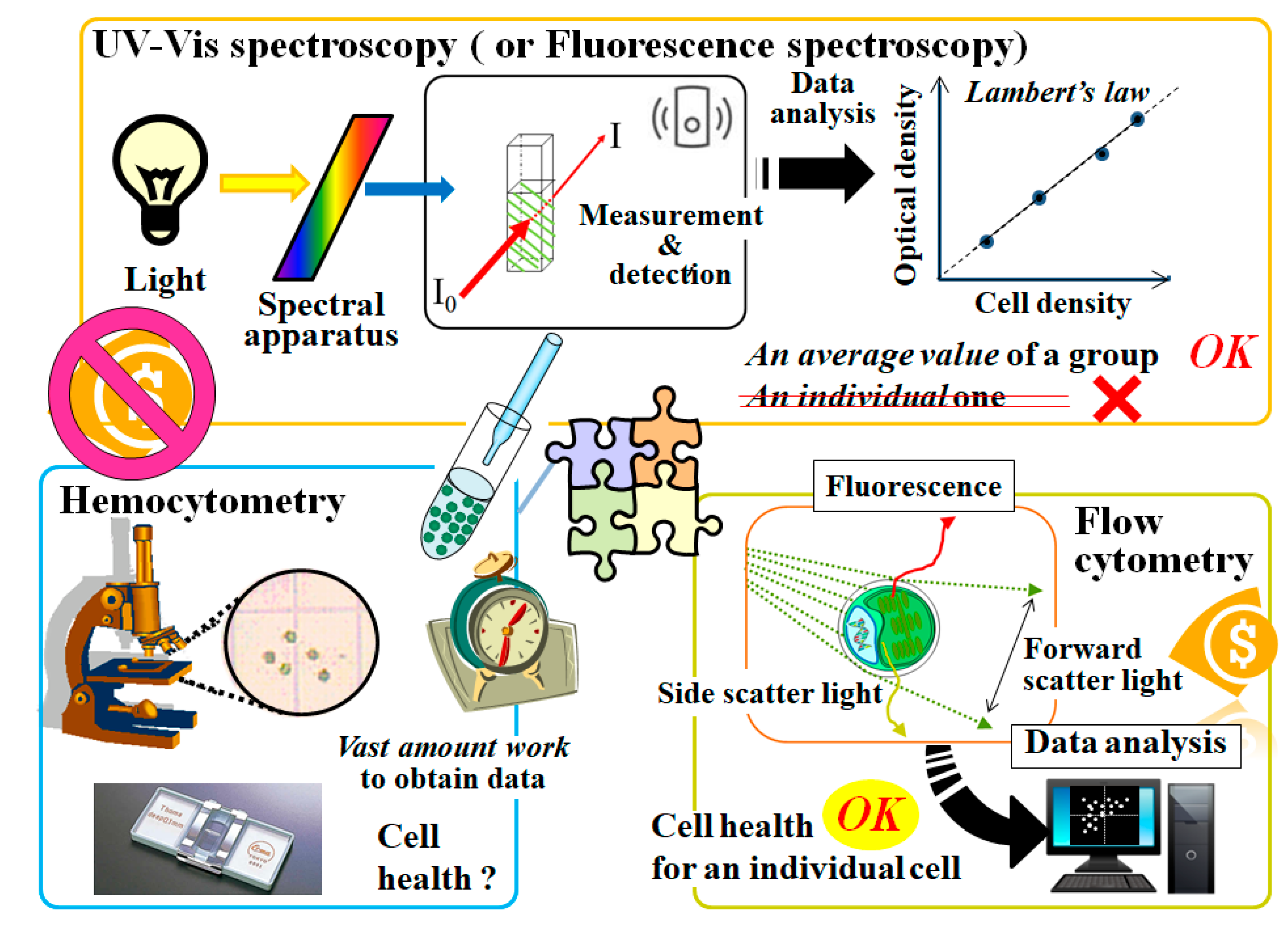
2. Standard Methods to Evaluate Cells
2.1. Conventional Methods to Evaluate Cells
2.1.1. Microscopy and Hemocytometry
2.1.2. Spectroscopy and a Method Based on a Chlorophyll Meter
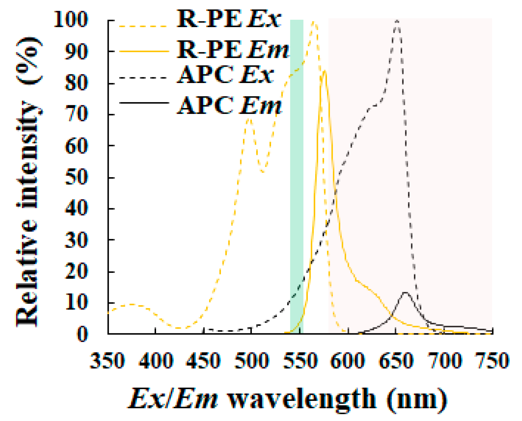
2.1.3. Spectrofluorometry and Flow Cytometry
2.1.4. Dry Weight Measurement of Biomass
2.2. A Recently Visualization-Based Automated Cell Counter to Evaluate Cells Such as Cultured Animal Cells
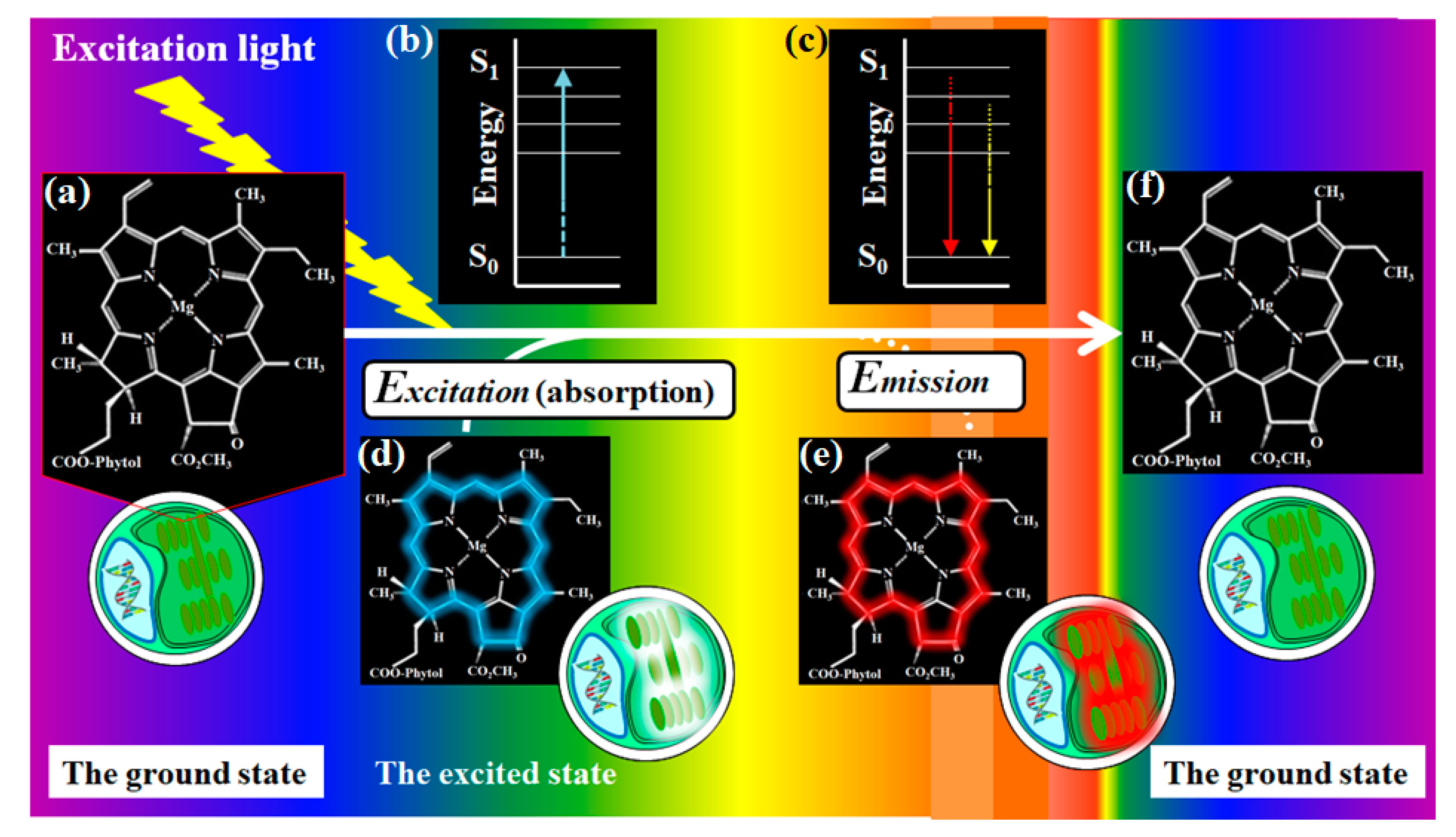
3. A New Method to Detect and Evaluate Microalgae Using Chlorophyll Autofluorescence
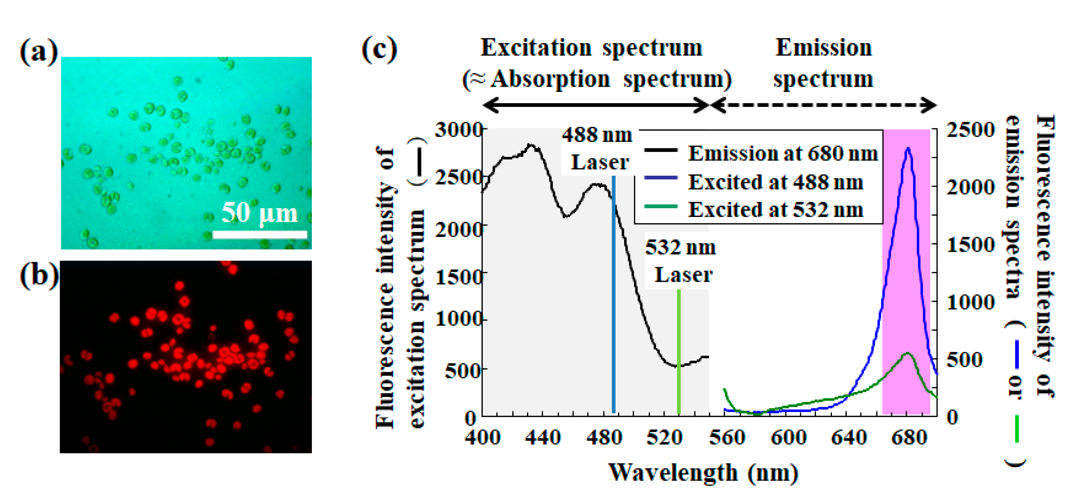
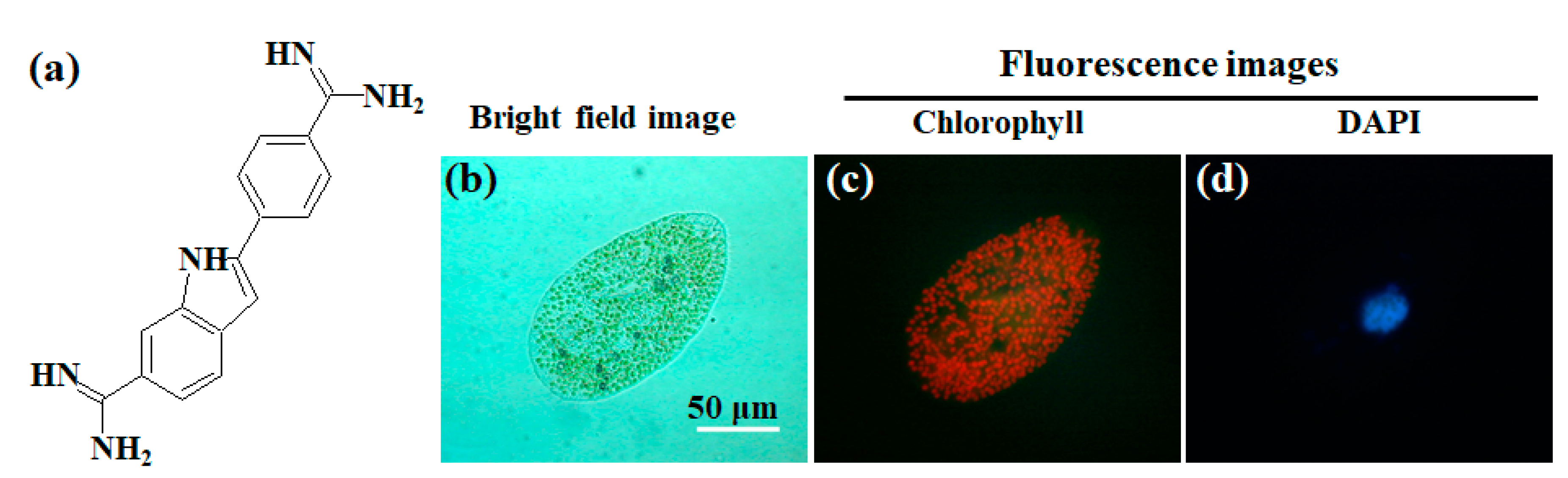
3.1. Advantages to Drawing on Chlorophyll Fluorescence from Chloroplast(s) on a Microalgal Management
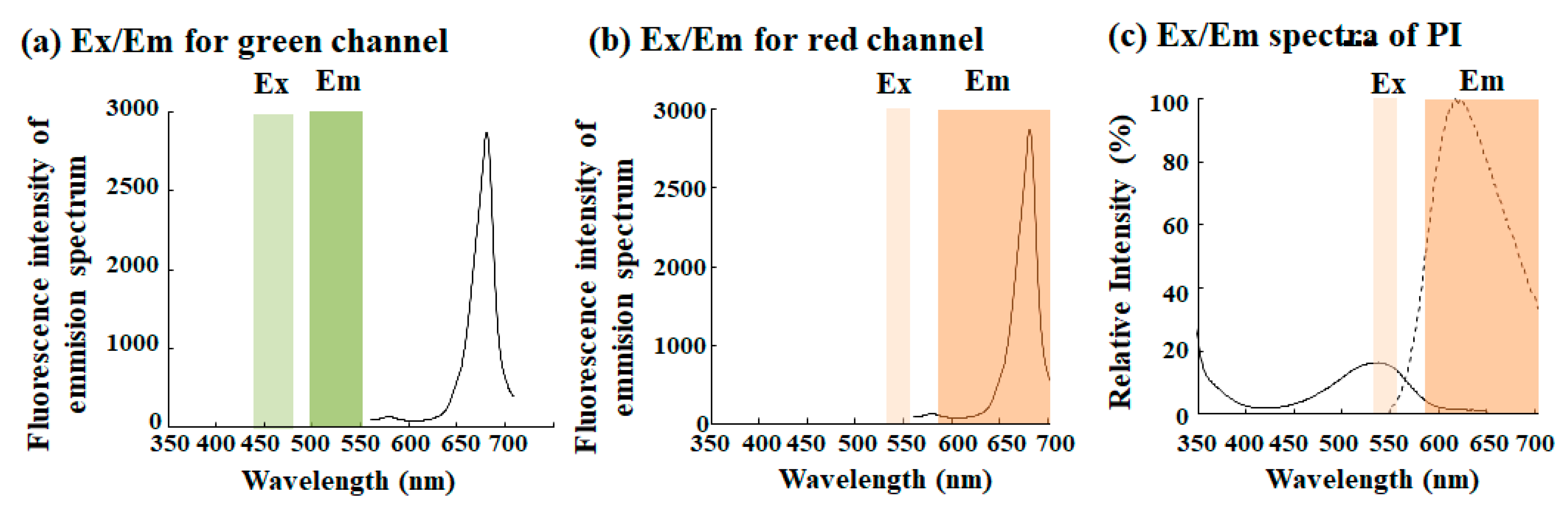
3.2. Standard Automated Cell Counter Equipped with a Fluorescence Filter
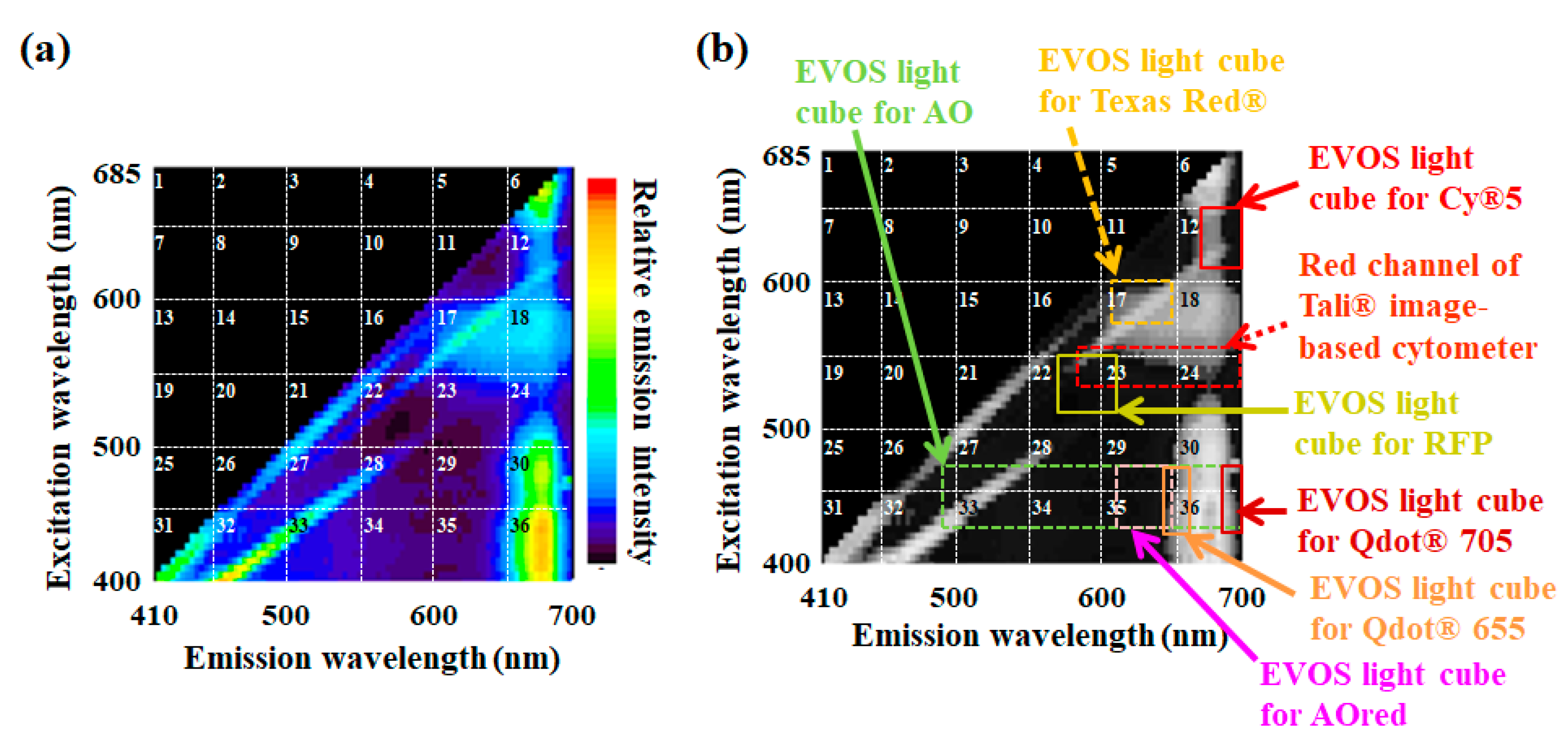
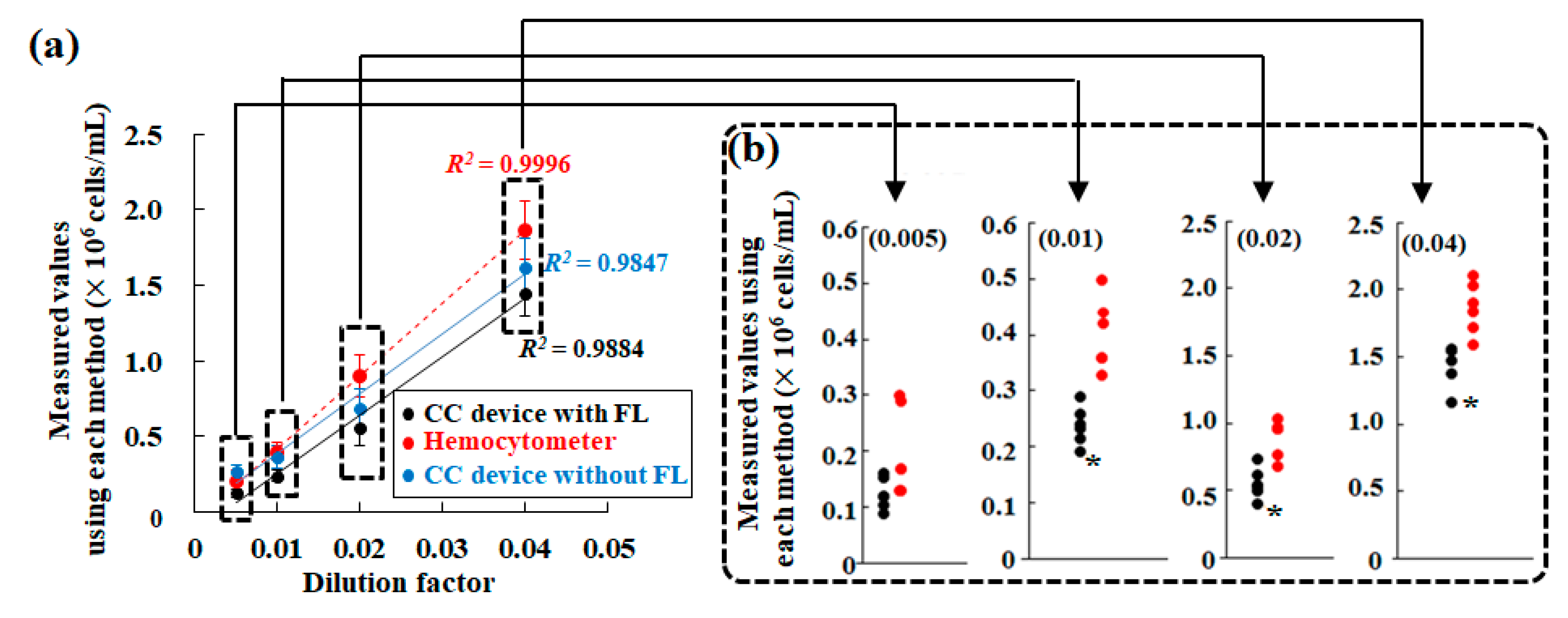
3.3. Automated Cell Counter Equipped with a Fluorescence Filter to Detect Chlorophyll Fluorescence
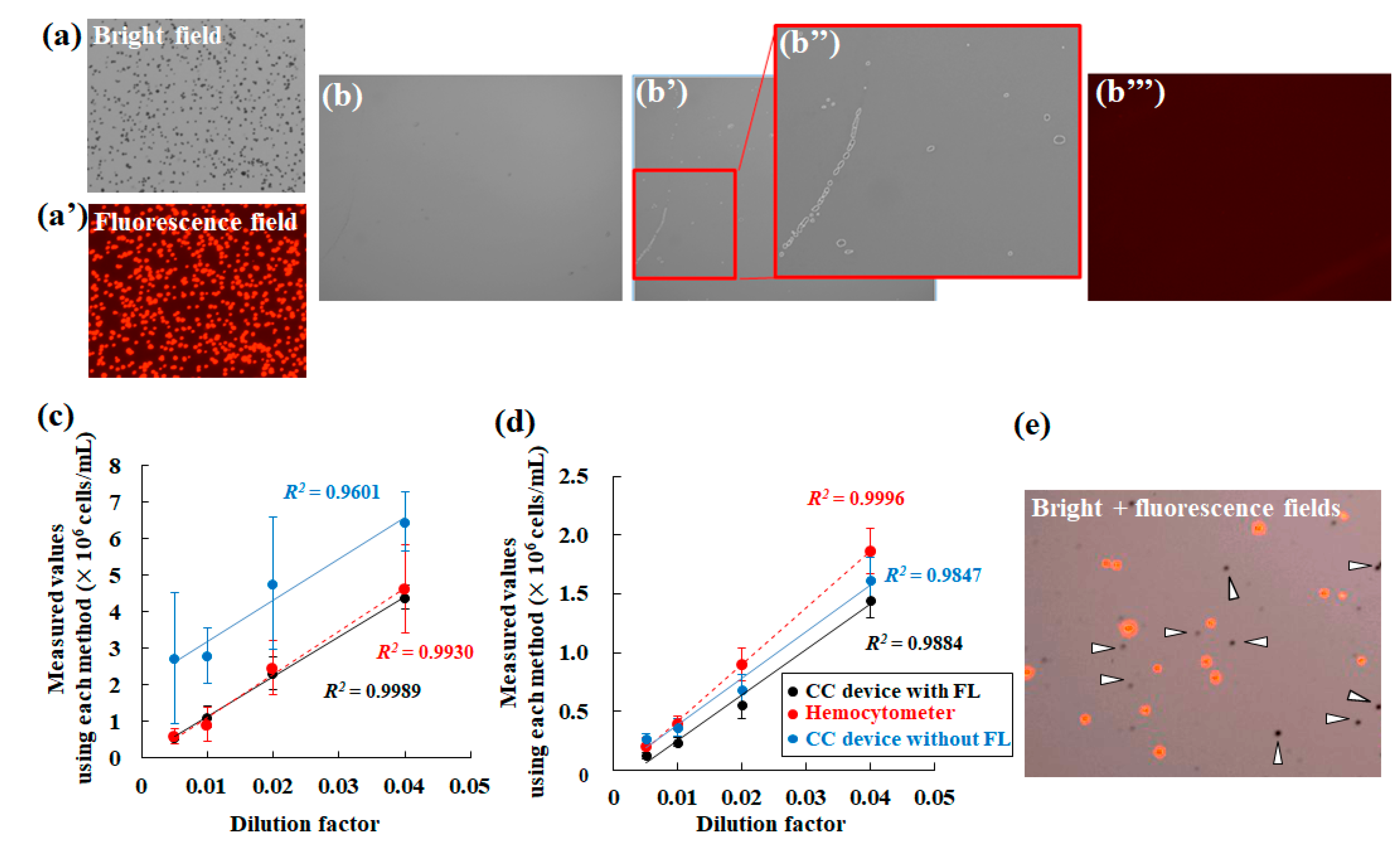
3.4. Measurements of Microalgal Numbers Using the Automated Cell Counter Equipped with Fluorescence Filter for Chlorophyll Fluorescence
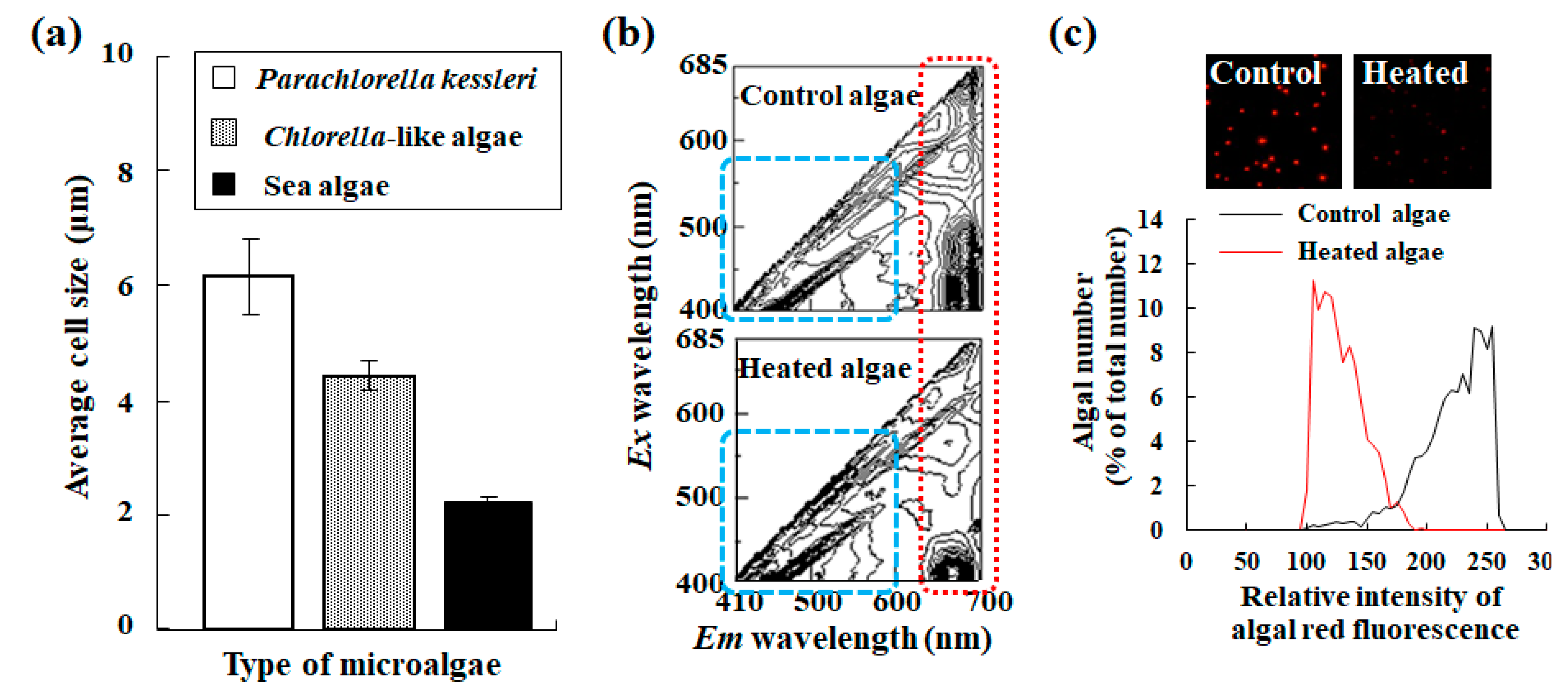
3.5. Evaluation of Microalgal Size and their Status Using the Automated Cell Counter Equipped with the Fluorescence Filter for Chlorophyll Fluorescence
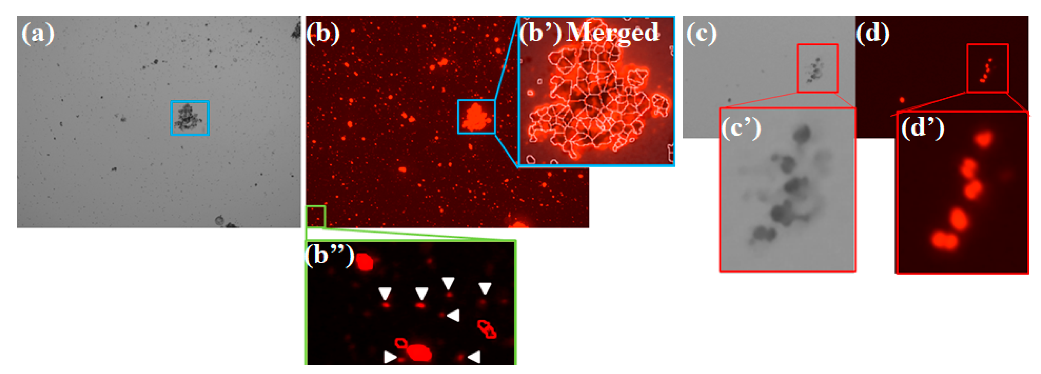
3.6. Remaining Issues about Application of the Automated Cell Counter to Microalgal Researches
4. Conclusions
Funding
Conflicts of Interest
References
- Arashida, R. Characteristics of the microalgae Euglena and its applications in foods and ecological fields. Jap. Soc. Photosynth. Res. 2012, 22, 33–38. [Google Scholar]
- Chaturvedi, V.; Nikhil, K. Effect of algal bio-fertilizer on the Vigna radiate: A Critical Review. Int. J. Eng. Res. Appl. 2016, 6, 85–94. [Google Scholar]
- Chisti, Y. Biodiesel from microalgae. Biotechnol. Adv. 2007, 25, 294–306. [Google Scholar] [CrossRef] [PubMed]
- Faheed, F.A.; Fattah, Z.A.-E. Effects of Chlorella vulgaris as bio-fertilizer on growth parameters and metabolic aspects of lettuce plant. J. Agricul. Soc. Sci. 2008, 4, 165–169. [Google Scholar]
- Ghayal, M.S.; Pandya, M.T. Microalgae biomass: A renewable source of energy. Energy Procedia. 2013, 32, 242–250. [Google Scholar] [CrossRef] [Green Version]
- Hernándes-García, A.; Velásquez-Orta, S.B.; Novelo, E.; Yáñez-Noguez, I.; Monje-Ramírez, I.; Ledesma, M.T.O. Wastewater-leachate treatment by microalgae: Biomass, carbohydrate and lipid production. Ecotoxicol. Environ. Safety 2019, 174, 435–444. [Google Scholar] [CrossRef] [PubMed]
- Seyidoglu, N.; Inan, S.; Aydin, C. A Prominent superfood: Spirulina platensis. In Superfood and Functional Food-Development of Superfood and Its Role in Medicine, 1st ed.; Shiomi, N., Waisundara, V.Y., Eds.; InTechOpen: Rijeka, Croatia, 2017; pp. 1–27. [Google Scholar]
- Spolaore, P.; Joannis-Cassan, C.; Duran, E.; Isambert, A. Commercial applications of microalgae. J. Biosci. Bioeng. 2006, 101, 87–96. [Google Scholar] [CrossRef] [Green Version]
- Williams, P.J.l.B.; Laurens, L.M.L. Microalgae as biodiesel & biomass feedstocks: Review & analysis of the biochemistry, energetics & economics. Energy Environ. Sci. 2010, 3, 554–590. [Google Scholar]
- Kafarski, P. Rainbow code of biotechnology. CHEMIK 2012, 66, 811–816. [Google Scholar]
- Takahashi, T. Applicability of automated cell counter with a chlorophyll detector in routine management of microalgae. Sci. Rep. 2018, 8, 4967. [Google Scholar] [CrossRef]
- Takahashi, T. Quality assessment of microalgae exposed to trace metals using flow cytometry. In Superfood and Functional Food-Development of Superfood and Its Role in Medicine, 1st ed.; Shiomi, N., Waisundara, V.Y., Eds.; InTechOpen: Rijeka, Croatia, 2017; pp. 29–45. [Google Scholar]
- Kaplan, D. Absorption and adsorption of heavy metals by microalgae. In Handbook of Microalgal Culture: Applied Phycology and Biotechnology, 2nd ed.; Richmond, A., Hu, Q., Eds.; John Wiley & Sons, Ltd.: Hoboken, NJ, USA, 2013; pp. 602–611. [Google Scholar]
- Takahashi, T.; Yokoyama, S. Bioassay of components eluted from electric arc furnace steel slag using microalgae Chlorella. ISIJ Int. 2016, 56, 1495–1503. [Google Scholar] [CrossRef] [Green Version]
- Ongena, K.; Das, C.; Smith, J.L.; Gil, S.; Johnston, G. Determining cell number during cell culture using the Scepter cell counter. J. Vis. Exp. 2010, 45, e2204. [Google Scholar] [CrossRef] [PubMed]
- Koch, A.L. Turbidity measurements of bacterial cultures in some available commercial instruments. Anal. Biochem. 1970, 38, 252–259. [Google Scholar] [CrossRef]
- Sutton, S. Measurement of microbial cells by optical density. J. Validation Technol. 2011, 17, 46–49. [Google Scholar]
- Takahashi, T. Direct evaluation of endosymbiotic status in Paramecium bursaria using capillary flow cytometry. Cytometry Part A 2014, 85, 911–914. [Google Scholar] [CrossRef] [PubMed]
- Takahashi, T. Application of phytoplankton. In Corrosion Control and Surface Finishing–Environmentally Friendly Approaches, 1st ed.; Kanematsu, H., Barry, D.M., Eds.; Springer Japan: Tokyo, Japan, 2016; pp. 213–224. [Google Scholar]
- Chiocciodi, M.; Hankamer, B.; Ross, I.L. Flow cytometry pulse width data enables rapid and sensitive estimation of biomass dry weith in the microalgae Chlamydomonas reinhardtii and Chlorella vulgaris. PLoS ONE 2014, 9, e97269. [Google Scholar]
- Masojídek, J.; Torzillo, G.; Koblízek, M.; Kopecký, J.; Bernardini, P.; Sacchi, A.; Komenda, J. Photoadaptation of two members of the Chlorophyta (Scenedesmus and Chlorella) in laboratory and outdoor cultures: Changes in chlorophyll fluorescence quenching and the xanthophyll cycle. Planta 1999, 209, 126–135. [Google Scholar]
- Deamici, K.M.; Santos, L.O.; Costa, J.A.V. Magnetic field action on outdoor and indoor cultures of Spirulina: Evaluation of growth, medium consumption and protein profile. Bioresour. Technol. 2018, 249, 168–174. [Google Scholar] [CrossRef]
- Berglund, D.L.; Eversman, S. Flow cytometric measurement of pollutant stresses on algal cells. Cytometry 1988, 9, 150–155. [Google Scholar] [CrossRef]
- Gerashchenko, B.I.; Kosaka, T.; Hosoya, H. Optical compartmentation of vegetating algae species as a basis for their growth-specific characterization. Cytometry 2002, 48, 153–158. [Google Scholar] [CrossRef]
- Gerashchenko, B.I.; Takahashi, T.; Kosaka, T.; Hosoya, H. Life Cycle Analysis of Unicellular Algae. Curr. Protoc. Cytom. 2010, 52, 11.19.1–11.19.6. [Google Scholar] [CrossRef] [PubMed]
- Takahashi, T.; Ogura, Y.; Ogawa, A.; Kanematsu, H.; Yokoyama, S. An effective and economic strategy to restore acidified freshwater ecosystems with steel industrial byproducts. J. Wat. Environ. Technol. 2012, 10, 347–362. [Google Scholar] [CrossRef] [Green Version]
- Marie, D.; Rigaut-Jalabert, F.; Vaulot, D. An improved protocol for flow cytometry analysis of phytoplankton cultures and natural samples. Cytometry Part A 2014, 85A, 962–968. [Google Scholar] [CrossRef] [PubMed] [Green Version]
- Büscher, M. Flow cytometry instrumentation-An overview. Curr. Protoc. Cytom. 2019, 87, e52. [Google Scholar] [CrossRef]
- Takahashi, T. Life cycle analysis of endosymbiotic algae in an endosymbiotic situation with Paramecium bursaria using capillary flow cytometry. Energies 2017, 10, 1413. [Google Scholar] [CrossRef] [Green Version]
- Hu, Q.; Sommerfeld, M.; Jarvis, E.; Ghirardi, M.; Posewitz, M.; Seibert, M.; Darzins, A. Microalgal triacylglycerols as feedstocks for biofuel production: Perspectives and advances. Plant. J. 2008, 54, 621–639. [Google Scholar] [CrossRef]
- Leeuw, T.; Boss, E.S.; Wright, D.L. In situ measurements of phytoplankton fluorescence using low cost electronics. Sensors 2013, 13, 7872–7883. [Google Scholar] [CrossRef] [Green Version]
- Zeng, L.; Li, D. Development of In Situ sensors for chlorophyll concentration measurement. J. Sens. 2015, 2015, 903509. [Google Scholar] [CrossRef] [Green Version]
- Vítavá, M.; Hendrychová, J.; Cepák, V.; Zachleder, V. Visualization of DNA-containing structures in various species of Chlorophyta, Rhodophyta and Cyanophyta using SYBR Green I dye. Folia Microbiol. 2005, 50, 333–340. [Google Scholar] [CrossRef]
- Estep, K.W.; MacIntyre, F.; Hjörleifsson, E.; Sieburth, J.M. MacImage: A user-friendly image-analysis system for the accurate mensuration of marine organisms. Mar. Ecol. Prog. Ser. 1986, 33, 243–253. [Google Scholar] [CrossRef]
- Albertano, P.; Somma, D.D.; Capucci, E. Cyanobacterial picoplankton from the central Baltic sea: Cell size classification by mage-analyzed fluorescence microscopy. J. Plankton Res. 1997, 19, 1405–1416. [Google Scholar] [CrossRef] [Green Version]
- Congestri, R.; Federici, R.; Albertano, P. Evaluating biomass of Baltic filamentous cyanobacteria by image analysis. Aquat. Microb. Ecol. 2000, 22, 283–290. [Google Scholar] [CrossRef]
- Congestri, R.; Capucci, E.; Albertano, P. Morphometric variability of the genus Nodularia (Cyanobacteria) in Baltic natural communities. Aquat. Microb. Ecol. 2003, 32, 251–259. [Google Scholar] [CrossRef] [Green Version]
- Gandola, E.; Antonioli, M.; Traficante, A.; Franceschini, S.; Scardi, M.; Cangestri, R. ACQUA: Atutomated cyanobacterial quantification algorithm for toxic filamentous genera using spline curves, pattern recognition and machine learning. J. Microbiol. Methods 2016, 124, 48–56. [Google Scholar] [CrossRef]
- Koç, E.; Çelik-Uzuner, S.; Uzuner, U.; Çakmak, R. The detailed comparison of cell death detected by annexin V-PI counterstain using fluorescence microscope, flow cytometer and automated cell counter in mammalian and microalgae cells. J. Fluoresc. 2018, 28, 1393–1404. [Google Scholar]
- MacColl, R. Cyanobacteria phycobilisomes. J. Struct. Biol. 1998, 124, 311–334. [Google Scholar] [CrossRef]
- Gupa, G.D.; Sonani, R.R.; Sharma, M.; Patel, K.; Rastogi, R.P.; Madamwar, D.; Kumar, V. Crystal structure analysis of phycocyanin from chromatically adapted Phormidium rubidium A09DM. RSC Adv. 2016, 6, 77898–77907. [Google Scholar] [CrossRef]
- ThermoFisher Scientific Inc. EVOS Light Cube Selection Guide. Available online: https://www.thermofisher.com/content/dam/LifeTech/migration/files/cell-analysis/pdfs.par.1324.file.dat/co27602-evos-light-cube-tables-tech-lit.pdf (accessed on 4 November 2019).
- Takahashi, T. Efficient interpretation of multiparametric data using principal component analysis as an example of quality assessment of microalgae. In Multidimensional Flow Cytometry Techniques for Novel Highly Informative Assays, 1st ed.; Gemiei, M., Ed.; InTechOpen: London, UK, 2018; pp. 81–97. [Google Scholar]
- Hosseini Tafreshi, A.; Shariati, M. Dunaliella biotechnology: Methods and applications. J. Appl. Microbiol. 2009, 107, 14–35. [Google Scholar] [CrossRef]
- De Jesus Raposo, M.F.; de Morais, A.M.M.B.; de Morais, R.M.S.C. Carotenoids from marine microalgae: A valuable natural source for the prevention of chronic diseases. Mar. Drugs 2015, 13, 5128–5155. [Google Scholar] [CrossRef]
- Shiho, M.; Kawachi, M.; Horioka, K.; Nishita, Y.; Ohashi, K.; Kaya, K.; Watanabe, M. Business evaluation of a green microalgae Botryococcus braunii oil production system. Procedia Environ. Sci. 2012, 15, 90–109. [Google Scholar] [CrossRef] [Green Version]
- Kronick, M.N.; Grossman, P.D. Immunoassay techniques with fluorescent phycobiliprotein conjugates. Chin. Chem. 1983, 29, 1582–1588. [Google Scholar]

| Filter | Wavelength Region of Excitation (nm) | Wavelength Region of Emission (nm) |
|---|---|---|
| EVOS light cube for AO (Acridine Orange) | 419–465 | 488-longer wavelength than 488 nm 1 |
| EVOS light cube for AOred | 419–465 | 612–644 |
| EVOS light cube for RFP | 511–551 | 573–613 |
| EVOS light cube for Texas Red® | 570.5–599.5 | 604–644 |
| EVOS light cube for Cy®5 | 608–648 | 672–712 |
| EVOS light cube for Qdot® 655 | 422.5–467.5 | 647.5–662.5 |
| EVOS light cube for Qdot® 705 | 422.5–467.5 | 685–725 |
| Red channel of TaliTM Image-based Cytometer | 521–565 | 580-longer wavelength than 580 nm 1 |
© 2019 by the author. Licensee MDPI, Basel, Switzerland. This article is an open access article distributed under the terms and conditions of the Creative Commons Attribution (CC BY) license (http://creativecommons.org/licenses/by/4.0/).
Share and Cite
Takahashi, T. Routine Management of Microalgae Using Autofluorescence from Chlorophyll. Molecules 2019, 24, 4441. https://doi.org/10.3390/molecules24244441
Takahashi T. Routine Management of Microalgae Using Autofluorescence from Chlorophyll. Molecules. 2019; 24(24):4441. https://doi.org/10.3390/molecules24244441
Chicago/Turabian StyleTakahashi, Toshiyuki. 2019. "Routine Management of Microalgae Using Autofluorescence from Chlorophyll" Molecules 24, no. 24: 4441. https://doi.org/10.3390/molecules24244441
APA StyleTakahashi, T. (2019). Routine Management of Microalgae Using Autofluorescence from Chlorophyll. Molecules, 24(24), 4441. https://doi.org/10.3390/molecules24244441