
Phenolic Profile, Antioxidant Activity, and Anti-obesogenic Bioactivity of Mao Luang Fruits (Antidesma bunius L.)


Abstract
1. Introduction
2. Results
2.1. The Different Phytochemical Contents and Anti-oxidation Properties Among the Samples
2.2. Inhibitory Activity Against α-Amylase and Lipase Enzymes
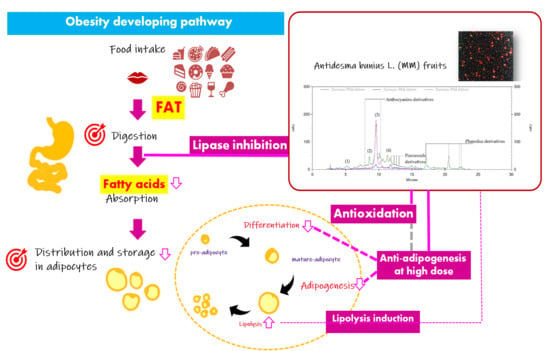
2.3. Anti-obesity Potential via Interruption on Adipocyte Life Cycle
2.3.1. Effect on Pre-adipocyte Viability by MTT Assay
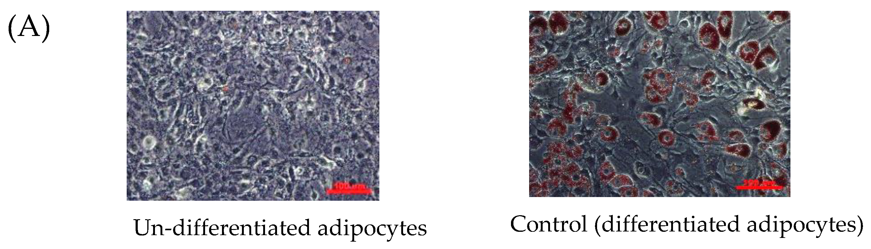
2.3.2. Anti-differentiation as Showing Reduction of Oil Red O Staining
2.3.3. Inhibition of Lipid Accumulation and Reduction of Triglyceride Content
2.3.4. Adipocyte lipolysis Induction as Increasing of Released Glycerol Content
3. Discussion
4. Materials and Methods
4.1. Materials
4.2. Sample Preparation
4.3. Determination of Total Phytochemical Contents
4.3.1. Total Phenolics Content Using the Folin Ciocalteu Assay
4.3.2. Total Flavonoids Content Using Aluminum-flavonoid Assay
4.3.3. Total Anthocyanins Content Using pH-differential Assay
4.4. Determination of Anti-oxidation Activity Using DPPH and TBARS Assays
4.5. HPLC-UV Analysis of the Phytochemical Contents
4.6. Determination of Inhibitory Activity on Lipase and Amylase Enzyme
4.6.1. Effect on Lipase Enzyme
4.6.2. Effect on α-Amylase Enzyme
4.7. Determination of Anti-obesity Potential via Interruption of Adipocyte Cell Cycle
4.7.1. Cell Viability Using MTT Assay
4.7.2. Anti-adipogenesis in 3T3-L1 Adipocytes
Culture Conditions for Pre-adipocyte Maintenance and Differentiation
Oil Red O Staining
Triglyceride (Triacylglycerol) Determination
4.7.3. Lipolysis Induction by Glycerol Released Assay
4.8. Statistical Analysis
5. Conclusions
Author Contributions
Funding
Acknowledgments
Conflicts of Interest
References
- Yoon, K.-H.; Lee, J.-H.; Kim, J.-W.; Cho, J.H.; Choi, Y.-H.; Ko, S.-H.; Zimmet, P.; Son, H.-Y. Epidemic obesity and type 2 diabetes in Asia. Lancet 2006, 368, 1681–1688. [Google Scholar] [CrossRef]
- World Health Organization. Obesity: Prevention and Managing the Global Epidemic; World Health Organization: Geneva, Switzerland, 2000; p. 253. [Google Scholar]
- Worsztynowicz, P.; Napierała, M.; Białas, W.; Grajek, W.; Olkowicz, M. Pancreatic α-amylase and lipase inhibitory activity of polyphenolic compounds present in the extract of black chokeberry (Aronia melanocarpa L.). Process Biochem. 2014, 49, 1457–1463. [Google Scholar] [CrossRef]
- Ghaben, A.L.; Scherer, P.E. Adipogenesis and metabolic health. Nat. Rev. Mol. Cell Bio. 2019, 20, 242–258. [Google Scholar] [CrossRef]
- Guo, X.; Liu, J.; Cai, S.; Wang, O.; Ji, B. Synergistic interactions of apigenin, naringin, quercetin and emodin on inhibition of 3T3-L1 preadipocyte differentiation and pancreas lipase activity. Obes. Res. Clin. Pract. 2016, 10, 327–339. [Google Scholar] [CrossRef]
- El-Tantawy, W.H.; Soliman, N.D.; El-naggar, D.; Shafei, A. Investigation of antidiabetic action of Antidesma bunius extract in type 1 diabetes. Arch. Insect. Biochem. Physiol. 2015, 121, 116–122. [Google Scholar] [CrossRef]
- Kassem, M.E.; Hashim, A.N.; Hassanein, H.M. Bioactivity of Antidesma bunius leaves (Euphorbiaceae) and their major phenolic constituents. Eur. Sci. J. ESJ 2013, 9. [Google Scholar]
- Islam, S.; Koly, S. A review on phytochemical and pharmacological potentials of Antidesma bunius. J. Anal. Pharm. Res. 2018, 7, 602–604. [Google Scholar]
- Jorjong, S.; Butkhup, L.; Samappito, S. Phytochemicals and antioxidant capacities of Mao-Luang (Antidesma bunius L.) cultivars from Northeastern Thailand. Food Chem. 2015, 181, 248–255. [Google Scholar] [CrossRef] [PubMed]
- Lawag, I.L.; Aguinaldo, A.M.; Naheed, S.; Mosihuzzaman, M. α-Glucosidase inhibitory activity of selected Philippine plants. J. Ethnopharmacol. 2012, 144, 217–219. [Google Scholar] [CrossRef] [PubMed]
- Harnly, J.M.; Bhagwat, S.; Lin, L.-Z. Profiling methods for the determination of phenolic compounds in foods and dietary supplements. Anal. Bioanal. Chem. 2007, 389, 47–61. [Google Scholar] [CrossRef]
- Pool-Zobel, B.L.; Bub, A.; Schröder, N.; Rechkemmer, G. Anthocyanins are potent antioxidants in model systems but do not reduce endogenous oxidative DNA damage in human colon cells. Eur. J. Nutr. 1999, 38, 227–234. [Google Scholar] [CrossRef] [PubMed]
- Scott, B.C.; Butler, J.; Halliwell, B.; Aruoma, O.I. Evaluation of the Antioxidant Actions of Ferulic Acid and Catechins. Free Radical Res. Commun. 1993, 19, 241–253. [Google Scholar] [CrossRef] [PubMed]
- Gooda Sahib, N.; Abdul Hamid, A.; Saari, N.; Abas, F.; Pak Dek, M.S.; Rahim, M. Anti-Pancreatic Lipase and Antioxidant Activity of Selected Tropical Herbs. Int. J. Food Prop. 2012, 15, 569–578. [Google Scholar] [CrossRef]
- Butkhup, L.; Samappito, S. Analysis of Anthocyanin, Flavonoids, and Phenolic Acids in Tropical Bignay Berries. Int. J. Fruit Sci. 2008, 8, 15–34. [Google Scholar] [CrossRef]
- Fernández-Sánchez, A.; Madrigal-Santillán, E.; Bautista, M.; Esquivel-Soto, J.; Morales-González, Á.; Esquivel-Chirino, C.; Durante-Montiel, I.; Sánchez-Rivera, G.; Valadez-Vega, C.; Morales-González, J.A. Inflammation, Oxidative Stress, and Obesity. Int. J. Mol. Sci. 2011, 12, 3117–3132. [Google Scholar] [CrossRef]
- Furukawa, S.; Fujita, T.; Shimabukuro, M.; Iwaki, M.; Yamada, Y.; Nakajima, Y.; Nakayama, O.; Makishima, M.; Matsuda, M.; Shimomura, I. Increased oxidative stress in obesity and its impact on metabolic syndrome. J. Clin. Investig. 2017, 114, 1752–1761. [Google Scholar] [CrossRef]
- Lee, S.-E.; Lee, E.-H.; Lee, T.-J.; Kim, S.-W.; Kim, B.-H. Anti-obesity effect and action mechanism of Adenophora triphylla root ethanol extract in C57BL/6 obese mice fed a high-fat diet. Biosci. Biotechnol. Biochem. 2013, 77, 544–550. [Google Scholar] [CrossRef]
- You, J.S.; Lee, Y.J.; Kim, K.S.; Kim, S.H.; Chang, K.J. Ethanol extract of lotus (Nelumbo nucifera) root exhibits an anti-adipogenic effect in human pre-adipocytes and anti-obesity and anti-oxidant effects in rats fed a high-fat diet. Nutr. Res. 2014, 34, 258–267. [Google Scholar] [CrossRef]
- Samout, N.; Ettaya, A.; Bouzenna, H.; Ncib, S.; Elfeki, A.; Hfaiedh, N. Beneficial effects of Plantago albicans on high-fat diet-induced obesity in rats. Biomed. Pharmacother. 2016, 84, 1768–1775. [Google Scholar] [CrossRef]
- Udomkasemsab, A.; Ngamlerst, C.; Adisakwattana, P.; Aroonnual, A.; Tungtrongchitr, R.; Prangthip, P. Maoberry (Antidesma bunius) ameliorates oxidative stress and inflammation in cardiac tissues of rats fed a high-fat diet. BMC Complement. Altern. Med. 2018, 18, 344. [Google Scholar] [CrossRef]
- Janda, E.; Lascala, A.; Martino, C.; Ragusa, S.; Nucera, S.; Walker, R.; Gratteri, S.; Mollace, V. Molecular mechanisms of lipid- and glucose-lowering activities of bergamot flavonoids. PharmaNutrition 2016, 4, S8–S18. [Google Scholar] [CrossRef]
- Semaan, D.G.; Igoli, J.O.; Young, L.; Marrero, E.; Gray, A.I.; Rowan, E.G. In vitro anti-diabetic activity of flavonoids and pheophytins from Allophylus cominia Sw. on PTP1B, DPPIV, alpha-glucosidase and alpha-amylase enzymes. J. Ethnopharmacol. 2017, 203, 39–46. [Google Scholar] [CrossRef] [PubMed]
- Zeng, P.; Zhang, Y.; Pan, C.; Jia, Q.; Guo, F.; Li, Y.; Zhu, W.; Chen, K. Advances in studying of the pharmacological activities and structure–activity relationships of natural C-glycosylflavonoids. Acta Pharm. Sinica B 2013, 3, 154–162. [Google Scholar] [CrossRef][Green Version]
- Sosnowska, D.; Podsędek, A.; Kucharska, A.Z.; Redzynia, M.; Opęchowska, M.; Koziołkiewicz, M. Comparison of in vitro anti-lipase and antioxidant activities, and composition of commercial chokeberry juices. Eur. Food Res. Technol. 2016, 242, 505–515. [Google Scholar] [CrossRef]
- Chowtivannakul, P.; Srichaikul, B.; Talubmook, C. Hypoglycemic and Hypolipidemic Effects of Seed Extract from Antidesma bunius (L.) Spreng in Streptozotocin-induced Diabetic Rats. Pakistan J. Bio. Sci. PJBS 2016, 19, 211–218. [Google Scholar]
- Gulua, L.; Nikolaishvili, L.; Jgenti, M.; Turmanidze, T.; Dzneladze, G. Polyphenol content, anti-lipase and antioxidant activity of teas made in Georgia. Ann. Agrar. Sci. 2018, 16, 357–361. [Google Scholar] [CrossRef]
- Qin, B.; Anderson, R.A. An extract of chokeberry attenuates weight gain and modulates insulin, adipogenic and inflammatory signalling pathways in epididymal adipose tissue of rats fed a fructose-rich diet. Brit. J. Nutr. 2012, 108, 581–587. [Google Scholar] [CrossRef]
- Matsukawa, T.; Villareal, M.O.; Motojima, H.; Isoda, H. Increasing cAMP levels of preadipocytes by cyanidin-3-glucoside treatment induces the formation of beige phenotypes in 3T3-L1 adipocytes. J. Nutr. Biochem. 2017, 40, 77–85. [Google Scholar] [CrossRef]
- Butkhup, L.; Samappito, S. Changes in physico-chemical properties, polyphenol compounds and antiradical activity during development and ripening of maoluang (Antidesma bunius L. Spreng) fruits. J. Fruit Ornam. Plant. Res. 2011, 19, 85–99. [Google Scholar]
- Butkhup, L.; Samappito, S. An analysis on flavonoids contents in Mao Luang fruits of fifteen cultivars (Antidesma bunius), grown in northeast Thailand. Pak. J. Biol. Sci. PJBS 2008, 11, 996–1002. [Google Scholar]
- Furuyashiki, T.; Nagayasu, H.; Aoki, Y.; Bessho, H.; Hashimoto, T.; Kanazawa, K.; Ashida, H. Tea Catechin Suppresses Adipocyte Differentiation Accompanied by Down-regulation of PPARγ2 and C/EBPα in 3T3-L1 Cells. Biosci. Biotechnol. Biochem. 2004, 68, 2353–2359. [Google Scholar] [CrossRef] [PubMed]
- Matsukawa, T.; Inaguma, T.; Han, J.; Villareal, M.O.; Isoda, H. Cyanidin-3-glucoside derived from black soybeans ameliorate type 2 diabetes through the induction of differentiation of preadipocytes into smaller and insulin-sensitive adipocytes. J. Nutr. Biochem. 2015, 26, 860–867. [Google Scholar] [CrossRef] [PubMed]
- Guo, H.; Guo, J.; Jiang, X.; Li, Z.; Ling, W. Cyanidin-3-O-β-glucoside, a typical anthocyanin, exhibits antilipolytic effects in 3T3-L1 adipocytes during hyperglycemia: Involvement of FoxO1-mediated transcription of adipose triglyceride lipase. Food Chem. Toxicol. 2012, 50, 3040–3047. [Google Scholar] [CrossRef] [PubMed]
- Yoshikawa, M.; Shimoda, H.; Nishida, N.; Takada, M.; Matsuda, H. Salacia reticulata and Its Polyphenolic Constituents with Lipase Inhibitory and Lipolytic Activities Have Mild Antiobesity Effects in Rats. J. Nutr. 2002, 132, 1819–1824. [Google Scholar] [CrossRef]
- Singleton, V.L.; Rossi, J.A. Colorimetry of Total Phenolics with Phosphomolybdic-Phosphotungstic Acid Reagents. Am. J. Enology Vitic. 1965, 16, 144. [Google Scholar]
- Chaiittianan, R.; Chayopas, P.; Rattanathongkom, A.; Tippayawat, P.; Sutthanut, K. Anti-obesity potential of corn silks: Relationships of phytochemicals and antioxidation, anti-pre-adipocyte proliferation, anti-adipogenesis, and lipolysis induction. J. Funct. Foods 2016, 23, 497–510. [Google Scholar] [CrossRef]
- Lee, J.; Durst, R.W.; Wrolstad, R.E. Determination of Total Monomeric Anthocyanin Pigment Content of Fruit Juices, Beverages Natural Colorants, and Wines by the pH Differential Method: Collaborative Study. J. AOAC Int. 2005, 88, 1269–1278. [Google Scholar]
- Chu, Y.; Chang, C.; Hsu, H. Flavonoid content of several vegetables and their antioxidant activity. J.Sci. Food Agricult. 2000, 80, 561–566. [Google Scholar] [CrossRef]
- Lee, Y.M.; Kim, Y.S.; Lee, Y.; Kim, J.; Sun, H.; Kim, J.H.; Kim, J.S. Inhibitory activities of pancreatic lipase and phosphodiesterase from Korean medicinal plant extracts. Phytother. Res. 2012, 26, 778–782. [Google Scholar] [CrossRef]
Sample Availability: Samples of the compounds are not available from the authors. |


| Phytochemical Contents | Equivalent Weight to Reference Compound (mg eq/g Extract) | ||
|---|---|---|---|
| Total phenolics content | 11.57 ± 1.13 mg GA eq/g extract | ||
| Total flavonoids content | 0.30 ± 0.00 mg QE eq/g extract | ||
| Total anthocyanins content | 3.76 ± 0.19 mg C3G eq/g extract | ||
| Antioxidation activities | IC50 (µg/mL) | Equivalent weight to reference compound (mg eq/g extract) | |
| DPPH assay (Vit C, IC50 14.47 ± 0.42 µg/mL) | 652.30 ± 5.56 | 21.81 ± 3.50 mg Vit C eq/g extract | |
| TBAR assay (BHT, IC50 2.16 ± 0.18 µg/mL) | >1000 | 3.55 ± 0.42 mg BHT eq/g extract | |
 | |||||
|---|---|---|---|---|---|
| Peak# | Identification | RT (min) | λmax (nm) | Linear Equation y = ax + b (r2) | Content (mg/g Extract) |
| 1 | Gallic acid | 5.167 | 203/224**/271* | y = 39115x − 26163 (0.999) | 0.26 ± 0.01 |
| 2 | Catechin | 8.250 | 206/234*/279** | y = 6513x − 5080 (0.999) | 2.26 ± 0.61 |
| 3 | Cynidin-3-glucoside | 9.600 | 244/276**/529* | y = 17572x (0.995) | 13.38 ± 0.72 |
| 4 | Protocatechuic acid | 11.833 | 202/261*/295** | y = 73467x − 40696 (0.999) | 0.24 ± 0.01 |
| Parameters | Lipase Enzyme | Amylase Enzyme |
|---|---|---|
| Reference drug | Orlistat (IC50 = 1.30 ± 0.17 µg/mL) | Acarbose (IC50 = 13.83 ± 0.38 µg/mL) |
| IC50 | 90.75 ± 4.12 µg/mL † | n.d. (Maximal inhibition 44.25 ± 0.86%§) |
| Reference drug equivalent weight | 14.33 ± 4.12 µg Orlistat eq/mg extract | 0.45 ± 0.05 µg Acarbose eq/mg extract |
© 2019 by the authors. Licensee MDPI, Basel, Switzerland. This article is an open access article distributed under the terms and conditions of the Creative Commons Attribution (CC BY) license (http://creativecommons.org/licenses/by/4.0/).
Share and Cite
Krongyut, O.; Sutthanut, K. Phenolic Profile, Antioxidant Activity, and Anti-obesogenic Bioactivity of Mao Luang Fruits (Antidesma bunius L.). Molecules 2019, 24, 4109. https://doi.org/10.3390/molecules24224109
Krongyut O, Sutthanut K. Phenolic Profile, Antioxidant Activity, and Anti-obesogenic Bioactivity of Mao Luang Fruits (Antidesma bunius L.). Molecules. 2019; 24(22):4109. https://doi.org/10.3390/molecules24224109
Chicago/Turabian StyleKrongyut, Ornnicha, and Khaetthareeya Sutthanut. 2019. "Phenolic Profile, Antioxidant Activity, and Anti-obesogenic Bioactivity of Mao Luang Fruits (Antidesma bunius L.)" Molecules 24, no. 22: 4109. https://doi.org/10.3390/molecules24224109
APA StyleKrongyut, O., & Sutthanut, K. (2019). Phenolic Profile, Antioxidant Activity, and Anti-obesogenic Bioactivity of Mao Luang Fruits (Antidesma bunius L.). Molecules, 24(22), 4109. https://doi.org/10.3390/molecules24224109