
The Microenvironment in Immobilized Enzymes: Methods of Characterization and Its Role in Determining Enzyme Performance


Abstract
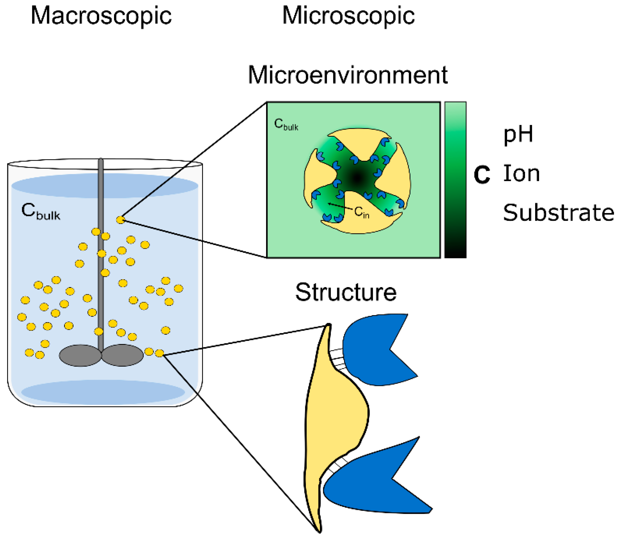
1. Introduction
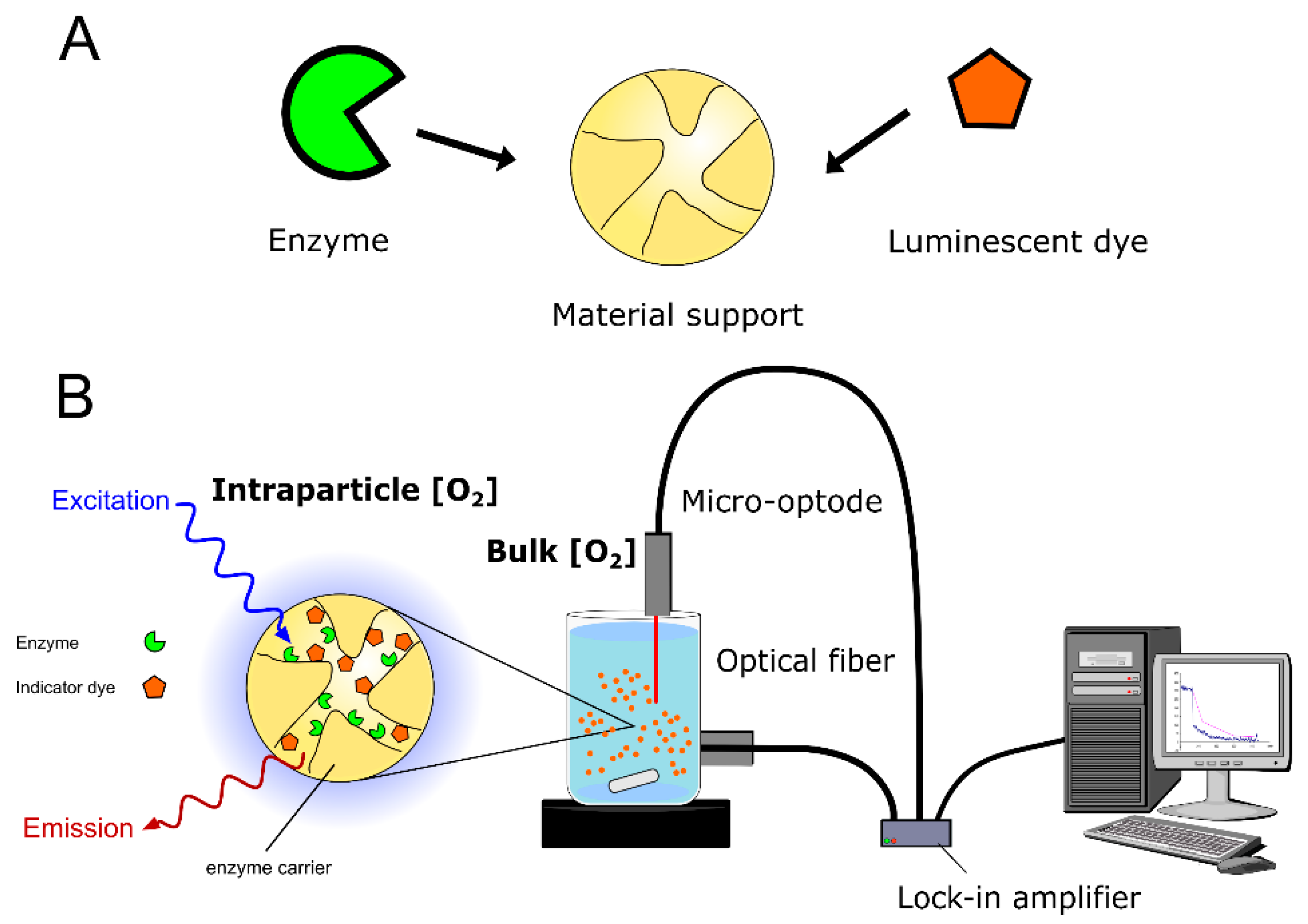
2. Opto-Chemical Sensing within Solid Particles
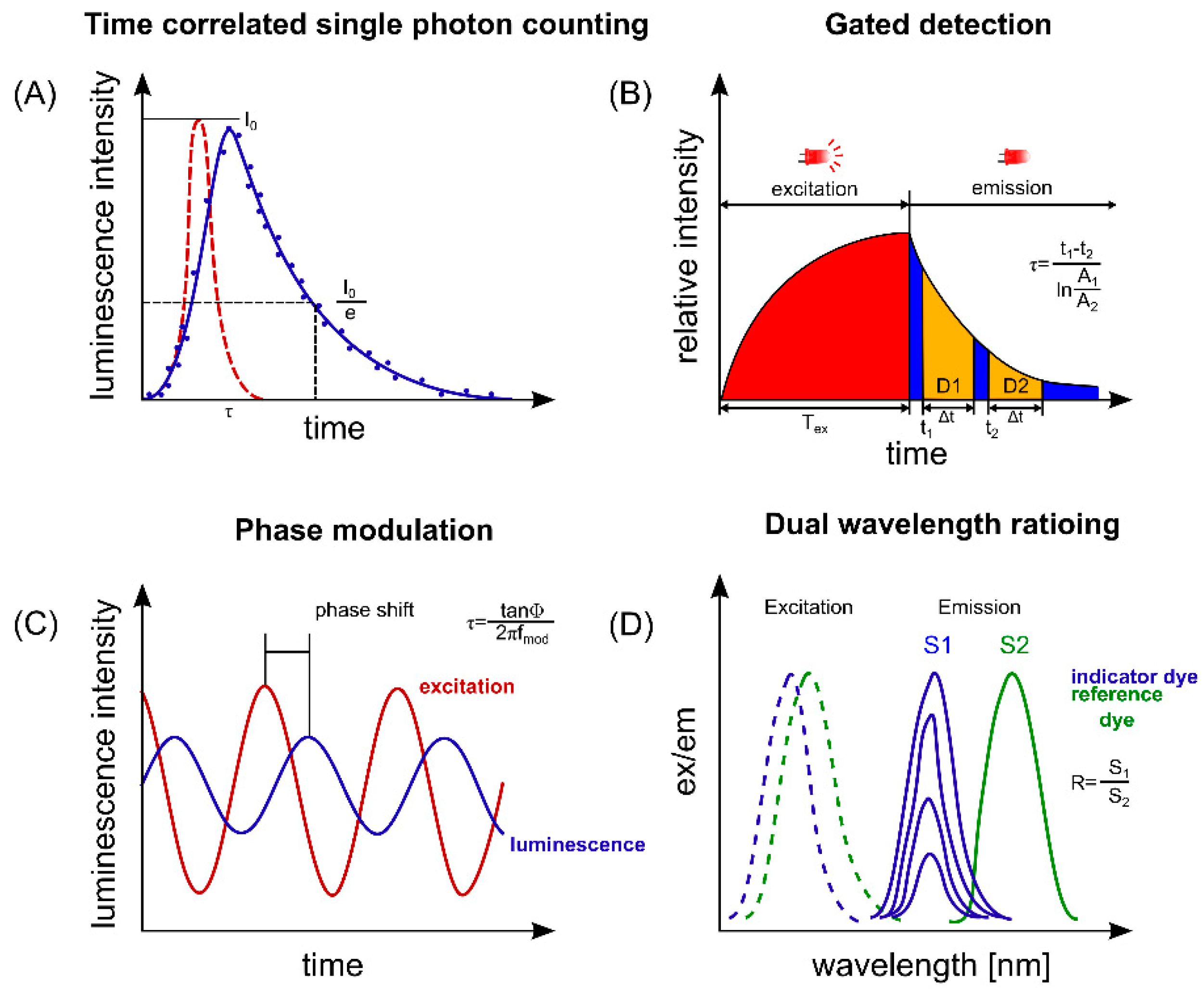
2.1. The Principles of Opto-Chemical Sensing
2.2. Luminescence Labeling of the Solid Support
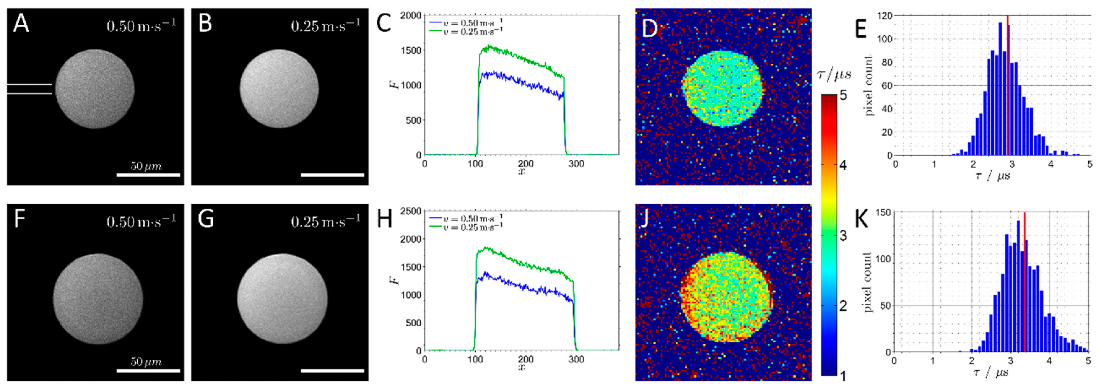
2.3. Choice of Set-Up and the Degree of Spatial and Temporal Resolution
3. The Application of Intraparticle Sensing for the Development of Immobilized Enzymes as Biocatalysts
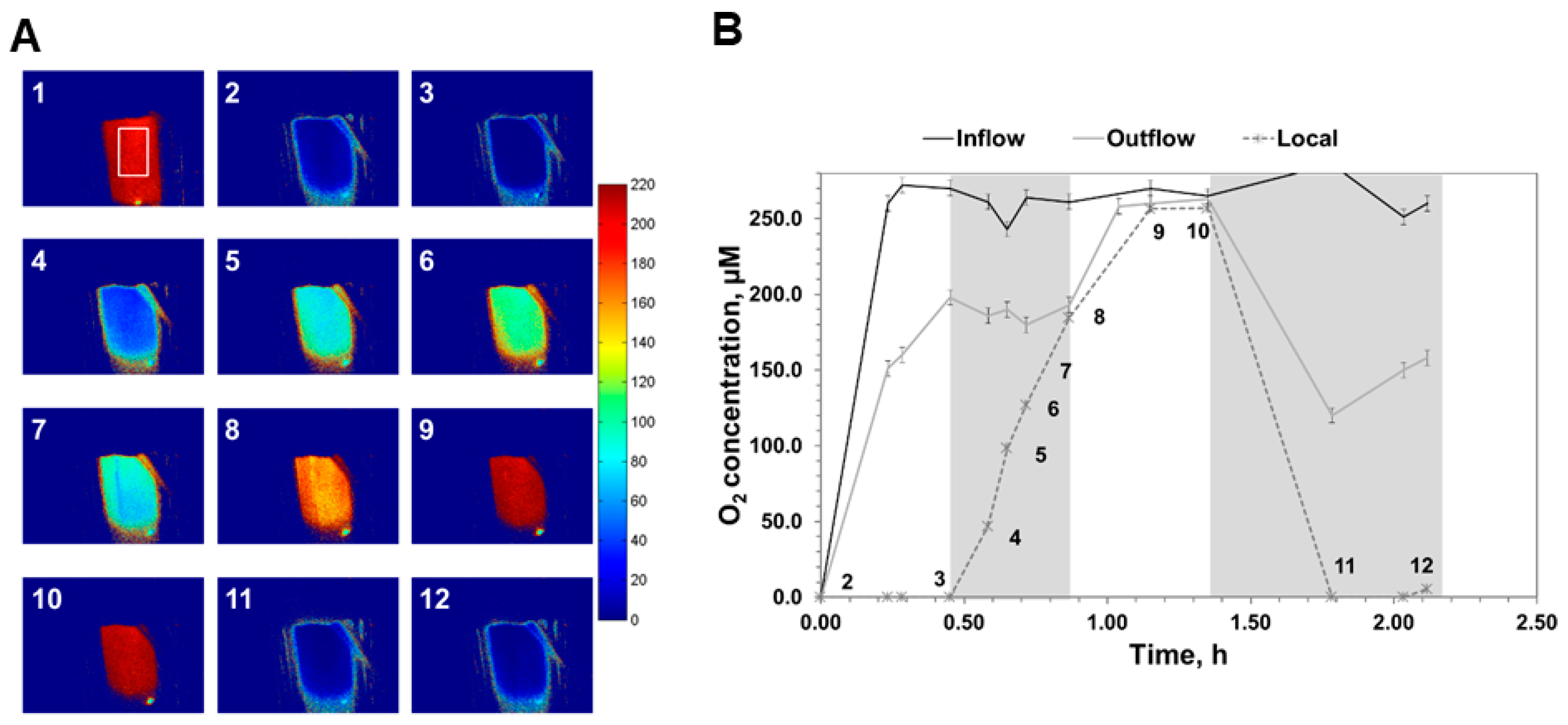
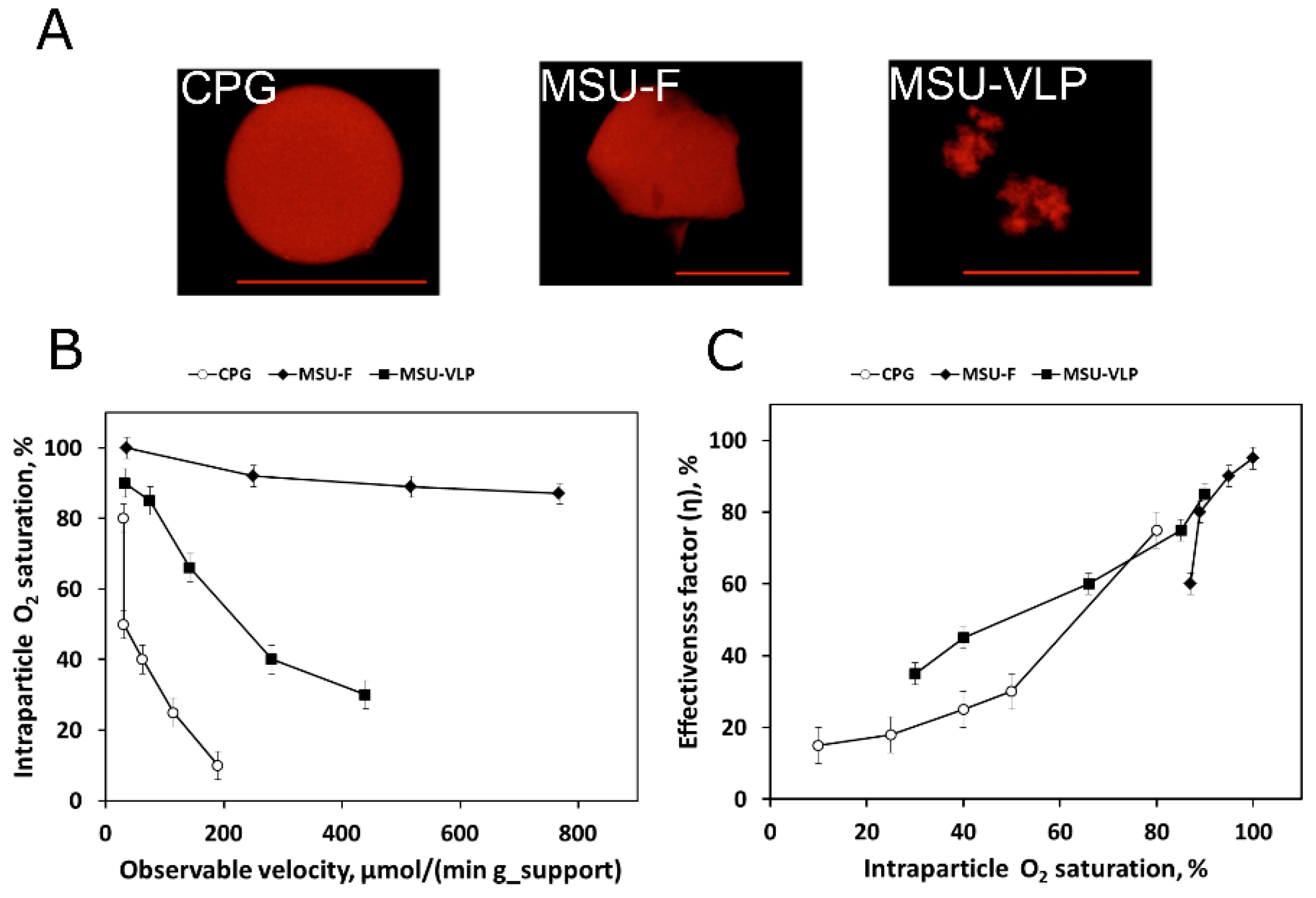
3.1. Identification and Quantification of Diffusion Limitations
3.2. Dissection of the Influence of Physical and Biochemical Factors on Catalytic Effectiveness
3.3. Screening of Suitable Immobilization Procedures
3.4. Guidance for Biocatalytic Process Intensification
3.5. The use of Reaction-Diffusion Modeling for Biocatalytic Process Development
3.6. Biocatalytic Process Design
3.7. New Strategies for Control of the Reaction
3.8. Sensing in Microreactors
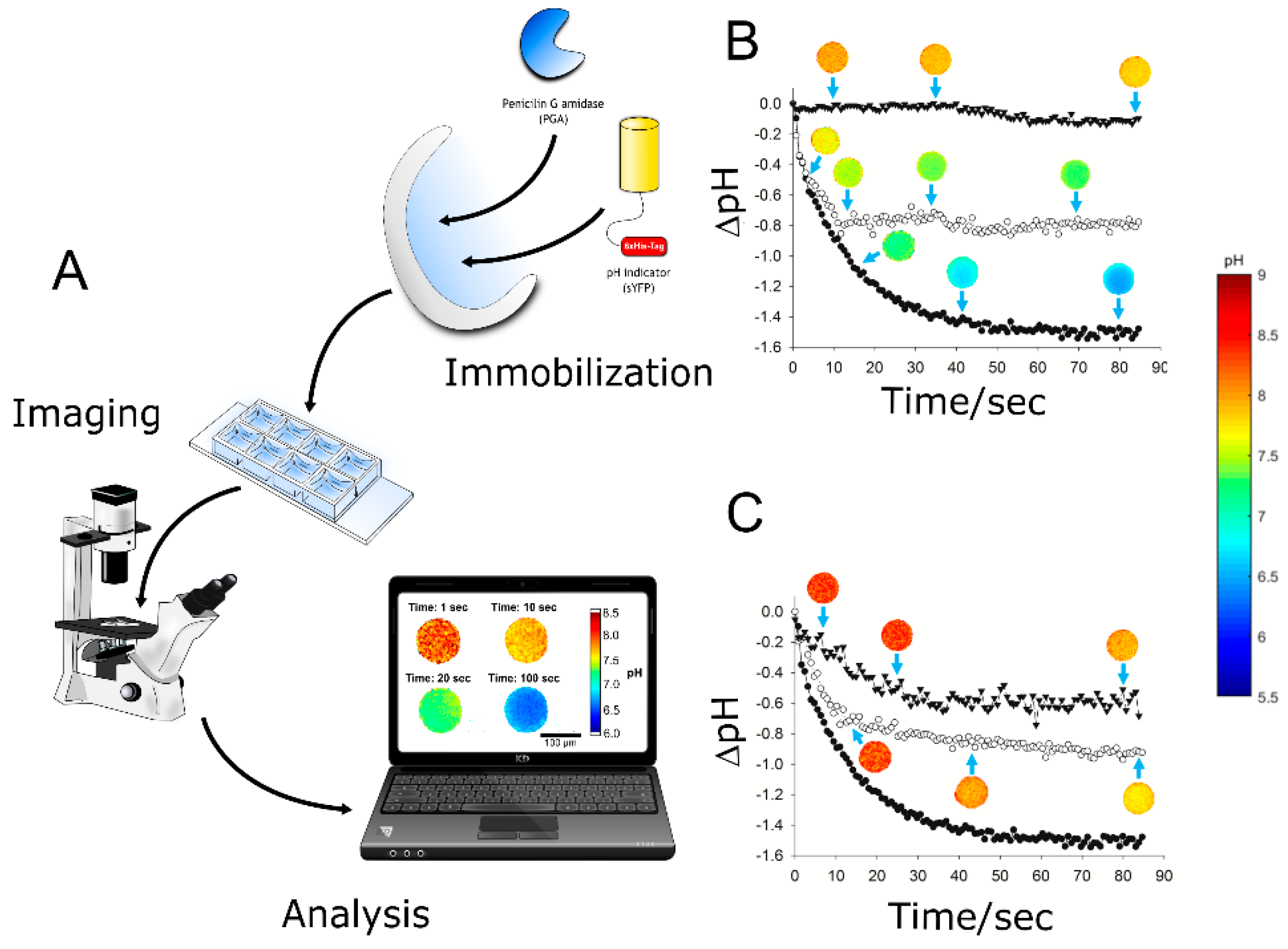
4. Spatiotemporal Sensing of Internal pH and Oxygen: An Update
5. Conclusions
Acknowledgments
Conflicts of Interest
References
- DiCosimo, R.; McAuliffe, J.; Poulose, A.J.; Bohlmann, G. Industrial use of immobilized enzymes. Chem. Soc. Rev. 2013, 42, 6437. [Google Scholar] [CrossRef] [PubMed]
- Reetz, M.T. Biocatalysis in organic chemistry and biotechnology: Past, present, and future. J. Am. Chem. Soc. 2013, 135, 12480–12496. [Google Scholar] [CrossRef] [PubMed]
- Choi, J.-M.; Han, S.-S.; Kim, H.-S. Industrial applications of enzyme biocatalysis: Current status and future aspects. Biotechnol. Adv. 2015, 33, 1443–1454. [Google Scholar] [CrossRef] [PubMed]
- Prasad, S.; Roy, I. Converting enzymes into tools of industrial importance. Recent Pat. Biotechnol. 2017, 12, 33–56. [Google Scholar] [CrossRef] [PubMed]
- Garcia-Galan, C.; Berenguer-Murcia, Á.; Fernandez-Lafuente, R.; Rodrigues, R.C. Potential of different enzyme immobilization strategies to improve enzyme performance. Adv. Synth. Catal. 2011, 353, 2885–2904. [Google Scholar] [CrossRef]
- Rodrigues, R.C.; Ortiz, C.; Berenguer-Murcia, Á.; Torres, R.; Fernández-Lafuente, R. Modifying enzyme activity and selectivity by immobilization. Chem. Soc. Rev. 2013, 42, 6290–6307. [Google Scholar] [CrossRef] [PubMed]
- Guisan, J.M. (Ed.) Immobilization of Enzymes and Cells, 3rd ed.; Humana Press: New York, NY, USA, 2013; ISBN 978-1-62703-549-1. [Google Scholar]
- Zdarta, J.; Meyer, A.S.; Jesionowski, T.; Pinelo, M. Developments in support materials for immobilization of oxidoreductases: A comprehensive review. Adv. Colloid Interface Sci. 2018, 258, 1–20. [Google Scholar] [CrossRef] [PubMed]
- Cantone, S.; Ferrario, V.; Corici, L.; Ebert, C.; Fattor, D.; Spizzo, P.; Gardossi, L. Efficient immobilisation of industrial biocatalysts: Criteria and constraints for the selection of organic polymeric carriers and immobilisation methods. Chem. Soc. Rev. 2013, 42, 6262–6276. [Google Scholar] [CrossRef]
- Santos, J.C.S.D.; Barbosa, O.; Ortiz, C.; Berenguer-Murcia, A.; Rodrigues, R.C.; Fernandez-Lafuente, R. Importance of the support properties for immobilization or purification of enzymes. ChemCatChem 2015, 7, 2413–2432. [Google Scholar] [CrossRef]
- Küchler, A.; Yoshimoto, M.; Luginbühl, S.; Mavelli, F.; Walde, P. Enzymatic reactions in confined environments. Nat. Nanotechnol. 2016, 11, 409–420. [Google Scholar] [CrossRef]
- Bolivar, J.M.; Eisl, I.; Nidetzky, B. Advanced characterization of immobilized enzymes as heterogeneous biocatalysts. Catal. Today 2016, 259, 66–80. [Google Scholar] [CrossRef]
- Bilal, M.; Cui, J.; Iqbal, H.M.N. Tailoring enzyme microenvironment: State-of-the-art strategy to fulfill the quest for efficient bio-catalysis. Int. J. Biol. Macromol. 2019, 130, 186–196. [Google Scholar] [CrossRef] [PubMed]
- Bolivar, J.M.; Consolati, T.; Mayr, T.; Nidetzky, B. Shine a light on immobilized enzymes: Real-time sensing in solid supported biocatalysts. Trends Biotechnol. 2013, 31, 194–203. [Google Scholar] [CrossRef] [PubMed]
- Secundo, F. Conformational changes of enzymes upon immobilisation. Chem. Soc. Rev. 2013, 42, 6250–6261. [Google Scholar] [CrossRef] [PubMed]
- Talbert, J.N.; Goddard, J.M. Enzymes on material surfaces. Colloids Surf. B Biointerfaces 2012, 93, 8–19. [Google Scholar] [CrossRef] [PubMed]
- Hoarau, M.; Badieyan, S.; Marsh, E.N.G. Immobilized enzymes: Understanding enzyme – surface interactions at the molecular level. Org. Biomol. Chem. 2017, 15, 9539–9551. [Google Scholar] [CrossRef] [PubMed]
- Carlsson, N.; Gustafsson, H.; Thörn, C.; Olsson, L.; Holmberg, K.; Åkerman, B. Enzymes immobilized in mesoporous silica: A physical-chemical perspective. Adv. Colloid Interface Sci. 2014, 205, 339–360. [Google Scholar] [CrossRef]
- Mohamad, N.R.; Marzuki, N.H.C.; Buang, N.A.; Huyop, F.; Wahab, R.A. An overview of technologies for immobilization of enzymes and surface analysis techniques for immobilized enzymes. Biotechnol. Biotechnol. Equip. 2015, 29, 205–220. [Google Scholar] [CrossRef]
- Benítez-Mateos, A.I.; Nidetzky, B.; Bolivar, J.M.; López-Gallego, F. Single-particle studies to advance the characterization of heterogeneous biocatalysts. ChemCatChem 2018, 10, 654–665. [Google Scholar] [CrossRef]
- Yamaguchi, A.; Namekawa, M.; Kamijo, T.; Itoh, T.; Teramae, N. Acid-base equilibria inside amine-functionalized mesoporous silica. Anal. Chem. 2011, 83, 2939–2946. [Google Scholar] [CrossRef]
- Sun, X.; Xie, J.; Xu, J.; Higgins, D.A.; Hohn, K.L. Single-Molecule studies of acidity distributions in mesoporous aluminosilicate thin films. Langmuir 2015, 31, 5667–5675. [Google Scholar] [CrossRef] [PubMed]
- Illanes, A. (Ed.) Enzyme Biocatalysis: Principles and Applications; Springer: Berlin/Heidelberg, Germany, 2008; ISBN 978-1-4020-8360-0. [Google Scholar]
- Liese, A.; Hilterhaus, L. Evaluation of immobilized enzymes for industrial applications. Chem. Soc. Rev. 2013, 42, 6236. [Google Scholar] [CrossRef] [PubMed]
- Zhang, Y.; Ge, J.; Liu, Z. Enhanced activity of immobilized or chemically modified enzymes. ACS Catal. 2015, 5, 4503–4513. [Google Scholar] [CrossRef]
- Mojet, B.L.; Ebbesen, S.D.; Lefferts, L. Light at the interface: The potential of attenuated total reflection infrared spectroscopy for understanding heterogeneous catalysis in water. Chem. Soc. Rev. 2010, 39, 4643–4655. [Google Scholar] [CrossRef] [PubMed]
- De Cremer, G.; Sels, B.F.; De Vos, D.E.; Hofkens, J.; Roeffaers, M.B.J. Fluorescence micro(spectro)scopy as a tool to study catalytic materials in action. Chem. Soc. Rev. 2010, 39, 4703–4717. [Google Scholar] [CrossRef] [PubMed]
- Gladden, L.F.; Mantle, M.D.; Sederman, A.J. Magnetic resonance imaging of catalysts and catalytic processes. In Advances in Catalysis; Elsevier: Amsterdam, The Netherlands, 2006; Volume 50, pp. 1–75. ISBN 978-0-12-007850-9. [Google Scholar]
- Buljubasich, L.; Blümich, B.; Stapf, S. Monitoring mass transport in heterogeneously catalyzed reactions by field-gradient NMR for assessing reaction efficiency in a single pellet. J. Magn. Reson. 2011, 212, 47–54. [Google Scholar] [CrossRef] [PubMed]
- Lysova, A.A.; Koptyug, I.V. Magnetic resonance imaging methods for in situ studies in heterogeneous catalysis. Chem. Soc. Rev. 2010, 39, 4585–4601. [Google Scholar] [CrossRef] [PubMed]
- Küppers, M.; Heine, C.; Han, S.; Stapf, S.; Blümich, B. In situ observation of diffusion and reaction dynamics in gel microreactors by chemically resolved NMR microscopy. Appl. Magn. Reson. 2002, 22, 235–246. [Google Scholar] [CrossRef]
- Kwak, S.; Lafleur, M. Raman spectroscopy as a tool for measuring mutual-diffusion coefficients in hydrogels. Appl. Spectrosc. 2003, 57, 768–773. [Google Scholar] [CrossRef]
- Holzberg, T.R.; Watson, V.; Brown, S.; Andar, A.; Ge, X.; Kostov, Y.; Tolosa, L.; Rao, G. Sensors for biomanufacturing process development: Facilitating the shift from batch to continuous manufacturing. Curr. Opin. Chem. Eng. 2018, 22, 115–127. [Google Scholar] [CrossRef]
- Demuth, C.; Varonier, J.; Jossen, V.; Eibl, R.; Eibl, D. Novel probes for pH and dissolved oxygen measurements in cultivations from millilitre to benchtop scale. Appl. Microbiol. Biotechnol. 2016, 100, 3853–3863. [Google Scholar] [CrossRef] [PubMed]
- Stich, M.I.J.; Fischer, L.H.; Wolfbeis, O.S. Multiple fluorescent chemical sensing and imaging. Chem. Soc. Rev. 2010, 39, 3102–3114. [Google Scholar] [CrossRef] [PubMed]
- Borisov, S.M.; Klimant, I. New luminescent oxygen-sensing and temperature-sensing materials based on gadolinium(III) and europium(III) complexes embedded in an acridone-polystyrene conjugate. Anal. Bioanal. Chem. 2012, 404, 2797–2806. [Google Scholar] [CrossRef] [PubMed]
- Beutel, S.; Henkel, S. In situ sensor techniques in modern bioprocess monitoring. Appl. Microbiol. Biotechnol. 2011, 91, 1493–1505. [Google Scholar] [CrossRef] [PubMed]
- Berezin, M.Y.; Achilefu, S. Fluorescence lifetime measurements and biological imaging. Chem. Rev. 2010, 110, 2641–2684. [Google Scholar] [CrossRef] [PubMed]
- Velasco-Lozano, S.; Benítez-Mateos, A.I.; López-Gallego, F. Co-immobilized phosphorylated cofactors and enzymes as self-sufficient heterogeneous biocatalysts for chemical processes. Angew. Chem. Int. Ed. 2017, 56, 771–775. [Google Scholar] [CrossRef]
- Borisov, S.M.; Klimant, I. Optical nanosensors—smart tools in bioanalytics. Analyst 2008, 133, 1302–1307. [Google Scholar] [CrossRef] [PubMed]
- Borisov, S.M.; Mayr, T.; Mistlberger, G.; Waich, K.; Koren, K.; Chojnacki, P.; Klimant, I. Precipitation as a simple and versatile method for preparation of optical nanochemosensors. Talanta 2009, 79, 1322–1330. [Google Scholar] [CrossRef]
- Barczak, M.; McDonagh, C.; Wencel, D. Micro-and nanostructured sol-gel-based materials for optical chemical sensing (2005–2015). Microchim. Acta 2016, 183, 2085–2109. [Google Scholar] [CrossRef]
- Wang, X.; Wolfbeis, O.S. Optical methods for sensing and imaging oxygen: Materials, spectroscopies and applications. Chem. Soc. Rev. 2014, 43, 3666. [Google Scholar] [CrossRef]
- Wencel, D.; Abel, T.; McDonagh, C. Optical Chemical pH Sensors. Anal. Chem. 2014, 86, 15–29. [Google Scholar] [CrossRef] [PubMed]
- Wang, X.; Wolfbeis, O.S. Fiber-optic chemical sensors and biosensors (2013–2015). Anal. Chem. 2016, 88, 203–227. [Google Scholar] [CrossRef] [PubMed]
- Gruber, P.; Marques, M.P.C.; Szita, N.; Mayr, T. Integration and application of optical chemical sensors in microbioreactors. Lab Chip 2017, 17, 2693–2712. [Google Scholar] [CrossRef] [PubMed]
- Gruber, P.; Marques, M.P.C.; Sulzer, P.; Wohlgemuth, R.; Mayr, T.; Baganz, F.; Szita, N. Real-time pH monitoring of industrially relevant enzymatic reactions in a microfluidic side-entry reactor (μSER) shows potential for pH control. Biotechnol. J. 2017, 12, 1600475. [Google Scholar] [CrossRef] [PubMed]
- Bolivar, J.M.; Consolati, T.; Mayr, T.; Nidetzky, B. Quantitating intraparticle O2 gradients in solid supported enzyme immobilizates: Experimental determination of their role in limiting the catalytic effectiveness of immobilized glucose oxidase. Biotechnol. Bioeng. 2013, 110, 2086–2095. [Google Scholar] [CrossRef] [PubMed]
- Spiess, A.; Schlothauer, R.; Hinrichs, J.; Scheidat, B.; Kasche, V. pH gradients in immobilized amidases and their influence on rates and yields of beta-lactam hydrolysis. Biotechnol. Bioeng. 1999, 62, 267–277. [Google Scholar] [CrossRef]
- Spiess, A.C.; Kasche, V. Direct measurement of pH profiles in immobilized enzyme carriers during kinetically controlled synthesis using CLSM. Biotechnol. Prog. 2001, 17, 294–303. [Google Scholar] [CrossRef] [PubMed]
- Luo, H.; Zhu, L.; Chang, Y.; Liu, X.; Liu, Z.; Sun, H.; Li, X.; Yu, H.; Shen, Z. Microenvironmental pH changes in immobilized cephalosporin C acylase during a proton-producing reaction and regulation by a two-stage catalytic process. Bioresour. Technol. 2017, 223, 157–165. [Google Scholar] [CrossRef]
- Heinemann, M.; Limper, U.; Büchs, J. New insights in the spatially resolved dynamic pH measurement in macroscopic large absorbent particles by confocal laser scanning microscopy. J. Chromatogr. A 2004, 1024, 45–53. [Google Scholar] [CrossRef][Green Version]
- Spiess, A.C.; Zavrel, M.; Ansorge-Schumacher, M.B.; Janzen, C.; Michalik, C.; Schmidt, T.W.; Schwendt, T.; Büchs, J.; Poprawe, R.; Marquardt, W. Model discrimination for the propionic acid diffusion into hydrogel beads using lifetime confocal laser scanning microscopy. Chem. Eng. Sci. 2008, 63, 3457–3465. [Google Scholar] [CrossRef]
- Kuwana, E.; Liang, F.; Sevick-Muraca, E.M. Fluorescence lifetime spectroscopy of a pH-sensitive dye encapsulated in hydrogel beads. Biotechnol. Prog. 2004, 20, 1561–1566. [Google Scholar] [CrossRef]
- Huang, H.Y.; Shaw, J.; Yip, C.; Wu, X.Y. Microdomain pH gradient and kinetics inside composite polymeric membranes of pH and glucose sensitivity. Pharm. Res. 2008, 25, 1150–1157. [Google Scholar] [CrossRef]
- Stanzel, M.; Brilmayer, R.; Langhans, M.; Meckel, T.; Andrieu-Brunsen, A. Systematic study on FRET-pair functionalization of mesoporous thin films for correlation of pH-sensing and ionic mesopore accessibility. Microporous Mesoporous Mater. 2019, 282, 29–37. [Google Scholar] [CrossRef]
- Kovaleva, E.G.; Molochnikov, L.S.; Venkatesan, U.; Marek, A.; Stepanova, D.P.; Kozhikhova, K.V.; Mironov, M.A.; Smirnov, A.I. Acid–Base Properties of Nanoconfined Volumes of Anodic Aluminum Oxide Pores by EPR of pH-Sensitive Spin Probes. J. Phys. Chem. C 2016, 120, 2703–2711. [Google Scholar] [CrossRef]
- Begemann, J.; Spiess, A.C. Dual lifetime referencing enables pH-control for oxidoreductions in hydrogel-stabilized biphasic reaction systems. Biotechnol. J. 2015, 10, 1822–1829. [Google Scholar] [CrossRef]
- Boniello, C.; Mayr, T.; Bolivar, J.M.; Nidetzky, B. Dual-lifetime referencing (DLR): A powerful method for on-line measurement of internal pH in carrier-bound immobilized biocatalysts. BMC Biotechnol. 2012, 12, 11. [Google Scholar] [CrossRef]
- Hanefeld, U.; Gardossi, L.; Magner, E. Understanding enzyme immobilisation. Chem. Soc. Rev. 2009, 38, 453. [Google Scholar] [CrossRef]
- Hilterhaus, L.; Minow, B.; Müller, J.; Berheide, M.; Quitmann, H.; Katzer, M.; Thum, O.; Antranikian, G.; Zeng, A.P.; Liese, A. Practical application of different enzymes immobilized on sepabeads. Bioprocess Biosyst. Eng. 2008, 31, 163–171. [Google Scholar] [CrossRef]
- Boniello, C.; Mayr, T.; Klimant, I.; Koenig, B.; Riethorst, W.; Nidetzky, B. Intraparticle concentration gradients for substrate and acidic product in immobilized cephalosporin C amidase and their dependencies on carrier characteristics and reaction parameters. Biotechnol. Bioeng. 2010, 106, 528–540. [Google Scholar] [CrossRef]
- Zucca, P.; Fernandez-Lafuente, R.; Sanjust, E. Agarose and its derivatives as supports for enzyme immobilization. Molecules 2016, 21, 1577. [Google Scholar] [CrossRef]
- Petrášek, Z.; Bolivar, J.M.; Nidetzky, B. Confocal Luminescence lifetime imaging with variable scan velocity and its application to oxygen sensing. Anal. Chem. 2016, 88, 10736–10743. [Google Scholar] [CrossRef]
- Bolivar, J.M.; Schelch, S.; Mayr, T.; Nidetzky, B. Mesoporous silica materials labeled for optical oxygen sensing and their application to development of a silica-supported oxidoreductase biocatalyst. ACS Catal. 2015, 5, 5984–5993. [Google Scholar] [CrossRef]
- Bolivar, J.M.; Schelch, S.; Mayr, T.; Nidetzky, B. Dissecting physical and biochemical factors of catalytic effectiveness in immobilized D-Amino acid Oxidase by real-time sensing of O2 availability inside porous Carriers. ChemCatChem 2014, 6, 981–986. [Google Scholar] [CrossRef]
- Consolati, T.; Bolivar, J.M.; Petrasek, Z.; Berenguer, J.; Hidalgo, A.; Guisán, J.M.; Nidetzky, B. Biobased, Internally pH-Sensitive Materials: Immobilized yellow fluorescent protein as an optical sensor for spatiotemporal mapping of pH inside porous matrices. ACS Appl. Mater. Interfaces 2018, 10, 6858–6868. [Google Scholar] [CrossRef]
- Valeur, B. Molecular Fluorescence: Principles and Applications, 1st ed.; Wiley-VCH: Weinheim, Germany, 2001; ISBN 3-527-29919-X. [Google Scholar]
- Lakowicz, J.R. Principles of Fluorescence Spectroscopy; Springer: New York, NY, USA, 2006; ISBN 978-0-387-46312-4. [Google Scholar]
- Kermis, H.R.; Kostov, Y.; Harms, P.; Rao, G. Dual excitation ratiometric fluorescent pH sensor for noninvasive bioprocess monitoring: Development and application. Biotechnol. Prog. 2002, 18, 1047–1053. [Google Scholar] [CrossRef]
- Liebsch, G.; Klimant, I.; Krause, C.; Wolfbeis, O.S. Fluorescent imaging of pH with optical sensors using time domain dual lifetime referencing. Anal. Chem. 2001, 73, 4354–4363. [Google Scholar] [CrossRef]
- Mayr, T.; Klimant, I.; Wolfbeis, O.S.; Werner, T. Dual lifetime referenced optical sensor membrane for the determination of copper(II) ions. Anal. Chim. Acta 2002, 462, 1–10. [Google Scholar] [CrossRef]
- Schwendt, T.; Michalik, C.; Zavrel, M.; Dennig, A.; Spiess, A.C.; Poprawe, R.; Janzen, C. Determination of temporal and spatial concentration gradients in hydrogel beads using multiphoton microscopy techniques. Appl. Spectrosc. 2010, 64, 720–726. [Google Scholar] [CrossRef]
- Zavrel, M.; Michalik, C.; Schwendt, T.; Schmidt, T.; Ansorge-Schumacher, M.; Janzen, C.; Marquardt, W.; Büchs, J.; Spiess, A.C. Systematic determination of intrinsic reaction parameters in enzyme immobilizates. Chem. Eng. Sci. 2010, 65, 2491–2499. [Google Scholar] [CrossRef]
- Pédelacq, J.-D.; Cabantous, S.; Tran, T.; Terwilliger, T.C.; Waldo, G.S. Engineering and characterization of a superfolder green fluorescent protein. Nat. Biotechnol. 2006, 24, 79–88. [Google Scholar] [CrossRef]
- Aliye, N.; Fabbretti, A.; Lupidi, G.; Tsekoa, T.; Spurio, R. Engineering color variants of green fluorescent protein (GFP) for thermostability, pH-sensitivity, and improved folding kinetics. Appl. Microbiol. Biotechnol. 2015, 99, 1205–1216. [Google Scholar] [CrossRef]
- Thörn, C.; Carlsson, N.; Gustafsson, H.; Holmberg, K.; Åkerman, B.; Olsson, L. A method to measure pH inside mesoporous particles using protein-bound SNARF1 fluorescent probe. Microporous Mesoporous Mater. 2013, 165, 240–246. [Google Scholar] [CrossRef]
- Bolivar, J.M.; Schelch, S.; Pfeiffer, M.; Nidetzky, B. Intensifying the O2-dependent heterogeneous biocatalysis: Superoxygenation of solid support from H2O2 by a catalase tailor-made for effective immobilization. J. Mol. Catal. B Enzym. 2016, 134, 302–309. [Google Scholar] [CrossRef]
- Zahel, T.; Boniello, C.; Nidetzky, B. Real-time measurement and modeling of intraparticle pH gradient formation in immobilized cephalosporin C amidase. Process Biochem. 2013, 48, 593–604. [Google Scholar] [CrossRef]
- Chang, Y.; Tong, S.; Luo, H.; Liu, Z.; Qin, B.; Zhu, L.; Sun, H.; Yu, H.; Shen, Z. Application of ammonium bicarbonate buffer as a smart microenvironmental pH regulator of immobilized cephalosporin C acylase catalysis in different reactors. Biotechnol. Prog. 2019, e2846. [Google Scholar] [CrossRef]
- Bolivar, J.M.; Nidetzky, B. Multiphase biotransformations in microstructured reactors: Opportunities for biocatalytic process intensification and smart flow processing. Green Process. Synth. 2013, 2, 541–559. [Google Scholar] [CrossRef]
- Sun, S.; Ungerböck, B.; Mayr, T. Imaging of oxygen in microreactors and microfluidic systems. Methods Appl. Fluoresc. 2015, 3, 034002. [Google Scholar] [CrossRef]
- Bolivar, J.M.; Krämer, C.E.M.; Ungerböck, B.; Mayr, T.; Nidetzky, B. Development of a fully integrated falling film microreactor for gas-liquid-solid biotransformation with surface immobilized O2-dependent enzyme. Biotechnol. Bioeng. 2016, 113, 1862–1872. [Google Scholar] [CrossRef]
- Viefhues, M.; Sun, S.; Valikhani, D.; Nidetzky, B.; Vrouwe, E.X.; Mayr, T.; Bolivar, J.M. Tailor-made resealable micro(bio)reactors providing easy integration of in situ sensors. J. Micromechanics Microengineering 2017, 27, 065012. [Google Scholar] [CrossRef]


| Analyte | Methodology | System | Spatiotemporal Resolution | Relevance and Comments | Ref. |
|---|---|---|---|---|---|
| pH |
|
|
|
| [49] |
| pH |
|
|
|
| [50] |
| pH |
|
|
|
| [55] |
| pH |
|
|
|
| [59,62] |
| O2 |
|
|
|
| [48] |
| O2 |
|
|
|
| [66] |
| 3,5-Dimethoxybenzaldehyde-) |
|
|
|
| [73,74] |
| O2 |
|
|
|
| [65] |
| O2 |
|
|
|
| [78] |
| pH |
|
|
|
| [58] |
| pH |
|
|
|
| [51] |
| pH |
|
|
|
| [77] |
| pH |
|
|
|
| [79] |
| O2 |
|
|
|
| [64] |
| pH |
|
|
|
| [80] |
| pH |
|
|
|
| [67] |
© 2019 by the authors. Licensee MDPI, Basel, Switzerland. This article is an open access article distributed under the terms and conditions of the Creative Commons Attribution (CC BY) license (http://creativecommons.org/licenses/by/4.0/).
Share and Cite
Bolivar, J.M.; Nidetzky, B. The Microenvironment in Immobilized Enzymes: Methods of Characterization and Its Role in Determining Enzyme Performance. Molecules 2019, 24, 3460. https://doi.org/10.3390/molecules24193460
Bolivar JM, Nidetzky B. The Microenvironment in Immobilized Enzymes: Methods of Characterization and Its Role in Determining Enzyme Performance. Molecules. 2019; 24(19):3460. https://doi.org/10.3390/molecules24193460
Chicago/Turabian StyleBolivar, Juan M., and Bernd Nidetzky. 2019. "The Microenvironment in Immobilized Enzymes: Methods of Characterization and Its Role in Determining Enzyme Performance" Molecules 24, no. 19: 3460. https://doi.org/10.3390/molecules24193460
APA StyleBolivar, J. M., & Nidetzky, B. (2019). The Microenvironment in Immobilized Enzymes: Methods of Characterization and Its Role in Determining Enzyme Performance. Molecules, 24(19), 3460. https://doi.org/10.3390/molecules24193460