Article

Insight into the Influence of Cultivar Type, Cultivation Year, and Site on the Lignans and Related Phenolic Profiles, and the Health-Promoting Antioxidant Potential of Flax (Linum usitatissimum L.) Seeds

by
Laurine Garros
1,2,3,†,
Samantha Drouet
1,2,†,
Cyrielle Corbin
1,2,
Cédric Decourtil
1,2,
Thibaud Fidel
1,2,
Julie Lebas de Lacour
1,2,
Emilie A. Leclerc
1,2,
Sullivan Renouard
1,2,
Duangjai Tungmunnithum
1,2,4,
Joël Doussot
1,2,5,
Bilal Haider Abassi
1,2,6,
Benoit Maunit
2,3,
Éric Lainé
1,2,
Ophélie Fliniaux
7,
François Mesnard
7 and
Christophe Hano
1,2,*
1
Laboratoire de Biologie des Ligneux et des Grandes Cultures (LBLGC) EA1207 INRA USC1328, Plant LIGNANS Team, Université d’Orléans, 28000 Chartres, France
2
COSM’ACTIFS, Bioactifs et Cosmétiques, CNRS GDR3711, 45067 Orléans Cedex 2, France
3
Institut de Chimie Organique et Analytique (ICOA) UMR7311, Université d’Orléans-CNRS, 45067 Orléans CEDEX 2, France
4
Department of Pharmaceutical Botany, Faculty of Pharmacy, Mahidol University, 447 Sri-Ayuthaya Road, Rajathevi, Bangkok 10400, Thailand
5
Le CNAM, Ecole Sciences Industrielles et Technologies de l’Information (SITI), Chimie Alimentation Santé Environnement Risque (CASER), 75141 Paris Cedex 3, France
6
Department of Biotechnology, Quaid-i-Azam University, 45320 Islamabad, Pakistan
7
Biologie des Plantes et Innovation (BIOPI) EA 3900, Université de Picardie Jules Verne, 80000 Amiens, France
*
Author to whom correspondence should be addressed.
These two authors contributed equally to this work and should be considered both as first authors.
Molecules 2018, 23(10), 2636; https://doi.org/10.3390/molecules23102636
Submission received: 23 September 2018 / Revised: 10 October 2018 / Accepted: 11 October 2018 / Published: 14 October 2018
(This article belongs to the Special Issue Lignans)

Abstract

:
Flaxseeds are a functional food representing, by far, the richest natural grain source of lignans, and accumulate substantial amounts of other health beneficial phenolic compounds (i.e., flavonols, hydroxycinnamic acids). This specific accumulation pattern is related to their numerous beneficial effects on human health. However, to date, little data is available concerning the relative impact of genetic and geographic parameters on the phytochemical yield and composition. Here, the major influence of the cultivar over geographic parameters on the flaxseed phytochemical accumulation yield and composition is evidenced. The importance of genetic parameters on the lignan accumulation was further confirmed by gene expression analysis monitored by RT-qPCR. The corresponding antioxidant activity of these flaxseed extracts was evaluated, both in vitro, using ferric reducing antioxidant power (FRAP), oxygen radical absorbance capacity (ORAC), and iron chelating assays, as well as in vivo, by monitoring the impact of UV-induced oxidative stress on the lipid membrane peroxidation of yeast cells. Our results, both the in vitro and in vivo studies, confirm that flaxseed extracts are an effective protector against oxidative stress. The results point out that secoisolariciresinol diglucoside, caffeic acid glucoside, and p-coumaric acid glucoside are the main contributors to the antioxidant capacity. Considering the health benefits of these compounds, the present study demonstrates that the flaxseed cultivar type could greatly influence the phytochemical intakes and, therefore, the associated biological activities. We recommend that this crucial parameter be considered in epidemiological studies dealing with flaxseeds.

Graphical Abstract

1. Introduction

The consumption of fruit, vegetables, and grains has been associated with lower risks of chronic and degeneration diseases [1]. Considering their numerous beneficial effects on human health, during the last decades, there has been an increasing interest in their uses, and flaxseeds are, therefore, considered as functional food [2]. Flaxseeds are the richest natural grain source of lignan and accumulate a substantial amount of other phenolic compounds (e.g., flavonols, hydroxycinnamic acids). In flaxseed, the foremost part of these phytochemicals is accumulated under the form of a macromolecular complex (also known as lignan macromolecule) composed of the lignan secoisolariciresinol diglucoside (SDG, Figure 1A) as the main component, and of flavonol herbacetin diglucoside (HDG, Figure 1A), as well as hydroxycinnamic acid derivatives: p-coumaric acid glucoside (CouG, Figure 1A), caffeic acid glucoside (CafG, Figure 1A), and ferulic acid glucoside (FerG, Figure 1A), ester-linked together to hydroxymethylglutaryl spacers (Figure 1B) [3,4].
The beneficial effects of lignans on human health are well recognized [5,6]. Particularly, the chemopreventive actions of SDG toward cancer, diabetes mellitus, and cardiovascular diseases have been largely described [5,7,8]. The pharmacological activity of this compound is thought to be due to its high antioxidant capacity [9,10,11] and to its phytoestrogenic activity [12]. Flavonols and hydroxycinnamic acids, the other constituents of the flaxseed lignan macromolecule, also display a wide range of health-promoting effects. The favorable actions on cardiovascular health of vegetable-rich diets have been ascribed to flavonols, and hydroxycinnamic acids have revealed powerful antioxidant properties and might be of particular interest for dermatologic applications [13,14].
Although both in vivo and in vitro data are globally in favor of a chemopreventive effect of lignans, epidemiological studies are much less conclusive, and the mechanism by which phytoestrogenic lignans prevent cancers still remains unclear [7] and requires further elucidation. This could be explained by the fact that our current knowledge concerning the genetic and environmental factors affecting productivity and yield stability of these phenolic compounds, in flaxseeds, remains partial, and little is known about the variation in antioxidant capacities of different flaxseed cultivars. Moreover, no study has put efforts toward linking the lignan content of different cultivars and the expression of genes involved in their biosynthetic pathway.
Herein we present a complete dataset concerning the relative impact of cultivar, edaphic, and climatic parameters on productivity of the main constituents of the lignan macromolecule of flaxseeds, in relation to their antioxidant capacities determined using both in vitro and in vivo systems. Such data could be useful to predict, more precisely, the accumulation and, therefore, the nutritional intakes of these compounds, with health benefits for pharmaceutical, nutraceutical, and/or cosmetic applications.

2. Materials and Methods

2.1. Chemicals

All chemicals were of analytical grade quality and purchased from Thermo (Illkirch, France). The deionized water was produced using a milli-Q water purification system (Merck Millipore, Molsheim, France). SDG and HDG standard were purchased from LGC Standards (Molsheim, France). The hydroxycinnamic acid glucosides—p-coumaric acid glucoside, caffeic acid glucoside, ferulic acid glucoside—were synthesized according to Beejmohun et al. (2004) [15] and Beejmohun et al. (2006) [16]. Prior to their use for HPLC or LC-MS analysis, all solutions were filtered through 0.45 µm nylon syringe membranes (Merck Millipore, Molsheim, France).

2.2. Plant Materials and Cultivation

Flax cultivars Astral, Baïkal, Baladin, Barbara, and Oliver were provided by Laboulet Semences (Airaines, France), Coopérative Linière Terre de Lin (Saint-Pierre-le-Viger, France) and Arvalis-Institut Technique du Lin (Boigneville, France). Flax was grown up to seed at the following locations in France: Eure (Gamaches-en-Vexin, GAM, 49°16′14′’N/1°37′02′’E/89 m), Somme (Airaines, AIR, 49°57′57″N/1°56′39″/70 m), and Eure-et-Loir (Chartres, CHA, 48°27′21.05″N/1°29′3.06″E/141 m). Sowings were performed on March 30th of each year, with 450 seeds per m2. Fields were fertilized, immediately after sowing, with 80 units of nitrogen, 60 units of potassium, and 60 units of phosphorus per hectare (Figure S1). The soils of these sites were of clay loam type balanced, well-structured with a granulometry of ca. 25% 2000–63 µm, 50% 63–2 µm, and 25% <2 µm particles, and a pH around 7.8. The final harvest took place on August 15th of each year at the same ripening stage for each cultivar. Throughout the experiments, no visible disease or insect attack occurred at either location. During the growing period, the experimental stations received 182.8 mm (year 2003), 305.0 mm (year 2004), 269.8 mm (year 2005) for GAM, 387.4 mm for AIR (year 2005), and 308.6 mm for CHA (year 2005) of rainfall over the growing period. The day temperatures at an elevation of 2 m averaged 15.72 °C (year 2003), 13.67 °C (year 2004), 13.96 °C (year 2005) for GAM, 13.48 °C for AIR (year 2005), and 14.45 °C for CHA (year 2005) over the growing period. All these meteorological characteristics are displayed in Table S1 and Figure S2.

2.3. Gene Expression Analysis by RT-qPCR

Total RNA was extracted from 100 mg of frozen plant material in liquid nitrogen as described by Hano et al. (2006) [17]. Expression patterns of LuDIR5, LuPLR1, and LuUGT74S1 were analyzed using RT-qPCR, using specific primers described by Dalisay et al. (2015) [18]. For reverse transcription, 50 ng of total RNA was incubated for 60 min at 50 °C with 1× RT buffer, 0.5 mM of each dNTP, 1 μM of oligo-dT primers, 1 unit of RiboLock, and 4 units of Omniscript Reverse Transcriptase in a total volume of 20 μL (Qiagen, Hilden, Germany). qPCR was performed with a PikoReal™ Real-Time PCR System (Thermo Fisher Scientific, Villebon-sur-Yvette, France) using DyNAmo ColorFlash SYBR Green qPCR (ThermoScientific) and specific primers. Two reference genes (CYC and ETIF5A) were used for data normalization [19]. The qPCR parameters were as follows: an initial denaturation at 95 °C for 5 min, then 40 three-step cycles of 94 °C for 10 s, primer annealing at 65 °C for 10 s, and extension at 72 °C for 30 s. After 40 cycles, an additional extension step was performed at 72 °C for 90 s. The presence of a single amplicon was confirmed by the observation of a single peak in the melting curve obtained after amplification. Expression levels were calculated and normalized using 2−ΔΔCt method [20]. Reactions were performed in three biological and two technical replicates.

2.4. Extraction, HPLC, and LC-ESI-MS Analysis

Extractions (4 biological and 2 technical replicates), quantification of compounds was carried out on a Varian liquid chromatographic system (Agilent Technology, Les Ulis, France), as well as LC-ESI-MS analyses using a Waters 2695 Alliance coupled with a single quadrupole mass spectrometer ZQ (Waters-Micromass, Manchester, UK), equipped with an electrospray ion source (ESI-MS), were performed as described in Corbin et al. (2015) [21].

2.5. Determination of the Ferric-Reducing Antioxidant Power (FRAP)

Ferric-reducing antioxidant power (FRAP) was measured as described by Benzie & Strain, (1996) [22] with little modification. Briefly, 10 μL of the extracted sample was mixed with 190 μL of FRAP (10 mM TPTZ; 20 mM FeCl3∙6H2O, and 300 mM acetate buffer pH 3.6; ratio 1:1:10 (v/v/v)). Incubation lasted 15 min at room temperature. Absorbance of the reaction mixture was measured at 630 nm with a BioTek ELX800 Absorbance Microplate Reader (Thermo Fisher Scientific, Villebon-sur-Yvette, France). Assays were made in triplicate and antioxidant capacity was expressed as Trolox C equivalent antioxidant capacity (TAEC).

2.6. Determination of Oxygen Radical Absorbance Capacity (ORAC)

Oxygen radical absorbance capacity (ORAC) assay was performed as described by Prior et al. (2003) [23]. Briefly, 10 μL of the extracted sample was mixed with 190 μL of fluorescein (0.96 µM) in 75 mM phosphate buffer pH 7.4, and incubated for at least 20 minutes at 37 °C with intermittent shaking. Then, 20 µL of 119.4 mM 2,2′-azobis-amidinopropane (ABAP, Sigma Aldrich, Saint-Quentin Fallavier, France) was added and the fluorescence intensity was measured every 5 min for 2.5 h at 37 °C using a fluorescence spectrophotometer (Bio-Rad, Marnes-la-Coquette, France) set with an excitation at 485 nm and emission at 535 nm. Assays were made in triplicate, and antioxidant capacity was expressed as Trolox C equivalent antioxidant capacity (TAEC).

2.7. Determination of the Iron-Chelating Capacity

The iron-chelating capacity was determined as described by Mladenka et al. (2011) [24]. Briefly, 10 µL of extract sample were mixed with ferrous iron at a final concentration of 50 μM in HEPES (pH 6.8) buffer and 50 µL ferrozine (5 mM aqueous solution). All experiments were performed in 96-well microplates. Each sample was measured with and without (blank) the addition of ferrozine. Absorbance was measured at 550 nm immediately after addition of ferrozine, and 5 min later with a BioTek ELX800 Absorbance Microplate Reader (Thermo Fisher Scientific, Villebon-sur-Yvette, France). Chelating activity values were expressed in µM of fixed iron.

2.8. Yeast Cells Cultivation and Treatments

Yeast (Saccharomyces cerevisiae) strain MAV203 (Invitrogen, Thermo Fisher Scientific Villebon-sur-Yvette, France) were used. Cells were grown aerobically at 30 °C in an orbital shaker (150 rpm) in complete 2.0% (w/v) glucose YPD medium (Sigma Aldrich, Saint-Quentin Fallavier, France). All extracts evaporated under nitrogen flow, dissolved in DMSO at 50 µg/mL, and added to the cells 6 h before oxidative stress induction at a final concentration of 1 mg/mL. The final concentration of DMSO applied on the cell was 1 % (v/v). For the control sample, DMSO to 0.1% of the final volume, was added. Cells were irradiated with 106.5 J/m2 UV-C (254 nm) under a Vilber VL-6.C filtered lamp (Thermo Fisher Scientific, Villebon-sur-Yvette, France), as described by Bisquert et al. (2018) [25], and then incubated overnight at 30 °C before membrane lipid peroxidation determination.

2.9. Determination of Membrane Lipid Peroxidation Using Thiobarbituric Acid-Reactive Substances (TBARS) Assay

Measurement of membrane lipid peroxide was carried out with the thiobarbituric acid (TBA; Sigma Aldrich, Saint-Quentin Fallavier, France) method described by Hano et al. (2008) [26]. Briefly, ca. 107 cells were ground using a mortar and pestle in distilled water, and centrifuged at 10,000×g for 10 min. Supernatant fractions (75 µL) were mixed with 25 µL of 3% (w/v) SDS, 50 µL of 3% TBA (w/v) in 50 mM NaOH, and 50 µL of 23% (v/v) of HCl throughout mixing between each addition. The mixture was heated at 80 °C for 20 min. After cooling on ice, the absorbance at 532 nm (A532) was measured, and non-specific absorbance at 600 nm (A600) was subtracted.

2.10. Statistical Treatment of Data

All data presented in this study are the means and the standard deviations of at least three independent replicates. ANOVAs and Pearson correlations were performed using R software version 3.0.2. PCA was performed with XL-STAT2017 software (Addinsoft, Paris, France), with each parameter considered as a discrete variable; the initial dataset was then converted into principal components (PCs), and it was possible to graphically display the relationships among the considered parameters. Gene expression and SDG content were represented using MeV4 software. All statistical tests were considered significant at p < 0.05.

3. Results and Discussion

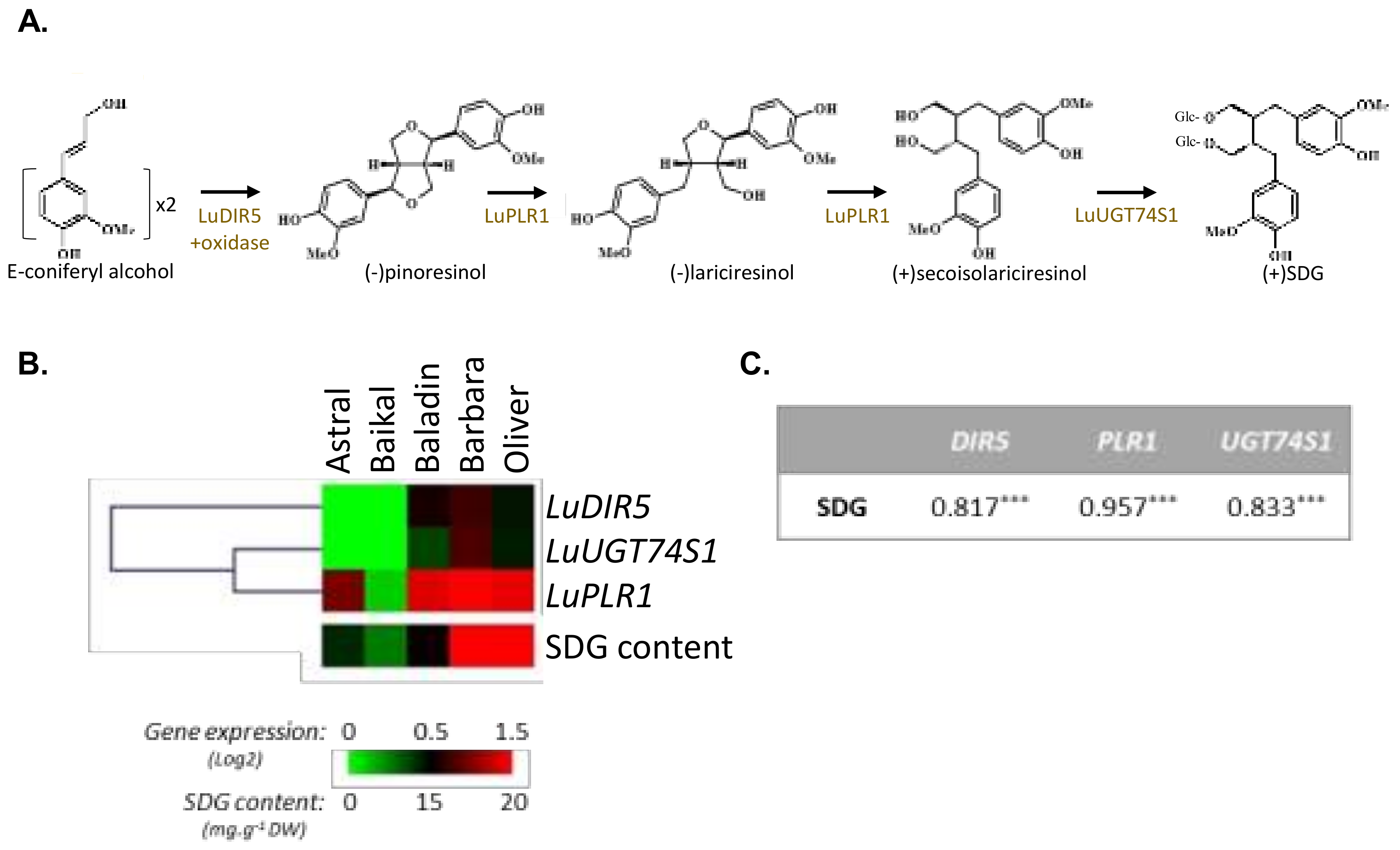
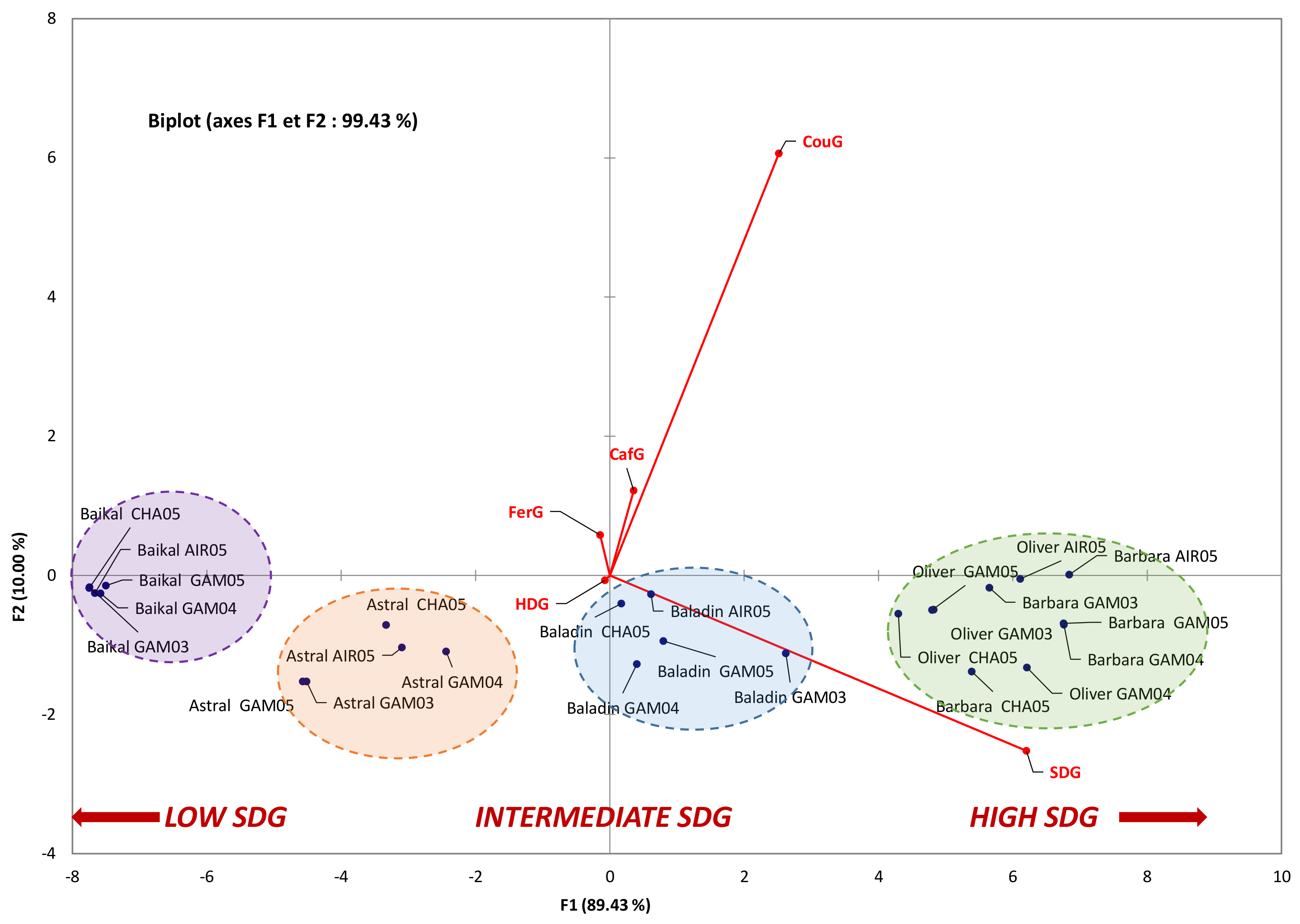
3.1. Influence of Genetic Variations on the Accumulation of the Main Constituents of the Lignan Macromolecule

The flax cultivars, herein studied, showed an SDG content ranging from 8.23 to 21.85 mg/g of dry weight (DW) (Table 1). Barbara and Oliver are high SDG-producing cultivars, Baladin presents an intermediate content, whereas Astral and Baïkal are poor in SDG, as compared to the other cultivars. A similar range of variation in SDG content has been reported in a flax germplasm collection by Diederichsen and Fu (2008) [27]. Lower SDG content was reported by Zimmermann et al. (2007, 2006) [28,29] for cultivars grown in Spain and Germany. Nevertheless, it should be noted that these authors employed an extraction method based on acid hydrolysis, which is known to be potentially destructive for SDG [30], leading to a possible underestimation in the actual contents. SDG is the main component of the lignan macromolecule accumulated in flaxseed, but other compounds, such as hydroxycinnamic acid glucosides (caffeic acid glucoside (CafG), p-coumaric acid glucoside (CouG), and ferulic acid glucoside (FerG), as well as the flavonol herbacetin diglucoside (HDG), are also incorporated in substantial amounts in this macromolecule [4,31,32]. Here, the whole set of these compounds was assayed. In our hands, CouG contents ranged from 4.78 to 10.48 mg/g DW, and FerG content from 1.03 to 2.28 mg/g DW (Table 1). These results sound consistent with those described by Westcott and Muir (1996), Jonhson et al. (2000), and Eliasson et al. (2003) [33,34,35] for cultivars grown respectively in Canada, Denmark, and Sweden. To date, only semi-quantitative evaluation of the HDG variations in flax cultivars have been studied through NMR [36], therefore, to the best of our knowledge, the present work is the first study focusing on the quantitative variations in HDG contents in linseed cultivars. Concerning the quantitative variations in CafG, only Wang et al. (2017) [37] reported very low contents ranging from 2.40 to 8.70 µg/g DW for Chinese cultivars. Here, HDG content ranged from 0.75 to 1.18 mg/g DW, and CafG contents from 0.80 to 1.90 mg/g DW (Table 1).
In flaxseed, the lignan biosynthesis involves the dirigent protein (LuDIR5; Figure 2A)-mediated stereoselective coupling of two E-coniferyl alcohol moieties, resulting in the formation of (−)-pinoresinol [18,38]. The two following reaction steps leading to the conversion of (−)-pinoresinol to (−)-lariciresinol, and (−)-lariciresinol to (+)-secoisolariciresinol, are catalyzed by the same bifunctional enzyme pinoresinol–lariciresinol reductase (LuPLR1, Figure 2A) [17,39,40]. Secoisolariciresinol is then glycosylated into SDG under the control of UDP-glycosyltransferase (LuUGT74S1, Figure 2A) glycosylating the C-9 and C-9’ hydroxyl positions [41,42]. SDG is stored as a 3-hydroxy-3-methylglutaryl ester-linked complex (HMG-SDG), as shown in Figure 1. Formation of the HMG–SDG ester-linked oligomers, occurs by linking hydroxylmethylglutaryl (HMG) to C-6a and C-6a’ position, via action of HMG CoA-transferase [43].
Correlation analysis between the different constituents of the flax lignan macromolecule revealed significant positive correlations between the CafG, CouG, and SDG contents, on the one hand, and between HDG and FerG, on the other hand (Table 2). This correlation was in agreement with our previous results [36]. On the contrary, significant negative correlations were noted between the SDG vs HDG and FerG yields (Table 2), which confirmed our previous observations [36]. From a metabolic point of view, p-coumaric acid (Figure 1A) is a branch point leading to the biosynthesis of either flavonoids or lignans [44]. Therefore, caffeic acid and p-coumaric acid (Figure 1A) could be considered as more direct precursors for the HDG biosynthesis, whereas ferulic acid (Figure 1A) constitutes a precursor for SDG biosynthesis. These biosynthetic links could explain, in part, the observed correlations. Studying the possible metabolic channel regulation of the carbon allocation between these two branches, during flaxseed development, could be of particular interest.
As a step forward, lignan biosynthetic gene expression analysis performed on immature flaxseed (developmental stage 2; [17]) by RT-qPCR using the 3 specific genes involved in SDG biosynthesis (LuDIR5, LuPLR1, and LuUGT74S1; Figure 2A,B) appeared in good agreement with the HPLC quantification (Figure 2C). High expression of LuPLR1 was detected in high SDG-producing cultivars, Barbara and Oliver, whereas Astral and Baïkal cultivars, accumulating lower SDG content, showed a lower expression of these biosynthetic genes (Table 1). The steady state levels of the key LuPLR1 transcripts [40], and the two other biosynthetic genes (LuDIR5 and LuUGT74S1) are correlated with the SDG content measured in the corresponding mature seeds (Figure 2C), confirming the great influence of genetic parameters (i.e., the cultivar), and indicated that most of the regulation occurred at transcriptional level.

3.2. Influence of Geographic Parameters on the Accumulation of the Main Constituents of Lignan Macromolecule

It is well accepted that environmental conditions, such as the climate of the culture year and the location (soil conditions), could also greatly affect the accumulation of phenolic compounds, as previously observed by Oomah et al. (1996) [45] for the accumulation of total flavonoids in flaxseeds. Here, three different locations have been selected to provide access to the potential influence of edaphic condition on lignan accumulation in flaxseed. Bordered by four different seas, three mountain ranges, and the edge of the central European lowlands, France is known to be a country with very diverse climatic conditions, resulting in very different weather patterns. Here, the three selected experimental sites are representative of the major flax-growing areas in France, i.e., the western part, and the present contrasting weather patterns. The CHA site is characterized by the highest temperatures and the lowest rainfall during the seed maturation phase. On the contrary, AIR location presents the lowest temperatures and the highest rainfall observed during the same period. The last location, GAM, is considered as an intermediate in terms of climate. The impact of these different conditions, on the composition and amount of the main constituents of the lignan macromolecule accumulated in the seed of the five selected cultivars, are presented in Table 1. Analysis of the variance revealed that cultivar was the main contributor for the observed variability (cultivars, C, Table 1). Edaphic factor (location L, Table 1) has no significant effect on the accumulation of these phytochemicals, whereas significant interactions with genetic factor were noted, but evidenced the prominent effect of genetic background at a particular location according to F values (Table 1).
Nonetheless, the location constitutes a complex variable, differing by both climatic and edaphic parameters, thus, to evaluate the sole contribution of climate, we decided to compare flaxseeds grown at the same site, GAM (i.e., the same edaphic parameters) but in different cultivation years (i.e., different climatic parameters). Here, we chose to consider three consecutive years with very contrasting weather patterns and, for this reason, the 2003–2005 period was selected. Indeed, it must be noted that the summer of 2003 was the hottest and driest in recent decades, and must be regarded as extremely unusual. The 2003–2005 period was also the warmest period recorded in France since 1950, whereas the low rainfall observed from June 2004 to December 2005 led to a dramatic soil water deficit for 2005, with a soil humidity index close to 0.25 for GAM region (considering that a soil humidity index of 1 is for water-saturated soil whereas 0 is for water-depleted soil; see Table S1, Figure S2 for complete meteorological condition descriptions). As flax is known to be a water-demanding crop during its flowering period (i.e., June), we therefore decided to evaluate how these climate changes, leading to water deficiency during this period, have affected the flaxseed metabolism. The results are reported in Table 1, and the analysis of the variance evidenced the genetic background (cultivars, C, Table 1) as the sole significant factor influencing the SDG, FerG, CouG, and CafG content (Table 1). The climatic parameters considered here (cultivation year, Y, Table 1) did not influence the accumulation of any molecules in the analyzed cultivars, whereas significant interaction between genetic and climatic parameters (C × Y, Table 1) was noted, but with lower F values as compared to genetic parameter alone (C, Table 1), indicating that the main contributions have to be attributed to this latter parameter. Our results are in good agreement with the results of Saastamoinen et al. (2013) [46], who also reported a lower impact of the cultivation year compared to the cultivar parameter on SDG accumulation. On the contrary, Wescott et al. (2002) [47] reported that the cultivation year could also influence SDG yield. This apparently contradictory result can be due to the complexity of the climatic variable, that could also be influenced by the nature of the soil considered (edaphic parameters). The nature of the soil could greatly affect the influence of the drought period as its ability to retain water greatly relied on its composition and granulometry. Indeed, a high soil ability to retain water could alleviate the effect of temporary drought, and differences in this feature could explain such apparent discrepancies, moreover, the rainfall regime differs between Scandinavian and Canadian summers, making it more probable that drought occurs during the latter.
All these phytochemical profiles were subjected to principal component analysis. The resulting biplot representation accounts for 99.43% (F1 + F2) of the initial variability of the data (Figure 3). Discrimination occurs mainly in the first dimension, and SDG content was the main contributor for this F1 axis that explains 89.43% of the initial variability. The concentrations of hydroxycinnamic acid glucosides (particularly CouG) were the main contributors for the second dimension (F2 axis), accounting for only 10% of the initial variability (Figure 3). PCA showed a significant grouping of samples as a function of their SDG content. Using this analysis, the different cultivars could also be easily discriminated. This PCA confirmed the prominence of the genetic background over the environmental (edaphic and climatic) factors studied here.

3.3. Evaluation and Comparison of In Vitro and In Vivo Antioxidant Capacities

To evaluate the influence of genetic and edaphic variables on the health benefit potential of these flaxseeds, the antioxidant capacity of the corresponding extracts was then evaluated using both in vitro and in vivo assays. On the basis of the chemical reaction involved, the major antioxidant capacity assays can be roughly divided into two categories: i) hydrogen atom transfer (HAT) reaction-based assay, such as ORAC assay, or ii) electron transfer (ET) reaction-based assay, such as FRAP assay (Table 3).
In our hands, the antioxidant capacity of our flaxseed extracts revealed by these two different assays ranged from 217.35 (Baladin, AIR_05) to 355.75 (Oliver, GAM_05) µM of Trolox C equivalent antioxidant capacity (TEAC) using FRAP assay, and from 269.97 (Baïkal, GAM_04) to 375.76 (Oliver, GAM_05) µM TEAC using ORAC assay (Table 3). The antioxidant capacity of polyphenolic compounds, such as lignans, has been previously attributed to their capacity for HAT, from their OH groups to the free radicals [48]. However, the radical scavenging capacity of these extracts occurring through an ET-based mechanism cannot be excluded, according to the high antioxidant values calculated from the FRAP assay (Table 3). Here these two in vitro antioxidant assays were significantly correlated with the presence of SDG, CouG and CafG (Table 2).
Besides these two mechanisms involved in the scavenging of reactive oxygen species, transient metal ion chelation is also considered as an antioxidant mechanism, since the Fenton reaction, responsible for the hydroxyl radical formation and, subsequently, radical chain reaction propagation, could be inhibited through this chelating mechanism [49,50]. Here, we evidenced that flaxseed extracts displayed an efficient iron (Fe2+)-chelating activity, ranging from 7.18 µM (Baïkal, GAM_03) to 14.72 µM (Oliver, GAM_04) of fixed iron (Table 3), that could also contribute to their antioxidant activity, largely described in the literature [9,10]. In good agreement with the recent rationalization of the iron-chelating capacity of SDG and its aglycone form secoisolariciresinol, high SDG quantities associated with elevated contents of CouG and CafG, appeared to significantly contribute to the development of a high iron-chelating capacity of the corresponding flaxseed extracts (Table 2).
It is necessary to emphasize that the assays described herein are strictly predictive results based on the chemical reaction in vitro, however, they not necessary bear a great similarity to biological systems. The validity of these data has to be, therefore, considered as limited to a strict chemical sense with context interpretation. For this reason, in order to better reflect the in vivo situation, the antioxidant activity of these extracts was further investigated for their capacity to inhibit membrane lipid peroxidation induced by UV-C in yeast cells. Yeast cells have been proven to be an excellent model to evaluate in vivo antioxidant capacity in a cellular oxidative stress context [51]. Indeed, baker’s yeast (Saccharomyces cerevisiae) is an attractive and reliable model. This organism is a true eukaryote, and the mechanisms of defense and adaptation to oxidative stress are well understood [25,52]. The in vivo anti-lipoperoxidation activity (inhibition of malondialdehyde (MDA) formation), determined using the TBARS assay, ranged from 21.68% (Baïkal, GAM_04) to 47.02% (Oliver, AIR_05) (Table 3). Interestingly, a strong and significant correlation was observed between this cellular antioxidant capacity and the SDG (PCC = 0.867), CouG (PCC = 0.806), and CafG (PCC = 0.721) contents (Table 2). However, we can note that, since the contents of SDG, CouG, and CafG are highly correlated, these parameters are not independent, and it is, therefore, difficult to definitely judge their respective contribution to this biological activity (cellular antioxidant capacity) by single correlation analysis. In yeast models, a similar protective effect against oxidative stress was previously observed on yeast cells treated with thiamine [52] and melatonin [25]. To the best of our knowledge, this the first time that this system is applied to characterize a flax extract. Our results are in agreement with those obtained using in vitro assays, and highlighted the great in vivo antioxidant potential of flaxseed extracts as already proposed by Wang et al. (2017) [37], using another cellular antioxidant assay in HepG2 cells.

4. Conclusions

During the last decades, flaxseeds have emerged as one of the key sources of antioxidant phytochemicals. Knowledge about the variation in the accumulation of these valuable constituents is, hence, of particular interest. This study constitutes the first work devoted to the influence of genetic, edaphic, and climatic parameters on the main compounds constituting the so-called lignan macromolecule of flaxseeds, and the antioxidant activities of the obtained extracts. Our results evidenced the predominant influence of genetic factors (cultivar) on the accumulation of the constituents of the lignan macromolecule in flaxseeds. The results of gene expression suggest a transcriptional regulation of this accumulation, knowledge of which would help to manipulate the phenolic contents of flax. Elucidating the complete transcription regulation of lignan biosynthesis in flax would, therefore, help to better control their accumulation. In our hands, other environmental parameters, such as geographic and climatic variables, did not result in significant changes in the lignan macromolecule accumulation. Both in vitro and in vivo antioxidant activity relied on SDG, CafG, and CouG accumulations. Future works using purified compounds will be conducted to further elucidate their respective contribution to the cellular antioxidant capacity observed with flaxseed extracts. Considering the health benefits of these compounds, the present study evidenced the importance of a better knowledge of the flax cultivar type that could greatly influence the phytochemical intakes and the associated biological activities. Therefore, we recommend that this crucial parameter be considered in epidemiological studies dealing with flaxseeds.

Supplementary Materials

The following are available online, Table S1: Meteorological characteristics of the cultivation site; Figure S1: Scheme describing cultivation conditions; Figure S2: Climatic data for the trial sites Airaines (AIR), Gamaches-en-Vexin (GAM) and Chartres (CHA) for the years 2003 (03), 2004 (04), and 2005 (05). Precipitations are expressed as cumulative monthly rainfall in mm, and temperatures are an average of daily temperature in °C.

Author Contributions

Conceptualization, C.H., F.M. and E.L.; Methodology, L.G., S.D., C.C., C.D., J.L.L., E.A.L.; Software, T.F.; Validation, C.H., E.A.L., S.R., E.L. and F.M.; Formal Analysis, C.H., D.T., J.D., B.H.A., B.M. and O.F.; Investigation L.G., S.D., and C.C.; Resources, C.H. and E.L.; Data Curation, C.H., E.L. and S.R.; Writing—Original Draft Preparation, C.H.; Writing—Review & Editing, C.H., O.F., D.T., S.R., E.L., B.H.A and F.M.; Visualization, L.G., S.D. and C.H.; Supervision, C.H., B.M. and E.L.; Project Administration, C.H.; Funding Acquisition, C.H., E.L. and B.M.

Funding

This research was supported by Cosmetosciences, a global training and research program dedicated to the cosmetic industry. Located in the heart of the Cosmetic Valley, this program led by University of Orleans is funded by the Region Centre-Val de Loire. This research was also supported by the Conseil Departemental d’Eure et Loir. BHA acknowledges research fellowship of Le Studium-Institute for Advanced Studies, Loire Valley, Orléans, France. DT gratefully acknowledges the support of French government via the French Embassy in Thailand in the form of Junior Research Fellowship Program 2018.

Acknowledgments

The authors wish to thank Laboulet Semences, Coopérative Linière Terre de Lin, Arvalis-Institut Technique du Lin, and Graines de Lin 28 for the donation of flaxseed used in this study and agronomical data on cultivars and access to experimental fields.

Conflicts of Interest

The authors declare no conflict of interest.

References

  1. Nayak, B.; Liu, R.H.; Tang, J. Effect of Processing on Phenolic Antioxidants of Fruits, Vegetables, and Grains—A Review. Crit. Rev. Food Sci. Nutr. 2015, 55, 887–919. [Google Scholar] [CrossRef] [PubMed]
  2. Oomah, B.D. Flaxseed as a functional food source. J. Sci. Food Agric. 2001, 81, 889–894. [Google Scholar] [CrossRef]
  3. Kamal-Eldin, A.; Peerlkamp, N.; Johnsson, P.; Andersson, R.; Andersson, R.E.; Lundgren, L.N.; Åman, P. An oligomer from flaxseed composed of secoisolariciresinoldiglucoside and 3-hydroxy-3-methyl glutaric acid residues. Phytochemistry 2001, 58, 587–590. [Google Scholar] [CrossRef]
  4. Struijs, K.; Vincken, J.-P.; Doeswijk, T.G.; Voragen, A.G.J.; Gruppen, H. The chain length of lignan macromolecule from flaxseed hulls is determined by the incorporation of coumaric acid glucosides and ferulic acid glucosides. Phytochemistry 2009, 70, 262–269. [Google Scholar] [CrossRef] [PubMed]
  5. Westcott, N.; Muir, A. Flax seed lignan in disease prevention and health promotion. Phytochem. Rev. 2003, 2, 401–417. [Google Scholar] [CrossRef]
  6. McCann, M.J.; Gill, C.I.R.; McGlynn, H.; Rowland, I.R. Role of Mammalian Lignans in the Prevention and Treatment of Prostate Cancer Mark. Nutr. Cancer 2005, 52, 1–14. [Google Scholar] [CrossRef] [PubMed]
  7. Lainé, E.; Hano, C.; Lamblin, F.F. Phytoestrogens: Lignans; Knasmüller, S., DeMarini, D.M., Johnson, I., Gerhäuser, C., Eds.; WILEY-VCH: Weinheim, Germany, 2009; ISBN 9783527320585. [Google Scholar]
  8. Hano, C.; Renouard, S.; Molinié, R.; Corbin, C.; Barakzoy, E.; Doussot, J.; Lamblin, F.; Lainé, E. Flaxseed (Linum usitatissimum L.) extract as well as (+)-secoisolariciresinol diglucoside and its mammalian derivatives are potent inhibitors of α-amylase activity. Bioorg. Med. Chem. Lett. 2013, 23, 3007–3012. [Google Scholar] [CrossRef] [PubMed]
  9. Prasad, K. Hydroxyl radical-scavenging property of secoisolariciresinol diglucoside (SDG) isolated from flax-seed. Mol. Cell. Biochem. 1997, 168, 117–123. [Google Scholar] [CrossRef] [PubMed]
  10. Kitts, D.D.; Yuan, Y.V.; Wijewickreme, A.N.; Thompson, L.U. Antioxidant activity of the flaxseed lignan secoisolariciresinol diglycoside and its mammalian lignan metabolites enterodiol and enterolactone. Mol. Cell. Biochem. 1999, 202, 91–100. [Google Scholar] [CrossRef] [PubMed]
  11. Hano, C.; Corbin, C.; Drouet, S.; Quéro, A.; Rombaut, N.; Savoire, R.; Molinié, R.; Thomasset, B.; Mesnard, F.; Lainé, E. The lignan (+)-secoisolariciresinol extracted from flax hulls is an effective protectant of linseed oil and its emulsion against oxidative damage. Eur. J. Lipid Sci. Technol. 2017, 119. [Google Scholar] [CrossRef]
  12. Adlercreutz, H.; Mousavi, Y.; Clark, J.; Höckerstedt, K.; Hämäläinen, E.; Wähälä, K.; Mäkela, T.; Hase, T. Dietary phytoestrogen and cancer: In vitro and In vivo studies. J. Steroid Biochem. Mol. Biol. 1992, 41, 8012–8020. [Google Scholar] [CrossRef]
  13. Schoenrock, U.; Untiedt, S.; Kux, U.; Inoue, K. Application of Ferulic Acid Glucosides as Anti-irritants in Cosmetic and Topical Dermatological Preparations. German Patent AN 1997:708582, 1997. [Google Scholar]
  14. Kosinska, A.; Penkacik, K.; Wiczkowski, W.; Amarowicz, R. Presence of caffeic acid in flaxseed lignan macromolecule. Plant Foods Hum. Nutr. 2011, 66, 270–274. [Google Scholar] [CrossRef] [PubMed]
  15. Beejmohun, V.; Grand, E.; Mesnard, F.; Fliniaux, M.A.; Kovensky, J. First synthesis of (1,2-13C2)-monolignol glucosides. Tetrahedron Lett. 2004, 45, 8745–8747. [Google Scholar] [CrossRef]
  16. Beejmohun, V.; Grand, E.; Lesur, D.; Mesnard, F.; Fliniaux, M.A.; Kovensky, J. Synthesis and purification of [1,2-13C2]coniferin. J. Label. Compd. Radiopharm. 2006, 49, 463–470. [Google Scholar] [CrossRef]
  17. Hano, C.; Martin, I.; Fliniaux, O.; Legrand, B.; Gutierrez, L.; Arroo, R.R.J.; Mesnard, F.; Lamblin, F.; Lainé, E. Pinoresinol-lariciresinol reductase gene expression and secoisolariciresinol diglucoside accumulation in developing flax (Linum usitatissimum) seeds. Planta 2006, 224, 1291–1301. [Google Scholar] [CrossRef] [PubMed]
  18. Dalisay, D.S.; Kim, K.W.; Lee, C.; Yang, H.; Rübel, O.; Bowen, B.P.; Davin, L.B.; Lewis, N.G. Dirigent Protein-Mediated Lignan and Cyanogenic Glucoside Formation in Flax Seed: Integrated Omics and MALDI Mass Spectrometry Imaging. J. Nat. Prod. 2015, 78, 1231–1242. [Google Scholar] [CrossRef] [PubMed]
  19. Huis, R.; Hawkins, S.; Neutelings, G. Selection of reference genes for quantitative gene expression normalization in flax (Linum usitatissimum L.). BMC Plant Biol. 2010, 10, 71. [Google Scholar] [CrossRef] [PubMed]
  20. Livak, K.J.; Schmittgen, T.D. Analysis of relative gene expression data using real-time quantitative PCR and the 2(-Delta Delta C(T)) Method. Methods 2001, 25, 402–408. [Google Scholar] [CrossRef] [PubMed]
  21. Corbin, C.; Fidel, T.; Leclerc, E.A.; Barakzoy, E.; Sagot, N.; Falguiéres, A.; Renouard, S.; Blondeau, J.; Ferroud, C.; Doussot, J.; et al. Development and validation of an efficient ultrasound assisted extraction of phenolic compounds from flax (Linum usitatissimum L.) seeds. Ultrason. Sonochem. 2015, 26, 176–185. [Google Scholar] [CrossRef] [PubMed]
  22. Benzie, I.; Strain, J. The ferric reducing ability of plasma (FRAP) as a measure of “antioxidant power”: The FRAP assay. Anal. Biochem. 1996, 239, 70–76. [Google Scholar] [CrossRef] [PubMed]
  23. Prior, R.L.; Hoang, H.; Gu, L.; Wu, X.; Bacchiocca, M.; Howard, L.; Hampsch-Woodill, M.; Huang, D.; Ou, B.; Jacob, R. Assays for hydrophilic and lipophilic antioxidant capacity (oxygen radical absorbance capacity (ORACFL)) of plasma and other biological and food samples. J. Agric. Food Chem. 2003, 51, 3273–3279. [Google Scholar] [CrossRef] [PubMed]
  24. Mladěnka, P.; MacÁková, K.; Filipský, T.; Zatloukalová, L.; Jahodář, L.; Bovicelli, P.; Silvestri, I.P.; Hrdina, R.; Saso, L. In vitro analysis of iron chelating activity of flavonoids. J. Inorg. Biochem. 2011, 105, 693–701. [Google Scholar] [CrossRef] [PubMed]
  25. Bisquert, R.; Muñiz-Calvo, S.; Guillamón, J.M. Protective role of intracellular Melatonin against oxidative stress and UV radiation in Saccharomyces cerevisiae. Front. Microbiol. 2018, 9, 1–11. [Google Scholar] [CrossRef] [PubMed]
  26. Hano, C.; Addi, M.; Fliniaux, O.; Bensaddek, L.; Duverger, E.; Mesnard, F.; Lamblin, F.; Lainé, E. Molecular characterization of cell death induced by a compatible interaction between Fusarium oxysporum f. sp. linii and flax (Linum usitatissimum) cells. Plant Physiol. Biochem. 2008, 46, 590–600. [Google Scholar] [CrossRef] [PubMed]
  27. Diederichsen, A.; Fu, Y.B. Flax Genetic Diversity as the Raw Material for Future Success. In Proceedings of the International Conference on Flax and Other Bast Plants, Saskatoon, SK, Canada, 21–23 July 2008; pp. 270–279. [Google Scholar]
  28. Zimmermann, R.; Bauermann, U.; Morales, F. Effects of growing site and nitrogen fertilization on biomass production and lignan content of linseed (Linum usitatissimum L.). J. Sci. Food Agric. 2006, 86, 415–419. [Google Scholar] [CrossRef]
  29. Zimmermann, R.; Bauermann, U.; Spedding, C. Effects of nitrogen fertilisation and two growing sites on biomass production and lignan content of linseed (Linum usitatissimum L.): Second year. Acta Agron. Hungarica 2007, 55, 173–181. [Google Scholar] [CrossRef]
  30. Li, H.B.; Wong, C.C.; Cheng, K.W.; Chen, F. Antioxidant properties in vitro and total phenolic contents in methanol extracts from medicinal plants. LWT-Food Sci. Technol. 2008, 41, 385–390. [Google Scholar] [CrossRef]
  31. Struijs, K.; Vincken, J.P.; Verhoef, R.; van Oostveen-van Casteren, W.H.M.; Voragen, A.G.J.; Gruppen, H. The flavonoid herbacetin diglucoside as a constituent of the lignan macromolecule from flaxseed hulls. Phytochemistry 2007, 68, 1227–1235. [Google Scholar] [CrossRef] [PubMed]
  32. Struijs, K.; Vincken, J.P.; Verhoef, R.; Voragen, A.G.J.; Gruppen, H. Hydroxycinnamic acids are ester-linked directly to glucosyl moieties within the lignan macromolecule from flaxseed hulls. Phytochemistry 2008, 69, 1250–1260. [Google Scholar] [CrossRef] [PubMed]
  33. Westcott, N.D.; Muir, A.D. Variation in the concentration of the flax seed lignan concentration with variety, location and year. In Proceedings of the 56th Flax Institute of the United States Conference, Fargo, ND, USA, 20–22 March 1996; pp. 77–80. [Google Scholar]
  34. Johnsson, P.; Kamal-Eldin, A.; Lundgren, L.N.; Aman, P. HPLC method for analysis of secoisolariciresinol diglucoside in flaxseeds. J. Agric. Food Chem. 2000, 48, 5216–5219. [Google Scholar] [CrossRef] [PubMed]
  35. Eliasson, C.; Kamal-Eldin, A.; Andersson, R.; Aman, P. High-performance liquid chromatographic analysis of secoisolariciresinol diglucoside and hydroxycinnamic acid glucosides in flaxseed by alkaline extraction. J. Chromatogr. A 2003, 1012, 151–159. [Google Scholar] [CrossRef]
  36. Ramsay, A.; Fliniaux, O.; Fang, J.; Molinie, R.; Roscher, A.; Grand, E.; Guillot, X.; Kovensky, J.; Fliniaux, M.A.; Schneider, B.; et al. Development of an NMR metabolomics-based tool for selection of flaxseed varieties. Metabolomics 2014, 10, 1258–1267. [Google Scholar] [CrossRef]
  37. Wang, H.; Wang, J.; Qiu, C.; Ye, Y.; Guo, X.; Chen, G.; Li, T.; Wang, Y.; Fu, X.; Liu, R.H. Comparison of phytochemical profiles and health benefits in fiber and oil flaxseeds (Linum usitatissimum L.). Food Chem. 2017, 214, 227–233. [Google Scholar] [CrossRef] [PubMed]
  38. Corbin, C.; Drouet, S.; Markulin, L.; Auguin, D.; Lainé, É.; Davin, L.B.; Cort, J.R.; Lewis, N.G.; Hano, C. A genome-wide analysis of the flax (Linum usitatissimum L.) dirigent protein family: From gene identification and evolution to differential regulation. Plant Mol. Biol. 2018, 97, 73–101. [Google Scholar] [CrossRef] [PubMed]
  39. von Heimendahl, C.B.I.; Schäfer, K.M.; Eklund, P.; Sjöholm, R.; Schmidt, T.J.; Fuss, E. Pinoresinol–lariciresinol reductases with different stereospecificity from Linum album and Linum usitatissimum. Phytochemistry 2005, 66, 1254–1263. [Google Scholar] [CrossRef] [PubMed]
  40. Renouard, S.; Tribalatc, M.; Lamblin, F.; Mongelard, G.; Fliniaux, O.; Corbin, C.; Marosevic, D.; Pilard, S.; Demailly, H.; Gutierrez, L.; et al. RNAi-mediated pinoresinol lariciresinol reductase gene silencing in flax (Linum usitatissimum L.) seed coat: Consequences on lignans and neolignans accumulation. J. Plant Physiol. 2014, 171, 1372–1377. [Google Scholar] [CrossRef] [PubMed]
  41. Ghose, K.; Selvaraj, K.; McCallum, J.; Kirby, C.W.; Sweeney-Nixon, M.; Cloutier, S.J.; Deyholos, M.; Datla, R.; Fofana, B. Identification and functional characterization of a flax UDP-glycosyltransferase glucosylating secoisolariciresinol (SECO) into secoisolariciresinol monoglucoside (SMG) and diglucoside (SDG). BMC Plant Biol. 2014, 14, 82. [Google Scholar] [CrossRef] [PubMed]
  42. Fofana, B.; Ghose, K.; McCallum, J.; You, F.M.; Cloutier, S. UGT74S1 is the key player in controlling secoisolariciresinol diglucoside (SDG) formation in flax. BMC Plant Biol. 2017, 17, 1–13. [Google Scholar] [CrossRef] [PubMed]
  43. Ford, J.D.; Huang, K.; Wang, H.; Davin, L.B.; Lewis, N.G. Biosynthetic Pathway to the Cancer Chemopreventive Secoisolariciresinol Diglucoside-Hydroxymethyl Glutaryl Ester-Linked Lignan Oligomers in Flax (Linum usitatissimum) Seed. J. Nat. Prod. 2001, 2, 1388–1397. [Google Scholar] [CrossRef]
  44. Żuk, M.; Kulma, A.; Dymińska, L.; Szołtysek, K.; Prescha, A.; Hanuza, J.; Szopa, J. Flavonoid engineering of flax potentiate its biotechnological application. BMC Biotechnol. 2011, 11, 10. [Google Scholar] [CrossRef] [PubMed]
  45. Dave Oomah, B.; Mazza, G.; Kenaschuk, E.O. Flavonoid content of flaxseed. Influence of cultivar and environment. Euphytica 1996, 90, 163–167. [Google Scholar] [CrossRef]
  46. Saastamoinen, M.; Pihlava, J.M.; Eurola, M.; Klemola, A.; Jauhiainen, L.; Hietaniemi, V. Yield, SDG lignan, cadmium, lead, oil and protein contents of linseed (Linum usitatissimum L.) cultivated in trials and at different farm conditions in the south-western part of Finland. Agric. Food Sci. 2013, 22, 296–306. [Google Scholar] [CrossRef]
  47. Westcott, N.D.; Muir, A.D.; Lafond, G.; McAndrew, D.W.; May, W.; Irvine, B.; Grant, C.; Shirtliffe, S.; Bruulsema, T.W. Factors Affecting the Concentration of a Nutraceutical Lignan in Flaxseed. In Proceedings of the Symposium on Fertilizing Crops for Functional Food, Indianapolis, IN, USA, 11 November 2002; pp. 1–3. [Google Scholar]
  48. Podloucká, P.; Berka, K.; Fabre, G.; Paloncýová, M.; Duroux, J.L.; Otyepka, M.; Trouillas, P. Lipid bilayer membrane affinity rationalizes inhibition of lipid peroxidation by a natural lignan antioxidant. J. Phys. Chem. B 2013, 117, 5043–5049. [Google Scholar] [CrossRef] [PubMed]
  49. Donoso-Fierro, C.; Becerra, J.; Bustos-Concha, E.; Silva, M. Chelating and antioxidant activity of lignans from Chilean woods (Cupressaceae). Holzforschung 2009, 63, 559–563. [Google Scholar] [CrossRef]
  50. Fucassi, F.; Heikal, A.; Mikhalovska, L.I.; Standen, G.; Allan, I.U.; Mikhalovsky, S.V.; Cragg, P.J. Metal chelation by a plant lignan, secoisolariciresinol diglucoside. J. Incl. Phenom. Macrocycl. Chem. 2014, 80, 345–351. [Google Scholar] [CrossRef]
  51. Steels, E.L.; Learmonth, R.P.; Watson, K. Stress tolerance and membrane lipid unsaturation in Saccharomyces cerevisiae grown aerobically or anaerobically. Microbiology 1994, 140, 569–576. [Google Scholar] [CrossRef] [PubMed]
  52. Wolak, N.; Kowalska, E.; Kozik, A.; Rapala-Kozik, M. Thiamine increases the resistance of baker’s yeast Saccharomyces cerevisiae against oxidative, osmotic and thermal stress, through mechanisms partly independent of thiamine diphosphate-bound enzymes. FEMS Yeast Res. 2014, 14, 1249–1262. [Google Scholar] [CrossRef] [PubMed]
Figure 1. Structure of phenolic compounds involved in the lignan macromolecular complex. (A) Structure of the complex components: (1) secoisolariciresinol (R = H), or secoisolariciresinol diglucoside (SDG, R = β-d-glucose), (2) herbacetin (R = H) or herbacetin diglucoside (HDG, R = β-d-glucose), (3) p-coumaric acid (R = H), or p-coumaric acid glucoside (CouG, R = β-d-glucose), (4) caffeic acid (R = H) or caffeic acid glucoside (CafG, R = β-d-glucose), (5) ferulic acid (R = H) or ferulic acid glucoside (FerG, R = β-d-glucose), (6) hydroxymethylglutaric acid (HMGA). (B) Schematic representation of lignan macromolecule, where a unit of SDG or HDG is ester-linked to another unit, thanks to HMGA, which can be replaced by one hydroxycinnamic acid glucoside (HCAG) unit (CouG, CafG, or FerG) in terminal position of the chain.
Molecules 23 02636 g001
Figure 2. Expression profile of flax lignan biosynthetic gene and SDG accumulation in five flaxseed cultivars. (A) Biosynthetic pathway leading to the formation of (+)-SDG in flaxseed. (B) Expression of LuDIR5, LuPLR1, and LuUGT74S1 determined by RT-qPCR (normalized with CYC and ETIF5A reference genes) visualized using MeV4 (n = 3) and SDG content measured by HPLC and visualized using MeV4 (n = 3). (C) Pearson correlation matrix between (+)-SDG accumulation and the corresponding biosynthetic gene expression. Significance level: * p < 0.05; ** p < 0.01; *** p < 0.001.
Molecules 23 02636 g002
Figure 3. Correlation circle for principal component analysis. The SDG, HDG, CafG, CouG, and FerG contents for 5 cultivars (Astral, Baïkal, Baladin, Barbara, and Oliver) growing at 3 different locations (GAM, AIR, and CHA) and over 3 different years (03 (2003), 04 (2004), or 05 (2005)) were submitted for analysis by the PCA algorithm in Excel-XLSTAT software, using the Pearson correlation matrix (at a significance level of p < 0.05).
Molecules 23 02636 g003
Table 1. Influence of the cultivar (C), cultivation site (L), and year (Y) on the accumulation of the main constituents of the lignan macromolecule in flaxseeds.
CultivarLocation_Year SDG aHDG aFerG aCouG aCafG a
AstralAIR_0512.85 ± 0.140.98 ± 0.061.65 ± 0.065.88 ± 0.090.85 ± 0.04
CHA_0512.53 ± 0.111.05 ± 0.031.90 ± 0.086.05 ± 0.080.98 ± 0.06
GAM_0311.73 ± 0.060.88 ± 0.041.58 ± 0.074.80 ± 0.081.23 ± 0.06
GAM_0411.68 ± 0.091.10 ± 0.051.58 ± 0.044.78 ± 0.051.25 ± 0.05
GAM_0513.48 ± 0.130.85 ± 0.011.57 ± 0.026.07 ± 0.220.87 ± 0.02
BarbaraAIR_0521.68 ± 0.170.93 ± 0.061.95 ± 0.0310.48 ± 0.121.83 ± 0.04
CHA_0520.88 ± 0.071.05 ± 0.032.18 ± 0.078.63 ± 0.651.63 ± 0.04
GAM_0320.63 ± 0.100.95 ± 0.031.78 ± 0.039.95 ± 0.111.33 ± 0.06
GAM_0421.85 ± 0.340.85 ± 0.031.78 ± 0.029.85 ± 0.261.53 ± 0.06
GAM_0521.85 ± 0.810.88 ± 0.011.66 ± 0.019.84 ± 0.071.53 ± 0.01
BaladinAIR_0516.03 ± 0.111.15 ± 0.032.10 ± 0.057.88 ± 0.071.48 ± 0.07
CHA_0515.68 ± 0.191.18 ± 0.032.28 ± 0.077.58 ± 0.101.43 ± 0.09
GAM_0318.20 ± 0.381.03 ± 0.032.03 ± 0.047.88 ± 0.121.33 ± 0.02
GAM_0416.45 ± 0.131.08 ± 0.072.08 ± 0.027.33 ± 0.211.40 ± 0.08
GAM_0516.20 ± 0.380.95 ± 0.011.92 ± 0.016.91 ± 0.041.19 ± 0.01
BaïkalAIR_058.33 ± 0.131.03 ± 0.041.75 ± 0.074.85 ± 0.070.85 ± 0.01
CHA_058.23 ± 0.071.18 ± 0.021.75 ± 0.054.85 ± 0.061.05 ± 0.03
GAM_038.23 ± 0.171.15 ± 0.051.88 ± 0.074.90 ± 0.070.80 ± 0.05
GAM_048.45 ± 0.101.05 ± 0.081.98 ± 0.034.98 ± 0.080.98 ± 0.04
GAM_058.40 ± 0.040.98 ± 0.011.79 ± 0.024.88 ± 0.050.83 ± 0.01
OliverAIR_0521.00 ± 0.250.75 ± 0.031.55 ± 0.0310.15 ± 0.091.90 ± 0.05
CHA_0519.50 ± 0.190.85 ± 0.031.33 ± 0.069.03 ± 0.261.75 ± 0.06
GAM_0319.95 ± 0.110.98 ± 0.041.18 ± 0.069.38 ± 0.071.33 ± 0.06
GAM_0419.93 ± 0.111.03 ± 0.071.03 ± 0.039.35 ± 0.091.45 ± 0.05
GAM_0521.55 ± 0.370.83 ± 0.011.06 ± 0.019.12 ± 0.081.37 ± 0.01
F valuesCultivar (C)284.62 ***4.06 *19.18 ***95.33 ***14.76 ***
Location (L)0.021.211.040.130.61
Year (Y)0.032.090.680.020.48
C × L194.69 ***3.57 *21.90 ***75.96 ***11.94 ***
C × Y198.99 ***4.58 *17.25 ***59.81 ***11.19 ***
Y × L0.021.4840.5360.0620.441
C × L × Y148.70 ***4.23 *16.18 ***51.26 ***9.62 ***
a All contents are given in mg/g DW. Values are mean ± SD of 4 independent replicates. ANOVA, F represents the effect. Significance level: * p < 0.05; ** p < 0.01; *** p < 0.001.
Table 2. Correlation analysis using Pearson correlation coefficient (PCC).
VariablesSDGHDGFerGCouGCafGFRAPORACIron ChelationMDA inhibition
SDG
HDG−0.515 **
FerG−0.231 ns0.510 **
CouG0.966 ***−0.476 *−0.215 ns
CafG0.835 ***−0.360 ns−0.063 ns0.832 ***
FRAP0.676 ***−0.624 ***−0.545 **0.639 ***0.671 ***
ORAC0.669 ***−0.325 ns−0.170 ns0.676 ***0.717 ***0.573 **
Iron Chelation0.758 ***−0.627 ***−0.692 ***0.768 ***0.661 ***0.817 ***0.665 ***
MDA inhibition0.867 ***−0.666 ***−0.482 *0.806 ***0.721 ***0.774 ***0.617 ***0.875 ***
Significance level: * p < 0.05; ** p < 0.01; *** p < 0.001; ns: not significant.
Table 3. Influence of the cultivar (C), cultivation site (L), and year (Y) on the in vitro and in vivo antioxidant activities of flaxseed extracts.
CultivarLocation_YearFRAP aORAC aIron Chelation bMDA Inhibition c
AstralAIR_05252.55 ± 2.45281.55 ± 11.169.57 ± 0.2537.10 ± 0.28
CHA_05264.41 ± 10.28317.87 ± 8.0010.11 ± 0.4136.95 ± 0.43
GAM_03322.81 ± 1.41263.13 ± 11.919.66 ± 0.1238.47 ± 0.75
GAM_04276.68 ± 4.33312.34 ± 1.129.31 ± 0.5337.71 ± 1.18
GAM_05252.55 ± 5.23263.92 ± 4.569.57 ± 0.2238.17 ± 1.07
BarbaraAIR_05332.55 ± 4.57339.45 ± 1.9511.97 ± 0.1945.95 ± 2.26
CHA_05317.61 ± 9.33341.29 ± 16.0911.35 ± 0.7244.58 ± 1.08
GAM_03292.81 ± 5.84329.45 ± 8.6511.79 ± 0.0643.05 ± 3.23
GAM_04296.41 ± 8.20336.55 ± 2.1412.32 ± 0.5346.41 ± 0.43
GAM_05331.48 ± 5.23309.45 ± 3.7210.99 ± 0.2845.65 ± 1.94
BaladinAIR_05217.35 ± 4.67286.82 ± 2.798.69 ± 0.3133.44 ± 2.16
CHA_05240.55 ± 6.31334.18 ± 5.958.24 ± 0.4433.28 ± 0.97
GAM_03254.01 ± 2.30307.61 ± 4.188.16 ± 0.4332.06 ± 0.64
GAM_04288.41 ± 11.27321.82 ± 2.608.87 ± 0.0628.85 ± 0.43
GAM_05255.08 ± 14.24281.03 ± 2.707.80 ± 0.5934.50 ± 2.16
BaïkalAIR_05262.01 ± 3.21278.66 ± 5.768.42 ± 0.3725.34 ± 1.18
CHA_05274.95 ± 3.49281.03 ± 2.798.16 ± 0.4024.89 ± 1.19
GAM_03227.61 ± 1.41305.24 ± 2.327.18 ± 0.5623.66 ± 1.62
GAM_04243.75 ± 5.42269.97 ± 4.097.62 ± 0.5521.68 ± 3.13
GAM_05243.78 ± 1.27286.29 ± 2.518.07 ± 0.1226.11 ± 0.86
OliverAIR_05350.68 ± 0.81368.66 ± 21.3914.45 ± 0.2547.02 ± 2.16
CHA_05306.28 ± 5.27374.45 ± 6.4214.54 ± 0.4446.87 ± 1.19
GAM_03302.41 ± 6.17278.92 ± 5.3913.74 ± 0.3843.21 ± 0.75
GAM_04355.75 ± 8.20326.55 ± 2.7014.72 ± 0.4745.34 ± 2.37
GAM_05343.08 ± 5.28375.76 ± 7.9114.10 ± 0.3846.87 ± 1.40
F valuesGenetic (C)11.91 ***5.37 ***188.00 ***161.33 ***
Location (L)0.021.050.040.03
Year (Y)0.060.150.040.08
C × L7.20 **4.59 **133.03 ***105.31 ***
C × Y7.34 **3.40 *133.16 ***130.60 ***
Y × L0.030.640.040.06
C × L × Y4.91 *3.41 *114.80 ***109.27 ***
a expressed in mM of Trolox C equivalent antioxidant capacity (TEAC); b expressed in µM of fixed Fe2+; c expressed in % inhibition of MDA formation relative to control cells; values are mean ± SD of 4 independent replicates. ANOVA, F represents the effect. Significance level: * p < 0.05; ** p < 0.01; *** p < 0.001.

