Article

Synthesis and Evaluation of Novel Ligustrazine Derivatives as Multi-Targeted Inhibitors for the Treatment of Alzheimer’s Disease

Key Laboratory of Molecular Target & Clinical Pharmacology, School of Pharmaceutical Sciences, Guangzhou Medical University, Guangzhou 511436, Guangdong, China
*
Authors to whom correspondence should be addressed.
These authors contributed equally to this work.
Molecules 2018, 23(10), 2540; https://doi.org/10.3390/molecules23102540
Submission received: 11 September 2018 / Revised: 25 September 2018 / Accepted: 1 October 2018 / Published: 5 October 2018
(This article belongs to the Special Issue Design and Synthesis of Bioactive Compounds)

Abstract

:
A series of novel ligustrazine derivatives 8ar were designed, synthesized, and evaluated as multi-targeted inhibitors for anti-Alzheimer’s disease (AD) drug discovery. The results showed that most of them exhibited a potent ability to inhibit both ChEs, with a high selectivity towards AChE. In particular, compounds 8q and 8r had the greatest inhibitory abilities for AChE, with IC50 values of 1.39 and 0.25 nM, respectively, and the highest selectivity towards AChE (for 8q, IC50 BuChE/IC50 AChE = 2.91 × 10⁶; for 8r, IC50 BuChE/IC50 AChE = 1.32 × 10⁷). Of note, 8q and 8r also presented potent inhibitory activities against Aβ aggregation, with IC50 values of 17.36 µM and 49.14 µM, respectively. Further cellular experiments demonstrated that the potent compounds 8q and 8r had no obvious cytotoxicity in either HepG2 cells or SH-SY5Y cells, even at a high concentration of 500 μM. Besides, a combined Lineweaver-Burk plot and molecular docking study revealed that these compounds might act as mixed-type inhibitors to exhibit such effects via selectively targeting both the catalytic active site (CAS) and the peripheral anionic site (PAS) of AChEs. Taken together, these results suggested that further development of these compounds should be of great interest.

1. Introduction

Alzheimer’s disease (AD) is a progressive neurodegenerative brain disorder that is manifested as dementia, cognitive impairment, memory loss, severe behavioral abnormalities, and ultimately death [1,2,3,4]. To date, AD is thought to be a complex, multifactorial syndrome, with many related molecular lesions contributing to its pathogenesis. Based on the existing hypothesis, AD is characterized as amyloid plaques, neurofibrillary tangles, inflammatory intermediates, and reactive oxygen species (ROS), and imposes neuronal death via a complex array of networked pathways [5].
According to the cholinergic hypothesis, the cognitive and memory deterioration of AD is due to a loss of cholinergic function in the central nervous system. Acetylcholinesterase (AChE) and butyrylcholinesterase (BuChE) are two major cholinesterases (ChE) involved in the hydrolysis and regulation of choline in vertebrates. Indeed, current treatment of AD mainly focuses on the inhibition of AChE activity in order to rectify the deficiency of cerebral acetylcholine [6,7]. However, the role of BuChE in the progression of AD has recently been proved. For instance, BuChE inhibitors could recover cholinergic activity through restoring the AChE/BuChE activity ratios, as shown in a healthy brain [8], and BuChE may play a role in AD plaque due to its contribution to a subpopulation of Aβ plaques maturation by immunostaining analysis of AD brain tissues [9]. Nevertheless, many studies have showed that both ChEs could facilitate amyloid fibril formation to yield stable ChE-Aβ complexes, which are more toxic than a single Aβ peptide [10,11]. The complex of AChE, through interaction with Aβ, promotes amyloid fibril formation through a spanning hydrophobic sequence exposed on the surface of AChE, which is close to the peripheral anionic binding site (PAS) and interacts with liposomes. Besides, an AChE-derived 35-residue peptide corresponding to the above hydrophobic sequence is incorporated into the growing Aβ-fibrils. Therefore, drugs capable of inhibiting ChE might have beneficial therapy effects on the cognitive, functional, and behavioural symptoms of AD.
As a result, the development of the inhibitor targeting ChE, especially for AChE, has been an intensive research focus and a large number of AChE inhibitors have been reported thus far. Among them, those named “dual-site” AChE inhibitors occupy a particularly prominent position for the treatment of AD since they can interact with both the CAS and the PAS of AChE, leading to the potent activation of the cholinergic system, as well as the efficient inhibition of AChE-promoted Aβ production and aggregation [12].
On the other hand, the multi-target approach has been proposed as particularly suitable for combating the heterogeneity and the multifactorial nature of AD [13]. Compared to single-targeted drugs, several pieces of literature have disclosed that, the candidate compound, possessing two or more distinct pharmacological properties closely related to the neurodegenerative process, is more effective in AD therapy [14,15,16,17,18,19]. Building on this strong foundation, ever-increasing efforts toward multi-target drug discovery are being made in the field for the treatment of AD [20].
Ligustrazine (tetramethylpyrazine (TMP), 1), one of the major efficacious components of the Chinese traditional medicine herb chuanxiong (Ligusticum wallichii), is widely used in China as a novel calcium channel antagonist for the treatment of coronary atherosclerotic cardiovascular disease and ischemic cerebrocardiac vascular disease [21]. TMP is also used with a combination of Rhizoma Curcumae Longae and Calculus bovis to treat cancer [22]. A preliminary pharmacokinetic investigation revealed that TMP could be rapidly absorbed into the blood and then crosses the blood-brain and blood-labyrinth barriers [23]. Recently, it was also reported that TMP could inhibit AChE, self-induced Aβ aggregation, by acting as the potential antioxidant [24,25,26].
Motivated by the aforementioned information and in continuation of our interest in developing multi-target inhibitors as potent candidates for the treatment of AD [27], we herein disclose the design, synthesis, and biological evaluation of a series of novel ligustrazine-based derivatives as multi-targeted inhibitors against AChE, BChE, and Aβ aggregation for potent treatment of AD. Furthermore, the structure–activity relationships (SARs) are discussed and the inhibition mechanism, the inhibitory kinetics, and the cytotoxicity of selected compounds are studied systematically. Taken together, our present results demonstrate that such compounds can serve as a promising candidate for the treatment of AD and establish a rational foundation for future design of new pharmacological agents as multi-target inhibitors of AChE, BChE, and Aβ aggregation.

2. Results and Discussion
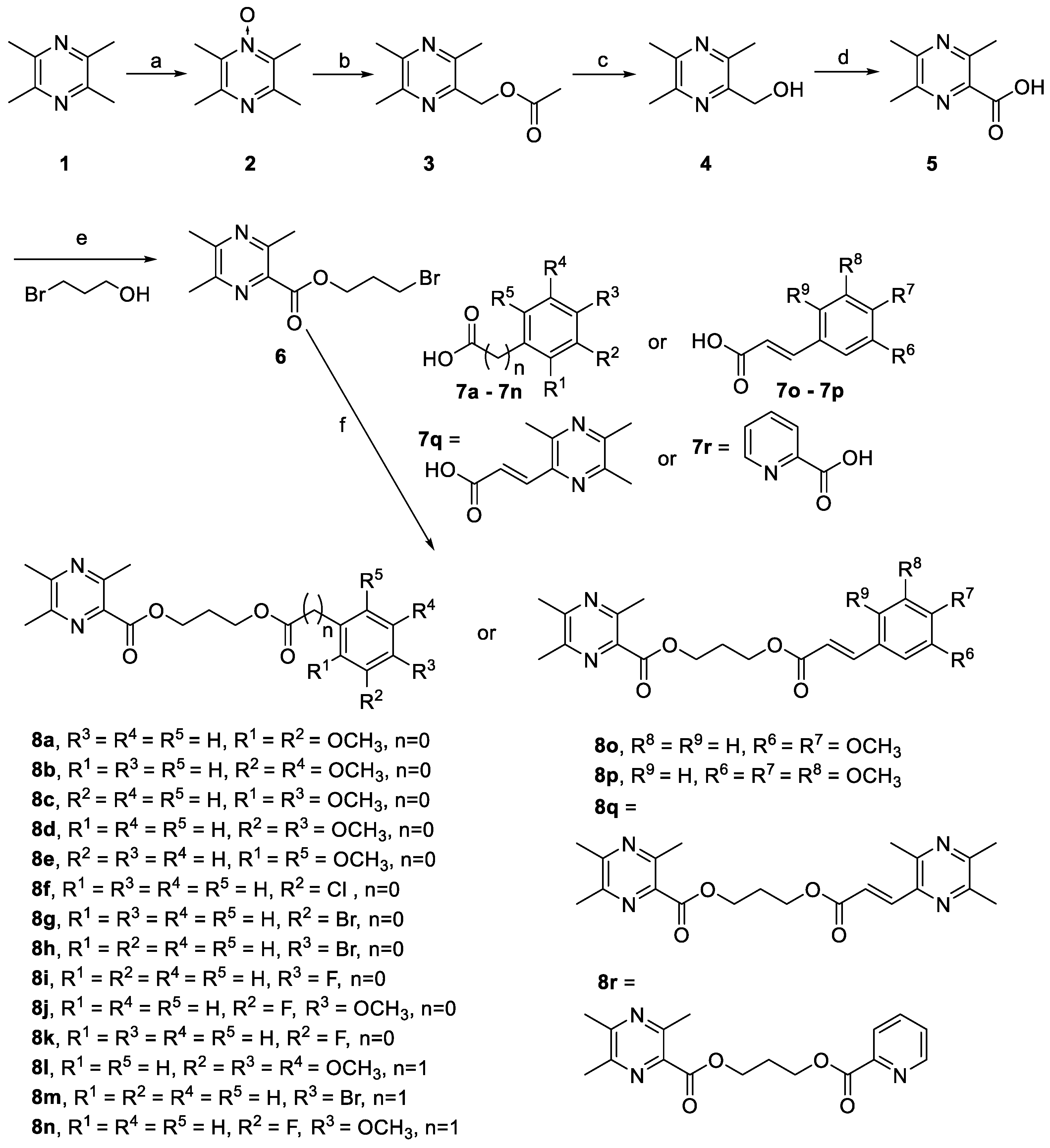
2.1. Chemistry

The ligustrazine hybrids were conveniently synthesized according to the synthetic routes shown in Scheme 1. First, compound 5 was prepared based on a previous reported method [24]. Then, it was treated with 3-bromopropan-1-ol in the presence of EDC·HCl and DMAP to yield 3-bromopropyl 3,5,6-trimethylpyrazine-2-carboxylate 6 in a yield of 73%. Finally, intermediate 6 was reacted with the corresponding carboxylic acid derivatives 7ar in the presence of DMF and anhydrous K2CO3 to afford the desired products 8ar. The structures of synthesized compounds were confirmed by 1H and 13C-NMR and HRMS. It should be noted that 3-(3,5,6-trimethylpyrazin-2-yl)acrylic acid (7q) was smoothly synthesized according to the literature, with a total yield of 60% [28].

2.2. In Vitro Inhibition Studies on AChE, BuChE and Aβ (1-42) Self-Induced Aggregation.

To determine the potential of the target compounds 8ar for the treatment of AD, their AChE (from electric eel) and BuChE (from equine serum) inhibitory activities were evaluated by using the method of Ellman [29], in which tacrine and galanthamine were employed as the reference compounds. The IC50 values of all compounds for ChEs (AChE and BuChE) and the affinity ratios were summarized as shown in Table 1. The results demonstrated that most of the tested compounds had a potent capability to inhibit AChE, even with an IC50 value at the nanomolar level. In sharp contrast, these compounds showed relatively weak inhibitory activities towards BuChE (with an IC50 value in the millimol grade). Obviously, these compounds displayed a good selectivity for AChE over BuChE, and the ratios of IC50 BuChE/IC50 AChE affinity values ranged from 1.14 × 10² to 1.32 × 10⁷.
As shown in Table 1, the reference compound-tacrine had a potent inhibitory activity, with an IC50 value of 73.36 nM, which was in good agreement with previous reports [27,30]. Interestingly, among these, compound 8r, bearing a picolinic acid group, showed the most potent inhibition for AChE, with an IC50 value of 0.25 nM, and the potency was 293-times stronger than tacrine. However, its IC50 value against BuChE was 3.30 mM, which was much weaker than AChE. Moreover, the greatest selectivity index was also obtained for this compound, with an IC50 BuChE/IC50 AChE value of 1.32 × 10⁷. It indicated that compound 8r had an exclusive selectivity for AChE over BuChE. Furthermore, an SAR analysis suggested that the electron density of the benzene ring moiety in the product played a significant role in determining the inhibitory activity of AChE, and in general, the compounds bearing the electron-donating substituent exhibited more potent AChE inhibitory activity than those bearing the electron-withdrawing group. For instance, compounds 8a8e, in which the two methoxyl substituents were installed in different positions of the benzene ring, showed strong AChE inhibitory activities, with ranges of IC50 values from 4.12 to 387.9 nM. However, the homogeneous class of compounds 8gh and 8m, bearing a bromo substituent in the benzene ring, exhibited relatively low AChE inhibitory activities compared with 8ae.
Moreover, compounds 8ln exhibited AChE inhibitory effects, with IC50 values that ranged from 303.4 µM to 7.16 mM, which was less active than 8ak (from 4.12 nM to 993.39 µM), suggesting that the introduction of the methylene part between the ester group and the benzene ring was harmful to AChE inhibitory activity. Interestingly, the introduction of the conjugated vinyl group into 8d that gave the compound 8o, demonstrated potent AChE inhibitory effects, with an IC50 value of 3.24 nM. A similar conclusion was observed in comparison with 8l and 8p. The results revealed that the introduction of a proper and relatively rigid group between the ester group and the benzene ring was beneficial to AChE inhibitory activity. Of note, replacement of the benzene ring moiety with the N-heterocyclic ring, such as pyrazine or pyridine, led to a dramatic increase in inhibitory activity, since the obtained compounds 8q and 8r were found to be the most potent inhibitors of AChE, with excellent selectivity towards AChE (for 8q, IC50 = 1.39 nM, IC50 BuChE/IC50 AChE = 1.32 × 10⁷; for 8r, IC50 = 0.25 nM, IC50 BuChE/IC50 AChE = 2.91 × 10⁶), indicating that the introduction of the nitrogen atom contributed to AChE inhibitory activities, probably due to the additional affinity with the active pocket of AChE.
Inspired by the above results, these synthesized compounds were further tested for their abilities to inhibit self-mediated aggregation of Aβ (1-42) by using a thioflavin-T fluorescence method [29] and employing the well-known tacrine as a standard reference. As summarized in Table 1, the results showed that most of the ligustrazine derivatives apparently prevented self-mediated Aβ aggregation. In particular, compounds 8b (IC50 = 3.66 µM), 8j (IC50 = 7.12 µM), and 8k (IC50 = 5.10 µM) showed higher potency than that of the reference compound tacrine (12.21 µM). Gratefully, the two best potent AChE inhibitors 8q and 8r also possessed acceptable inhibitory activities against self-induced Aβ (1-42) aggregation, with IC50 values of 17.36 µM and 49.14 µM, respectively. Taken together, the data from the ChEs and Aβ aggregation test revealed that these developed compounds might act as novel multi-targeted “hits” for potent anti-AD drug discovery. Thus, further development of such compounds should be of great interest.

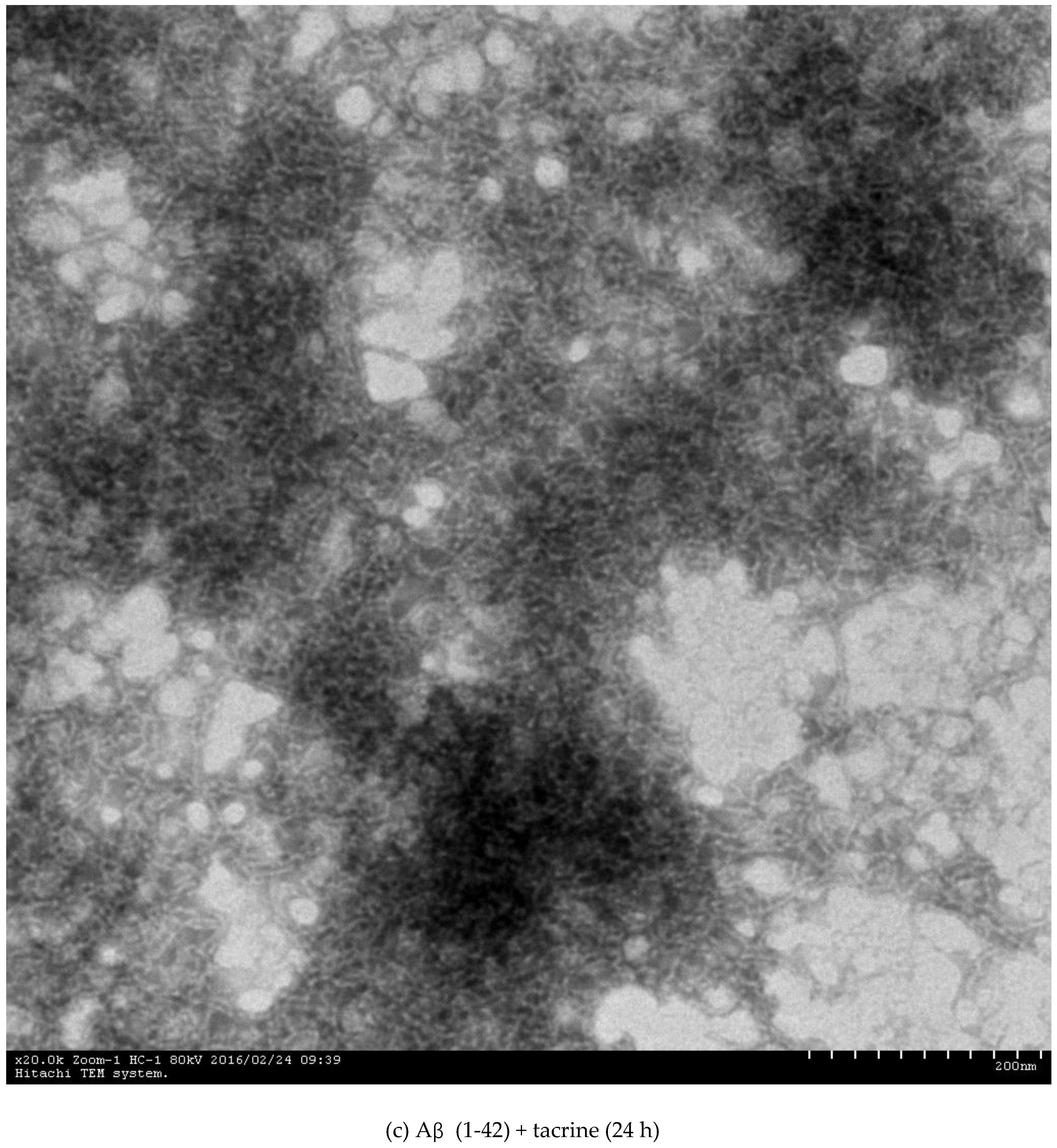
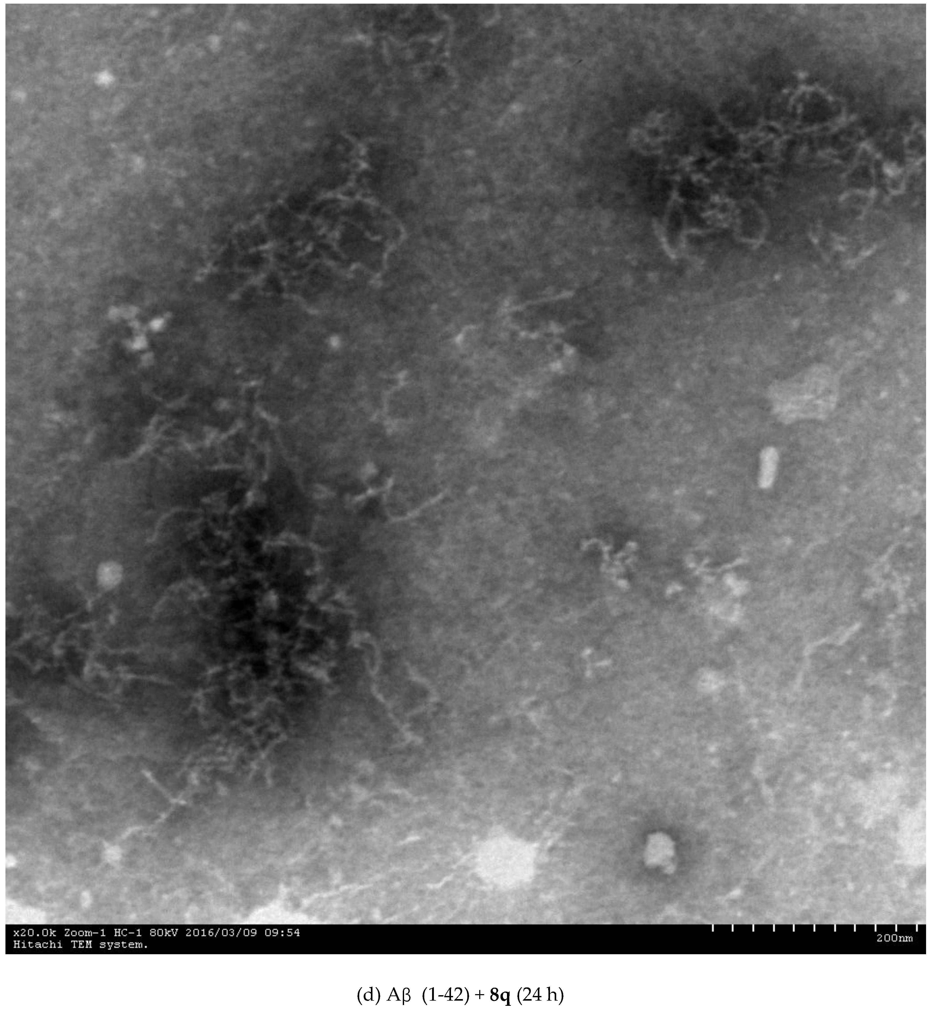
2.3. Inhibition of Aβ (1-42) Fibril Formation Monitored by Transmission Electron Microscopy (TEM)

To further confirm the ability of these compounds in inhibiting Aβ (1-42) aggregation, the inhibitory activity of the selected compound 8q was monitored by using TEM [31]. As shown in Figure 1, after 24 h of incubation at 37 °C, Aβ (1-42) alone aggregated into well-defined Aβ fibrils and amorphous deposits were observed (Figure 1a,b). In sharp contrast, no obvious Aβ fibril was detected in the presence of either tacrine or 8q (Figure 1c,d) under identical conditions. Moreover, compound 8q could obviously alleviate the formation of Aβ amorphous deposits compared with tacrine. These results were in agreement with the data from the thioflavin-T fluorescence experiment, also suggesting that 8q can serve as a promising candidate for the treatment of AD.

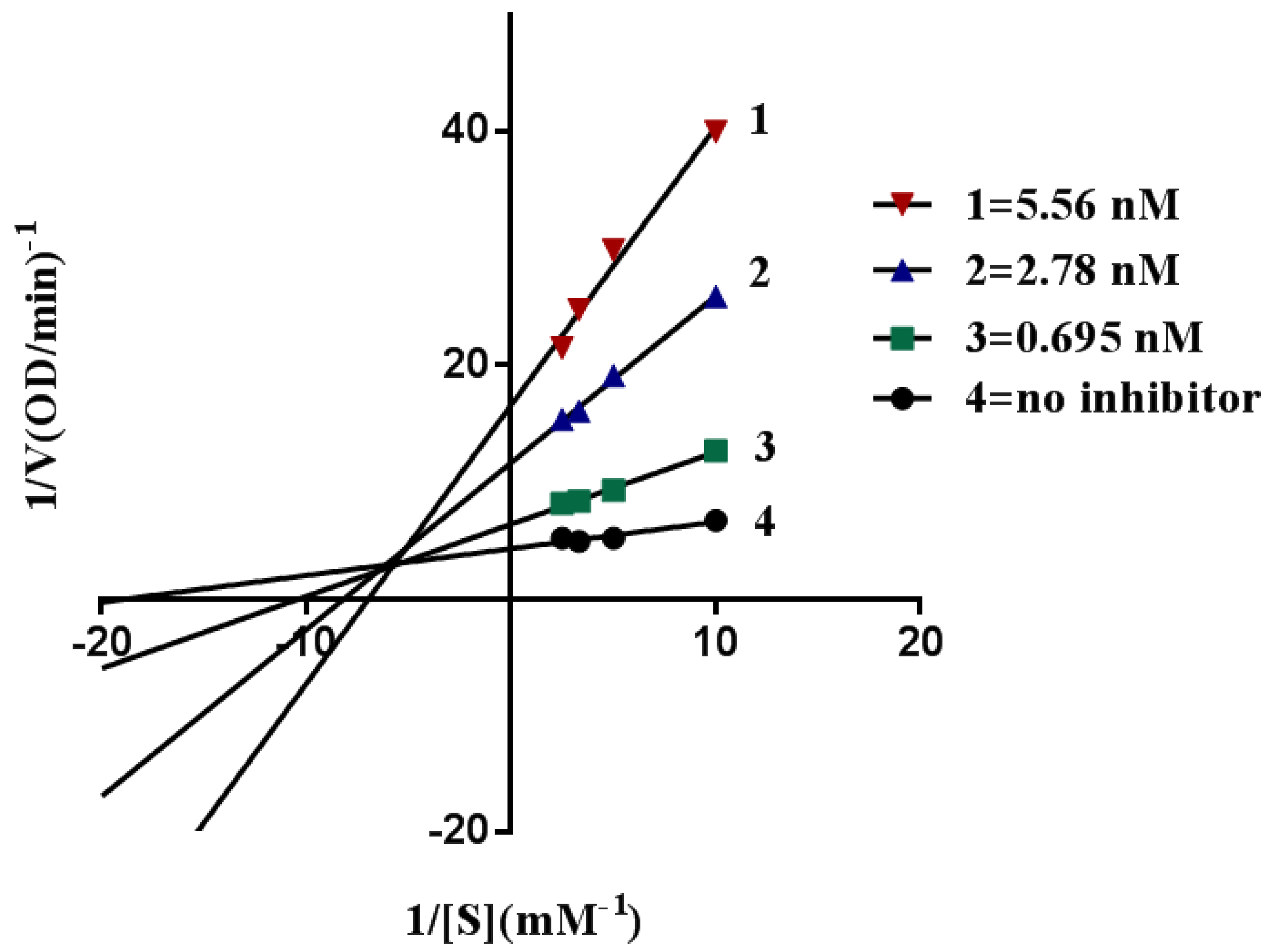
2.4. Kinetic Characterization of AChE Inhibition

Encouraged by the above results, we further selected the most potent AChE inhibitor 8q for kinetic analysis to uncover the inhibition mechanism by using the graphical analysis of the reciprocal Lineweaver–Burk plot. As demonstrated in Figure 2, the results showed that the plots of 1/V versus 1/[S] gave a family of straight lines, with different slopes that intersected one another in the second quadrant. With the increase of the concentration of 8q, the values of Vmax descended, but the values of Km kept increasing, indicating that it was a mixed-type inhibitor of AChE.

2.5. Molecular Modeling Study of AChE

To gain further insight into the molecular basis, the most active compounds 8q and 8r were selected to dock into the active site of TcAChE. It is well-known that, in previous crystallographic studies, significant conformation changes were observed in the active site of TcAChE [31,32,33,34,35]. For example, bis-tacrine derivatives with a 7-carbon spacer (PDB code 2ckm) induced the rotation of side chains of Tyr70 and Trp279 to promote the sandwich interaction with the active PAS site in the pocket of AChE, while their analogues with a 5-carbon spacer (PDB code 2cmf) caused the structural alteration of Trp279-Ser291’s loop in the CAS site [32]. Based on this information, we herein used 2ckm and 2cmf as molecular modeling for 8q containing a 7-carbon spacer and 8r with a 5-carbon spacer, respectively. The results showed that no large-scale structural displacement occurred in 8q or 8r binding to AChE, mainly due to their small molecular size compared with tacrine derivatives. Alternatively, the crystal structure of the HupA-TcAChE complex (PDB code 1vot), where most residues were retained in the native conformations, was chosen as the molecular docking model [33]. Gratefully, re-docking of the original ligand into the active site gave an acceptable RMSD difference of 0.5384, which verified the reliability of our strategy. Figure 3 shows that both 8q and 8r spanned the active gorge with an extended conformation. The proximal pyrazine ring was bound in the CAS at the bottom, stacking against the side chain of Trp84, while the distal pyrazine/pyridine ring was present in the PAS near the mouth, forming a π-π interaction with Trp279. Moreover, some hydrophobic or van der Waals interactions might occur between 8q or 8r and the surrounding residues, such as Tyr70, Asp72, Asn85, Gly119, Tyr121, Ser122, Gly123, Tyr130, Leu282, Ser286, Ile287, Phe288, Phe290, Phe330, Phe331, Tyr334, Gly335, Trp432, His440, and Tyr442. Such interactions all contributed to their potent affinities with AChE. It should be noted that, owing to the fact that BuChE did not have the PAS [36], these compounds could not form effective binding interactions with the active pocket of BuChE, thereby giving a specific selectivity towards AChE. Finally, the result also revealed that compound 8q could bind to both the CAS and the PAS of AChE.

2.6. Cytotoxicity Assay for 8q and 8r

One of the best documented side-effects of the drug discovery and development of AD is cytotoxicity [37,38,39]. To probe whether such a type of target compounds involves cytotoxic effects, we respectively treated HepG2 cells and SH-SY5Y cells with several concentrations of the corresponding compound. Cell viability was determined by a CCK-8 assay. As reported, the referenced compound tacrine showed dose-dependent cytotoxicity (Table 2 and Table 3). Excitedly, our potent “hits” 8q and 8r displayed no obvious cytotoxicity in the two cell lines, even at a high concentration of 500 μM, suggesting that potent compounds 8q and 8r have a potent safe window for the next stage of the development of anti-AD drug discovery.

3. Materials and Methods

3.1. Chemistry

All commercially available compounds and dried solvents were used without further purification. The NMR (1H and 13C) spectra were recorded on a Bruker Avnace III 400 spectrometer (Bruker Avnace, Billerica, MA, USA) or Varian Mercury–Plus 300 spectrometer (Palo Alto, CA, USA). Chemical shifts (δ) were referenced to TMS as an internal standard. Coupling constants are given in Hz. MS spectra were obtained on an Agilent 6330 Ion Trap Mass Spectrometer (Palo Alto, CA, USA). HRMS spectra were recorded on a Shimazu LCMS-IT-TOF mass spectrometer (Kyoto, Japan). Microscopy images were captured on a HITACHI H-7650 transmission electron microscope (Tokyo, Japan). TLC was monitored by precoated silica gel GF254 plates purchased from Merck& Co., (Darmstadt, Germany) and the spots were detected through a UV lamp (Yuhua Co. Ltd., Guangzhou, China) at 254 nm. Flash column chromatography was performed with silica gel (200–300 mesh) purchased from Qingdao Haiyang Chemical Co. Ltd., Qingdao, China.

3.2. Synthesis of Intermediate 6

3-Bromopropan-1-ol was added dropwise to an ice-cold solution of 5 (20.0 mmol), DMAP (10 mg), and EDC·HCl (52 mmol) in CH2Cl2 (20 mL). After the addition had been completed, the mixture was allowed to adjust to room temperature and was stirred for 24 h. When the reaction had been monitored by TLC, the solvent was removed in vacuum. The obtained brown residue was dissolved by adding distilled water, extracted with EtOAc three times (50 mL × 3), and washed with water. The organic layer was washed with brine dried over anhydrous Na2SO4. It was concentrated under reduced pressure to give compound 6 (2.08 g) as tan jelly without further purification. The yield was 73%.

3.3. General Procedures for the Preparation of 8ar

To a solution of 7 (1.22 mmol) in DMF (2 mL), anhydrous K2CO3 (1.62 mmol) was first added, 6 (0.81 mmol) in DMF (2 mL) was then added dropwise, and the reaction mixture was stirred at 30 °C for 3 h. When the reaction was completed, the mixture was diluted with water (20 mL) and extracted with EtOAc (25 mL × 3). The organic layer was washed with brine, dried over anhydrous Na2SO4, and concentrated under reduced pressure. The obtained residue was purified by silica gel chromatography to afford the desire product.
3-[(2,3-Dimethoxybenzoyl)oxy]propyl 3,5,6-trimethylpyrazine-2-carboxylate (8a). Compound 6 was treated with 2,3-dimethoxybenzoic acid (7a) according to general procedure to give the desired product 8a as a white solid. Yield 48%, 1H-NMR (CDCl3, 300 MHz) δ (ppm): 2.29 (p, J = 6.3 Hz, 2H), 2.56 (s, 6H), 2.74 (s, 3H), 3.90 (d, J = 8.5 Hz, 6H), 4.48 (t, J = 6.2 Hz, 2H), 4.58 (t, J = 6.5 Hz, 2H), 7.06 (d, J = 3.9 Hz, 2H), 7.38–7.27 (m, 1H).13C-NMR (CDCl3, 75 MHz) δ (ppm): 21.55, 22.17, 22.57, 28.07, 56.05, 61.58 (d, J = 14.0 Hz), 62.56, 115.86, 122.19, 123.80, 125.95, 139.40, 149.18 (d, J = 16.3 Hz), 151.13, 153.52, 154.50, 165.91, 166.18. ESI-HRMS Calcd. for C20H24N2O6 [M + Na]+: 411.1527; found: 411.1532.
3-[(3,5-Dimethoxybenzoyl)oxy]propyl 3,5,6-trimethylpyrazine-2-carboxylate (8b). Compound 6 was treated with 3,5-dimethoxybenzoic acid (7b) according to general procedure to give the desired product 8b as a white solid. Yield 46%, 1H-NMR (CDCl3, 300 MHz) δ (ppm): 2.30 (p, J = 6.3 Hz, 2H), 2.54(s, 3H), 2.56 (s, 3H), 2.73 (s, 3H), 3.82 (s, 6H), 4.49 (t, J = 6.2 Hz, 1H), 4.58 (t, J = 6.4 Hz, 1H), 6.63 (t, J = 2.4 Hz, 1H), 7.15 (d, J = 2.4 Hz, 2H).13C-NMR (CDCl3, 75 MHz) δ (ppm): 21.53, 22.15, 22.57, 28.10, 55.54, 61.87, 62.60, 105.61, 107.14(d, J = 4.4 Hz), 131.87, 139.29, 149.26, 151.19,154.52, 160.58, 165.86, 166.19. ESI-HRMS Calcd. for C20H24N2O6 [M + Na]+: 411.1527; found: 411.1535.
3-[(2,4-Dimethoxybenzoyl)oxy]propyl 3,5,6-trimethylpyrazine-2-carboxylate (8c). Compound 6 was treated with 2,4-dimethoxybenzoic acid (7c) according to general procedure to give the desired product 8c as a white solid. Yield 48%, 1H-NMR (CDCl3, 300 MHz) δ (ppm):2.26 (m, 2H), 2.54 (s, 6H), 2.71 (s, 3H), 3.86 (s, 3H), 3.89(s, 3H), 4.45 (t, J = 6.2 Hz, 2H), 4.55 (t, J = 6.5 Hz, 2H), 7.03 (s, 1H), 7.04 (d, J = 1.7 Hz, 1H), 7.28 (dd, J = 5.5, 3.9 Hz, 1H). 13C-NMR (CDCl3, 75 MHz) δ (ppm): 21.53, 22.14, 22.54, 28.05, 56.02, 61.55, 62.53, 115.87, 122.20, 123.78, 125.92, 139.37, 149.15, 151.09, 153.50, 154.47, 165.88, 166.15. ESI-HRMS Calcd. for C20H24N2O6 [M + Na]+: 411.1527; found: 411.1538.
3-[(3,4-Dimethoxybenzoyl)oxy]propyl 3,5,6-trimethylpyrazine-2-carboxylate (8d). Compound 6 was treated with 3,4-dimethoxybenzoic acid (7d) according to general procedure to give the desired product 8d as a white solid. Yield 41%, 1H-NMR (d6-DMSO, 400 MHz) δ (ppm): 2.18 (m, 2H), 2.43 (s, 3H), 2.48 (s, 3H), 2.57 (s, 3H), 3.77 (s, 3H), 3.83 (s, 3H), 4.38 (t, J = 6.1 Hz, 2H), 4.47 (t, J = 6.2 Hz, 2H), 7.00 (d, J = 8.5 Hz, 1H), 7.38 (d, J = 2.0 Hz, 1H), 7.54 (dd, J = 8.4, 2.0 Hz, 1H). 13C-NMR (d6-DMSO, 101 MHz) δ (ppm): 21.42, 22.15, 22.31, 28.08, 55.90, 56.15, 62.11, 62.95, 111.40, 112.02, 122.25, 123.55, 139.51, 148.76, 149.47, 150.16, 153.34, 154.77, 165.89, 165.92. ESI-HRMS Calcd. for C20H24N2O6 [M + Na]+: 411.1527; found: 411.1528.
3-[(2,6-Dimethoxybenzoyl)oxy]propyl 3,5,6-trimethylpyrazine-2-carboxylate (8e). Compound 6 was treated with 2,6-dimethoxybenzoic acid (7e) according to general procedure to give the desired product 8e as a white solid. Yield 51%, 1H-NMR (CDCl3, 300 MHz) δ (ppm): 2.25 (m, 2H), 2.56 (s, 6H), 2.73 (s, 3H), 3.81 (s, 6H), 4.49 (t, J = 6.1 Hz, 2H), 4.55 (t, J = 6.5 Hz, 2H), 6.54 (d, J = 8.4 Hz, 2H), 7.27 (t, J = 8.4 Hz, 1H). 13C-NMR (CDCl3, 75 MHz) δ21.54, 22.14, 22.55, 27.99, 55.94, 61.59, 62.53, 103.83, 112.82, 131.15, 139.59, 149.27, 150.93, 154.39, 157.33, 165.99, 166.42. ESI-HRMS Calcd. for C20H24N2O6 [M + Na]+: 411.1527; found: 411.1526.
3-[(3-Chlorobenzoyl)oxy]propyl 3,5,6-trimethylpyrazine-2-carboxylate (8f). Compound 6 was treated with 3-chlorobenzoic acid (7f) according to general procedure to give the desired product 8f as a white solid. Yield 51%, 1H-NMR (CDCl3, 300 MHz) δ (ppm): 2.30 (m, 2H), 2.53 (s, 3H), 2.55 (s, 3H), 2.72 (s, 3H), 4.49 (t, J = 6.2 Hz, 2H), 4.57 (t, J = 6.4 Hz, 2H), 7.34 (t, J = 7.9 Hz, 1H),7.50 (ddd, J = 8.0, 2.2, 1.1 Hz, 1H), 7.88 (dt, J = 7.7, 1.3 Hz, 1H), 7.95 (t, J = 1.8 Hz, 1H). 13C-NMR (CDCl3, 75 MHz) δ (ppm): 21.54, 22.16, 22.57, 28.04, 62.17, 62.55, 127.66, 129.61 (d, J = 7.1 Hz), 131.73, 132.96, 134.44, 139.21, 149.27, 151.21, 154.56, 165.19, 165.84. ESI-HRMS Calcd. for C18H19N2O4Cl [M + Na]+: 385.0926; found: 385.0936.
3-[(3-Bromobenzoyl)oxy]propyl 3,5,6-trimethylpyrazine-2-carboxylate (8g). Compound 6 was treated with 3-bromobenzoic acid (7g) according to general procedure to give the desired product 8g as a white solid. Yield 42%, 1H-NMR (d6-DMSO, 400 MHz) δ (ppm): 2.19 (m, 2H), 2.41 (s, 3H), 2.47 (s, 3H), 2.56 (s, 3H), 4.43 (t, J = 5.9 Hz, 2H), 4.48 (t, J = 6.0 Hz, 2H), 7.43 (t, J = 7.3 Hz, 1H), 7.81 (d, J = 7.9 Hz, 1H), 7.94 (d, J = 7.6 Hz, 1H), 8.04 (s, 1H). 13C-NMR (d6-DMSO, 101 MHz) δ (ppm): 165.83, 164.89, 150.22, 139.35, 138.38, 136.39, 132.50, 131.92, 131.45, 131.26, 128.55, 122.25, 63.02, 57.68, 31.86, 27.88, 22.29, 22.14, 21.40. ESI-HRMS Calcd. for C18H19N2O4Br [M + H]+: 407.0601; found: 407.0605.
3-[(4-Bromobenzoyl)oxy]propyl 3,5,6-trimethylpyrazine-2-carboxylate (8h). Compound 6 was treated with 4-bromobenzoic acid (7h) according to general procedure to give the desired product 8h as a white solid. Yield 42%, 1H-NMR (d6-DMSO, 400 MHz) δ (ppm): 2.19 (m, 2H), 2.43 (s, 3H), 2.49 (s, 3H), 2.57 (s, 3H), 4.42 (t, J = 6.0 Hz, 2H), 4.47 (t, J = 6.2 Hz, 2H), 7.66 (d, J = 8.5 Hz, 2H), 7.81 (d, J = 8.5 Hz, 2H). 13C-NMR (d6-DMSO, 101 MHz) δ (ppm): 21.43, 22.21, 22.32, 27.92, 31.93, 57.68, 62.80, 63.01, 127.74, 129.26, 131.48, 132.18, 139.45, 149.48, 154.81, 165.46, 165.90. ESI-HRMS Calcd. for C18H19N2O4Br [M + H]+: 407.0601; found: 407.0616.
3-[(4-Fluorobenzoyl)oxy]propyl 3,5,6-trimethylpyrazine-2-carboxylate (8i). Compound 6 was treated with 4-fluorobenzoic acid (7i) according to general procedure to give the desired product 8i as a white solid. Yield 42%, 1H-NMR (d6-DMSO, 400 MHz) δ (ppm):2.19 (m, 2H), 2.44 (s, 3H), 2.49 (s, 3H), 2.57 (s, 3H), 4.41 (t, J = 6.1 Hz, 2H), 4.48 (t, J = 6.2 Hz, 2H), 7.29 (t, J = 8.8 Hz, 2H), 7.98 (dd, J = 8.8, 5.6 Hz, 1H). 13C-NMR (d6-DMSO, 101 MHz) δ (ppm): 21.44, 22.17, 22.30, 27.97, 31.98, 57.70, 62.56, 62.92, 116.08, 116.30, 132.39, 132.48, 139.48, 149.52, 150.20, 154.82, 165.21, 165.92. ESI-HRMS Calcd. for C18H19N2O4F [M + H]+: 347.1402; found: 347.1411.
3-[(3-Fluoro-4-methoxybenzoyl)oxy]propyl 3,5,6-trimethylpyrazine-2-carboxylate (8j). Compound 6 was treated with 3-fluoro-4-methoxybenzoic acid (7j) according to general procedure to give the desired product 8j as a white solid. Yield 43%, 1H-NMR (d6-DMSO, 400 MHz) δ (ppm): 2.18 (m, 2H), 2.44 (s, 3H), 2.48 (s, 3H), 2.57 (s, 3H), 3.91 (s, 3H), 4.39 (t, J = 6.0 Hz, 2H), 4.47 (t, J = 6.2 Hz, 2H), 7.22 (t, J = 8.6 Hz, 1H), 7.61 (dd, J = 11.9, 2.1 Hz, 1H), 7.74 (ddd, J = 8.6, 2.1, 1.2 Hz, 1H). 13C-NMR (d6-DMSO, 101 MHz) δ (ppm): 21.42, 22.15, 22.30, 27.98, 56.77, 62.55, 63.03, 113.81, 116.55, 116.75, 122.53, 122.59, 127.14, 127.17, 139.48, 149.48, 150.18, 151.68, 151.78, 152.42, 154.77, 164.99, 165.91. ESI-HRMS Calcd. for C19H21N2O7F [M + H]+: 377.1507; found: 377.1515.
3-[(3-Fluorobenzoyl)oxy]propyl 3,5,6-trimethylpyrazine-2-carboxylate (8k). Compound 6 was treated with 3-fluoro-4-methoxybenzoic acid (7k) according to general procedure to give the desired product 8k as a white solid. Yield 48%, 1H-NMR (CDCl3, 300 MHz) δ (ppm): 2.30 (m, 2H), 2.53 (s, 3H), 2.55 (s, 3H), 2.72 (s, 3H), 4.49 (t, J = 6.1 Hz, 2H), 4.57 (t, J = 6.3 Hz, 2H), 7.34 (t, J = 7.9 Hz, 1H), 7.50 (ddd, J = 8.0, 2.2, 1.1 Hz, 1H), 7.88 (dt, J = 7.7, 1.4 Hz, 1H), 7.95 (t, J = 1.9 Hz, 1H). 13C-NMR (d6-DMSO, 101 MHz) δ (ppm): 164.69, 162.86, 160.42, 154.07, 149.95, 148.81, 138.20, 129.30, 129.22, 124.15, 118.86, 114.83, 62.00, 61.66, 26.84, 20.22, 19.70, 18.84. ESI-HRMS Calcd. for C18H19N2O4F [M + Na]+: 369.1221; found: 369.1231.
3-{[(3,4,5-Trimethoxyphenyl)acetyl]oxy}propyl 3,5,6-trimethylpyrazine-2-carboxylate (8l). Compound 6 was treated with (3,4,5-trimethoxyphenyl)acetic acid (7l) according to general procedure to give the desired product 8l as a white solid. Yield 43%, 1H-NMR (CDCl3, 300 MHz) δ (ppm): 2.17 (m, 2H), 2.57 (s, 6H), 2.73 (s, 3H), 3.57 (s, 2H), 3.83 (s, 3H), 3.86 (s, 6H), 4.27 (t, J = 6.2 Hz, 2H), 4.48 (t, J = 6.5 Hz, 2H), 6.51 (s, 2H).13C-NMR (CDCl3, 75 MHz) δ (ppm): 21.55, 22.16, 22.54, 27.95, 41.46, 56.06, 60.79, 61.48, 62.28, 106.23, 129.37, 137.04, 139.27, 149.29, 151.14, 153.20, 154.60, 165.84, 171.44. ESI-HRMS Calcd. for C22H28N2O7 [M + Na]+: 455.1789; found: 455.1783.
3-{[(4-Bromophenyl)acetyl]oxy}propyl 3,5,6-trimethylpyrazine-2-carboxylate (8m). Compound 6 was treated with (4-bromophenyl)acetic acid (7m) according to general procedure to give the desired product 8m as a white solid. Yield 47%, 1H-NMR (d6-DMSO, 400 MHz) δ (ppm): 2.04 (m, 2H), 2.47 (s, 3H), 2.51 (s, 3H), 2.59 (s, 3H), 3.68 (s, 2H), 4.17 (t, J = 6.3 Hz, 1H), 4.35 (t, J = 6.4 Hz, 1H), 7.22 (d, J = 8.4 Hz, 2H), 7.49 (d, J = 8.4 Hz, 2H). 13C-NMR (d6-DMSO, 101 MHz) δ (ppm):21.48, 22.22, 22.31, 27.93, 31.94, 57.62, 61.77, 62.50, 120.49, 131.62, 132.10, 134.26, 134.40, 139.54, 149.60, 150.18, 154.89, 165.90, 171.26. ESI-HRMS Calcd. for C19H21N2O4Br [M + H]+: 443.0577; found: 443.0581.
3-{[(3-Fluoro-4-methoxyphenyl)acetyl]oxy}propyl 3,5,6-trimethylpyrazine-2-carboxylate (8n). Compound 6 was treated with (3-fluoro-4-methoxyphenyl)acetic acid (7n) according to general procedure to give the desired product 8n as a white solid. Yield 47%. 1H-NMR (d6-DMSO, 400 MHz) δ (ppm): 6.86 (d, J = 7.6 Hz, 1H), 6.67 (d, J = 9.6 Hz, 1H), 6.41 (t, J = 8.5 Hz, 1H), 3.67 (t, J = 6.1 Hz, 3H), 3.60 (t, J = 6.1 Hz, 3H), 2.42 (s, 2H), 1.75 (s, 3H), 1.63 (s, 3H), 1.59 (s, 3H), 1.37–1.43 (m, 2H). 13C-NMR (d6-DMSO, 101 MHz) δ (ppm): 18.88, 19.73, 20.23, 26.74, 38.58, 54.50, 60.53, 61.33, 112.46, 115.53, 115.71, 124.09, 126.24, 126.31, 138.27, 148.89, 149.92, 154.13, 164.72, 170.97. ESI-HRMS Calcd. for C20H23N2O5F [M + H]+: 413.1483; found: 413.1484.
3-{[(2E)-3-(3,4-Dimethoxyphenyl)prop-2-enoyl]oxy}propyl 3,5,6-trimethyl-pyrazine-2-carboxylate (8o). Compound 6 was treated with (2E)-3-(3,4-dimethoxyphenyl)prop-2-enoic acid (7o) according to general procedure to give the desired product 8o as a white solid. Yield 53%, 1H-NMR (d6-DMSO,400 MHz) δ (ppm): 2.12 (m, 2H), 2.45 (s, 3H), 2.46 (s, 3H), 2.61 (s, 3H), 3.79 (s, 3H), 3.80 (s, 3H), 4.28 (t, J = 6.2 Hz, 2H), 4.43 (t, J = 6.3 Hz, 2H), 6.50 (d, J = 15.9 Hz, 1H), 6.98 (d, J = 8.3 Hz, 1H), 7.18 (dd, J = 8.3, 2.0 Hz, 1H), 7.30 (d, J = 1.9 Hz, 1H), 7.54 (d, J = 15.9 Hz, 1H). 13C-NMR (d6-DMSO, 101 MHz) δ (ppm): 21.47, 22.16, 22.34, 28.11, 56.04, 56.06, 61.41, 62.83, 110.72, 111.93, 115.77, 123.43, 127.24, 139.54, 145.20, 149.41, 149.55, 150.20, 151.45, 154.83, 165.94, 166.94. ESI-HRMS Calcd. for C22H26N2O6 [M + Na]+:437.1683; found: 437.1686.
3-{[(2E)-3-(3,4,5-Trimethoxyphenyl)prop-2-enoyl]oxy}propyl 3,5,6-tri-methylpyrazine-2-carboxylate (8p). Compound 6 was treated with (2E)-3-(3,4,5-trimethoxyphenyl)prop-2-enoic acid (7p) according to general procedure to give the desired product 8p as a white solid. Yield 53%, 1H-NMR (CDCl3, 300 MHz,) δ (ppm): 2.18–2.25 (m, 2H), 2.53(s, 6H), 2.73(s, 3H), 3.88(s, 9H), 4.34-4.38(t, J = 6.2 Hz, 2H), 4.52-4.56(t, J = 6.4 Hz,2H), 6.31 (d, J = 15.9 Hz, 1H), 6.72(s, 2H), 7.56 (d, J = 15.9 Hz,1H). 13C-NMR (d6-DMSO, 101 MHz) δ (ppm): 21.46, 22.15, 22.36, 28.08, 32.11, 56.51, 57.76, 60.56, 61.55, 61.83, 62.83, 106.41, 117.60, 117.89, 130.11, 139.91, 145.09, 145.23, 149.57, 153.52, 154.85, 165.96,166.93. ESI- HRMS Calcd. for C23H28N2O7 [M + Na]+: 467.1789; found: 467.1759.
3-{[(2E)-3-(3,5,6-Trimethylpyrazin-2-yl)prop-2-enoyl]oxy}propyl 3,5,6-trimethylpyrazine-2-carboxylate (8q). Compound 6 was treated with (2E)-3-(3,5,6-trimethylpyrazin-2-yl)prop-2-enoic acid (7q) according to general procedure to give the desired product 8q as a white solid. Yield 53%, 1H-NMR (CDCl3, 300 MHz,) δ (ppm): 2.22 (m, 2H), 2.54–2.50 (m, 12H), 2.59 (s, 3H), 2.73 (s, 3H), 4.37 (t, J = 6.2 Hz, 2H), 4.53 (t, J = 6.4 Hz, 2H), 7.02 (d, J = 15.3 Hz, 1H), 7.83(d, J = 15.3 Hz, 1H). 13C-NMR (CDCl3, 126 MHz) δ (ppm): 166.84,166.53, 165.64, 154.41, 152.49, 151.02, 149.84, 149.20, 148.70, 142.38, 138.70, 122.75, 61.58, 58.45, 31.61, 22.41, 21.95, 21.76, 21.55, 21.34, 20.43. ESI-HRMS Calcd. for C21H26N4O4 [M + Na]+: 421.1846; found: 421.1848.
3-[(Pyridine-2-carbonyl)oxy]propyl 3,5,6-trimethylpyrazine-2-carboxylate (8r). Compound 6 was treated with pyridine-2-carboxylic acid (7r) according to general procedure to give the desired product 8r as a white solid. Yield 53%, 1H-NMR (CDCl3, 300 MHz,) δ (ppm): 2.31 (m, 2H), 2.54 (s, 3H), 2.55 (s, 3H), 2.73 (s, 3H), 4.52 (t, J = 6.2 Hz, 2H), 4.57 (t, J = 6.3 Hz, 2H), 7.37 (dd, J = 8.0, 4.9 Hz, 1H), 8.27 (dt, J = 8.0, 1.9 Hz, 1H), 8.76 (dd, J = 4.8, 1.6 Hz, 1H), 9.19 (d, J = 1.9 Hz, 1H). 13C-NMR (CDCl3, 75 MHz) δ (ppm): 21.55, 22.17, 22.56, 28.04, 62.27 (d, J = 17.9 Hz), 123.26, 125.91, 137.00, 139.18, 149.29, 150.86, 151.23, 153.44, 154.62, 165.12, 165.84. ESI-HRMS Calcd. for C17H19N3O4 [M + Na]+: 352.1268; found: 352.1271.

3.4. Biological Activity

3.4.1. Inhibitory Activities of AChE and BuChE

Acetylcholinesterase (AChE, E.C. 3.1.1.7, from electric eel), butyrylcholinesterase (BuChE, E.C. 3.1.1.8, from equine serum), acetylthiocholine chloride (ATC), butyrylthiocholine chloride (BTC), and 5,5′-dithiobis-(2-nitrobenzoic acid) (Ellman’s reagent, DTNB) were purchased from Merck & Co. Tacrine hydrochloride (Darmstadt, Germany) and galanthamine hydrobromide were used as the reference and purchased from Sigma-Aldrich (Santa Clara, CA. USA,). Photometric 96-well microplates made from polystyrene (Thermo Scientific, Waltham, CA. USA) were used for measurement purposes.
AChE and BuChE inhibitory activities were measured by the spectrophotometric method of Ellman, with slight modification [29]. Briefly, all in vitro assays were performed in 0.1 M NaH2PO4/Na2HPO4 buffer, pH 7.4, using a THERMO Enzyme-labeled Instrument. The enzyme solutions were prepared to obtain 2.0 units/mL in 2.0 mL aliquots. The assay medium (1 mL) consisted of phosphate buffer (pH 7.4), 10 μL of the enzyme, 30 μL of 0.01 M DTNB, and 30 μL of substrate (ATC or BTC) at a concentration of 0.01 M. The test and reference compounds were first dissolved in DMSO (5‰) and fetal bovine serum (1‰), and then diluted in 0.1 M NaH2PO4/Na2HPO4 buffer (pH 7.4) to provide a final concentration range. The inhibitors were added to the assay solution containing enzyme, buffer, and DTNB with pre-incubation for 20 min at 37 °C, prior to the addition of the substrate. The activity was determined by measuring the increase in absorbance at 410 nm at 1 min intervals at 37 °C. The data were calculated according to the method described by Ellman et al. Each concentration was assayed in triplicate. Blanks containing all of the components except AChE were included in the analyses.

3.4.2. Inhibition of Self-Mediated Aβ (1-42) Aggregation

A Thioflavin T-based fluorometric assay was performed in order to investigate the self-induced Aβ (1-42) aggregation [27]. Briefly, compounds to be tested were firstly dissolved in DMSO to obtain a 10 mM solution and then diluted in phosphate buffer (pH 7.4) to provide a final concentration range. Aβ (1-42) peptide (Sigma-Aldrich) was prepared in 10 mM phosphate buffer (pH 7.4) to give a final concentration of 20 μM solution and incubated at 37 °C for 24 h with the inhibitors at different concentrations (0, 5, 10, 20, 50, 100, 150, 200, 250, 300 μM). After incubation, thioflavin-T (5 μM in 50 mM glycine-NaOH buffer, pH 8.0) was added into the above 96-well microplate and the samples were diluted to a final volume of 150 μL. Fluorescence was measured at 450 nm (λex) and 485 nm (λem) on a Varioskan Flash Multimode Reader (Thermo Scientific). Each inhibitor was run in triplicate.

3.4.3. Transmission Electron Microscopy (TEM) Assay

Aβ (1-42) peptide (Sigma-Aldrich) was added into a 96-well microplate and then diluted by phosphate buffer (10 mM, pH 7.4) to provide a concentration of 100 μM solution at 4 °C before use and the sample preparation procedure was the same as that used for the Thioflavin T assay. Aliquots (5 μL) of the samples were placed on a carbon-coated copper/rhodium grid for 2 min at room temperature. Each grid was stained with phosphomolybdic acid solution (3%, 5 μL) for 2 min. After draining off the excess staining solution, the specimen was transferred for imaging by transmission electron microscopy.

3.4.4. Kinetic Study of AChE Inhibition

The assay solution (100 μL) was afforded by the addition of 30 μL of DTNB (0.01 M), 10 μL of 2 units/mL AChE, and 30 μL of substrate (ATC) to the phosphate buffer (0.1 M, pH 7.0). Three different concentrations of 8q were added to the assay solution with the AChE and pre-incubated for 20 min at 37 °C, followed by the addition of substrate in different concentrations. Kinetic characterization of the hydrolysis of ATC catalysed by AChE was carried out at 410 nm. A parallel control was made with the assay solution of no inhibitor for each time. The plots were assessed by a weighted least square analysis that assumed the variance of V to be a constant percentage of V for the entire data set. Slopes of these reciprocal plots were then plotted against the concentration of 8q in a weighted analysis, and Km was determined as the intercept on the negative x-axis.

3.4.5. In Vitro Cytotoxicity of Tacrine and Ligustrazine Hybrids 8q, 8r in HepG2 Cells

HepG2 cells (human hepatocellular liver carcinoma cell line from American Type Culture Collection, ATCC) were cultured in Eagle’s minimum essential medium (EMEM) supplemented with 10% heat-inactivated fetal bovine serum (FBS), and 100 U/mL of penicillin/streptomycin (Gino Biomedical Technology Co. LTD., Hangzhou, China). Cultures were seeded into flasks containing supplemented medium and maintained at 37 °C in a humidified atmosphere of 5% CO2. For assays, cells (1.0 × 10⁵ cells/well) were seeded in a 96-well plate in complete medium, the medium was removed after 24 h, and cells were exposed to the increasing concentrations of compounds 8q or 8r (0, 25, 50, 125, 200, 300, 400, and 500 μM) in DMEM with no serum for a further 24 h. Cell survival was measured through a CCK-8 assay. All compounds were dissolved in pure DMSO.

3.4.6. In Vitro Cytotoxicity of Tacrine and Compounds 8q, 8r in SH-SY5Y Cells

SH-SY5Y cells (human neuroblastoma cell line from American Type Culture Collection, ATCC, Manassas, VA, USA) were seeded into 96-well plates at a density of 4 × 10⁴ cells/mL in Dulbecco’s modified Eagle’s medium (DMEM) supplemented with 10% FBS and 100 U/mL of penicillin/streptomycin (Gino Biomedical Technology Co. LTD., Hangzhou, China), and incubated in a humidified atmosphere containing 5% CO2 at 37 °C. For the experiments, cells (1.0 × 10⁵ cells/well) were seeded in a 96-well plate in complete medium; after 24 h, cells were treated with the test compound in different concentrations (0, 25, 50, 125, 200, 300, 400, and 500 μM) at 37 °C for 24 h. After this incubation, 10 μL/well of CCK-8 (5 mg/mL) was added and incubated at 37 °C for 4 h. The optical density (OD) of each well was measured using an Epoch Microplate Spectrophotometer (BioTek Instruments, Inc., Winooski, VT, USA) with a test wavelength of 450, 630 nm.

3.5. Molecular Docking

For the docking studies, the Surflex dock in Sybyl X-2.1.1 (Certara, L.P., Princeton, NJ, USA) was used. The structures of 8q and 8r were built in Sybyl and the energy was minimized with the conjugate gradient method. For the protein, all the water molecules were extracted and the AMBER FF99 force field was applied. The standard docking procedure was used with the parameters set in default. Re-docking the original ligand into the crystal structure was performed to test the reliability of the docking strategy. Binding modes and interactions were analyzed in Pymol.

4. Conclusions

In summary, a series of novel ligustrazine derivatives 8a8r have been designed, synthesized, and evaluated for the first time as multi-targeted inhibitors for anti-AD drug discovery. The results demonstrated that: (1) most of the target compounds exhibited potent AChE inhibitory activities, even at a nanomolar level, with excellent selectivity towards AChE. In particular, compounds 8q and 8r were found to be the most active inhibitors against AChE, with IC50 values of 1.39 and 0.25 nM, respectively, and the highest selectivity for AChE (for 8q, IC50 BuChE/IC50 AChE = 2.91 × 10⁶; for 8r, IC50 BuChE/IC50 AChE = 1.32 × 10⁷). Additionally, (2) the two most potent AChE inhibitors 8q and 8r possessed acceptable inhibitory activities against self-induced Aβ (1-42) aggregation, with IC50 values of 17.36 µM and 49.14 µM, respectively. The SAR analysis indicated that the introduction of a proper and relatively rigid group between the ester group and the benzene ring was beneficial to AChE inhibitory activity, and the replacement of the benzene ring moiety with a pyrazine unit or pyridine core also led to a dramatic increase in AChE inhibitory activity. A combined Lineweaver-Burk plot and molecular docking study revealed that these compounds might act as mixed-type inhibitors to exhibit such effects via selectively targeting both the CAS and the PAS of AChE. Moreover, the investigations of both the HepG2 cells and the SH-SY5Y cells showed that the potent “hits” 8q and 8r have a potent safe window for the next stage of the development of anti-AD drug discovery. Taken together, these results suggested that 8q and 8r could serve as promising candidates for the treatment of AD disorders and also provide a rational basis for the development of future multi-targeted inhibitors as anti-AD drugs with validated advantages over the classical anti-AD drug tacrine.

Supplementary Materials

The following are available online.

Author Contributions

W.W., G.X., L.C., W.L., and G.L. carried out the chemical synthesis; X.L., P.Z., and L.Y. performed the biological assay; X.Z. and H.J. performed the investigation of molecular docking. W.W., C.Z., and W.Y. analyzed the results and wrote the manuscript. All of the authors read and approved the final version of the manuscript.

Funding

This research was funded by the Foundation of Educational Commission of Guangdong Province of China (grant number: 2013KJCX0151, 2014KTSCX096 and 2016KQNCX129); Guangzhou science and technology project (grant number: 201707010008).

Conflicts of Interest

The authors declare no conflict of interest.

References

  1. Mazon, J.N.; Mello, A.H.; Ferreira, G.K.; Rezin, G.T. The impact of obesity on neurodegenerative diseases. Life Sci. 2017, 182, 22–28. [Google Scholar] [CrossRef] [PubMed]
  2. Fox, M. ‘Evolutionary medicine’ perspectives on Alzheimer’s Disease: Review and new directions. Ageing Res. Rev. 2018, 47, 140–148. [Google Scholar] [CrossRef] [PubMed]
  3. Alzheimer’s, A. 2018 Alzheimer’s disease facts and figures. Alzheimer’s Dement. 2018, 14, 367–429. [Google Scholar]
  4. Robertson, D.S. Proposed biochemistry of Parkinson’s and Alzheimer′s diseases. Med. Hypotheses 2017, 109, 131–138. [Google Scholar] [CrossRef] [PubMed]
  5. Soler-Lopez, M.; Badiola, N.; Zanzoni, A.; Aloy, P. Towards Alzheimer’s root cause: ECSIT as an integrating hub between oxidative stress, inflammation and mitochondrial dysfunction. Hypothetical role of the adapter protein ECSIT in familial and sporadic Alzheimer′s disease pathogenesis. Bioessays 2012, 34, 532–541. [Google Scholar] [CrossRef] [PubMed]
  6. Querfurth, H.W.; LaFerla, F.M. Alzheimer’s disease. N. Engl. J. Med. 2010, 362, 329–344. [Google Scholar] [CrossRef] [PubMed]
  7. Mohamed, T.; Osmana, W.; Tina, G.; Raoa, P.P. Selective inhibition of human acetylcholinesterase by xanthine derivatives: In vitro inhibition and molecular modeling investigations. Bioorg. Med. Chem. Lett. 2013, 23, 4336–4341. [Google Scholar] [CrossRef] [PubMed]
  8. Karlsson, D.; Fallarero, A.; Brunhofer, G.; Mayer, C.; Prakash, O.; Mohan, C.G.; Vuorela, P.; Erker, T. The exploration of thienothiazines as selective butyrylcholinesterase inhibitors. Eur. J. Pharm. Sci. 2012, 47, 190–205. [Google Scholar] [CrossRef] [PubMed]
  9. Darvesh, S.; Cash, M.K.; Reid, G.A.; Martin, E.; Mitnitski, A.; Geula, C. Butyrylcholinesterase is associated with β-amyloid plaques in the transgenic APP SWE/PSEN1dE9 mouse model of alzheimer disease. J. Neuropathol. Exp. Neurol. 2012, 71, 2–14. [Google Scholar] [CrossRef] [PubMed]
  10. Han, X.; He, G. Toward a Rational Design to Regulate β-Amyloid Fibrillation for Alzheimer’s Disease Treatment. ACS Chem. Neurosci. 2018, 9, 198–210. [Google Scholar] [CrossRef] [PubMed]
  11. Nalivaeva, N.N.; Turner, A.J. AChE and the amyloid precursor protein (APP)–Cross-talk in Alzheimer’s disease. Chem. Biol. Interact. 2018, 259B, 301–306. [Google Scholar] [CrossRef] [PubMed]
  12. Kumar, D.; Gupta, S.K.; Ganeshpurkar, A.; Gutti, G.; Krishnamurthy, S.; Modi, G.; Singh, S.K. Development of Piperazinediones as dual inhibitor for treatment of Alzheimer’s disease. Eur. J. Med. Chem. 2018, 150, 87–101. [Google Scholar] [CrossRef] [PubMed]
  13. Bolognesi, M.L. Polypharmacology in a single drug: Multitarget drugs. Curr. Med. Chem. 2013, 20, 1639–1645. [Google Scholar] [CrossRef] [PubMed]
  14. Khanam, H.; Ali, A.; Asif, M. Neurodegenerative diseases linked to misfolded proteins and their therapeutic approaches: A review. Eur. J. Med. Chem. 2016, 124, 1121–1141. [Google Scholar] [CrossRef] [PubMed]
  15. Sahoo, A.K.; Dandapat, J.; Dash, U.C.; Kanhar, S. Features and outcomes of drugs for combination therapy as multi-targets strategy to combat Alzheimer’s disease. J. Ethnopharmacol. 2018, 215, 42–73. [Google Scholar] [CrossRef] [PubMed]
  16. Geldenhuys, W.J.; Van der Schyf, C.J. Rationally designed multi-targeted agents against neurodegenerative diseases. Curr. Med. Chem. 2013, 20, 1662–1672. [Google Scholar] [CrossRef] [PubMed]
  17. Leon, R.; Garcia, A.G.; Marco-Contelles, J. Recent advances in the multitarget-directed ligands approach for the treatment of Alzheimer’s disease. Med. Res. Rev. 2013, 33, 139–189. [Google Scholar] [CrossRef] [PubMed]
  18. Ramsay, R.R.; Popovic-Nikolic, M.R.; Nikolic, K.; Uliassi, E.; Bolognesi, M.L. A perspective on multi-target drug discovery and design for complex diseases. Clin. Transl. Med. 2018, 7, 3–17. [Google Scholar] [CrossRef] [PubMed]
  19. Dias, K.S.; Viegas, J.C. Multi-Target Directed Drugs: A Modern Approach for Design of New Drugs for the treatment of Alzheimer’s Disease. Curr. Neuropharmacol. 2014, 12, 239–255. [Google Scholar] [CrossRef] [PubMed]
  20. Peters, J.U. Polypharmacology-foe or friend? J. Med. Chem. 2013, 56, 8955–8971. [Google Scholar] [CrossRef] [PubMed]
  21. Zheng, X.; Wu, F.; Lin, X.; Shen, L.; Feng, Y. Developments in drug delivery of bioactive alkaloids derived from traditional Chinese medicine. Drug Deliv. 2018, 25, 398–416. [Google Scholar] [CrossRef] [PubMed]
  22. Xu, K.; Wang, P.; Xu, X.; Chu, F.; Lin, J.; Zhang, Y.; Lei, H. An overview on structural modifications of ligustrazine and biological evaluation of its synthetic derivatives. Res. Chem. Intermed. 2015, 41, 1385–1411. [Google Scholar] [CrossRef]
  23. Zhu, X.L.; Xiong, L.Z.; Wang, Q.; Liu, Z.G.; Ma, X.; Zhu, Z.H.; Hu, S.; Gong, G.; Chen, S.Y. Therapeutic time window and mechanism of tetramethylpyrazine on transient focal cerebral ischemia/reperfusion injury in rats. Neurosci. Lett. 2009, 449, 24–27. [Google Scholar] [CrossRef] [PubMed]
  24. Zhang, C.; Yan, W.; Zhao, R.; Xu, B.; Fang, X.; Yan, M.; Zhang, Y.; Wang, P.; Lei, H. Design, synthesis and evaluation of new ligustrazine derivatives as potential plasma-stable neuroprotective agents. MedChemComm 2017, 8, 652–656. [Google Scholar] [CrossRef] [PubMed]
  25. Wang, M.; Qin, H.L.; Leng, J.; Ameeduzzafar, Z.; Amjad, M.W.; Raja, M.A.G.; Hussain, M.A.; Bukhari, S.N.A. Synthesis and biological evaluation of new tetramethylpyrazine-based chalcone derivatives as potential anti-Alzheimer agents. Chem. Biol. Drug. Des. 2018. [Google Scholar] [CrossRef] [PubMed]
  26. Li, G.; Hong, G.; Li, X.; Zhang, Y.; Xu, Z.; Mao, L.; Feng, X.; Liu, T. Synthesis and activity towards Alzheimer’s disease in vitro: Tacrine, phenolic acid and ligustrazine hybrids. Eur. J. Med. Chem. 2018, 14, 238–254. [Google Scholar] [CrossRef] [PubMed]
  27. Zhang, C.; Du, Q.Y.; Chen, L.D.; Wu, W.H.; Liao, S.Y.; Yu, L.H.; Liang, X.T. Design, synthesis and evaluation of novel tacrine-multialkoxybenzene hybrids as multi-targeted compounds against Alzheimer’s disease. Eur. J. Med. Chem. 2016, 116, 200–209. [Google Scholar] [CrossRef] [PubMed]
  28. Zhang, C.; Chen, L.D.; Liang, X.T.; Liu, W.X.; Wu, W.H. Synthesis and Biological Evaluation of Ligustrazine Derivatives. Chem. Nat. Comp. 2017, 53, 114–117. [Google Scholar] [CrossRef]
  29. Ellman, G.L.; Courtney, K.D.; Andres Jr., V.; Feather-Stone, R.M. A new and rapid colorimetric determination of acetylcholinesterase activity. Biochem. Pharmacol. 1961, 7, 88–95. [Google Scholar] [CrossRef] [Green Version]
  30. Huang, L.; Miao, H.; Sun, Y.; Meng, F.; Li, X. Discovery of indanone derivatives as multi-target-directed ligands against Alzheimer’s disease. Eur. J. Med. Chem. 2014, 87, 429–439. [Google Scholar] [CrossRef] [PubMed]
  31. Sharma, A.K.; Pavlova, S.T.; Kim, J.; Finkelstein, D.; Hawco, N.J.; Rath, N.P.; Kim, J.; Mirica, L.M. Bifunctional compounds for controlling metal-mediated aggregation of the aβ42 peptide. J. Am. Chem. Soc. 2012, 134, 6625–6636. [Google Scholar] [CrossRef] [PubMed]
  32. Nepovimova, E.; Uliassi, E.; Korabecny, J.; Peña-Altamira, L.E.; Samez, S.; Pesaresi, A.; Garcia, G.E.; Bartolini, M.; Andrisano, V.; Bergamini, C.; et al. Multitarget drug design strategy: Quinone-tacrine hybrids designed to block amyloid-β aggregation and to exert anticholinesterase and antioxidant effects. J. Med. Chem. 2014, 57, 8576–8589. [Google Scholar] [CrossRef] [PubMed]
  33. Colletier, J.P.; Sanson, B.; Nachon, F.; Gabellieri, E.; Fattorusso, C.; Campiani, G.; Weik, M. Conformational flexibility in the peripheral site of Torpedo californica acetylcholinesterase revealed by the complex structure with a bifunctional inhibitor. J. Am. Chem. Soc. 2006, 128, 4526–4527. [Google Scholar] [CrossRef] [PubMed]
  34. Harel, M.; Schalk, I.; Ehretsabatier, L.; Bouet, F.; Goeldner, M.; Hirth, C.; Axelsen, P.H.; Silman, I.; Sussman, J.L. Quaternary Ligand Binding to Aromatic Residues in the Active-Site Gorge of Acetylcholinesterase. Proc. Natl. Acad. Sci. USA 1993, 90, 9031–9035. [Google Scholar] [CrossRef] [PubMed]
  35. Rydberg, E.H.; Brumshtein, B.; Greenblatt, H.M.; Wong, D.M.; Shaya, D.; Williams, L.D.; Carlier, P.R.; Pang, Y.P.; Silman, I.; Sussman, J.L. Complexes of alkylene-linked tacrine dimers with Torpedo californica acetylcholinesterase: Binding of Bis5-tacrine produces a dramatic rearrangement in the active-site gorge. J. Med. Chem. 2006, 49, 5491–5500. [Google Scholar] [CrossRef] [PubMed]
  36. Raves, M.L.; Harel, M.; Pang, Y.P.; Silman, I.; Kozikowski, A.P.; Sussman, J.L. Structure of acetylcholinesterase complexed with the nootropic alkaloid, (−)-huperzine A. Nat. Struct. Biol. 1997, 4, 57–63. [Google Scholar] [CrossRef] [PubMed]
  37. Esquivias-Pérez, M.; Maalej, E.; Romero, A.; Chabchoub, F.; Samadi, A.; Marco-Contelles, J.; Oset-Gasque, M.J. Nontoxic and neuroprotective β-naphthotacrines for Alzheimer’s disease. Chem. Res. Toxicol. 2013, 26, 986–992. [Google Scholar] [CrossRef] [PubMed]
  38. Shi, C.; Zhao, L.; Zhu, B.; Li, Q.; Yew, D.T.; Yao, Z.; Xu, J. Protective effects of Ginkgo biloba extract (EGb761) and its constituents quercetin and ginkgolide B against β-amyloid peptide-induced toxicity in SH-SY5Y cells. Chem. Biol. Interact. 2009, 181, 115–123. [Google Scholar] [CrossRef] [PubMed]
  39. González-Muñoz, G.C.; Arce, M.P.; López, B.; Pérez, C.; Romero, A.; del Barrio, L.; Martìn-de-Saavedra, M.D.; Egea, J.; León, R.; Villarroya, M.; et al. N-acylaminophenothiazines: Neuroprotective agents displaying multifunctional activities for a potential treatment of Alzheimer’s disease. Eur. J. Med. Chem. 2011, 46, 2224–2235. [Google Scholar] [CrossRef] [PubMed]
Sample Availability: Not available
Scheme 1. Synthesis of novel ligustrazine hybrids. Reagents and conditions: (a) 30% H2O2, HOAc, 90 °C, 4 h; (b) Ac2O, reflux, 105 °C, 2.5 h; (c) NaOH, THF: MeOH: H2O, r.t., 1 h; (d) KMnO4, H2O, 50 °C, 12 h; (e) EDC.HCl, DMAP, CH2Cl2, r.t., 24 h; (f) anhydrous K2CO3, DMF, 30 °C, 12 h.
Molecules 23 02540 sch001
Figure 1. TEM image analysis of Aβ (1-42) aggregation. (a) Aβ (1-42) alone (20 µM) was incubated at 37 °C for 0 h; (b) Aβ (1-42) alone (20 µM) was incubated at 37 °C for 24 h; (c)Aβ (1-42) (20 µM) and tacrine (50 µM) were incubated at 37 °C for 24 h; (d) Aβ (1-42) (20 µM) and 8q (50 µM) were incubated at 37 °C for 24 h.
Molecules 23 02540 g001aMolecules 23 02540 g001bMolecules 23 02540 g001cMolecules 23 02540 g001d
Figure 2. Determination of the inhibitory type of selected compound 8q on AChE. Merged Lineweaver-Burk reciprocal plots of AChE initial velocity with increasing substrate concentrations (0.1–0.4 mM) in the absence or presence of 8q. Lines were derived from a weighted least-squares analysis of data points.
Molecules 23 02540 g002
Figure 3. Binding modes of 8q (green carbon) and 8r (cyan carbon) obtained from molecular docking. Surrounding residues are depicted in grey (carbon), red (oxygen), and blue (nitrogen).
Molecules 23 02540 g003
Table 1. Inhibition of ChEs activity, affinity ratios, and inhibition of Aβ (1-42) self-induced aggregation a,b.
CompoundsIC50 c for AChE (nM)IC50 for BuChE (mM)Selectivity Index fIC50 for Aβ (1-42) Aggregation (µM) g
8a167.9 ± 0.393.63 ± 0.072.16 × 104NA
8b4.43 ± 0.355.64 ± 0.381.27 × 1063.66 ± 0.104
8c387.9 ± 0.452.70 ± 0.035.16 × 10322.54 ± 0.014
8d4.12 ± 0.244.29 ± 0.121.02 × 10651.81 ± 0.047
8e143.8 ± 0.250.34 ± 0.032.36 × 10355.86 ± 0.035
8f6.61 ± 0.133.27 ± 0.064.95 × 10545.88 ± 0.024
8gNA d21.13 ± 0.466.23NA
8hNA2.49 ± 0.292.62NA
8iNA112.74 ± 0.471.14 × 102211.18 ± 0.039
8jNA0.857 ± 0.0022.977.12 ± 0.012
8kNA5.49 ± 0.159.495.10 ± 0.040
8l303.4 ± 0.263.96 ± 0.261.31 × 10477.45 ± 0.029
8mNA373.52 ± 0.7852.17200.84 ± 0.016
8nNA3.06 ± 0.3420.67NA
8o3.24 ± 0.103.97 ± 0.101.23 × 10645.29 ± 0.033
8p93.51 ± 0.204.02 ± 0.034.30 × 10471.28 ± 0.044
8q1.39 ± 0.334.04 ± 0.912.91 × 10617.36 ± 0.027
8r0.25 ± 0.393.30 ± 0.061.32 × 10749.14 ± 0.025
Tacrine73.36 ± 0.2214.45 ± 0.06 e0.2012.21 ± 0.02
Galanthamine23.74 ± 0.060.180 ± 0.0047.58 × 103NA
a Data was expressed as mean ± SEM of at least three independent experiments. b All tested compounds were soluble at a final concentration in each assay. c IC50, inhibitor concentration for 50% inactivation of AChE. d No potent inhibitory activity. e Unit of nM (nanomolar). f Selectivity index = IC50 (BChE)/IC50 (AChE). g The thioflavin-T fluorescence method was used and the measurements were carried out in the presence of 0, 5, 10, 20, 50, 100, 150, 200, 250, and 300 μM tested compounds, respectively.
Table 2. In vitro cytotoxicity of tacrine and ligustrazine hybrids 8q and 8r in HepG2 cells a,b.
CompoundsControl25 µM50 µM125 µM200 µM300 µM400 µM500 µM
8q100 ± 0.8296.81 ± 3.05 ns91.09 ± 0.99 ns81.19 ± 1.48 **64.81 ± 0.54 **66.75 ± 1.11 **68.89 ± 0.51 **70.10 ± 1.98 **
8r100 ± 0.86102.83 ± 0.57 ns88.05 ± 1.12 ns81.72 ± 0.87 **71.10 ± 1.06 **71.10 ± 0.80 **68.85 ± 1.11 **68.31 ± 0.53 **
Tacrine100 ± 2.8799.78 ± 1.07 ns95.02 ± 2.30 ns83.72 ± 0.73 **81.80 ± 1.86 **74.32 ± 3.93 **23.61 ± 0.05 **19.92 ± 0.05 **
a Cell viability was determined by a Cell Counting Kit-8 (CCK-8) assay and all compounds were assayed at increasing concentrations (1–500 µM, as indicated); b Data are normalized as a percentage of control and are expressed as the means ± SEM of triplicates from at least three different cultures. ** p ≤ 0.01 ns Not significant with respect to the control group.
Table 3. In vitro cytotoxicity of tacrine and compounds 8q and 8r in SH-SY5Y cells a,b.
CompoundsControl25 µM50 µM125 µM200µM300 µM400 µM500 µM
8q100 ± 0.28104.30 ± 0.29 ns96.08 ± 0.78 ns80.25 ± 1.53 **72.17 ±0.54 **58.92 ± 0.70 **55.14 ± 0.50 **55.28 ± 0.55 **
8r100 ± 1.7594.51 ± 0.45 ns91.46 ± 0.29 ns83.23 ± 0.78 *83.26 ± 2.74 *68.43 ± 0.48 **62.68 ± 0.56 **66.38 ± 1.57 **
Tacrine100 ± 1.8687.89 ± 0.85 ns88.17 ± 1.55 ns75.42 ± 0.78 **48.75 ± 0.43 **43.39 ± 2.69 **34.69 ± 2.86 **27.93 ± 1.44 **
a Cell viability was determined by a Cell Counting Kit-8 (CCK-8) assay and all compounds were assayed at increasing concentrations (1–500 µM, as indicated); b Data are normalized as a percentage of the control and are expressed as the means ± SEM of triplicates from at least three different cultures. ** p ≤ 0.01. * p ≤ 0.05 ns Not significant with respect to the control group.

