Article

Synthesis, Biological Evaluation, and Molecular Docking Studies of Novel Isatin-Thiazole Derivatives as α-Glucosidase Inhibitors

College of Chemistry and Chemical Engineering, Hunan Engineering Laboratory for Analyse and Drugs Development of Ethnomedicine in Wuling Mountains, Jishou University, Jishou 416000, China
*
Author to whom correspondence should be addressed.
Molecules 2017, 22(4), 659; https://doi.org/10.3390/molecules22040659
Submission received: 29 March 2017 / Revised: 14 April 2017 / Accepted: 18 April 2017 / Published: 20 April 2017
(This article belongs to the Section Medicinal Chemistry)

Abstract

:
A series of novel isatin-thiazole derivatives were synthesized and screened for their in vitro α-glucosidase inhibitory activity. These compounds displayed a varying degree of α-glucosidase inhibitory activity with IC50 ranging from 5.36 ± 0.13 to 35.76 ± 0.31 μm as compared to the standard drug acarbose (IC50 = 817.38 ± 6.27 μm). Among the series, compound 6p bearing a hydroxyl group at the 4-position of the right phenyl and 2-fluorobenzyl substituent at the N1-positions of the 5-methylisatin displayed the highest inhibitory activity with an IC50 value of 5.36 ± 0.13 μm. Molecular docking studies revealed the existence of hydrophobic interaction, CH-π interaction, arene-anion interaction, arene-cation interaction, and hydrogen bond between these compounds and α-glucosidase enzyme.

Graphical Abstract

1. Introduction

Diabetes mellitus is a group of metabolic disorders of carbohydrate metabolism characterized by high blood glucose levels (hyperglycemia), resulting from defects in insulin secretion, insulin action, or both [1]. Currently, there are an estimated 422 million people have diabetes mellitus in the world, according to the latest 2016 data from the World Health Organization (WHO) [2]. In diabetic patients, untreated and chronic hyperglycemia can cause serious complications, such as heart disease, stroke, blindness, high blood pressure, kidney disease, and nervous system disease [3]. α-Glucosidase is a membrane-bound enzyme at the epithelium of the small intestine and hydrolyzes terminal non-reducing 1–4 linked α-glucose residues to release monomeric glucose molecules which is mainly responsible to cause hyperglycemia [4]. Inhibition of α-glucosidase activity can delay carbohydrate absorption and have been used as one of the therapeutic approaches for the treatment of diabetes [4,5]. Some α-glucosidase inhibitors (acarbose, miglitol, and voglibose) have been approved for clinical use and also used as anticancer [6], anti-HIV [7], and anti-hepatitis agents [8]. Therefore, design and synthesis of small molecules as α-glucosidase inhibitors is an important research area in medicinal chemistry.
Isatin (1H-indole-2,3-dione, I) is the reference compound of an important class of nitrogen-containing aromatic heterocyclic compounds, which have been found in many plants and human blood and tissue [9]. Isatin has emerged as a promising nucleus and attracted increasing attention in medicinal chemistry and drug discovery over the past decade [10]. Previous literature reports indicated that isatin and its derivatives have diverse types of biological activity, including anticancer [11], antibacterial [12], antiviral [13], anticonvulsant [14], anti-inflammatory [15], and antifungal activity [16]. Notably, some isatin derivatives have been approved for clinic use such as sunitinib, toceranib, and nintedanib. Furthermore, Rahim et al. reported the synthesis of isatin based Schiff bases II (Figure 1) which showed excellent inhibitory potential many fold better than the standard acarbose [17]. Sun et al. reported the synthesis of tetracyclic oxindole derivatives III (Figure 1) and the most active compound (IC50 = 0.7 μm) was about 170 times as active as acarbose [18]. Recently, we have also synthesized a series of coumarin-isatin derivatives IV (Figure 1), and some of the obtained compound exhibited excellent α-glucosidase inhibition activity [19].
On the other hand, thiazole derivatives are considered as another important class of heterocyclic compounds, which displayed a wide range of pharmacological activities such as anti-inflammatory [20], anticancer [21], anticonvulsant [22], and antibacterial [23]. It is interesting that numerous studies pointed out thiazole could be used as a useful moiety in the design of potent α-glucosidase inhibitors [24,25,26,27,28]. For example, compound series V [25], VI [28], and VII [26] displayed potent α-glucosidase inhibitory activity (Figure 1).
Over the years, molecular hybrid-based approaches had been exploited by researchers to discover some promising chemical architectures which containing two or more bioactive pharmacophores [29,30]. Using this approach, and as part of our ongoing effort to develop potent α-glucosidase inhibitors [31,32,33,34,35], herein we report the design and synthesis of a series of novel isatin-thiazole derivatives containing isatin and thiazole moieties. The synthesized compounds were evaluated for their inhibitory activity against α-glucosidase. Furthermore, molecular docking was also performed to investigate the interaction of inhibitors with enzymes.

2. Results and Discussion
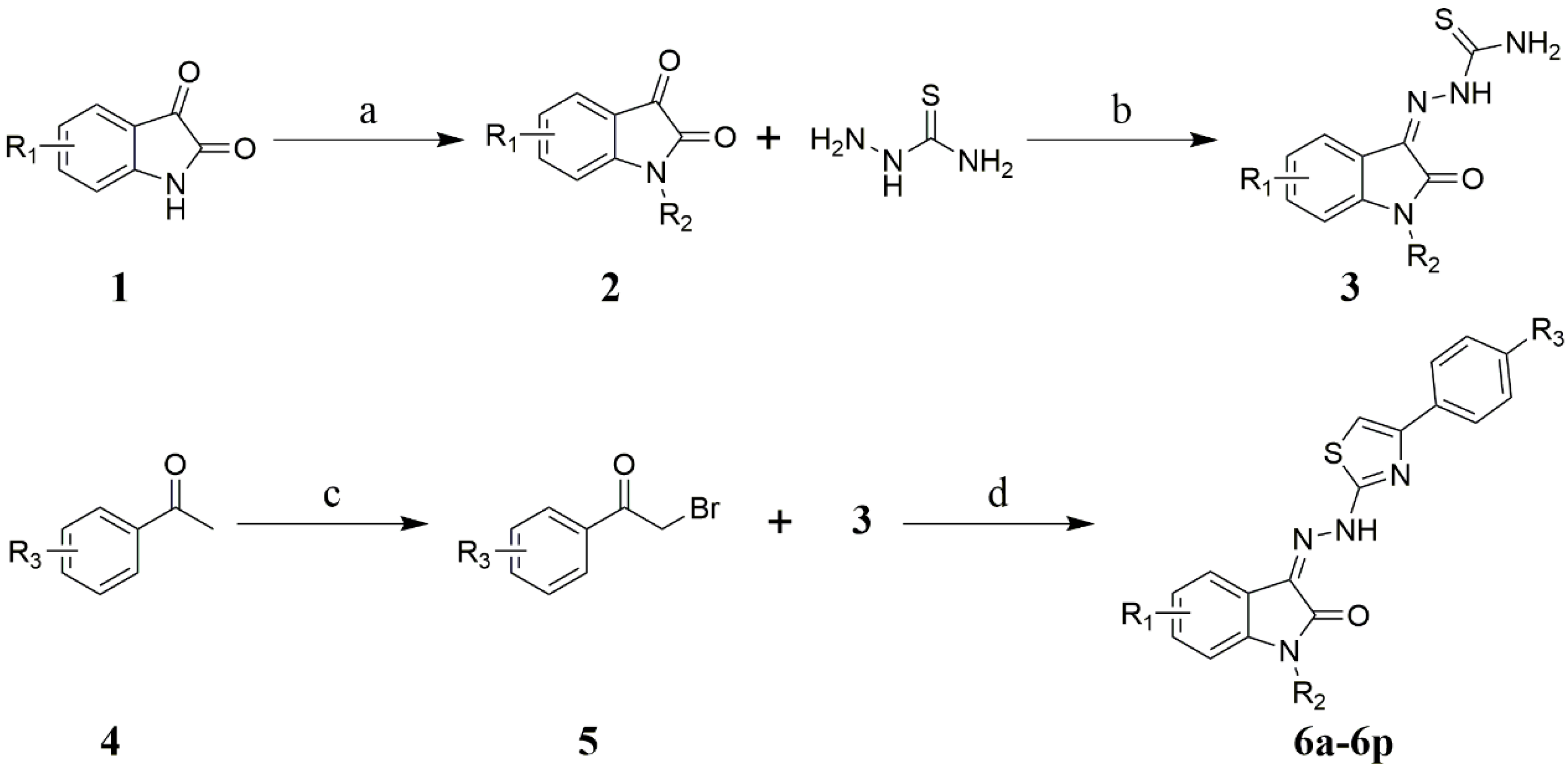
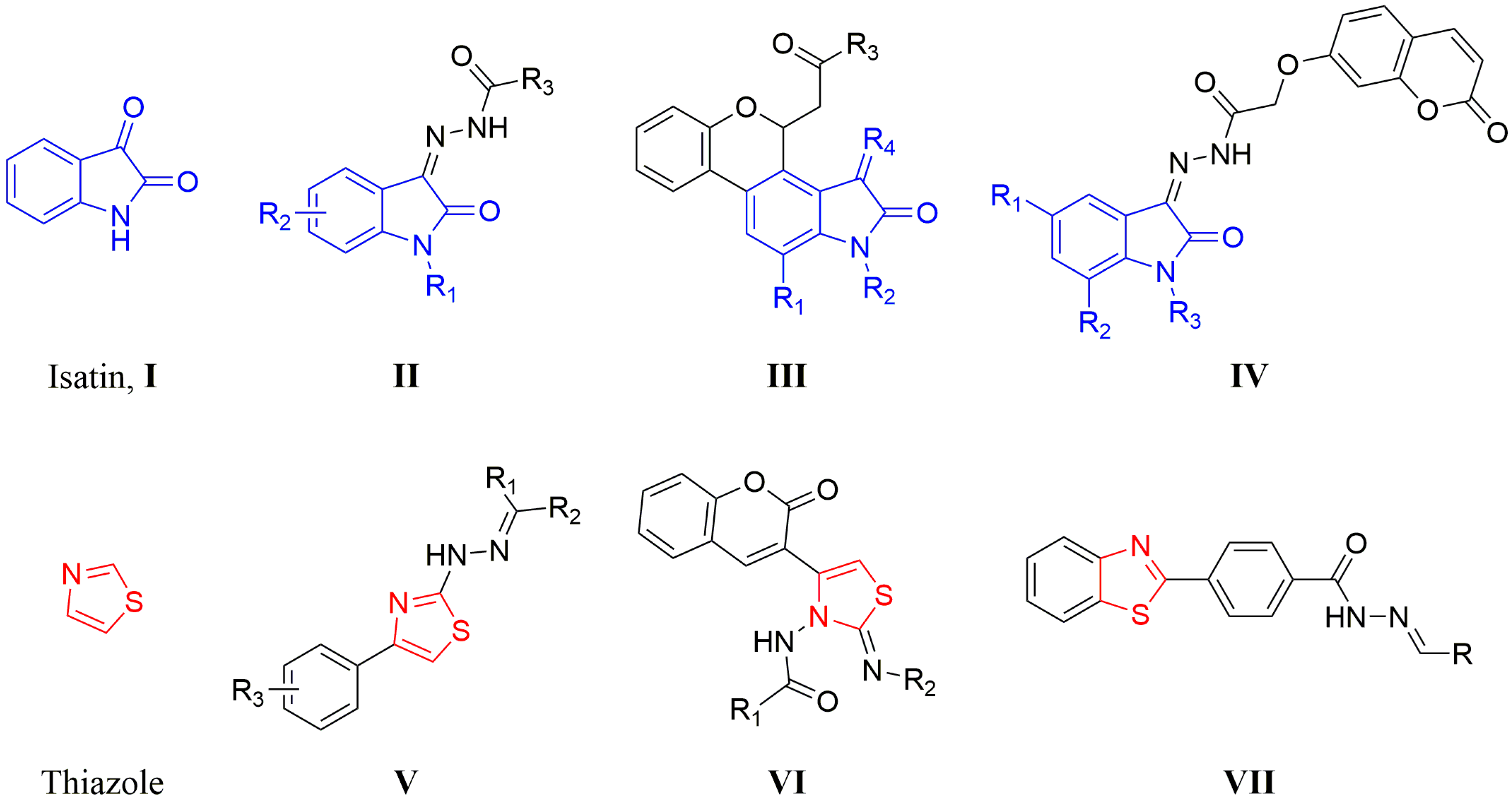
2.1. Chemistry

The synthesis of isatin-thiazole derivatives 6a6p was shown in Scheme 1. Reaction of commercially available isatins 1 with various alkyl halides in the presence of K2CO3 in DMF provided N-alkyl isatins 2. Isatins (1 and 2) were stirred with thiosemicarbazide in ethanol at 45 °C for 3 h to provide the isatin thiosemicarbazones 3. Various substitutions of acetophenone 4 were treated with NBS in the presence of p-toluenesulfonic acid in acetonitrile to give α-bromoacetophenone 5. α-bromoacetophenone 5, with appropriate isatin thiosemicarbazones 3, was condensed in refluxing ethanol for 2 h to afford the corresponding isatin-thiazole derivatives 6a6p in moderate to high yield (52.4–78.4%). All of the title compounds 6a6p have not yet been reported in the literature.
The structures of all of the title compounds 6a6p were characterized by 1H-NMR spectra. The 1H-NMR spectrum of 6a exhibited two singlet signals at 11.36 and 13.33 ppm, attributed to the protons of NH-indolin-2-one and =NNH group, respectively. Two doublet signals at 6.97 ppm (J = 8.4 Hz) and 7.52 ppm (J = 2.0 Hz) were attributed to C7-H and C4-H of isatin ring, respectively. The double-double peak of C6-H of the isatin ring was observed at 7.36 ppm with a coupling constant of 8.4 Hz and 2.0 Hz. Two double peaks at 7.61 and 7.85 ppm with coupling constant of 8.4 Hz were attributed to the aromatic protons of C3,5-H and C2,6-H, respectively. The hydrogen atom of thiazole ring appeared as a singlet signal at 7.74 ppm. The 1H-NMR spectrum of all new compounds consistent with their structures. Moreover, in their 13C-NMR spectra, the number of signals equals the number of different carbons in the molecule (Supplementary Materials).

2.2. α-Glucosidase Inhibition Assay

All the synthetic compounds 6a6p were screened for their in vitro α-glucosidase inhibitory activity (Table 1). The results were shown that all the tested compounds displayed potent to moderate α-glucosidase inhibitory activity with IC50 ranging from 5.36 ± 0.13 to 35.76 ± 0.31 μm more potent than the standard drug acarbose (IC50 = 817.38 ± 6.27 μm [17,36]). Among the series, compounds 6a, 6g, 6h, 6i, 6j, 6l, 6m, 6n, and 6p displayed potent inhibitory activity with IC50 values of 6.87 ± 0.14, 5.98 ± 0.12, 6.12 ± 0.15, 7.51 ± 0.17, 6.51 ± 0.13, 8.33 ± 0.18, 7.17 ± 0.15, 6.36 ± 0.12, and 5.36 ± 0.13 μm. In particular, compound 6p (IC50 = 5.36 ± 0.13 μm) with a hydroxyl group at the 4-position of the right phenyl ring and methyl and 2-fluorobenzyl groups at 5- and N1-positions of the isatin ring, was found to be the most active compound. In comparison to compound 6p, a decrease in activity was observed for 6i (IC50 = 7.51 ± 0.17) in which 2-fluorobenzyl groups is replaced with hydrogen at N1-positions of the isatin ring. Compounds 6c, 6k, and 6o also displayed good inhibition with IC50 value 10.34 ± 0.17, 15.68 ± 0.24, and 11.78 ± 0.21 μm, respectively. Other compounds displayed low α-glucosidase inhibitory activity. The binding interactions of the most active compounds with α-glucosidase were confirmed through molecular docking studies.

2.3. Homology Model

The crystallographic structure of Saccharomyces cerevisiae α-glucosidase enzyme has not been reported. To understand the ligand-enzyme interactions, the 3D structure of α-glucosidase was built by means of modeller 9.15 homology modeling software (http://salilab.org/modeller/). The sequence in FASTA format of α-glucosidase was retrieved from UniProt (access code P53341). The crystallographic structure of Saccharomyces cerevisiae isomaltase (PDB ID: 3AJ7, Resolution 1.30 Å) with 72.4% of sequence identity with the target was selected as the template for homology modeling [37]. The quality of homology model was verified by PROCHECK (http://services.mbi.ucla.edu/PROCHECK/). The result was shown that the model could be used to study the interactions between this class of compounds and the active site of α-glucosidase [35].

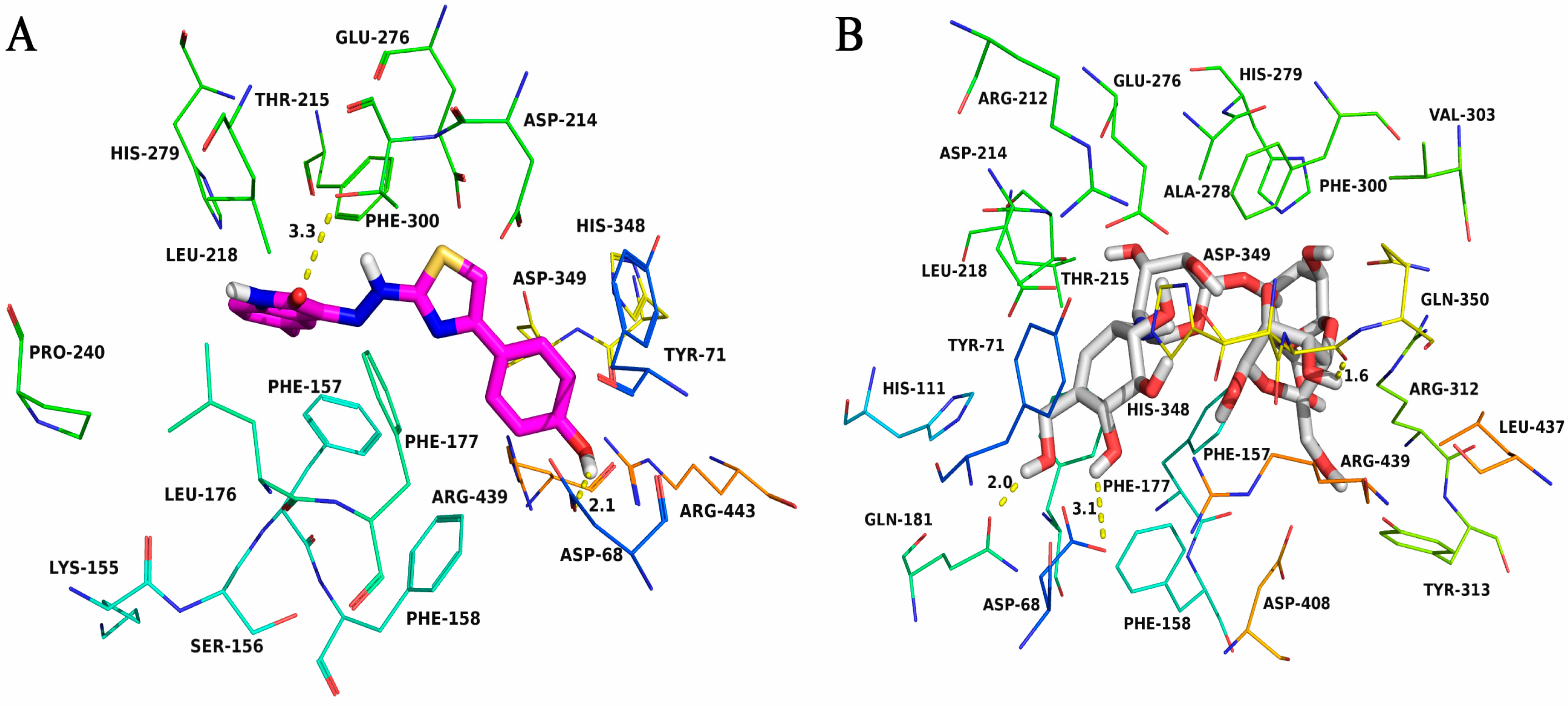
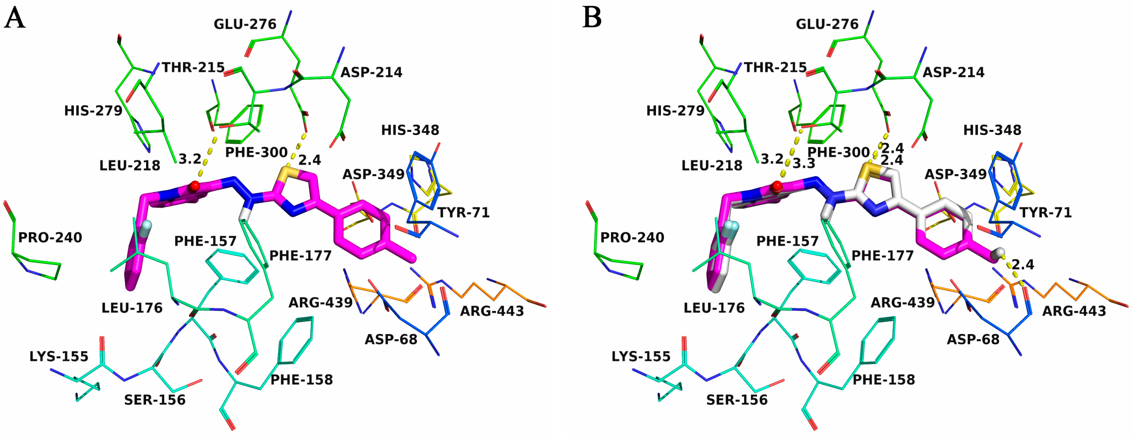
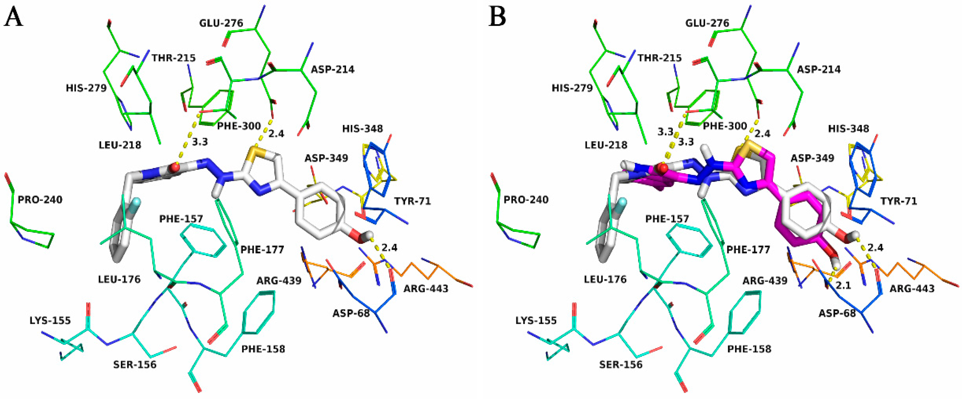
2.4. Molecular Docking

The theoretical binding mode between 6i and Saccharomyces cerevisiae α-glucosidase was shown in Figure 2. Compound 6i adopted a “V-shaped” conformation in the pocket of the α-glucosidase. The indolin-2-one scaffold of 6i located at the hydrophobic pocket, surrounded by the residues Phe-157, Leu-176, Pro-240, Phe-300, and Leu-218, forming a stable hydrophobic binding. Detailed analysis showed that the indolin-2-one scaffold of 6i formed CH-π interaction with the residue Phe-157. In addition, the 4-hydroxylphenyl group of 6i formed CH-π interactions with the residues Phe-158 and Tyr-71, and arene-anion interactions with the residues Asp-68 and Asp-349. Also, the arene-cation interactions were observed between the 4-hydroxylphenyl group of 6i and the residues Arg-439 and Arg-443. It was shown that the residues Thr-215 (bond length: 3.3 Å) and Asp-68 (length: 2.1 Å) formed two hydrogen bonds with 6i, which was the main interaction between 6i and α-glucosidase. On the other hand, molecular docking study of the standard drug acarbose with α-glucosidase was also performed (Figure 2B). The results were shown that compound 6i (binding energy was about −9.2 kcal mol−1) has a similar binding affinity as compared to standard drug acarbose (binding energy was about −6.8 kcal mol−1).
To explain the activity order of 6i and 6p against α-glucosidase in the molecular level, 6p was further docked into the binding pocket of α-glucosidase, and the theoretical binding mode between 6p and α-glucosidase was shown in Figure 3A. The interaction between 6p and α-glucosidase was nearly the same as the compound 6i (Figure 3B). The main difference was that the 2-fluorophenyl group of 6p formed extra hydrophobic interactions with the residues Phe-157, Leu-176, Pro-240, and Leu-218, and formed two extra hydrogen bonds with the residue Glu-276 (length: 2.4 Å) and Asp-68 (length: 2.4 Å), which made 6p was more active than 6i against α-glucosidase (Figure 3B). In addition, the estimated binding energies were −9.2 kcal mol−1 for 6i and −10.1 kcal mol−1 for 6p, respectively, which was consistent with the results of the in vitro α-glucosidase inhibitory activity.
To explain the activity order of 6b and 6p against α-glucosidase in the molecular level, 6b was further docked into the binding pocket of α-glucosidase, and the theoretical binding mode between 6b and α-glucosidase was shown in Figure 4A. Compound 6b adopted a ‘V-shaped’ conformation in the pocket of the α-glucosidase. The 2-fluorophenylindolin-2-one group of 6b stretched into the hydrophobic pocket that consisted of Phe-157, Leu-176, Pro-240, Phe-300, and Leu-218, forming a stable hydrophobic binding. Detailed analysis showed that the indolin-2-one scaffold of 6b formed CH-π interaction with the residue Phe-157. In addition, the 4-methylphenyl group of 6b formed CH-π interactions with the residues Phe-158 and Tyr-71, and arene-anion interactions with the residues Asp-68 and Asp-349, respectively. Also, the arene-cation interactions were observed between the 4-methylphenyl group of 6b and the residues Arg-439 and Arg-443. It was shown that the residues Thr-215 (bond length: 3.2 Å) and Glu-276 (length: 2.4 Å) formed two hydrogen bonds with 6b, which was the main interaction between 6b and α-glucosidase. The interaction between 6p and α-glucosidase was nearly the same as the compound 6b (Figure 4B). The main difference was that the 4-methoxyphenyl group of 6p formed an extra hydrogen bond with the residue Asp-68 (length: 2.4 Å), which made 6p was more active than 6b against α-glucosidase (Figure 4B). In addition, the estimated binding energies were −9.5 kcal mol−1 for 6b and −10.1 kcal mol−1 for 6p, respectively, which was consistent with the results of the in vitro α-glucosidase inhibitory activity.

3. Experimental Section

3.1. General

All starting materials and reagents were purchased from commercial suppliers. TLC was performed on 0.20 mm Silica Gel 60 F254 plates (Qingdao Ocean Chemical Factory, Shandong, China). 1H- and 13C-NMR spectra were recorded were recorded on a Bruker spectrometer (400 MHz) with TMS as an external reference and reported in parts per million.

3.2. General Procedures for the Synthesis of 6a6p

A mixture of 3 (1.0 mmol) and 5 (1.2 mmol) in EtOH (10 mL) was stirred at reflux for 2 h. After the completion of the reaction, the precipitates that formed were collected by filtration and washed with ethanol (3 × 10 mL) to give the desired products 6a6p. The spectroscopic and analytical data of compounds are as follows:
(Z)-3-(2-(4-(4-Bromophenyl)thiazol-2-yl)hydrazono)-5-chloroindolin-2-one (6a). Orange solid, yield 77.5%, 1H-NMR (d6-DMSO, 400 MHz) δ: 6.97 (d, 1H, J = 8.4 Hz, ArH), 7.36 (dd, 1H, J = 8.4 Hz, 2.0 Hz, ArH), 7.52 (d, 1H, J = 2.0 Hz, ArH), 7.61 (d, 2H, J = 8.4 Hz, ArH), 7.74 (s, 1H, CH-thiazole), 7.85 (d, 2H, J = 8.4 Hz, ArH), 11.36 (s, 1H, NH), 13.33 (s, 1H, NH); 13C-NMR (d6-DMSO, 100 MHz) δ: 108.6, 113.0, 119.8, 121.5, 121.9, 127.1, 128.1, 128.2, 130.3, 131.6, 132.1, 132.2, 133.6, 140.4, 150.4, 163.4, 166.5.
(Z)-1-(2-Fluorobenzyl)-5-methyl-3-(2-(4-(p-tolyl)thiazol-2-yl)hydrazono) indolin-2-one (6b). Red solid, yield 52.5%, 1H-NMR (d6-DMSO, 400 MHz) δ: 2.33 (s, 6H, CH3), 5.06 (s, 2H, CH2), 6.93 (d, 1H, J = 8.4 Hz, ArH), 7.14–7.19 (m, 2H, ArH), 7.23–7.26 (m, 3H, ArH), 7.32–7.37 (m, 2H, ArH), 7.44 (s, 1H, ArH), 7.58 (s, 1H, ArH), 7.79 (d, 2H, J = 8.0 Hz, ArH), 13.22 (s, 1H, NH); 13C-NMR (d6-DMSO, 100 MHz) δ: 21.0, 21.3, 37.4, 106.6, 110.3, 115.9 (d, 1C, J = 20.9 Hz), 119.7, 120.6, 122.9, 123.0, 125.2 (d, 1C, J = 3.4 Hz), 126.0, 126.1, 129.7, 129.9, 130.0, 130.1, 130.3 (d, 1C, J = 8.1 Hz), 131.2, 131.4, 131.8, 132.9, 137.8, 139.7, 151.7, 159.4 (d, 1C, J = 243.9 Hz), 161.8, 166.3; MS (ESI, m/z): 457.11 [M + H]+.
(Z)-1-(2-Fluorobenzyl)-3-(2-(4-(4-methoxyphenyl)thiazol-2-yl)hydrazono)-5-methylindolin-2-one (6c). Red solid, yield 77.2%, 1H-NMR (d6-DMSO, 400 MHz) δ: 2.32 (s, 3H, CH3), 3.79 (s, 3H, OCH3), 5.06 (s, 2H, CH2), 6.92 (d, 1H, J = 8.0 Hz, ArH), 6.98 (d, 2H, J = 8.0 Hz, ArH), 7.13–7.18 (m, 2H, ArH), 7.22–7.26 (m, 1H, ArH), 7.32–7.37 (m, 2H, ArH), 7.43 (s, 1H, ArH), 7.48 (s, 1H, CH-thiazole), 7.83 (d, 2H, J = 8.0 Hz, ArH), 13.21 (s, 1H, NH); 13C-NMR (d6-DMSO, 100 MHz) δ: 21.0, 37.4, 55.6, 105.3, 110.3, 114.5, 114.7, 115.9 (d, 1C, J = 20.9 Hz), 119.7, 120.5, 122.9, 123.0, 125.2 (d, 1C, J = 3.5 Hz), 127.3, 127.6, 130.0, 130.1, 130.3 (d, 1C, J = 8.1 Hz), 131.2, 131.3, 132.8, 139.7, 151.5, 159.4 (d, 1C, J = 243.9 Hz), 159.6, 161.7; MS (ESI, m/z): 473.09 [M + H]+.
(Z)-1-Methyl-3-(2-(4-(p-tolyl)thiazol-2-yl)hydrazono)indolin-2-one (6d). Red solid, yield 75.4%, 1H-NMR (d6-DMSO, 400 MHz) δ: 2.33(s, 3H, ArCH3), 3.27 (s, 3H, NCH3), 7.15–7.19 (m, 2H, ArH), 7.23 (d, 2H, J = 8.0 Hz, ArH), 7.44 (t, 1H, J = 8.0 Hz, ArH), 7.57 (s, 1H, CH-thiazole), 7.57 (d, 1H, J = 8.0 Hz, ArH), 7.79 (d, 2H, J = 8.0 Hz, ArH), 13.26 (s, 1H, NH); 13C-NMR (d6-DMSO, 100 MHz) δ: 21.3, 26.2, 106.4, 110.2, 119.5, 119.9, 123.3, 126.1, 129.7, 130.8, 131.6, 131.8, 137.8, 143.0, 151.6, 161.8, 166.3.
(Z)-3-(2-(4-(4-Fluorophenyl)thiazol-2-yl)hydrazono)-1-methylindolin-2-one (6e). Orange solid, yield 70.3%, 1H-NMR (d6-DMSO, 400 MHz) δ: 3.27 (s, 3H, NCH3), 7.15–7.18 (m, 2H, ArH), 7.26 (t, 2H, J = 8.8 Hz, ArH), 7.44 (t, 1H, J = 8.0 Hz, ArH), 7.57 (d, 1H, J = 8.0 Hz, ArH), 7.62 (s, 1H, CH-thiazole), 7.93 (dd, 2H, J = 8.8 Hz, 5.6 Hz, ArH), 13.25 (s, 1H, NH); 13C-NMR (d6-DMSO, 100 MHz) δ: 26.2, 107.1, 110.3, 115.9 (d, 1C, J = 21.5 Hz), 119.5, 120.0, 123.4, 128.2 (d, 1C, J = 8.1 Hz), 130.9, 131.0 (d, 1C, J = 2.9 Hz), 131.1, 131.9, 143.1, 150.5, 161.1 (d, 1C, J = 243.7 Hz), 161.8, 166.6.
(Z)-3-(2-(4-(4-Methoxyphenyl)thiazol-2-yl)hydrazono)-1-methylindolin-2-one (6f). Red solid, yield 69.9%, 1H-NMR (d6-DMSO, 400 MHz) δ: 3.27 (s, 3H, NCH3), 3.80 (s, 3H, OCH3), 6.98 (d, 2H, J = 8.8 Hz, ArH), 7.15–7.19 (m, 2H, ArH), 7.44 (t, 1H, J = 8.0 Hz, ArH), 7.48 (s, 1H, CH-thiazole), 7.58 (d, 1H, J = 7.2 Hz, ArH), 7.83 (d, 2H, J = 8.8 Hz, ArH), 13.26 (s, 1H, NH); 13C-NMR (d6-DMSO, 100 MHz) δ: 26.2, 55.6, 105.2, 110.3, 114.5, 114.7, 119.5, 119.9, 123.4, 127.3, 127.5, 127.6, 130.8, 131.6, 143.0, 151.5, 159.6, 161.8, 166.3.
(Z)-3-(2-(4-(4-Chlorophenyl)thiazol-2-yl)hydrazono)indolin-2-one (6g). Orange solid, yield 66.9%, 1H-NMR (d6-DMSO, 400 MHz) δ: 6.96 (d, 1H, J = 8.8 Hz, ArH), 7.10 (t, 1H, J = 8.8 Hz, ArH), 7.35 (t, 1H, J = 8.8 Hz, ArH), 7.47 (d, 2H, J = 8.8 Hz, ArH), 7.54 (d, 1H, J = 8.8 Hz, ArH), 7.70 (s, 1H, CH-thiazole), 7.92 (d, 2H, J = 8.8 Hz, ArH), 11.26 (s, 1H, NH-indolin-2-one),13.35 (s, 1H, NH); 13C-NMR (d6-DMSO, 100 MHz) δ: 108.0, 111.5, 120.2, 120.3, 122.9, 127.9, 129.1, 131.0, 132.7, 132.9, 133.3, 141.8, 150.3, 163.6, 166.7.
(Z)-3-(2-(4-(4-Bromophenyl)thiazol-2-yl)hydrazono)-5,7-dimethylindolin-2-one (6h). Orange solid, yield 64.4%, 1H-NMR (d6-DMSO, 400 MHz) δ: 2.21 (s, 3H, ArCH3), 2.29 (s, 3H, ArCH3), 6.99 (s, 1H, ArH), 7.19 (s, 1H, ArH), 7.61 (d, 2H, J = 8.8 Hz, ArH), 7.71 (s, 1H, CH-thiazole), 7.85 (d, 2H, J = 8.8 Hz, ArH), 11.22 (s, 1H, NH-indolin-2-one), 13.40 (s, 1H, NH); 13C-NMR (d6-DMSO, 100 MHz) δ: 16.3, 20.9, 107.9, 118.1, 119.9, 120.6, 121.4, 128.2, 131.9, 132.0, 132.9, 133.2, 133.7, 138.2, 150.3, 164.2, 166.7.
(Z)-3-(2-(4-(4-Hydroxyphenyl)thiazol-2-yl)hydrazono)-5-methylindolin-2-one (6i). Orange solid, yield 52.4%, 1H-NMR (d6-DMSO, 400 MHz) δ: 2.33 (s, 3H, ArCH3), 6.80 (d, 2H, J = 8.8 Hz, ArH), 6.85 (d, 1H, J = 8.0 Hz, ArH), 7.15 (d, 1H, J = 8.0 Hz, ArH), 7.36 (s, 2H, CH-thiazole and ArH), 7.71 (d, 2H, J = 8.8 Hz, ArH), 11.16 (s, 1H, NH-indolin-2-one), 13.35 (s, 1H, NH); 13C-NMR (d6-DMSO, 100 MHz) δ: 21.0, 104.2, 111.3, 115.6, 115.9, 120.2, 120.6, 125.6, 127.6, 129.9, 131.4, 131.9, 132.7, 139.5, 151.5, 157.9, 163.7, 166.3.
(Z)-3-(2-(4-(4-Fluorophenyl)thiazol-2-yl)hydrazono)-5,7-dimethylindolin-2-one (6j). Orange solid, yield 59.6%, 1H-NMR (d6-DMSO, 400 MHz) δ: 2.21 (s, 3H, ArCH3), 2.29 (s, 3H, ArCH3), 6.99 (s, 1H, ArH), 7.19 (s, 1H, ArH), 7.26 (t, 2H, J = 8.8 Hz), 7.61 (s, 1H, CH-thiazole), 7.93 (d, 2H, J = 8.8 Hz, 5.6 Hz, ArH), 11.22 (s, 1H, NH-indolin-2-one), 13.39 (s, 1H, NH); 13C-NMR (d6-DMSO, 100 MHz) δ: 16.3, 20.9, 106.8, 115.8 (d, 1C, J = 21.5 Hz), 118.1, 119.9, 120.6, 128.2 (d, 1C, J = 8.1 Hz), 131.1 (d, 1C, J = 2.7 Hz), 131.8, 132.8, 133.1, 138.2, 150.5, 161.1 (d, 1C, J = 243.7 Hz), 164.2, 166.6.
(Z)-5,7-Dimethyl-3-(2-(4-(p-tolyl)thiazol-2-yl)hydrazono)indolin-2-one (6k). Orange solid, yield 62.1%, 1H-NMR (d6-DMSO, 400 MHz) δ: 2.21 (s, 3H, ArCH3), 2.29 (s, 3H, ArCH3), 2.34 (s, 3H, ArCH3), 6.99 (s, 1H, ArH), 7.20 (s, 1H, ArH), 7.23 (d, 2H, J = 8.0 Hz, ArH), 7.55 (s, 1H, CH-thiazole), 7.79 (d, 2H, J = 8.0 Hz, ArH),11.22 (s, 1H, NH-indolin-2-one), 13.39 (s, 1H, NH); 13C-NMR (d6-DMSO, 100 MHz) δ: 16.3, 20.9, 21.3, 106.2, 118.1, 119.9, 120.6, 126.1, 129.7, 131.9, 132.8, 133.0, 137.7, 138.2, 151.6, 164.2, 166.4.
(Z)-5-Fluoro-3-(2-(4-(p-tolyl)thiazol-2-yl)hydrazono)indolin-2-one (6l). Orange solid, yield 60.8%, 1H-NMR (d6-DMSO, 400 MHz) δ: 2.24 (s, 3H, ArCH3), 6.96 (dd, 1H, J = 8.4 Hz, 4.4 Hz, ArH), 7.19 (dt, 1H, J = 8.4 Hz, 2.4 Hz, ArH), 7.23 (d, 2H, J = 8.0 Hz, ArH), 7.35 (dd, 1H, J = 8.4 Hz, 2.4 Hz, ArH), 7.59 (s, 1H, CH-thiazole), 7.79 (d, 2H, J = 8.0 Hz, ArH), 11.28 (s, 1H, NH-indolin-2-one), 13.39 (s, 1H, NH); 13C-NMR (d6-DMSO, 100 MHz) δ: 21.3, 106.7, 107.1, 107.4, 112.5 112.6, 117.0 (d, 1C, J = 24.5 Hz), 121.4 (d, 1C, J = 8.9 Hz), 125.9, 126.1, 129.7, 129.9, 131.7, 131.9 (d, 1C, J = 3.1 Hz), 137.8, 137.9, 151.7, 157.6 (d, 1C, J = 236.4 Hz), 163.8, 166.1.
(Z)-5-Fluoro-3-(2-(4-(4-fluorophenyl)thiazol-2-yl)hydrazono)indolin-2-one (6m). Orange solid, yield 57.7%, 1H-NMR (d6-DMSO, 400 MHz) δ: 6.95 (dd, 1H, J = 8.4 Hz, 4.4 Hz, ArH), 7.19 (dt, 1H, J = 8.4 Hz, 2.4 Hz, ArH), 7.25 (d, 2H, J = 8.8 Hz, ArH), 7.35 (dd, 1H, J = 8.4 Hz, 2.4 Hz, ArH), 7.66 (s, 1H, CH-thiazole), 7.94 (dd, 2H, J = 8.8 Hz, 5.6 Hz, ArH), 11.28 (s, 1H, NH-indolin-2-one), 13.39 (s, 1H, NH); 13C-NMR (d6-DMSO, 100 MHz) δ: 107.2, 107.4, 107.4, 112.5, 112.6, 115.9 (d, 1C, J = 21.4 Hz), 116.2 (d, 1C, J = 21.5 Hz), 117.1, 117.3, 121.4, 121.5, 128.2, 128.3, 128.4, 131.0 (d, 1C, J = 2.9 Hz), 132.0 (d, 1C, J = 3.6 Hz), 138.0, 150.5, 157.6 (d, 1C, J = 236.3 Hz), 161.1 (d, 1C, J = 243.6 Hz), 163.8, 166.3.
(Z)-3-(2-(4-(4-Fluorophenyl)thiazol-2-yl)hydrazono)indolin-2-one (6n). Orange solid, yield 78.4%, 1H-NMR (d6-DMSO, 400 MHz) δ: 6.97 (d, 1H, J = 8.0 Hz, ArH), 7.10 (t, 1H, J = 8.0 Hz, ArH), 7.26 (d, 2H, J = 8.8 Hz, ArH), 7.36 (t, 1H, J = 8.0 Hz, ArH), 7.54 (d, 1H, J = 8.0 Hz, ArH), 7.62 (s, 1H, CH-thiazole), 7.94 (dd, 2H, J = 8.8 Hz, 5.6 Hz, ArH), 11.27 (s, 1H, NH-indolin-2-one), 13.35 (s, 1H, NH); 13C-NMR (d6-DMSO, 100 MHz) δ: 117.0, 111.5, 115.9 (d, 1C, J = 21.4 Hz), 120.2, 120.3, 122.9, 128.2 (d, 1C, J = 8.1 Hz), 130.9, 131.1 (d, 1C, J = 2.9 Hz), 132.6, 141.8, 150.5, 161.1(d, 1C, J = 243.6 Hz), 163.6, 166.6.
(Z)-3-(2-(4-(p-Tolyl)thiazol-2-yl)hydrazono)indolin-2-one (6o). Red solid, yield 73.3%, 1H-NMR (d6-DMSO, 400 MHz) δ: 2.34 (s, 3H, ArCH3), 6.97 (d, 1H, J = 8.0 Hz, ArH), 7.10 (t, 1H, J = 8.0 Hz, ArH), 7.23 (d, 2H, J = 8.0 Hz, ArH), 7.35 (t, 1H, J = 8.0 Hz, ArH), 7.54–7.56 (m, 2H, CH-thiazole and ArH), 7.79 (d, 2H, J = 8.4 Hz, ArH), 11.27 (s, 1H, NH-indolin-2-one), 13.35 (s, 1H, NH); 13C-NMR (d6-DMSO, 100 MHz) δ: 21.3, 106.3, 111.5, 120.2, 120.3, 122.9, 126.1, 129.7, 129.8, 130.9, 131.8, 132.5, 137.8, 141.7, 151.6, 163.7, 166.4.
(Z)-1-(2-Fluorobenzyl)-3-(2-(4-(4-hydroxyphenyl)thiazol-2-yl)hydrazono)-5-methylindolin-2-one (6p). Red solid, yield 53.1%, 1H-NMR (d6-DMSO, 400 MHz) δ: 2.33 (s, 3H, ArCH3), 5.07 (s, 2H, ArCH2), 6.81 (d, 2H, J = 7.2 Hz, ArH), 6.93 (d, 1H, J = 7.2 Hz, ArH), 7.12–7.25 (m, 3H, ArH), 7.36–7.44 (m, 3H), 7.72 (d, 2H, J = 7.2 Hz, ArH), 9.62 (s, 1H, OH), 13.21 (s, 1H, NH); 13C-NMR (d6-DMSO, 100 MHz) δ: 21.0, 37.4, 104.4, 110.3, 115.9, 115.9 (d, 1C, J = 23.0 Hz), 119.8, 120.5, 122.9, 123.0, 125.2, 125.8, 127.6, 130.0, 130.2 (d, 1C, J = 8.1 Hz), 131.1, 132.8, 139.6, 151.9, 157.9, 159.4 (d, 1C, J = 237.9 Hz), 166.0; MS (ESI, m/z): 459.06 [M + H]+.

3.3. In Vitro Assay of α-Glucosidase Inhibitory Activity

According to the literature procedure [36], α-Glucosidase inhibitory activity was assayed by using 0.1 M phosphate buffer (pH 6.8) at 37 °C. The enzyme (0.1 U/mL) in phosphate buffer saline was incubated with various concentrations of test compounds at 37 °C for 15 min. Then 1.25 mM p-nitrophenyl α-d-glucopyranoside was added to the mixture as a substrate, after further incubation at 37 °C for 30 min. The absorbance was measured spectrophotometrically at 405 nm. The sample solution was replaced by DMSO as a control. Acarbose was used as a positive control. All experiments were carried out in triplicates. The % inhibition has been obtained using the formula: inhibition (%) = (1 − ∆Asample/∆Acontrol) × 100%. IC50 value is defined as a concentration of samples inhibiting 50% of α-glucosidase activity under the stated assay conditions.

3.4. Molecular Docking

Molecular docking studies were performed to investigate the binding mode between the compounds 6b, 6i, 6p, and α-glucosidase using Autodock vina 1.1.2 [38]. The 3D structures of 6i and 6p were obtained by ChemBioDraw Ultra 14.0 and ChemBio3D Ultra 14.0 softwares. The AutoDockTools 1.5.6 package was employed to generate the docking input files [39,40]. The search grid of α-glucosidase was identified as center_x: −19.676, center_y: −7.243, and center_z: −21.469 with dimensions size_x: 15, size_y: 15, and size_z: 15. The value of exhaustiveness was set to 20. For Vina docking, the default parameters were used if it was not mentioned. The best-scoring poses as judged by the Vina docking score were chosen and visually analyzed using PyMOL 1.7.6 software (Schrödinger®, New York, NY, USA) (http://www.pymol.org/).

4. Conclusions

In conclusion, we designed and synthesized a novel series of α-glucosidase inhibitor based on the molecular hybrid-based approaches. All the target compounds displayed potent to moderate α-glucosidase inhibitory activity with IC50 ranging from 5.36 ± 0.13 to 35.76 ± 0.31 μm as compared to the standard drug acarbose (IC50 = 817.38 ± 6.27 μm). Among the series, compound 6p bearing a hydroxyl group at the 4-position of the right phenyl and 2-fluorobenzyl substituent at the N1-positions of the 5-methylisatin displayed the highest inhibitory activity with an IC50 value of 5.36 ± 0.13 μm. Furthermore, molecular docking studies revealed the existence of hydrophobic interaction, CH-π interaction, arene-anion interaction, arene-cation interaction, and hydrogen bond between these compounds and α-glucosidase enzyme.

Supplementary Materials

Supplementary materials are available online.

Acknowledgments

This work was supported by Hunan Provincial Natural Science Foundation of China (2015JJ3099), Scientific Research Fund of Hunan Provincial Education Department (15B194); The college students’ research learning and innovative experiment plan project of Jishou University and Hunan Province (Zhenzhen Xie).

Author Contributions

Guangcheng Wang designed and wrote the paper; Zhenzhen Xie, Jing Wang, Ming Chen, Yaping Peng, Luyao Li, Bing Deng, Shan Chen, and Wenbiao Li carried out the experiments. All authors have read and approved the final manuscript.

Conflicts of Interest

The authors confirm that this article content has no conflict of interest.

References

  1. DeFronzo, R.A. Pathogenesis of type 2 diabetes mellitus. Med. Clin. N. Am. 2004, 88, 787–835. [Google Scholar] [CrossRef] [PubMed]
  2. World Health Organization. Global Report on Diabetes; WHO: Geneva, Switzerland, 30 August 2016. [Google Scholar]
  3. Deshpande, A.D.; Harris-Hayes, M.; Schootman, M. Epidemiology of Diabetes and Diabetes-Related Complications. Phys. Ther. 2008, 88, 1254–1264. [Google Scholar] [CrossRef] [PubMed]
  4. Van de Laar, F.A. α-glucosidase inhibitors in the early treatment of type 2 diabetes. Vasc. Health Risk Manag. 2008, 4, 1189–1195. [Google Scholar] [CrossRef] [PubMed]
  5. Joshi, S.R.; Standl, E.; Tong, N.; Shah, P.; Kalra, S.; Rathod, R. Therapeutic potential of α-glucosidase inhibitors in type 2 diabetes mellitus: An evidence-based review. Expert Opin. Pharmacother. 2015, 16, 1959–1981. [Google Scholar] [CrossRef] [PubMed]
  6. Pili, R.; Chang, J.; Partis, R.A.; Mueller, R.A.; Chrest, F.J.; Passaniti, A. The α-glucosidase I inhibitor castanospermine alters endothelial cell glycosylation, prevents angiogenesis, and inhibits tumor growth. Cancer Res. 1995, 55, 2920–2926. [Google Scholar] [PubMed]
  7. Mehta, A.; Zitzmann, N.; Rudd, P.M.; Block, T.M.; Dwek, R.A. α-glucosidase inhibitors as potential broad based anti-viral agents. FEBS Lett. 1998, 430, 17–22. [Google Scholar] [CrossRef]
  8. Zitzmann, N.; Mehta, A.S.; Carrouée, S.; Butters, T.D.; Platt, F.M.; McCauley, J.; Blumberg, B.S.; Dwek, R.A.; Block, T.M. Imino sugars inhibit the formation and secretion of bovine viral diarrhea virus, a pestivirus model of hepatitis C virus: Implications for the development of broad spectrum anti-hepatitis virus agents. Proc. Natl. Acad. Sci. USA 1999, 96, 11878–11882. [Google Scholar] [CrossRef] [PubMed]
  9. Hamaue, N.; Minami, M.; Hirafuji, M.; Terado, M.; Machida, M.; Yamazaki, N.; Yoshioka, M.; Ogata, A.; Tashiro, K. Isatin, an endogenous MAO inhibitor, as a new biological modulator. CNS Drug Rev. 1999, 5, 331–346. [Google Scholar] [CrossRef]
  10. Khan, F.A.; Maalik, A.; Noor, T.; Zaidi, A.; Farooq, U.; Bukhari, S.M. Advances in Pharmacology of Isatin and its Derivatives: A Review. Trop. J. Pharm. Res. 2015, 14, 1937–1942. [Google Scholar] [CrossRef]
  11. Nguyen-Hai, N.; Tran Lan, H.; Do Thi Mai, D.; Phan Thi Phuong, D.; Dao Thi Kim, O.; Do, Q.; Le Thi, T.; Park, S.H.; Kim, K.R.; Han, B.W.; et al. Novel isatin-based hydroxamic acids as histone deacetylase inhibitors and antitumor agents. Eur. J. Med. Chem. 2013, 70, 477–486. [Google Scholar]
  12. Zhang, X.-M.; Guo, H.; Li, Z.-S.; Song, F.-H.; Wang, W.-M.; Dai, H.-Q.; Zhang, L.-X.; Wang, J.-G. Synthesis and evaluation of isatin-beta-thiosemicarbazones as novel agents against antibiotic-resistant Gram-positive bacterial species. Eur. J. Med. Chem. 2015, 101, 419–430. [Google Scholar] [CrossRef] [PubMed]
  13. Bal, T.R.; Anand, B.; Yogeeswari, P.; Sriram, D. Synthesis and evaluation of anti-HIV activity of isatin beta-thiosemicarbazone derivatives. Bioorg. Med. Chem. Lett. 2005, 15, 4451–4455. [Google Scholar] [CrossRef] [PubMed]
  14. Xie, C.; Tang, L.-M.; Li, F.-N.; Guan, L.-P.; Pan, C.-Y.; Wang, S.-H. Structure-based design, synthesis, and anticonvulsant activity of isatin-1-N-phenylacetamide derivatives. Med. Chem. Res. 2014, 23, 2161–2168. [Google Scholar] [CrossRef]
  15. Sharma, P.K.; Balwani, S.; Mathur, D.; Malhotra, S.; Singh, B.K.; Prasad, A.K.; Len, C.; Van der Eycken, E.V.; Ghosh, B.; Richards, N.G.J.; et al. Synthesis and anti-inflammatory activity evaluation of novel triazolyl-isatin hybrids. J. Enzym. Inhib. Med. Chem. 2016, 31, 1520–1526. [Google Scholar] [CrossRef] [PubMed]
  16. Raj, A.A.; Raghunathan, R.; SrideviKumari, M.R.; Raman, N. Synthesis, antimicrobial and antifungal activity of a new class of spiro pyrrolidines. Bioorg. Med. Chem. 2003, 11, 407–419. [Google Scholar] [CrossRef]
  17. Rahim, F.; Malik, F.; Ullah, H.; Wadood, A.; Khan, F.; Javid, M.T.; Taha, M.; Rehman, W.; Rehman, A.U.; Khan, K.M. Isatin based Schiff bases as inhibitors of α-glucosidase: Synthesis, characterization, in vitro evaluation and molecular docking studies. Bioorg. Chem. 2015, 60, 42–48. [Google Scholar] [CrossRef] [PubMed]
  18. Sun, H.; Zhang, Y.; Ding, W.; Zhao, X.; Song, X.; Wang, D.; Li, Y.; Han, K.; Yang, Y.; Ma, Y.; et al. Inhibitory activity evaluation and mechanistic studies of tetracyclic oxindole derivatives as α-glucosidase inhibitors. Eur. J. Med. Chem. 2016, 123, 365–378. [Google Scholar] [CrossRef] [PubMed]
  19. Wang, G.; Wang, J.; He, D.; Li, X.; Li, J.; Peng, Z. Synthesis, in vitro evaluation and molecular docking studies of novel coumarin-isatin derivatives as α-glucosidase inhibitors. Chem. Biol. Drug Des. 2016, 89, 456–463. [Google Scholar] [CrossRef] [PubMed]
  20. Aggarwal, R.; Kumar, S.; Kaushik, P.; Kaushik, D.; Gupta, G.K. Synthesis and pharmacological evaluation of some novel 2-(5-hydroxy-5-trifluoromethyl-4,5-dihydropyrazol-1-yl)-4-(coumarin-3-yl )thiazoles. Eur. J. Med. Chem. 2013, 62, 508–514. [Google Scholar] [CrossRef] [PubMed]
  21. Azizmohammadi, M.; Khoobi, M.; Ramazani, A.; Emami, S.; Zarrin, A.; Firuzi, O.; Miri, R.; Shafiee, A. 2H-chromene derivatives bearing thiazolidine-2,4-dione, rhodanine or hydantoin moieties as potential anticancer agents. Eur. J. Med. Chem. 2013, 59, 15–22. [Google Scholar] [CrossRef] [PubMed]
  22. Siddiqui, N.; Ahsan, W. Triazole incorporated thiazoles as a new class of anticonvulsants: Design, synthesis and in vivo screening. Eur. J. Med. Chem. 2010, 45, 1536–1543. [Google Scholar] [CrossRef] [PubMed]
  23. Qin, Y.-J.; Wang, P.-F.; Makawana, J.A.; Wang, Z.-C.; Wang, Z.-N.; Yan, G.; Jiang, A.-Q.; Zhu, H.-L. Design, synthesis and biological evaluation of metronidazole-thiazole derivatives as antibacterial inhibitors. Bioorg. Med. Chem. Lett. 2014, 24, 5279–5283. [Google Scholar] [CrossRef] [PubMed]
  24. Koppireddi, S.; Komsani, J.R.; Tiwari, A.K.; Ali, A.Z.; Yadla, R. Synthesis and biological evaluation of new derivatives of tertiary thiazolamines. Pharm. Chem. 2014, 6, 136–143. [Google Scholar]
  25. Rahim, F.; Ullah, H.; Javid, M.T.; Wadood, A.; Taha, M.; Ashraf, M.; Shaukat, A.; Junaid, M.; Hussain, S.; Rehman, W.; et al. Synthesis, in vitro evaluation and molecular docking studies of thiazole derivatives as new inhibitors of α-glucosidase. Bioorg. Chem. 2015, 62, 15–21. [Google Scholar] [CrossRef] [PubMed]
  26. Taha, M.; Ismail, N.H.; Lalani, S.; Fatmi, M.Q.; Atia-tul-Wahab; Siddiqui, S.; Khan, K.M.; Imran, S.; Choudhary, M.I. Synthesis of novel inhibitors of α-glucosidase based on the benzothiazole skeleton containing benzohydrazide moiety and their molecular docking studies. Eur. J. Med. Chem. 2015, 92, 387–400. [Google Scholar] [CrossRef] [PubMed]
  27. Khan, K.M.; Qurban, S.; Salar, U.; Taha, M.; Hussain, S.; Perveen, S.; Hameed, A.; Ismail, N.H.; Riaz, M.; Wadood, A. Synthesis, in vitro α-glucosidase inhibitory activity and molecular docking studies of new thiazole derivatives. Bioorg. Chem. 2016, 68, 245–258. [Google Scholar] [CrossRef] [PubMed]
  28. Salar, U.; Taha, M.; Khan, K.M.; Ismail, N.H.; Imran, S.; Perveen, S.; Gul, S.; Wadood, A. Syntheses of new 3-thiazolyl coumarin derivatives, in vitro α-glucosidase inhibitory activity, and molecular modeling studies. Eur. J. Med. Chem. 2016, 122, 196–204. [Google Scholar] [CrossRef] [PubMed]
  29. Viegas-Junior, C.; Danuello, A.; da Silva Bolzani, V.; Barreir, E.J.; Manssour Fraga, C.A. Molecular hybridization: A useful tool in the design of new drug prototypes. Curr. Med. Chem. 2007, 14, 1829–1852. [Google Scholar] [CrossRef] [PubMed]
  30. Shaveta; Mishra, S.; Singh, P. Hybrid molecules: The privileged scaffolds for various pharmaceuticals. Eur. J. Med. Chem. 2016, 124, 500–536. [Google Scholar]
  31. Wang, G.; Wang, J.; He, D.; Li, X.; Li, J.; Peng, Z. Synthesis and biological evaluation of novel 1,2,4-triazine derivatives bearing carbazole moiety as potent α-glucosidase inhibitors. Bioorg. Med. Chem. Lett. 2016, 26, 2806–2809. [Google Scholar] [CrossRef] [PubMed]
  32. Wang, G.; Wang, J.; He, D.; Li, X.; Li, J.; Peng, Z. One-pot and three-component synthesis, characterization and biological evaluation of some new 1,2,4-triazine-coumarins. Heterocycles 2016, 92, 1430–1439. [Google Scholar] [CrossRef]
  33. Wang, G.; Peng, Z.; Wang, J.; Li, J.; Li, X. Synthesis and biological evaluation of novel 2,4,5-triarylimidazole–1,2,3-triazole derivatives via click chemistry as α-glucosidase inhibitors. Bioorg. Med. Chem. Lett. 2016, 26, 5719–5723. [Google Scholar] [CrossRef] [PubMed]
  34. Wang, G.; Peng, Z.; Wang, J.; Li, J.; Li, X. Synthesis, biological evaluation and molecular docking study of N-arylbenzo[d]oxazol-2-amines as potential α-glucosidase inhibitors. Bioorg. Med. Chem. 2016, 24, 5374–5379. [Google Scholar] [CrossRef] [PubMed]
  35. Wang, G.; Peng, Z.; Wang, J.; Li, X.; Li, J. Synthesis, in vitro evaluation and molecular docking studies of novel triazine-triazole derivatives as potential α-glucosidase inhibitors. Eur. J. Med. Chem. 2017, 125, 423–429. [Google Scholar] [CrossRef] [PubMed]
  36. Niaz, H.; Kashtoh, H.; Khan, J.A.J.; Khan, A.; Atiatul, W.; Alam, M.T.; Khan, K.M.; Perveen, S.; Choudhary, M.I. Synthesis of diethyl 4-substituted-2,6-dimethyl-1,4-dihydropyridine-3,5-dicarboxylates as a new series of inhibitors against yeast α-glucosidase. Eur. J. Med. Chem. 2015, 95, 199–209. [Google Scholar] [CrossRef] [PubMed]
  37. Yamamoto, K.; Miyake, H.; Kusunoki, M.; Osaki, S. Crystal structures of isomaltase from Saccharomyces cerevisiae and in complex with its competitive inhibitor maltose. FEBS J. 2010, 277, 4205–4214. [Google Scholar] [CrossRef] [PubMed]
  38. Trott, O.; Olson, A.J. Software news and update AutoDock Vina: Improving the speed and accuracy of docking with a new scoring function, efficient optimization, and multithreading. J. Comput. Chem. 2010, 31, 455–461. [Google Scholar] [PubMed]
  39. Sanner, M.F. Python: A programming language for software integration and development. J. Mol. Graph. Model. 1999, 17, 57–61. [Google Scholar] [PubMed]
  40. Morris, G.M.; Ruth, H.; Lindstrom, W.; Sanner, M.F.; Belew, R.K.; Goodsell, D.S.; Olson, A.J. Software news and updates AutoDock4 and AutoDockTools4: Automated docking with selective receptor flexibility. J. Comput. Chem. 2009, 30, 2785–2791. [Google Scholar] [CrossRef] [PubMed]
Sample Availability: Samples of the compounds 6a6p are available from the authors.
Figure 1. The chemical structures of the reported α-glucosidase inhibitors containing isatin or thiazole moiety.
Molecules 22 00659 g001
Scheme 1. Reagents and conditions: (a) R2X, K2CO3, DMF, room temperature, 2 h; (b) EtOH, 45 °C, 3 h; (c) p-MeC6H4SO3H, NBS, CH3CN, reflux, 2 h; (d) EtOH, reflux, 2 h.
Molecules 22 00659 sch001
Figure 2. Compound 6i (A) and acarbose (B) was docked to the binding pocket of the Saccharomyces cerevisiae α-glucosidase.
Molecules 22 00659 g002
Figure 3. (A) Compound 6p was docked to the binding pocket of the Saccharomyces cerevisiae α-glucosidase. (B) Compounds 6i and 6p were docked to the binding pocket of the Saccharomyces cerevisiae α-glucosidase (overlapped).
Molecules 22 00659 g003
Figure 4. (A) Compound 6b was docked to the binding pocket of the Saccharomyces cerevisiae α-glucosidase. (B) Compounds 6b and 6p were docked to the binding pocket of the Saccharomyces cerevisiae α-glucosidase (overlapped).
Molecules 22 00659 g004
Table 1. α-Glucosidase inhibitory activity of novel isatin-thiazole derivatives (6a6p).
CompoundR1R2R3IC50 (μm) a
6a5-ClHBr6.87 ± 0.14
6b5-Me2-F-benzylMe35.76 ± 0.31
6c5-Me2-F-benzylMeO10.34 ± 0.17
6dHMeMe32.17 ± 0.29
6eHMeF33.20 ± 0.24
6fHMeMeO24.17 ± 0.28
6gHHCl5.98 ± 0.12
6h5.7-Me2HBr6.12 ± 0.15
6i5-MeHOH7.51 ± 0.17
6j5.7-Me2HF6.51 ± 0.13
6k5.7-Me2HMe15.68 ± 0.24
6l5-FHMe8.33 ± 0.18
6m5-FHF7.17 ± 0.15
6nHHF6.36 ± 0.12
6oHHMe11.78 ± 0.21
6p5-Me2-F-benzylOH5.36 ± 0.13
Acarbose 817.38 ± 6.27
a Acarbose is standard for α-glucosidase inhibition activity.

