Article

Aqueous Extracts of the Edible Gracilaria tenuistipitata are Protective Against H2O2-Induced DNA Damage, Growth Inhibition, and Cell Cycle Arrest

by
Jing-Iong Yang
1,
Chi-Chen Yeh
1,
Jin-Ching Lee
2,
Szu-Cheng Yi
2,
Hurng-Wern Huang
3,
Chao-Neng Tseng
4,* and
Hsueh-Wei Chang
4,5,*
1
Department of Seafood Science, National Kaohsiung Marine University, Kaohsiung 811, Taiwan
2
Department of Biotechnology, Kaohsiung Medical University, Kaohsiung 807, Taiwan
3
Institute of Biomedical Science, National Sun Yat-Sen University, Kaohsiung 804, Taiwan
4
Department of Biomedical Science and Environmental Biology, Graduate Institute of Natural Products, Center of Excellence for Environmental Medicine, Kaohsiung Medical University, Kaohsiung 807, Taiwan
5
Cancer Center, Kaohsiung Medical University Hospital, Kaohsiung Medical University, Kaohsiung 807, Taiwan
*
Authors to whom correspondence should be addressed.
Molecules 2012, 17(6), 7241-7254; https://doi.org/10.3390/molecules17067241
Submission received: 16 April 2012 / Revised: 5 June 2012 / Accepted: 7 June 2012 / Published: 13 June 2012

Abstract

:
Potential antioxidant properties of an aqueous extract of the edible red seaweed Gracilaria tenuistipitata (AEGT) against oxidative DNA damage were evaluated. The AEGT revealed several antioxidant molecules, including phenolics, flavonoids and ascorbic acid. In a cell-free assay, the extract exhibited 1,1-diphenyl-2-picrylhydrazyl (DPPH) radical scavenging activity that significantly reduced H2O2-induced plasmid DNA breaks in a dose-response manner (P < 0.001). The AEGT also suppressed H2O2-induced oxidative DNA damage in H1299 cells by reducing the percentage of damaged DNA in a dose-response manner (P < 0.001) as measured by a modified alkaline comet-nuclear extract (comet-NE) assay. The MTT assay results showed that AEGT confers significant protection against H2O2-induced cytotoxicity and that AEGT itself is not cytotoxic (P < 0.001). Moreover, H2O2-induced cell cycle G2/M arrest was significantly released when cells were co-treated with different concentrations of AEGT (P < 0.001). Taken together, these findings suggest that edible red algae Gracilaria water extract can prevent H2O2-induced oxidative DNA damage and its related cellular responses.

1. Introduction

The red algal genus Gracilaria is distributed worldwide and is the main source of large scale production of food grade agar and phycocolloids [1]. The global use and the importance of Gracilaria are well documented [2]. Currently, Taiwan is one of the few countries that produces Gracilaria. Because Taiwan can produce more than 30,000 tons of Gracilaria annually, this inexpensive algae is now an important aquaculture species [1]. In Taiwan, Gracilaria species have been cultivated since 1961 [3]. The major Gracilaria species produced by open sea cultivation are Gracilariatenuistipitata, Gracilaria coforvoides, Gracilaria gigas, Gracilaria chorda and Gracilaria compressa.
To date, algae represent about 9% of marine biomedical compounds [4]. Moreover, seaweeds are well known sources of bioactive primary and secondary metabolites [5]. Many Gracilaria seaweeds, such as G. gigas [6], G. dura [7] and others [8], contain abundant amino acids, fatty acids, vitamins, minerals, polyphenolic compounds and carbohydrates.
Bioactivities of marine algae of the genus Gracilaria have been extensively studied [8]. The many health-promoting properties of genus Gracilaria seaweed extracts include the anti-hypercholesterolemic properties of G.tenuistipitata [9], the antioxidative properties of G. tenuistipitata [10], G. edulis [11], G. salicornia [12], G. birdiae and G. cornea [13], the anti-inflammatory properties of G. verrucosa [14] and G. cornea [15], and the antimicrobial properties of G. salicornia [12] and G. tenuistipitata [16]. Of these, the antioxidant properties may be helpful for modulating the effects of reactive oxygen species (ROS) generated by cellular metabolism or environmental factors. However, the potential protective effects of Gracilaria against ROS are seldom addressed.
The ROS induce formation of 8-oxoguanine, the accumulation of which can induce base pairing mismatch, protein miscoding, DNA mutation and even further genome instability [17]. Excessive free radicals are known to inflict cellular injuries such as gene dysregulation, protein function alteration, lipid oxidation, DNA damage and mutation, and cell growth retardation [18]. Accordingly, reduction of its ROS content and attenuation of its associated effects are important research issues.
This study hypothesized that Gracilaria extract has the potential to modulate H2O2-induced DNA damage and its related cellular responses. The hypothesis was tested by using hydrogen peroxide (H2O2) as a DNA damaging agent [19] to examine the antioxidative properties of aqueous extracts of G. tenuistipitata (AEGT) and its role in recovery from plasmid and cellular DNA damage, cytotoxicity, and cell cycle arrest.

2. Results

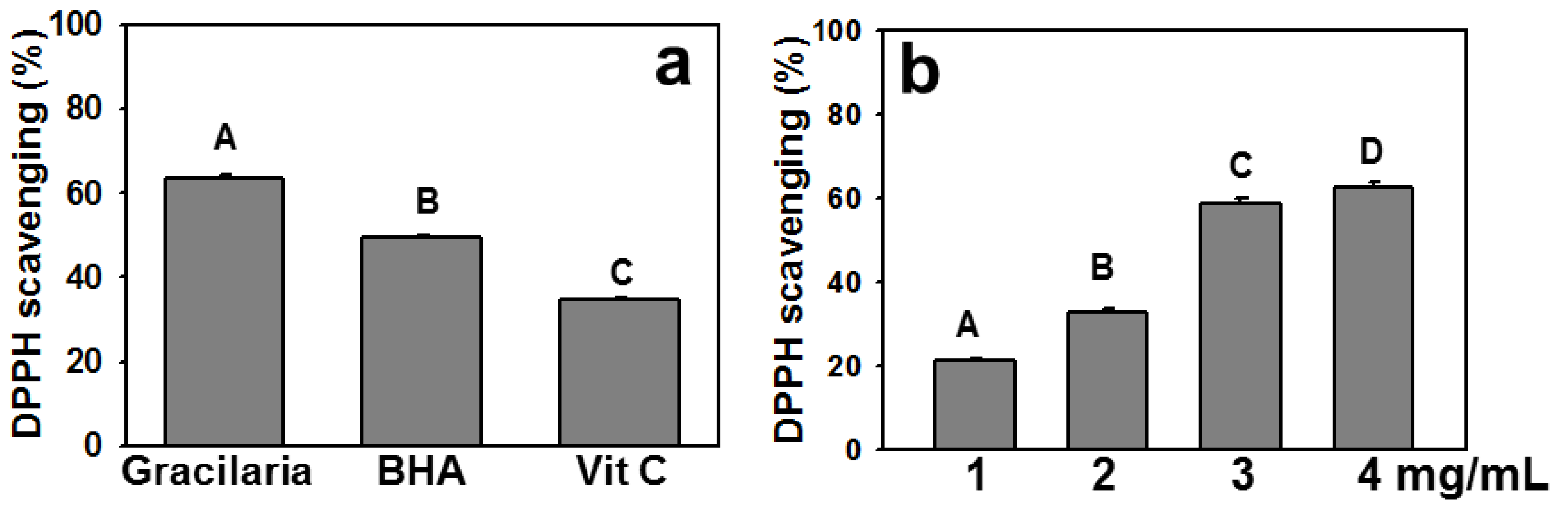
2.1. Polyphenols, Flavonoids, and Ascorbic Acid Contents of AEGT and Its DPPH Radical Scavenging Activity

The amounts of total polyphenol, flavonoid, and ascorbic acid were recorded as 98.94 ± 2.43 μg gallic acid equivalent, 22.59 ± 1.08 μg quercetin equivalent and 1.59 ± 0.18 μg ascorbic acid/mg dry extract, respectively. The DPPH scavenging activity of 4 mg/mL AEGT exceeded 60% (63.37 ± 0.91%), which was significantly higher than those of 10 ppm BHA and 100 ppm ascorbic acid (P < 0.0001) (Figure 1a). Moreover, AEGT significantly (P < 0.05) increased DPPH scavenging activity in a dose-dependent manner (Figure 1b).
Figure 1. DPPH radical scavenging activity of AEGT. (a) Comparison of DPPH radical scavenging in AEGT (4 mg/mL), positive control BHA (10 ppm) and positive control Vit. C (100 ppm). (b) DPPH radical scavenging activity of AEGT at different concentrations (1–4 mg/mL). Data are means ± S.D. (n = 3). Levels not connected by same big letter significantly differed. With the exception of A vs. D (P < 0.05), all comparisons revealed significant differences (P < 0.0001).
Molecules 17 07241 g001

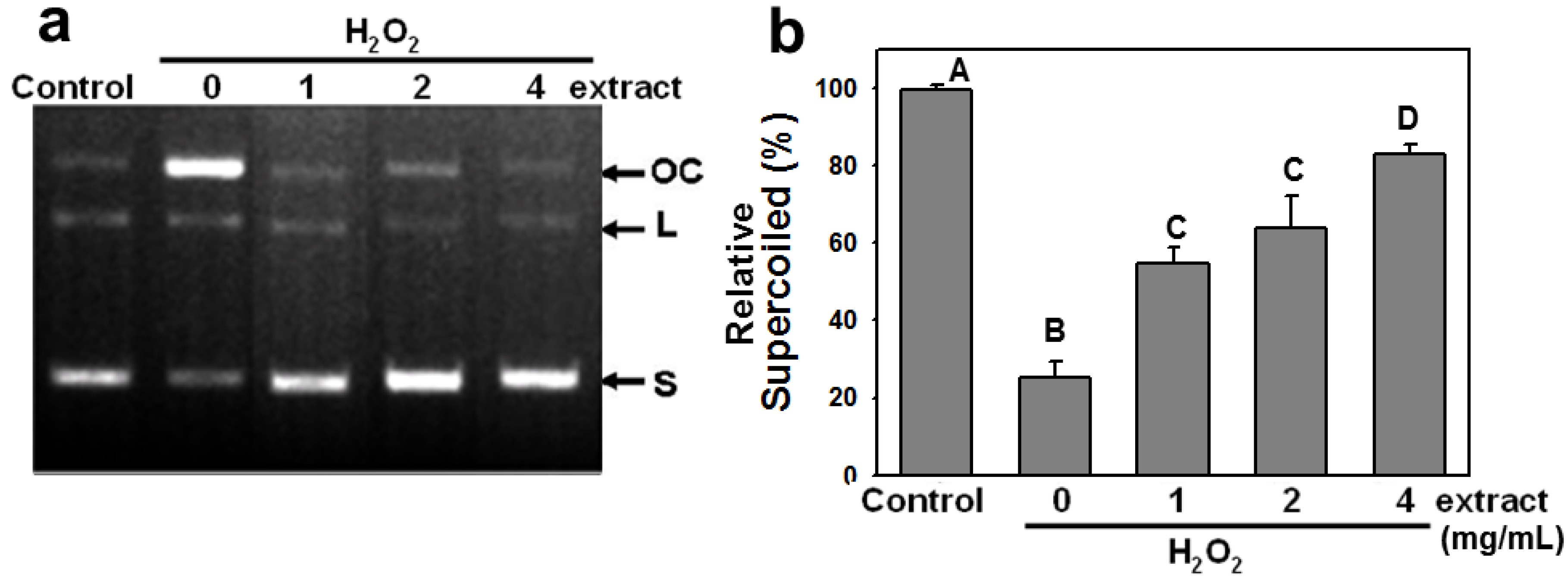
2.2. AEGT Modulates H2O2-Induced Plasmid DNA Strand Breaks

This study assessed the protective effect of AEGT against pBR322 plasmid DNA cleavage induced by H2O2 treatment. In the absence of H2O2, plasmid DNA appeared mainly in supercoiled form (S) (Figure 2a, control). Adding FeSO4 and H2O2 converted the supercoiled form to the relaxed open-circular (OC) and linear forms (L) (Figure 2a, lane 2). However, 1 to 4 mg/mL AEGT retained the supercoiled form of pBR322 in a dose-dependent manner (Figure 2a, lanes 3–5). The relative percentages of supercoiled plasmid DNA decreased to 25.2 ± 4.3 after H2O2 treatment and recovered to 54.6 ± 4.1, 63.7 ± 8.3, and 82.9 ± 2.5 in the presence of 1, 2, and 4 mg/mL AEGT, respectively (P < 0.02 to 0.001 between H2O2 alone and 1 to 4 mg/mL AEGT treatment) (Figure 2b). These results indicate that AEGT stabilizes DNA molecules by neutralizing oxidative DNA damage by H2O2.
Figure 2. Modulating effect of AEGT on H2O2-induced plasmid conformational change. (a) Gel view of plasmid DNA cleavage assay. The OC, L and S indicate the open circular, linear, and supercoiled plasmid forms, respectively. (b) The supercoiled content. Plasmid DNA pBR322 was incubated at 37 °C for 30 min with 0.003% H2O2 with and without varying AEGT doses (1, 2, and 4 mg/mL). Control was plasmid alone without treatment with H2O2 or AEGT. Data are means ± S.D. (n = 3). Levels not connected by same big letter significantly differed. Except A vs. D (P < 0.02), all comparisons among different letters showed significant differences (P < 0.005).
Molecules 17 07241 g002

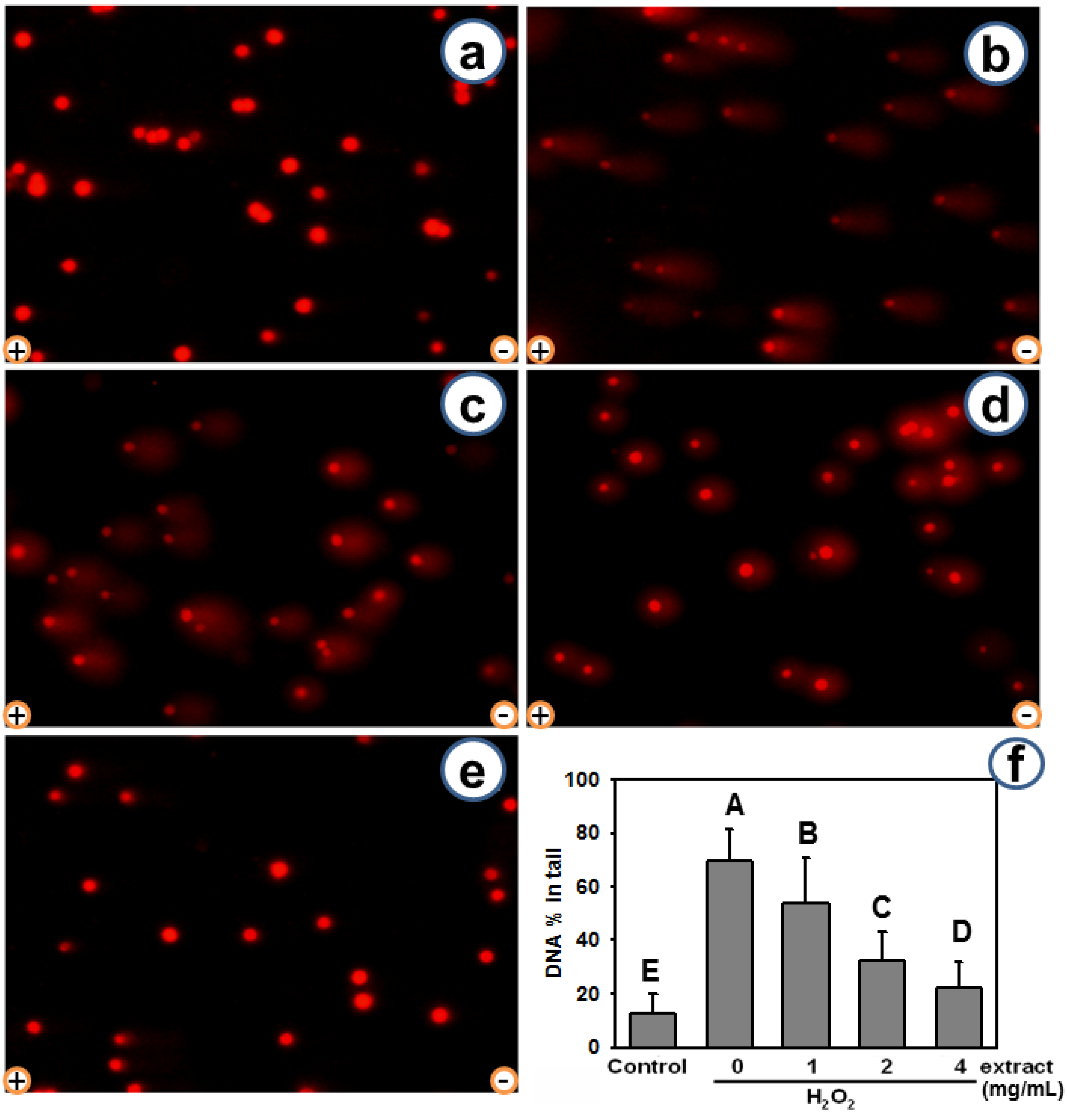
2.3. AEGT Modulates H2O2-Induced Cellular DNA Damage

Next, the protective effect of AEGT against oxidative DNA damage was verified by comet-NE assay in H1299 cells. Few “tails” were observed in the untreated controls (Figure 3a). While DNA trailing was evident in the presence of H2O2 (Figure 3b, H2O2 only), it was reduced by the AEGT co-treatments (Figure 3c–e). The average % in tail values (mean ± S.D.) for NE buffer control, H2O2alone, and AEGT treatments (1, 2, and 4 mg/mL) were 12.4 ± 7.4, 69.6 ± 11.9, 53.6 ± 17.1, 32.5 ± 10.5, and 22.1 ± 1.0 (n = 100; in duplicate), respectively (Figure 3f, P < 0.0001 between H2O2 only and 1–4 mg/mL AEGT-treated cells). Again, AEGT showed a dose-dependent protective effect against oxidative DNA damage. These results indicate the DNA modulating effect of AEGT in the cellular DNA context.

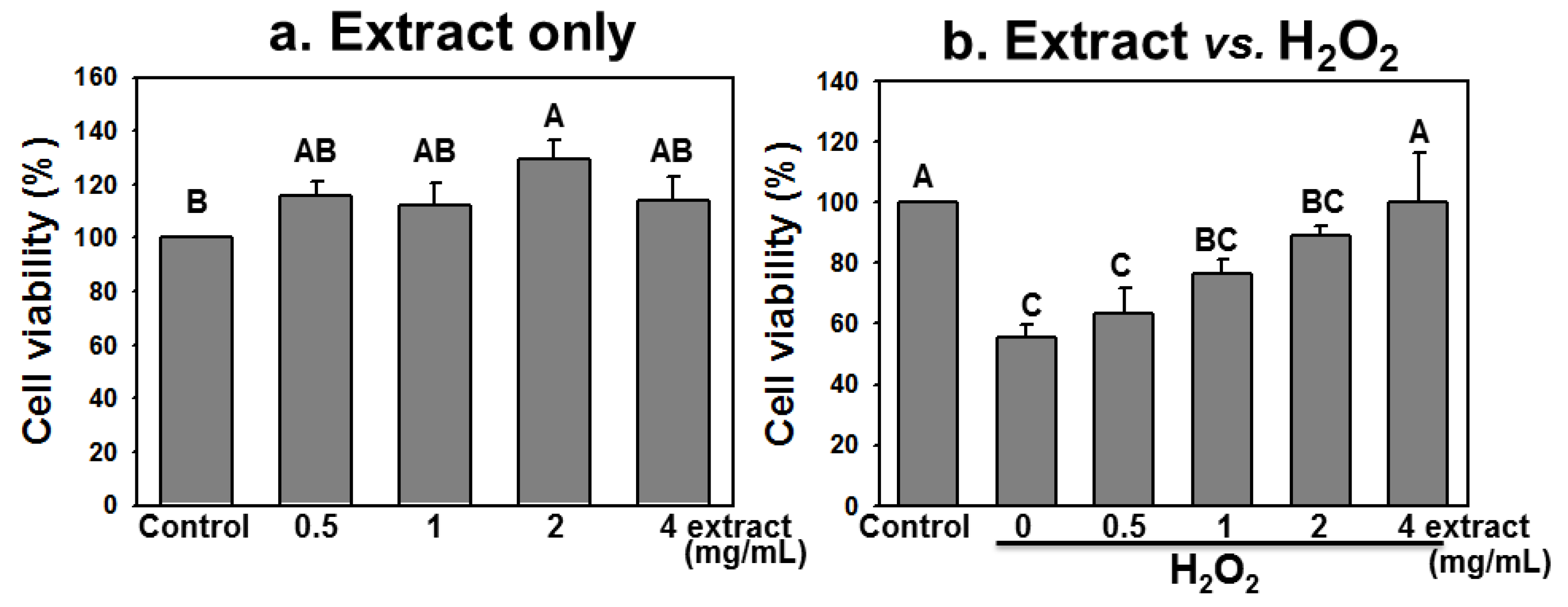
2.4. AEGT Promotes Cell Survival under H2O2 Treatment

We further tested whether the modulating effect of AEGT also promotes H1299 cell survival under H2O2 treatment. The H1299 cells were treated with or without H2O2 in the presence of 0.5, 1, 2, and 4 mg/mL of AEGT for 24 h. The cell viability was then determined by MTT assay. Treating H1299 with 0.5, 1, and 4 mg/mL AEGT alone showed no significant adverse effects on cell viability (P > 0.05 versus control) (Figure 4a). In the presence of H2O2–induced oxidative DNA damage, AEGT increased cell viability in a dose-dependent manner. From a baseline cell viability of 55.7 ± 3.8, treatment with 0.5, 1, 2 and 4 mg/mK AEGT significantly increased the cell viability of H2O2-treated cells to 63.3 ± 8.3, 76.5 ± 4.7, 89.1 ± 3.2 and 100.3 ± 16.0, respectively (P < 0.05 to 0.005, Figure 4b). Notably, the viability of H1299 cells treated with both H2O2 and 4 mg/mL AEGT did not significantly differ from that in control cells (P > 0.05, Figure 4b).
Figure 3. Modulating effect of AEGT on H2O2-induced cellular DNA strand breaks revealed by comet assay. After 2 h pretreatment with 1, 2, and 4 mg/mL AEGT, H1299 cells were co-incubated with or without 0.003% H2O2 at 37 °C for 2 h. (ae) Demonstration of PI staining results for H1299 cells incubated with or without 0.003% H2O2 and AEGT. (a) Negative control. (b) Positive control (H2O2 alone). (ce) H2O2 treatment with 1, 2, and 4 mg/mL AEGT, respectively. (f) Average % of tail DNA for H1299 cells. Data are means ± S.D. (n = 2; 100 cell counts per experiment with two individual replicates). Levels not connected by same big letter significantly differed (P < 0.0001). Electrode polarity is also shown.
Molecules 17 07241 g003
Figure 4. Cell viability was determined by MTT assay. The H1299 cells were treated for 24 h with AEGT (0.5, 1, 2, and 4 mg/mL) (a) without H2O2 and (b) with 0.003% H2O2. Data are means ± S.D. (n = 3). Levels not connected by same big letter significantly differed. Except (b) A vs. BC (P < 0.05), comparisons between other different letters in (a) and (b) showed significant differences (P < 0.005).
Molecules 17 07241 g004

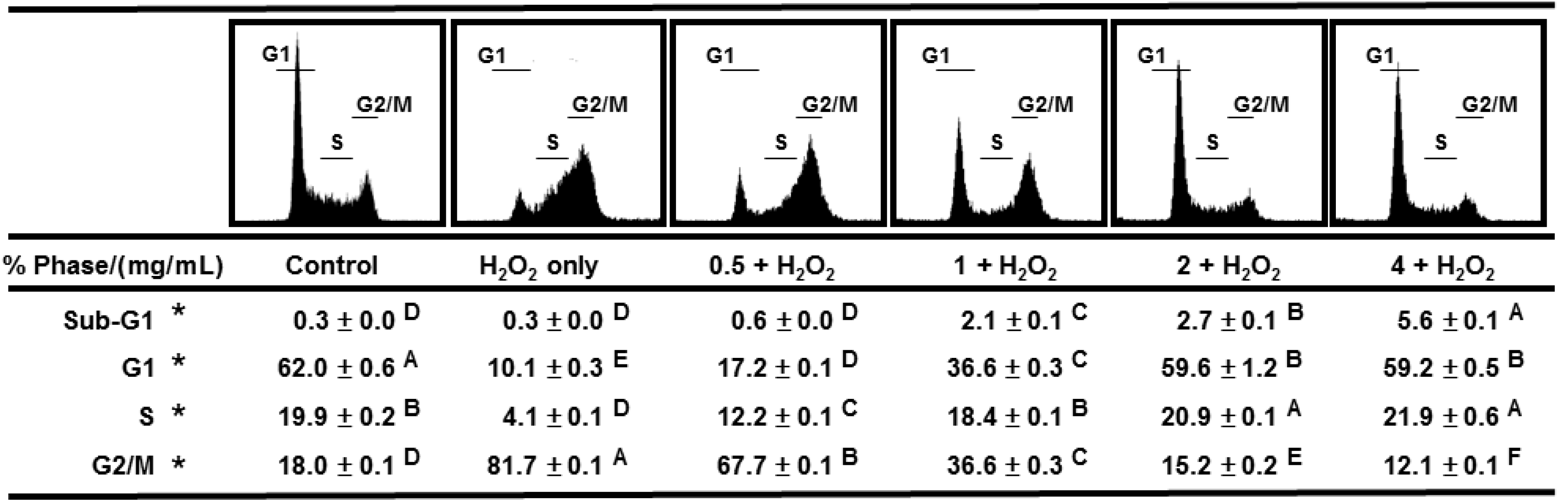
2.5. AEGT Prevents Cell Cycle Arrest by H2O2

After 24 h treatment with H2O2 in the presence of 0.5, 1, 2, and 4 mg/mL AEGT, the cell cycle distributions of H1299 cells were further analyzed by flow cytometry. The control H1299 displayed a major G1 peak and a minor G2/M peak comprising 62.0% and 18.0% of the cell population, respectively (Figure 5, panel 1). However, the H2O2-treated H1299 showed a significant increase in the G2/M population to 81.7%, which clearly indicated cell cycle arrest caused by DNA damage (Figure 5, panel 2) (P < 0.001). However, this H2O2-induced G2/M arrest was gradually and significantly diminished by co-incubation with AEGT and was virtually eliminated in the presence of 2-4 mg/mL AEGT (Figure 5, panels 3-6) (P < 0.01). This result indicates that AEGT can prevent DNA damage and ensure a normal cell cycle progression.
Figure 5. Cell cycle distribution. The H1299 cells were treated with AEGT (0.5, 1, 2, and 4 mg/mL) without and with 0.003% H2O2 for 24 h. Data are means ± S.D. (n = 3). Levels not connected by same big letter significantly differed (P < 0.01 to 0.0001). * The statistical analyses were performed on the same cell cycle phase % between different treatments (control, H2O2 alone, and AEGT/H2O2) without comparison between different phases.
Molecules 17 07241 g005

2.6. Discussion

This study investigated the antioxidant activity of AEGT in terms of its biochemical characteristics, DNA damage protection, cell growth, and cell cycle recovery after treatment with H2O2 alone or with H2O2 and AEGT. The selected damaging agent in this study was H2O2 because it exhibits either direct (oxidation of its target) or indirect (involving peroxiredoxins) signaling [18] and induction of oxidative stress [20,21,22]. The AEGT effectively suppressed ROS-induced DNA strand break in cell-free assay (Figure 2). Sensitive comet-NE assay of cellular DNA damage (Figure 3) and cell cycle profiling (Figure 5) further showed that AEGT increases H1299 cell survival by modulating oxidative stress (Figure 4). In accordance with many other reports of H2O2 treatment in various cell lines [23,24,25], H2O2-treated H1299 also showed prominent G2/M arrest, and AEGT conferred effective recovery from H2O2-induced G2/M arrest although the sub-G1 population in H2O2/4 mg/mL AEGT is mildly increased (Figure 5). Earlier reports that ROS correlate with immunity [26], anti-hepatoma activity [27], HCV-related activity [28], and Alzheimer’s disease [29] suggest that AEGT has potential applications in testing recovery from ROS-related cellular responses.
Typical phenolic compounds known to exhibit antioxidant activity include phenolic acids and flavonoids [30,31]. In addition to phenolic compounds, tannins [32], ascorbic acid [33], and pigments [34] are other potential antioxidant compounds contained in seaweeds. In the current study, polyphenols, flavonoids, and ascorbic acid were also detected in AEGT. Algal polyphenols are known to confer the major antioxidant activity of seaweed extracts [35]. Analysis of DPPH radical scavenging is largely performed to assess the free radical scavenging effect of specific compounds or extracts and can quickly indicate antioxidant activity [36,37]. The DPPH radical scavenging and phenolic content reportedly have a strong correlation in plant-based foods such as herbs [38] and seaweeds [39]. Moreover, flavonoids [40] and ascorbic acid [36] also exhibit free radical scavenging activities.
Oxidative DNA damage can result from ROS that are generated by cellular metabolism or environmental stress and it is believed to contribute to aging, carcinogenesis and other diseases [41]. For In vitro assays, the conversion of the supercoiled form of plasmid DNA into either open-circular or linear form has been used as a standard indicator of DNA damage [42].
The literature shows that boiled G. tenuistipitata extract exhibits radical scavenging activities with an IC50 of 24.22 mg/mL in DPPH radical scavenging assay [10]. This study shows that AEGT exhibits an even more potent antioxidant activity. A 60% DPPH scavenging activity was observed at only 3 mg/mL. Considering the rapid growth of seaweed and the extensive use of seaweed extracts in the food and cosmetics industries, AEGT represents an attractive multifunctional alternative for such applications. Future studies to identify the bioactive fraction of AEGT would further enhance the economic value of red algal extract. The potential protective role of AEGT to other types of cancer cells warrants further investigation.
Moreover, many algal extracts reportedly exhibit other interesting biological effects, such as the protective effects of enzymatic extracts from microalgae against H2O2-induced DNA damage [43] and the protective effects of Phaeodactylum tricornutum lipid-rich algae extract against proteasome activity [44]. Similarly, we found that AEGT protects against H2O2-induced plasmid and cellular DNA damage, cytotoxicity, and cell cycle arrest without affecting cell viability as observed in AEGT alone.

3. Experimental

3.1. Raw Materials

Specimens of G.tenuistipitata collected during spring 2009 from a culture farm at Kouhu beach, Yunlin County, Taiwan, were delivered to the laboratory at 0 °C. In the laboratory, the seaweeds were washed with running tap-water to remove epiphytes and encrusting material, immersed twice in distilled water, and then lyophilized. After pulverizing the dried sample and passing it through a 60-mesh sieve, the lyophilized sample was ground to fine powder and stored at −40 °C.

3.2. Extraction and Isolation of Seaweed G. tenuistipitata

After the addition of 1,000 mL deionized water, the dried samples (50 g) were agitated in a mechanical shaker at room temperature for 24 h. The extract was then filtered with Whatman No. 1 filter paper. The filtrate solution was evaporated to dryness at 40 ± 2 °C in a rotary evaporator (Buchi Laboratoriums-Technik, Buchs, Switzerland) and then lyophilized. The lyophilized extract was stored in a sealed container at −40 °C until use.

3.3. Determination of Total Phenolics, Flavonoid, and Ascorbic Acid of AEGT

The total phenolic compounds in AEGT extracts were determined with Folin-Ciocalteu reagent as described by Singleton and Rossi [45] using gallic acid as standard. The total phenolic content was expressed as gallic acid equivalent in μg per mg of dry sample. Ascorbic acid was quantitatively determined using the 2,6-dichloroindophenol-Na dye method as described by Jones and Hurghes [46]. Results were presented on a dry matter basis (μg ascorbic acid per mg of dry sample). Flavonoid content was determined by the colorimetric method described by Woisky and Salatino [47]. Total flavonoid content was calculated in quercetin equivalents based on a calibration curve and expressed as μg quercetin equivalents per mg of dry sample.

3.4. Free Radical Scavenging Activity

The ability of AEGT to scavenge 1,1-diphenyl-2-picrylhydrazyl (DPPH) radicals was determined as described earlier [48]. Briefly, a 1 mM methanolic solution of DPPH (1 mL) was mixed with solution of each extract (1, 2, and 4 mg/mL, 3 mL). After vigorous vortexing, the mixture was kept in darkness for 30 min at room temperature. Absorbance was measured at 517 nm, and activity was expressed as percentage of DPPH scavenging compared to control. The percentage of scavenging activity was calculated as [(Ac − As)/Ac] × 100 where As is the absorbance measured with the extract sample in the assay and Ac is the absorbance of control (without extract sample). Butylated hydroxyanisole (BHA) and ascorbic acid (Vit. C) were used as positive controls.

3.5. Cell Cultures

Cells were routinely maintained in complete RPMI-1640 (Gibco, Grand Island, NY, USA) supplemented with 10% fetal bovine serum (FBS) (BenchMark, GEMINI, Bio-Products, West Sacramento, CA, USA), 100 U/mL penicillin, 100 μg/mL streptomycin and 0.03% glutamine (Gibco, Invitrogen Ltd., Carlsbad, CA, USA). Cells were kept at 37 °C in a humidified atmosphere containing 5% CO2. The H1299 (human lung adenocarcinoma) cell line was used to test cytotoxicity in the lung cancer cell line.

3.6. Plasmid DNA Cleavage Assay

Conversion of the supercoiled (S) form of plasmid DNA to the open-circular (OC) and/or further linear (L) forms was analyzed as an indicator of DNA strand breaks [49]. Reaction mixtures (10 μL) containing 150 ng of pBR322 plasmid DNA, 0.1 mM FeSO4, 0.05% H2O2 and AEGT at concentrations of 0, 1, 2, and 4 mg/mL were incubated at 37 °C for 30 min. After stopping the reaction by adding 2 μL of 6 × gel loading dye (0.05% bromophenol blue, 40 mM EDTA and 50% glycerol (v/v), electrophoresis was performed on 0.8% agarose gel in 0.5 × TAE buffer at 50 V for 1–2 h. The DNA in the gel was stained with ethidium bromide (final concentration 0.8 μg/mL) and then visualized and photographed under ultraviolet light. The formula used for calculating the percentage of supercoiled DNA was as follows: S% = (band density of S / band density of (S + L + OC), where the band density was determined by the Gel-pro Analyzer 4.0 (Media Cybernetics, Bethesda, MD, USA). Relative S% = S% of test sample/S% of control.

3.7. Comet-NE Assay

The comet-NE is more sensitive than the traditional comet assay [50,51]. The comet-NE assay using nuclear extracts (NEs) prepared from NB4 cells (human acute promyelocytic leukemia) cell line [52,53,54] was performed using a protocol described previously [52,53,54]. Aliquots (100 μL) of H1299 cell suspensions (1 × 10⁶ cells/mL in PBS) were mixed with equal volumes of 1.2% low-melting agarose (in PBS, pH 7.4) and immediately pipetted onto a glass slide precoated with 1% regular agarose (in distilled water). The slides were then immersed in freshly prepared ice-cold cell lysis solution (2.5 M NaCl, 100 mM EDTA, 10 mM Tris pH 10, 1% N-laurylsarcosine, 1% Triton X-100 and 10% dimethylsulfoxide or DMSO) incubated at 4 °C for 2 h and then rinsed three times with deionized water. A 20 μL excision mixture containing 0.6 μg NE, 50 mM Hepes-KOH (pH 7.9), 70 mM KCl, 5 mM MgCl2, 0.4 mM EDTA, 2 mM ATP, 40 mM phosphocreatine and 2.5 mM creatine phosphokinase was then added to each slide. After applying the cover slips, the slides were incubated at 37 °C for 2 h in a humidified space for NE digestion. The slides were denatured in 0.3 N NaOH, 1 mM EDTA for 20 min and then electrophoresed at 20 V, 300 mA for 25 min. After washing with deionized water, the slides were neutralized in 0.4 M Tris-HCl, pH 7.5 and stained with 40 μL propidium iodide (PI, 50 μg/mL). Under a fluorescence microscope (TE2000-U; Nikon, Tokyo, Japan), the migration of DNA from the nucleus of each cell was measured with the CometScore [55] software program. The % tail DNA parameter [56,57] was calculated as the percentage of DNA in the comet tail (sum of intensities of pixels in the tail). The formula used for calculating the % tail DNA was as follows: Tail % DNA = 100 − Head % DNA, where Head % DNA = (Head Optic Intensity / (Head Optic Intensity + Tail Optic Intensity)) × 100.

3.8. Cell Viability Assay

The 3-(4,5-Dimethylthiazol-2-yl)-2,5-diphenyltetrazolium bromide (MTT) assay was performed as described previously [58]. Briefly, fresh medium (100 μL) containing 0.5 mg/mL MTT was added into each well of a 96-well plate containing 5 × 10³ cells/well and incubated for 2 h at 37 °C. After removing MTT-containing medium, 100 μL of DMSO was added into each well to dissolve the purple formazan crystal. The plates were then shaken gently for 20 min in darkness and then read at 595 nm on a microtiter plate reader.

3.9. Cell Cycle Histogram Obtained by Propidium Iodide Staining in Flow Cytometry

The cell cycle histogram was determined as described previously [59]. Briefly, cells with 5 × 10⁵ cells/100-mm Petri-dish were plated. After recovery, cells were treated for 24 h with 0, 1, 2, and 4 mg/mL AEGT with or without 0.003% H2O2. After treatment, cells were collected, washed twice with PBS, and fixed in 70% ethanol overnight. The cells were then centrifuged at 700 rpm for 5 min at 4 °C and then resuspended in PBS buffer containing 10 μg/mL PI (Sigma, St Louis, MO, USA) and 10 μg/mL RNase A. After 15 min incubation in darkness at room temperature, the cells were analyzed with a FACScan flow cytometer (Becton-Dickinson, Mansfield, MA, USA) in cell counts of 10,000 with gated setting (forward light scatter versus side light scatter such that only single cells were assayed) [60], and the gated data were analyzed by Cell-Quest and Modfit softwares (Becton-Dickinson, Mansfield, MA, USA).

3.10. Statistical Analysis

All data were presented as means ± SEM. Experimental groups were compared by one-way ANOVA with Tukey HSD Post Hoc Test using JMP® 9 software [61]. Levels not connected by the same big letter significantly differed.

4. Conclusions

Taken together, the experimental results in this study confirmed the hypothesis that edible red algae Gracilaria extract prevents ROS-induced DNA damage and its related cellular responses.

Acknowledgments

This work was supported by Ministry of Education of ROC under the grant 98D1024 of marine technology research and development center to J. I. Yang and was also supported by grants from the Kaohsiung Medical University Research Foundation, the Department of Health, Executive Yuan, R.O.C. (TAIWAN) (DOH101-TD-C-111-002), the Kaohsiung Medical University Research Foundation (KMUER014 and KMU-M110001) and the NSYSU-KMU JOINT RESEARCH PROJECT (#NSYSUKMU 101-006).

Conflicts of Interest

The authors declare no conflict of interest.

References

  1. Chiang, Y.M.; Lin, J.L. Nitrate uptake by nitrogen-starved plants of the red alga Gracilaria tenuistipitata var. Liui. Jpn. J. Phycol. 1989, 37, 187–193. [Google Scholar]
  2. Armisen, R. World-wide use and importance of Gracilaria. J. Appl. Phycol. 1995, 7, 231–243. [Google Scholar] [CrossRef]
  3. Ajisaka, T.; Chiang, Y.M. Recent status of Gracilaria cultivation in Taiwan. Hydrobiologia 1993, 260/261, 335–338. [Google Scholar] [CrossRef]
  4. Jha, R.K.; Zi-rong, X. Biomedical compounds from marine organisms. Mar. Drugs 2004, 2, 123–146. [Google Scholar] [CrossRef]
  5. Faulkner, D.J. Marine natural products. Nat. Prod. Rep. 2002, 19, 1–48. [Google Scholar]
  6. Hsu, B.Y.; Tsao, C.Y.; Chiou, T.K.; Hwang, P.A.; Hwang, D.F. HPLC determination for prostaglandins from seaweed Gracilaria gigas. Food Control 2007, 18, 639–645. [Google Scholar] [CrossRef]
  7. Marinho-Soriano, E.; Bourret, E. Polysaccharides from the red seaweed Gracilaria dura (Gracilariales, Rhodophyta). Bioresour. Technol. 2005, 96, 379–382. [Google Scholar] [CrossRef]
  8. de Almeida, C.L.; Falcao Hde, S.; Lima, G.R.; Montenegro Cde, A.; Lira, N.S.; de Athayde-Filho, P.F.; Rodrigues, L.C.; de Souza Mde, F.; Barbosa-Filho, J.M.; Batista, L.M. Bioactivities from marine algae of the genus gracilaria. Int. J. Mol. Sci. 2011, 12, 4550–4573. [Google Scholar] [CrossRef]
  9. Lin, Y.H.; Tsai, J.S.; Hung, L.B.; Pan, B.S. Hypocholesterolemic effect of compounded freshwater clam protein hydrolysate and Gracilaria. Food Chem. 2010, 123, 395–399. [Google Scholar] [CrossRef]
  10. Yangthong, M.; Hutadilok-Towatana, N.; Phromkunthong, W. Antioxidant activities of four edible seaweeds from the southern coast of Thailand. Plant Foods Hum. Nutr. 2009, 64, 218–223. [Google Scholar] [CrossRef]
  11. Ganesan, P.; Kumar, C.S.; Bhaskar, N. Antioxidant properties of methanol extract and its solvent fractions obtained from selected Indian red seaweeds. Bioresour. Technol. 2008, 99, 2717–2723. [Google Scholar] [CrossRef]
  12. Vijayavel, K.; Martinez, J.A. In vitro antioxidant and antimicrobial activities of two Hawaiian marine Limu: Ulva fasciata (Chlorophyta) and Gracilaria salicornia (Rhodophyta). J. Med. Food 2010, 13, 1494–1499. [Google Scholar] [CrossRef]
  13. Souza, B.W.; Cerqueira, M.A.; Martins, J.T.; Quintas, M.A.; Ferreira, A.C.; Teixeira, J.A.; Vicente, A.A. Antioxidant potential of two red seaweeds from the Brazilian coasts. J. Agric. Food Chem. 2011, 59, 5589–5594. [Google Scholar]
  14. Dang, H.T.; Lee, H.J.; Yoo, E.S.; Shinde, P.B.; Lee, Y.M.; Hong, J.; Kim, D.K.; Jung, J.H. Anti-inflammatory constituents of the red alga Gracilaria verrucosa and their synthetic analogues. J. Nat. Prod. 2008, 71, 232–240. [Google Scholar] [CrossRef]
  15. Coura, C.O.; de Araujo, I.W.; Vanderlei, E.S.; Rodrigues, J.A.; Quindere, A.; Fontes, B.P.; de Queiroz, I.N.; de Menezes, D.B.; Bezerra, e Silver, A.A.; et al. Antinociceptive and anti-inflammatory activities of sulphated polysaccharides from the red seaweed gracilaria cornea. Basic Clin. Pharmacol. Toxicol. 2011, 110, 335–341. [Google Scholar]
  16. Yeh, S.T.; Lin, Y.C.; Huang, C.L.; Chen, J.C. White shrimp Litopenaeus vannamei that received the hot-water extract of Gracilaria tenuistipitata showed protective innate immunity and up-regulation of gene expressions after low-salinity stress. Fish Shellfish Immunol. 2010, 28, 887–894. [Google Scholar] [CrossRef]
  17. Martinet, W.; Knaapen, M.W.; De Meyer, G.R.; Herman, A.G.; Kockx, M.M. Oxidative DNA damage and repair in experimental atherosclerosis are reversed by dietary lipid lowering. Circ. Res. 2001, 88, 733–739. [Google Scholar] [CrossRef]
  18. Rigoulet, M.; Yoboue, E.D.; Devin, A. Mitochondrial ROS generation and its regulation: Mechanisms involved in H(2)O(2) signaling. Antioxid. Redox Signal. 2011, 14, 459–468. [Google Scholar] [CrossRef]
  19. Cao, W.; Chen, W.J.; Suo, Z.R.; Yao, Y.P. Protective effects of ethanolic extracts of buckwheat groats on DNA damage caused by hydroxyl radicals. Food Res. Int. 2008, 41, 924–929. [Google Scholar] [CrossRef]
  20. Wijeratne, S.S.; Cuppett, S.L.; Schlegel, V. Hydrogen peroxide induced oxidative stress damage and antioxidant enzyme response in Caco-2 human colon cells. J. Agric. Food Chem. 2005, 53, 8768–8774. [Google Scholar] [CrossRef]
  21. Wood, J.M.; Decker, H.; Hartmann, H.; Chavan, B.; Rokos, H.; Spencer, J.D.; Hasse, S.; Thornton, M.J.; Shalbaf, M.; Paus, R.; et al. Senile hair graying: H2O2-mediated oxidative stress affects human hair color by blunting methionine sulfoxide repair. FASEB J. 2009, 23, 2065–2075. [Google Scholar] [CrossRef]
  22. Spencer, J.D.; Gibbons, N.C.; Rokos, H.; Peters, E.M.; Wood, J.M.; Schallreuter, K.U. Oxidative stress via hydrogen peroxide affects proopiomelanocortin peptides directly in the epidermis of patients with vitiligo. J. Invest. Dermatol. 2007, 127, 411–420. [Google Scholar] [CrossRef]
  23. Seomun, Y.; Kim, J.T.; Kim, H.S.; Park, J.Y.; Joo, C.K. Induction of p21Cip1-mediated G2/M arrest in H2O2-treated lens epithelial cells. Mol. Vis. 2005, 11, 764–774. [Google Scholar]
  24. Thorn, T.; Gniadecki, R.; Petersen, A.B.; Vicanova, J.; Wulf, H.C. Differences in activation of G2/M checkpoint in keratinocytes after genotoxic stress induced by hydrogen peroxide and ultraviolet A radiation. Free Radic. Res. 2001, 35, 405–416. [Google Scholar] [CrossRef]
  25. Chien, M.; Rinker-Schaeffer, C.; Stadler, W.M. A G2/M growth arrest response to low-dose intermittent H2O2 in normal uroepithelial cells. Int. J. Oncol. 2000, 17, 425–432. [Google Scholar]
  26. Lu, S.; Guo, X.; Zhao, P. Effect of Ginkgo biloba extract 50 on immunity and antioxidant enzyme activities in ischemia reperfusion rats. Molecules 2011, 16, 9194–9206. [Google Scholar] [CrossRef]
  27. Li, Q.; Jiang, C.; Zu, Y.; Song, Z.; Zhang, B.; Meng, X.; Qiu, W.; Zhang, L. SFE-CO2 extract from Typhonium giganteum Engl. tubers, induces apoptosis in human hepatoma SMMC-7721 cells involvement of a ROS-mediated mitochondrial pathway. Molecules 2011, 16, 8228–8242. [Google Scholar] [CrossRef]
  28. Simula, M.P.; De Re, V. Hepatitis C virus-induced oxidative stress and mitochondrial dysfunction: A focus on recent advances in proteomics. Proteom. Clin. Appl. 2010, 4, 782–793. [Google Scholar] [CrossRef]
  29. Dumont, M.; Beal, M.F. Neuroprotective strategies involving ROS in Alzheimer disease. Free Radic. Biol. Med. 2011, 51, 1014–1026. [Google Scholar] [CrossRef]
  30. de Oliveira, C.B.; Comunello, L.N.; Lunardelli, A.; Amaral, R.H.; Pires, M.G.; da Silva, G.L.; Manfredini, V.; Vargas, C.R.; Gnoatto, S.C.; de Oliveira, J.R.; et al. Phenolic enriched extract of Baccharis trimera presents anti-inflammatory and antioxidant activities. Molecules 2012, 17, 1113–1123. [Google Scholar] [CrossRef] [Green Version]
  31. Pieroni, L.G.; de Rezende, F.M.; Ximenes, V.F.; Dokkedal, A.L. Antioxidant activity and total phenols from the methanolic extract of Miconia albicans (Sw.) Triana leaves. Molecules 2011, 16, 9439–9450. [Google Scholar] [CrossRef]
  32. Heo, S.J.; Jeon, Y.J.; Lee, J.; Kim, H.T.; Lee, K.W. Antioxidant effect of enzymatic hydrolyzate from a Kelp, Ecklonia cava. Algae 2003, 18, 341–347. [Google Scholar] [CrossRef]
  33. Munda, I.M. Preliminary information on the ascorbic acid content in some Adriatic seaweeds. Hydrobiologia 1987, 151/152, 477–481. [Google Scholar] [CrossRef]
  34. Siriwardhana, N.; Lee, K.W.; Kim, S.H.; Ha, J.W.; Park, G.T.; Jeon, Y.J. Lipid peroxidation inhibitory effects of Hizikia fusiformis methanolic extract on fish oil and linoleic acid. Food Sci. Technol. Int. 2004, 10, 65–72. [Google Scholar] [CrossRef]
  35. Nakamura, T.; Nagayama, K.; Uchida, K.; Tanaka, R. Antioxidant activity of phlorotannins from the brown alga Eisenia bicyclis. Fish. Sci. 1996, 62, 923–926. [Google Scholar]
  36. Scalzo, R.L. Organic acids influence on DPPH scavenging by ascorbic acid. Food Chem. 2008, 107, 40–43. [Google Scholar] [CrossRef]
  37. Yang, J.I.; Ho, H.Y.; Chu, Y.J.; Chow, C.J. Characteristic and antioxidant activity of retorted gelatin hydrolysate from cobia (Rachycentron canadum) skin. Food Chem. 2008, 110, 128–136. [Google Scholar] [CrossRef]
  38. Zheng, W.; Wang, S.Y. Antioxidant activity and phenolic compounds in selected herbs. J. Agric. Food Chem. 2001, 49, 5165–5170. [Google Scholar] [CrossRef]
  39. Jimenez-Escrig, A.; Rincon, M.; Pulido, R.; Saura-Calixto, F. Guava fruit (Psidium guajava L.) as a new source of antioxidant dietary fiber. J. Agric. Food Chem. 2001, 49, 5489–5493. [Google Scholar] [CrossRef]
  40. Robards, K.; Prenzler, P.D.; Tucker, G.; Swatsitang, P.; Glover, W. Phenolic compounds and their role in oxidative process in fruits. Food Chem. 1999, 66, 401–436. [Google Scholar] [CrossRef]
  41. Cooke, M.S.; Evans, M.D.; Dizdaroglu, M.; Lunec, J. Oxidative DNA damage: Mechanisms, mutation, and diseas. FASEB J. 2003, 17, 1195–1214. [Google Scholar] [CrossRef]
  42. Onuki, J.; Medeiros, M.H.; Bechara, E.J.; Di Mascio, P. 5-Aminolevulinic acid induces single-strand breaks in plasmid pBR322 DNA in the presence of Fe2+ ions. Biochim. Biophys. Acta 1994, 1225, 259–263. [Google Scholar] [CrossRef]
  43. Karawita, R.; Senevirathne, M.; Athukorala, Y.; Affan, A.; Lee, Y.J.; Kim, S.K.; Lee, J.B.; Jeon, Y.J. Protective effect of enzymatic extracts from microalgae against DNA damage induced by H2O2. Mar. Biotechnol. (NY) 2007, 9, 479–490. [Google Scholar] [CrossRef]
  44. Nizard, C.; Poggioli, S.; Heusele, C.; Bulteau, A.L.; Moreau, M.; Saunois, A.; Schnebert, S.; Mahe, C.; Friguet, B. Algae extract protection effect on oxidized protein level in human stratum corneum. Ann. NY Acad. Sci. 2004, 1019, 219–222. [Google Scholar] [CrossRef]
  45. Singleton, V.L.; Rossi, J.A. Colorimetry of total phenolics with phosphomolybdic-phosphotungstic acid reagents. Am. J. Enol. Vitic. 1965, 16, 144–158. [Google Scholar]
  46. Jones, E.; Hurghes, R.E. Foliar ascorbic acid in some angiosperms. Phytochemistry 1983, 22, 2493–2499. [Google Scholar] [CrossRef]
  47. Woisky, R.G.; Salatino, A. Analysis of propolis: Some parameters and procedures for chemical quality control. J. Agric. Food Chem. 1998, 37, 99–105. [Google Scholar]
  48. Blois, M.S. Antioxidant determinations by the use of a stable free radical. Nature 1958, 26, 1199–1200. [Google Scholar] [CrossRef]
  49. Chen, B.H.; Chang, H.W.; Huang, H.M.; Chong, I.W.; Chen, J.S.; Chen, C.Y.; Wang, H.M. (−)-Anonaine induces DNA damage and inhibits growth and migration of human lung carcinoma H1299 cells. J. Agric. Food Chem. 2011, 59, 2284–2290. [Google Scholar] [CrossRef]
  50. Li, P.Y.; Chang, Y.C.; Tzang, B.S.; Chen, C.C.; Liu, Y.C. Antibiotic amoxicillin induces DNA lesions in mammalian cells possibly via the reactive oxygen species. Mutat. Res. 2007, 629, 133–139. [Google Scholar] [CrossRef]
  51. Wang, A.S.; Ramanathan, B.; Chien, Y.H.; Goparaju, C.M.; Jan, K.Y. Comet assay with nuclear extract incubation. Anal. Biochem. 2005, 337, 70–75. [Google Scholar]
  52. Chang, Y.C.; Liao, C.B.; Hsieh, P.Y.; Liou, M.L.; Liu, Y.C. Expression of tumor suppressor p53 facilitates DNA repair but not UV-induced G2/M arrest or apoptosis in Chinese hamster ovary CHO-K1 cells. J. Cell. Biochem. 2008, 103, 528–537. [Google Scholar] [CrossRef]
  53. Chang, Y.C.; Jan, K.Y.; Cheng, C.A.; Liao, C.B.; Liu, Y.C. Direct involvement of the tumor suppressor p53 in nucleotide excision repair. DNA Repair (Amst) 2008, 7, 751–761. [Google Scholar] [CrossRef]
  54. Cemeli, E.; Mirkova, E.; Chiuchiarelli, G.; Alexandrova, E.; Anderson, D. Investigation on the mechanisms of genotoxicity of butadiene, styrene and their combination in human lymphocytes using the Comet assay. Mutat. Res. 2009, 664, 69–76. [Google Scholar] [CrossRef]
  55. CometScore, version 1.5. Available online: http://www.tritekcorp.com (accessed on 11 June 2012).
  56. Wong, V.W.C.; Szeto, Y.T.; Collins, A.R.; Benzie, I.F.F. The comet assay: A biomonitoring tool for nutraceutical research. Curr. Top. Nutraceut. Res. 2005, 3, 1–14. [Google Scholar]
  57. Collins, A.R. The comet assay for DNA damage and repair: Principles, applications, and limitations. Mol. Biotechnol. 2004, 26, 249–261. [Google Scholar] [CrossRef]
  58. Huang, C.Y.; Chen, J.Y.; Wu, J.E.; Pu, Y.S.; Liu, G.Y.; Pan, M.H.; Huang, Y.T.; Huang, A.M.; Hwang, C.C.; Chung, S.J.; et al. Ling-Zhi polysaccharides potentiate cytotoxic effects of anticancer drugs against drug-resistant urothelial carcinoma cells. J. Agric. Food Chem. 2010, 58, 8798–8805. [Google Scholar]
  59. Chiu, C.C.; Li, C.H.; Fuh, T.S.; Chen, W.L.; Huang, C.S.; Chen, L.J.; Ung, W.H.; Fang, K. The suppressed proliferation and premature senescence by ganciclovir in p53-mutated human non-small-lung cancer cells acquiring herpes simplex virus-thymidine kinase cDNA. Cancer Detect. Prev. 2005, 29, 286–293. [Google Scholar] [CrossRef]
  60. Nunez, R. DNA measurement and cell cycle analysis by flow cytometry. Curr. Issues Mol. Biol. 2001, 3, 67–70. [Google Scholar]
  61. JMP® 9 software. Available online: http://www.jmp.com/ (accessed on 11 June 2012).
  • Sample Availability: Samples of aqueous extract of Gracilaria tenuistipitata (AEGT) are available upon request from Dr. Jing-Iong Yang.

