Article

Antioxidant Activity, Phenolic Profile, and Nephroprotective Potential of Anastatica hierochuntica Ethanolic and Aqueous Extracts against CCl4-Induced Nephrotoxicity in Rats

by
Tariq I. Almundarij
1,
Yousef M. Alharbi
1,
Hassan A. Abdel-Rahman
2 and
Hassan Barakat
3,4,*
1
Department of Veterinary Medicine, College of Agriculture and Veterinary Medicine, Qassim University, Buraydah 51452, Saudi Arabia
2
Department of Physiology, Faculty of Veterinary Medicine, Sadat City University, Menofia 32897, Egypt
3
Department of Food Science and Human Nutrition, College of Agriculture and Veterinary Medicine, Qassim University, Buraydah 51452, Saudi Arabia
4
Food Technology Department, Faculty of Agriculture, Benha University, Moshtohor 13736, Qaliuobia, Egypt
*
Author to whom correspondence should be addressed.
Nutrients 2021, 13(9), 2973; https://doi.org/10.3390/nu13092973
Submission received: 17 July 2021 / Revised: 21 August 2021 / Accepted: 24 August 2021 / Published: 26 August 2021
(This article belongs to the Section Phytochemicals and Human Health)

Abstract

:
Kaff-e-Maryam (Anastatica hierochuntica L.) is extensively used to treat a range of health problems, most notably to ease childbirth and alleviate reproductive system-related disorders. This study aimed to evaluate the effect of A. hierochuntica ethanolic (KEE), and aqueous (KAE) extracts on CCl4-induced oxidative stress and nephrotoxicity in rats using the biochemical markers for renal functions and antioxidant status as well as histopathological examinations of kidney tissue. A. hierochuntica contained 67.49 mg GAE g−1 of total phenolic compounds (TPC), 3.51 µg g−1 of total carotenoids (TC), and 49.78 and 17.45 mg QE g−1 of total flavonoids (TF) and total flavonols (TFL), respectively. It resulted in 128.71 µmol of TE g−1 of DPPH-RSA and 141.92 µmol of TE g−1 of ABTS-RSA. A. hierochuntica presented superior antioxidant activity by inhibiting linoleic acid radicals and chelating oxidation metals. The HPLC analysis resulted in 9 and 21 phenolic acids and 6 and 2 flavonoids in KEE and KAE with a predominance of sinapic and syringic acids, respectively. Intramuscular injection of vit. E + Se and oral administration of KEE, KAE, and KEE + KAE at 250 mg kg−1 body weight significantly restored serum creatinine, urea, K, total protein, and albumin levels. Additionally, they reduced malondialdehyde (MOD), restored reduced-glutathione (GSH), and enhanced superoxide dismutase (SOD) levels. KEE, KAE, and KEE + KAE protected the kidneys from CCl4-nephrotoxicity as they mainly attenuated induced oxidative stress. Total nephroprotection was about 83.27%, 97.62%, and 78.85% for KEE, KAE, and KEE + KAE, respectively. Both vit. E + Se and A. hierochuntica extracts attenuated the histopathological alteration in CCl4-treated rats. In conclusion, A. hierochuntica, especially KAE, has the potential capability to restore oxidative stability and improve kidney function after CCl4 acute kidney injury better than KEE. Therefore, A. hierochuntica has the potential to be a useful therapeutic agent in the treatment of drug-induced nephrotoxicity.

Graphical Abstract

1. Introduction

Kidney disease is the 9th leading cause of death with more than 1 in 7, that is, 15% of US adults or 37 million people, are estimated to have chronic kidney disease (CKD) [1]. Remarkably, the most common cause of CKD as recorded in 2015 is diabetes mellitus followed by high blood pressure and glomerulonephritis [2]. Other causes of CKD include idiopathic (often associated with small kidneys on renal ultrasound) [3]. Previously, CCl4 was used for metal degreasing, dry cleaning, fabric spotting, fire extinguisher fluids, and grain fumigation [4]. It causes severe disorders in the liver, lungs, and testes as well as in blood by generating active free radicals [5]. According to the findings of Ogeturk et al. [6], exposure to this solvent produces acute and chronic kidney damage. Free radical-induced lipid peroxidation is believed to be one of the primary causes of cell membrane damage, leading to a variety of pathological conditions [7]. The generation of reactive radicals has been implicated in CCl4-induced nephrotoxicity, which is involved in lipid peroxidation and accumulation of dysfunctional proteins, leading to injuries in kidneys [8]. Amazingly, traditional uses of medicinal plants have grown in recent years, and numerous investigations have confirmed their therapeutic role against several illnesses [9,10,11,12].
Kaff-e-Maryam (Anastatica hierochuntica) is a desert medicinal herb that belongs to the Brassicaceae family. It grows in various regions over the world, especially in Arab countries (e.g., Saudi Arabia, Egypt, Oman, Libya, Iraq, the United Arab Emirates, Kuwait) as well as some Asian, European, and African countries. A. hierochuntica is believed to have superior medical potentials and is preferably consumed for various medical conditions [13]. It is mainly used to ease the process of childbirth and for treating reproductive system-related disorders and metabolic disorders, mainly diabetes mellitus [14]. It is used as an analgesic and as a treatment for epilepsy, gastrointestinal disorders, arthritis, bronchial asthma, mouth ulcers, malaria, and mental depression [15,16,17]. A. hierochuntica contains significant amounts of minerals, such as Mg, Ca, Mn, Fe, Cu, and Zn, which are comparable to or greater than those of many herbal plants, which may give metals chelating properties [18]. Yoshikawa et al. [19] concluded that the presence of flavanones, such as Anastatin A and B, correlates with potent hepatoprotective effects by inhibiting D-galactosamine-induced cytotoxicity in primary cultured mouse hepatocytes. The production of reactive oxygen species (ROS) and cytokines such as tumor necrosis factor and interleukin-1 by Kupffer cells in the liver contribute to hepatocyte destruction in D-galactosamine hepatotoxicity [20]. The antioxidant and anti-inflammatory properties of A. hierochuntica components may help to reduce D-galactosamine-induced hepatotoxicity [21]. A. hierochuntica can afford extract-depending protection against CCl4-hepatotoxicity [22]. However, despite the literature showing promising potentialities related to the use of A. hierochuntica, the nephroprotective potential of A. hierochuntica ethanolic (KEE) and aqueous (KAE) extracts needs to be carefully examined. Moreover, the literature review mainly highlighted the hepatoprotective efficiency of A. hierochuntica, but the nephroprotective potential has not been studied so far, thus motivating this work. Therefore, the current study aims to observe the changes in the antioxidative defense enzymes, detect the alterations of renal microscopy after CCl4 administration in rats, and investigate the possible protective effects of A. hierochuntica extracts against CCl4-induced renal damage.

2. Materials and Methods

2.1. Sample Preparation

A sample of the Kaff-e-Maryam (A. hierochuntica L.) plant was purchased from a native market in Buraydah city, Qassim region, Saudi Arabia. The plant material was authenticated by the Department of Plant Production and Protection, College of Agriculture and Veterinary Medicine, Qassim University, Saudi Arabia. The sample was washed with clean tap water to remove sand and dirt from the leaves and then air-dried plant material (at 28 ± 1 °C for 48 h.) was mechanically powdered and kept in opaque polyethylene bags at 4 ± 1 °C until use.

2.2. Preparation of Ethanolic and Aqueous Extracts

Approximately 200 g of dried A. hierochuntica were extracted with 300 mL 70% ethanol in a Soxhlet extractor to prepare ethanolic extraction (KEE). The extract was concentrated by a rotary evaporator at 40 °C to evaporate the remaining solvent, then to dryness under an N2 stream. The aqueous extraction (KAE) was carried out as described by Asuzu [23] with minor modifications. Two hundred grams of dried plant material were added to 500 mL of hot sterile distilled water. The mixture was then shaken well and allowed to stand for 1 h. Then a reflux condenser was attached to the flask and then heated until boiling gently for 10 min, cooled, shaken well, and filtered through Whatman No. 1 filter paper. The filtrate was evaporated by a rotary evaporator, then to dryness under an N2 stream. The alcoholic and aqueous extracts (250 mg mL−1) were freshly formulated in distilled water to be used for oral administration.

2.3. Total Phenolic Content (TPC)

The TPC content of A. hierochuntica was determined according to the adapted method by Bettaieb et al. [24]. The results were compared to a plotted gallic acid (GA) standard curve made in the range of 50–500 mg mL−1 (R2 = 0.99), and the TPC was calculated as mg of gallic acid equivalent (GAE) per gram of A. hierochuntica (mg of GAE g−1).

2.4. Total Carotenoids (TC), Total Flavonoids (TF), and Total Flavonols (TFL)

As reported by Al-Qabba et al. [10], 5 g of A. hierochuntica was extracted repeatedly with acetone and petroleum ether mixture (1:1, v/v). Total carotenoids (TC) content was spectrophotometrically determined at 451 nm. TC was expressed as mg g−1 dw. The TF content of A. hierochuntica was assayed according to described protocol by Mohdaly et al. [25]. The TF content was calculated as mg quercetin equivalent (QE) per 100 g−1 dw. In the same context, the TFL content was carried out [26]. The absorbance at 440 nm was recorded, and TFL was calculated as mg quercetin equivalent (QE) per 100 g−1 dw.

2.5. Antioxidant Capacity Determination

DPPH radical scavenging assay: The RSA was tested spectrophotometrically depending on bleaching of DPPH purple-colored solution according to an altered technique by Lu et al. [27]. The antiradical capacity was presented as µmol of Trolox equivalents (TE) per gram of A. hierochuntica (µmoL TE g−1). ABTS radical scavenging activity: The RSA of A. hierochuntica against ABTS radicals was tested by the adapted method of Lu et al. [27]. The results were expressed as µmoL TE g−1. β-carotene–linoleic acid bleaching assay: The antioxidant percentage of A. hierochuntica was assessed in terms of β-carotene bleaching in comparison to butylated hydroxyanisole (BHA) applying an adapted spectrophotometric protocol designated by Koleva et al. [28]; the results were given as BHA-related percentage. Chelating action of A. hierochuntica on ferrous ions: The chelating activity of A. hierochuntica was measured as protocoled by Zhao et al. [29]. The inhibition % of ferrozine–Fe2+ complex creation as metal chelating action was measured and presented as mg mL−1 when ethylenediaminetetraacetic acid (EDTA) as a positive control was used.

2.6. Polyphenolic Compound Fractionation of A. hierochuntica Aqueous and Ethanolic Extracts

Determination of polyphenols from ethanolic and aqueous extracts was performed by an HP1100 (Agilent Technologies, Palo Alto, CA, USA) HPLC system equipped with an auto-sampler, quaternary pump, and diode array detector (Hewlett Packard 1050) using a column (Alltima C18 150 × 4.6 mm, 5 µm) with a 5 mm Altima C18 guard column (Alltech, Nettetal, Germany) according to Goupy et al. [30]. The solvent system used was a gradient of A (acetic acid 2.5%), B (acetic acid 8%), and C (acetonitrile). The solvent flow rate was 1 mL min−1, and separation was performed at 35 °C. The injected volume was 10 µL. Phenolic compounds were assayed by external standard calibration and expressed as mg g−1 dw of equivalents (+)-catechin for flavan-3-ols and equivalent coumarin for polar aromatic compounds. A variability of 8% was determined on five extractions of phenolics from the same sample. All values were the mean of duplicate injections. Polyphenols and their derivatives were identified and quantified at 280 and 320 nm, while flavonoids were identified at 360 nm.

2.7. Experimental Design

All experiments were approved by the Institutional Animal Ethics Committee (IAEC) of QU (No. 15-4-2017), KSA, which is regulated by the Purpose of the Control and the Supervision of Experiments on Animals (CPCSEA) Committee under the National Committee of BioEthics (NCBE), Implementing Regulations of the Law of Ethics of Research on Living Creatures. A total of 36 male albino rats were used in the current study and divided into 6 groups of 6 animals each and treated as follows: Group I (Control) received an intraperitoneal injection (i.p.) of olive oil (1.0 mL kg−1 twice a week) and 0.5 mL distilled water orally/daily for 21 successive days. Group II received i.p. injection of a fresh mixture of equal volumes of CCl4 and olive oil (at a dose of 1.0 mL kg−1 twice a week) and 0.5 mL distilled water orally/daily according to Al-Qabba et al. [10] with minor modifications. Group III (reference group) received an intramuscular injection (i.m.) of 50 mg kg−1 vit. E + Se (Selepherol, Vetoquinol Co., Magny-Vernois, France) twice a week, according to Asuku et al. [31] and El-Desoky et al. [32], and 0.5 mL distilled water orally/daily. Group IV served as a test and received 250 mg kg−1 of KEE orally/daily along with CCl4 i.p. twice a week. Group V received 250 mg kg−1 of KAE orally/daily along with CCl4 i.p. twice a week. Group VI received 250 mg kg−1 of KEE + KAE (1:1) orally/daily along with CCl4 i.p. twice a week. Twenty-four hours after the last treatment (day 21), the rats were anesthetized by the mixture (alcohol:chloroform:ether, 1:2:3). Blood samples from heart puncture were collected for all animals, and serum was separated by centrifugation at 4000 rpm for 10 min and kept at −20 °C for biochemical examination.

2.7.1. Kidney Biochemical Analysis

Serum creatinine, urea, total protein, and albumin concentrations were determined by automated spectrophotometric methods (BM/Hitachi autoanalyzer-911; Boehringer Mannheim, Germany) according to the instructions of the manufacturer. Potassium levels were determined by flame photometry at 766 nm.

2.7.2. Estimation of Renal Antioxidant Activity

After the collection of blood samples, animals of all groups were sacrificed; right kidneys were rapidly isolated and rinsed with ice-cold saline. The tissue was then clipped, rinsed in cold saline, blotted dry, and placed on ice immediately. Using an electrical tissue homogenizer, portions of the tissue (1.0 g) were weighed and homogenized with 9 volumes of ice-cold 0.05 M phosphate buffer at pH 7.4. Cell debris was removed by centrifugation at 12,000 rpm (4 °C) for 20 min to collect supernatants for determination of malondialdehyde (MDA) concentration [33], superoxide dismutase (SOD) activity [34], and reduced glutathione (GSH) content [35]. Protein concentration in kidney homogenate was determined using the Bradford method [36].

2.7.3. Nephroprotection Percentage

The nephroprotection (F) percentages of vit. E + Se, KEE, KAE, and KEE + KAE were calculated for each biochemical parameter separately according to Wakchaure et al. [37] using the following equation:
F % = [ 1 ( T N ) ( C N ) ] × 100
where T = mean value of treatment group, C = mean value of the positive control group, and N= mean value of the negative control group. Moreover, the total nephroprotection percentage (TFP %) was compared to vit. E + Se as follows:
TFP % = Sum   of   F %   of   the   biochemical   parameters   of   each   extract sum   of   F %   of   the   biochemical   parameters   of   vit . E + Se × 100

2.7.4. Histopathological Studies

Autopsy samples were collected from the left kidney of separate groups of rats and fixed in 10% formalin saline for 24 h. Washing with tap water was followed by dehydration with serial dilutions of alcohol (methyl, ethyl, and absolute ethyl). Specimens were cleaned in xylene and embedded in paraffin for 24 h at 56 °C in a hot air oven. Paraffin bees wax tissue blocks were prepared for sectioning at 4-micron thickness using a sled microtome. Tissue slices were collected on glass slides, deparaffinized, and stained with hematoxylin and eosin for regular inspection under a light electric microscope [38].

2.8. Statistical Analysis

The results are shown as mean ± standard error (SE). The significance of differences between means in various groups was examined using a one-way analysis of variance (ANOVA) followed by Duncan’s test, and a p-value among means was given at the p < 0.05 level [39].

3. Results

3.1. Phytochemicals and Antioxidant Capacity of A. hierochuntica

The quantitative analysis of A. hierochuntica phytochemicals and related antioxidant activities using DPPH and ABTS radical scavenging, β-carotene–linoleic acid bleaching activities, and chelating ability (CA) were performed. As can clearly be seen in Table 1, TPC content was 67.49 mg GAE g−1. The TC content was 3.51 µg g−1. The TF and TL contents were 49.78 and 17.45 mg QE g−1, respectively. Moreover, DPPH-RSA and ABTS-RSA were used to measure the progression of antioxidant activities. Results indicated 128.71 µmol of TE g−1 and 141.92 µmol of TE g−1 for DPPH-RSA and ABTS-RSA, respectively. Additionally, the antioxidant activity (AOA) of A. hierochuntica is presented in Table 1. The inhibition percentage of linoleic acid radicals was calculated as 45.74% comparing to BHA using β-Carotene bleaching (β-CB) assay. Furthermore, evaluation of the metal-chelating activity revealed 42.89 mg g−1, which seems to be proficient in interfering with Fe2+–ferrozine complex formation, indicating its capability to chelate oxidation metals.

3.2. Quantification of A. hierochuntica Phenolic Compounds

The quantitative analysis of phenolic compounds for KEE and KAE by HPLC analysis was carried out, and data are tabulated in Table 2. Nine separated phenolic acids and six flavonoids were identified in detectable amounts from the KEE of A. hierochuntica. The most abundant phenolic acids were hydroxycinnamic acids such as sinapic acid (28.704 mg 100 g−1) followed by caffeic acid (6.621 mg 100 g−1), rosmarinic acid (2.884 mg 100 g−1), ferulic acid (1.854 mg 100 g−1), and cinnamic acid (0.094 mg 100 g−1); and hydroxy-benzoic acids such as p-hydroxybenzoic acid (3.440 mg 100 g−1), protocatechuic acid (1.811 mg 100 g−1), vanillic acid (3.326 mg 100 g−1), and syringic acid (1.083 mg 100 g−1). Flavonoids such as myricetin (16.269 mg 100 g−1), D-catechin (2.410 mg 100 g−1), kaempferol (0.434 mg 100 g−1), rutin (0.539 mg 100 g−1), apigenin-7-glucoside (0.192 mg 100 g−1), and quercetin (0.184 mg 100 g−1) in valuable amounts were detected. The phenolic compounds in KAE of A. hierochuntica were also determined, and data are tabulated in Table 2. Syringic acid was recorded as the highest phenolic acid among the 21 identified phenolics. Catechol and pyrogallol were 2.526 and 1.589 mg 100 g−1, respectively. Data indicated that some phenolic acids such as caffeic, catechin, chlorogenic, epicatechin, e-vanillic, p-hydroxybenzoic, and protocatechuic acids were detected in the moderate amounts of 0.725, 0.256, 0.136, 0.193, 0.443, 0.223, and 0.454 mg 100 g−1, respectively. In the same context, low amounts of 3,4,5-trimethoxycinnamic, 4-aminobenzoic, benzoic, cinnamic, coumarin, ellagic, ferulic, gallic, iso-ferulic, α-coumaric, p-coumaric, and salicylic acids were quantified after being identified. Epicatechin and D-catechin as flavonoids were quantified in KAE of A. hierochuntica as well.

3.3. Serum Creatinine, Urea, K, Total Protein, and Albumin Levels

CCl4 injection substantially raised serum creatinine, urea, and k levels in GII rats when compared to control rats (GI). Conversely, total protein and albumin levels were significantly decreased in CCl4-treated rats (Table 3). Vit. E + Se and A. hierochuntica extracts (G III, IV, V, and VI) substantially reduced the alterations in creatinine and urea caused by CCl4 injection, while they increased albumin and total proteins to be close to normal values in GI (Table 3). Serum k level was markedly increased in CCl4-treated rats (GII) when compared to GI (Table 3). The injection of vit. E + Se and administration of A. hierochuntica alcoholic and aqueous extracts (G IV, V, and VI) was also positively improve the k level when compared to GI (Table 3).

3.4. Renal Antioxidant Biomarkers

As shown in Table 4, administration of CCl4 significantly reduced SOD and GSH levels and increased the MDA level in GII kidney homogenate tissue. However, when compared to GI, rats treated with both vit. E + Se and A. hierochuntica extracts (GIII, VI, V, and VI) exhibited a substantial improvement in the activity of antioxidant enzymes SOD and GSH, as well as a reduction in MDA levels (Table 4). A. hierochuntica alcoholic extract (GIV) outperformed A. hierochuntica aqueous extract (GV) and combined A. hierochuntica alcoholic and aqueous extracts in attenuating antioxidant levels, and combating the autoxidation process resulted in low MDA levels when compared to GI.

3.5. Nephroprotection Percentage

The nephroprotection percentage (relative to the negative control (GI) and positive (GII) groups) of kidney functions such as creatinine, urea, k, TP, and albumin as well as antioxidant activities in kidney homogenate (MDA, SOD, GSH) is illustrated in Table 5. The nephroprotection % recorded the highest value as creatinine, urea, k in GIII, TP, and albumin in GV, MDA, and GSH in GIII and SOD in GV (Table 5). The total nephroprotection % relative to vit. E + Se treatment registered maximum levels in the KAE treated group (GV, 97.62%), then KEE (GIV, 83.27%), and then KEE + KAE (GVI, 78.85%), as revealed in Table 5.

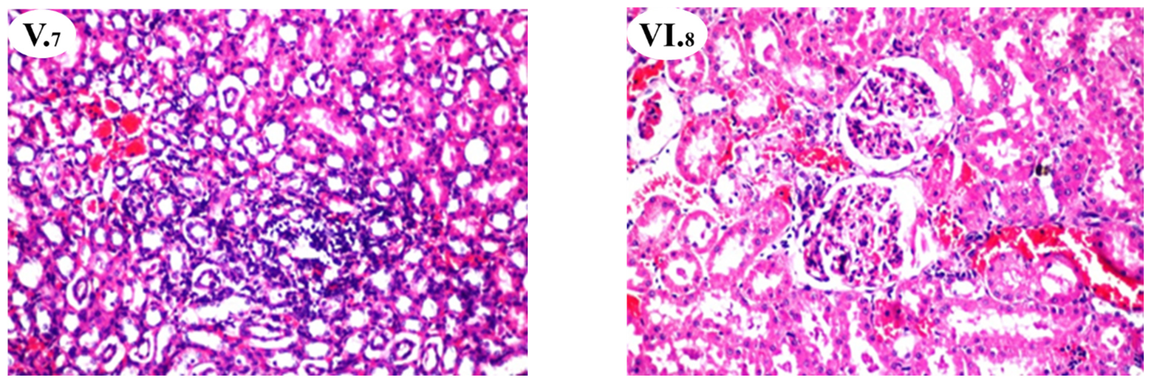
3.6. Effects of A. hierochuntica Extracts on Renal Histoarchitecture

The results of the biochemical investigations were supported by histopathological examination. Table 6 and Figure 1 show the degree of histological changes in the underlying structure of the rat’s kidneys in various experimental groups treated with A. hierochuntica extracts. In the current investigation, the kidney of the control group (GI) was found to have a normal histological structure (Figure 1(I.1)). The histoarchitecture of the CCl4-treated rats (GII) showed focal inflammatory cell infiltration (++) between the tubules at the cortex, congestion (++) of blood vessels between the tubules (Figure 1(II.2)), multiple eosinophilic cast (++) formations in the lumen of some tubules, and focal hemorrhage (++) between the degenerated tubules at the corticomedullary portion (Figure 1(II.3), Table 6). In GIII, injecting vit. E + Se, administrating the alcoholic extract of A. hierochuntica (GIV), aqueous extract of A. hierochuntica (GV), and a combination of A. hierochuntica extracts (GVI) attenuated the cytotoxic effects of CCl4 and recorded mild (+) to moderate (++) congestion in the blood vessels among tubules at the cortex (Figure 1(III.4–VI.8), Table 6) with well-developed Bowman’s capsule with glomerulus and convoluted tubules enlarged. Additionally, the aqueous extract of A. hierochuntica (GV) recorded focal inflammatory cell infiltration (++) at the corticomedullary portion (Figure 1(V.7), Table 6).

4. Discussion

Phytochemicals are mostly effective free radical scavengers and are considered plant-based superior antioxidant agents. Polyphenolic substances are thought to have anti-carcinogenic and anti-mutagenic properties in humans [40]. A valuable TPC content in A. hierochuntica was slightly higher than that obtained by Mohamed et al. [21] as 51.97 mg GAE g−1 in A. hierochuntica herb and by AlGamdi et al. [41], who found 4 mmol L−1 GAE in A. hierochuntica seeds. Recently, Zin et al. [42] indicated the presence of tannins in A. hierochuntica as a bioactive compound and recommended its bioactivity, which needed to be deeply investigated. The β-carotene content, as a part of total carotenoids, was 2.27 μg g−1 as mentioned by Mohamed et al. [21], and even current results presented total carotenoids as 3.51 μg g−1. Similar findings in flavonoid and flavonol contents have been indicated by Mohamed et al. [21]. Biologically active components, such as phenolic compounds, present antioxidant activity as breakdowns of lipid oxidation chain reactions by giving hydrogen to active free radicals. This scavenging potential of phenolics to inhibit radicals was elucidated by their phenolic hydroxyl groups [8,10,22]. This phenolic acid has been described as an effective antioxidant component, including hydrogen peroxide, hydroxyl radical, and superoxide anion [43]. A. hierochuntica metal chelating activity seems to be proficient in interfering with “Fe2+–ferrozine” complex construction, suggesting its ability to capture “ferrous” ions before “ferrozine”. A positive relationship between an increase in their contents of phenolic compounds is directly indicated with their antioxidant capacity [42]. Andjelković et al. [44] established the activity of numerous phenolic acids to form complexes with metals. The valuable TPC and relevant antioxidant activities using different measuring approaches give a clear plate from and confirm the bioactivity of A. hierochuntica as a medicinal herb for food or beverage applications.
Biologically active components, such as phenolic compounds, present antioxidant activity as breakdowns of lipid oxidation chain reactions by donating hydrogen to active free radicals [45]. This scavenging potential of phenolics to inhibit radicals was elucidated by their phenolic hydroxyl groups [46]. This phenolic acid has been described as an effective antioxidant component, including hydrogen peroxide, hydroxyl radical, and superoxide anion [43,45,47]. The identified and quantified compounds by HPLC in KAE of A. hierochuntica were higher than the number of identified compounds in KEE, but identified compounds in KEE of A. hierochuntica were presented in higher amounts [22]. The results reflect that the consuming A. hierochuntica could present many components in both polar and non-polar forms. These results are similarly presented by AlGamdi et al. [41] as they identified and quantified 20 polyphenolic compounds in seeds of A. hirerochuntica. The extract contained chlorogenic acids and hydroxybenzoic acids, but the main components were flavone C-glycosides, C-diglycosides, O-glycosides, and O-glycoside-C-glycosides occurring predominantly as luteolin conjugates. In addition, 14 of the 20 compounds in A. hierochuntica extract exhibited antioxidant activity using an HPLC-on-line antioxidant detection system [41]. Interestingly, current data confirmed that A. hierochuntica is rich in phytochemicals compounds and is a good source of natural antioxidants with potential health benefits, as has been scarcely highlighted before in seeds [41]. Hence, tea prepared from the whole plant powder is the traditional form of consumption; data illustrated new identified bioactive compounds in KEE and KAE of A. hirerochuntica, which differed from those found in AlGamdi et al. [41]
In numerous studies, CCl4-induced nephrotoxicity is utilized as a model system for testing the nephroprotective effect of plant extracts/drugs [48,49]. The current study looked at the effect of A. hierochuntica extracts on CCl4-induced kidney damage, as well as its nephroprotection and antioxidant potential in rats. In the current study, the CCl4 treatment (GII) group significantly increased creatinine, urea, and k levels and decreased total protein and albumin concentrations when compared to GI. This might be because CCl4 intoxication is a major source of free radical production in numerous organs, including the liver, kidneys, lungs, brain, and blood [50]. It has also been observed that following CCl4 injection in rats, the concentration of CCl4 is distributed more evenly in the kidneys than in the liver [51], since the kidney has a high affinity for CCl4 and contains cytochrome P450, predominantly in the cortex. The most common free radicals from CCl4 are trichloromethyl radical (CCl3) and trichloromethyl peroxyl radical (CCl3O2) [52]. These radicals attach to an intracellular protein, cell membrane lipids, and DNA, causing protein denaturation, lipid peroxidation, and oxidative DNA damage that leads to cell death [53]. In contrast, treating CCl4-rats with vit. E + Se (GIII) and A. hierochuntica extracts (GVI: VI) efficiently attenuated these rises in creatinine and urea levels as well as increased serum albumin and total proteins to be very close to their levels in GI. This may be due to the antioxidant properties and rich phenolic content of A. hierochuntica extracts and antioxidant capacity and chelating activity of vit. E + Se, which scavenges free radicals thereby inhibiting the renal damage. Phytochemicals are the most highly effective free radical scavengers and are considered superior antioxidant agents from plants [54]. The most abundant phenolic compounds were hydroxycinnamic acids, such as sinapic acid, among the nine identified phenolic compounds in KEE, while syringic acid was the highest phenolic acid among the 21 identified phenolic acids in KAE. Six flavonoids were identified in KEE and two in KAE using HPLC analysis [55]. Furthermore, as an antioxidant, vit. E is believed to protect tissues from harm caused by reactive oxygen metabolites. Selenium is also well recognized to be an essential trace mineral for human health, shielding cells from the damaging effects of free radicals [22].
In the current study, CCl4 administration markedly decreased GSH and SOD and increased MDA levels in kidney homogenates relative to GI. Vit. E + Se and A. hierochuntica extracts ameliorated the diverse effects of CCl4 by restoring the altered activity of antioxidant agents such as SOD and GSH and may deactivate the process of producing the MDA, as was recently reported [15,21,40,41]. GSH is a non-enzymatic antioxidant that is found in all mammalian cells. With its oxidized form, GSSG, GSH acts as a cofactor for numerous detoxifying enzymes (GPx, GST, and others) against oxidative stress and maintains cellular redox balance [47]. This finding is in accordance with those of Khan and Siddique [56] and Makni et al. [57], who reported that CCl4 decreased the GSH level in rat kidneys. Treatment with vit. E + Se and A. hierochuntica extracts showed protection against reduction in GSH level triggered by CCl4. In the same context, SOD catalyzes the dismutation of two molecules of superoxide anion (*O2) to hydrogen peroxide (H2O2) and molecular oxygen (O2), consequently rendering the potentially harmful superoxide anion less hazardous [58,59]. CCl4 intoxication alters the gene expression level by depleting renal SOD [60]. A decrease in SOD activity is a sensitive index of cellular damage. Our tested A. hierochuntica extracts ameliorated renal toxicity by alleviating the level of SOD. It participates in various enzymatic processes to reduce the concentration of detoxification reactions [61]. MDA is the first product of lipid peroxidation and is one of the important markers of oxidative stress. A. hierochuntica extracts diminished the increase in MDA levels and restored total antioxidant power in the CCl4-treated rat kidneys. These protective effects may be due to the powerful antioxidative activity of A. hierochuntica extracts [15,21,40,41]. These results also suggest that A. hierochuntica extracts may attenuate oxidative stress by decreasing levels of lipid peroxide in CCl4-exposed rat kidneys and prevent renal damage. These results agreed with the results of the antioxidative activities of Zn on CCl4-induced acute nephrotoxicity [62,63].
A. hirerochuntica extracts presented valuable nephroprotection capacity regarding kidney function tests (creatinine, urea, K, TP, and albumin) and kidney homogenate antioxidant activities (GSH, SOD, MDA) in GIV, V, and IV, respectively. The total nephroprotection % relative to vit. E + Se treatment registered maximum levels in the KAE treated group (GV, 97.62%), then KEE (GIV, 83.27%), and then KEE + KAE (GVI, 78.85%), respectively, in descending order. This may be due to differences in quantity and quality of phenolic and antioxidant contents of A. hirerochuntica extracts, which have a relation to antioxidant capacity [15,19,22,40,42].
The histopathological findings in kidneys are consistent with the biochemical estimations of the experimental groups investigated. CCl4 administration (GII) caused a glomerular and tubular lesion with vasocongestion in the kidneys. Dogukan et al. [64] discovered a similar histological pattern in rat renal tissue in response to prolonged CCl4 treatment. It is also considered that histological changes are caused by functional overloading of nephrons, which leads to renal dysfunction [65], and/or are due to the destruction of tissue provoked as a consequence of free radical generation via CCl4 metabolism [56,66]. The effect of vit. E + Se and A. hierochuntica extracts to repair and restore kidneys destruction effects of CCl4 were notable. This may be because vit. E + Se (as a potent antioxidant) acts on ROS induced by CCl4 [67]. A. hierochuntica extracts suppress CCl4-induced acute nephrotoxicity due to the antioxidative role and free radical scavenging properties of the phenolic compounds present in A. hirerochuntica extracts [22]. Our findings are consistent with those of other researchers who have shown that various plant derivatives have pharmacological effects by eliminating CCl4 abuses and restoring to normality [6].

5. Conclusions

Results of this study clearly demonstrated that A. hierochuntica plant is rich in polar and nonpolar phenolic compounds with a superior antioxidant capacity, which is directly related to the phytochemical content. A. hierochuntica (particularly aqueous extract) protects rats against CCl4-induced oxidative stress and acute kidney injury, as evidenced by a significant drop in MDA levels and increased GSH and SOD activity, as well as the cessation of biochemical and histological alterations in the kidneys. The protective efficacy might arise from the antioxidant and free radical scavenging properties of the phenolic compounds present in the A. hierochuntica extracts. These characteristics help to explain the plant’s medicinal efficacy as a herbal medication. More research is needed to completely describe the active principles in A. hierochuntica, and this study is meant to stimulate more comprehensive related research to offer sufficient data and recommendations for defining its mechanisms and safe doses.

Author Contributions

Conceptualization, T.I.A., Y.M.A. and H.B.; methodology, investigation, H.B. and H.A.A.-R.; data curation, T.I.A. and Y.M.A.; writing—original draft preparation, review, and editing; H.B. and H.A.A.-R. All authors have read and agreed to the published version of the manuscript.

Funding

The researchers would like to thank the Deanship of Scientific Research, Qassim University, for funding the publication of this project.

Institutional Review Board Statement

Not applicable.

Informed Consent Statement

Not applicable.

Data Availability Statement

The data presented in this study are available on request from the corresponding author.

Conflicts of Interest

The authors declare no conflict of interest.

Abbreviations

ABTS: 2,2’-azino-bis(3-ethylbenzothiazoline-6-sulfonic acid); AOA: antioxidant activity; BHA: butylated hydroxyanisole; BHA: butylated hydroxyanisole; DPPH: 1,1-diphenyl-2-picryl hydrazine; dw: dry weight; GA: gallic acid; GAE: gallic acid equivalent; GSH: reduced-glutathione; HPLC-DAD: high-performance liquid chromatography diode array detection; KAE: A. hierochuntica aqueous extract; KEE: A. hierochuntica ethanolic extract; MDA: malonaldehyde; QE: quercetin equivalent; RAA: relative antioxidant activity; ROS: reactive oxygen species; RSA: radical scavenging activity; Se: selenium; SE: standard error; SOD: superoxide dismutase; TC: total carotenoids; TC: total carotenoids; TF: total flavonoids; TE: trolox equivalents; TFL: total flavonols; TPC: total phenolic compounds.

References

  1. Statistics, K.D. Chronic Kidney Disease in the United States. 2021. Available online: https://www.cdc.gov/kidneydisease/pdf/Chronic-Kidney-Disease-in-the-US-2021-h.pdf (accessed on 20 July 2021).
  2. Ku, E.; Glidden, D.V.; Johansen, K.L.; Sarnak, M.; Tighiouart, H.; Grimes, B.; Hsu, C.Y. Association between strict blood pressure control during chronic kidney disease and lower mortality after onset of end-stage renal disease. Kidney Int. 2015, 87, 1055–1060. [Google Scholar] [CrossRef] [PubMed] [Green Version]
  3. Tumlin, J.A.; Madaio, M.P.; Hennigar, R. Idiopathic IgA nephropathy: Pathogenesis, histopathology, and therapeutic options. Clin. J. Am. Soc. Nephrol. 2007, 2, 1054–1061. [Google Scholar] [CrossRef] [PubMed] [Green Version]
  4. Olah, G.; Reddy, V.P.; Prakash, G.S.; Reactions, F.C. Kirk-Othmer encyclopedia of chemical technology. Contact Lenses 1978, 720–742. [Google Scholar]
  5. Ozturk, F.; Ucar, M.; Ozturk, I.C.; Vardi, N.; Batcioglu, K. Carbon tetrachloride-induced nephrotoxicity and protective effect of betaine in Sprague-Dawley rats. Urology 2003, 62, 353–356. [Google Scholar] [CrossRef]
  6. Ogeturk, M.; Kus, I.; Colakoglu, N.; Zararsiz, I.; Ilhan, N.; Sarsilmaz, M. Caffeic acid phenethyl ester protects kidneys against carbon tetrachloride toxicity in rats. J. Ethnopharmacol. 2005, 97, 273–280. [Google Scholar] [CrossRef]
  7. Slater, T.F. Free Radical Mechanisms in Tissue Injury. In Cell Function and Disease; Cañedo, L.E., Todd, L.E., Packer, L., Jaz, J., Eds.; Springer: Boston, MA, USA, 1988; pp. 209–218. [Google Scholar] [CrossRef]
  8. Khan, M.R.; Rizvi, W.; Khan, G.N.; Khan, R.A.; Shaheen, S. Carbon tetrachloride-induced nephrotoxicity in rats: Protective role of Digera muricata. J. Ethnopharmacol. 2009, 122, 91–99. [Google Scholar] [CrossRef]
  9. Afsar, T.; Khan, M.R.; Razak, S.; Ullah, S.; Mirza, B. Antipyretic, anti-inflammatory and analgesic activity of Acacia hydaspica R. Parker and its phytochemical analysis. BMC Complement. Altern. Med. 2015, 15, 1–12. [Google Scholar] [CrossRef] [Green Version]
  10. Al-Qabba, M.M.; El-Mowafy, M.A.; Althwab, S.A.; Alfheeaid, H.A.; Aljutaily, T.; Barakat, H. Phenolic profile, antioxidant activity, and ameliorating efficacy of Chenopodium quinoa sprouts against CCl4-induced oxidative stress in rats. Nutrients 2020, 12, 2904. [Google Scholar] [CrossRef]
  11. Al Zhrani, M.M.; Althwab, S.A.; Aljutaily, T.; Alfheeaid, H.A.; Ashoush, I.S.; Barakat, H. Protective effect of moringa-based beverages against hyperlipidemia and hyperglycemia in type 2 diabetes-induced rats. Food Res. 2021, 5, 279–289. [Google Scholar] [CrossRef]
  12. Khalifa, I.; Nawaz, A.; Sobhy, R.; Althwab, S.A.; Barakat, H. Polyacylated anthocyanins constructively network with catalytic dyad residues of 3CLpro of 2019-nCoV than monomeric anthocyanins: A structural-relationship activity study with 10 anthocyanins using in-silico approaches. J. Mol. Graph. Model. 2020, 100, 107690. [Google Scholar] [CrossRef]
  13. Yusof, J.; Mahdy, Z.A.; Noor, R.M. Use of complementary and alternative medicine in pregnancy and its impact on obstetric outcome. Complement. Ther. Clin. Pract. 2016, 25, 155–163. [Google Scholar] [CrossRef]
  14. Salah, S.H.; Abdou, H.S.; Abd El-Azeem, A.; Abdel-Rahim, E. The antioxidative effects of some medicinal plants as hypoglycemic agents on chromosomal aberration and abnormal nucleic acids metabolism produced by diabetes stress in male adult albino rats. J. Diabetes Mellit. 2011, 1, 6–14. [Google Scholar] [CrossRef] [Green Version]
  15. Daur, I. Chemical properties of the medicinal herb Kaff Maryam (Anastatica hierochuntica L.) and its relation to folk medicine use. Afr. J. Microbiol. Res. 2012, 6, 5048–5051. [Google Scholar]
  16. Shah, A.H.; Bhandari, M.; Al-Harbi, N.O.; Al-Ashban, R.M. Kaff-E-Maryam (Anastatica Hierochuntica L.): Evaluation of gastro-protective activity and toxicity in different experimental models. Biol. Med. 2014, 6, 1–10. [Google Scholar]
  17. Kim Sooi, L.; Lean Keng, S. Herbal medicines: Malaysian women’s knowledge and practice. Evid-Based Complement. Alternat. Med. 2013, 2013, 438139. [Google Scholar] [CrossRef] [PubMed] [Green Version]
  18. Karadaş, C.; Kara, D. Chemometric approach to evaluate trace metal concentrations in some spices and herbs. Food Chem. 2012, 130, 196–202. [Google Scholar] [CrossRef]
  19. Yoshikawa, M.; Xu, F.; Morikawa, T.; Ninomiya, K.; Matsuda, H. Anastatins A and B, new skeletal flavonoids with hepatoprotective activities from the desert plant Anastatica hierochuntica. Bioorganic Med. Chem. Lett. 2003, 13, 1045–1049. [Google Scholar] [CrossRef]
  20. Padmanabhan, P.; Jangle, S. Hepatoprotective activity of herbal preparation (HP-4) against D-galactosamine induced hepatotoxicity in mice. Int. J. Pharm. Sci. Drug Res. 2014, 6, 31–37. [Google Scholar]
  21. Mohamed, A.A.; Khalil, A.A.; El-Beltagi, H.E. Antioxidant and antimicrobial properties of kaff maryam (Anastatica hierochuntica) and doum palm (Hyphaene thebaica). Grasas Y Aceites 2010, 61, 67–75. [Google Scholar] [CrossRef] [Green Version]
  22. Barakat, H.; Almundarij, T.I. Phenolic compounds and hepatoprotective potential of Anastatica hierochuntica ethanolic and aqueous extracts against CCl4-induced hepatotoxicity in rats. J. Tradit. Chin. Med. 2020, 40, 947–955. [Google Scholar]
  23. Asuzu, I.U. Pharmacological evaluation of the folklore use of Sphenostylis stenocarpa. J. Ethnopharmacol. 1986, 16, 263–267. [Google Scholar] [CrossRef]
  24. Bettaieb, I.; Bourgou, S.; Wannes, W.A.; Hamrouni, I.; Limam, F.; Marzouk, B. Essential oils, phenolics, and antioxidant activities of different parts of cumin (Cuminum cyminum L.). J. Agric. Food Chem. 2010, 58, 10410–10418. [Google Scholar] [CrossRef] [PubMed]
  25. Mohdaly, A.A.A.; Hassanien, M.F.R.; Mahmoud, A.; Sarhan, M.A.; Smetanska, I. Phenolics extracted from potato, sugar beet, and sesame processing by-products. Int. J. Food Prop. 2013, 16, 1148–1168. [Google Scholar] [CrossRef]
  26. Khalifa, I.; Barakat, H.; El-Mansy, H.A.; Soliman, S.A. Optimizing bioactive substances extraction procedures from guava, olive and potato processing wastes and evaluating their antioxidant capacity. J. Food Chem. Nanotechnol. 2016, 2, 170–177. [Google Scholar] [CrossRef]
  27. Lu, J.; Zhao, H.; Chen, J.; Fan, W.; Dong, J.; Kong, W.; Sun, J.; Cao, Y.; Cai, G. Evolution of phenolic compounds and antioxidant activity during malting. J. Agric. Food Chem. 2007, 55, 10994–11001. [Google Scholar] [CrossRef] [PubMed]
  28. Koleva, I.I.; Van Beek, T.A.; Linssen, J.P.; Groot, A.d.; Evstatieva, L.N. Screening of plant extracts for antioxidant activity: A comparative study on three testing methods. Phytochem. Anal. 2002, 13, 8–17. [Google Scholar] [CrossRef] [PubMed]
  29. Zhao, H.; Dong, J.; Lu, J.; Chen, J.; Li, Y.; Shan, L.; Lin, Y.; Fan, W.; Gu, G. Effects of extraction solvent mixtures on antioxidant activity evaluation and their extraction capacity and selectivity for free phenolic compounds in barley (Hordeum vulgare L.). J. Agric. Food Chem. 2006, 54, 7277–7286. [Google Scholar] [CrossRef]
  30. Goupy, P.; Hugues, M.; Boivin, P.; Amiot, M.J. Antioxidant composition and activity of barley (Hordeum vulgare) and malt extracts and of isolated phenolic compounds. J. Sci. Food Agric. 1999, 79, 1625–1634. [Google Scholar] [CrossRef]
  31. Asuku, O.; Atawodi, S.E.; Onyike, E. Antioxidant, hepatoprotective, and ameliorative effects of methanolic extract of leaves of Grewia mollis Juss. on carbon tetrachloride–treated albino rats. J. Med. Food 2012, 15, 83–88. [Google Scholar] [CrossRef] [PubMed]
  32. El-Desoky, G.; Abdelreheem, M.; Abdulaziz, A.-O.; ALOthman, Z.; Mahmoud, M.; Yusuf, K. Potential hepatoprotective effects of vitamin E and selenium on hepatotoxicity induced by malathion in rats. Afr. J. Pharm. Pharmacol. 2012, 6, 806–813. [Google Scholar] [CrossRef]
  33. Draper, H.H.; Hadley, M. Malondialdehyde determination as index of lipid peroxidation. Methods Enzymol. 1990, 186, 421–431. [Google Scholar]
  34. Giannopolitis, C.N.; Ries, S.K. Superoxide dismutases: I. Occurrence in higher plants. Plant Physiol. 1977, 59, 309–314. [Google Scholar] [CrossRef] [PubMed]
  35. Chanarin, I. Textbook of Laboratory Haematology: An Account of Laboratory Techniques; Churchill Livingstone: New York, NY, USA, 1989. [Google Scholar]
  36. Bradford, M.M. A rapid and sensitive method for the quantitation of microgram quantities of protein utilizing the principle of protein-dye binding. Anal. Biochem. 1976, 72, 248–254. [Google Scholar] [CrossRef]
  37. Wakchaure, D.; Jain, D.; Singhai, A.K.; Somani, R. Hepatoprotective activity of Symplocos racemosa bark on carbon tetrachloride-induced hepatic damage in rats. J. Ayurveda Integr. Med. 2011, 2, 137. [Google Scholar]
  38. Banchroft, J.D.; Stevans, A.; Turnes, D.R. Theory and Practice of Histological Techniques, 4th ed.; Livingstone: Edinburgh, UK; London, UK; Melbourne, Australia; New York, NY, USA; Tokyo, Japan, 1996. [Google Scholar]
  39. Steel, R.; Torrie, J.; Dickey, D. Principles and Procedures of Statistics: A Biometrical Approach; WCB/McGraw-Hill: Columbus, OH, USA, 1997. [Google Scholar]
  40. Md Zin, S.R.; Mohamed, Z.; Alshawsh, M.A.; Wong, W.F.; Kassim, N.M. Mutagenicity evaluation of Anastatica hierochuntica L. aqueous extract in vitro and in vivo. Exp. Biol. Med. 2018, 243, 375–385. [Google Scholar] [CrossRef] [PubMed]
  41. AlGamdi, N.; Mullen, W.; Crozier, A. Tea prepared from Anastatica hirerochuntica seeds contains a diversity of antioxidant flavonoids, chlorogenic acids and phenolic compounds. Phytochemistry 2011, 72, 248–254. [Google Scholar] [CrossRef] [PubMed]
  42. Zin, S.R.M.; Kassim, N.M.; Alshawsh, M.A.; Hashim, N.E.; Mohamed, Z. Biological activities of Anastatica hierochuntica L.: A systematic review. Biomed. Pharmacother. 2017, 91, 611–620. [Google Scholar] [CrossRef]
  43. Erkan, N.; Ayranci, G.; Ayranci, E. Antioxidant activities of rosemary (Rosmarinus officinalis L.) extract, blackseed (Nigella sativa L.) essential oil, carnosic acid, rosmarinic acid and sesamol. Food Chem. 2008, 110, 76–82. [Google Scholar] [CrossRef]
  44. Andjelković, M.; Van Camp, J.; De Meulenaer, B.; Depaemelaere, G.; Socaciu, C.; Verloo, M.; Verhe, R. Iron-chelation properties of phenolic acids bearing catechol and galloyl groups. Food Chem. 2006, 98, 23–31. [Google Scholar] [CrossRef]
  45. Shahidi, F.; Janitha, P.K.; Wanasundara, P.D. Phenolic antioxidants. Crit. Rev. Food Sci. Nutr. 1992, 32, 67–103. [Google Scholar] [CrossRef]
  46. Sawa, T.; Nakao, M.; Akaike, T.; Ono, K.; Maeda, H. Alkylperoxyl radical-scavenging activity of various flavonoids and other phenolic compounds: Implications for the anti-tumor-promoter effect of vegetables. J. Agric. Food Chem. 1999, 47, 397–402. [Google Scholar] [CrossRef]
  47. Valko, M.; Rhodes, C.; Moncol, J.; Izakovic, M.; Mazur, M. Free radicals, metals and antioxidants in oxidative stress-induced cancer. Chem.-Biol. Interact. 2006, 160, 1–40. [Google Scholar] [CrossRef]
  48. Ogeturk, M.; Kus, I.; Kavakli, A.; Oner, J.; Kukner, A.; Sarsilmaz, M. Reduction of carbon tetrachloride-induced nephropathy by melatonin administration. Cell Biochem. Funct. Cell. Biochem. its Modul. by Act. Agents or Dis. 2005, 23, 85–92. [Google Scholar] [CrossRef] [PubMed]
  49. Safhi, M.M. Nephroprotective effect of zingerone against CCl4-induced renal toxicity in swiss albino mice: Molecular mechanism. Oxid. Med. Cell Longev. 2018, 2018, 2474831. [Google Scholar] [CrossRef] [Green Version]
  50. Hamed, M.A.; Ali, S.A.; Saba El-Rigal, N. Therapeutic potential of ginger against renal injury induced by carbon tetrachloride in rats. Sci. World J. 2012, 2012, 840421. [Google Scholar] [CrossRef] [PubMed] [Green Version]
  51. Sanzgiri, U.; Bruckner, J.J.F.; Toxicology, A. Effect of Emulphor, an emulsifier, on the pharmacokinetics and hepatotoxicity of oral carbon tetrachloride in the rat. Fundam. Appl. Nematol. 1997, 36, 54–61. [Google Scholar] [CrossRef]
  52. Ali, S.A.E.-m.; Abdelaziz, D.H.A. The protective effect of date seeds on nephrotoxicity induced by carbon tetrachloride in rats. Int. J. Pharm. Sci. Rev. Res. 2014, 26, 62–68. [Google Scholar]
  53. Hismiogullari, A.A.; Hismiogullari, S.E.; Karaca, O.; Sunay, F.B.; Paksoy, S.; Can, M.; Kus, I.; Seyrek, K.; Yavuz, O.J.P.R. The protective effect of curcumin administration on carbon tetrachloride CCl4-induced nephrotoxicity in rats. Pharmacol. Rep. 2015, 67, 410–416. [Google Scholar] [CrossRef] [PubMed]
  54. Soong, Y.-Y.; Barlow, P.J. Antioxidant activity and phenolic content of selected fruit seeds. Food Chem. 2004, 88, 411–417. [Google Scholar] [CrossRef]
  55. El-Demerdash, F.M. Antioxidant effect of vitamin E and selenium on lipid peroxidation, enzyme activities and biochemical parameters in rats exposed to aluminium. J. Trace. Elem. Med. Biol. 2004, 18, 113–121. [Google Scholar] [CrossRef] [PubMed]
  56. Khan, M.R.; Siddique, F. Antioxidant effects of Citharexylum spinosum in CCl4 induced nephrotoxicity in rat. Exp. Toxicol. Pathol. 2012, 64, 349–355. [Google Scholar] [CrossRef] [PubMed]
  57. Makni, M.; Chtourou, Y.; Garoui, E.; Boudawara, T.; Fetoui, H. Carbon tetrachloride-induced nephrotoxicity and DNA damage in rats: Protective role of vanillin. Hum. Exp. Toxicol. 2012, 31, 844–852. [Google Scholar] [CrossRef]
  58. Fridovich, I. Superoxide radical and superoxide dismutases. Annu. Rev. Biochem. 1995, 64, 97–112. [Google Scholar] [CrossRef] [PubMed]
  59. Dringen, R.; Pawlowski, P.G.; Hirrlinger, J. Peroxide detoxification by brain cells. J. Neurosci. Res. 2005, 79, 157–165. [Google Scholar] [CrossRef]
  60. Szymonik-Lesiuk, S.; Czechowska, G.; Stryjecka-Zimmer, M.; Słomka, M.; MĄldro, A.; Celiński, K.; Wielosz, M. Catalase, superoxide dismutase, and glutathione peroxidase activities in various rat tissues after carbon tetrachloride intoxication. J. Hepato-Biliary-Pancreat. Surg. 2003, 10, 309–315. [Google Scholar] [CrossRef] [PubMed]
  61. Kadiiska, M.B.; Gladen, B.C.; Baird, D.D.; Dikalova, A.E.; Sohal, R.S.; Hatch, G.E.; Jones, D.P.; Mason, R.P.; Barrett, J.C. Biomarkers of oxidative stress study: Are plasma antioxidants markers of CCl4 poisoning? Free Radic. Biol. Med. 2000, 28, 838–845. [Google Scholar] [CrossRef]
  62. McCall, K.A.; Huang, C.-C.; Fierke, C.A. Function and mechanism of zinc metalloenzymes. J. Nutr. 2000, 130, 1437S–1446S. [Google Scholar] [CrossRef] [Green Version]
  63. Yoshioka, H.; Usuda, H.; Fukuishi, N.; Nonogaki, T.; Onosaka, S. Carbon tetrachloride-induced nephrotoxicity in mice is prevented by pretreatment with zinc sulfate. Biol. Pharm. Bull. 2016, 39, 1042–1046. [Google Scholar] [CrossRef] [PubMed] [Green Version]
  64. Doğukan, A.; Akpolat, N.; Çeliker, H.; Ilhan, N.; Bahçecioğlu, H.; Günal, A.I. Protective effect of interferon-alpha on carbon tetrachloride-induced nephrotoxicity. J. Nephrol. 2003, 16, 81–84. [Google Scholar]
  65. Adewole, S.; Salako, A.; Doherty, O.; Naicker, T. Effect of melatonin on carbon tetrachloride-induced kidney injury in Wistar rats. Afr. J. Biomed. Res. 2007, 10, 153–164. [Google Scholar] [CrossRef]
  66. Jan, S.; Khan, M.R. Protective effects of Monotheca buxifolia fruit on renal toxicity induced by CCl4 in rats. BMC Complement. Altern. Med. 2016, 16, 1–15. [Google Scholar] [CrossRef] [PubMed] [Green Version]
  67. Venkatanarayana, G.; Sudhakara, G.; Sivajyothi, P.; Indira, P. Protective effects of curcumin and vitamin E on carbon tetrachloride-induced nephrotoxicity in rats. EXCLI J. 2012, 11, 641–650. [Google Scholar] [PubMed]
Figure 1. Histopathological findings of rat kidney protection by A. hierochuntica extracts (Hematoxylin-Eosin (HE), 200X). Histopathological nephrotoxicity induction. GI: Histopathological structure of control group with normal histological structure of the glomeruli and tubules of rat kidney (I.1). GII: Kidney of a rat receiving only CCl4 showing focal inflammatory cell infiltration in between the tubules at the cortex (II.2). There was congestion in the blood vessels between the tubules, and multiple eosinophilic casts formed in the lumen of some tubules at the cortex. The corticomedullary portion showed focal hemorrhage and hemolysis in between the degenerated tubules (II.3). GIII: Kidney of a rat receiving CCl4 + vit. E + Se showing congestion in the blood vessels in between the tubules at the cortex (III.4). GIV: Kidney of a rat receiving CCl4 + KEE viewing congestion in the cortical blood vessels (IV.5). GV: rat kidney receiving CCl4 + KAE showing congestion in the cortical blood vessels (V.6). The corticomedullary junction showed focal inflammatory cells infiltration with eosinophilic renal casts formation in the lumen of some tubules (V.7). GVI: kidney of rats receiving CCl4 + KEE + KAE showing congestion in the cortical blood vessels and glomeruli (VI.8).
Nutrients 13 02973 g001aNutrients 13 02973 g001b
Table 1. Total phenolic content, total carotenoids, total flavonoids, total flavonols, and relative potential antioxidant activities of A. hierochuntica (mean ± SE), n = 6.
ItemA. hierochuntica
TPC (mg GAE g−1)67.49 ± 3.33
TC (µg g−1)3.51 ± 0.91
TF (mg QE g−1)49.78 ± 2.62
TFL (mg QE g−1)17.45 ± 0.83
DPPH (µmol of TE g−1)128.71 ± 3.55
ABTS (µmol of TE g−1)141.92 ± 4.67
β-CB * (RAA) %45.74 ± 4.80
CA (mg g−1)42.89 ± 2.11
Note: *: relatively calculated based on BHA as 100%, RAA: relative antioxidant activity.
Table 2. Quantitative analysis of phenolic compounds from KEE and KAE of A. hierochuntica by HPLC-DAD.
ItemNo.CompoundEthanolic Extract (KEE) (mg 100 g−1)Aqueous Extract (KAE) (mg 100 g−1)
Phenolic acids13,4,5-trimethoxycinnamic acid-0.042
24-Aminobenzoic acid-0.012
3Benzoic acid-0.005
4Caffeic acid6.6210.725
5Catechol-2.526
6Chlorogenic acid-0.136
7Cinnamic acid0.0940.001
8Coumarin-0.036
9Ellagic acid-0.039
10e-Vanillic acid-0.443
11Ferulic acid1.8540.037
12Gallic acid-0.041
13Iso-ferulic acid-0.005
14α-Coumaric acid-0.039
15p-Coumaric acid-0.009
16p-Hydroxybenzoic acid3.4400.223
17Protocatechuic acid1.8110.454
18Pyrogallol-1.589
19Rosmarinic acid2.884-
20Salicylic acid-0.089
21Sinapic acid28.704-
22Syringic acid1.0831.959
23Vanillic acid3.3261.406
Flavonoids1Apigenin-7-glucoside0.192-
2D-Catechin2.4100.256
3Epicatechin-0.193
4Kaempferol0.434-
5Myricetin1.627-
6Quercetin0.184-
7Rutin0.539-
Notes: KEE: Anastatica hierochuntica ethanolic extract; KAE: Anastatica hierochuntica aaqueous extract.
Table 3. Effect of oral administration of A. hierochuntica extracts on biochemical kidney markers in CCl4-induced toxicity in rats (mean ± SE), n = 6.
Kidney FunctionsExperimental Groups
GIGIIGIIIGIVGVGVI
Creatinine (mg dL−1)0.88 ± 0.09 a1.30 ± 0.11 b0.87 ± 0.11 a0.99 ± 0.07 a1.08 ± 0.03 a0.91 ± 0.11 a
Urea (mg dL−1)77.59 ± 2.60 a117.00 ± 3.98 b77.53 ± 10.11 a73.60 ± 5.35 a78.65 ± 12.69 a70.33 ± 8.37 a
K (mEq L−1)4.18 ± 0.21 a5.55 ± 0.68 bc4.57 ± 0.23 ab4.78 ± 0.21 b5.00 ± 0.21 b5.48 ± 0.23 c
Total proteins (g dL−1)8.71 ± 0.92 c5.04 ± 0.36 a7.54 ± 0.45 b7.89 ± 0.44 bc8.59 ± 0.18 c5.89 ± 1.43 ab
Albumin (g dL−1)3.91 ± 0.13 b3.28 ± 0.09 a3.79 ± 0.31 b3.68 ± 0.16 b4.34 ± 0.17 c3.71 ± 0.14 b
GI: control negative group, GII: control positive group received CCl4 (i.p.), GIII: CCl4-rats received 50 mg kg−1 vit. E + Se twice a week (i.m.), GIV: CCl4-rats received KEE as 250 mg kg−1 per oral (p.o.) daily, GV: CCl4-rats received KAE as 250 mg kg−1 (p.o.) daily and GVI: CCl4-rats received KEE + KAE (1:1) as 250 mg kg−1 (p.o.) daily. a–c: values with the same superscript letter in the same raw are not significantly different at p ≤ 0.05.
Table 4. Effects of oral administration of A. hierochuntica extracts on kidney oxidative damage in CCl4-induced toxicity in rats (mean ± SE), n = 6.
Oxidative Stress MarkersExperimental Groups
GIGIIGIIIGIVGVGVI
MDA
nmol/mg protein
131.68 ± 10.83 a308.58 ± 18.27 c125.01 ± 12.40 a151.46 ± 9.01 a242.06 ± 40.81 b285.75 ± 20.47 b
SOD
nmol/mg protein
22.66 ± 0.54 c11.47 ± 2.01 a18.16 ± 0.99 b16.32 ± 1.51 b21.98 ± 0.97 c20.16 ± 1.87 bc
GSH
nmol/mg protein
3.64 ± 0.15 b2.42 ± 0.25 a3.83 ± 0.55 b3.40 ± 0.15 b3.48 ± 0.18 b3.82 ± 0.26 b
MDA: malondialdehyde, SOD = superoxide dismutase, GSH: reduced glutathione, GI: control negative group, GII: control positive group received CCl4 (i.p.), GIII: CCl4-rats received 50 mg kg−1 vit. E + Se twice a week (i.m.), GIV: CCl4-rats received KEE as 250 mg kg−1 (p.o.) daily, GV: CCl4-rats received KAE as 250 mg kg−1 (p.o.) daily and GVI: CCl4-rats received KEE + KAE (1:1) as 250 mg kg−1 (p.o.) daily. a–c: values with the same superscript letter in the same raw are not significantly different at p ≤ 0.05.
Table 5. Nephroprotection percentage of A. hierochuntica extracts in CCl4-induced toxicity in rats.
ParametersExperimental Groups
GIIIGIVGVGVI
Creatinine97.6273.8052.3892.29
Urea99.8589.8897.3181.58
K71.5356.9640.155.11
Total proteins68.1177.6696.7323.16
Albumin80.9563.49168.2568.25
MDA96.2388.8137.6012.90
SOD59.7943.3493.9277.65
GSH115.5780.3286.8985.25
* TFP%10083.2797.6278.85
MDA: malondialdehyde, SOD: superoxide dismutase, GSH: reduced glutathione, * TFP%: total nephroprotection % calculated relatively based on vit. E and Se treatment, GIII: CCl4-rats received 50 mg kg−1 vit. E + Se twice a week (i.m.), GIV: CCl4-rats received KEE as 250 mg kg−1 (p.o) daily, GV: CCl4-rats received KAE as 250 mg kg−1 (p.o.) daily and GVI: CCl4-rats received KEE + KAE (1:1) as 250 mg kg−1 (p.o.) daily.
Table 6. The severity of histopathological alteration in rat kidney’s underlying structure of different experimental groups treated by A. hierochuntica extracts.
GIGIIGIIIGIVGVGVI
Focal inflammatory cells
Infiltration between the tubule
++++
Eosinophilic renal cast++
Congestion+++++++++
Focal hemorrhage++
+++ = severe, ++ = moderate, + = mild, − = nil, GI: control negative group, GII: control positive group received CCl4 (i.p.), GIII: CCl4-rats received 50 mg kg−1 vit. E + Se twice a week (i.m.), GIV: CCl4-rats received KEE as 250 mg kg−1 (p.o.) daily, GV: CCl4-rats received KAE as 250 mg kg−1 (p.o.) daily and GVI: CCl4-rats received KEE + KAE (1:1) as 250 mg kg−1 (p.o.) daily.
Publisher’s Note: MDPI stays neutral with regard to jurisdictional claims in published maps and institutional affiliations.

