Review

Research Progress of Maternal Metabolism on Cardiac Development and Function in Offspring

1
Obstetrics and Gynecology Hospital, Institute of Reproduction and Development, Fudan University, Shanghai 200001, China
2
Research Units of Embryo Original Diseases, Chinese Academy of Medical Sciences, Shanghai 200032, China
3
Shanghai First Maternity and Infant Hospital, Shanghai 200126, China
*
Author to whom correspondence should be addressed.
Nutrients 2023, 15(15), 3388; https://doi.org/10.3390/nu15153388
Submission received: 19 June 2023 / Revised: 18 July 2023 / Accepted: 27 July 2023 / Published: 30 July 2023
(This article belongs to the Section Pediatric Nutrition)

Abstract

:
The developmental origin of health and disease (DOHaD) hypothesis refers to the adverse effects of suboptimal developmental environments during embryonic and early fetal stages on the long-term health of offspring. Intrauterine metabolic perturbations can profoundly impact organogenesis in offspring, particularly affecting cardiac development and giving rise to potential structural and functional abnormalities. In this discussion, we contemplate the existing understanding regarding the impact of maternal metabolic disorders, such as obesity, diabetes, or undernutrition, on the developmental and functional aspects of the offspring’s heart. This influence has the potential to contribute to the susceptibility of offspring to cardiovascular health issues. Alteration in the nutritional milieu can influence mitochondrial function in the developing hearts of offspring, while also serving as signaling molecules that directly modulate gene expression. Moreover, metabolic disorders can exert influence on cardiac development-related genes epigenetically through DNA methylation, levels of histone modifications, microRNA expression, and other factors. However, the comprehensive understanding of the mechanistic underpinnings of these phenomena remains incomplete. Further investigations in this domain hold profound clinical significance, as they can contribute to the enhancement of public health and the prevention of cardiovascular diseases.

1. Introduction

The developmental origin of health and disease (DOHaD) hypothesis, first proposed by Baker in the late twentieth century, posits that early-life environmental insults, including those occurring during conception, gestation, and the perinatal period, can significantly impact an individual’s susceptibility to disease later in life [1]. The prevalence of maternal obesity and diabetes has risen dramatically over the past several decades and constitutes a major public health concern [2]. Maternal hyperglycemia has been linked to an elevated risk of numerous birth defects and long-term health issues in offspring. Several large-scale cohort studies have demonstrated that maternal metabolic status, including diabetes, obesity, and undernutrition, can impact both the morphological and functional development of offspring hearts, as well as their long-term cardiac health [3,4]. Despite these findings, further research is required to elucidate the precise effects of maternal metabolism on cardiac development and function in offspring, as well as the underlying molecular and cellular mechanisms involved.
The heart is one of the earliest organs to develop during embryonic development, with cardiogenesis beginning at approximately 15–19 days of human pregnancy and embryonic day 6.5–7.5 (E6.5–7.5) in mice. Initially, mesodermal cells form a heart-forming region known as the cardiac crescent, which contains myocardial precursor cells. These cells can be distinguished based on their temporal expression of various marker genes and are classified as either first heart field (FHF) or second heart field (SHF) cells. FHF cells ultimately become the heart tube, which then forms the definitive left ventricle and atrioventricular canal, while SHF cells give rise to the right ventricle, outflow tract, and atria. The primary heart tube, which is a looped tube with only one cavity, forms from around 20 to 25 days of human development and 9 days of mouse development. The heart tube then loops and twists to ensure proper cardiac segment alignment, in preparation for chamber septation. Atrial and ventricular septation begins at 31–35 days of human pregnancy and E10.5 in mice. By E13.5–14.5, the basic structure of the four chambers has formed [1,2]. After birth, cardiomyocytes retain some proliferative capacity, but this capability diminishes rapidly during the first week [3]. Therefore, precise regulation of gene expression is crucial for correct cardiac morphogenesis and differentiation, requiring the coordinated activity of many signaling pathways, such as HIF [4], Notch [5], and Hippo [6]. Any dysregulation of these pathways during embryonic development may disrupt the spatiotemporal regulation of complex three-dimensional heart structures, leading to morphological abnormalities, such as congenital heart diseases (CHDs), or cardiac dysfunction in offspring. The heart has a high energy demand to maintain a constant cardiac impulse, and thus its metabolic pattern is unique and worthy of attention. During the embryonic stage, the fetal heart primarily depends on glycolysis to acquire energy [7]. One unique characteristic of heart metabolism is that shortly after birth, it undergoes a dramatic transformation and primarily depends on fatty acid oxidation (FAO) to provide energy [8]. Some studies have suggested that adverse maternal environments may lead to early onset of this transformation and influence offspring’s cardiac metabolism and health [9].
Although a considerable body of evidence indicates that maternal nutritional status can influence fetal development, the specific effects on cardiac development and the underlying mechanisms remain poorly understood. Therefore, the objective of this review is to comprehensively assess the current state of research pertaining to maternal metabolism and its impact on cardiac development and function in offspring. By examining and synthesizing the existing literature, this review aims to provide an updated overview of the relationship between maternal nutritional status and cardiac outcomes in offspring, shedding light on the existing knowledge gaps and future research directions.

2. Methods

2.1. Search Strategy

We performed a systematic literature search on PubMed (1955 to 2023) and Web of Science Core Collection to identify relevant papers using the search strategy ‘maternal AND (hyperglycemia OR diabetes OR gestational diabetes OR pregestational diabetes OR Obesity OR BMI OR overweight OR undernutrition OR famine) AND (offspring OR neonatal OR birth OR fetal OR embryonic OR next generation) AND (heart OR cardiac OR cardiomyocytes OR cardiovascular)’. Additional articles were identified by reviewing reference lists of articles.

2.2. Inclusion/Exclusion Criteria

Studies were initially eligible if they met the following criteria: 1. Studies were published in English; 2. Studies were observational studies, randomized controlled trials, or observational studies were included. Reviews, meeting abstracts, and books were excluded. 3. The exposures of interest were maternal overweight, diabetes, and undernutrition. Diabetes includes type I diabetes mellitus, type II diabetes mellitus, and gestational diabetes mellitus. 4. The outcomes of interest was any kinds of cardiovascular diseases (CVDs) including CHDs, cardiometabolic risk factors, and diseases following ICD-8 and ICD-10 codes for CVDs, such as ischemic heart disease, cerebrovascular disease, stroke, heart failure, atrial fibrillation, hypertensive disease, deep vein thrombosis, pulmonary embolism, and other types of CVDs. Finally, 83 studies were included in this review (Figure 1).

3. Results

3.1. Human Studies of the Effect of Maternal Metabolism on Cardiac Development and Function in Offspring

3.1.1. Maternal Diabetes

Exposure to hyperglycemia during early stages of life can have a profound impact on the cardiac health of offspring across their entire lifespan, ranging from the fetal stage to adulthood [10]. In fact, even during fetal development, the cardiac function of offspring can be adversely affected by hyperglycemia. Studies using fetal echocardiography have demonstrated that fetuses of diabetic mothers exhibit a range of morphological and functional changes across all stages of gestation, including septal hypertrophy and ventricular dysfunction [11,12].
Maternal diabetes is associated with an increased risk of birth defects, including CHDs, which are one of the most prevalent congenital anomalies and a common cause of perinatal mortality [13]. Extensive evidence has shown that both type 1 and type 2 diabetes enhance the risk of all subtypes of CHDs, such as septal and conotruncal defects [11,13,14,15,16,17,18]. For example, a cohort study following over 2 million births for 34 years showed that maternal pre-gestational diabetes mellitus (PGDM) was linked to a fourfold increase in offspring CHD incidence, with both types of diabetes showing similar elevated risks of CHDs. Moreover, mothers with acute diabetic complications conferred even higher increases, by up to eightfold, in CHD risk in offspring. Glucose regulation and insulin treatment does not reduce the odds of CHD in offspring of diabetic mothers [14]. Subclinical abnormalities in glucose levels are also related to an increased risk of CHDs. Priest et al. found that maternal glucose levels during the second trimester were strongly associated with the risk of tetralogy of Fallot compared with women who delivered infants without structural malformations, indicating that blood glucose level itself might be a continuous variable affecting offspring’s cardiac structure development [15]. Therefore, the prevention and treatment of maternal diabetes during pregnancy may play a critical role in reducing the incidence of CHDs in offspring.
Offspring born without CHDs can still exhibit altered cardiometabolic and functional phenotypes throughout their lifespan due to exposure to maternal hyperglycemia. A study reported that maternal hyperglycemia during the first half of pregnancy was associated with lower childhood left ventricular mass and left ventricular end-diastolic volume [5]. Offspring of mothers with gestational diabetes mellitus (GDM) exhibited higher blood pressure and cardiac structural changes during early childhood [19,20]. Kaseva et al. assessed cardiometabolic biomarkers and blood pressure in adult offspring of mothers with GDM and found increased markers of insulin resistance and a more atherogenic lipid profile [21]. Moreover, a cohort study indicated that intrauterine exposure to hyperglycemia was associated with higher mortality and an increased risk of cardiovascular diseases during early adulthood [22]. The longest cohort study to date, spanning 40 years, found that offspring of mothers with diabetes had an increased risk of early onset of various subtypes of cardiovascular diseases, such as hypertensive disease, heart failure, stroke, and ischemic heart disease [10]. Collectively, these findings suggest that intrauterine exposure to maternal diabetes can have lasting effects on the offspring’s cardiometabolic and functional health not only during the intrauterine stage but also throughout their lifespan.

3.1.2. Maternal Obesity

Observational studies have consistently reported an association between maternal obesity during pregnancy and an increased risk of obesity, coronary heart disease, stroke, and other diseases in offspring [23]. Additionally, maternal obesity has been implicated in an increased incidence of CHDs in offspring. A population-based cohort study conducted in Sweden comprising 2,050,491 live singleton fetuses born between 1992 and 2012 found that the adjusted incidence of various CHDs, such as tetralogy of Fallot (ToF), transposition of the great arteries, and atrioventricular septal defects, was significantly higher in the obese group and correlated with higher maternal body mass index (BMI) [24]. Similarly, a systematic review of 24 studies revealed a dose-dependent relationship between maternal overweight, mild and severe obesity, and the incidence of all types of CHDs in offspring [25]. However, a Mendelian randomization study did not demonstrate a causal association between maternal BMI and the incidence of CHDs in offspring [26]. The conflicting results of these studies suggest that the effect of maternal obesity on the offspring’s heart remains uncertain, and additional robust evidence and analytical methods are necessary to control for potential confounding factors.
In a cohort study comprising 37,709 individuals, it was found that offspring of mothers with a BMI exceeding 30 exhibited elevated all-cause mortality compared to those born to mothers with normal BMI. Moreover, the offspring of obese mothers were at a higher risk of hospital admission for cardiovascular diseases in adulthood according to Persson et al. [27] Another study assessing neonatal cardiac structure and function found that newborns of obese mothers displayed an increased heart rate, reduced heart rate variability, and smaller left ventricular volume [28]. A follow-up echocardiography of infants born to overweight mothers at birth, three months, six months, and 12 months old, indicated that those born to overweight mothers, particularly in the third trimester, exhibited a thicker posterior left ventricular wall and elevated stroke volumes throughout the first year of life [29]. These early-life modifications could potentially suggest an increased risk of cardiovascular disease in adulthood.

3.1.3. Maternal Undernutrition

Maternal undernutrition during gestation can have long-lasting effects on offspring health later in life [30]. Studies conducted on humans born around 1960 during the Chinese famine found that early-life exposure to famine, particularly during infancy, increased the risk of hypertension in adulthood [31,32]. Similarly, many studies have focused on the 1944–1945 Dutch famine and its effects on offspring health [33,34]. A historical cohort study conducted in Amsterdam, including 721 singleton men and women born during the Dutch famine, evaluated blood pressure response to stress and showed that gestational famine exposure was associated with a greater increase in blood pressure [35]. Two cohort studies that focused on the Dutch famine evaluated the prevalence of coronary heart diseases in offspring and suggested that maternal malnutrition may increase the occurrence of coronary heart diseases in adult offspring. However, given that both sample sizes were small, more evidence is needed to confirm this finding [36,37]. Researchers followed fetuses with growth restriction identified in fetal life and found that, during childhood (around the age of 5), children with fetal growth restriction exhibited a different cardiac shape and reduced stroke volume, which could explain their increased susceptibility to cardiovascular diseases in adult life [38]. Another study examined cardiac risk markers of adult men and women exposed to prenatal famine compared to their unexposed siblings. Their findings suggest that no increase was observed in the exposed group after adjusting for age and sex [39]. Ekamper et al. examined 41,096 men born around the time of the Dutch famine and evaluated the relationship between early life stage exposure to famine and mortality up to age 63 years. They found no increase in mortality from cardiovascular diseases compared to the control group [40]. Considering the challenges of acquiring human samples, more evidence and logical statistical methods are required to confirm the relationship between maternal malnutrition and offspring cardiac influence and eliminate confounding factors.
In the human studies above, it is evident that intrauterine exposure to metabolic disorders, including hyperglycemia, obesity, and maternal undernutrition, exerts long-term structural and functional influences on offspring’s cardiac health (Table 1). These findings underscore the significant impact of maternal metabolic disturbances during pregnancy on the developmental trajectory and functional integrity of the offspring’s cardiovascular system over an extended period.

3.2. Animal Models of the Effect of Maternal Metabolism on Cardiovascular Disease in Offspring

3.2.1. Maternal Metabolism and the Risk of Congenital Heart Diseases in Offspring

Similar to findings from human studies, animal studies have also demonstrated the detrimental effects of maternal metabolic disorders on fetal heart development, leading to a significantly increased risk of CHDs. In rodent models, diabetes or obesity can be induced by streptozocin injection or a high-fat diet to mimic the overnutrition state in humans. Single-cell transcriptomic profiling of embryonic hearts showed that exposure to hyperglycemia environment as early as E9.5 and E11.5 led to diverse cellular responses, with differentially expressed genes (DEGs) enriched in pathways related to cell differentiation, voltage-gated calcium channels, potassium channels, regulators of cardiac contractility, and transcriptional and chromatin regulators. These changes were strongly perturbed in genes related to cardiomyocyte lineage and may be related to the spectrum of conotruncal defects observed in hyperglycemia-exposed fetuses [41].
Studies have also shown that diabetic pregnancies have deleterious effects on whole heart development, leading to various cardiac malformations, including atrial septal defect (ASD), ventricular septal defect (VSD), atrioventricular septal defect (AVSD), transposition of great arteries (TGA), double outlet right ventricle (DORV), and TOF [42,43,44,45,46,47,48,49,50,51,52,53,54,55]. At E13.5, when the four-chamber structure is normally formed, offspring of diabetic mothers displayed VSD, persistent truncus arteriosus (PTA), outflow tract defects, and disarranged myocardium, indicating congenital defects or delayed cardiac formation [46,55]. Prenatal and neonatal heart analysis revealed a spectrum of CHDs in offspring of diabetic mothers, with the majority of them being VSD [47,49,50], which is consistent with human cohort studies [14]. Additionally, even those without congenital morphological malformations, newborns of diabetic mice showed diminished systolic and diastolic function, with a significantly lower mean heart rate, shortening fraction, and E:A ratio [56].
Animal studies have also established maternal obesity models to investigate the influence of maternal body weight on offspring’s heart structure and function. Maternal obesity led to disrupted Ca2+ homeostasis and contractile dysfunction in fetal cardiomyocytes [57]. In miniature pig models, high-fat diets during gestation resulted in larger ventricular mass and volume, glycogen accumulation, and low oxidase activity at birth [29]. In mice offspring of mothers exposed to a high-fat diet during pregnancy, there was an increase in absolute heart weight, left ventricular wall, and septal thickness, and signs of impaired cardiac function such as decreased ejection fraction in adulthood (8 weeks of age) [58].
Maternal undernutrition also interferes with fetal heart development. To compensate for the lack of evidence from human studies, scientists have created a number of animal models to simulate the nutritionally restricted environment of early development. A low protein diet in dams before embryo implantation leads to early blastocysts of offspring exhibiting slow cell growth and hypertension at twelve weeks [59]. Elevated expression of IGF2/IGF2R signaling associated with hypertrophy in the fetal right ventricle was found to cause ventricular remodeling using a model of nutritional restriction during pregnancy in sheep [60]. Cardiomyocytes of intrauterine growth restriction (IUGR) fetuses exhibited a smaller size, less maturity, and reduced activity in the cell cycle [61]. The primate baboon model demonstrates that the left ventricular mitochondria of male zygotes are affected, as evidenced by increased mtDNA content, reduced mitochondrial number, and disrupted internal structure, which may affect the energy metabolism of the fetal heart [62].

3.2.2. Maternal Metabolism and Long-Term Cardiac Diseases in Offspring

Offspring of diabetic mothers who do not exhibit morphological abnormalities may still suffer from impaired cardiac function throughout their lives (Table 2). Newborns of diabetic mothers have been shown to have lower birth weight, cardiomyopathy, and diminished systolic and diastolic function. Although their cardiac function improves after birth and into adulthood, poorer cardiac function re-emerges in aged offspring, particularly in male offspring [63]. Hypertension has been observed in male offspring of mothers with diabetes at the age of 6 months [64]. While many studies have demonstrated that cardiac function in adult offspring is not obviously influenced under baseline conditions, it is more susceptible to environmental stimulation or cardiovascular diseases. For example, one study found that before dietary challenges, echocardiography showed no significant alterations comparing the diabetic adult group and the control group. However, after 28 weeks of a high-fat diet, the diabetic group exhibited cardiomyocyte hypertrophy, increased inflammation reaction, and cardiovascular risks, while the control group showed no such changes [65]. Offspring exposed to intrauterine hyperglycemia also showed a diminished tolerance to myocardial ischemia. Our previous research found that after myocardial ischemia insult and reperfusion, male offspring of the diabetic group had a larger infarct size and aggravated cell apoptosis [66]. Consistent with our study, Chen et al. demonstrated that after 24 h and seven days of ischemic insult, male offspring showed greater susceptibility and cardiac dysfunction than the control group [67]. Nevertheless, the mechanism of the persistent effects of maternal hyperglycemia is still not fully understood.
Another study revealed that adult offspring of obese mice showed left ventricular diastolic dysfunction that worsened progressively in females, but not in males [68]. Offspring exposed to maternal obesity exhibited mild cardiac dysfunction, which further worsened under hypertension stress, leading to severe cardiac remodeling and malfunction [69]. Maternal high-fat diet induced cardiac hypertrophy only in male offspring, but not in females. Both sexes showed no impairment in systolic and diastolic function, but male offspring exhibited increased susceptibility to ischemia-reperfusion injury in adulthood [70]. Male offspring of mothers on a low-protein diet exhibited elevated systolic blood pressure at 9 and 15 weeks of age, and at 21 weeks of age for both sexes [71].

3.3. Mechanisms of the Effect of Maternal Metabolism on Cardiac Development and Function in Offspring

3.3.1. Maternal Metabolism Influences Cardiac Mitochondria in Offspring

Cardiomyocytes from neonatal rats born to diabetic mothers exhibited reduced mitochondrial function, lower mitochondrial DNA copy number, and abnormal structure and membrane potential [56,72]. Transcriptomics analysis revealed changes in the expression of several mitochondrial-specific genes due to maternal hyperglycemia and high-fat diet exposure [73]. Raji and colleagues monitored the cardiac health of offspring over an extended period and reported that male adult offspring from diabetic pregnancies showed decreased mitochondrial respiration and increased autophagy despite no significant differences observed in the weaning stage (21 days) [74]. Another study investigated cardiomyocyte activity from birth to old age, showing lower mitochondrial capacity in the diabetic group at birth, which improved after birth and showed no differences at 10 weeks. However, mitochondrial respiratory activity decreased again in aged (12-month-old) offspring, preceding cardiac dysfunction, which suggests that mitochondria may play a crucial role in enhancing susceptibility to cardiovascular insults in adult offspring [63].

3.3.2. Nutritional Molecular Signals Can Directly Affect Cardiac Gene Expression

Glucose can act as a signaling molecule that directly interacts with RNA binding proteins to modulate gene expression and tissue differentiation [75]. The expression of GAB1, a gene involved in PI3K/Akt signaling, was found to be reduced in the fetal heart tissue of diabetic rats, contributing to an increased risk of congenital heart defects [76]. Additionally, genes associated with cardiac lipid metabolism, such as PPARα and PGC-1, were downregulated in the fetal and neonatal hearts of diabetic dams, potentially affecting heart energy utilization and cardiomyocyte cell cycle regulation [77,78]. Nutritional disturbances during intrauterine development can also lead to cardiac dysfunction, potentially via dysregulated AMPK/PPARα signaling and impaired vascular endothelial function in offspring of rats with gestational diabetes [79].
Transcriptional analysis of fetuses exposed to maternal obesity has identified alterations in the expression of several metabolic genes, including Pparg and Cd36, which are involved in lipid metabolism. In male fetuses, there was an increase in lipid synthesis and metabolism of membrane lipid derivatives, while in female fetuses, there was an enhancement in the absorption of monosaccharides and carbohydrates [68]. In cardiac progenitors of offspring from obese mothers, genes associated with extracellular matrix remodeling, metabolism, and TGF-β signaling were found to be dysregulated. Moreover, the expression of Nkx2-5, a crucial regulator of heart development, was correlated with maternal obesity in fetal mice and persisted in adult hearts [69]. Male offspring of obese mothers also showed increased cardiac angiotensin II receptor type 2 (Agtr2) mRNA and protein abundance, which might contribute to heightened cardiac ischemic vulnerability [70].
Maternal malnutrition can have significant effects on the nutrition usage and gene expression of offspring’s hearts. A low-protein diet during pregnancy may lead to compensatory increased metabolism during fetal and perinatal periods, mediated through adjustments in endocytosis during early fetal stages [80]. To compensate for the shortage of nutrients in the intrauterine environment, the offspring’s visceral nutritional transport cavity undergoes alterations, which might increase their susceptibility to cardiovascular diseases later in life [81]. Researchers have also found that males with IUGR show decreased expression of AMPK and ACC, which are crucial for fatty acid activation in the sarcoplasm and their transport into the mitochondria [82].

3.3.3. Reactive Oxygen Species Could Mediate Modulating Reactions towards Environmental Changes

Reactive oxygen species (ROS) are produced as byproducts of normal mitochondrial metabolism and homeostasis, which include oxygen free radicals, such as the superoxide anion radical and the hydroxyl radical, as well as non-radical oxidants such as hydrogen peroxide [83]. The accumulation of potentially toxic levels of ROS and oxidative stress can have a detrimental effect on fetal development [84]. Metabolic disorders such as hyperglycemia can increase ROS production through various mechanisms, including increased polyol pathway flux, intracellular advanced glycation end products (AGEs), hexosamine pathway flux, and protein kinase C activation [85]. Studies have shown a significant increase in superoxide levels in the embryonic and neonatal hearts of diabetic dams compared to the control group [47,56], and these levels remained elevated during the adolescent period [67].
ROS can impact the cardiac health of offspring through various biological pathways. In adult offspring exposed to intrauterine hyperglycemia, an increased susceptibility to hypertension associated with arterial dysfunction has been observed. This effect is attributed to the inhibition of the nitric oxide (NO) pathway caused by elevated ROS levels [64]. The reduction of NO induced by hyperglycemia leads to an upregulation of Jarid2, an epigenetic repressor of Notch1. Consequently, diminished expression of Notch1 below the threshold required for normal cardiac development increases the risk of CHD in the offspring of diabetic mothers [46].
Furthermore, downregulation of sirtuin 1 (Sirt1), a protective factor against cardiovascular diseases such as myocardial ischemia, has been observed in adult male offspring of mothers with GDM. This downregulation promotes myocardial mitochondrial autophagy, thereby contributing to the susceptibility of these individuals to myocardial ischemia. It was found that antioxidant treatment can restore Sirt1 repression and rescue the cardiac ischemia-sensitive phenotype induced by GDM [67].
Numerous studies have shown that antioxidants have the potential to decrease the incidence of CHDs and enhance cardiac health in offspring. For instance, administering tetrahydrobiopterin, a cofactor of endothelial NO synthase, to diabetic mice led to a significant reduction in CHD incidence, dropping from 59% to 27%, and prevented major abnormalities [45]. Similarly, N-acetylcysteine, which promotes glutathione synthesis and curbs ROS production, was able to decrease the occurrence of CHDs in the offspring of pregestational diabetic mothers [50]. Furthermore, supplementation of zinc during gestation demonstrates effective prevention of CHDs induced by maternal diabetes. This beneficial effect can be attributed to the ability of zinc to reduce lipid peroxidation, superoxide ions, and oxidized glutathione levels in the developing heart [53]. These findings strongly suggest that ROS reduction represents a promising therapeutic avenue for improving the cardiac health of offspring born to diabetic mothers.

3.3.4. Epigenetic Regulation Plays a Vital Role in Cardiac Changes Induced by Maternal Nutritional Disorders

Epigenetics refers to the heritable changes in gene expression that occur without altering the underlying DNA sequence. There are three common forms of epigenetic regulation: DNA methylation, histone modification, and non-coding RNA [86]. Epigenetic regulation of cardiac genes holds significant importance in cardiac development [87]. Moreover, maternal nutritional dysfunction can exert a profound influence on offspring health through the mediation of epigenetic mechanisms [88].
DNA methylation. DNA methylation is a well-studied epigenetic mechanism during fetal development. Recent human studies have demonstrated that genome-wide DNA methylation changes occur in umbilical cord blood from diabetic mothers [89,90]. In addition, the global DNA methylation level of the placenta and peripheral blood was increased in children of mothers with GDM, with 48 differentially methylated CpG sites being identified [91,92]. Chen et al. found that the DNA methylation level was significantly increased in 6-week-old male offspring of diabetic mice, leading to decreased expression of Sirt1 and increased susceptibility to myocardial ischemia [67]. Furthermore, cardiomyocytes exposed to a hyperglycemic environment displayed altered chromatin accessibility, as determined by ATAC-seq profiling [46]. Analysis of DNA methylation in whole blood after exposure to prenatal malnutrition during the Dutch Famine revealed differentially methylated regions (DMRs) that mainly occurred at regulatory regions and mapped to genes enriched for differential expression during early development. These genes, such as CDH23, CMAD7, INSR, KLF13, CPT1A, and RFTN1, are involved in many metabolic and growth pathways [93]. When evaluating the DNA methylation level of adult sheep muscle tissue after periconceptional undernutrition, scientists identified 686 DMRs affecting genes related to the development and function of the muscular system and steroid hormone receptor activity, suggesting that maternal nutritional status may induce long-lasting epigenetic changes in the offspring, leading to cardiac dysfunction [93].
Histone modification. Histone modification is another common form of epigenetic change that regulates gene expression through post-translational modifications of histone proteins, such as methylation, sumoylation, phosphorylation, and acetylation [94]. By measuring histone marks and global DNA methylation levels, Blin et al. found that maternal exposure to a high-fat diet induced long-term derepressive chromatin marks in the adult offspring heart. Evidence suggests that maternal high-fat exposure upregulates cardiac developing genes, such as isl lim homeobox 1 (Isl1) and six homeobox 1 (Six1), by decreasing di- and trimethylated histone H3 and ubiquitinated histone H2A levels [95]. Chromatin immunoprecipitation sequencing studies have shown that the offspring of obese mothers exhibit a differential peak distribution on gene promoters related to the acetylation of lysine 9 and 14 and the trimethylation of lysine 4 and 27 in histone H3. Many of these genes are associated with metabolic processes and cardiac disease susceptibility [96].
MicroRNA and other non-coding RNA. MicroRNA and other non-coding RNA play a role in cardiovascular disease development [97]. The expression of microRNA is influenced by adverse intrauterine environments, which can affect fetal heart development [98]. MicroRNA profiling showed that in the offspring of pregestational diabetic mothers, 149 mapped microRNAs were altered, affecting 2111 potential microRNA target genes associated with cardiac development, such as STAT3 and IGF-1 and transcription factors Cited2, Zeb2, Mef2c, Smad4, and Ets1 [48]. In rats exposed to maternal high-fat diets, a downregulated subset of microRNAs was identified, many of which were related to FGFβ expression, suggesting that altered microRNA expression might be related to TGFβ-mediated cardiac fibrosis and hypertrophy induced by early exposure to an altered intrauterine environment [99]. In the heart tissue of young offspring exposed to maternal obesity, miR-133 was up-regulated, simulating the MAPK pathways and leading to cardiac hypertrophy at an early age [100]. Altered microRNA expression levels might explain transgenerational epigenetic changes in another way [98]. In a baboon model of moderate maternal undernutrition, 56 cardiac miRNAs were dysregulated in female offspring and 38 in male offspring [101]. Although other non-coding RNA such as long non-coding RNA, siRNA, and piRNA are also related to heart development [102], no evidence has linked them with metabolic disorders, requiring further investigations.

4. Conclusions and Future Directions

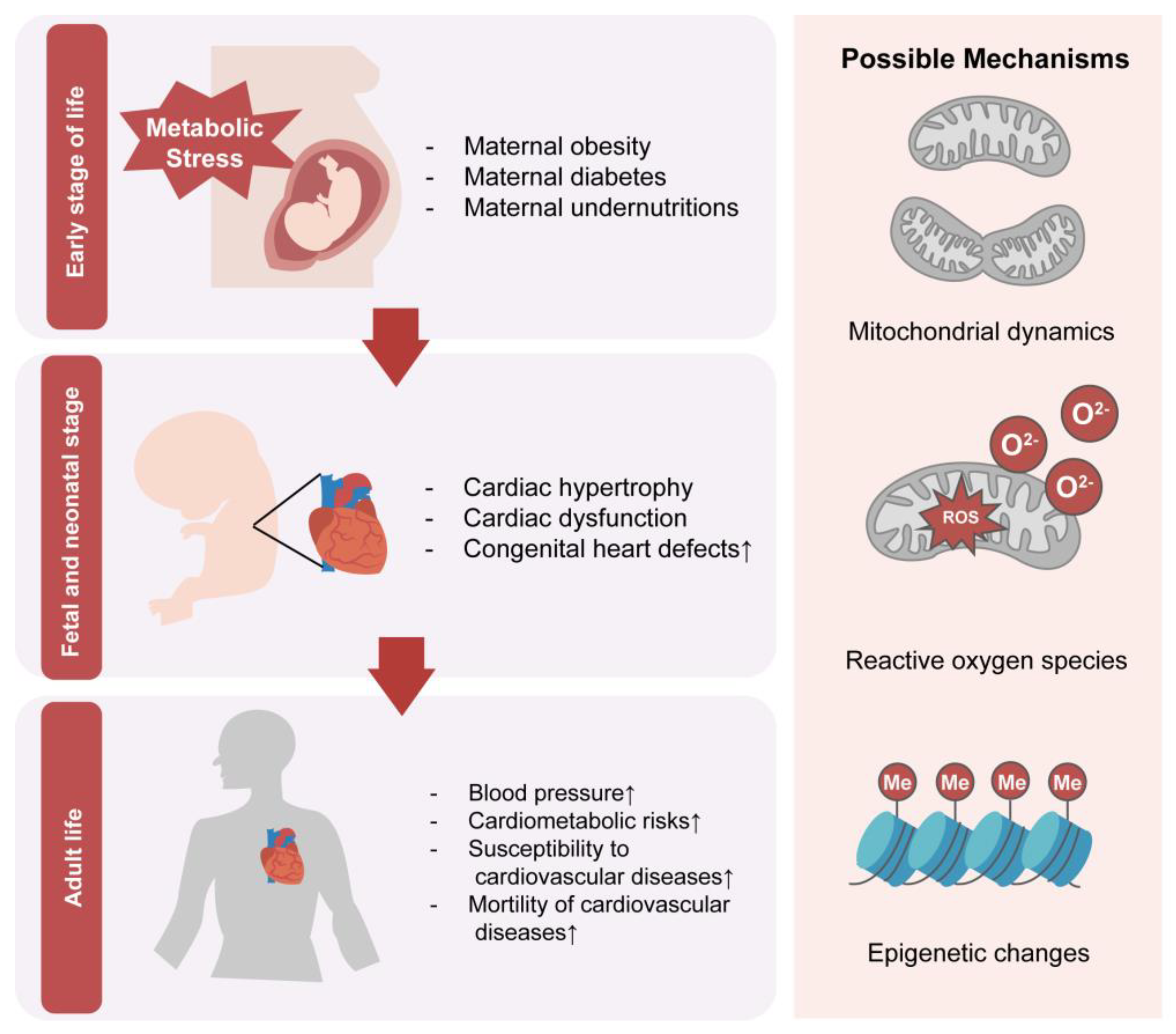
Alterations in the intrauterine environment and nutrient availability can have significant impacts on offspring growth, development, and adult health. Changes in the accessibility of glucose, lipids, and proteins during fetal growth can affect cardiac development and increase the risk of long-term cardiovascular disease (Figure 2). However, the precise effects and underlying mechanisms of these relationships remain unclear due to limitations in human studies. Additional research is necessary to elucidate the pathways through which nutritional molecules influence fetal development, persist into adulthood, and contribute to distant morbidity and mortality. This would enable development of novel biomarkers and possible new intervention strategies. The identification of epigenetic markers as possible biomarkers has instructive effects on changing maternal lifestyles and possible supplements or diets, and blocking pathways related to cardiovascular disease in early life has significant implications for improving public health and preventing cardiovascular disease.

Author Contributions

Conceptualization, G.D.; writing—original draft preparation, Z.R.; writing—review and editing, S.L., J.C. and Y.T.; visualization, Z.R.; supervision, G.D.; funding acquisition, H.H. and G.D. All authors have read and agreed to the published version of the manuscript.

Funding

This research was funded by the National Key R&D Program of China (2021YFC2701102, 2022YFC2703500), National Natural Science Foundation of China (81971458, 82088102, 82271722), CAMS Innovation Fund for Medical Sciences (2019-I2M-5-064), Collaborative Innovation Program of Shanghai Municipal Health Commission (2020CXJQ01), Shanghai Talent Development Fund Project (2021056), Shanghai Clinical Research Center for Gynecological Diseases (22MC1940200), Shanghai Urogenital System Diseases Research Center (2022ZZ01012), and Shanghai Frontiers Science Research Base of Reproduction and Development.

Institutional Review Board Statement

Not applicable.

Informed Consent Statement

Not applicable.

Data Availability Statement

No new data were created.

Conflicts of Interest

The authors declare no conflict of interest.

References

  1. Houyel, L.; Meilhac, S.M. Heart Development and Congenital Structural Heart Defects. Annu. Rev. Genom. Hum. Genet. 2021, 22, 257–284. [Google Scholar] [CrossRef] [PubMed]
  2. Sizarov, A.; Ya, J.; de Boer, B.A.; Lamers, W.H.; Christoffels, V.M.; Moorman, A.F. Formation of the building plan of the human heart: Morphogenesis, growth, and differentiation. Circulation 2011, 123, 1125–1135. [Google Scholar] [CrossRef] [PubMed] [Green Version]
  3. DeLaughter, D.M.; Bick, A.G.; Wakimoto, H.; McKean, D.; Gorham, J.M.; Kathiriya, I.S.; Hinson, J.T.; Homsy, J.; Gray, J.; Pu, W.; et al. Single-Cell Resolution of Temporal Gene Expression during Heart Development. Dev. Cell 2016, 39, 480–490. [Google Scholar] [CrossRef] [Green Version]
  4. Knutson, A.K.; Williams, A.L.; Boisvert, W.A.; Shohet, R.V. HIF in the heart: Development, metabolism, ischemia, and atherosclerosis. J. Clin. Investig. 2021, 131, e137557. [Google Scholar] [CrossRef]
  5. MacGrogan, D.; Münch, J.; de la Pompa, J.L. Notch and interacting signalling pathways in cardiac development, disease, and regeneration. Nat. Rev. Cardiol. 2018, 15, 685–704. [Google Scholar] [CrossRef]
  6. Zhou, Q.; Li, L.; Zhao, B.; Guan, K.L. The hippo pathway in heart development, regeneration, and diseases. Circ. Res. 2015, 116, 1431–1447. [Google Scholar] [CrossRef] [PubMed]
  7. Kolwicz, S.C., Jr.; Purohit, S.; Tian, R. Cardiac metabolism and its interactions with contraction, growth, and survival of cardiomyocytes. Circ. Res. 2013, 113, 603–616. [Google Scholar] [CrossRef]
  8. Gibb, A.A.; Hill, B.G. Metabolic Coordination of Physiological and Pathological Cardiac Remodeling. Circ. Res. 2018, 123, 107–128. [Google Scholar] [CrossRef]
  9. Maréchal, L.; Sicotte, B.; Caron, V.; Brochu, M.; Tremblay, A. Fetal Cardiac Lipid Sensing Triggers an Early and Sex-related Metabolic Energy Switch in Intrauterine Growth Restriction. J. Clin. Endocrinol. Metab. 2021, 106, 3295–3311. [Google Scholar] [CrossRef]
  10. Yu, Y.; Arah, O.A.; Liew, Z.; Cnattingius, S.; Olsen, J.; Sørensen, H.T.; Qin, G.; Li, J. Maternal diabetes during pregnancy and early onset of cardiovascular disease in offspring: Population based cohort study with 40 years of follow-up. BMJ 2019, 367, l6398. [Google Scholar] [CrossRef] [Green Version]
  11. Zhang, T.N.; Huang, X.M.; Zhao, X.Y.; Wang, W.; Wen, R.; Gao, S.Y. Risks of specific congenital anomalies in offspring of women with diabetes: A systematic review and meta-analysis of population-based studies including over 80 million births. PLoS Med. 2022, 19, e1003900. [Google Scholar] [CrossRef]
  12. Ghaderian, M.; Hemmat, M.; Behdad, S.; Saeedi, M.; Shahsanaei, F. Fetal Cardiac Functional Abnormalities Assessed by Echocardiography in Mothers Suffering Gestational Diabetes Mellitus: A Systematic Review and Meta-analysis. Curr. Probl. Cardiol. 2021, 46, 100658. [Google Scholar] [CrossRef] [PubMed]
  13. Wu, Y.; Liu, B.; Sun, Y.; Du, Y.; Santillan, M.K.; Santillan, D.A.; Snetselaar, L.G.; Bao, W. Association of Maternal Prepregnancy Diabetes and Gestational Diabetes Mellitus With Congenital Anomalies of the Newborn. Diabetes Care 2020, 43, 2983–2990. [Google Scholar] [CrossRef] [PubMed]
  14. Øyen, N.; Diaz, L.J.; Leirgul, E.; Boyd, H.A.; Priest, J.; Mathiesen, E.R.; Quertermous, T.; Wohlfahrt, J.; Melbye, M. Prepregnancy Diabetes and Offspring Risk of Congenital Heart Disease: A Nationwide Cohort Study. Circulation 2016, 133, 2243–2253. [Google Scholar] [CrossRef]
  15. Priest, J.R.; Yang, W.; Reaven, G.; Knowles, J.W.; Shaw, G.M. Maternal Midpregnancy Glucose Levels and Risk of Congenital Heart Disease in Offspring. JAMA Pediatr. 2015, 169, 1112–1116. [Google Scholar] [CrossRef]
  16. Zhao, M.; Diao, J.; Huang, P.; Li, J.; Li, Y.; Yang, Y.; Luo, L.; Zhang, S.; Chen, L.; Wang, T.; et al. Association of Maternal Diabetes Mellitus and Polymorphisms of the NKX2.5 Gene in Children with Congenital Heart Disease: A Single Centre-Based Case-Control Study. J. Diabetes Res. 2020, 2020, 3854630. [Google Scholar] [CrossRef]
  17. Tinker, S.C.; Gilboa, S.M.; Moore, C.A.; Waller, D.K.; Simeone, R.M.; Kim, S.Y.; Jamieson, D.J.; Botto, L.D.; Reefhuis, J. Specific birth defects in pregnancies of women with diabetes: National Birth Defects Prevention Study, 1997–2011. Am. J. Obstet. Gynecol. 2020, 222, 176.e1–176.e11. [Google Scholar] [CrossRef]
  18. Leirgul, E.; Brodwall, K.; Greve, G.; Vollset, S.E.; Holmstrøm, H.; Tell, G.S.; Øyen, N. Maternal Diabetes, Birth Weight, and Neonatal Risk of Congenital Heart Defects in Norway, 1994–2009. Obstet. Gynecol. 2016, 128, 1116–1125. [Google Scholar] [CrossRef]
  19. Tam, W.H.; Ma, R.C.W.; Ozaki, R.; Li, A.M.; Chan, M.H.M.; Yuen, L.Y.; Lao, T.T.H.; Yang, X.; Ho, C.S.; Tutino, G.E.; et al. In Utero Exposure to Maternal Hyperglycemia Increases Childhood Cardiometabolic Risk in Offspring. Diabetes Care 2017, 40, 679–686. [Google Scholar] [CrossRef] [Green Version]
  20. Li, Z.; Wu, Y.; Du, B.; Yu, X.; Wang, H.; Niu, Y.; Wang, J.; Chen, S.; Sun, K. Associations of maternal gestational diabetes mellitus with alterations in cardiovascular system in early childhood. Diabetes Metab. Res. Rev. 2022, 38, e3551. [Google Scholar] [CrossRef]
  21. Kaseva, N.; Vääräsmäki, M.; Sundvall, J.; Matinolli, H.M.; Sipola, M.; Tikanmäki, M.; Heinonen, K.; Lano, A.; Wehkalampi, K.; Wolke, D.; et al. Gestational Diabetes But Not Prepregnancy Overweight Predicts for Cardiometabolic Markers in Offspring Twenty Years Later. J. Clin. Endocrinol. Metab. 2019, 104, 2785–2795. [Google Scholar] [CrossRef] [PubMed] [Green Version]
  22. Guillemette, L.; Wicklow, B.; Sellers, E.A.C.; Dart, A.; Shen, G.X.; Dolinsky, V.W.; Gordon, J.W.; Jassal, D.S.; Nickel, N.; Duhamel, T.A.; et al. Intrauterine exposure to diabetes and risk of cardiovascular disease in adolescence and early adulthood: A population-based birth cohort study. Can. Med. Assoc. J. 2020, 192, E1104–E1113. [Google Scholar] [CrossRef]
  23. Godfrey, K.M.; Reynolds, R.M.; Prescott, S.L.; Nyirenda, M.; Jaddoe, V.W.; Eriksson, J.G.; Broekman, B.F. Influence of maternal obesity on the long-term health of offspring. Lancet Diabetes Endocrinol. 2017, 5, 53–64. [Google Scholar] [CrossRef] [PubMed] [Green Version]
  24. Persson, M.; Razaz, N.; Edstedt Bonamy, A.K.; Villamor, E.; Cnattingius, S. Maternal Overweight and Obesity and Risk of Congenital Heart Defects. J. Am. Coll. Cardiol. 2019, 73, 44–53. [Google Scholar] [CrossRef]
  25. Cai, G.J.; Sun, X.X.; Zhang, L.; Hong, Q. Association between maternal body mass index and congenital heart defects in offspring: A systematic review. Am. J. Obstet. Gynecol. 2014, 211, 91–117. [Google Scholar] [CrossRef] [PubMed]
  26. Taylor, K.; Wootton, R.E.; Yang, Q.; Oddie, S.; Wright, J.; Yang, T.C.; Magnus, M.; Andreassen, O.A.; Borges, M.C.; Caputo, M.; et al. The effect of maternal BMI, smoking and alcohol on congenital heart diseases: A Mendelian randomisation study. BMC Med. 2023, 21, 35. [Google Scholar] [CrossRef]
  27. Persson, M.; Cnattingius, S.; Villamor, E.; Söderling, J.; Pasternak, B.; Stephansson, O.; Neovius, M. Risk of major congenital malformations in relation to maternal overweight and obesity severity: Cohort study of 1.2 million singletons. BMJ 2017, 357, j2563. [Google Scholar] [CrossRef] [Green Version]
  28. Groves, A.M.; Price, A.N.; Russell-Webster, T.; Jhaveri, S.; Yang, Y.; Battersby, E.E.; Shahid, S.; Costa Vieira, M.; Hughes, E.; Miller, F.; et al. Impact of maternal obesity on neonatal heart rate and cardiac size. Arch. Dis. Child. Fetal Neonatal Ed. 2022, 107, 481–487. [Google Scholar] [CrossRef]
  29. Guzzardi, M.A.; Liistro, T.; Gargani, L.; Ait Ali, L.; D’Angelo, G.; Rocchiccioli, S.; La Rosa, F.; Kemeny, A.; Sanguinetti, E.; Ucciferri, N.; et al. Maternal Obesity and Cardiac Development in the Offspring: Study in Human Neonates and Minipigs. JACC Cardiovasc. Imaging 2018, 11, 1750–1755. [Google Scholar] [CrossRef]
  30. Fleming, T.P.; Watkins, A.J.; Velazquez, M.A.; Mathers, J.C.; Prentice, A.M.; Stephenson, J.; Barker, M.; Saffery, R.; Yajnik, C.S.; Eckert, J.J.; et al. Origins of lifetime health around the time of conception: Causes and consequences. Lancet 2018, 391, 1842–1852. [Google Scholar] [CrossRef]
  31. Wang, Z.; Li, C.; Yang, Z.; Zou, Z.; Ma, J. Infant exposure to Chinese famine increased the risk of hypertension in adulthood: Results from the China Health and Retirement Longitudinal Study. BMC Public Health 2016, 16, 435. [Google Scholar] [CrossRef] [Green Version]
  32. Li, C.; Lumey, L.H. Exposure to the Chinese famine of 1959-61 in early life and long-term health conditions: A systematic review and meta-analysis. Int. J. Epidemiol. 2017, 46, 1157–1170. [Google Scholar] [CrossRef] [PubMed] [Green Version]
  33. Painter, R.C.; Roseboom, T.J.; Bleker, O.P. Prenatal exposure to the Dutch famine and disease in later life: An overview. Reprod. Toxicol. 2005, 20, 345–352. [Google Scholar] [CrossRef]
  34. Roseboom, T.J.; Painter, R.C.; van Abeelen, A.F.; Veenendaal, M.V.; de Rooij, S.R. Hungry in the womb: What are the consequences? Lessons from the Dutch famine. Maturitas 2011, 70, 141–145. [Google Scholar] [CrossRef]
  35. Painter, R.C.; de Rooij, S.R.; Bossuyt, P.M.; Phillips, D.I.; Osmond, C.; Barker, D.J.; Bleker, O.P.; Roseboom, T.J. Blood pressure response to psychological stressors in adults after prenatal exposure to the Dutch famine. J. Hypertens. 2006, 24, 1771–1778. [Google Scholar] [CrossRef] [PubMed]
  36. Roseboom, T.J.; van der Meulen, J.H.; Osmond, C.; Barker, D.J.; Ravelli, A.C.; Schroeder-Tanka, J.M.; van Montfrans, G.A.; Michels, R.P.; Bleker, O.P. Coronary heart disease after prenatal exposure to the Dutch famine, 1944–1945. Heart 2000, 84, 595–598. [Google Scholar] [CrossRef] [Green Version]
  37. Painter, R.C.; de Rooij, S.R.; Bossuyt, P.M.; Simmers, T.A.; Osmond, C.; Barker, D.J.; Bleker, O.P.; Roseboom, T.J. Early onset of coronary artery disease after prenatal exposure to the Dutch famine. Am. J. Clin. Nutr. 2006, 84, 322–327; quiz 466–467. [Google Scholar] [CrossRef]
  38. Crispi, F.; Bijnens, B.; Figueras, F.; Bartrons, J.; Eixarch, E.; Le Noble, F.; Ahmed, A.; Gratacós, E. Fetal growth restriction results in remodeled and less efficient hearts in children. Circulation 2010, 121, 2427–2436. [Google Scholar] [CrossRef] [Green Version]
  39. Lumey, L.H.; Martini, L.H.; Myerson, M.; Stein, A.D.; Prineas, R.J. No relation between coronary artery disease or electrocardiographic markers of disease in middle age and prenatal exposure to the Dutch famine of 1944–5. Heart 2012, 98, 1653–1659. [Google Scholar] [CrossRef]
  40. Ekamper, P.; van Poppel, F.; Stein, A.D.; Bijwaard, G.E.; Lumey, L.H. Prenatal famine exposure and adult mortality from cancer, cardiovascular disease, and other causes through age 63 years. Am. J. Epidemiol. 2015, 181, 271–279. [Google Scholar] [CrossRef] [Green Version]
  41. Manivannan, S.; Mansfield, C.; Zhang, X.; Kodigepalli, K.M.; Majumdar, U.; Garg, V.; Basu, M. Single-cell transcriptomic profiling unveils dysregulation of cardiac progenitor cells and cardiomyocytes in a mouse model of maternal hyperglycemia. Commun. Biol. 2022, 5, 820. [Google Scholar] [CrossRef] [PubMed]
  42. Su, D.; Li, Y.; Guan, L.; Li, Q.; Shi, C.; Ma, X.; Song, Y. Elevated MST1 leads to apoptosis via depletion of YAP1 in cardiomyocytes exposed to high glucose. Mol. Med. 2021, 27, 13. [Google Scholar] [CrossRef] [PubMed]
  43. Luo, Z.; Xu, L.; Lu, J.; Shen, Y.; Tang, Y.; Wang, X.; Wu, Y.; Sun, H.; Guo, T. Down-regulation of the insulin signaling pathway by SHC may correlate with congenital heart disease in Chinese populations. Clin. Sci. 2020, 134, 349–358. [Google Scholar] [CrossRef] [Green Version]
  44. Saiyin, T.; Engineer, A.; Greco, E.R.; Kim, M.Y.; Lu, X.; Jones, D.L.; Feng, Q. Maternal voluntary exercise mitigates oxidative stress and incidence of congenital heart defects in pre-gestational diabetes. J. Cell. Mol. Med. 2019, 23, 5553–5565. [Google Scholar] [CrossRef]
  45. Engineer, A.; Saiyin, T.; Lu, X.; Kucey, A.S.; Urquhart, B.L.; Drysdale, T.A.; Norozi, K.; Feng, Q. Sapropterin Treatment Prevents Congenital Heart Defects Induced by Pregestational Diabetes Mellitus in Mice. J. Am. Heart Assoc. 2018, 7, e009624. [Google Scholar] [CrossRef] [Green Version]
  46. Basu, M.; Zhu, J.Y.; LaHaye, S.; Majumdar, U.; Jiao, K.; Han, Z.; Garg, V. Epigenetic mechanisms underlying maternal diabetes-associated risk of congenital heart disease. JCI Insight 2017, 2, e95085. [Google Scholar] [CrossRef] [Green Version]
  47. Wu, Y.; Reece, E.A.; Zhong, J.; Dong, D.; Shen, W.B.; Harman, C.R.; Yang, P. Type 2 diabetes mellitus induces congenital heart defects in murine embryos by increasing oxidative stress, endoplasmic reticulum stress, and apoptosis. Am. J. Obstet. Gynecol. 2016, 215, 366.e1–366.e10. [Google Scholar] [CrossRef] [Green Version]
  48. Dong, D.; Zhang, Y.; Reece, E.A.; Wang, L.; Harman, C.R.; Yang, P. microRNA expression profiling and functional annotation analysis of their targets modulated by oxidative stress during embryonic heart development in diabetic mice. Reprod. Toxicol. 2016, 65, 365–374. [Google Scholar] [CrossRef] [Green Version]
  49. Wang, F.; Wu, Y.; Quon, M.J.; Li, X.; Yang, P. ASK1 mediates the teratogenicity of diabetes in the developing heart by inducing ER stress and inhibiting critical factors essential for cardiac development. Am. J. Physiol. Endocrinol. Metab. 2015, 309, E487–E499. [Google Scholar] [CrossRef] [Green Version]
  50. Moazzen, H.; Lu, X.; Ma, N.L.; Velenosi, T.J.; Urquhart, B.L.; Wisse, L.J.; Gittenberger-de Groot, A.C.; Feng, Q. N-Acetylcysteine prevents congenital heart defects induced by pregestational diabetes. Cardiovasc. Diabetol. 2014, 13, 46. [Google Scholar] [CrossRef] [Green Version]
  51. Vijaya, M.; Manikandan, J.; Parakalan, R.; Dheen, S.T.; Kumar, S.D.; Tay, S.S. Differential gene expression profiles during embryonic heart development in diabetic mice pregnancy. Gene 2013, 516, 218–227. [Google Scholar] [CrossRef]
  52. Scott-Drechsel, D.E.; Rugonyi, S.; Marks, D.L.; Thornburg, K.L.; Hinds, M.T. Hyperglycemia slows embryonic growth and suppresses cell cycle via cyclin D1 and p21. Diabetes 2013, 62, 234–242. [Google Scholar] [CrossRef] [PubMed] [Green Version]
  53. Kumar, S.D.; Vijaya, M.; Samy, R.P.; Dheen, S.T.; Ren, M.; Watt, F.; Kang, Y.J.; Bay, B.H.; Tay, S.S. Zinc supplementation prevents cardiomyocyte apoptosis and congenital heart defects in embryos of diabetic mice. Free Radic. Biol. Med. 2012, 53, 1595–1606. [Google Scholar] [CrossRef]
  54. Ejdesjö, A.; Wentzel, P.; Eriksson, U.J. Influence of maternal metabolism and parental genetics on fetal maldevelopment in diabetic rat pregnancy. Am. J. Physiol. Endocrinol. Metab. 2012, 302, E1198–E1209. [Google Scholar] [CrossRef] [Green Version]
  55. Kumar, S.D.; Dheen, S.T.; Tay, S.S. Maternal diabetes induces congenital heart defects in mice by altering the expression of genes involved in cardiovascular development. Cardiovasc. Diabetol. 2007, 6, 34. [Google Scholar] [CrossRef] [Green Version]
  56. Mdaki, K.S.; Larsen, T.D.; Wachal, A.L.; Schimelpfenig, M.D.; Weaver, L.J.; Dooyema, S.D.; Louwagie, E.J.; Baack, M.L. Maternal high-fat diet impairs cardiac function in offspring of diabetic pregnancy through metabolic stress and mitochondrial dysfunction. Am. J. Physiol. Heart Circ. Physiol. 2016, 310, H681–H692. [Google Scholar] [CrossRef] [PubMed] [Green Version]
  57. Wang, Q.; Zhu, C.; Sun, M.; Maimaiti, R.; Ford, S.P.; Nathanielsz, P.W.; Ren, J.; Guo, W. Maternal obesity impairs fetal cardiomyocyte contractile function in sheep. FASEB J. 2019, 33, 2587–2598. [Google Scholar] [CrossRef] [Green Version]
  58. Loche, E.; Blackmore, H.L.; Carpenter, A.A.; Beeson, J.H.; Pinnock, A.; Ashmore, T.J.; Aiken, C.E.; de Almeida-Faria, J.; Schoonejans, J.M.; Giussani, D.A.; et al. Maternal diet-induced obesity programmes cardiac dysfunction in male mice independently of post-weaning diet. Cardiovasc. Res. 2018, 114, 1372–1384. [Google Scholar] [CrossRef] [Green Version]
  59. Kwong, W.Y.; Wild, A.E.; Roberts, P.; Willis, A.C.; Fleming, T.P. Maternal undernutrition during the preimplantation period of rat development causes blastocyst abnormalities and programming of postnatal hypertension. Development 2000, 127, 4195–4202. [Google Scholar] [CrossRef]
  60. Darby, J.R.T.; McMillen, I.C.; Morrison, J.L. Maternal undernutrition in late gestation increases IGF2 signalling molecules and collagen deposition in the right ventricle of the fetal sheep heart. J. Physiol. 2018, 596, 2345–2358. [Google Scholar] [CrossRef] [Green Version]
  61. Jonker, S.S.; Kamna, D.; LoTurco, D.; Kailey, J.; Brown, L.D. IUGR impairs cardiomyocyte growth and maturation in fetal sheep. J. Endocrinol. 2018, 239, 253–265. [Google Scholar] [CrossRef] [Green Version]
  62. Pereira, S.P.; Tavares, L.C.; Duarte, A.I.; Baldeiras, I.; Cunha-Oliveira, T.; Martins, J.D.; Santos, M.S.; Maloyan, A.; Moreno, A.J.; Cox, L.A.; et al. Sex-dependent vulnerability of fetal nonhuman primate cardiac mitochondria to moderate maternal nutrient reduction. Clin. Sci. 2021, 135, 1103–1126. [Google Scholar] [CrossRef]
  63. Louwagie, E.J.; Larsen, T.D.; Wachal, A.L.; Gandy, T.C.T.; Eclov, J.A.; Rideout, T.C.; Kern, K.A.; Cain, J.T.; Anderson, R.H.; Mdaki, K.S.; et al. Age and Sex Influence Mitochondria and Cardiac Health in Offspring Exposed to Maternal Glucolipotoxicity. iScience 2020, 23, 101746. [Google Scholar] [CrossRef] [PubMed]
  64. Yu, C.; Chen, S.; Wang, X.; Wu, G.; Zhang, Y.; Fu, C.; Hu, C.; Liu, Z.; Luo, X.; Wang, J.; et al. Exposure to maternal diabetes induces endothelial dysfunction and hypertension in adult male rat offspring. Microvasc. Res. 2021, 133, 104076. [Google Scholar] [CrossRef]
  65. Schütte, T.; Kedziora, S.M.; Haase, N.; Herse, F.; Alenina, N.; Müller, D.N.; Bader, M.; Schupp, M.; Dechend, R.; Golic, M.; et al. Diabetic pregnancy as a novel risk factor for cardiac dysfunction in the offspring-the heart as a target for fetal programming in rats. Diabetologia 2021, 64, 2829–2842. [Google Scholar] [CrossRef]
  66. Gao, L.; Zhao, Y.C.; Liang, Y.; Lin, X.H.; Tan, Y.J.; Wu, D.D.; Li, X.Z.; Ye, B.Z.; Kong, F.Q.; Sheng, J.Z.; et al. The impaired myocardial ischemic tolerance in adult offspring of diabetic pregnancy is restored by maternal melatonin treatment. J. Pineal Res. 2016, 61, 340–352. [Google Scholar] [CrossRef]
  67. Chen, Z.; Gong, L.; Zhang, P.; Li, Y.; Liu, B.; Zhang, L.; Zhuang, J.; Xiao, D. Epigenetic Down-Regulation of Sirt 1 via DNA Methylation and Oxidative Stress Signaling Contributes to the Gestational Diabetes Mellitus-Induced Fetal Programming of Heart Ischemia-Sensitive Phenotype in Late Life. Int. J. Biol. Sci. 2019, 15, 1240–1251. [Google Scholar] [CrossRef]
  68. Vaughan, O.R.; Rosario, F.J.; Chan, J.; Cox, L.A.; Ferchaud-Roucher, V.; Zemski-Berry, K.A.; Reusch, J.E.B.; Keller, A.C.; Powell, T.L.; Jansson, T. Maternal obesity causes fetal cardiac hypertrophy and alters adult offspring myocardial metabolism in mice. J. Physiol. 2022, 600, 3169–3191. [Google Scholar] [CrossRef]
  69. Ahmed, A.; Liang, M.; Chi, L.; Zhou, Y.Q.; Sled, J.G.; Wilson, M.D.; Delgado-Olguín, P. Maternal obesity persistently alters cardiac progenitor gene expression and programs adult-onset heart disease susceptibility. Mol. Metab. 2021, 43, 101116. [Google Scholar] [CrossRef]
  70. Xue, Q.; Chen, P.; Li, X.; Zhang, G.; Patterson, A.J.; Luo, J. Maternal High-Fat Diet Causes a Sex-Dependent Increase in AGTR2 Expression and Cardiac Dysfunction in Adult Male Rat Offspring. Biol. Reprod. 2015, 93, 49. [Google Scholar] [CrossRef]
  71. Watkins, A.J.; Wilkins, A.; Cunningham, C.; Perry, V.H.; Seet, M.J.; Osmond, C.; Eckert, J.J.; Torrens, C.; Cagampang, F.R.; Cleal, J.; et al. Low protein diet fed exclusively during mouse oocyte maturation leads to behavioural and cardiovascular abnormalities in offspring. J. Physiol. 2008, 586, 2231–2244. [Google Scholar] [CrossRef]
  72. Larsen, T.D.; Sabey, K.H.; Knutson, A.J.; Gandy, T.C.T.; Louwagie, E.J.; Lauterboeck, L.; Mdaki, K.S.; Baack, M.L. Diabetic Pregnancy and Maternal High-Fat Diet Impair Mitochondrial Dynamism in the Developing Fetal Rat Heart by Sex-Specific Mechanisms. Int. J. Mol. Sci. 2019, 20, 3090. [Google Scholar] [CrossRef] [PubMed] [Green Version]
  73. Preston, C.C.; Larsen, T.D.; Eclov, J.A.; Louwagie, E.J.; Gandy, T.C.T.; Faustino, R.S.; Baack, M.L. Maternal High Fat Diet and Diabetes Disrupts Transcriptomic Pathways That Regulate Cardiac Metabolism and Cell Fate in Newborn Rat Hearts. Front. Endocrinol. 2020, 11, 570846. [Google Scholar] [CrossRef]
  74. Raji, S.R.; Nandini, R.J.; Ashok, S.; Anand, R.C.; Vivek, P.V.; Karunakaran, J.; Sreelatha, H.V.; Manjunatha, S.; Gopala, S. Diminished substrate-mediated cardiac mitochondrial respiration and elevated autophagy in adult male offspring of gestational diabetic rats. IUBMB Life 2021, 73, 676–689. [Google Scholar] [CrossRef] [PubMed]
  75. Miao, W.; Porter, D.F.; Lopez-Pajares, V.; Siprashvili, Z.; Meyers, R.M.; Bai, Y.; Nguyen, D.T.; Ko, L.A.; Zarnegar, B.J.; Ferguson, I.D.; et al. Glucose dissociates DDX21 dimers to regulate mRNA splicing and tissue differentiation. Cell 2023, 186, 80–97.e26. [Google Scholar] [CrossRef]
  76. Su, D.; Zhou, Y.; Hu, S.; Guan, L.; Shi, C.; Wang, Q.; Chen, Y.; Lu, C.; Li, Q.; Ma, X. Role of GAB1/PI3K/AKT signaling high glucose-induced cardiomyocyte apoptosis. Biomed. Pharmacother. 2017, 93, 1197–1204. [Google Scholar] [CrossRef]
  77. Lindegaard, M.L.; Nielsen, L.B. Maternal diabetes causes coordinated down-regulation of genes involved with lipid metabolism in the murine fetal heart. Metabolism 2008, 57, 766–773. [Google Scholar] [CrossRef]
  78. Cardoso, A.C.; Lam, N.T.; Savla, J.J.; Nakada, Y.; Pereira, A.H.M.; Elnwasany, A.; Menendez-Montes, I.; Ensley, E.L.; Petric, U.B.; Sharma, G.; et al. Mitochondrial Substrate Utilization Regulates Cardiomyocyte Cell Cycle Progression. Nat. Metab. 2020, 2, 167–178. [Google Scholar] [CrossRef]
  79. Luo, H.; Lan, C.; Fan, C.; Gong, X.; Chen, C.; Yu, C.; Wang, J.; Luo, X.; Hu, C.; Jose, P.A.; et al. Down-regulation of AMPK/PPARδ signalling promotes endoplasmic reticulum stress-induced endothelial dysfunction in adult rat offspring exposed to maternal diabetes. Cardiovasc. Res. 2022, 118, 2304–2316. [Google Scholar] [CrossRef]
  80. Sun, C.; Velazquez, M.A.; Marfy-Smith, S.; Sheth, B.; Cox, A.; Johnston, D.A.; Smyth, N.; Fleming, T.P. Mouse early extra-embryonic lineages activate compensatory endocytosis in response to poor maternal nutrition. Development 2014, 141, 1140–1150. [Google Scholar] [CrossRef] [Green Version]
  81. Watkins, A.J.; Ursell, E.; Panton, R.; Papenbrock, T.; Hollis, L.; Cunningham, C.; Wilkins, A.; Perry, V.H.; Sheth, B.; Kwong, W.Y.; et al. Adaptive responses by mouse early embryos to maternal diet protect fetal growth but predispose to adult onset disease. Biol. Reprod. 2008, 78, 299–306. [Google Scholar] [CrossRef]
  82. Botting, K.J.; Loke, X.Y.; Zhang, S.; Andersen, J.B.; Nyengaard, J.R.; Morrison, J.L. IUGR decreases cardiomyocyte endowment and alters cardiac metabolism in a sex- and cause-of-IUGR-specific manner. Am. J. Physiol. Regul. Integr. Comp. Physiol. 2018, 315, R48–R67. [Google Scholar] [CrossRef]
  83. Zorov, D.B.; Juhaszova, M.; Sollott, S.J. Mitochondrial reactive oxygen species (ROS) and ROS-induced ROS release. Physiol. Rev. 2014, 94, 909–950. [Google Scholar] [CrossRef] [Green Version]
  84. Torres-Cuevas, I.; Parra-Llorca, A.; Sánchez-Illana, A.; Nuñez-Ramiro, A.; Kuligowski, J.; Cháfer-Pericás, C.; Cernada, M.; Escobar, J.; Vento, M. Oxygen and oxidative stress in the perinatal period. Redox Biol. 2017, 12, 674–681. [Google Scholar] [CrossRef] [PubMed]
  85. Giacco, F.; Brownlee, M. Oxidative stress and diabetic complications. Circ. Res. 2010, 107, 1058–1070. [Google Scholar] [CrossRef]
  86. Block, T.; El-Osta, A. Epigenetic programming, early life nutrition and the risk of metabolic disease. Atherosclerosis 2017, 266, 31–40. [Google Scholar] [CrossRef]
  87. Robinson, E.L.; Anene-Nzelu, C.G.; Rosa-Garrido, M.; Foo, R.S.Y. Cardiac epigenetics: Driving signals to the cardiac epigenome in development and disease. J. Mol. Cell. Cardiol. 2021, 151, 88. [Google Scholar] [CrossRef]
  88. Agarwal, P.; Morriseau, T.S.; Kereliuk, S.M.; Doucette, C.A.; Wicklow, B.A.; Dolinsky, V.W. Maternal obesity, diabetes during pregnancy and epigenetic mechanisms that influence the developmental origins of cardiometabolic disease in the offspring. Crit. Rev. Clin. Lab. Sci. 2018, 55, 71–101. [Google Scholar] [CrossRef]
  89. Canouil, M.; Khamis, A.; Keikkala, E.; Hummel, S.; Lobbens, S.; Bonnefond, A.; Delahaye, F.; Tzala, E.; Mustaniemi, S.; Vääräsmäki, M.; et al. Epigenome-Wide Association Study Reveals Methylation Loci Associated With Offspring Gestational Diabetes Mellitus Exposure and Maternal Methylome. Diabetes Care 2021, 44, 1992–1999. [Google Scholar] [CrossRef]
  90. Weng, X.; Liu, F.; Zhang, H.; Kan, M.; Wang, T.; Dong, M.; Liu, Y. Genome-wide DNA methylation profiling in infants born to gestational diabetes mellitus. Diabetes Res. Clin. Pract. 2018, 142, 10–18. [Google Scholar] [CrossRef]
  91. Reichetzeder, C.; Dwi Putra, S.E.; Pfab, T.; Slowinski, T.; Neuber, C.; Kleuser, B.; Hocher, B. Increased global placental DNA methylation levels are associated with gestational diabetes. Clin. Epigenet. 2016, 8, 82. [Google Scholar] [CrossRef] [Green Version]
  92. Chen, P.; Piaggi, P.; Traurig, M.; Bogardus, C.; Knowler, W.C.; Baier, L.J.; Hanson, R.L. Differential methylation of genes in individuals exposed to maternal diabetes in utero. Diabetologia 2017, 60, 645–655. [Google Scholar] [CrossRef] [Green Version]
  93. Tobi, E.W.; Goeman, J.J.; Monajemi, R.; Gu, H.; Putter, H.; Zhang, Y.; Slieker, R.C.; Stok, A.P.; Thijssen, P.E.; Müller, F.; et al. DNA methylation signatures link prenatal famine exposure to growth and metabolism. Nat. Commun. 2014, 5, 5592. [Google Scholar] [CrossRef] [Green Version]
  94. Bošković, A.; Rando, O.J. Transgenerational Epigenetic Inheritance. Annu. Rev. Genet. 2018, 52, 21–41. [Google Scholar] [CrossRef]
  95. Blin, G.; Liand, M.; Mauduit, C.; Chehade, H.; Benahmed, M.; Simeoni, U.; Siddeek, B. Maternal Exposure to High-Fat Diet Induces Long-Term Derepressive Chromatin Marks in the Heart. Nutrients 2020, 12, 181. [Google Scholar] [CrossRef] [PubMed] [Green Version]
  96. Upadhyaya, B.; Larsen, T.; Barwari, S.; Louwagie, E.J.; Baack, M.L.; Dey, M. Prenatal Exposure to a Maternal High-Fat Diet Affects Histone Modification of Cardiometabolic Genes in Newborn Rats. Nutrients 2017, 9, 407. [Google Scholar] [CrossRef] [Green Version]
  97. Barwari, T.; Joshi, A.; Mayr, M. MicroRNAs in Cardiovascular Disease. J. Am. Coll. Cardiol. 2016, 68, 2577–2584. [Google Scholar] [CrossRef] [PubMed] [Green Version]
  98. Lock, M.C.; Botting, K.J.; Tellam, R.L.; Brooks, D.; Morrison, J.L. Adverse Intrauterine Environment and Cardiac miRNA Expression. Int. J. Mol. Sci. 2017, 18, 2628. [Google Scholar] [CrossRef] [Green Version]
  99. Siddeek, B.; Mauduit, C.; Chehade, H.; Blin, G.; Liand, M.; Chindamo, M.; Benahmed, M.; Simeoni, U. Long-term impact of maternal high-fat diet on offspring cardiac health: Role of micro-RNA biogenesis. Cell Death Discov. 2019, 5, 71. [Google Scholar] [CrossRef] [PubMed] [Green Version]
  100. Fernandez-Twinn, D.S.; Blackmore, H.L.; Siggens, L.; Giussani, D.A.; Cross, C.M.; Foo, R.; Ozanne, S.E. The programming of cardiac hypertrophy in the offspring by maternal obesity is associated with hyperinsulinemia, AKT, ERK, and mTOR activation. Endocrinology 2012, 153, 5961–5971. [Google Scholar] [CrossRef] [Green Version]
  101. Muralimanoharan, S.; Li, C.; Nakayasu, E.S.; Casey, C.P.; Metz, T.O.; Nathanielsz, P.W.; Maloyan, A. Sexual dimorphism in the fetal cardiac response to maternal nutrient restriction. J. Mol. Cell. Cardiol. 2017, 108, 181–193. [Google Scholar] [CrossRef] [PubMed]
  102. Moore-Morris, T.; van Vliet, P.P.; Andelfinger, G.; Puceat, M. Role of Epigenetics in Cardiac Development and Congenital Diseases. Physiol. Rev. 2018, 98, 2453–2475. [Google Scholar] [CrossRef] [PubMed] [Green Version]
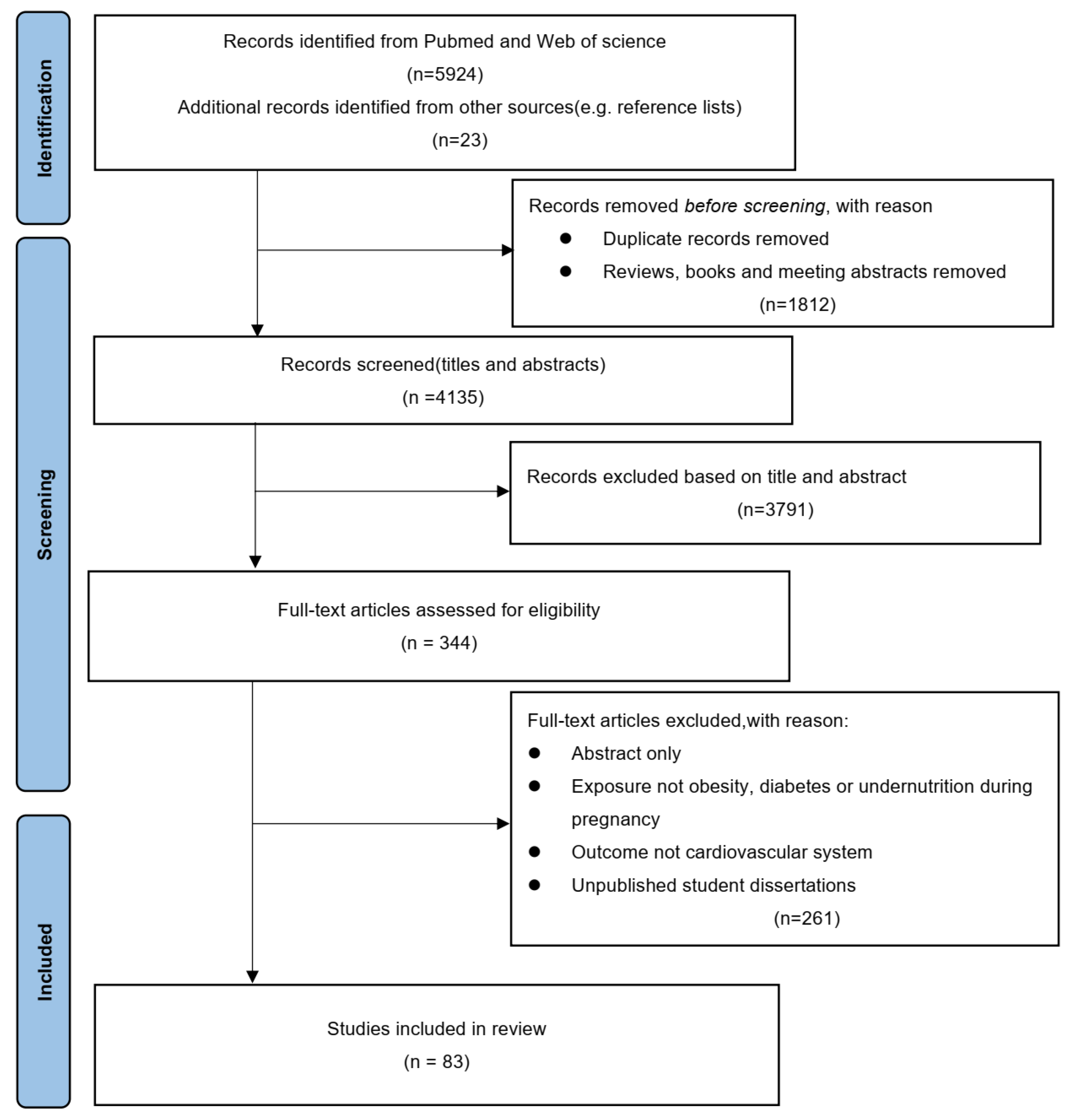
Figure 1. The methodology used for the literature search.
Nutrients 15 03388 g001
Figure 2. Maternal metabolism and cardiovascular diseases in offspring. Intrauterine exposure to nutritional disorders could influence offspring’s health through all stages of life. Possible mechanisms including mitochondrial changes, increase of reactive oxygen species, and epigenetic changes such as DNA methylation and histone modification.
Nutrients 15 03388 g002
Table 1. Human studies of the effect of maternal metabolism on cardiac development and function in offspring.
StudyStudy PopulationExposuresFollowing YearsOutcomesMajor Findings
Øyen et al. [14]2,025,727 persons born alive in Denmark between 1978 and 2011
  • PGDM
  • GDM
Birth defectsAll types of congenital heart diseases
  • CHD risk increased nor differed by diabetes subtype
  • Diabetes complication related with higher risk
Priest et al. [15]277 pregnant women in southern and central California
  • Maternal diabetes
Birth defectsCardiac malformations, including ToF, d-transposition of the great arteries (dTGA)
  • Odds of ToF increased
  • dTGA not changed
Tam et al. [19]970 mothers who joined the Hyperglycemia and Adverse Pregnancy Outcome study and their children
  • GDM
7 yearsCardiometabolic risk
  • Associated with offspring’s risk of abnormal glucose tolerance, obesity, and higher blood pressure
  • Sex dimorphism, apparent in girls
Kaseva et al. [21]906 pregnant women and their offspring from Uusimaa and northern Finland
  • GDM
  • Obesity
24.1 ± 1.3 yearsCardiometabolic risk
  • Increased markers of insulin resistance in GDM offspring
  • Impaired glucose regulation in obesity offspring
Guillemette et al. [22]293,546 people born between 1979 and 2005 in Manitoba, Canada
  • PGDM (type II)
  • GDM
Up to 35 yearsCardiovascular disease,
including cardiac arrest, myocardial infarction, ischemic heart disease, and cerebral infarction
  • Higher morbidity and higher risk of cardiovascular disease
Yu et al. [10]All 2,432,000 liveborn children without congenital
heart disease in Denmark during 1977–2016
  • PGDM (including type I and type II)
  • GDM
40 yearsEarly onset CVD
including ischemic heart disease, cerebrovascular disease, stroke, heart failure, atrial fibrillation, hypertensive disease, deep vein thrombosis, pulmonary embolism, other CVDs
  • Increased rates of early onset CVD from childhood to early adulthood
Persson et al. [24]2,050,491 live singleton infants born between 1992 and 2012 in Sweden
  • Overweight/obesity
Birth defectsCongenital heart defect
  • Aortic branch defects, ASD, and persistent ductus arteriosus increase
Groves et al. [28]87 neonates in UK
  • Obesity
NewbornsHeart rates
Heart variability
Cardiac function
  • Increased heart rate
  • Decreased heart rate variability
  • Decreased left ventricular volumes
Guzzardi et al. [29]91 pregnant women in Italy
  • Overweight
12 monthsCardiac function
  • Cardiac morphology changed
Wang et al. [31]1966 adults born between 1956 and 1964 in China
  • Maternal malnutrition
45 yearsHypertension
  • Increased risk of hypertension
Painter et al. [35]721 men and women born as term singletons in Amsterdam at about the time of the Dutch 1944–1945 famine
  • Maternal malnutrition
58 yearsHypertension
  • Increased blood pressure in famine exposed individuals
Roseboom et al. [36]912 singletons born in Amsterdam at about the time of the Dutch 1944–1945 famine
  • Maternal malnutrition
50 yearsCoronary artery disease (CAD)
  • Increased risk of coronary heart diseases
Painter et al. [37]837 singletons born in Amsterdam at about the time of the Dutch 1944–1945 famine
  • Maternal malnutrition
50 yearsCAD
  • Increased risk of coronary heart disease
Lumey et al. [39]1075 men and women born around the Dutch 1944–1945
  • Maternal malnutrition
58 yearsCAD
  • No relation between prenatal famine and CAD
Ekamper et al. [40]41,096 men born in 1944–1947 in the Netherlands
  • Maternal malnutrition
63 yearsMortality of heart diseases
  • No increase in mortality from cardiovascular diseases
Table 2. Long-term cardiac effects of offspring exposed to maternal overnutrition in early stage of life.
StudySpeciesGenderAgeEarly ExposureSecond HitMajor Findings
Pereia et al. [63]Rat
(Sprague Dawley)
MP1, 3 W, 10 W, 6 W, 12 WHF group:
Gestational diabetes group:
-Declined cardiac function in aged offspring
Louwagie et al. [64]Rat
(Sprague Dawley)
M12 W, 16 W, 20 W, 24 WStreptozotocin, 35 mg/kg, IP, at E0-Higher blood pressure
Yu et al. [65]Rat
(Sprague Dawley)
M8 W,
29 W–36 W
Transgenic Tet29 female dams, 1.5 mg/kg DOX in drinking waterHigh-fat diet challenge for 28 weeksCardiac dysfunction
LV hypertrophy
Altered proinflammatory status
Schütte et al. [66]Mouse
(C57BL/6)
M8–10 WSTZ, IP, 80 mg/kg for 3 days at 8 weeks oldMyocardial ischemia/reperfusion injury at 8–10 weeks oldLarger infarct size
Augmented cardiac dysfunction
Augmented myocardial apoptosis
Gao et al. [67]Rat
(Sprague Dawley)
M and F6 WSTZ, SC, 50 mg/kg at E12Heart ischemia for 24 h at 6 weeks-oldLarger infarct size
LV dysfunction in male offspring
Chen et al. [68]Mouse
(C57BL/6)
M and FE18.5, 3 W, 6 W, 9 W, 24 WObesogenic diet until body weight increased for 25%-Altered embryonic metabolic genes transcription
Diminished cardiac diastolic function
Female cardiac function worsens with age
Vaughan et al. [69]Mouse
(Chimeric)
ME18.5, 16 W, 32 WObesogenic diet from 4 weeks old through whole lifeIsoproterenol (60 mg/kg/day) in 8-week-old mice for 14 daysExacerbated cardiac remodeling
Altered gene expression of cardiac progenitors
Ahmed et al. [70]Rat
(Sprague Dawley)
M and F3 WHigh-fat diet from E0 to E21Myocardial ischemia/reperfusion injuryCardiac hypertrophy in male
Increased infract size in male
M, Male; F, Female; P, postnatal day; W, weeks; E, embryonic day; M, month; lv, left ventricular.
Disclaimer/Publisher’s Note: The statements, opinions and data contained in all publications are solely those of the individual author(s) and contributor(s) and not of MDPI and/or the editor(s). MDPI and/or the editor(s) disclaim responsibility for any injury to people or property resulting from any ideas, methods, instructions or products referred to in the content.