Article

Mortality in Hemodialysis Patients with COVID-19, the Effect of Paricalcitol or Calcimimetics

by
María Dolores Arenas Jimenez
1,2,*,
Emilio González-Parra
3,
Marta Riera
1,
Abraham Rincón Bello
4,
Ana López-Herradón
4,
Higini Cao
1,
Sara Hurtado
4,
Silvia Collado
1,
Laura Ribera
4,
Francesc Barbosa
1,
Fabiola Dapena
5,
Vicent Torregrosa
6,
José-Jesús Broseta
5,
Carlos Soto Montañez
6,
Juan F. Navarro-González
7,8,9,
Rosa Ramos
4,
Jordi Bover
10,
Xavier Nogués-Solan
11,
Marta Crespo
1,
Adriana S. Dusso
12,13 and
Julio Pascual
1
add Show full author list remove Hide full author list
1
Department of Nephrology, Hospital del Mar, IMIM Hospital del Mar Medical Research Institute, RD16/0009/0013 (ISCIII FEDER REDinREN), 08003 Barcelona, Spain
2
Fundación Renal Iñigo Alvarez de Toledo, 28003 Madrid, Spain
3
Fundación Jimenez Díaz, 28040 Madrid, Spain
4
Fresenius Medical Care, Dirección Médica FMC, 28760 Madrid, Spain
5
Department of Nephrology, Consorci Sanitari Alt Penedes Garraf, 08800 Barcelona, Spain
6
Department of Nephrology and Kidney Transplantation, Hospital Clinic, 08036 Barcelona, Spain
7
Research Division and Department of Nephrology, Hospital Nuestra Señora de la Candelaria, 38010 Santa Cruz de Tenerife, Spain
8
Instituto de Tecnologías Biomédicas, Universidad de La Laguna, 38010 Tenerife, Spain
9
Red de Investigación Renal (REDINREN–RD16/0009/0022), Instituto de Salud Carlos III, 28029 Madrid, Spain
10
Department of Nephrology, Hospital Can Ruti, 08916 Barcelona, Spain
11
Department of Internal Medicine, Hospital del Mar, Institut Mar for Medical Research, CIBERFES, 08003 Barcelona, Spain
12
Bone and Mineral Research Unit, Instituto de Investigaciones Sanitarias del Principado de Asturias, 33011 Oviedo, Spain
13
Department of Internal Medicine, Division of Endocrinology, Metabolism and Lipid Research, Washington University School of Medicine, St. Louis, MO 63110, USA
*
Author to whom correspondence should be addressed.
Nutrients 2021, 13(8), 2559; https://doi.org/10.3390/nu13082559
Submission received: 22 June 2021 / Revised: 15 July 2021 / Accepted: 20 July 2021 / Published: 26 July 2021
(This article belongs to the Section Nutritional Epidemiology)

Abstract

:
Background. In COVID-19 patients, low serum vitamin D (VD) levels have been associated with severe acute respiratory failure and poor prognosis. In regular hemodialysis (HD) patients, there is VD deficiency and markedly reduced calcitriol levels, which may predispose them to worse outcomes of COVID-19 infection. Some hemodialysis patients receive treatment with drugs for secondary hyperparathyroidism, which have well known pleiotropic effects beyond mineral metabolism. The aim of this study was to evaluate the impact of VD status and the administration of active vitamin D medications, used to treat secondary hyperparathyroidism, on survival in a cohort of COVID-19 positive HD patients. Methods. A cross-sectional retrospective observational study was conducted from 12 March to 21 May 2020 in 288 HD patients with positive PCR for SARS-CoV2. Patients were from 52 different centers in Spain. Results. The percent of HD patients with COVID-19 was 6.1% (288 out of 4743). Mortality rate was 28.4% (81/285). Three patients were lost to follow-up. Serum 25(OH)D (calcidiol) level was 17.1 [10.6–27.5] ng/mL and was not significantly associated to mortality (OR 0.99 (0.97–1.01), p = 0.4). Patients receiving active vitamin D medications (16/94 (17%) vs. 65/191(34%), p = 0.003), including calcimimetics (4/49 (8.2%) vs. 77/236 (32.6%), p = 0.001), paricalcitol or calcimimetics (19/117 (16.2%) vs. 62/168 (36.9%); p < 0.001), and also those on both paricalcitol and calcimimetics, to treat secondary hyperparathyroidism (SHPTH) (1/26 (3.8%) vs. 80/259 (30.9%), p < 0.001) showed a lower mortality rate than patients receiving no treatment with either drug. Multivariate Cox regression analysis confirmed this increased survival. Conclusions. Our findings suggest that the use of paricalcitol, calcimimetics or the combination of both, seem to be associated with the improvement of survival in HD patients with COVID-19. No correlation was found between serum VD levels and prognosis or outcomes in HD patients with COVID-19. Prospective studies and clinical trials are needed to support these findings.

1. Introduction

The world is experiencing its third major epidemic of coronavirus (CoV) infection [1]. A new coronavirus-induced pneumonia, which appeared in late November 2019 in Wuhan, China, was named coronavirus 2019 disease (COVID-19) by the World Health Organization (WHO) on 11 February 2020 [2]. The clinical manifestations of COVID-19 patients were similar to those of SARS-CoV and MERS-CoV infections [3,4]. There is a wide spectrum of manifestations [5], ranging from asymptomatic patients [3] to acute respiratory distress syndrome (ARDS), which is the main cause of death [4]. ARDS is, in part, the result of the storm of cytokines and chemo-attractant molecules that follow viral infection [6], which is aggravated by age and pre-existing conditions, including hypertension and diabetes [7]. Despite the prevalence of variable clinical presentation and outcomes in COVID-19 infected HD patients [8,9,10], the main factors driving the severity of disease progression in these specific patients remain to be elucidated.
Recent evidence supports a direct association between low serum vitamin D (VD) levels with the severity of acute respiratory failure and COVID-19 prognosis [11], possibly mediated by VD anti-viral, anti-hypertensive and/or anti-inflammatory properties [12]. Meta-analyses of randomized clinical trials revealed protective effects of VD against acute respiratory infections, although effects were modest and heterogenous [13]. Airway diseases are associated with deregulated VD metabolism [14]; however, the data are contradictory [15], and more studies are required to evaluate such a possibility. In addition to circulating VD levels, interventions to attenuate the activation of the renin-angiotensin system (RAS), either with angiotensin converting enzyme inhibitors (ACEI) or angiotensin-2 receptor blockers (ARB), have been associated with improved COVID-19 clinical evolution and prognosis [16].
In HD patients, there is a high prevalence of hypertension, diabetes and cardiovascular disease, which concurs not only with systemic VD deficiency but also with a severely depressed 1-alpha-hydroxylation capacity to synthesize calcitriol, both renal and extrarenal. Uremic toxins are responsible for decreased 1-alpha-hydroxylase activity in cells other than kidney proximal tubular cells, suggesting an increased susceptibility for a faster progression of COVID-19 to acute lung injury.
KDIGO [17] and Spanish guidelines [18] for chronic kidney disease (CKD) mineral and bone disorders support VD supplementation and/or calcitriol replacement therapy to treat secondary hyperparathyroidism in these patients. The aim of the present study was to evaluate the impact of VD status and the administration of VD/calcitriol and calcimimetics, used to treat secondary hyperparathyroidism, on the survival of a cohort of COVID-19 positive HD patients.

2. Materials and Methods

2.1. Patients and Variables

This is a cross-sectional retrospective observational study of 288 HD patients with SARS-CoV2 positive PCR, from 52 Spanish HD facilities from 12 March 2020 to 21 May 2020. This sample is representative of the Spanish HD population, in terms of age and sex. The study was performed under the principles of the Declaration of Helsinki and was approved by the hospital ethics committee at participant institutions.
Demographics, clinical, laboratory and radiological data were obtained from medical records launched at the beginning of the epidemic. After the diagnosis, all patients were followed for at least 14 days, for a maximum of 35 days or until death. Three patients were lost to follow-up. All related-to-mortality statistical analyses were conducted in 285 patients. Parameters that were recorded included serum concentrations of 25-hydroxyvitamin-D (25-OH-D3), PTH, calcium, phosphate and treatment with 25-OH-D3 (calcifediol), active VD (calcitriol; paricalcitol) and/or calcimimetics shortly (20 days) before COVID-19 infection.
Other variables that were recorded were age; sex; race; body mass index (BMI); type of vascular access; comorbidities; baseline treatments, including angiotensin converting enzyme inhibitors (ACEIs), angiotensin receptors blockers (ARBs) or nonsteroidal anti-inflammatory drugs (NSAIDs); clinical symptoms; chest X-Ray findings; standard laboratory parameters; specific treatments for COVID-19; and outcomes. Laboratory parameters were recorded at two points: at baseline (regular control tests performed immediately prior to the time of COVID-19 diagnosis (median 20 days, interquartile range–IQR-12.5–25 days)) and at hospital admission.

2.2. SARS-CoV-2 Testing

Testing for SARS-CoV-2 virus infection was undertaken using qualitative reverse transcription PCR (RT-PCR), following M2000® RNA Extraction Kit according to the manufacturer’s instructions (Abbott, Chicago, IL, USA). Several molecular diagnostic RT-PCR platforms were used, including Roche Cobas® and Abbott® Real-time SARS-CoV2 Assay®, (Chicago, IL, USA), which provide the quantification of viremia ≥20 copies/mL. Screening tests looked for gen E (envelope protein), and confirmatory genes of SARS-CoV-2 were RdRp (RNA-dependent RNA polymerase) and N (nucleocapsid protein).

2.3. Statistical Analysis
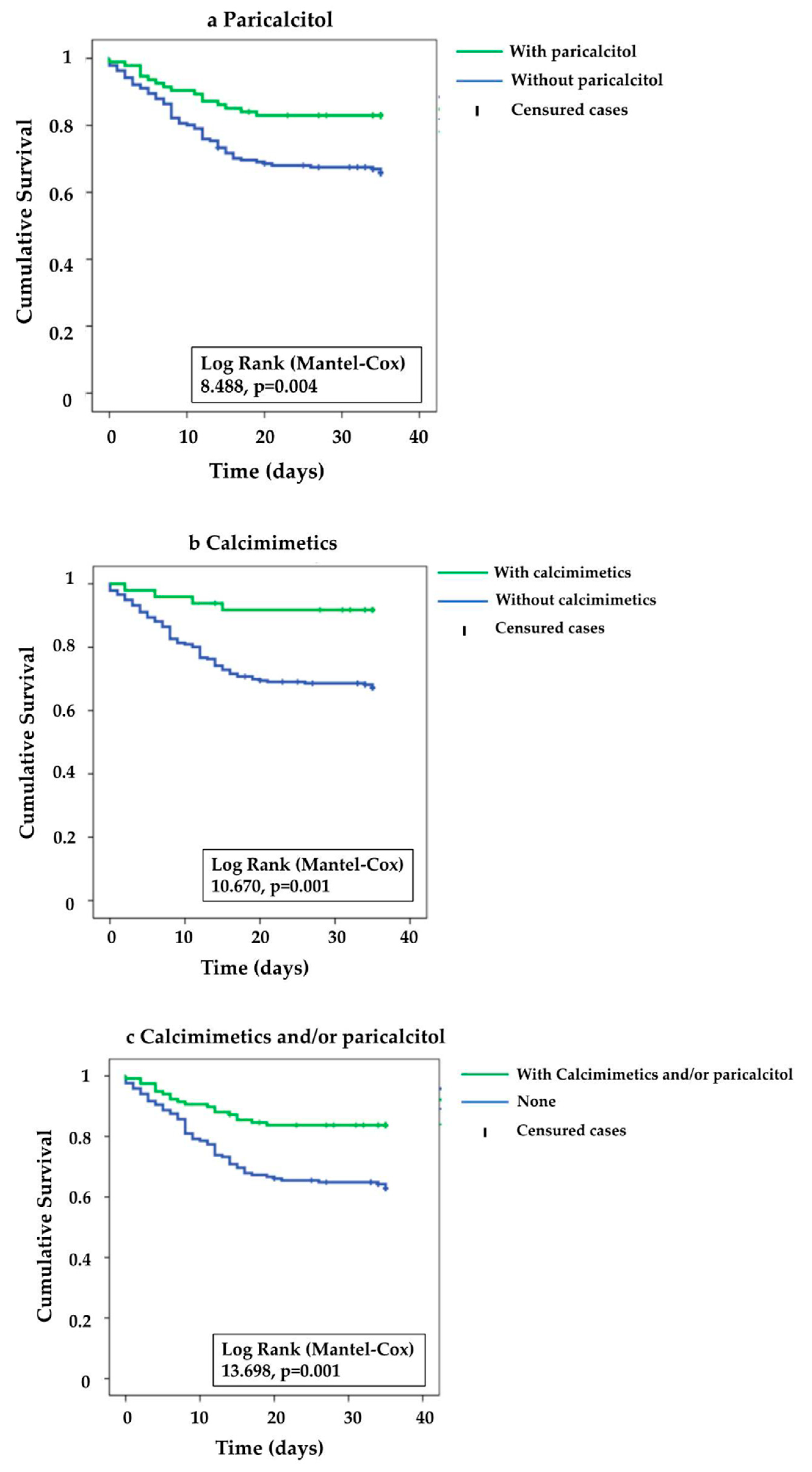
Data are presented as mean and standard deviation (SD) for normally distributed variables; median and IQR for non-normally distributed variables; and percentages (%) for categorical variables.
Univariant logistic regression analyses were used to evaluate the association between circulating VD levels and hospital admission or medical care requirements.
For survival analysis, we used Log-Rank Tests to compare Kaplan–Meier probability of death curves and performed univariate and multivariable Cox regression analyses. Based on the sample size and the number of events, variables significantly related with mortality in univariate analyses were prioritized and included in a multivariate Cox model to obtain the corresponding hazard ratios (HRs) and 95% confidence intervals (CIs). The “log minus log” (LML) plots were used to assess the proportional hazards assumption for categorical variables. For quantitative variables, we created interactions of predictors and a function of survival time to generate the time-dependent covariates. Variables and their time-dependent covariates were included in Cox models to assess the proportional hazards assumption.
Continuous predictors were categorized into quartiles and tertiles and included in multiple univariate models to assess whether linearity assumption was reasonable. Only variables with linear effects were included as continuous predictors. For predictors with non-linear effects, several cut-off values were chosen to generate balanced groups in a clinically relevant manner: age < 70, 70–80 and >80 years; vitamin D < 20, 20–30 and >30 ng/mL; C-reactive protein < 5.6, 5.6–24, >24 mg/L; leukocytes < 4500, 4500–7000 and >7000 n/mm3; lymphocytes < 700, 700–1200 and > 1200 n/mm3; ferritin < 400, 400–800 and > 800 µg/L; D-dimer <700, 700–1500 and >1500 ng/mL; LDH <200, 200–300 and >300 IU/L; and finally, procalcitonin < 0.5, 0.5–1 and >1 ng/mL. The Endocrine Society Clinical Practice Guideline on the Evaluation, Treatment and Prevention of Vitamin D Deficiency defined VD deficiency as a serum 25(OH)D level < 20 ng/mL and insufficiency between 20 and 29 ng/mL: a target of >30 ng/mL is suggested for optimal health [19,20].
All p-values were two-sided. Statistical significance was set at p < 0.05. Statistical analyses were performed using IBM® SPSS® Statistics version 19 (IBM Corp., Armonk, NY, USA).

3. Results

COVID-19 was diagnosed through a positive RT-PCR test for SARS-CoV-2 in 288 out of 4743 HD patients (6.1%). Demographic parameters, comorbidities and baseline treatments and analytics in COVID-19 HD patients are shown in Table 1. Median [p25–75] [percentile 25-75]serum 25-OH-VD (calcidiol) level was 17.1 [10.6–27.5], with 43.6% of patients with levels <20 ng/mL, 36.5% of patients within the 20–30 ng/mL range and only 19.9% with levels higher than >30 ng/mL. The outcomes, clinical profile and treatments of COVID-19 maintenance hemodialysis patients are presented in Table 2. By the end of the follow-up period, 28.4% (81/285) of patients had died. The most frequent symptoms were fever (67.9%), cough (52.4%) and dyspnea (32%). Only 12.6% of our patients were asymptomatic. Seventy-three per cent of patients with chest X-ray in our cohort presented with pneumonia (194/265), and 80% underwent hospital admission. Regarding specific treatments for COVID-19 patients (Table 2), hydroxychloroquine (73.9%), azithromycin (39.3%), lopinavir/ritonavir (8.7%), steroids (32.3%), tocilizumab (8.7%) and beta-interferon (5%), were the most frequently prescribed drugs. Ten percent of patients did not receive any specific treatment.

Serum VD Levels and Outcomes

No significant associations were observed between circulating 25-OH-VD levels and hospital or intensive care unit (ICU) admission, non-invasive/mechanical ventilation requirements (Supplementary Table S1) or mortality (Log Rank (Mantel-Cox) 0.341; p = 0.843).
Variables that were associated to mortality also included treatment of secondary hyperparathyroidism.
In order to elucidate the possible risk factors which could lead to a fatal progression in hemodialysis patients, we performed different univariate Cox regression analyses. An increase in age or in the serum levels of inflammatory markers (PCR, CK, D-dimer and procalcitonin), as well as the appearance of dyspnea or pneumonia, was significantly associated with a fatal outcome. However, other factors, such as PTH and albumin levels, BMI, and, interestingly, previous treatment with paricalcitol or calcimimetics, were associated with increase survival. (Table 3) (Figure 1). Within the group of patients treated with calcitriol, mortality was not different in patients with VD deficiency or insufficiency, compared to patients with VD sufficiency (Data not shown).
Mortality associated with COVID-19 was significantly lower in patients treated with any form of vitamin D (27/136, 19.9%), compared to those that were untreated (54/149; 36.2%) p: 0.002. Only patients treated with paricalcitol or calcimimetics showed significantly lower mortality, compared to those who received calcidiol or calcitriol (Table 3) (Supplementary Table S2).
Then, we compared possible differences between baseline characteristics of COVID-19 HD patients treated with no active vitamin D medication, paricalcitol, calcimimetics, paricalcitol or calcimimetics, to those who were not treated with either drug (Supplementary Tables S2–S6, respectively). Patients treated with paricalcitol and/or calcimimetics had a significantly higher serum PTH; while phosphate was significantly higher in patients treated with calcimimetics. Calcimimetics treated patients were younger and their dialysis vintage was lower than those who did not receive them. No significant differences between treated and non-treated patients were observed in hospital admission, non-invasive mechanical ventilation or endotracheal intubation. (Supplementary Tables S2–S6).
The multivariate Cox regression model displays some independent predictive factors for fatal outcomes: age 70–80 y.o. (HR 2.911; 95%CI 1.532–5.532), age > 80 y.o. (HR 2.372; 95%CI 1.195–4.707) and PCR > 24 mg/L (HR 2.417; 95%CI 1.284–4.55) were independent risk factors for mortality in COVID-19 HD patients. Cox regression analysis confirmed an increased survival in HD patients treated with paricalcitol (HR 0.499; 95%CI 0.278–0.898) and/or calcimimetics (HR 0.332; 95%CI 0.12–0.921) and with higher serum albumin levels (HR 0.668; 95%CI 0.496–0.9) (Table 4).

4. Discussion

Among the classical risk factors of COVID-19 associated mortality in the general population [3], only age was independently associated with mortality in our cohort of Spanish HD patients. Common biomarkers of severity and/or poor prognosis also markedly differed. Moreover, we did not find a significant negative association between serum baseline 25-OH-VD levels prior to the diagnosis of COVID-19 with mortality in our HD cohort. However, we did find a remarkable association with improved survival, not only with an active VD analog (paricalcitol) treatment, but also with the use of calcimimetics. Therefore, we describe that the impact of COVID-19 mortality risk prognostic markers is different in this specific population, and that the alleged and well described plausible benefits of VD pleiotropic effects for COVID-19 may be extended to other anti-hyperparathyroidism agents, such as calcimimetics.
The prevalence of confirmed COVID-19 in our cohort was 6.1%, lower than that described in other studies (range between 10–40%) [7,8,9]. There is a heterogeneous prevalence and mortality rate, according to different geographic areas, including within Spain [21]. The average number of patients undertaking HD with diabetes mellitus (DM) is around a 16–17% in the Spanish Registry. In the Spanish COVID-19 registry, 22% of COVID-19 patients had DM. The prevalence of DM in this cohort is higher than that reported for the whole country. It is important to emphasize that there are major differences in DM according to the geographic area of the country, and this particular HD cohort includes the Canary Islands, one of the areas with the highest incidence of DM in Spain [21]. Similar to other series, the most frequent symptoms observed were fever, cough and dyspnea [7,8,9,22]. Laboratory findings, incidence of pneumonia, hospital admission and mortality rates in our cohort are consistent with other published data on COVID-19 HD patients [7,8,9].
Phosphate (4.1 mg/dL), calcium (8.8 mg/dL) and PTH (240 pg/dL) levels within limit reflects an optimal control of MBD in this cohort. [17,18]. Patients treated with calcimimetics were younger and their dialysis vintage was lower than those who did not receive them. Dialysis vintage is a key determinant of the severity of secondary hyperparathyroidism. A direct relation between dialysis vintage and the severity of hyperparathyroidism has been demonstrated [23]. On the other hand, difficulties for the treatment of SHPT in clinical practice are frequently encountered, from the occasional inadequacy on dosage by the clinicians or the frequent non-compliance to the therapy by CKD patients. This is more frequent in younger patients, in which the use of calcimimetics could the best option, as hyperphostatemia is a limitation of the use of vitamin D analogs [24]. In fact, serum phosphate levels were higher in patients treated with calcimimetics.
VD deficiency is highly prevalent in patients on dialysis, and a low serum 25(OH)D level has been significantly associated with a higher risk of all-cause and cardiovascular mortality [25]. Some authors defend the hypothesis that low VD status is a consequence of poor health, rather than its cause [26]. Nevertheless, recent reviews hypothesized that VD insufficiency may compromise respiratory immune function, thereby increasing the risk of COVID-19 severity and mortality [27]. Some retrospective studies demonstrated a correlation between VD status and COVID-19 severity and mortality [28], while other studies did not find any correlation after the adjustment for confounding variables [29,30]. In our study, we did not find significant associations between circulating 25-OH-VD levels and outcomes in COVID-19 HD patients, including hospital admission, ICU admission, requirement of noninvasive mechanical ventilation, time from admission to resolution, and mortality, similar to that shown in other studies [28]. One potential explanation is the well-recognized population-dependent requirements of circulating 25(OH)D levels for the efficacious health benefits of non-classical vitamin D actions, including defeating COVID-19 infection and decreasing the risk of hospitalization. Indeed, while in the general population, vitamin D deficiency increases by 45% the risk of infection and by 95% the risk of hospitalization [31], circulating levels of calcidiol from 30 to 40 ng/mL cannot protect otherwise healthy African Americans from COVID-19 infection. Their risk of infection with these serum concentrations remains 2.45-fold higher than individuals with serum calcidiol >40 ng/mL [32]. Therefore, it is critical to personalize the levels of calcidiol in hemodialysis patients, to confer effective protection against COVID-19. Medical record keeping during the pandemic should help provide a first step in achieving an efficacious correction of vitamin D deficiency in Covid19 patients.
An effective attenuation of COVID-19 disease severity from adequate VD supplementation, or from treatment with VD receptor activators (calcitriol, paricalcitol) may be expected in HD patients. The arguments for this are the well-recognized VD anti-inflammatory, anti-viral, anti-hypertensive properties and its seemingly efficacious protection against respiratory infections [13], in addition to active VD efficaciously attenuating the progression of experimental renal or cardiovascular disease [30,33,34,35,36]. Moreover, a recent pilot study performed on non-CKD patients suggested that the administration of a high dose of calcifediol (25-OH-D3) reduced the need for intensive care treatments of COVID-19 patients requiring hospitalization [37].
An important finding in our study is the significantly lower COVID-19 mortality in patients treated with VD. Remarkably, only patients treated with paricalcitol but not calcitriol or calcifediol, showed a significantly lower mortality; however, the number of patients treated with calcitriol or calcifediol was small [11], and, therefore, we may lack sufficient power to discriminate between these two forms of active and moderately active endogenous VD metabolites.
From a mechanistic point of view, there are good reasons to postulate that VD may favorably modulate host responses to SARS-CoV-2, both in the early viraemic and later hyper-inflammatory phases of COVID-19. Regarding potential VD specific actions against COVID-19, calcitriol inhibits Skp2, the protein induced by SARS-COV2 to hijack Beclin1-driven autophagy, inhibits viral replication [38]. An additional and critical calcitriol action against COVID-19 is the induction of ACE2, which the downregulation by SARS-CoV-2 exacerbates the adverse impact on the lungs of the excessive inflammatory response, initiated by the high activity of the renin–angiotensin II system [39,40,41]. ACE2 converts angiotensin II into angiotensin 1–7, which binds its Mas receptor to initiate anti-fibrotic, anti-inflammatory signals that protect lung integrity by counteracting the deleterious effects of RAS activation [42]. The inhibition of ADAM17 expression and activity by VD receptor activators may contribute to attenuating ACE2 shedding, an ADAM17-driven cleavage; in addition, it also reduces the pro-inflammatory and pro-fibrotic signals downstream from angiotensin II-induced increases in ADAM17 activity at the cell surface of viral infected cells [43]. Pre-existing hypertension increases the risk of ICU admission, mechanical ventilation and death in patients with COVID-19. Calcitriol or paricalcitol simultaneous suppression of renin and induction of ACE2 may support a protective effect on lung injury, attenuating mortality. Paricalcitol pro-survival benefits in COVID-19 HD patients were dose independent and did not differ between VD deficient or sufficient individuals. In a nested case-control analysis in dialysis patients, low VD levels were associated with increased mortality, but a significant interaction was noted between VD levels, subsequent active VD therapy (mainly paricalcitol) and survival [42]. Patients treated during their first 9–12 months of hemodialysis with active forms of VD (mainly paricalcitol) had better survival, compared to those untreated, regardless of 25-OH-VD levels [44]. A potential survival advantage of paricalcitol over calcitriol in HD patients is also possible [45], but the mechanisms responsible for the different efficacy of these two VD receptor activators remain incompletely characterized.
Active VD targeting of ADAM17 [46] and consequently, the attenuation of ACE2-cleavage to maintain its protective pathways, may not be able to fully answer the questions regarding COVID-19 protection [47]. Unexpectedly, our results from univariate and multivariate models also suggest a protective effect of calcimimetic treatment on mortality in HD patients. The anti-hypertensive [48,49] and FGF-23 [46] suppressive properties of calcimimetic drugs may contribute to their survival advantage in COVID-19 patients. The benefits of attenuating RAS activation have been already discussed for VD and paricalcitol. On the other hand, extremely high levels of FGF-23 have been described in HD patients, and have been strongly associated with mortality [50]. Therefore, the proven efficacy of calcimimetics in reducing high FGF-23 levels could have played a role in attenuating either the cardiac damage [51], or FGF23-mediated suppression of macrophage calcitriol production [52] necessary for autocrine-paracrine anti-viral and anti-inflammatory actions by immune cells. It is possible that calcimimetics also mitigate the increases in macrophage TNFα, thereby attenuating the cytokine storm associated with COVID-19 [53], or preventing an impaired neutrophil activation to mount an appropriate anti-viral response [54]. Even though all of the above are well recognized deleterious actions of high FGF-23 levels, and treatment with calcimimetics efficaciously reduced serum FGF-23 in several studies, the absence of FGF-23 measurements in this study renders these proposed mechanisms as purely speculative. On the other hand, it is well known that VD increases FGF-23, but, at the same time, VD treatment attenuates cardiac FGF3/FGR4 signaling and hypertrophy in uremic rats [33].
The cross-sectional and retrospective nature of this analysis has limitations, such as the limited patient number, the lack of all analytical variables in some patients and the inherent associated selection bias and residual confounding, thus no conclusions on causality can be drawn. The absence of randomization led to some imbalances between groups; however, proper multivariant analysis, including the best-known variables to date, should have corrected these imbalances. Neither serum 25-OH-VD nor FGF-23 levels were obtained at the time of hospital admission, but our “baseline” values may offer a different perspective of the significance of VD deficiency, shortly before COVID-19 versus during active treatment when COVID-19 is ongoing. Moreover, we underline that other commonly used drugs in HD patients may also be of value. Among the strengths of our study, we describe novel associations of COVID-19 mortality in a significant cohort of HD patients. We did not only underline that clinical and biochemical classical mortality risk factors may differ from those described for the general population, but also that other antiparathyroid agents with pleiotropic effects beyond active forms of VD may offer some degree of protection. These findings may open new perspectives and novel pathways, which may improve the care of this complex and specific population.
In summary, no association was found between serum 25-OH-VD levels, prognosis and evolution of COVID-19 in our cohort of HD patients. Paricalcitol and/or calcimimetics was associated with improved survival in the complex COVID-19 HD patients. In fact, calcimimetics and paricalcitol have well known pleiotropic effects beyond mineral metabolism, that may be of value in the current scenario of extreme mortality in this population. This remains to be proven, and prospective studies and/or clinical trials are needed to validate and extend our observations.

Supplementary Materials

The following are available online at https://www.mdpi.com/article/10.3390/nu13082559/s1. Table S1. Univariant logistic regression analyses of hospital admission and care requirements according to serum VD levels in COVID-19 positive HD patients; Table S2. Baseline characteristics of patients treated with paricalcitol; Table S3. Baseline characteristics of patients treated with calcimimetics; Table S4. Baseline characteristics of patients treated with calcimimetics or paricalcitol; Table S5; Baseline characteristics of patients treated with paricalcitol and calcimimetics.

Author Contributions

Conceptualization: M.D.A.J.; Data curation: E.G.-P., M.R., A.L.-H., S.H., L.R., F.B., F.D., V.T., J.-J.B., J.F.N.-G., R.R.; Formal analysis: A.R.B., A.L.-H., C.S.M.; Writing the original draft: M.D.A.J. and J.P.; Writing, review & editing: E.G.-P., M.R., H.C., S.C., F.B., R.R., J.B., X.N.-S., M.C., A.S.D. All authors have read and agreed to the published version of the manuscript.

Funding

This research received no external funding.

Institutional Review Board Statement

Ethical review and approval were waived for this study, due to COVID19 pandemic and the retrospective nature of the data analysis.

Informed Consent Statement

Ethical review and approval were waived for this study, due to COVID19 pandemic and retrospective analysis of data.

Acknowledgments

We are indebted to all physicians and nurses taking care of these patients.

Conflicts of Interest

The authors declare no conflict of interest except M.D.A.J. and J.B. who declare receipt of advisory and/or lecture fees from Amgen, Sanofi-Genzyme, Shire, and Vifor-Fresenius-Pharma.

References

  1. Wu, J.T.; Leung, K.; Leung, G.M. Nowcasting and forecasting the potential domestic and international spread of the 2019-nCoV outbreak originating in Wuhan, China: A modelling study. Lancet 2020, 395, 689–697. [Google Scholar] [CrossRef] [Green Version]
  2. Cucinotta, D.; Vanelli, M. WHO Declares COVID-19 a Pandemic. Acta Biomed. 2020, 91, 157–160. [Google Scholar]
  3. Huang, C.; Wang, Y.; Li, X.; Ren, L.; Zhao, J.; Hu, Y.; Zhang, L.; Fan, G.; Xu, J.; Gu, X.; et al. Clinical features of patients infected with 2019 novel coronavirus in Wuhan, China. Lancet 2020, 395, 497–506. [Google Scholar] [CrossRef] [Green Version]
  4. Peiris, J.S.; Guan, Y.; Yuen, K.Y. Severe acute respiratory syndrome. Nat. Med. 2004, 10 (Suppl. 12), S88–S97. [Google Scholar] [CrossRef] [PubMed]
  5. Zhou, F.; Yu, T.; Du, R.; Fan, G.; Liu, Y.; Liu, Z.; Xiang, J.; Wang, Y.; Song, B.; Gu, X.; et al. Clinical course and risk factors for mortality of adult inpatients with COVID-19 in Wuhan, China: A retrospective cohort study. Lancet 2020, 395, 1054–1062. [Google Scholar] [CrossRef]
  6. McGonagle, D.; Sharif, K.; O’Regan, A.; Bridgewood, C. The Role of Cytokines including Interleukin-6 in COVID-19 induced Pneumonia and Macrophage Activation Syndrome-Like Disease. Autoimmun. Rev. 2020, 19, 102537. [Google Scholar] [CrossRef]
  7. Zheng, Z.; Peng, F.; Xu, B.; Zhao, J.; Liu, H.; Peng, J.; Li, Q.; Jiang, C.; Zhou, Y.; Liu, S.; et al. Risk factors of critical & mortal COVID-19 cases: A systematic literature review and meta-analysis. J. Infect. 2020, 81, e16–e25. [Google Scholar] [CrossRef]
  8. Arenas, M.D.; Crespo, M.; Pérez-Sáez, M.J.; Collado, S.; Redondo-Pachón, D.; Llinàs-Mallol, L.; Montero, M.M.; Villar-García, J.; Arias-Cabrales, C.; Barbosa, F.; et al. Clinical Profiles in Renal Patients with COVID-19. J. Clin. Med. 2020, 9, 2665. [Google Scholar] [CrossRef]
  9. Goicoechea, M.; Cámara, L.A.S.; Macías, N.; de Morales, A.M.; Rojas, Á.G.; Bascuñana, A.; Arroyo, D.; Vega, A.; Abad, S.; Verde, E.; et al. COVID-19: Clinical course and outcomes of 36 hemodialysis patients in Spain. Kidney Int. 2020, 98, 27–34. [Google Scholar] [CrossRef]
  10. Rincón, A.; Moreso, F.; López-Herradón, A.; Fernández-Robres, M.A.; Cidraque, I.; Nin, J.; Méndez, O.; López, M.; Pájaro, C.; Satorra, À.; et al. The keys to control a COVID-19 outbreak in a haemodialysis unit. Clin. Kidney J. 2020, 13, 542–549. [Google Scholar] [CrossRef]
  11. Carpagnano, G.E.; Di Lecce, V.; Quaranta, V.N.; Zito, A.; Buonamico, E.; Capozza, E.; Palumbo, A.; Di Gioia, G.; Valerio, V.N.; Resta, O. Vitamin D deficiency as a predictor of poor prognosis in patients with acute respiratory failure due to COVID-19. J. Endocrinol. Investig. 2021, 44, 765–771. [Google Scholar] [CrossRef] [PubMed]
  12. Dancer, R.C.A.; Parekh, D.; Lax, S.; D’Souza, V.; Zheng, S.; Bassford, C.R.; Park, D.; Bartis, D.G.; Mahida, R.; Turner, A.M.; et al. Vitamin D deficiency contributes directly to the acute respiratory distress syndrome (ARDS). Thorax 2015, 70, 617–624. [Google Scholar] [CrossRef] [Green Version]
  13. Martineau, A.R.; Jolliffe, D.A.; Hooper, R.L.; Greenberg, L.; Aloia, J.F.; Bergman, P.; Dubnov-Raz, G.; Esposito, S.; Ganmaa, D.; Ginde, A.A.; et al. Vitamin D supplementation to prevent acute respiratory tract infections: Systematic review and meta-analysis of individual participant data. BMJ 2017, 356, i6583. [Google Scholar] [CrossRef] [Green Version]
  14. Jolliffe, D.A.; Stefanidis, C.; Wang, Z.; Kermani, N.Z.; Dimitrov, V.; White, J.H.; McDonough, J.; Janssens, W.; Pfeffer, P.; Griffiths, C.J.; et al. Vitamin D Metabolism Is Dysregulated in Asthma and Chronic Obstructive Pulmonary Disease. Am. J. Respir. Crit. Care Med. 2020, 202, 371–382. [Google Scholar] [CrossRef] [PubMed]
  15. Pilz, S.; Zittermann, A.; Trummer, C.; Theiler-Schwetz, V.; Lerchbaum, E.; Keppel, M.H.; Grübler, M.R.; März, W.; Pandis, M. Vitamin D testing and treatment: A narrative review of current evidence. Endocr. Connect. 2019, 8, 27–43. [Google Scholar] [CrossRef] [PubMed] [Green Version]
  16. Zhang, H.; Penninger, J.M.; Li, Y.; Zhong, N.; Slutsky, A.S. Angiotensin-converting enzyme 2 (ACE2) as a SARS-CoV-2 receptor: Molecular mechanisms and potential therapeutic target. Intensive Care Med. 2020, 46, 586–590. [Google Scholar] [CrossRef] [Green Version]
  17. Kidney Disease: Improving Global Outcomes (KDIGO) CKD-MBD Update Work Group. KDIGO 2017 Clinical Practice Guideline Update for the Diagnosis, Evaluation, Prevention, and Treatment of Chronic Kidney Disease-Mineral and Bone Disorder (CKD-MBD). Kidney Int. Suppl. 2017, 7, 1–59. [Google Scholar] [CrossRef] [Green Version]
  18. Torregrosa, J.V.; Bover, J.; Andía, J.C.; Lorenzo, V.; de Francisco, A.L.M.; Martínez, I.; Portillo, M.R.; Arenas, L.; Parra, E.G.; Caravaca, F.; et al. Recomendaciones de la Sociedad Española de Nefrología para el manejo de las alteraciones del metabolismo óseo-mineral en los pacientes con enfermedad renal crónica (S.E.N.-MM). Nefrologia 2011, 31 (Suppl. 1), 3–32. [Google Scholar] [PubMed]
  19. Holick, M.F.; Binkley, N.C.; Bischoff-Ferrari, H.A.; Gordon, C.M.; Hanley, D.A.; Heaney, R.P.; Murad, M.H.; Weaver, C.M. Evaluation, treatment, and prevention of vitamin D deficiency: An Endocrine Society clinical practice guideline. J. Clin. Endocrinol. Metab. 2011, 96, 1911–1930. [Google Scholar] [CrossRef] [Green Version]
  20. Jean, G.; Souberbielle, J.C.; Chazot, C. Vitamin D in Chronic Kidney Disease and Dialysis Patients. Nutrients 2017, 9, 328. [Google Scholar] [CrossRef]
  21. Sánchez-Álvarez, J.E.; Pérez Fontán, M.; Jiménez Martín, C.; Pelícano, M.B.; Reina, C.J.C.; Prieto, Á.M.S.; Melilli, E.; Barrios, M.C.; Heras, M.M.; del Pino y Pino, M.D. SARS-CoV-2 infection in patients on renal replacement therapy. Report of the COVID-19 Registry of the Spanish Society of Nephrology (SEN). Nefrologia 2020, 40, 272–278. [Google Scholar] [CrossRef] [PubMed]
  22. Rodriguez-Morales, A.J.; Cardona-Ospina, J.A.; Gutiérrez-Ocampo, E.; Villamizar-Peña, R.; Holguin-Rivera, Y.; Antezana, J.P.E.; Alvarado-Arnez, L.E.; Bonilla-Aldana, D.K.; Franco-Paredes, C.; Henao-Martínez, A.; et al. Clinical, laboratory and imaging features of COVID-19: A systematic review and meta-analysis. Travel Med. Infect. Dis. 2020, 34, 101623. [Google Scholar] [CrossRef] [PubMed]
  23. Chertow, G.M.; Plone, M.; Dillon, M.A.; Burke, S.K.; Slatopolsky, E. Hyperparathyroidism and dialysis vintage. Clin. Nephrol. 2000, 54, 295–300. [Google Scholar]
  24. Alfieri, C.; Regalia, A.; Zanoni, F.; Vettoretti, S.; Cozzolino, M.; Messa, P. The Importance of Adherence in the Treatment of Secondary Hyperparathyroidism. Blood Purif. 2019, 47, 37–44. [Google Scholar] [CrossRef] [PubMed]
  25. Zhang, Y.; Darssan, D.; Pascoe, E.M.; Johnson, D.W.; Pi, H.; Dong, J. Vitamin D status and mortality risk among patients on dialysis: A systematic review and meta-analysis of observational studies. Nephrol. Dial. Transplant. 2018, 33, 1742–1751. [Google Scholar] [CrossRef] [PubMed]
  26. Autier, P.; Mullie, P.; Macacu, A.; Dragomir, M.; Boniol, M.; Coppens, K.; Pizot, C.; Boniol, M. Effect of vitamin D supplementation on non-skeletal disorders: A systematic review of meta-analyses and randomised trials. Lancet Diabetes Endocrinol. 2017, 5, 986–1004. [Google Scholar] [CrossRef]
  27. Grant, W.B.; Lahore, H.; McDonnell, S.L.; Baggerly, C.A.; French, C.B.; Aliano, J.L.; Bhattoa, H.P. Evidence that Vitamin D Supplementation Could Reduce Risk of Influenza and COVID-19 Infections and Deaths. Nutrients 2020, 12, 988. [Google Scholar] [CrossRef] [Green Version]
  28. Ilie, P.C.; Stefanescu, S.; Smith, L. The role of vitamin D in the prevention of coronavirus disease 2019 infection and mortality. Aging Clin. Exp. Res. 2020, 32, 1195–1198. [Google Scholar] [CrossRef]
  29. Ali, N. Role of vitamin D in preventing of COVID-19 infection, progression and severity. J. Infect. Public Health 2020, 13, 1373–1380. [Google Scholar] [CrossRef]
  30. Hastie, C.E.; Mackay, D.F.; Ho, F.; Celis-Morales, C.A.; Katikireddi, S.V.; Niedzwiedz, C.L.; Jani, B.D.; Welsh, P.; Mair, F.S.; Gray, S.R.; et al. Vitamin D concentrations and COVID-19 infection in UK Biobank. Diabetes Metab. Syndr. 2020, 14, 561–565. [Google Scholar] [CrossRef]
  31. Merzon, E.; Tworowski, D.; Gorohovski, A.; Vinker, S.; Cohen, A.G.; Green, I.; Frenkel-Morgenstern, M. Low plasma 25(OH) vitamin D level is associated with increased risk of COVID-19 infection: An Israeli population-based study. FEBS J. 2020, 287, 3693–3702. [Google Scholar] [CrossRef] [PubMed]
  32. Meltzer, D.O.; Best, T.J.; Zhang, H.; Vokes, T.; Arora, V.M.; Solway, J. Association of Vitamin D Levels, Race/Ethnicity, and Clinical Characteristics With COVID-19 Test Results. JAMA Netw. Open 2021, 4, e214117. [Google Scholar] [CrossRef]
  33. Guerrero, F.; De Oca, A.M.; Aguilera-Tejero, E.; Zafra, R.; Rodriguez, M.; López, I. The effect of vitamin D derivatives on vascular calcification associated with inflammation. Nephrol. Dial. Transplant. 2012, 27, 2206–2212. [Google Scholar] [CrossRef] [PubMed] [Green Version]
  34. Riera, M.; Anguiano, L.; Clotet, S.; Ho, H.R.; Rebull, M.; Pascual, J.; Soler, M.J. Paricalcitol modulates ACE2 shedding and renal ADAM17 in NOD mice beyond proteinuria. Am. J. Physiol. Renal Physiol. 2016, 310, F534–F546. [Google Scholar] [CrossRef] [Green Version]
  35. Leifheit-Nestler, M.; Grabner, A.; Hermann, L.; Richter, B.; Schmitz, K.; Fischer, D.-C.; Yanucil, C.; Faul, C.; Haffner, D. Vitamin D treatment attenuates cardiac FGF23/FGFR4 signaling and hypertrophy in uremic rats. Nephrol. Dial. Transplant. 2017, 32, 1493–1503. [Google Scholar] [CrossRef]
  36. Panizo, S.; Carrillo-López, N.; Naves-Díaz, M.; Solache-Berrocal, G.; Martínez-Arias, L.; Rodrigues-Díez, R.R.; Fernández-Vázquez, A.; Martínez-Salgado, C.; Ruiz-Ortega, M.; Dusso, A.; et al. Regulation of miR-29b and miR-30c by vitamin D receptor activators contributes to attenuate uraemia-induced cardiac fibrosis. Nephrol. Dial. Transplant. 2017, 32, 1831–1840. [Google Scholar] [CrossRef] [PubMed] [Green Version]
  37. Castillo, M.E.; Costa, L.M.E.; Barrios, J.M.V.; Díaz, J.F.A.; Miranda, J.L.; Bouillon, R.; Gomez, J.M.Q. Effect of calcifediol treatment and best available therapy versus best available therapy on intensive care unit admission and mortality among patients hospitalized for COVID-19: A pilot randomized clinical study. J. Steroid Biochem. Mol. Biol. 2020, 203, 105751. [Google Scholar] [CrossRef]
  38. Gassen, N.C.; Niemeyer, D.; Muth, D.; Corman, V.M.; Martinelli, S.; Gassen, A.; Hafner, K.; Papies, J.; Mösbauer, K.; Zellner, A.; et al. SKP2 attenuates autophagy through Beclin1-ubiquitination and its inhibition reduces MERS-Coronavirus infection. Nat. Commun. 2019, 10, 5770. [Google Scholar] [CrossRef] [PubMed]
  39. Xu, J.; Yang, J.; Chen, J.; Luo, Q.; Zhang, Q.; Zhang, H. Vitamin D alleviates lipopolysaccharide-induced acute lung injury via regulation of the renin-angiotensin system. Mol. Med. Rep. 2017, 16, 7432–7438. [Google Scholar] [CrossRef] [Green Version]
  40. Cui, C.; Xu, P.; Li, G.; Qiao, Y.; Han, W.; Geng, C.; Liao, D.; Yang, M.; Chen, D.; Jiang, P. Vitamin D receptor activation regulates microglia polarization and oxidative stress in spontaneously hypertensive rats and angiotensin II-exposed microglial cells: Role of renin-angiotensin system. Redox Biol. 2019, 26, 101295. [Google Scholar] [CrossRef] [PubMed]
  41. Dijkman, R.; Jebbink, M.F.; Deijs, M.; Milewska, A.; Pyrc, K.; Buelow, E.; Van Der Bijl, A.; Van Der Hoek, L. Replication-dependent downregulation of cellular angiotensin-converting enzyme 2 protein expression by human coronavirus NL63. J. Gen. Virol. 2012, 93 Pt 9, 1924–1929. [Google Scholar] [CrossRef]
  42. Sodhi, C.P.; Wohlford-Lenane, C.; Yamaguchi, Y.; Prindle, T.; Fulton, W.B.; Wang, S.; McCray, P.B., Jr.; Chappell, M.; Hackam, D.J.; Jia, H.; et al. Attenuation of pulmonary ACE2 activity impairs inactivation of des-Arg9 bradykinin/BKB1R axis and facilitates LPS-induced neutrophil infiltration. Am. J. Physiol. Lung Cell. Mol. Physiol. 2018, 314, L17–L31. [Google Scholar] [CrossRef]
  43. Patel, V.; Clarke, N.; Wang, Z.; Fan, D.; Parajuli, N.; Basu, R.; Putko, B.; Kassiri, Z.; Turner, A.J.; Oudit, G.Y. Angiotensin II induced proteolytic cleavage of myocardial ACE2 is mediated by TACE/ADAM-17: A positive feedback mechanism in the RAS. J. Mol. Cell. Cardiol. 2014, 66, 167–176. [Google Scholar] [CrossRef]
  44. Wolf, M.; Shah, A.; Gutierrez, O.; Ankers, E.; Monroy, M.; Tamez, H.; Steele, D.; Chang, Y.; Camargo, C.; Tonelli, M.; et al. Vitamin D levels and early mortality among incident hemodialysis patients. Kidney Int. 2007, 72, 1004–1013. [Google Scholar] [CrossRef] [Green Version]
  45. Teng, M.; Wolf, M.; Lowrie, E.; Ofsthun, N.; Lazarus, J.M.; Thadhani, R. Survival of Patients Undergoing Hemodialysis with Paricalcitol or Calcitriol Therapy. N. Engl. J. Med. 2003, 349, 446–456. [Google Scholar] [CrossRef] [PubMed]
  46. Arcidiacono, M.V.; Yang, J.; Fernandez, E.; Dusso, A. The induction of C/EBPβ contributes to vitamin D inhibition of ADAM17 expression and parathyroid hyperplasia in kidney disease. Nephrol. Dial. Transplant. 2014, 30, 423–433. [Google Scholar] [CrossRef] [PubMed] [Green Version]
  47. Palau, V.; Riera, M.; Soler, M.J. ADAM17 inhibition may exert a protective effect on COVID-19. Nephrol. Dial. Transplant. 2020, 35, 1071–1072. [Google Scholar] [CrossRef] [PubMed]
  48. Ostroverkhova, D.S.; Hu, J.; Tarasov, V.V.; Melnikova, T.I.; Porozov, Y.B.; Mutig, K. Calcium-Sensing Receptor and Regulation of WNK Kinases in the Kidney. Cells 2020, 9, 1644. [Google Scholar] [CrossRef]
  49. Atchison, D.K.; Ortiz-Capisano, M.C.; Beierwaltes, W.H. Acute activation of the calcium-sensing receptor inhibits plasma renin activity in vivo. Am. J. Physiol. Regul. Integr. Comp. Physiol. 2010, 299, R1020–R1026. [Google Scholar] [CrossRef] [PubMed] [Green Version]
  50. Moe, S.M.; Chertow, G.M.; Parfrey, P.S.; Kubo, Y.; Block, G.A.; Correa-Rotter, R.; Drüeke, T.B.; Herzog, C.A.; London, G.M.; Mahaffey, K.W.; et al. Cinacalcet, Fibroblast Growth Factor-23, and Cardiovascular Disease in Hemodialysis: The Evaluation of Cinacalcet HCl Therapy to Lower Cardiovascular Events (EVOLVE) Trial. Circulation 2015, 132, 27–39. [Google Scholar] [CrossRef] [Green Version]
  51. Isakova, T.; Wahl, P.; Vargas, G.S.; Gutiérrez, O.M.; Scialla, J.; Xie, H.; Appleby, D.; Nessel, L.; Bellovich, K.; Chen, J.; et al. Fibroblast growth factor 23 is elevated before parathyroid hormone and phosphate in chronic kidney disease. Kidney Int. 2011, 79, 1370–1378. [Google Scholar] [CrossRef] [PubMed] [Green Version]
  52. Han, X.; Li, L.; Yang, J.; King, G.; Xiao, Z.; Quarles, L.D. Counter-regulatory paracrine actions of FGF-23 and 1,25(OH)2D in macrophages. FEBS Lett. 2016, 590, 53–67. [Google Scholar] [CrossRef] [PubMed] [Green Version]
  53. Mehta, P.; McAuley, D.; Brown, M.; Sanchez, E.; Tattersall, R.S.; Manson, J.J. COVID-19: Consider cytokine storm syndromes and immunosuppression. Lancet 2020, 395, 1033–1034. [Google Scholar] [CrossRef]
  54. Rossaint, J.; Oehmichen, J.; Van Aken, H.; Reuter, S.; Pavenstädt, H.J.; Meersch, M.; Unruh, M.; Zarbock, A. FGF23 signaling impairs neutrophil recruitment and host defense during CKD. J. Clin. Investig. 2016, 126, 962–974. [Google Scholar] [CrossRef] [Green Version]
Figure 1. COVID-19 survival in HD patients according to their previous use of anti-secondary hyperparathyroidism agents. (a). With paricalcitol N:94, without paricalcitol N:194; (b). With calcimimetics N: 49, without calcimimetics N: 239; (c). With calcimimetics and/or paricalcitol N: 117, without calcimimetics and/or Paricalcitol N: 171; (d). With calcimimetics and/or paricalcitol N: 171, calcimimetics N: 49 and paricalcitol N:94.
Nutrients 13 02559 g001aNutrients 13 02559 g001b
Table 1. Baseline characteristics of COVID-19 maintenance hemodialysis patients (N: 288).
Demography
Age (years, mean ± SD)72.4 ± 12.6
Body mass index (kg/m2, mean ± SD)25.6 ± 5.5
Time on Hemodialysis (years, median [IQR])2.5 [1.2–5.2]
Sex
Female84 (29.2)
Race
Caucasian208 (72.2)
Other11 (3.8)
Unknown69 (23.9)
Vascular access (N: 261) **
Arteriovenous fistulae154 (59)
Central venous catheter107 (41)
Cause of renal failure
Diabetic Nephropathy85 (29.5)
Vascular37 (12.8)
Glomerular34 (11.8)
Hereditary18 (6.2)
Tubulointerstitial 15 (5.2)
Unknown78 (27.1)
Other21 (7.2)
Toxic habits (N: 117) **
Current smoking102 (49)
Alcohol abuse17 (9.6)
Risk factors and comorbidities (N: 288)
Diabetes mellitus137 (47.6)
Hypertension129 (44.8)
Dyslipemia106 (36.8)
Ischemic heart disease94 (32.6)
Lung disease66 (22.9)
Previous trasplantation20 (6.9)
Tuberculosis5 (1.7)
Previous treatment for secondary hyperparathyroidism (N: 288)
No treatment 128 (44.4)
Any treatment (Vitamin D and/or calcimetics) 160 (55.5)
Any calcimimetic 49 (17.0)
Cinacalcet33 (11.5)
Etelcalcetide16 (5.6)
No vitamin D treatments 151 (52.4)
Any vitamin D treatments 137 (47.6)
Calcifediol (25-OH-Vitamin D)33 (11.5)
Calcitriol22 (7.6)
Paricalcitol94 (32.6)
Two kind of Vitamin D 12(4.2)
One kind of vitamin D125 (43.4)
Calcimimetics or paricalcitol 117 (40.6)
Both calcimimetics and paricalcitol 26 (9.0)
Calcidiol dose (mcg/month, median, [IQR])399 [399–798]
Calcitriol dose (mcg/week median, [IQR])1.75 [0.99–1.83]
Paricalcitol dose (mcg/week median, [IQR])4.10 [2.25–5.60]
Other concomitant medication
ACEIs63 (21.9)
ARBs32 (11.1)
NSAIDs21 (7.3)
Baseline laboratory data ***
Time from baseline Blood test and PCR positive (days, median [p25–75])20.0 [12.5–25]
Hemoglobin (gr/dL, mean (SD))11.0 (1.3)
White blood cells (×10 3/uL, median [p25–75])6.4 (5–8.3)
Lymphocytes (×10 3/uL, median [p25–75])1.07 (0.76–1.4)
Neutrophils (×10 3/uL, median [p25–75])4.26 (3.2–5.5)
Platelets (×10 3/uL, median [p25–75])190 (146–227)
Fibrinogen (UI/L, mean (SD))433 (139)
AST (UI/L. median [p25–75])16 (12–20)
Albumin (g/dL. mean (SD))3.8 (0.3)
C-reactive protein (mg/L, median [p25–75])7.9 (3.1–19.4)
Ferritin (mcg/mL. median [p25–75])436 (265–586)
Transferrin Saturation Index (%. median [p25–75])26 (17.8–36)
Calcium (mg/dL. mean (SD))8.8 (0.7)
Phosphate (mg/dL, mean (SD))4.1 (1.1)
PTH (pg/dL, mean (SD))240 (133–398)
Hemoglobin (gr/dL, mean (SD))10.9 (1.6)
White blood cells (×10 3/uL, median [p25–75])5.5 (4.2–7.7)
Lymphocytes (×10 3/uL, median [p25–75])0.70 (0.4–1.0)
Neutrophils (×10 3/uL, median [p25–75])4.0 (2.8–5.6)
Platelets (×10 3/uL, median [p25–75])166 (120–204)
D-dimer (mcg/L, median [p25–75])982 (580–2000)
Fibrinogen (UI/L, mean (SD))593 (194)
AST (UI/L, median [p25–75])21 (16–28)
LDH (UI/L, mean (SD))353 (664)
IL6 (ng/L, median [p25–75])18 (8.9–59.9)
Albumin (g/dL, mean (SD))3.3 (0.5)
C-reactive protein (mg/L, median [p25–75])14.7 (4.3–61)
Ferritin (mcg/mL, median [p25–75])1000 (547–1566)
Transferrin Saturation Index (%, median [p25–75])20 (13.8–31)
Calcium (mg/dL, mean (SD))8.6 (0.7)
Phosphate (mg/dL, mean (SD)4.6 (1.5)
Data are expressed as n (%) or otherwise specified. SD: standard deviation; IQR: interquartile range; [p25–75]: percentile 25 and 75. ACEIs: angiotensin converting enzyme inhibitors; ARBs; anti-AT1 receptor antagonists, NSAIDs: non-steroidal anti-inflammatory agents. ** Variables without 100% of data. *** Baseline laboratory data: regular control tests performed immediately prior to the time of COVID-19 diagnosis (median 20 days, interquartile range–IQR-12.5–25 days).
Table 2. Outcomes and clinical profiles of COVID-19 maintenance hemodialysis patients (N: 285).
Hospital admission N, %
Admission required (N: 285)228 (80)
ICU admission (N: 257)15 (5.8)
Mechanical ventilation (N: 254)10 (3.9)
Outcome
Resolution and/or discharged (N: 285)169 (59.2)
Exitus (N: 285)81 (28.4)
Admission to discharge (time elapsed in days, median [p25–75])16.0 [10.8–28.0]
Admission to exitus (time elapsed in days days, median [p25–75])8.0 [ 4.7–14.2]
Approached COVID-19+ contact
Nursing home or relatives (N: 285)52 (18.2)
Symptoms
Asymptomatic (N: 276)35 (12.6)
Fever (N: 265)180 (67.9)
Cough (N: 265)139 (52.4)
Dyspnea (N: 265)85(32.07)
Other (sore throat, discomfort, muscle pain, fatigue) (N: 265)66 (24.9)
Diarrhea (N: 265)28 (10.5)
Ageusia (N: 93)4 (4.2)
Anosmia (N: 93)5 (5.2)
Lung disease (N: 265)
Pneumonia194 (73.2)
Covid-19 treatment
Hydroxychloroquine (N: 238)176 (73.9)
Azithromycin (N: 239)94 (39.3)
Lopinavir/Ritonavir (N: 239)21(8.7)
Steroids (N:284)92 (32.3)
Tocilizumab (N: 239)21 (8.7)
Beta-Interferon (N: 239)12 (5)
Remdesevir (N: 239)1 (0.4)
Other (Antibiotics.anticoagulants..etc) (N: 239)137 (57.3)
No treatment (N: 239)24 (10)
Data are expressed as n (%) or as otherwise specified.
Table 3. Clinical characteristics and associations with mortality in univariate Cox regression analyses.
HR [CI 95%]p
Demographics and Comorbidities
Age (years) < 70 (Ref.)
70–802.889 [1.569–5.321]0.001
>802.817 [1.529–5.187]0.001
Gender (female)1.195 [0.749–1.905]0.455
Race (caucasian)0.957 [0.738–1.241]0.742
Body mass index (kg/m2)0.929 [0.882–0.979]0.006
Dialysis vintage (years)1.011 [0.974–1.050]0.563
Renal etiology0.983 [0.908–1.065]0.682
Current Alcohol status0.347 [0.084–1.429]0.143
Current Smoking status1.019 [0.617–1.683]0.941
Native fistulae as vascular access0.679 [0.428–1.078]0.101
Previous transplant0.915 [0.369–2.271]0.848
Diabetes Mellitus1.080 [0.698–1.670]0.730
Arterial hypertension0.974 [0.629–1.510]0.908
Tuberculosis0.649 [0.090–4.672]0.668
Dyslipemia0.839 [0.529–1.332]0.456
Lung disease1.537 [0.959–2.463]0.074
Ischemic cardiopahty 1.181 [0.750–1.859]0.474
Mineral Metabolism
25-OH-Vitamin D. (ng/mL)0.994 [0.975–1.012]0.491
25-OH Vitamin D (ng/mL)
<20 (Ref.)1
20–301.185 [0.663–2.115]0.567
>301.077 [0.586–1.979]0.811
PTH (ng/L)0.998 [0.997–0.999]0.002
Calcium (mg/dL)0.875 [0.765–1.000]0.051
Phosphate (mg/dL)0.879 [0.758–1.020]0.089
Vitamin D derivatives use0.493 [0.310–0.782]0.003
Calcidiol use1.140 [0.591–2.222]0.686
Calcidiol dose (mcg/month)0.999 [0.999–1.000]0.653
Calcitriol use0.587 [0.215–1.605]0.260
Calcitriol dose (mcg/week)0.489 [0.110–2.175]0.348
Paricalcitol use0.455 [0.263 -0.787]0.002
Paricalcitol dose (mcg/week)0.915 [0.749–1.118]0.385
Calcimimetics use0.220 [0.080–0.601]0.003
Other Treatments
ACEIs0.903 [0.523–1.560]0.715
ARBs1.050 [0.525–2.099]0.891
NSAIDs0.821 [0.332–2.029]0.669
COVID-19 Clinical symptoms
Ageusia2.112 [0.499–8.945]0.310
Anosmia1.565 [0.370–6.625]0.543
Cough0.813 [0.515–1.283]0.374
Fever1.665 [0.998–2.779]0.051
Diarrhea0.832 [0.382–1.810]0.642
Dyspnea1.755 [1.110–2.774]0.016
Asthenia1.867 [1.165–2.993]0.009
Asymptomatic 0.259 [0.082–0.821]0.022
Pneumonia4.705 [2.037–10.867]<0.001
X-Ray compatible with COVID-195.629 [1.735–18.265]0.004
Pre-hospital admission for COVID-19–Laboratory findings
Hemoglobin (g/dL)0.937 [0.842–1.043]0.617
White blood cells (n/mm3) .
<4500 (Ref.)1
4500–70000.751 [0.443–1.272]0.287
>70000.797 [0.458–1.388]0.423
Lymphocytes (n/mm3), .
<700 (Ref.)1
700–12000.832 [0.507–1.367]0.468
>12000.634 [0.357–1.125]0.120
Platelets (n/mm3) (×100)1.000 [0.998–1.003]0.927
Neutrophils (n/mm3) (×100)0.988 [0.912–1.071]0.770
Albumin (g/dL)0.762 [0.630–0.923]0.005
C-reactive protein (mg/L)
<5.6 (Ref.)1
5.6–242.492 [1.349–4.602]0.004
>242.746 [1.495–5.043]0.001
D-dimer (ng/mL) .
<700 (Ref.)1
700–15001.02 [0.433–2.403]0.963
>15002.175 [1.049–4.513]0.037
Fibrinogen (mg/dL) .
<400 (Ref.)
400–6001.37 [0.613–3.063]0.444
>6001.192 [0.51–2.786]0.685
AST (IU/L)0.999 [0.995–1.003]0.678
Alanin aminotransferase (ALT) (IU/L)0.998 [0.988–1.009]0.681
GGT (IU/L)0.997 [0.992–1.002]0.870
LDH (IU/L) .
<200 (Ref.)1
200–3001.998 [ 0.922–4.331]0.079
>3001.798 [0.813–3.976]0.147
Troponin T (ng/mL)0.996 [0.982–1.01]0.336
Procalcitonin (ng/mL) .
<0.5 (Ref.)1
0.5–11.207 [0.479–3.04]0.690
>12.48 [1.085–5.672]0.031
IL-6 (ng/L)0.998 [0.988–1.008]0.744
Ferritin (μg/L) .
<400 (Ref.)1
400–8000.321 [1.092–0]0.093
>8001.218 [0.731–2.029]0.449
Transferrin Saturation Index (%)0.999 [0.984–1.015]0.958
COVID-19–Outcomes
Hospital admission5.295 [1.937–14.470]0.001
Oxygen saturation0.857 [0.809–0.908]<0.001
Non-invasive mechanical ventilation2.177 [1.342–3.529]0.002
ICU admission1.560 [0.718–3.392]0.262
Endotracheal intubation1.727 [0.697–4.276]0.238
COVID-19–Treatments
Hydroxychloroquine1.251 [0.692–2.263]0.459
Azithromycin1.268 [0.775–2.075]0.344
Lopinavir/Ritonavir1.938 [1.185–3.167]0.008
Steroids1.162 [0.738–1.831]0.516
Beta-Interferon3.663 [1.738–7.719]0.001
Tocilizumab0.872 [0.350–2.174]0.769
Antibiotics1.402 [0.841–2.336]0.195
No Treatment 0.599 [0.218–1.647]0.320
Expressed as percentages, mean ± SD or median [IQR p25–75]. SD: standard deviation; IQR: interquartile range; ACEI: angiotensin converting enzyme inhibitors; ARB; anti-AT1 receptor antagonists, NSAIDs: non-steroidal anti-inflammatory agents.
Table 4. Clinical and analytical characteristics associated to mortality in the multivariate Cox regression model (death: 81/285).
HR [IC95%]p
Age (years)
≤70 (Ref)1
70–802.911 [1.532–5.532]0.001
≥802.372 [1.195–4.707]0.014
Serum albumin (g/dL)0.668 [0.496–0.900]0.008
Paricalcitol use (Yes)0.499 [0.278–0.898]0.020
Calcimimetics use (Yes)0.332 [0.12–0.921]0.034
C-reactive protein (mg/dL)
<5.6 (Ref)1
5.6–241.576 [0.808–3.072]0.182
>242.417 [1.284–4.55]0.006
PTH (ng/L)0.999 [0.998–1.000]0.167
Serum calcium (mg/dL)1.046 [0.837–1.306]0.695
Serum phosphorus (mg/dL)1.037 [0.863–1.246]0.697
Publisher’s Note: MDPI stays neutral with regard to jurisdictional claims in published maps and institutional affiliations.

