Article

Nitrogen-Fixing Paenibacillus haidiansis and Paenibacillus sanfengchensis: Two Novel Species from Plant Rhizospheres

1
College of Biological Sciences, China Agricultural University, Beijing 100193, China
2
State Key Laboratory of Efficient Utilization of Arid and Semi-Arid Arable Land in Northern China, Key Laboratory of Microbial Resources Collection and Preservation, Institute of Agricultural Resources and Regional Planning, Chinese Academy of Agricultural Sciences, Beijing 100081, China
3
State Key Laboratory for Crop Stress Resistance and High-Efficiency Production, College of Life Sciences, Northwest A&F University, Yangling 712100, China
*
Author to whom correspondence should be addressed.
Microorganisms 2024, 12(12), 2561; https://doi.org/10.3390/microorganisms12122561
Submission received: 9 October 2024 / Revised: 8 December 2024 / Accepted: 10 December 2024 / Published: 12 December 2024
(This article belongs to the Special Issue Nitrogen-Fixing Microorganisms)

Abstract

:
Two strains, M1 and H32 with nitrogen-fixing ability, were isolated from the rhizospheres of different plants. Genome sequence analysis showed that a nif (nitrogen fixation) gene cluster composed of nine genes (nifB nifH nifD nifK nifE nifN nifX hesA nifV) was conserved in the two strains. Phylogenetic analysis based on the 16S rRNA gene sequence showed that strains M1 and H32 are members of the genus Paenibacillus. Strains M1 and H32 had 97% similarity in the 16S rRNA gene sequences. Strain M1 had the highest similarity (97.25%) with Paenibacillus vini LAM 0504T in the 16S rRNA gene sequences. Strain H32 had the highest similarity (97.48%) with Paenibacillus faecis TCIP 101062T in the 16S rRNA gene sequences. The average nucleotide identity (ANI) and digital DNA–DNA hybridization (dDDH) values between strain M1 and its closest member P. vini were 78.17% and 22.3%, respectively. ANI and dDDH values between strain H32 and its closest member P. faecis were 88.94% and 66.02%, respectively. The predominant fatty acid of both strains is anteiso-C15:0. The major polar lipids of both strains are DPG (diphosphatidylglycerol) and PG (phosphatidylglycerol). The predominant isoprenoid quinone of both strains is MK-7. With all the phylogenetic and phenotypic divergency, two novel species Paenibacillus haidiansis sp. nov and Paenibacillus sanfengchensis sp. nov are proposed with the type strain M1T [=GDMCC (Guangdong Culture Collection Centre of Microbiology) 1.4871 = JCM (Japan Collection of Microorganisms) 37487] and with type strain H32T (=GDMCC 1.4872 = JCM37488).

1. Introduction

Paenibacillus is a large genus of Gram-positive and endospore-forming bacteria. The genus Paenibacillus, which was originally included within the genus Bacillus, was reclassified as a separate genus in 1993 [1]. Paenibacillus (formerly Bacillus) polymyxa was proposed as the type species. At that time, the genus Paenibacillus encompassed 11 species, including the three N2-fixing species Paenibacillus polymyxa, Paenibacillus azotofixans and Paenibacillus macerans. The genus Paenibacillus currently comprises more than 400 named species (https://lpsn.dsmz.de/genus/paenibacillus, 1 September 2024). Most of the members of the genus Paenibacillus are non-nitrogen-fixing bacteria and only a few dozen species are able to fix nitrogen. For example, the nitrogen-fixing Paenibacillus species include Paenibacillus sinensis, Paenibacillus caui, Paenibacillus sonchi, Paenibacillus jilunlii, Paenibacillus sophorae, Paenibacillus zanthoxyli, Paenibacillus sabinae, Paenibacillus forsythiae, Paenibacillus triticisoli and Paenibacillus taohuashanense [2,3,4,5,6,7,8,9,10,11].
Paenibacillus is an important source of plant growth-promoting rhizosphere bacteria (PGPR), which can directly promote plant growth via nitrogen fixation, phosphate solubilization, phytohormone production and secretion of siderophores [12]. Some species of Paenibacillus produce antimicrobial compounds, such as Fusaricidins and Polymyxin, that can inhibit plant pathogenic fungi and also induce plant disease resistance [13,14,15]. Nitrogen-fixing Paenibacillus triticisoli, used as biofertilizer in wheat and maize, enhanced grain yields and changed the structure of microbial populations [16,17]. Comparative genome-sequencing analysis revealed that a nif (nitrogen fixation) gene cluster composed of 9–10 genes [nifB nifH nifD nifK nifE nifN nifX hesA (orf1) nifV] encoding Mo-nitrogenase is conserved in N2-fixing Paenibacillus strains [18].
Nitrogen is an essential nutrient element for plants. Cultivation of cereals (e.g., rice, wheat, maize), vegetables and fruit trees depend highly on chemical nitrogen fertilizer. However, overuse of chemical nitrogen fertilizer causes grave environmental problems. Biological nitrogen fixation is an alternative to the use of chemical nitrogen fertilizers. Nitrogen fixation performed by microorganisms in the rhizosphere is a significant source of nitrogen in terrestrial systems. Thus, we aim to isolate nitrogen-fixing Paenibacillus strains from the plant rhizosphere, so that the nitrogen-fixing bacteria can be used as bio-fertilizer in agriculture.
In this study, two nitrogen-fixing strains, M1 and H32, were isolated from the rhizospheres of Hosta plantaginea and Oryza sativa (rice), respectively. Nitrogenase activity and nifH gene sequencing analysis demonstrated that the two strains are N2-fixing bacteria. Both strains M1 and H32 are proposed as the two novel species of Paenibacillus genus, based on the data of 16S rDNA phylogenetic analysis, average nucleotide identity (ANI), digital DNA–DNA hybridization (dDDH), phenotypic characteristics and compositions of the cell fatty acids, the major polar lipids and the predomin isoprenoid quinone.

2. Materials and Methods

2.1. Isolation of Strains

Strain M1 was isolated from the rhizosphere soil of Hosta plantaginea in Haidian District of Beijing, China (39°57′52.84″ N 116°17′52.84″ E). The soil type is sandy loam. The pH is 6.0–7.5. The organic matter content is about 15 g/kg. Strain H32 was isolated from the rhizosphere soil of Oryza sativa L. ssp. Japonica cultivar Nipponbare (rice) in Hebei province of China (N 39°28′42″–32′, E 116°38′07″–44′06″). The pH of soil in paddy fields is 6.5–7.5. The organic matter content is about 29 g/kg. For isolation of N2-fixing strains, soil sample was incubated on nitrogen-free medium, containing (per liter) 20 g sucrose, 0.1 g K2HPO4, 0.4 g KH2PO4, 0.2 g MgSO4·7H2O, 0.1 g NaCl, 0.01 g FeCl3 and 0.002 g Na2MoO4. After 3 days incubation at 30 °C, strains M1 and H32 were screened by subculturing onto the same medium.

2.2. Nitrogenase Activity Assay

For nitrogenase activity assay, bacterial strains were grown in nitrogen-free medium. The nitrogenase activity was determined using the acetylene reduction assay and expressed as nmol C2H2/mg protein/h as described previously [4]. Paenibacillus polymyxa WLY78 was used as a positive control for nitrogenase activity assay [18].

2.3. nifH Gene Sequence and Phylogenetic Analysis

The nifH coding region of strains M1 and H32 was PCR amplified with primers nifH P1 (5′-GGCTGCGATCCVAAGGCCGAYTCVACCCG-3′) and nifH P2 (5′-CTGVGCCTTGTTYTCGCGGATSGGCATGGC-3′). The amplified nifH products were sequenced by SinoGenoMax in China. The sequencing results were then subjected to homology analysis using BLASTN on NCBI (https://blast.ncbi.nlm.nih.gov, 1 September 2024), and a phylogenetic tree was constructed by using the maximum likelihood (ML) in the software MEGA 7 [19].

2.4. 16S rRNA Gene Sequence and Phylogenetic Analysis

16S rRNA gene sequences of both strains M1 an H32 were amplified using forward primer 16S P1 (5′-AGAGTTTGATCCTGGCTCAGAACGAACGCT-3′) and reverse primer 16S P2 (5′-TACGGCTACCTTGTTACGACTTCACCCC-3′) [20]. The 16S rRNA gene products were sequenced by SinoGenoMax in Beijing, China. The closest phylogenetic species were derived from the 16S rRNA gene sequences in the NCBI server [21]. Generation of the phylogenetic tree was performed using the maximum likelihood (ML) in the software MEGA 7 [22].

2.5. Genome Sequencing and Analysis

Genomic DNA was extracted using the TIANamp Bacteria DNA Kit (DP302-02) provided by TIANGEN BIOTECH Co., Ltd., Beijing, China. Genomes of strains M1 and H32 were sequenced in Novogene Co., Beijing, China by using the Illumina PE150 platform. Fragments below 500 bp were filtered out, and contaminated samples were further decontaminated. The assembled genome was then evaluated, statistically analyzed, and subject to subsequent gene prediction. The GeneMarkS software (Version 4.17) was employed to predict protein-coding genes in the newly sequenced genome [23]. The protein sequences of predicted genes were aligned with various functional databases using Diamond (e value ≤ 1 × 10⁻⁵). The whole-genome similarity was assessed using the Average Nucleotide Identity (ANI) tool with the OrthoANIu algorithm [https://www.ezbiocloud.net/tools/ani, 1 September 2024]. Digital DNA–DNA hybridization (dDDH) values were computed at GGDC (Genome-to-Genome Distance Calculator) [24].

2.6. Morphological, Physiological and Biochemical Analysis

Bacterial strains were routinely grown on LB agar at 30 °C for 2–3 days. Transmission Electron Microscopy (TEM) was used for morphological observation. Gram staining was performed using the Color Gram 2 kit (bioMérieux, Tokyo, Japan). Physiological and biochemical analyses, such as NaCl tolerance and growth at different pH values, were performed as described by Li et al. [2].

2.7. Chemotaxonomic Characterization

The compositions of whole-cell fatty acids, polar lipids and respiratory quinones were analyzed at the Institute of Agricultural Resources and Regional Planning, Chinese Academy of Agricultural Sciences, Beijing, China. Polar lipid was extracted according to the method described by Minnikin et al. [25] and was identified by two-dimensional TLC (Thin Layer Chromatography). The compositions of cellular fatty acids were analyzed using the method described by Komagata and Suzuki [26] using Sherlock Identification System (MIDI) [27]. Cellular menaquinones and isoprenoid quinones were extracted and analyzed by HPLC (High Performance Liquid Chromatography) [28].

3. Results

3.1. Strain Isolation, Nitrogenase Activity and nifH Genes

Two strains M1 and H32 were isolated by screening on nitrogen-free medium from the rhizospheres of plants grown in different regions of China. Strain M1 was isolated from the rhizosphere of Hosta plantaginea in Beijing suburb, China, and strain H32 was isolated from the rhizosphere of rice in Hebei province, China.
To confirm the nitrogen-fixing ability of the two strains M1 and H32, nitrogenase activity and nifH gene were determined. Nitrogenase activity was determined by using acetylene-reduction assay. Strains M1 and H32 exhibited nitrogenase activity with 2950 (nmol C2H2/mg protein/h) and with 500 (nmol C2H2/mg protein/h), respectively. P. polymyxa WLY78 (positive control) had nitrogenase activity with 2100 (nmol C2H2/mg protein/h). PCR analysis showed that the two strains M1 and H32 had nifH genes. Phylogenetic analysis based on nifH gene sequences revealed that the two strains M1 and H32 clustered with the nifH genes of Paenibacillus strains (Figure 1).

3.2. Phylogenetic Analysis of 16S rRNA Gene

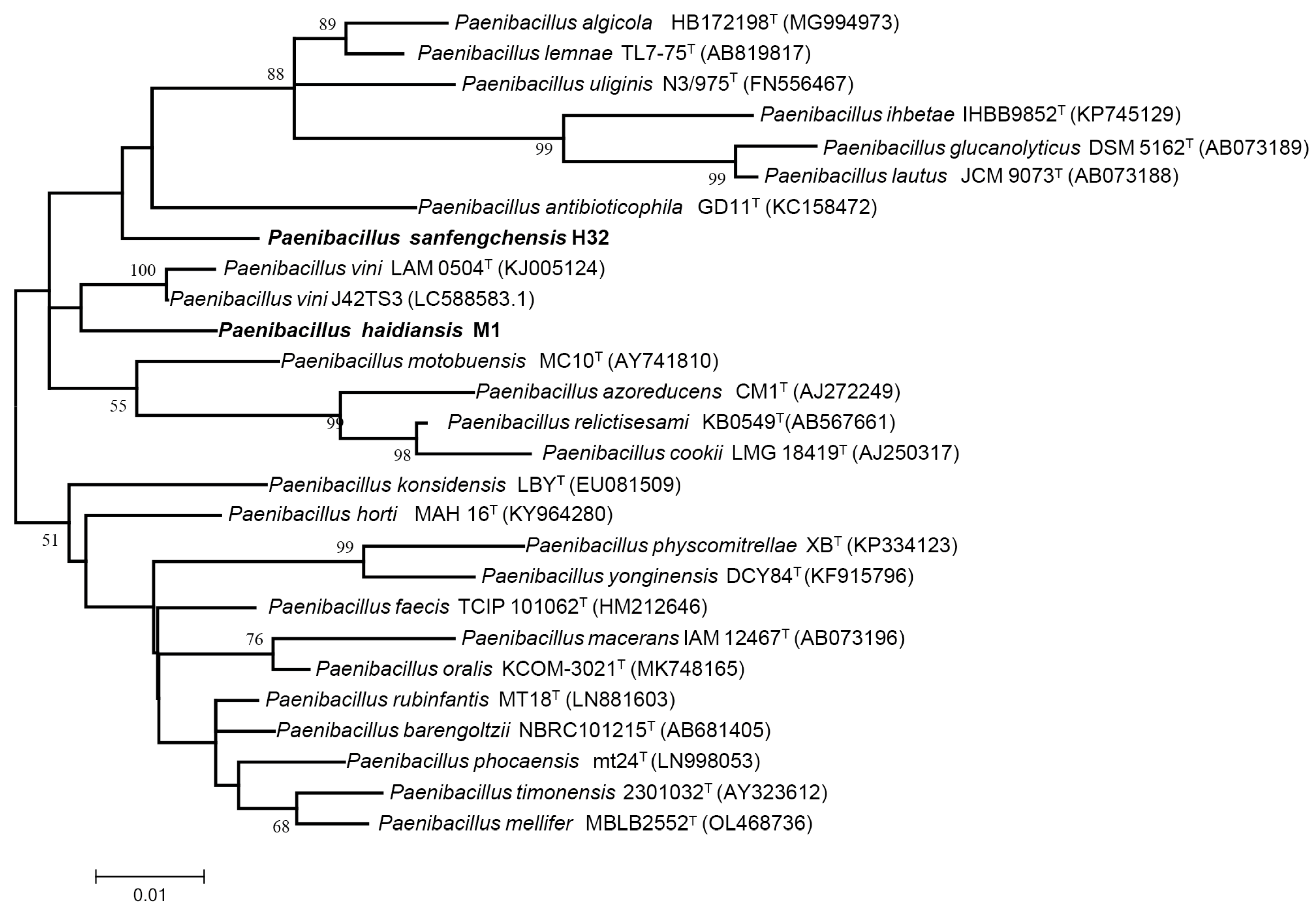
A nearly full-length 16S rRNA gene (ca. 1500 bp) was PCR amplified from each of the two strains M1 and H32 and then was sequenced. A comparison of the 16S rRNA gene sequences of the two strains M1 and H32 with the sequences held in the GenBank database revealed that the two strains belong to the genus Paenibacillus (Figure 2). Strain M1 showed a 97% identity with H32 at 16S rRNA gene sequences. Strain M1 had the highest (97.25%) identity with P. vini LAM 0504T and had a lower than 97% identity with other Paenibacillus species (such as Paenibacillus timonensis, Paenibacillus motobuensis, Paenibacillus azoreducens) at 16S rRNA gene sequences. Strain H32 had the highest (97.48%) identity with Paenibacillus faecis TCIP 101062T and had a lower than 97% identity with other Paenibacillus species (such as Paenibacillus physcomitrellae, Paenibacillus yonginensis, Paenibacillus konsidensis) at 16S rRNA gene sequences. Generally, 98.65% similarity of 16S rRNA gene sequences is suggested as the threshold to differentiate bacterial species [29]. The current results that the 16S rRNA gene sequence similarity of strains M1 and H32 with their closest species was below the threshold value of 98.65% indicate that both strains M1 and H32 are two novel species within the genus Paenibacillus.

3.3. Genome Sequence and Similarity Analysis

The two strains M1 and H32 were genome-sequenced to evaluate their relatedness to the closely related recognized species in the genus Paenibacillus. The genome size of strain M1 was 5.7 Mb which has 5426 genes. There are 87 tRNA genes and two 16S rRNA genes in the M1 strain. The DNA G+C content of strain M1 is 52% (Table 1). Whereas its closely related strain P. vini had a complete genome size of 5.6 Mb which has 5274 genes. The DNA G+C content of P. vini is 49%.
The genome size of strain H32 was 6.48 Mb carrying 5511 genes. The G+C content of strain H32 was 52.99% (Table 1). Whereas its closely related strain P. faecis had a complete genome size of 6.3 Mb comprising 5699 genes. The DNA G+C content of P. faecis is 53%. The genome sequences of the two strains M1 and H32 were deposited in GenBank under accession numbers GCA_036553665.1 and GCA_036864765.1, respectively.
The average nucleotide identity (ANI) and digital DNA-DNA hybridization (dDDH) values are generally used to define bacterial species [30]. The ANI for the species threshold is 95% and DDH for the species threshold is 70% [30,31]. The ANI and dDDH values between strain M1 and the reference strain P. vini are 78.17% and 22.3%, respectively. The ANI and dDDH values between strain H32 and the reference strain P. feacis DSM 23593 were 88.94% and 66.20%, respectively. These genomic relatedness data are below the thresholds of ANI (95.0%) and dDDH (70.0%), indicating that strains M1 and H32 are genomically distinct from their closest species of the genus Paenibacillus.

3.4. Analysis of Nitrogen Fixation and Nitrogen Metabolism Genes

Genome sequence analysis demonstrated that each genome of the two strains M1 and H32 contains a compact nif gene cluster composed of nine genes (nifB, nifH, nifD, nifK, nifE, nifN, nifX, hesA, nifV) encoding Mo-nitrogenase which is conserved in N2-fixing Paenibacillus strains [18]. The nif gene cluster carried in the genomes of the two strains M1 and H32 is consistent with the nitrogenase activity observed in both stains.
In addition to the nif genes, each genome of the two strains M1 and H32 contains two sets of nasE, nasD and nasB encoding nitrite reductase. There is a nirK encoding nitrite reductase, a nirC encoding nitrite transporter and a nasA encoding nitrate/nitrite transporter in both strains. There are two copies of glnA encoding glutamine synthetase, one of which is linked together with glnR as an operon glnRA, the other one of which is separated on the genome site [32]. GlnR encoded by glnR is a global transcription regulator controlling the expression of nif genes and some other genes involved in nitrogen metabolism in P. ploymyxa [32,33,34]. There is a gdhA encoding glutamate dehydrogenase in both strains M1 and H32, and there are gltD and gltB encoding glutamate synthase (NADPH) small chain and large chains, respectively. There is a gltC encoding transcription activator of glutamate synthase operon in both strains.

3.5. Phenotypic Characteristics

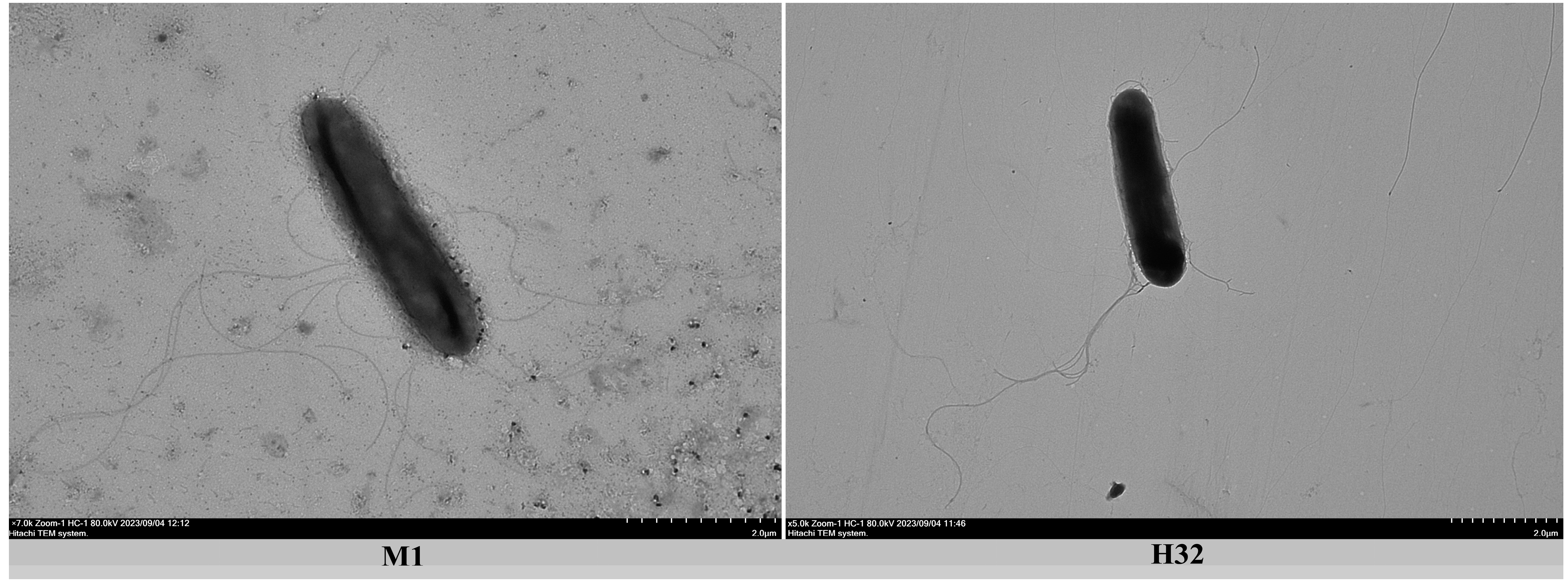
The two strains M1 and H32 are Gram-positive, facultatively anaerobic, motile and rod-shaped. Both strains have peritrichous flagella on the cell surface observed under a transmission electron microscope (Figure 3).
Strains M1 and H32, together with their reference strains P. vini LAM0504T and P. faecis 65684T, were tested for a range of physiological and biochemical characteristics. As shown in Table 2, Both strain M1 and the reference strain P. vini LAM0504T exhibited positive nitrate reductase activity. Both strain M1 and the reference strain P. vini LAM0504T utilized xylose, glucose, trehalose, galactose and maltose to produce acid. However, both strain M1 and the reference strain P. vini LAM0504T showed variation in fructose utilization and NaCl tolerance.
Both strain H32 and the reference strain P. faecis 65684T had nitrate reductase activity and utilized fructose, xylose, glucose, trehalose, galactose and maltose to produce acid. However, both strain H32 and the reference P. faecis 65684T showed variation in the utilization of D-mannitol and L-rhamnose and pH range tolerance.

3.6. Chemotaxonomic Analyses

Chemotaxonomic analyses included the cellular fatty acid, polar lipids and isoprenoid quinone that are used in the classification of bacteria [29,30,31,32]. As shown in Table 3, anteiso-C15:0 was the major fatty acid component of strains M1 (45.71%) and H32 (61.41%), and anteiso-C15:0 was also the predominant fatty acid of their reference strains P. vini LAM0504T (50.30%) and P. faecis DSM 23593T (45.42%). Our results are consistent with the findings that anteiso-C15:0 is the predominant fatty acid of the genus Paenibacillus [2,3,4,5,6,7,8,9,10,11]. Although anteiso-C15:0 is the major fatty acid for both strains (M1 and H32) and their reference strains, the fatty acid contents show significant variation among these strains (Table 3). These data indicate that strains M1 and H32 are different from each other and are distinguished from other members of the genus Paenibacillus.
The major polar lipids were detected by two-dimensional TLC. The major polar lipids of strain M1 are DPG (diphosphatidylglycerol), PG (phosphatidylglycerol), AL (aminolipid) and two unidentified phospholipids (PL3 and PL4). Whereas, the major polar lipids of strain H32 are DPG, PG, PE (phosphatidylethanolamine) and APL (aminophospholipids) (Figure S1). The polar lipid profiles of both strains M1 and H32 are similar to those of other species of the genus Paenibacillus [11]. Respiratory quinone is mainly composed of ubiquinone (Q) and menaquinone (MK). The major respiratory quinone component of both strains M1 and H32 is menaquinone-7 (MK-7) (Figure S2), in conformity with the genus Paenibacillus.

3.7. Description of Paenibacillus haidiansis sp. nov

Paenibacillus haidiansis (haidian.sis. L.gen. n. Hardian of Beijing, where the type strain M1 was isolated). Type strain M1 is Gram-positive, facultative anaerobic and rod-shaped (0.3–0.5 μm × 2.5–3.5 μm). Cells have peritrichous flagella. Ellipsoidal spores are located centrally in swollen sporangia. The colonies on LB medium are white, convex, circular and translucent. Cells grow at 25–35 °C (optimum, 30 °C), pH 6.0–8.0 (optimum, 7.0), and 0–1.0% NaCl concentrations. Nitrate is reduced to nitrite. Starch is hydrolyzed. The genome of strain M1 has nine genes (nifB, nifH, nifD, nifK, nifE, nifN, nifX, hesA, nifV) encoding Mo-nitrogenase. Strain M1 is able to reduce N2 to NH3 (i.e., biological nitrogen fixation). Able to utilize D-fructose, D-xylose, D-glucose, trehalose, D-galactose, maltose to produce acid, but not able to utilize L-rhamnose, D-mannitol, D-sorbitol and inositol. The predominant fatty acid is anteiso-C15:0. The major polar lipids are DPG, PG and AL. The only menaquinone is MK-7. The DNA G+C content of this strain is 52%.
The type strain, M1T (=CGMCC 1.4871 = JCM 37487, was isolated from the rhizosphere soil of Hosta plantantae in the Haidian District of Beijing, China. The GenBank accession numbers for the 16S rRNA sequence is PP204203.1 and for the draft genome sequence is GCA_036553665.1.

3.8. Description of Paenibacillus sanfengchensis nov sp. nov

Paenibacillus sanfengchensis (sanfeng′chen.sis. L.gen. n. sanfengchensis of Sanfeng Chen, named after Professor Sanfeng Chen, China). Type strain H32 is Gram-positive, facultatively anaerobic, and rod-shaped (0.3.5–0.5 μm × 2.5–3.0 μm). Cells have peritrichous flagella. Ellipsoidal spores are located centrally in swollen sporangia. The colonies on LB medium are white, convex, circular and translucent. The genome of strain H32 has nine genes (nifB, nifH, nifD, nifK, nifE, nifN, nifX, hesA, nifV) encoding Mo-nitrogenase. Strain H32 is able to reduce N2 to NH3 (i.e., biological nitrogen fixation). Cells grow at 20–40 °C (optimum, 30 °C), pH 5.0–9.0 (optimum, 7.0), and 0–1.0% NaCl concentrations. Starch is hydrolyzed. Nitrate is reduced to nitrite. Able to utilize D-fructose, D-xylose, D-glucose, trehalose, D-galactose, maltose, L-rhamnose and D-mannitol to produce acid, but not able to utilize D-sorbitol and inositol. The predominant fatty acid is anteiso-C15:0. The major polar lipids are DPG, PG, PE and APL. The only menaquinone is MK-7. The DNA G+C content of this strain is 52.5%.
The type strain, H32T (=CGMCC 1.4872 = JCM 37488, was isolated from the rhizosphere soil of Oryza sativa (rice) in Hebei province, China. The GenBank accession number for the 16S rRNA sequence is P204203.1 and for the draft genome sequence is GCA_036864765.1.

4. Discussion

Paenibacillus is a large genus of Gram-positive, facultative anaerobic and endospore-forming bacteria. N2-fixng Paenibacillus species and strains have potential uses as a bacterial fertilizer in agriculture.
In this study, two strains M1 and H32 were isolated from the rhizospheres of Hosta plantaginea and Oryza sativa (rice), respectively. The two strains are Gram-positive, facultative anaerobic and endospore-forming bacteria. Nitrogenase activity assay and nifH sequencing analysis showed that the two strains are N2-fixing bacteria. Genome sequence analysis revealed that each genome of the two strains has a nif gene cluster composed of nine genes (nifB nifH nifD nifK nifE nifN nifX hesA nifV) that are characteristics of N2-fixing Paenibacillus strains [18].
A comparison of the 16S rRNA gene sequences of the two strains M1 and H32 with the sequences held in the GenBank database revealed that the two strains belong to the genus Paenibacillus. Strain M1 showed 97% identity with strain H32 at 16S rRNA gene sequences. Strain M1 had the highest (97.25%) identity with P. vini and had a lower than 97% identity with other named Paenibacillus species at 16S rRNA gene sequences. Strain H32 had the highest (97.48%) identity with P. faecis and had a lower than 97% identity with other named Paenibacillus species. ANI and dDDH values between strain M1 and its closest member P. vini were 78.17% and 22.3%, respectively. The ANI and dDDH values between strain H32 and its closest member P. faecis were 88.94% and 66.02%, respectively. The predominant fatty acid is anteiso-C15:0 for both strain M1 (45.71%) and H32 (61.41%). The predominant isoprenoid quinone is MK-7 for both strains M1 and H32. The two strains M1 and H32 exhibited some variation with their reference strains in the contents and components of the cellular fatty acids and polar lipids. These data suggest that strains M1 and H32 are two novel species within the genus Paenibacillus. Thus, two novel species Paenibacillus haidiansis and Paenibacillus sanfengchensis are proposed.
It is known that most of the members of the genus Paenibacillus are non-nitrogen-fixing bacteria and only a few dozen species are able to fix nitrogen. In this study, two nitrogen-fixing novel species Paenibacillus haidiansis and Paenibacillus sanfengchensis are obtained, providing bacterial strains for study and utilization of biological nitrogen fixation. The genus Paenibacillus, which was originally included within the genus Bacillus, was reclassified as a separate genus in 1993 [1]. When the genus Paenibacillus was created, the three N2-fixing species Bacillus polymyxa, Bacillus azotofixans and Bacillus macerans were transferred to this new genus. Recently, we tried to isolate N2-fixing Bacillus strains but failed. The results support that the N2-fixing and endospore-forming bacteria mainly belong to the genus Paenibacillus. It is well known that most N2-fixing bacteria (e.g., Azotobacter vinelandii, Klebsiella oxytoca) are Gram-negative. In most of the Gram-negative and N2-fixing bacteria, the expression of nif genes is regulated by the transcription regulator NifA [35]. Whereas GlnR regulates expression of nif genes in Gram-positive and N2-fixing Paenibacillus strains. Gram-positive and N2-fixing Paenibacillus strains and the Gram-negative and N2-fixing bacteria also show some differences in colonization, nitrogen-fixation efficiency and growth rate.
It is now recognized that the free-living and plant-associated N2-fixing microorganisms play a contributing role in the growth of non-legumes by N2 fixation. Inoculation of non-legume plants with N2-fixing biofertilizers (also known as biological inoculants) could help to increase the available nitrogen content of the crop and reduce the usage amount of chemical nitrogen fertilizer in a variety of agricultural systems [36,37,38,39]. In the near future, we will utilize P. haidiansis M1 and P. sanfengchensis H32 to inoculate plants (e.g., rice, wheat, maize, cucumber, tomato) in a greenhouse, and then evaluate their effects on promoting plant growth through N2-fixation. If both strains or one of them have efficient effects on promoting plant growth through N2-fixation, both or one of them could be used as N2-fixing biofertilizers in agriculture on a large scale.

5. Conclusions

In this study, two strains M1 and H32 were isolated from the rhizospheres of Hosta plantaginea and Oryza sativa (rice), respectively. Both strains M1 and H32 are able to fix nitrogen. Based on the phenotypic, phylogenetic, genomic and chemotaxonomic dissimilarity, two novel species Paenibacillus haidiansis sp. nov and Paenibacillus sanfengchensis sp. nov are proposed with the type strain M1T = CGMCC 1.4871 = JCM 37487 and with the type strain H32T (=CGMCC 1.4872 = JCM 37488.

Supplementary Materials

The following supporting information can be downloaded at: https://www.mdpi.com/article/10.3390/microorganisms12122561/s1, Figure S1: Two-dimensional TLC plate of polar lipids extracted from strain M1 (A) and from strain H32 (B). The plate was sprayed with 10% (v/v) molybdophosphoric aicd to show all polar lipids. DPG, diphosphatidylglycerol; PG, phosphatidylglycerol; PE, phosphatidylethanolamine; APL, aminophospholipids; AL, aminolipid; PL, unidentified phosphoglycolipids; L, unknown polar lipids; APGL, unidentified aminophosphoglycolipid. Figure S2: HPLC analysis shows MK-7 as the major respiratory quinone component for strain M1 (A) and for strain H32 (B). Mobile phase, methanol: isopropanol = 65:35; Chromatographic column: Zorbax Eclipse XDB-C18 (4.6 * 250 mm, 5 μm; Agilent); Column temperature: 40 °C; Flow rate: 1.0 mL/min; Injection volume: 10 μL; Detection wavelength: 270 nm.

Author Contributions

Conceptualization, S.C.; methodology and investigation, W.Z. and R.H.; methodology; data curation, G.W., S.J. and M.G.; formal analysis, M.L. and P.L.; writing—original draft preparation, W.Z.; writing—reviewing and editing, S.C.; software, W.Z. and Y.S.; supervision, S.C.; project administration, S.C. and M.G.; funding acquisition, S.C. and M.G. All authors have read and agreed to the published version of the manuscript.

Funding

This work was supported by the National Key Research and Development Program of China Award (No. 2019YFA0904700) and by the China Agriculture Research System of MOF and MARA (CARS-23-B12).

Data Availability Statement

The original contributions presented in the study are included in the article/Supplementary Materials, further inquiries can be directed to the corresponding author.

Acknowledgments

We thank Yonghong Wang (Institute of Microbiology, Guangdong Academy of Science) for helping to preserve the bacterial strains.

Conflicts of Interest

The authors declare no conflicts of interest.

References

  1. Ash, C.; Priest, F.G.; Collins, M.D. Molecular identification of rRNA group 3 Bacilli (Ash, Farrow, Wallbanks and Collins) using a PCR probe test. Proposal for the creation of a new genus Paenibacillus. Antonie Van Leeuwenhoek 1993, 64, 253–260. [Google Scholar] [CrossRef] [PubMed]
  2. Li, Q.; Li, Y.; Liu, X.; Chen, S.F. Paenibacillus sinensis sp. nov., a nitrogen-fixing species isolated from plant rhizospheres. Antonie Van Leeuwenhoek 2022, 115, 7–18. [Google Scholar] [CrossRef] [PubMed]
  3. Li, Q.; Zuo, Y.Z.; Gao, M.; Chen, S.F. Paenibacillus caui sp. nov., a nitrogen-fixing Species Isolated from the Rhizosphere Soil of a Peach Tree. Int. J. Syst. Evol. Microbiol. 2022, 72, 005216. [Google Scholar] [CrossRef] [PubMed]
  4. Hong, Y.Y.; Ma, Y.C.; Zhou, Y.G.; Gao, F.; Liu, H.C.; Chen, S.F. Paenibacillus sonchi sp nov., a nitrogen-fixing species isolated from the rhizosphere of Sonchus oleraceus. Int. J. Syst. Evol. Microbiol. 2009, 59, 2656–2661. [Google Scholar] [CrossRef] [PubMed]
  5. Jin, H.J.; Zhou, Y.G.; Liu, H.C.; Chen, S.F. Paenibacillus jilunlii sp nov., a nitrogen-fixing species isolated from the rhizosphere of Begonia semperflorens. Int. J. Syst. Evol. Microbiol. 2011, 61, 1350–1355. [Google Scholar] [CrossRef]
  6. Jin, H.J.; Lv, J.; Chen, S.F. Paenibacillus sophorae sp. nov., a nitrogen-fixing species isolated from the rhizosphere of Sophora japonica. Int. J. Syst. Evol. Microbiol. 2011, 61, 767–771. [Google Scholar] [CrossRef]
  7. Ma, Y.C.; Zhang, J.; Chen, S.F. Paenibacillus zanthoxyli sp nov, a novel nitrogen-fixing species isolated from the rhizosphere of Zanthoxylum simulans. Int. J. Syst. Evol. Microbiol. 2007, 57, 873–877. [Google Scholar] [CrossRef]
  8. Ma, Y.C.; Xia, Z.Q.; Liu, X.M.; Chen, S.F. Paenibacillus sabinae sp nov., a nitrogen-fixing species isolated from the rhizosphere soils of shrubs. Int. J. Syst. Evol. Microbiol. 2007, 57, 6–11. [Google Scholar] [CrossRef]
  9. Ma, Y.C.; Chen, S.F. Paenibacillus forsythiae sp nov., a nitrogen-fixing species isolated from the rhizosphere soil of Forsythia mira. Int. J. Syst. Evol. Microbiol. 2008, 58, 319–323. [Google Scholar] [CrossRef]
  10. Wang, L.Y.; Li, J.; Li, Q.X.; Chen, S.F. Paenibacillus triticisoli, sp. nov., a nitrogen-fixing species isolated from wheat rhizosphere soil. Antonie Van Leeuwenhoek 2013, 104, 675–683. [Google Scholar] [CrossRef]
  11. Xie, J.B.; Zhang, L.H.; Zhou, Y.G.; Liu, H.C.; Chen, S.F. Paenibacillus taohuashanense sp. nov., a nitrogen-fixing species isolated from rhizosphere soil of the root of Caragana kansuensis pojark. Antonie Van Leeuwenhoek 2012, 102, 735–741. [Google Scholar] [CrossRef] [PubMed]
  12. Grady, E.N.; MacDonald, J.; Liu, L.; Richman, A.; Yuan, Z.C. Current knowledge and perspectives of Paenibacillus: A review. Microb. Cell Fact. 2016, 15, 203. [Google Scholar] [CrossRef] [PubMed]
  13. Mülner, P.; Schwarz, E.; Dietel, K.; Herfort, S.; Jähne, J.; Lasch, P.; Cernava, T.; Berg, G.; Vater, J. Fusaricidins, polymyxins and volatiles produced by Paenibacillus polymyxa strains DSM 32871 and M1. Pathogens 2021, 10, 1485. [Google Scholar] [CrossRef] [PubMed]
  14. Li, Y.; Chen, S.F. Fusaricidin Produced by Paenibacillus polymyxa WLY78 induces systemic resistance against Fusarium wilt of cucumber. Int. J. Mol. Sci. 2019, 20, 5240. [Google Scholar] [CrossRef] [PubMed]
  15. Li, Y.; Zhang, H.; Li, Y.; Chen, S.F. Fusaricidin biosynthesis is controlled via a KinB-Spo0A-AbrB signal pathway in Paenibacillus polymyxa WLY78. Mol. Plant Microbe Interact. 2021, 34, 1378–1389. [Google Scholar] [CrossRef]
  16. Li, Y.; Wang, M.; Chen, S.F. Application of N2-fixing Paenibacillus triticisoli BJ-18 changes the compositions and functions of the bacterial, diazotrophic, and fungal microbiomes in the rhizosphere and root/shoot endosphere of wheat under field conditions. Biol. Fertil. Soils 2021, 51, 347–362. [Google Scholar] [CrossRef]
  17. Li, Y.; Li, Q.; Chen, S.F. Diazotroph Paenibacillus triticisoli BJ-18 drives the variation in bacterial, diazotrophic and fungal communities in the rhizosphere and root/shoot endosphere of Maize. Int. J. Mol. Sci. 2021, 22, 1460. [Google Scholar] [CrossRef]
  18. Xie, J.B.; Du, Z.L.; Bai, L.Q.; Tian, C.F.; Zhang, Y.Z.; Xie, J.Y.; Wang, T.S.; Liu, X.M.; Chen, X.; Cheng, Q.; et al. Comparative genomic analysis of N2-fixing and non-N2-fixing Paenibacillus spp.: Organization, evolution and expression of the nitrogen fixation genes. PLoS Genet. 2014, 10, e1004231. [Google Scholar] [CrossRef]
  19. Kumar, S.; Stecher, G.; Li, M.; Knyaz, C.; Tamura, K. MEGA X: Molecular evolutionary genetics analysis across computing platforms. Mol. Biol. Evol. 2018, 35, 1547. [Google Scholar] [CrossRef]
  20. Frank, J.A.; Reich, C.I.; Sharma, S.; Weisbaum, J.S.; Wilson, B.A.; Olsen, G.J. Critical evaluation of two primers commonly used for amplification of bacterial 16S rRNA genes. Appl. Environ. Microbiol. 2008, 74, 2461–2470. [Google Scholar] [CrossRef]
  21. Yoon, S.H.; Ha, S.M.; Lim, J.; Kwon, S.; Chun, J. A large-scale evaluation of algorithms to calculate average nucleotide identity. Antonie Van Leeuwenhoek. 2017, 110, 1281–1286. [Google Scholar] [CrossRef] [PubMed]
  22. Yoon, S.H.; Ha, S.M.; Kwon, S.; Lim, J.; Kim, Y.; Seo, H.; Chun, J. Introducing EzBioCloud: A taxonomically united database of 16S rRNA gene sequences and whole-genome assemblies. Int. J. Syst. Evol. Microbiol. 2017, 67, 1613–1617. [Google Scholar] [CrossRef] [PubMed]
  23. Besemer, J.; Lomsadze, A.; Borodovsky, M. GeneMarkS: A self-training method for prediction of gene starts in microbial genomes. Implications for finding sequence motifs in regulatory regions. Nucleic Acids Res. 2001, 29, 2607–2618. [Google Scholar] [CrossRef] [PubMed]
  24. Meier-Kolthof, J.P.; Auch, A.F.; Klenk, H.P.; Göker, M. Genome sequence-based species delimitation with confidence intervals and improved distance functions. BMC Bioinform. 2013, 14, 60. [Google Scholar] [CrossRef]
  25. Minnikin, D.E.; O’donnell, A.G.; Goodfellow, M.; Alderson, G.; Athalye, M.; Schaal, A.; Parlett, J.H. An integrated procedure for the extraction of bacterial isoprenoid quinones and polar lipids. J. Microbiol. Methods 1984, 2, 233–241. [Google Scholar] [CrossRef]
  26. Komagata, K.; Suzuki, K. Lipid and cell-wall analysis in bacterial systematics. Methods Microbiol. 1987, 19, 161–207. [Google Scholar]
  27. Sasser, M.; Kunitsky, C.; Jackoway, G.; Ezzell, J.W.; Teska, J.D.; Harper, B.; Parker, S.; Barden, D.; Blair, H.; Breezee, J.; et al. Identification of Bacillus anthracis from culture using gas chromatographic analysis of fatty acid methyl esters. J. AOAC Int. 2005, 88, 178–181. [Google Scholar] [CrossRef]
  28. Collins, M.D.; Goodfellow, M.; Minnikin, D.E. Fatty acid, isoprenoid quinone and polar lipid composition in the classification of Curtobacterium and related taxa. J. Gen. Microbiol. 1980, 118, 29–37. [Google Scholar] [CrossRef]
  29. Kim, M.; Oh, H.S.; Park, S.C.; Chun, J. Towards a taxonomic coherence between average nucleotide identity and 16S rRNA gene sequence similarity for species demarcation of prokaryotes. Int. J. Syst. Evol. Microbiol. 2014, 64, 346–351. [Google Scholar] [CrossRef]
  30. Goris, J.; Konstantinidis, K.T.; Klappenbach, J.A.; Coenye, T.; Vandamme, P.; Tiedje, J.M. DNA-DNA hybridization values and their relationship to whole-genome sequence similarities. Int. J. Syst. Evol. Microbiol. 2007, 57, 81–91. [Google Scholar] [CrossRef]
  31. Konstantinidis, K.T.; Tiedje, J.M. Genomic insights that advance the species definition for prokaryotes. Proc. Natl. Acad. Sci. USA 2005, 102, 2567–2572. [Google Scholar] [CrossRef] [PubMed]
  32. Wang, T.; Zhao, X.; Shi, H.; Sun, L.; Li, Y.; Li, Q.; Zhang, H.; Chen, S.; Li, J. Positive and negative regulation of transferred nif genes mediated by indigenous GlnR in Gram-positive Paenibacillus polymyxa. PLoS Genet. 2018, 14, e1007629. [Google Scholar] [CrossRef]
  33. Wang, T.; Zhao, X.; Wu, X.; Chen, S.F. Genome-wide mapping of GlnR-binding sites reveals the global regulatory role of GlnR in controlling the metabolism of nitrogen and carbon in Paenibacillus polymyxa WLY78. BMC Genom. 2023, 24, 85. [Google Scholar] [CrossRef]
  34. Zhao, X.; Song, Y.; Wang, T.; Hua, C.; Hu, R.; Shang, Y.; Shi, H.; Chen, S.F. Glutamine synthetase and GlnR regulate nitrogen metabolism in Paenibacillus polymyxa WLY78. Appl. Environ. Microbiol. 2023, 89, e0013923. [Google Scholar] [CrossRef]
  35. Dixon, R.; Kahn, D. Genetic regulation of biological nitrogen fixation. Nat. Rev. Microbiol. 2004, 2, 621–631. [Google Scholar] [CrossRef] [PubMed]
  36. Ladha, J.K.; Peoples, M.B.; Reddy, P.M.; Biswas, J.C.; Bennett, A.; Jat, M.L.; Krupnik, T.J. Biological nitrogen fixation and prospects for ecological intensification in cereal-based cropping systems. Field Crops Res. 2022, 283, 108541. [Google Scholar] [CrossRef] [PubMed]
  37. Eskin, N.; Vessey, K.; Tian, L. Research progress and perspectives of nitrogen fixing bacterium, Gluconacetobacter diazotrophicus, in monocot plants. Int. J. Agron. 2014, 2014, 208383. [Google Scholar] [CrossRef]
  38. Bageshwar, U.K.; Srivastava, M.; Pardha-Saradhi, P.; Paul, S.; Gothandapani, S.; Jaat, R.S.; Shankar, P.; Yadav, R.; Biswas, D.R.; Kumar, P.A.; et al. An environmentally friendly engineered azotobacter strain that replaces a substantial amount of urea fertilizer while sustaining the same wheat yield. Appl. Environ. Microbiol. 2017, 83, e00590-17. [Google Scholar] [CrossRef]
  39. Li, Y.B.; Li, Y.L.; Zhang, H.W.; Wang, M.Y.; Chen, S.F. Diazotrophic Paenibacillus beijingensis BJ-18 provides nitrogen for plant and promotes plant growth, nitrogen uptake and metabolism. Front. Microbiol. 2019, 10, 1119. [Google Scholar] [CrossRef]
Figure 1. The maximum phylogenetic likelihood tree based on nifH gene sequences (300 bp) of the novel strains M1 and H32, together with their closely related taxonomic groups. Bootstrap analyses were performed with 1000 cycles. Numbers (50%) at nodes are bootstrap values. Bar 0.05 substitutions per nucleotide position.
Microorganisms 12 02561 g001
Figure 2. The maximum likelihood phylogenetic tree based on the 16S rRNA gene sequences of the novel strains M1 and H32, together with their closely related taxonomic groups. Bootstrap analyses were performed with 1000 cycles. Numbers (50%) at nodes are bootstrap values. Bar 0.05 substitutions per nucleotide position.
Microorganisms 12 02561 g002
Figure 3. Transmission electron microscopic images of vegetative cell and flagella of strains M1 and strain H32.
Microorganisms 12 02561 g003
Table 1. General genomic information of strains M1, H32 and their closely related type strains.
StrainsAccession No.Genome Size (Mb)DNA GC (%)Gene NumberRNAs
tRNA16S rRNAsncRNA (Non-Coding RNA)
Test strain
M1
GCA_036553665.15.75254628724
Type strain
Paenibacillus vini strain J42TS3
GCA_018403325.15.64952749324
Type strain
Paenibacillus vini strain CENA-BCM001
GCA_030412165.15.74952939324
Test strain
H32
GCA_036864765.16.4652.555117718
Type strain
Paenibacillus faecis strain J25TS5
GCA_008084145.16.35357438324
Type strain
Paenibacillus faecis DSM 23593(T)
GCA_008084145.16.35356996444
Table 2. Differential characteristic features of test strains M1 and H32 from their reference strains.
CharacteristicTest Strain Reference Strain Test StrainReference Strain
M1P. vini LAM0504TH32P. faecis 65684T
pH range6–86–85–96–9
NaCl0–1.0%0–3%0–1.0%0–3%
Growth temperature (°C)25–353020–4030
Nitrate reduction++++
Starch hydrolysis++++
Dihydroxyacetone production
Mobility++++++
Flagellum++++
Production of acid from following substrates:
D-fructose+++
D-xylose++++
D-glucose++++
Trehalose++++
D-galactose++++
Maltose++++
D-mannitol+
L-rhamnose+
D-sorbitol
Inositol
+, positive; −, negative.
Table 3. The cellular fatty acid content (% of the totals) of strains M1 and H32, together with their closely related reference strains P. vini LAM0504T and P. faecis DSM 23593T.
Fatty AcidM1P. vini LAM0504TH32P. faecis DSM 23593T
Saturated
C12:00.620.51.611.60
C14:03.163.301.101.77
C16:024.399.736.4721.56
C17:0 TR-TR1.50
Unsaturated
C14:1 ω5c-0.17TRTR
C19:0 cyclo ω8c--TRTR
C18:1 ω9c TRTR1.631.04
C20:4 ω6,9,12,15cTR---
Branched saturated
iso-C14:01.282.39TRTR
iso-C15:03.798.807.893.75
iso-C16:07.538.422.773.57
iso-C17:02.053.242.012.84
anteiso-C15:045.7150.3061.4145.42
anteiso-C17:07.525.217.9212.46
TR, trace amount (<1.0%); -, not detected.
Disclaimer/Publisher’s Note: The statements, opinions and data contained in all publications are solely those of the individual author(s) and contributor(s) and not of MDPI and/or the editor(s). MDPI and/or the editor(s) disclaim responsibility for any injury to people or property resulting from any ideas, methods, instructions or products referred to in the content.