
DNA Methylation in Offspring Conceived after Assisted Reproductive Techniques: A Systematic Review and Meta-Analysis

 ,
,  , ,
, ,  ,
,  , and
, and
Abstract
:1. Introduction
2. Methods
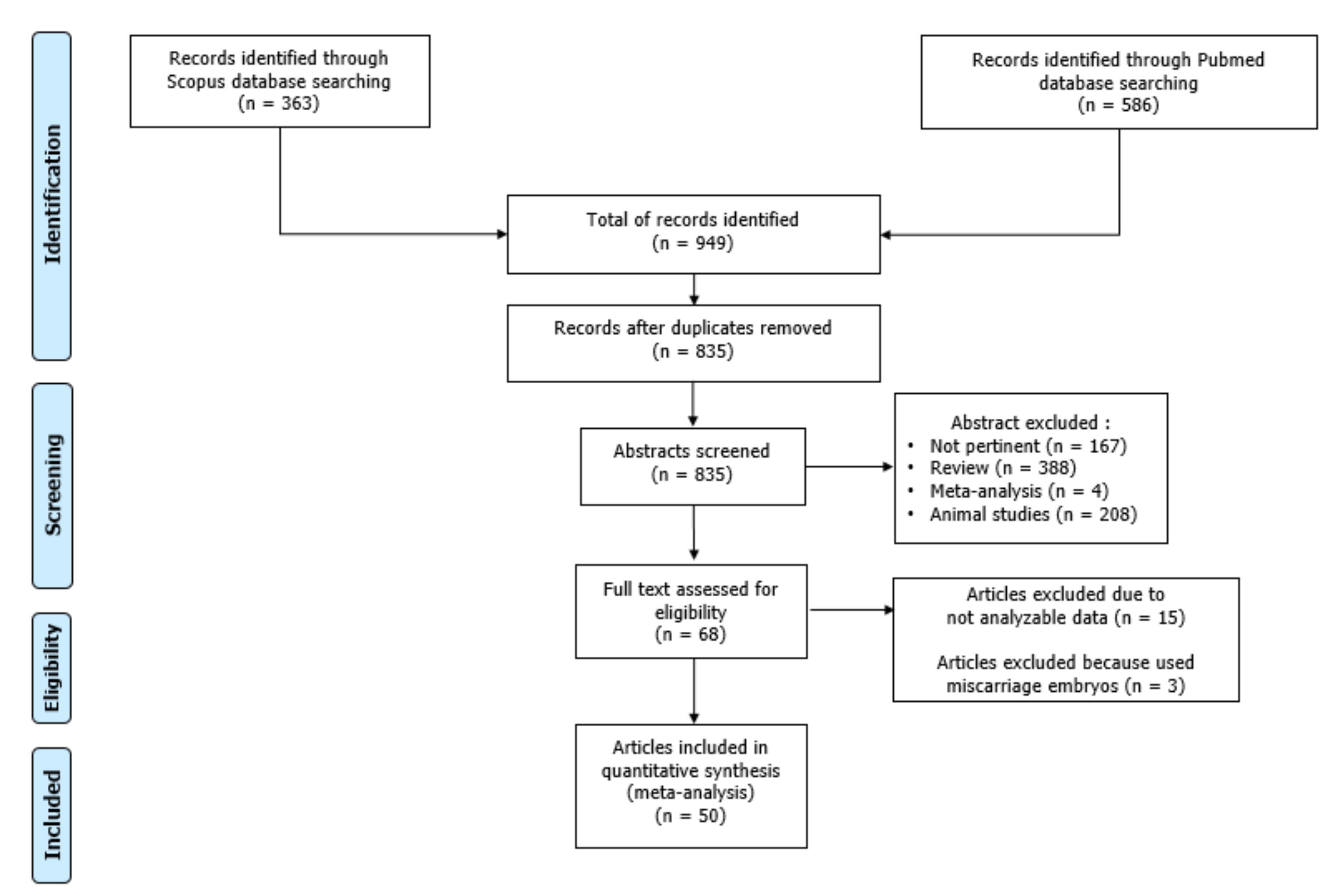
3. Results
3.1. Qualitative Synthesis
3.1.1. Global Methylation
3.1.2. Methylation of Imprinted Genes
3.1.3. Role of ART Protocol and Technique
3.1.4. Role of Parental Age
3.1.5. Role of the Etiology of Infertility
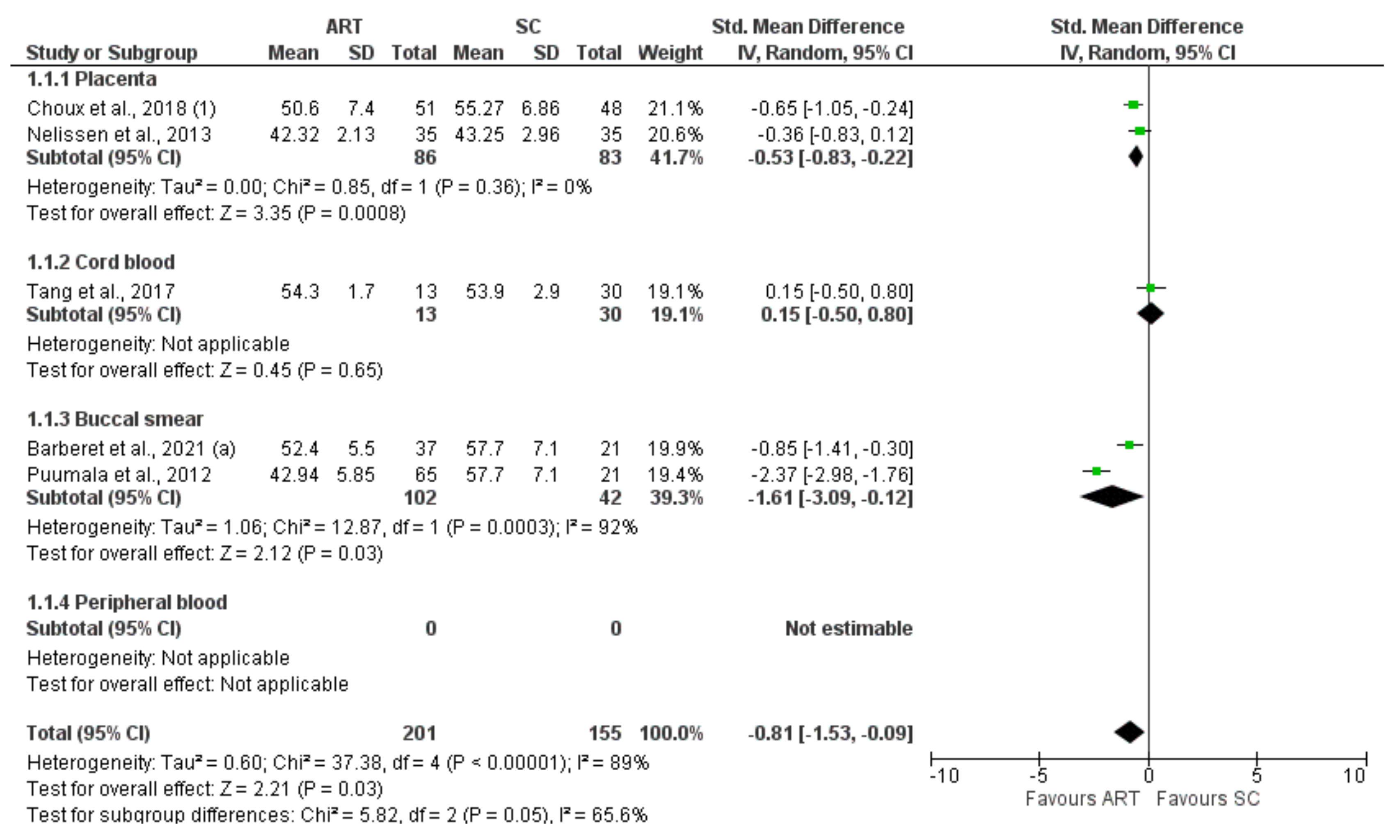
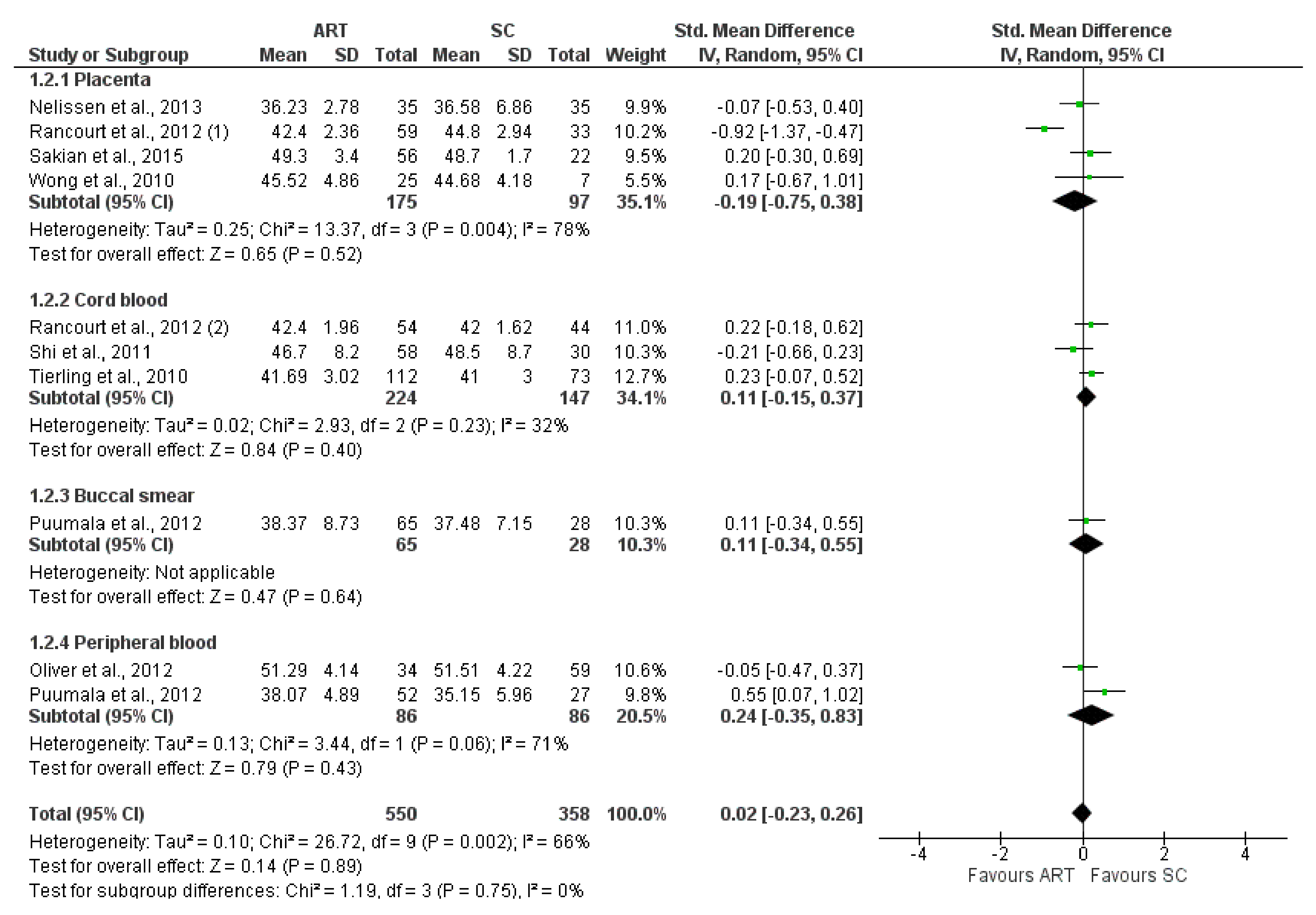
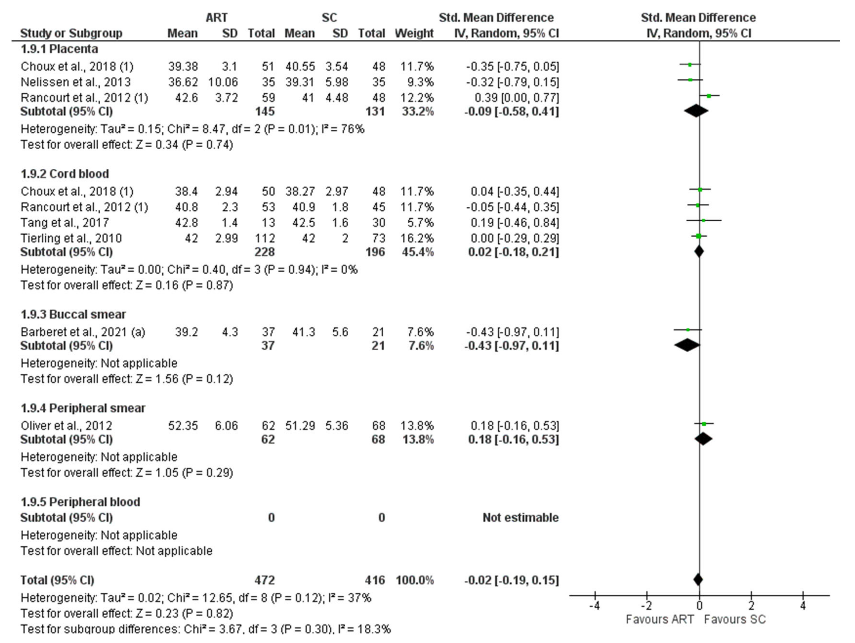
3.2. Quantitative Synthesis
4. Discussion
5. Conclusions
Supplementary Materials
Author Contributions
Funding
Institutional Review Board Statement
Informed Consent Statement
Data Availability Statement
Conflicts of Interest
References
- Mascarenhas, M.N.; Flaxman, S.R.; Boerma, T.; Vanderpoel, S.; Stevens, G.A. National, regional, and global trends in infertility prevalence since 1990: A systematic analysis of 277 health surveys. PLoS Med. 2012, 9, e1001356. [Google Scholar] [CrossRef]
- Lui Yovich, J. Founding pioneers of IVF update: Innovative researchers generating livebirths by 1982. Reprod. Biol. 2020, 20, 111–113. [Google Scholar] [CrossRef] [PubMed]
- Esteves, S.C. Who cares about oligozoospermia when we have ICSI. Reprod. Biomed. Online 2022, 44, 769–775. [Google Scholar] [CrossRef] [PubMed]
- Boulet, S.L.; Mehta, A.; Kissin, D.M.; Warner, L.; Kawwass, J.F.; Jamieson, J.D. Trends in use of and reproductive outcomes associated with intracytoplasmic sperm injection. JAMA 2015, 313, 255–263. [Google Scholar] [CrossRef] [PubMed]
- Chambers, G.M.; Wand, H.; Macaldowie, A.; Chapman, M.G.; Farquhar, C.M.; Bowman, M.; Molloy, D.; Ledger, W. Population trends and live birth rates associated with common ART treatment strategies. Hum. Reprod. 2016, 31, 2632–2641. [Google Scholar] [CrossRef] [PubMed]
- Wei, S.Q.; Luu, T.M.; Bilodeau-Bertrand, M.; Auger, N. Assisted reproductive technology and childhood morbidity: A longitudinal cohort study. Fertil. Steril. 2022, 118, 360–368. [Google Scholar] [CrossRef]
- Wijs, L.A.; Fusco, M.R.; Doherty, D.A.; Keelan, J.A.; Hart, R.J. Asthma and allergies in offspring conceived by ART: A systematic review and meta-analysis. Hum. Reprod. Update 2021, 28, 132–148. [Google Scholar] [CrossRef]
- Andreadou, M.T.; Katsaras, G.N.; Talimtzi, P.; Doxani, C.; Zintzaras, E.; Stefanidis, I. Association of assisted reproductive technology with autism spectrum disorder in the offspring: An updated systematic review and meta-analysis. Eur. J. Pediatr. 2021, 180, 2741–2755. [Google Scholar] [CrossRef]
- Zhang, Z.; Liu, X.; Wei, C.; Luo, J.; Shi, Y.; Lin, T.; He, D.; Wei, G. Assisted reproductive technologies and the risk of congenital urogenital tract malformations: A systematic review and meta-analysis. J. Pediatr. Urol. 2021, 17, 9–20. [Google Scholar] [CrossRef]
- Wijs, L.A.; Doherty, D.A.; Keelan, J.A.; Burton, P.; Yovich, J.L.; Beilin, L.; Mori, T.A.; Huang, R.C.; Adams, L.A.; Olynyk, J.K.; et al. Comparison of the cardiometabolic profiles of adolescents conceived through ART with those of a non-ART cohort. Hum. Reprod. 2022, 37, 1880–1895. [Google Scholar] [CrossRef]
- Wijs, L.A.; Doherty, D.A.; Keelan, J.A.; Panicker, V.; Burton, P.; Yovich, J.L.; Hart, R.J. Offspring conceived through ART have normal thyroid function in adolescence and as young adults. Hum. Reprod. 2022, 37, 1572–1580. [Google Scholar] [CrossRef]
- Argyraki, M.; Damdimopoulou, P.; Chatzimeletiou, K.; Grimbizis, G.F.; Tarlatzis, B.C.; Syrrou, M.; Lambropoulos, A. In-utero stress and mode of conception: Impact on regulation of imprinted genes, fetal development and future health. Hum. Reprod. Update 2019, 25, 777–801. [Google Scholar] [CrossRef]
- Sciorio, R.; Esteves, S.C. Contemporary Use of ICSI and Epigenetic Risks to Future Generations. J. Clin. Med. 2022, 11, 2135. [Google Scholar] [CrossRef]
- Monk, D.; Mackay, D.J.G.; Eggermann, T.; Maher, E.R.; Riccio, A. Genomic imprinting disorders: Lessons on how genome, epigenome and environment interact. Nat. Rev. Genet. 2019, 20, 235–248. [Google Scholar] [CrossRef]
- Morgan, H.D.; Santos, F.; Green, K.; Dean, W.; Reik, W. Epigenetic reprogramming in mammals. Hum. Mol. Genet. 2005, 14, R47–R58. [Google Scholar] [CrossRef]
- Agarwal, A.; Maldonado Rosas, I.; Anagnostopoulou, C.; Cannarella, R.; Boitrelle, F.; Munoz, L.V.; Finelli, R.; Durairajanayagam, D.; Henkel, R.; Saleh, R. Oxidative Stress and Assisted Reproduction: A Comprehensive Review of Its Pathophysiological Role and Strategies for Optimizing Embryo Culture Environment. Antioxidants 2022, 11, 477. [Google Scholar] [CrossRef]
- Santi, D.; De Vincentis, S.; Magnani, E.; Spaggiari, G. Impairment of sperm DNA methylation in male infertility: A meta-analytic study. Andrology 2017, 5, 695–703. [Google Scholar] [CrossRef]
- Cannarella, R.; Crafa, A.; Condorelli, R.A.; Mongioì, L.M.; La Vignera, S.; Calogero, A.E. Relevance of sperm imprinted gene methylation on assisted reproductive technique outcomes and pregnancy loss: A systematic review. Syst. Biol. Reprod. Med. 2021, 67, 251–259. [Google Scholar] [CrossRef]
- Barberet, J.; Ducreux, B.; Guilleman, M.; Simon, E.; Bruno, C.; Fauque, P. DNA methylation profiles after ART during human lifespan: A systematic review and meta-analysis. Hum. Reprod. Update 2022, dmac010. [Google Scholar] [CrossRef]
- Murray, J.; Farrington, D.P.; Eisner, M.P. Drawing conclusions about causes from systematic reviews of risk factors: The Cambridge Quality Checklists. J. Exp. Criminol. 2009, 5, 1–23. [Google Scholar] [CrossRef]
- Liu, Y.; Li, X.; Chen, S.; Wang, L.; Tan, Y.; Li, X.; Tang, L.; Zhang, J.; Wu, D.; Wu, Y.; et al. Comparison of Genome-Wide DNA Methylation Profiles of Human Fetal Tissues Conceived by in vitro Fertilization and Natural Conception. Front. Cell Dev. Biol. 2021, 9, 694769. [Google Scholar] [CrossRef] [PubMed]
- Zechner, U.; Pliushch, G.; Schneider, E.; El Hajj, N.; Tresch, A.; Shufaro, Y.; Seidmann, L.; Coerdt, W.; Müller, A.M.; Haaf, T. Quantitative methylation analysis of developmentally important genes in human pregnancy losses after ART and spontaneous conception. Mol. Hum. Reprod. 2010, 16, 704–713. [Google Scholar] [CrossRef] [PubMed]
- Zheng, H.Y.; Shi, X.Y.; Wu, F.R.; Wu, Y.Q.; Wang, L.L.; Chen, S.L. Assisted reproductive technologies do not increase risk of abnormal methylation of PEG1/MEST in human early pregnancy loss. Fertil. Steril. 2011, 96, 84–89.e2. [Google Scholar] [CrossRef] [PubMed]
- Argyraki, M.; Katafigiotis, S.; Vavilis, T.; Papadopoulou, Z.; Tzimagiorgis, G.; Haidich, A.B.; Chatzimeletiou, K.; Grimbizis, G.; Tarlatzis, B.; Syrrou, M.; et al. Influence of conception and delivery mode on stress response marker Oct4B1 and imprinted gene expression related to embryo development: A cohort study. Int. J. Reprod. Biomed. 2021, 19, 217–226. [Google Scholar] [CrossRef]
- Barberet, J.; Binquet, C.; Guilleman, M.; Doukani, A.; Choux, C.; Bruno, C.; Bourredjem, A.; Chapusot, C.; Bourc’his, D.; Duffourd, Y.; et al. Do assisted reproductive technologies and in vitro embryo culture influence the epigenetic control of imprinted genes and transposable elements in children? Hum. Reprod. 2021, 36, 479–492. [Google Scholar] [CrossRef]
- Barberet, J.; Romain, G.; Binquet, C.; Guilleman, M.; Bruno, C.; Ginod, P.; Chapusot, C.; Choux, C.; Fauque, P. Do frozen embryo transfers modify the epigenetic control of imprinted genes and transposable elements in newborns compared with fresh embryo transfers and natural conceptions? Fertil. Steril. 2021, 116, 1468–1480. [Google Scholar] [CrossRef]
- Camprubí, C.; Iglesias-Platas, I.; Martin-Trujillo, A.; Salvador-Alarcon, C.; Rodriguez, M.A.; Barredo, D.R.; Court, F.; Monk, D. Stability of genomic imprinting and gestational-age dynamic methylation in complicated pregnancies conceived following assisted reproductive technologies. Biol. Reprod. 2013, 89, 50. [Google Scholar] [CrossRef]
- Caramaschi, D.; Jungius, J.; Page, C.M.; Novakovic, B.; Saffery, R.; Halliday, J.; Lewis, S.; Magnus, M.C.; London, S.J.; Håberg, S.E.; et al. Association of medically assisted reproduction with offspring cord blood DNA methylation across cohorts. Hum. Reprod. 2021, 36, 2403–2413. [Google Scholar] [CrossRef]
- Castillo-Fernandez, J.E.; Loke, Y.J.; Bass-Stringer, S.; Gao, F.; Xia, Y.; Wu, H.; Lu, H.; Liu, Y.; Wang, J.; Spector, T.D.; et al. DNA methylation changes at infertility genes in newborn twins conceived by in vitro fertilisation. Genome Med. 2017, 9, 28. [Google Scholar] [CrossRef]
- Chen, X.J.; Chen, F.; Lv, P.P.; Zhang, D.; Ding, G.L.; Hu, X.L.; Feng, C.; Sheng, J.Z.; Huang, H.F. Maternal high estradiol exposure alters CDKN1C and IGF2 expression in human placenta. Placenta 2018, 61, 72–79. [Google Scholar] [CrossRef]
- Chen, W.; Peng, Y.; Ma, X.; Kong, S.; Tan, S.; Wei, Y.; Zhao, Y.; Zhang, W.; Wang, Y.; Yan, L.; et al. Integrated multi-omics reveal epigenomic disturbance of assisted reproductive technologies in human offspring. EBioMedicine 2020, 61, 103076. [Google Scholar] [CrossRef]
- Choufani, S.; Turinsky, A.L.; Melamed, N.; Greenblatt, E.; Brudno, M.; Bérard, A.; Fraser, W.D.; Weksberg, R.; Trasler, J.; Monnier, P. 3D cohort study group. Impact of assisted reproduction, infertility, sex and paternal factors on the placental DNA methylome. Hum. Mol. Genet. 2019, 28, 372–385. [Google Scholar] [CrossRef]
- Choux, C.; Binquet, C.; Carmignac, V.; Bruno, C.; Chapusot, C.; Barberet, J.; Lamotte, M.; Sagot, P.; Bourc’his, D.; Fauque, P. The epigenetic control of transposable elements and imprinted genes in newborns is affected by the mode of conception: ART versus spontaneous conception without underlying infertility. Hum. Reprod. 2018, 33, 331–340. [Google Scholar] [CrossRef]
- DeBaun, M.R.; Niemitz, E.L.; Feinberg, A.P. Association of in vitro fertilization with Beckwith-Wiedemann syndrome and epigenetic alterations of LIT1 and H19. Am. J. Hum. Genet. 2003, 72, 156–160. [Google Scholar] [CrossRef]
- El Hajj, N.; Haertle, L.; Dittrich, M.; Denk, S.; Lehnen, H.; Hahn, T.; Schorsch, M.; Haaf, T. DNA methylation signatures in cord blood of ICSI children. Hum. Reprod. 2017, 32, 1761–1769. [Google Scholar] [CrossRef]
- Estill, M.S.; Bolnick, J.M.; Waterland, R.A.; Bolnick, A.D.; Diamond, M.P.; Krawetz, S.A. Assisted reproductive technology alters deoxyribonucleic acid methylation profiles in bloodspots of newborn infants. Fertil. Steril. 2016, 106, 629–639.e10. [Google Scholar] [CrossRef]
- Feng, C.; Tian, S.; Zhang, Y.; He, J.; Zhu, X.M.; Zhang, D.; Sheng, J.Z.; Huang, H.F. General imprinting status is stable in assisted reproduction-conceived offspring. Fertil. Steril. 2011, 96, 1417–1423.e9. [Google Scholar] [CrossRef]
- Ghosh, J.; Coutifaris, C.; Sapienza, C.; Mainigi, M. Global DNA methylation levels are altered by modifiable clinical manipulations in assisted reproductive technologies. Clin. Epigenetics 2017, 9, 14. [Google Scholar] [CrossRef]
- Gomes, M.V.; Huber, J.; Ferriani, R.A.; Amaral Neto, A.M.; Ramos, E.S. Abnormal methylation at the KvDMR1 imprinting control region in clinically normal children conceived by assisted reproductive technologies. Mol. Hum. Reprod. 2009, 15, 471–477. [Google Scholar] [CrossRef]
- Ji, M.; Wang, X.; Wu, W.; Guan, Y.; Liu, J.; Wang, J.; Liu, W.; Shen, C. ART manipulation after controlled ovarian stimulation may not increase the risk of abnormal expression and DNA methylation at some CpG sites of H19, IGF2 and SNRPN in foetuses: A pilot study. Reprod. Biol. Endocrinol. 2018, 16, 63. [Google Scholar] [CrossRef] [Green Version]
- Jiang, Y.; Zhu, H.; Chen, H.; Yu, Y.C.; Xu, Y.T.; Liu, F.; He, S.N.; Sagnelli, M.; Zhu, Y.M.; Luo, Q. Elevated Expression of lncRNA MEG3 Induces Endothelial Dysfunction on HUVECs of IVF Born Offspring via Epigenetic Regulation. Front. Cardiovasc. Med. 2022, 8, 717729. [Google Scholar] [CrossRef] [PubMed]
- Katari, S.; Turan, N.; Bibikova, M.; Erinle, O.; Chalian, R.; Foster, M.; Gaughan, J.P.; Coutifaris, C.; Sapienza, C. DNA methylation and gene expression differences in children conceived in vitro or in vivo. Hum. Mol. Genet. 2009, 18, 3769–3778. [Google Scholar] [CrossRef] [PubMed]
- Li, L.; Wang, L.; Le, F.; Liu, X.; Yu, P.; Sheng, J.; Huang, H.; Jin, F. Evaluation of DNA methylation status at differentially methylated regions in IVF-conceived newborn twins. Fertil. Steril. 2011, 95, 1975–1979. [Google Scholar] [CrossRef] [PubMed]
- Lim, D.; Bowdin, S.C.; Tee, L.; Kirby, G.A.; Blair, E.; Fryer, A.; Lam, W.; Oley, C.; Cole, T.; Brueton, L.A.; et al. Clinical and molecular genetic features of Beckwith-Wiedemann syndrome associated with assisted reproductive technologies. Hum. Reprod. 2009, 24, 741–747. [Google Scholar] [CrossRef]
- Litzky, J.F.; Deyssenroth, M.A.; Everson, T.M.; Armstrong, D.A.; Lambertini, L.; Chen, J.; Marsit, C.J. Placental imprinting variation associated with assisted reproductive technologies and subfertility. Epigenetics 2017, 12, 653–661. [Google Scholar] [CrossRef]
- Liu, Z.; Chen, W.; Zhang, Z.; Wang, J.; Yang, Y.K.; Hai, L.; Wei, Y.; Qiao, J.; Sun, Y. Whole-Genome Methylation Analysis Revealed ART-Specific DNA Methylation Pattern of Neuro- and Immune-System Pathways in Chinese Human Neonates. Front. Genet. 2021, 12, 696840. [Google Scholar] [CrossRef]
- Loke, Y.J.; Galati, J.C.; Saffery, R.; Craig, J.M. Association of in vitro fertilization with global and IGF2/H19 methylation variation in newborn twins. J. Dev. Orig. Health Dis. 2015, 6, 115–124. [Google Scholar] [CrossRef]
- Lou, H.; Le, F.; Hu, M.; Yang, X.; Li, L.; Wang, L.; Wang, N.; Gao, H.; Jin, F. Aberrant DNA Methylation of IGF2-H19 Locus in Human Fetus and in Spermatozoa from Assisted Reproductive Technologies. Reprod. Sci. 2019, 26, 997–1004. [Google Scholar] [CrossRef]
- Mani, S.; Ghosh, J.; Lan, Y.; Senapati, S.; Ord, T.; Sapienza, C.; Coutifaris, C.; Mainigi, M. Epigenetic changes in preterm birth placenta suggest a role for ADAMTS genes in spontaneous preterm birth. Hum. Mol. Genet. 2019, 28, 84–95. [Google Scholar] [CrossRef]
- Manning, M.; Lissens, W.; Bonduelle, M.; Camus, M.; De Rijcke, M.; Liebaers, I.; Van Steirteghem, A. Study of DNA-methylation patterns at chromosome 15q11-q13 in children born after ICSI reveals no imprinting defects. Mol. Hum. Reprod. 2000, 6, 1049–1053. [Google Scholar] [CrossRef] [Green Version]
- Melamed, N.; Choufani, S.; Wilkins-Haug, L.E.; Koren, G.; Weksberg, R. Comparison of genome-wide and gene-specific DNA methylation between ART and naturally conceived pregnancies. Epigenetics 2015, 10, 474–483. [Google Scholar] [CrossRef]
- Nelissen, E.C.; Dumoulin, J.C.; Daunay, A.; Evers, J.L.; Tost, J.; van Montfoort, A.P. Placentas from pregnancies conceived by IVF/ICSI have a reduced DNA methylation level at the H19 and MEST differentially methylated regions. Hum. Reprod. 2013, 28, 1117–1126. [Google Scholar] [CrossRef]
- Nelissen, E.C.; Dumoulin, J.C.; Busato, F.; Ponger, L.; Eijssen, L.M.; Evers, J.L.; Tost, J.; van Montfoort, A.P. Altered gene expression in human placentas after IVF/ICSI. Hum Reprod. 2014, 29, 2821–2831. [Google Scholar] [CrossRef]
- Novakovic, B.; Lewis, S.; Halliday, J.; Kennedy, J.; Burgner, D.P.; Czajko, A.; Kim, B.; Sexton-Oates, A.; Juonala, M.; Hammarberg, K.; et al. Assisted reproductive technologies are associated with limited epigenetic variation at birth that largely resolves by adulthood. Nat. Commun. 2019, 10, 3922. [Google Scholar] [CrossRef]
- Oliver, V.F.; Miles, H.L.; Cutfield, W.S.; Hofman, P.L.; Ludgate, J.L.; Morison, I.M. Defects in imprinting and genome-wide DNA methylation are not common in the in vitro fertilization population. Fertil. Steril. 2012, 97, 147–153.e7. [Google Scholar] [CrossRef]
- Penova-Vaselinovic, B.; Melton, P.E.; Huang, R.C.; Yovich, J.L.; Burton, P.; Wijs, L.A.; Hart, R.J. DNA methylation patterns within whole blood of adolescents born from assisted reproductive technology are not different from adolescents born from natural conception. Hum. Reprod. 2021, 36, 2035–2049. [Google Scholar] [CrossRef]
- Pliushch, G.; Schneider, E.; Schneider, T.; El Hajj, N.; Rösner, S.; Strowitzki, T.; Haaf, T. In vitro maturation of oocytes is not associated with altered deoxyribonucleic acid methylation patterns in children from in vitro fertilization or intracytoplasmic sperm injection. Fertil. Steril. 2015, 103, 720–727.e1. [Google Scholar] [CrossRef]
- Puumala, S.E.; Nelson, H.H.; Ross, J.A.; Nguyen, R.H.; Damario, M.A.; Spector, L.G. Similar DNA methylation levels in specific imprinting control regions in children conceived with and without assisted reproductive technology: A cross-sectional study. BMC Pediatr. 2012, 12, 33. [Google Scholar] [CrossRef]
- Rancourt, R.C.; Harris, H.R.; Michels, K.B. Methylation levels at imprinting control regions are not altered with ovulation induction or in vitro fertilization in a birth cohort. Hum. Reprod. 2012, 27, 2208–2216. [Google Scholar] [CrossRef]
- Rossignol, S.; Steunou, V.; Chalas, C.; Kerjean, A.; Rigolet, M.; Viegas-Pequignot, E.; Jouannet, P.; Le Bouc, Y.; Gicquel, C. The epigenetic imprinting defect of patients with Beckwith-Wiedemann syndrome born after assisted reproductive technology is not restricted to the 11p15 region. J. Med. Genet. 2006, 43, 902–907. [Google Scholar] [CrossRef] [Green Version]
- Sakian, S.; Louie, K.; Wong, E.C.; Havelock, J.; Kashyap, S.; Rowe, T.; Taylor, B.; Ma, S. Altered gene expression of H19 and IGF2 in placentas from ART pregnancies. Placenta 2015, 36, 1100–1105. [Google Scholar] [CrossRef]
- Santos, F.; Hyslop, L.; Stojkovic, P.; Leary, C.; Murdoch, A.; Reik, W.; Stojkovic, M.; Herbert, M.; Dean, W. Evaluation of epigenetic marks in human embryos derived from IVF and ICSI. Hum. Reprod. 2010, 25, 2387–2395. [Google Scholar] [CrossRef]
- Shi, X.; Chen, S.; Zheng, H.; Wang, L.; Wu, Y. Abnormal DNA Methylation of Imprinted Loci in Human Preimplantation Embryos. Reprod. Sci. 2014, 21, 978–983. [Google Scholar] [CrossRef]
- Song, S.; Ghosh, J.; Mainigi, M.; Turan, N.; Weinerman, R.; Truongcao, M.; Coutifaris, C.; Sapienza, C. DNA methylation differences between in vitro- and in vivo-conceived children are associated with ART procedures rather than infertility. Clin. Epigenet. 2015, 7, 41. [Google Scholar] [CrossRef]
- Tang, L.; Liu, Z.; Zhang, R.; Su, C.; Yang, W.; Yao, Y.; Zhao, S. Imprinting alterations in sperm may not significantly influence ART outcomes and imprinting patterns in the cord blood of offspring. PLoS ONE. 2017, 12, e0187869. [Google Scholar] [CrossRef]
- Tierling, S.; Souren, N.Y.; Gries, J.; Loporto, C.; Groth, M.; Lutsik, P.; Neitzel, H.; Utz-Billing, I.; Gillessen-Kaesbach, G.; Kentenich, H.; et al. Assisted reproductive technologies do not enhance the variability of DNA methylation imprints in human. J. Med. Genet. 2010, 47, 371–376. [Google Scholar] [CrossRef]
- Turan, N.; Katari, S.; Gerson, L.F.; Chalian, R.; Foster, M.W.; Gaughan, J.P.; Coutifaris, C.; Sapienza, C. Inter- and intra-individual variation in allele-specific DNA methylation and gene expression in children conceived using assisted reproductive technology. PLoS Genet. 2010, 6, e1001033. [Google Scholar] [CrossRef]
- Vincent, R.N.; Gooding, L.D.; Louie, K.; Chan Wong, E.; Ma, S. Altered DNA methylation and expression of PLAGL1 in cord blood from assisted reproductive technology pregnancies compared with natural conceptions. Fertil. Steril. 2016, 106, 739–748.e3. [Google Scholar] [CrossRef] [PubMed]
- White, C.R.; Denomme, M.M.; Tekpetey, F.R.; Feyles, V.; Power, S.G.; Mann, M.R. High Frequency of Imprinted Methylation Errors in Human Preimplantation Embryos. Sci. Rep. 2015, 5, 17311. [Google Scholar] [CrossRef] [PubMed]
- Whitelaw, N.; Bhattacharya, S.; Hoad, G.; Horgan, G.W.; Hamilton, M.; Haggarty, P. Epigenetic status in the offspring of spontaneous and assisted conception. Hum. Reprod. 2014, 29, 1452–1458. [Google Scholar] [CrossRef] [PubMed] [Green Version]
- Wong, E.C.; Hatakeyama, C.; Robinson, W.P.; Ma, S. DNA methylation at H19/IGF2 ICR1 in the placenta of pregnancies conceived by in vitro fertilization and intracytoplasmic sperm injection. Fertil. Steril. 2011, 95, e1–e3. [Google Scholar] [CrossRef]
- Yoshida, H.; Abe, H.; Arima, T. Quality evaluation of IVM embryo and imprinting genes of IVM babies. J. Assist. Reprod. Genet. 2013, 30, 221–225. [Google Scholar] [CrossRef]
- Zhang, M.; Lu, L.; Zhang, Y.; Li, X.; Fan, X.; Chen, X.; Tang, J.; Han, B.; Li, M.; Tao, J.; et al. Methylation-reprogrammed AGTR1 results in increased vasoconstriction by angiotensin II in human umbilical cord vessel following in vitro fertilization-embryo transfer. Life Sci. 2019, 234, 116792. [Google Scholar] [CrossRef]
- Hu, T.; Zhu, X.; Pi, W.; Yu, M.; Shi, H.; Tuan, D. Hypermethylated LTR retrotransposon exhibits enhancer activity. Epigenetics 2017, 12, 226–237. [Google Scholar] [CrossRef]
- Xavier, M.J.; Roman, S.D.; Aitken, R.J.; Nixon, B. Transgenerational inheritance: How impacts to the epigenetic and genetic information of parents affect offspring health. Hum. Reprod. Update 2019, 25, 518–540. [Google Scholar] [CrossRef]
- De Mouzon, J.; Lancaster, P.; Nygren, K.G.; Sullivan, E.; Zegers-Hochschild, F.; Mansour, R.; Ishihara, O.; Adamson, D. International Committee for Monitoring Assisted Reproductive Technology. World collaborative report on Assisted Reproductive Technology, 2002. Hum. Reprod. 2009, 24, 2310–2320. [Google Scholar] [CrossRef]
- Barker, D.J. The origins of the developmental origins theory. J. Intern. Med. 2007, 261, 412–417. [Google Scholar] [CrossRef]
- Henningsen, A.A.; Gissler, M.; Rasmussen, S.; Opdahl, S.; Wennerholm, U.B.; Spangsmose, A.L.; Tiitinen, A.; Bergh, C.; Romundstad, L.B.; Laivuori, H.; et al. Imprinting disorders in children born after ART: A Nordic study from the CoNARTaS group. Hum. Reprod. 2020, 35, 1178–1184. [Google Scholar] [CrossRef]
- Uk, A.; Collardeau-Frachon, S.; Scanvion, Q.; Michon, L.; Amar, E. Assisted Reproductive Technologies and imprinting disorders: Results of a study from a French congenital malformations registry. Eur. J. Med. Genet. 2018, 61, 518–523. [Google Scholar] [CrossRef]


| Inclusion | Exclusion | |
|---|---|---|
| Population | Human offspring | / |
| Intervention | ART (including IVF, ICSI, IUI, FET, ET, COS, OI) | / |
| Comparison | SC | / |
| Outcome | Methylation statuses of both imprinted and non-imprinted genes, global DNA methylation, evaluated in any kind of tissue and at any age | Aborted embryos |
| Study type | Observational, cohort, cross-sectional, and case-control | Case reports, comments, letters to the editor, systematic or narrative reviews, in vitro studies, studies on animals |
| Author and Year | Study Design | Etiology of Infertility (M/F) | Paternal/ Maternal Age (y) | Tissue | Timing | ART Group | SC Group (Parents’ Fertility Status) | Outcome Assessed | Methylation Evaluation Method |
|---|---|---|---|---|---|---|---|---|---|
| Argyraki et al., 2021 [24] | Cross-sectional | NR | NR/35.2 ± 3.12 | Cord blood | Birth | 10 | 30 (10 delivered naturally, 10 by cesarean section in head position, 10 by cesarean section in breech position) (NS) | IGF2, MEST, PEG10 | Methylation-specific PCR |
| Barberet et al., 2021 [25] | Cross-sectional | NR | NR | Buccal smear | Childhood | 37 (16 IVF, 21 ICSI) | 21 (fertile) | H19, SNURF, PEG3 KCNQ1, LINE1, AluYa5 | Pyrosequencing and EPIC array |
| Barberet et al., 2021 [26] | Cross-sectional | NR | NR/ICSI-ET: 33.1 ± 3.9; ICSI-FET: 31.3 ± 5.1; SC: 29.1 ± 3.6 | Placenta Cord blood | Pregnancy Birth | 118 (66 IVF/ICSI-ET, 52 IVF/ICSI-FET) | 84 (fertile) | H19/IGF2, KCNQ1OT1, SNURF, LINE1, HERV-FRD | Pyrosequencing |
| Camprubì et al., 2013 [27] | Cross-sectional | NR | NR/ART: 36.2 ± 5.0; SC: 33.3 ± 5.4 | Placenta Cord blood | Birth | 73 | 121 (NS) | LINE1, AluYbU, a-satellite repeats, and the promoters of SLC2A3, PLA2G2A, and VEGFA | Illumina Goldengate methylation array and pyrosequencing |
| Caramaschi et al., 2011 [28] | Cross-sectional | NR | NR/ART: 29.65 ± 4.41; SC: 28.84 ± 4.83 | Placenta | Birth | 205 | 2439 (NS) | Global DNA methylation | Illumina Methylation 450k BeadChip Array |
| Castillo-Fernandez et al., 2017 [29] | Cross-sectional | NR | NR/NR in total sample | Cord blood | Birth | 47 | 60 (NS) | Global DNA methylation | MeDIP-sequencing |
| Chen et al., 2018 [30] | Cross-sectional | NR | NR/ART: 32.9 ± 3.3; SC: 31.5 ± 4.3 | Placenta | Birth | 35 (COS-FET) | 37 (NS) | CDKN1C, IGF2 | Bisulfite sequencing |
| Chen et al., 2020 [31] | Cross-sectional | NR | NR | Cord blood | Birth | NR | NR | Global DNA methylation | RRBs for DNA methylome and CHIP for histone modifications |
| Choufani et al., 2018 [32] | Cross-sectional | M in 12/40 F in 6/40 | ART: 34.5 ± 4.3; SC: 33.0 ± 3.8/ART: 34.7 ± 7.0; SC: 36.0 ± 5.3 | Placenta | Birth | 23 (18 ICSI, 5 IVF), 11 IUI, 10 (more than one technique) | 44 (fertile) | Global DNA methylation | Illumina Human Methylation 450 BeadChip array and pyrosequencing |
| Choux et al., 2018 [33] | Cross-sectional | NR | ART: 33.7 ± 5.7; SC: 31.9 ± 5.2/ART: 31.1 ± 5.3; SC: 29.4 ± 4.0 | Placenta Cord blood | Birth | 51 | 48 (fertile) | ERVFRD1, ERVW1, LINE1, AluYa5 | Bisulfite pyrosequencing |
| DeBaun et al., 2003 [34] | Observational uncontrolled | NR | NR | Peripheral blood | Children | 6 (ICSI) | / | LIT1, H19 | Southern blot |
| El Hajj et al., 2017 [35] | Cross-sectional | NR | NR/IVF: 34.3 ± 4.5; ICSI: 34.0 ± 3.9; SC: 30.2 ± 5.9 | Cord blood | Birth | 48 | 46 (NS) | Global DNA methylation | Illumina 450 k Methylation Array and pyrosequencing |
| Estill et al., 2016 [36] | Cross-sectional | NR | NR | Peripheral blood | Children | 76 (38 ICSI-ET, 38 ICSI-FET), 18 IUI | 43 (NS) | Global DNA Methylation | Illumina Infinium Human Methylation 450 BeadChip Array |
| Feng et al., 2011 [37] | Cross-sectional | NR | NR/IVF: 31.0 ± 3.7; ICSI: 29.1± 3.6; SC: 29.7 ± 4.2 | Cord blood | Birth | 60 (30 IVF, 30 ICSI) | 60 (NS) | L3MBTL | Bisulfite sequencing |
| Ghosh et al., 2017 [38] | Cross-sectional | NR | ART: 36.9 ± 5.7; SC: 33.3 ± 5.2/ART: 34.7 ± 3.6; SC: 32.2 ± 4.8 | Placenta | Birth | 182 | 77 (NS) | LINE 1 | Pyrosequencing for LINE1 and |
| Gomes et al., 2009 [39] | Cross-sectional | M = 7 F = 7 M + F = 4 | NR/ART: 32.3 ± 4.27 | CSV, cord blood, placenta, peripheral blood | Birth, children | 12, 6 | 8, 22, 3 (NS) | KvDMR1 | Methylation-specific PCR |
| Ji et al., 2018 [40] | Cross-sectional | NR | NR/Total: 30.1 ± 3.2 IVF-ET-D3: 31.6 ±3.5 IVF-FET-D3: 30.7 ± 4.6; IVF-FET-D5: 29.7 ± 0.6; ICSI-ET-D3: 28 ± 3.6; ICSI-FET-D3: 31.3 ± 4.7; COS: 29.33 ± 1.5 | Fetal fraction | Pregnancy | 3 (IVF fresh D3), 3 (IVF frozen D3), 3 (IVF frozen D5), 3 (ICSI fresh D3), 3 (ICSI frozen D3), 3 (COS) | / | H19, IGF2, SNRPN | Methylation-specific PCR and pyrosequencing |
| Jiang et al., 2022 [41] | Cross-sectional | NR | NR/ART: 32.7 ± 3.35; SC: 33.8 ± 3.05 | Cord blood | Birth | 21 | 22 (NS) | MEG3 | Pyrosequencing |
| Katari et al., 2009 [42] | Cross-sectional | F = 4; M = 2; M + F = 1; Unexp: 3 | ART 38.3 ± 5.85; SC: 33.4 ± 7.6/ART: 33.5 ± 7.6; SC: 32.5 ± 4.5 | Cord blood Placenta | Birth | 10 | 13 (fertile) | Global DNA methylation | Golden Gate Array |
| Li et al., 2011 [43] | Cross-sectional | NR | NR/ART: 31.7 ± 3.93; SC:28.9 ± 3.75 | Cord blood | Birth | 29 | 30 (NS) | KvDMR1, PEG1, H19/IGF2 | DNA bisulfite sequencing |
| Lim et al., 2009 [44] | Cross-sectional | NR | 41.8/36.7 | Peripheral blood | Children | 25 (11 IVF, 13 ICSI) | 87 (NS) | KvDMR1, ZAC, PEG1, SNRPN, DLK1 | Methylation-specific PCR, bisulfite sequencing, pyrosequencing |
| Litzky et al., 2017 [45] | Cross-sectional | NR | NR/31.5 ± 4.81 | Placenta | Birth | 18 IVF | 158 (NS) | Differences in DNA methylation among groups at the level of 108 imprinted genes | Illumina Infinium Human Methylation 450 array |
| Liu et al., 2021b [46] | Cross-sectional | NR | NR for al sample/ART: 32.3 ± 5.5; SC: 27.7 ± 2.5 | Cord blood | Birth | 12 (IVF-ET) | 12 (NS) | Global DNA methylation | Human Methylation 450k BeadChip array and bisulfite sequencing |
| Loke et al., 2015 [47] | Cross-sectional | NR | NR/IVF: 36.9 ± 4.9; SC: 32.2 ± 4.9 | Buccal smear | Children | 34 | 174 (fertile) | LINE1, AluYa5, H19/IGF2, H19 | Mass Array EpiTYPER |
| Lou et al., 2018 [48] | Cross-sectional | NR | NR | Fetal fraction | Pregnancy | 42 COS, 36 IVF, 20 ICSI | / | H19, IGF2, SNRPN | Methylation-specific PCR and pyrosequencing |
| Mani et al., 2018 [49] | Cross-sectional | NR | 35.0–40.5/33.0–36.7 | Placenta | Birth | 35 | 35 (NS) | Global DNA methylation | Illumina MethylationEPIC BeadChip array and validation with pyrosequencing |
| Manning et al., 2000 [50] | Prospective uncontrolled | M | NR | Peripheral blood | Children | 92 (ICSI) | / | DNA methylation at 15q11-q13 region (PWS/AS region) | Methyl-specific PCR |
| Melamed et al., 2015 [51] | Cross-sectional | NR | NR/ART: 38.2 ± 2.8; SC: 36.4 ± 2.3 | Cord blood | Birth | 10 | 8 (NS) | Global DNA Methylation | Infinium Illumina Methylation 27 Array; pyrosequencing for HOP gene |
| Nelissen et al., 2013 [52] | Cross-sectional | M = 28 F = 3 Unexpl = 4 | NR | Placenta | Birth | 35 (5 IVF, 30 ICSI) | 35 (fertile) | IGF2, H19, MEG3, MEST α and β, PEG3, SNRPN, KCNQ1OT1 | Pyrosequencing |
| Nelissen et al., 2014 [53] | Cross-sectional | NR | ART: 36.3 ± 5.8; SC: 33.5 ± 5.1/ART: 33.9 ± 4.1; SC: 31.1 ± 4.6 | Placenta | Birth | 81 (IVF/ICSI + ET) | 105 (fertile) | H19, IGF2, MEST α and β, PHLDA2, CDKN1C | Pyrosequencing |
| Novakovic et al., 2019 [54] | Cross-sectional | NR | NR | Peripheral blood | Children/Adults | 149 infants + 158 adults | 58 infants + 75 adults (NS) | Global DNA methylation | Infinium Illumina Methylation Epic Bead Chip array |
| Oliver et al., 2012 [55] | Cross-sectional | NR | NR | Peripheral blood | Children | 66 (34 IVF, 32 ICSI) | 69 (NS) | H19, KCNQ1OT1, SNRPN, IGF2, INSL5, ARHGAP24, STK19, NCRNA00282, JPH4, SYP, BEX1 | MSQ-PCR; Bisulfite Sequencing; MeDIP and promoter array; Sequenom MassARRAY EpyTIPER |
| Penova-Vaselinovic et al., 2021 [56] | Cross-sectional | M = 32.47% F = 43.29% Unexpl = 18.18% | NR/ART: 33.9 ± 3.9 SC: 28.5 ± 5.8 | Peripheral blood | Adults | 231 | 1188 (NS) | Global DNA methylation | In the ART group evaluated by and in the SC group by Illumina INfinium Human Methylation BeadChip Array |
| Pliushch et al., 2015 [57] | Cross-sectional | NR | IVM +ART: 36 ± 4; ART: 36.5 ± 4.5/IVM+ ART: 32.0 ± 1.5; ART: 35.0 ± 4.0 | CVS, cord blood | Birth | 30 (11 IVM + IVF/ICSI, 19 IVF/ICSI) | / | LIT1, MEST, MEG3, NESPas, PEG3, SNRPN, APC, ATM, BRCA1, RAD51C, TP53, NANOG, OCT4, LEP, NR3C1, LINE1, ALU | Bisulfite pyrosequencing |
| Puumala et al., 2012 [58] | Cross-sectional | M = 17.28% F = 21.34% M and F = 16.26% Unexpl. = 6.10% | NR/ART: 34.1 ± 3.9; SC: 29.6 ± 4.3 | Buccal smear | Children | 67 (IVF/ICSI) | 31 (fertile) | IGF2, H19, IGF2R, KvDMR | Pyrosequencing |
| Rancourt et al., 2012 [59] | Cross-sectional | NR | NR/IVF: 36.5 ± 4.5; OI: 34.5 ± 4.6; SC: 35.5 ± 4.7 | Placenta, cord blood | Children | 86 (27 OI, 59 IVF) | 61 (NS) | MEST, GRB10, KCNQ1, SNRPN, H19, IGF2 | Pyrosequencing |
| Rossignol et al., 2006 [60] | Cross-sectional | NR | NR | Peripheral blood | Children | 11 | 29 (NS) | H19, IGF2, SNRPN, PEG1/MEST | Southern blot Bisulfite sequencing |
| Sakian et al., 2015 [61] | Cross-sectional | NR | NR/IVF: 35.3 ± 3.9; ICSI: 34.1 ± 2.9; SC: 32.4 ± 8.7 | Placenta | Birth | 97 (56 IVF, 41 ICSI) | 22 (fertile) | H19 | Pyrosequencing |
| Santos et al., 2010 [62] | Cross-sectional | NR | NR | Embryo, blasts | / | 138 (75 IVF, 63 ICSI), 27 (14 IVF, 13 ICSI) | / | Global DNA methylation | Anti-5-methyl cytosine antibodies |
| Shi et al., 2014 [63] | Observational uncontrolled | M = 3/23 F = 20/23 | NR | Embryo | / | 254 | / | H19, PEG1, KvDMR | Bisulfite PCR and pyrosequencing |
| Song et al., 2015 [64] | Cross-sectional | NR | ART: 36.2 ± 5.3; SC: 34.9 ± 5.7/ART: 35.3 ± 3.7; SC: 34.5 ± 5.0 | Placenta | Birth | 88 | 49 (fertile) | DNA methylation of 37 CpG in 16 different genes (CCDC62, CRTAM, FLJ10260, FLJ90650, GRB10, GRIN2C, H19, IL5, LYST, MEST, NDN, PCDHGB7, PTPN20B, SNRPN, TCF2, TTR) | Bisulfite DNA and pyrosequencing |
| Tang et al., 2017 [65] | Cross-sectional | M | NR | Cord blood | Birth | 13 ICSI | 30 (fertile) | H19, SNRPN, KCQ1OT1 | Pyrosequencing |
| Tierling et al., 2010 [66] | Cross-sectional | NR | NR/IVF: 34.8 ± 4; ICSI: 35.3 ± 4.3; SC: 31.7 ± 5.7 | Peripheral blood | Children | 112 (35 IVF, 77 ICSI) | 73 (NS) | KvDMR1, H19, SNRPN, MEST, GRB10, DLK1/MEG | Bisulfite techniques (SNuPE assay with SIRPH, Homoduplex separation, pyrosequencing) |
| Turan et al., 2010 [67] | Cross-sectional | NR | NR/ART: 36 ± 4; SC: 31 ± 6 | Placenta, cord blood | Children | 45 | 56 (fertile) | IGF2/H19 | Pyrosequencing |
| Vincent et al., 2016 [68] | Cross-sectional | NR | NR/NR in total sample | CVS, cord blood | Birth | 150 (68 ICSI, 82 IVF) | 66 (NS) | PLAGL1, KvDMR1, PEG10, LINE1 | Bisulfite assay and pyrosequencing |
| White et al., 2015 [69] | Cross-sectional | NR | NR | Embryo, blasts | / | 24 + 29 | / | SNRPN, KCNQ1OT1, H19 | Bisulfite clonal sequencing |
| Whitelaw et al., 2014 [70] | Retrospective cohort | NR | NR/ART: 34.6 ± 3.3; SC: 34.1 ± 3.4 | Buccal smear | Children | 69 (49 IVF-ET, 20 ICSI-ET) | 89 (fertile) | LINE1, SNRPN, PEG3, INS, IGF2 | Pyrosequencing |
| Wong et al., 2010 [71] | Cross-sectional | NR | NR/ART: 36.4 ± 3.1; ICSI: 35.0 ± 4.8; SC: 33.0 ± 4.9 | Placenta, cord blood | Children | 77 (32 IVF, 45 ICSI) | 12 (NS) | H19 | MS-SNuPE |
| Yoshida et al., 2013 [72] | Cross-sectional | NR | NR | Placenta, cord blood | Children | 8 IVM + IVF | / | H19, GTL2, Zdbf2, PEG1, PEG3, LIT1, ZAC, SNRPN | Imprinted methylation Assay |
| Zhang et al., 2019 [73] | Cross-sectional | NR | NR | Cord blood | Birth | 33 | 43 (NS) | AGTR1 | Bisulfite sequencing |
| Author and Year of Publication | Checklist for Correlates | Checklist for Risk Factors | Checklist for Causal Risk Factors | Total |
|---|---|---|---|---|
| Argyraki et al., 2021 [24] | 2 | 1 | 2 | 5/15 |
| Barberet et al., 2021 [25] | 3 | 1 | 2 | 6/15 |
| Barberet et al., 2021 [26] | 2 | 1 | 2 | 5/15 |
| Camprubì et al., 2013 [27] | 3 | 1 | 2 | 6/15 |
| Caramaschi et al., 2011 [28] | 3 | 1 | 2 | 6/15 |
| Castillo-Fernandez et al., 2017 [29] | 2 | 1 | 2 | 5/15 |
| Chen et al., 2018 [30] | 2 | 1 | 2 | 5/15 |
| Chen et al., 2020 [31] | 3 | 1 | 2 | 6/15 |
| Choufani et al., 2018 [32] | 3 | 1 | 5 | 9/15 |
| Choux et al., 2018 [33] | 2 | 1 | 2 | 5/15 |
| DeBaun et al., 2003 [34] | 2 | 1 | 1 | 4/15 |
| El Hajj et al., 2017 [35] | 2 | 1 | 2 | 5/15 |
| Estill et al., 2016 [36] | 3 | 1 | 2 | 6/15 |
| Feng et al., 2011 [37] | 2 | 1 | 2 | 5/15 |
| Ghosh et al., 2017 [38] | 2 | 1 | 2 | 5/15 |
| Gomes et al., 2009 [39] | 1 | 1 | 2 | 4/15 |
| Ji et al., 2018 [40] | 2 | 1 | 1 | 4/15 |
| Jiang et al., 2022 [41] | 2 | 1 | 2 | 5/15 |
| Katari et al., 2009 [42] | 2 | 1 | 2 | 5/15 |
| Li et al., 2011 [43] | 2 | 1 | 2 | 5/15 |
| Lim et al., 2009 [44] | 2 | 1 | 2 | 5/15 |
| Litzky et al., 2017 [45] | 2 | 1 | 5 | 8/15 |
| Liu et al., 2021b [46] | 2 | 1 | 2 | 5/15 |
| Loke et al., 2015 [47] | 1 | 1 | 2 | 4/15 |
| Lou et al., 2018 [48] | 3 | 1 | 1 | 5/10 |
| Mani et al., 2018 [49] | 3 | 1 | 5 | 9/15 |
| Manning et al., 2000 [50] | 2 | 3 | 1 | 6/15 |
| Melamed et al., 2015 [51] | 3 | 1 | 2 | 6/15 |
| Nelissen et al., 2013 [52] | 2 | 1 | 2 | 5/15 |
| Nelissen et al., 2014 [53] | 3 | 1 | 2 | 6/15 |
| Novakovic et al., 2019 [54] | 3 | 1 | 2 | 6/15 |
| Oliver et al., 2012 [55] | 3 | 1 | 2 | 6/15 |
| Penova-Vaselinovic et al., 2021 [56] | 3 | 1 | 2 | 6/15 |
| Pliushch et al., 2015 [57] | 3 | 1 | 1 | 5/15 |
| Puumala et al., 2012 [58] | 2 | 1 | 2 | 5/15 |
| Rancourt et al., 2012 [59] | 2 | 1 | 2 | 5/15 |
| Rossignol et al., 2006 [60] | 3 | 1 | 2 | 6/15 |
| Sakian et al., 2015 [61] | 2 | 1 | 2 | 6/15 |
| Santos et al., 2010 [62] | 2 | 1 | 1 | 4/15 |
| Shi et al., 2014 [63] | 1 | 1 | 1 | 3/15 |
| Song et al., 2015 [64] | 1 | 1 | 2 | 4/15 |
| Tang et al., 2017 [65] | 2 | 1 | 2 | 5/15 |
| Tierling et al., 2010 [66] | 3 | 1 | 2 | 6/15 |
| Turan et al., 2010 [67] | 2 | 1 | 2 | 5/15 |
| Vincent et al., 2016 [68] | 2 | 1 | 2 | 5/15 |
| White et al., 2015 [69] | 2 | 1 | 1 | 4/15 |
| Whitelaw et al., 2014 [70] | 2 | 2 | 5 | 9/15 |
| Wong et al., 2010 [71] | 1 | 1 | 2 | 4/15 |
| Yoshida et al., 2013 [72] | 1 | 1 | 1 | 3/15 |
| Zhang et al., 2019 [73] | 2 | 1 | 2 | 5/15 |
Publisher’s Note: MDPI stays neutral with regard to jurisdictional claims in published maps and institutional affiliations. |
© 2022 by the authors. Licensee MDPI, Basel, Switzerland. This article is an open access article distributed under the terms and conditions of the Creative Commons Attribution (CC BY) license (https://creativecommons.org/licenses/by/4.0/).
Share and Cite
Cannarella, R.; Crafa, A.; Mongioì, L.M.; Leggio, L.; Iraci, N.; La Vignera, S.; Condorelli, R.A.; Calogero, A.E. DNA Methylation in Offspring Conceived after Assisted Reproductive Techniques: A Systematic Review and Meta-Analysis. J. Clin. Med. 2022, 11, 5056. https://doi.org/10.3390/jcm11175056
Cannarella R, Crafa A, Mongioì LM, Leggio L, Iraci N, La Vignera S, Condorelli RA, Calogero AE. DNA Methylation in Offspring Conceived after Assisted Reproductive Techniques: A Systematic Review and Meta-Analysis. Journal of Clinical Medicine. 2022; 11(17):5056. https://doi.org/10.3390/jcm11175056
Chicago/Turabian StyleCannarella, Rossella, Andrea Crafa, Laura M. Mongioì, Loredana Leggio, Nunzio Iraci, Sandro La Vignera, Rosita A. Condorelli, and Aldo E. Calogero. 2022. "DNA Methylation in Offspring Conceived after Assisted Reproductive Techniques: A Systematic Review and Meta-Analysis" Journal of Clinical Medicine 11, no. 17: 5056. https://doi.org/10.3390/jcm11175056
APA StyleCannarella, R., Crafa, A., Mongioì, L. M., Leggio, L., Iraci, N., La Vignera, S., Condorelli, R. A., & Calogero, A. E. (2022). DNA Methylation in Offspring Conceived after Assisted Reproductive Techniques: A Systematic Review and Meta-Analysis. Journal of Clinical Medicine, 11(17), 5056. https://doi.org/10.3390/jcm11175056