Article

Ultrasound-Assisted Synthesis of Substituted Chalcone-Linked 1,2,3-Triazole Derivatives as Antiproliferative Agents: In Vitro Antitumor Activity and Molecular Docking Studies

by
Manuel Cáceres
1,
Víctor Kesternich
1,*,
Marcia Pérez-Fehrmann
1,
Mariña Castroagudin
1,
Ronald Nelson
1,
Víctor Quezada
1,
Philippe Christen
2,
Alejandro Castro-Alvarez
3,* and
Juan G. Cárcamo
4,5
1
Departamento de Química, Facultad de Ciencias, Universidad Católica del Norte, Avda. Angamos 0610, Antofagasta 1270709, Chile
2
School of Pharmaceutical Sciences and Institute of Pharmaceutical Sciences of Western Switzerland, University of Geneva, 1205 Geneva, Switzerland
3
Departamento de Ciencias Preclínicas, Facultad de Medicina, Universidad de La Frontera, Temuco 4811230, Chile
4
Instituto de Bioquímica y Microbiología, Facultad de Ciencias, Universidad Austral de Chile, Valdivia 5091000, Chile
5
Centro FONDAP, Interdisciplinary Center for Aquaculture Research (INCAR), Valdivia 5091000, Chile
*
Authors to whom correspondence should be addressed.
Int. J. Mol. Sci. 2025, 26(7), 3389; https://doi.org/10.3390/ijms26073389
Submission received: 13 February 2025 / Revised: 31 March 2025 / Accepted: 1 April 2025 / Published: 4 April 2025
(This article belongs to the Special Issue Advances of Organic Synthesis in Drug Discovery)

Abstract

:
The synthesis of (E)-1-(1-benzyl-5-methyl-1H-1,2,3-triazol-4-yl)-3-phenyl-2-propen-1-one derivatives was carried out in two steps, using benzylic chloride derivatives as starting material. The structural determination of intermediates and final products was performed by spectroscopic methods: infrared spectroscopy, nuclear magnetic resonance spectroscopy and mass spectrometry (IR, NMR, and MS). In vitro evaluation of cytotoxic activity on adherent and non-adherent cells showed that triazole chalcones exhibited significant activity against three of the five cell lines studied: non-Hodgkin lymphoma U937, glioblastoma multiform tumor T98G, and gallbladder cancer cells Gb-d1. In contrast, the cytotoxic activity observed for cervical cancer HeLa and gallbladder adenocarcinoma G-415 was considerably lower. Additionally, in the cell lines where activity was observed, some compounds demonstrated an In vitro inhibitory effect superior to that of the control, paclitaxel. Molecular docking studies revealed specific interactions between the synthesized ligands and therapeutic targets in various cell lines. In U937 cells, compounds 4a and 4c exhibited significant inhibition of vascular endothelial growth factor receptor (VEGFR) kinase, correlating with their biological activity. This effect was attributed to favorable interactions with key residues in the binding site. In T98G cells, compounds 4r and 4w showed affinity for transglutaminase 2 (TG2) protein, driven by their ability to form hydrophobic interactions. In Gb-d1 cells, compounds 4l and 4p exhibited favorable interactions with mitogen-activated protein kinase (MEK) protein, similar to those observed with the known inhibitor selumetinib. In HeLa cells, compounds 4h and 4g showed activity against dihydrofolate reductase (DHFR) protein, driven by hydrogen bonding interactions and favorable aromatic ring orientations. On the other hand, compounds 4b and 4t exhibited no activity, likely due to unfavorable interactions related to halogen substitutions in the aromatic rings.

1. Introduction

The 1H-1,2,3-triazole ring is a stable and versatile structure widely used as a bioisostere, a pharmacophore, a linker, and a building block for synthesizing more complex chemical compounds, including pharmaceutical drugs [1,2]. Although the 1H-1,2,3-triazole ring does not directly correlate with biological activity, it is found in the structure of several drugs, such as cefatrizine, a cephalosporin antibiotic; tazobactam, a β-lactamase inhibitor; and rufinamide, an anticonvulsant [3]. The 1,2,3-triazole ring is a prominent pharmacophore system among nitrogen-containing heterocycles, and it can be easily synthesized using ‘click’ chemistry through copper- or ruthenium-catalyzed azide–alkyne cycloaddition reactions (Figure 1) [4].
In addition, the 1,2,3-triazole pharmacophore has been extensively reported in the medicinal chemistry literature for its diverse pharmacological profile. Some of its pharmacological activities include platelet antiaggregant [5,6], antitumor [7,8,9,10], anticonvulsant [11], potassium channel activator [12], antimicrobial [13] anti-inflammatory [14,15], antiviral [16,17,18], antichagasic [19,20], antituberculosis [21,22,23,24,25,26], antileishmanial [27], antibacterial [28], and antifungal [29] properties.
On the other hand, chalcones are α,β-unsaturated ketones that form the central core of numerous biologically active compounds and serve as precursors in flavonoid biosynthesis [30]. Structurally, they consist of two aromatic rings linked by a three-carbon α,β-unsaturated carbonyl system, which enables diverse chemical modifications and facilitates interactions with biological targets [31]. Furthermore, the pharmacological activities of chalcones are diverse, and are influenced by the position and type of substituents on their aromatic rings [32,33]. For instance, Mohammed et al. [34] reported the synthesis of 1,2,3-triazole-linked ciprofloxacin–chalcones, which exhibited remarkable antiproliferative activity against colon cancer cells. They attributed the antiproliferative effect to the presence of the 1,2,3-triazole ring, and the inhibitory activity on tubulin polymerization to the chalcone substructure moiety [34]. Zhang et al. reported the synthesis and antiproliferative activity of novel chalcone-1,2,3-triazole-azole hybrids, identifying a compound with exceptional efficacy against SK-N-SH cancer cells. Mechanistic investigations revealed that this compound induced morphological changes in cancer cells, likely through apoptosis activation, suggesting its potential as a therapeutic candidate for neuroblastoma treatment [35]. The hybrid framework, combining chalcone and triazole–azole pharmacophores, highlights a promising strategy for developing anticancer agents with enhanced activity profiles. Other analogous structures have also demonstrated efficient antitumor activity in vitro [36,37,38,39]. In addition to their anticancer properties, chalcones and their derivatives exhibit a wide range of pharmaceutical activities, including antibacterial, antifungal, antiparasitic, antioxidant, antimalarial, and antiviral activities, and anti-infective effects [40,41,42,43,44]. Furthermore, they have demonstrated efficacy as neuroprotectors against oxidative stress-induced neuronal cell damage (Figure 2) [45].
Motivated by the goal of discovering compounds with potent anticancer activity and the opportunity to further investigate their structure–activity relationships, this study focused on synthesizing substituted chalcone-linked 1,2,3-triazole derivatives by combining two widely studied pharmacophores. The anticancer activity of these compounds was evaluated against G-415 (Human gallbladder adenocarcinoma), Gbd1 (Human gallbladder adenocarcinoma), U-937 (human histiocytic lymphoma), HeLa (human cervix adenocarcinoma), and T98G (human glioblastoma multiforme) cell lines. Additionally, molecular docking studies were performed on VEGFR, TG2, MEK, and DHFR proteins to explore their potential mechanisms of action.

2. Results and Discussion

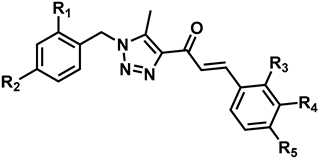
2.1. Synthesis of Chalcone Derivatives

For the synthesis of 1,2,3-triazole-linked chalcones, the Benzyl Acetyl Triazole derivatives (BAT) 2ad (Scheme 1) were first obtained as key intermediates. Their synthesis was carried out sequentially by substitution of benzyl chloride derivatives with NaN3 and subsequent cyclocondensation with acetylacetone in a basic medium, adapting procedures previously described by Chen et al. [46] and Nelson et al. [47]. Finally, the synthesis of the triazolic chalcone analogues 4av was achieved using the method of Shankarling et al., involving a KOH (40% w/v)-promoted, ultrasound-assisted (frequency: 50/60 Hz) cross-aldol condensation between BAT 2ad and benzaldehyde derivatives (Scheme 1 and Table 1) [48].
The structural determination of the synthesized products was carried out through spectroscopic 1D and 2D NMR experiments, IR spectroscopy, and MS spectrometry. In the IR spectra of the chalcones, intense and sharp bands, attributable to the conjugated ketone carbonyl groups, were observed between 1684 and 1653 cm−1. In the 13C-NMR spectra, the carbonyl group was observed between 183.4 and 184.8 ppm (Table 2). Additionally, in the 1H-NMR spectra, two doublet signals were observed at approximately 8.31 and 7.78 ppm, both with a coupling constant (J) of 16 Hz (Table 2). This observation indicates the presence of vinyl protons conjugated to a ketone carbonyl group in a trans-configuration.

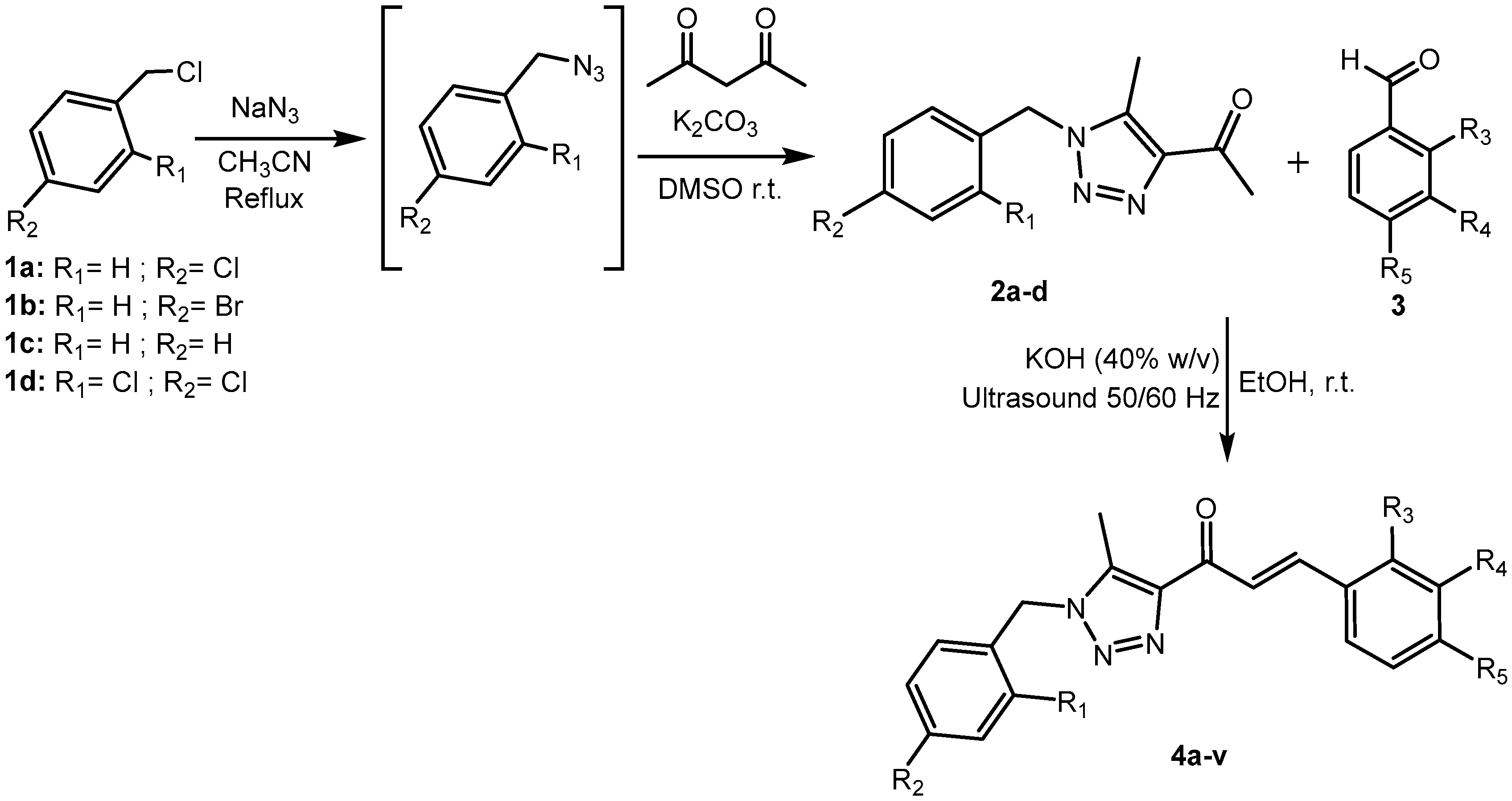
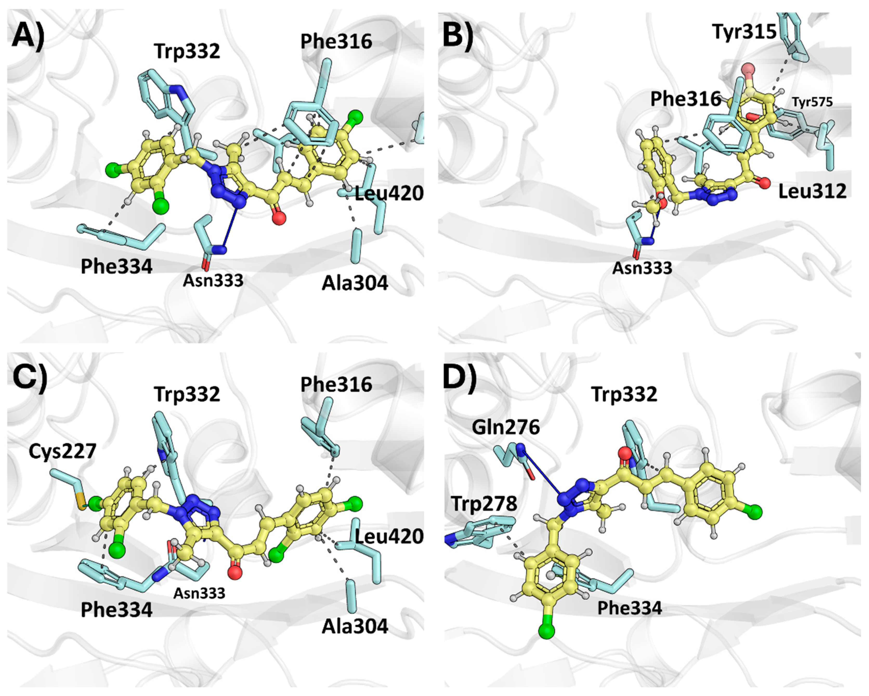
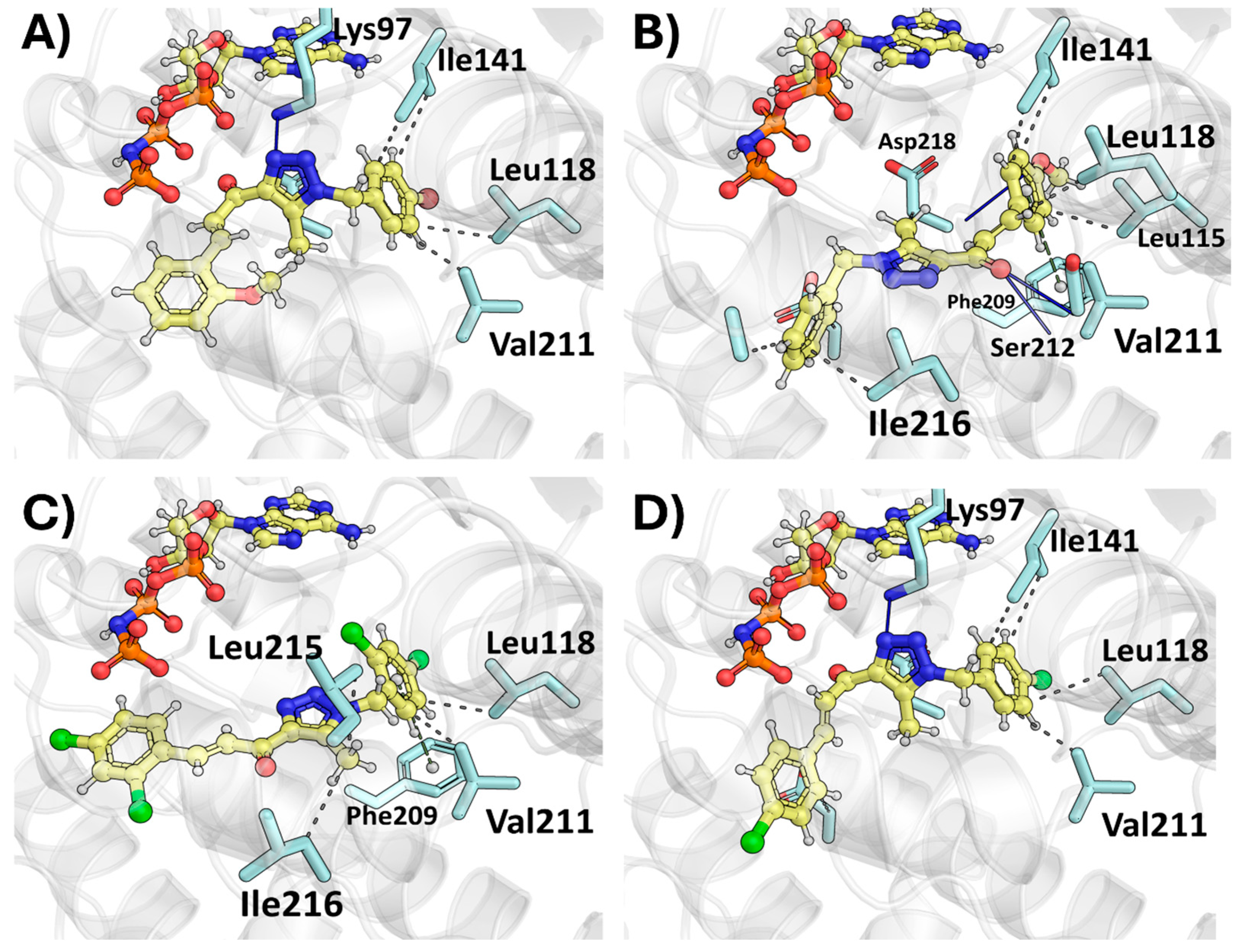
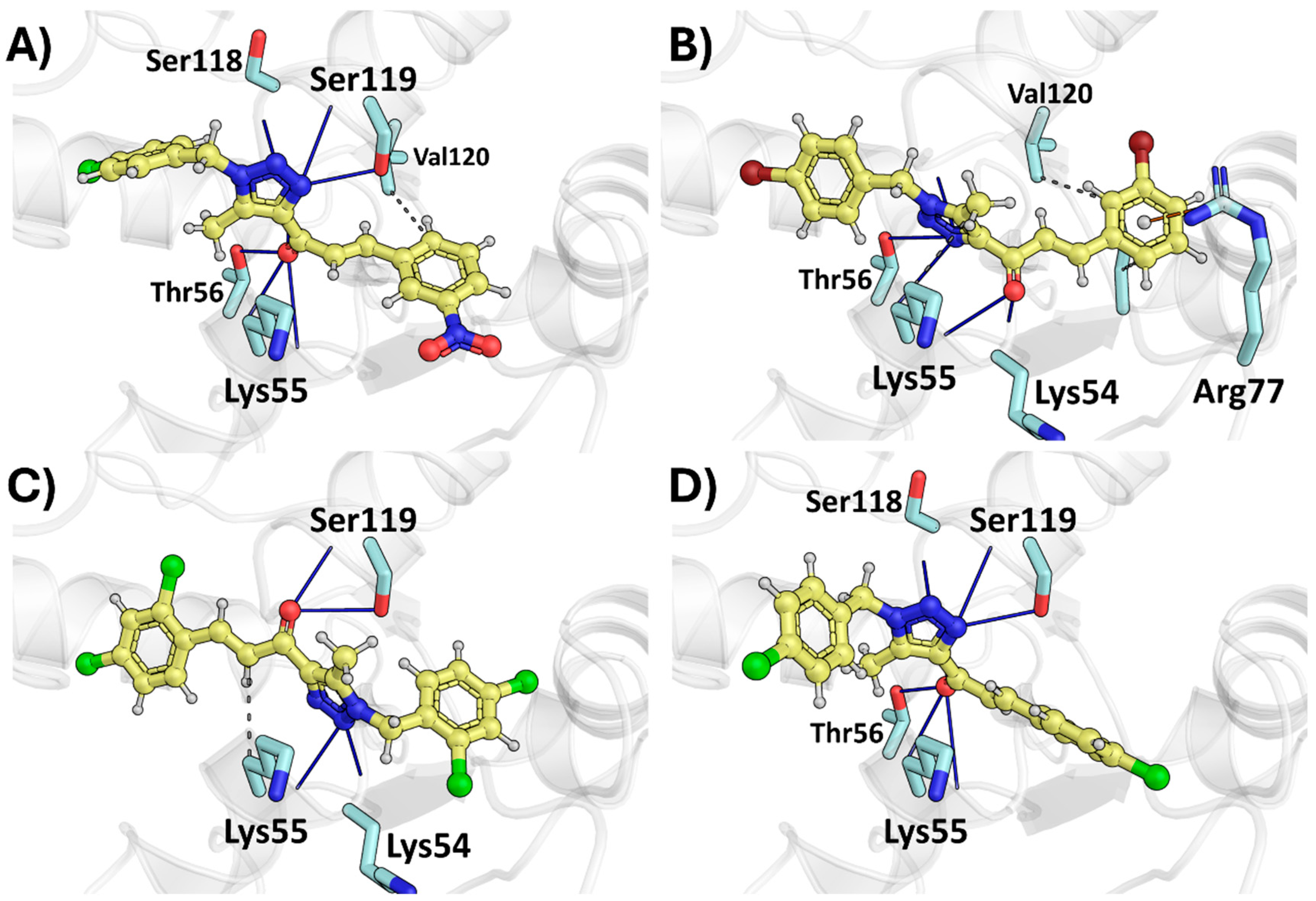
2.2. Molecular Docking

A molecular docking study was performed to identify the predominant interactions of the most active synthesized ligands in each of the cell lines studied. The analysis was organized according to the most representative therapeutic target for each cell line. The estimated energies were expressed in kcal/mol, and images were obtained using PyMol (Table 3).

2.2.1. Therapeutic Target Involved in U937—VEGFR Kinase

The VEFGR protein was selected as a target due to its reported overexpression in U937 and KG-1 cell lines [49]. One of the most relevant drugs for U937 is sorafenib, a multikinase inhibitor approved by the FDA, which has been shown to reduce the prevalence of refractory leukemia in its early stages [50]. VEFGR was obtained from the PDB (4ASD) [51]. The most active ligands, 4a and 4c (Figure 3A,B), were compared with the least active compounds, 4b and 4s (IC50 > 200 µM).
The docking poses were obtained by generating the top ten poses for each ligand. Compound 4c (Figure 3B) presented favorable interactions due to its optimal phenyl arrangement, which enhanced the carbonyl orientation, allowing a favorable hydrogen bond with the Cys919 residue, which interacted with sorafenib (co-crystallized ligand). In addition, ligand 4c oriented its phenylmethoxyl fragment towards the internal region of the binding site, establishing favorable interactions with Phe1047, Val91, and Val848. Furthermore, the chlorobenzyl group was positioned to form a π-stacking interaction with Phe918. In contrast, compound 4a (Figure 3A) oriented its chlorobenzyl group towards the innermost pocket, interacting with Val848. The triazole fragment was positioned to form a hydrogen bond with the amide proton of residue Asp1046, which interacted with Leu840, Leu1035, and Phe1047 to accommodate the ligand and enhance its Van der Waals interactions. These interactions were also observed in sorafenib, explaining the strong biological activity and binding affinity of the ligands for the target site.
On the other hand, the low activity of compounds 4b and 4s (Figure 3C,D) may be attributed to the presence of chlorine atoms on the phenyl rings, which hindered the formation of favorable interactions within the internal pocket of the binding site. In the case of compound 4s, its binding pose exposed the ligand to the solvent region of the binding site. In contrast, compound 4b exhibited a binding mode similar to compound 4a; however, the chlorophenyl group of the chalcone moiety does not engage in hydrophobic interactions as effectively as the bromobenzene group in 4a.

2.2.2. Therapeutic Target Involved in T98G—Tissue Transglutaminase, TG2

The therapeutic target studied was transglutaminase 2 (TG2), in accordance with Gundemir et al. [52], who related TG2 overexpression to cell survival and proliferation in glioblastomas (GMBs). For this study, the Protein Data Bank structure 3S3J [53] was used to identify the predominant interactions contributing to the biological activity of the most active compounds, particularly 4q and 4v, and to compare them with the less active compounds 4b and 4s (Figure 4).
The arrangement of the most active ligands revealed that the chalcone fragment oriented the para-halogenated phenyl group in the hydrophobic region, constituted by Leu420 and Phe316 in the case of compound 4q. On the other hand, compound 4v penetrated deeper into this pocket, establishing additional interactions with Tyr315, Tyr575, and Leu312, in addition to Phe316. This enhanced binding is attributed to the ortho-methoxy substitution on the benzyl group in 4v, which allowed the formation of a hydrogen bond with Asn333, facilitating its entry into a more hydrophobic region compared to 4q.
In contrast, the less active compounds 4b and 4s failed to establish similar hydrophobic interactions, due to the absence of hydrogen bond acceptor groups at the ortho-position of the benzyl group (Figure 4D). Additional substitutions, such as the dichlorinated phenyls in compound 4t, prevented the ligand from adopting a suitable orientation to interact with Phe316 and Leu420.

2.2.3. Therapeutic Target Involved in Gb-d1—MEK

MEK is an active kinase implicated in several gallbladder cancer cell lines [54]. The protein crystallized structure, obtained from the Protein Data Bank (PDB ID: 7M0T) [55], contains the ligand selumetinib, a kinase inhibitor whose binding site is located in the innermost region of the kinase, thereby inhibiting the active state of MEK.
The most active ligands, 4k and 4o, showed favorable interactions similar to those of selumetinib (SI). In the case of compound 4k, hydrogen bond interactions with Lys97 were favored, resulting in additional hydrophobic interactions with Ile141, Leu118, and Val211. Compound 4o primarily formed a hydrogen bond between the carbonyl group and amide of Ser212, enhancing its interaction with the hydrophobic region constituted by Ile141, Leu118, Leu115, and Phe209 (Figure 5).
In contrast, the less active compounds 4b and 4s exhibited poor interactions with the hydrophobic region. Although compound 4b possesses a single substituent on the aromatic ring, which could favor an interaction similar to that of 4q, the absence of a substituent at the ortho-position and the presence of chlorine at the para-position prevented the ligand from stabilizing in the innermost hydrophobic region of the binding site. For compound 4s, the disubstitution of the aromatic rings hindered the formation of a hydrogen bond at the binding site.

2.2.4. Therapeutic Target Involved in HeLa—Dihydrofolate Reductase, DHFR

According to our previously published work, one of the most significant therapeutic targets in HeLa cells is dihydrofolate reductase (DHFR) [56]. The docking poses obtained corresponded to compounds 4g and 4f, which exhibited activities of 42.5 and 44.5 μM, respectively. Similarly to the analysis in Section 2.2.2, these ligands were compared with the less active compounds 4b and 4s (Figure 6).
All four ligands formed hydrogen bonds with the binding site through their carbonyl and triazole groups. The most notable differences arose from the substitutions on the aromatic rings. In the case of compound 4g, the triazole arrangement allowed hydrogen bonding with Ser118 and Ser119, while the carbonyl interacted with Thr56. The aromatic rings enhanced these interactions, providing orientations that stabilized the carbonyl group, such as the arrangement of the m-nitrophenyl ring towards Val120. In the case of compound 4f, its orientation included an additional interaction with Arg77, suggesting that substitutions at the meta-position favored the positioning of the ring in the hydrophobic region.
In contrast, ligands 4b and 4s lacked these substitutions, and instead featured para-substituents, which hindered the optimal arrangement of the carbonyl group. As a result, in compound 4b, the rings were oriented towards the solvent-exposed region, whereas in compound 4s, the rings adopted an alternative conformation to facilitate hydrogen bonding between the carbonyl and Ser119, thereby losing crucial hydrophobic interactions.

2.3. Cytotoxic Activities

The cytotoxic effects of chalcones 4a4v were evaluated against five cancer cell lines (G415, Gbd1, T98G, HeLa, and U937) using the MTT colorimetric assay after 24 h of exposure (Table 4) [57]. The IC50 values were determined from dose–response curves, and ranged from 4.4 to >200 μM, indicating a variable cytotoxic effect, depending on both the structure of the chalcones and the specific cell line tested. Compounds with IC50 values greater than 200 μM were considered inactive.
Among the tested cell lines, the human non-Hodgkin lymphoma cell line (U937) was the most sensitive, with 16 chalcones exhibiting IC50 values below 20 μM. Notably, compounds 4a, 4c, 4d, 4e, 4f, 4g, 4h, 4i, 4j, 4k, 4l, 4m, 4n, 4o, 4t, and 4v showed IC50 values between 4.4 and 17.8 μM. The most potent compound against U937 was 4c (IC50 = 4.4 μM), followed by 4a (IC50 = 4.6 μM), both featuring substitutions at the para position. However, they differ in the nature of their substituents. Compound 4a has a bromine atom, whereas compound 4c contains a chlorine atom and a methoxy group at the para position of the aromatic ring of the chalcone. The human glioblastoma cell line (T98G) was sensitive to 4d, 4f, 4i, 4j, 4k, 4l, 4m, 4o, 4p, 4q, 4r, and 4v, with IC50 values ranging from 11 to 66.5 μM. The most active compounds were 4v (IC50 = 11 μM), 4r (IC50 = 14 μM), and 4q (IC50 = 12.3 μM), all characterized by the presence of bulky groups at R1 and R2, as well as halogen groups at the R3 or R5 positions. Compound 4v was the most active, likely due to the presence of substituents at the ortho position. This structural feature is absent in the less active compounds 4g and 4i, suggesting the importance of substituent position in modulating biological activity in this cell line. The human gallbladder carcinoma lines (Gb-d1) exhibited moderate sensitivity to 10 chalcones, with IC50 values ranging from 16 to 58.8 μM, comparable to the reference drug (paclitaxel, IC50 = 21 μM). The most potent compound was 4o (IC50 = 16 μM), followed by 4h (IC50 = 17.2 μM), both characterized by the presence of chlorine atoms at the R3 and R5 positions. The human cervical adenocarcinoma cell line (HeLa) was slightly sensitive to only four chalcones (4f, 4g, 4m, and 4o), with IC50 values ranging from 42.5 to 95.5 μM, indicating lower sensitivity compared to other cell lines. The main difference between these compounds and the others is the absence of substitutions at the para position of the aromatic rings. Compound 4g features chlorine substitutions at the ortho position and a nitro group at the ortho position of the chalcone fragment. A similar trend was observed with gallbladder carcinoma cell line (G-415), which was the least sensitive. Only three compounds (4f, 4g, and 4m) showed weak cytotoxicity, with IC50 values of 27.6, 51, and 26 μM, respectively.
Interestingly, chalcones with halogen substituents (Cl, Br) exhibited enhanced cytotoxic activity in multiple cell lines, particularly U937 and T98G, suggesting a possible influence of electronic effects on their mechanism of action. On the other hand, chalcones containing electron-donating groups, such as a methoxy group, showed moderate activity, especially in T98G and HeLa cell lines. When comparing the activity of chalcones with that of paclitaxel, a well-known anticancer agent, it is notable that compounds such as 4a, 4c, and 4d in U937, and 4v in T98G, exhibited IC50 values within a comparable range, suggesting their potential as lead compounds for further optimization. However, the presence of multiple chlorine atoms in compounds such as 4b and 4s was associated with reduced efficacy, negatively affecting their biological activity (4b and 4s). Overall, the cytotoxic activity of the chalcones appears to be influenced by both structural modifications and the specific cell line tested. Further investigations will be necessary to fully evaluate their therapeutic potential.

3. Materials and Methods

3.1. General Section

Melting points were determined on a Stuart SMP3 apparatus (Staffordshire UK), and are uncorrected. IR spectra were recorded on a Perkin-Elmer FT-IR Spectrometer Spectrum Two (Llantrisant, UK) with KBr. NMR spectra were acquired in DMSO-d6 or CDCl3 with a Varian Unity Inova 500 MHz spectrometer (Palo Alto, CA, USA). Chemical shifts are reported in parts per million (δ) relative to the residual solvent signals (DMSO-d6: δH 2.50, δC 39.5 or CDCl3; δH H 7.26, δC C 77.2) as internal standards for the 1H and 13C NMR spectra. Coupling constants (J) are expressed in Hz. HRMS spectra were recorded using a Micromass-LCT Premier Time-of-Flight ESI spectrometer, coupled with an ACQUITY UHPLC (Milford, Massachusetts, USA) (ultra-high-performance liquid chromatography) interface system. For copies of 1H, 13C NMR, HSQC, HMB, and HSMS-ESI, see Section 4 in the Supplementary Materials. The reactions were monitored by thin-layer chromatography (TLC), performed on silica gel Merck 60 F254. The components were visualized under UV light (254 and 365 nm), and/or by treatment with phosphomolybdic acid reagent, followed by heating. All starting materials and reagents were obtained from commercial suppliers [46,47,58,59,60,61].

3.2. General Procedure for Synthesis of Triazolic Chalcones (4a4v) (Exemplified for Synthesis of 4a)

Ijms 26 03389 i002
A mixture of 1.0 g (4.0 mmol) of 2a, 0.74 g (4.0 mmol) of 4-bromobenzaldehyde, and 40 mL of ethanol was subjected to ultrasound (50/60 Hz) for 2 min. Then, 7.0 mL of KOH (40% w/v) was added over 2 min. After completing the addition, the reaction mixture was subjected to ultrasound (50/60 Hz) at room temperature for 10 min. The resulting precipitate was filtered, washed with ethanol, and vacuum-dried, yielding 1.52 g (90% yield) of 4a as a white solid (Rf: 0.72, eluent: hexane/EtOAc, 1/1).
  • (E)-3-(4-bromophenyl)-1-(1-(4-chlorobenzyl)-5-methyl-1H-1,2,3-triazol-4-yl)-2-propen-1-one (4a).
White solid (90% yield), m.p.: 184-186 °C. Rf: 0.72, eluent: hexane/EtOAc, 1/1. IR (cm−1) v: 3063 and 3026 (CAr-H); 2988 and 2967 (Csp3-H); 1684 (C=O); 1601 (C=C); 1592 (CAr-CAr); 1554 (N=N). 1H-NMR (CDCl3, 500 MHz) δ (ppm): 2.55 (3H, s), 5.52 (2H, s), 7.14 (2H, d, J = 8.3 Hz, H4), 7.34 (2H, d, J = 8.4 Hz), 7.55 (4H, m), 7.80 (1H, d, J = 16.0 Hz), 8.02 (1H, d, J = 16.0 Hz). 13C-NMR (CDCl3, 125.7 MHz) δ (ppm): 9.5 (CH3), 51.2 (CH2), 123.6 (CH), 125.0 (C), 128.8 (2CH), 129.8 (2CH), 130.2 (2CH), 132.3 (2CH), 132.5 (C), 134.0 (C), 134.9 (C), 138.1 (C), 142.4 (CH), 144.4 (C), 184.2 (C). HRMS-ESI (m/z) calculated for C19H16ClBrN3O [M + H]+: 416.01652, found: 416.01669.
  • (E)-3-(4-chlorophenyl)-1-(1-(4-chlorobenzyl)-5-methyl-1H-1,2,3-triazol-4-yl)-2-propen-1-one (4b).
White solid (85% yield), m.p.: 176–178 °C. Rf: 0.77, eluent: hexane/EtOAc, 1/1. IR (cm−1) v: 3053 (CAr-H); 2984 and 2952 (Csp3-H); 1661 (C=O); 1601 (C=C); 1590 (CAr-CAr); 1567 (N=N). 1H-NMR (CDCl3, 500 MHz) δ (ppm): 2.55 (3H, s), 5.51 (2H, s), 7.14 (2H, d, J = 8.3 Hz), 7.34 (2H, d, J = 8.4 Hz), 7.38 (2H, d, J = 8.4 Hz), 7.63 (2H, d, J = 8.4 Hz), 7.82 (1H, d, J = 16.0 Hz), 8.00 (1H, d, J = 16.0 Hz). 13C-NMR (CDCl3, 125.7 MHz) δ (ppm): 9.5 (CH3), 51.2 (CH2), 123.5 (CH), 128.8 (2CH), 129.3 (2CH), 129.6 (2CH), 130.0 (2CH), 132.6 (C), 133.6 (C), 134.9 (C), 136.6 (C), 138.1 (C), 142.3 (CH), 144.4 (C), 184.2 (C). HRMS-ESI (m/z) calculated for C19H16Cl2N3O [M + H]+: 372.06703, found: 372.06772.
  • (E)-1-((4-chlorobenzyl)-5-methyl-1H-1,2,3-triazol-4-yl)-3-(4-methoxyphenyl)-2-propen-1-one (4c).
Yellow solid (91% yield), m.p.: 173–175 °C. Rf: 0.63, eluent: hexane/EtOAc, 1/1. IR (cm−1) v: 3084 nd 3051 (CAr-H); 2981; 2948 and 2849 (Csp3-H); 1656 (C=O); 1591 (C=C); 1570 (CAr-CAr); 1570 (N=N). 1H-NMR (CDCl3, 500 MHz) δ (ppm): 2.52 (3H, s), 3.82 (3H, s), 5.48 (2H, s), 6.91 (2H, d, J = 8.3 Hz), 7.11 (2H, d, J = 8.1 Hz), 7.30 (2H, d, J = 7.9 Hz), 7.63 (2H, d, J = 8.3 Hz), 7.82 (1H, d, J = 15.9 Hz), 7.90 (1H, d, J = 15.8 Hz). 13C-NMR (CDCl3, 125.7 MHz) δ (ppm): 9.4 (CH3), 51.0 (CH2), 55.4 (CH3), 114.4 (2CH), 120.6 (CH), 127.7 (C), 128.7 (2CH), 129.4 (2CH), 130.5 (2CH), 132.6 (C), 134.6 (C), 137.7 (C), 143.5 (CH), 144.4 (C), 161.8 (C), 184.3 (C). HRMS-ESI (m/z) calculated for C20H19ClN3O2 [M + H]+: 368.11657, found: 368.11703.
  • (E)-3-(2,4-dichlorophenyl)-1-(1-(4-chlorobenzyl)-5-methyl-1H-1,2,3-triazol-4-yl)-2-propen-1-ona (4d).
White solid (90% yield), m.p.: 181–182 °C. Rf: 0.75, eluent: hexane/EtOAc, 1/1. IR (cm−1) v: 3064 and 3025 (CAr-H); 2990 and 2952 (Csp3-H); 1669 (C=O); 1604 (C=C); 1583 (CAr-CAr); 1561(N=N). 1H-NMR (CDCl3, 500 MHz) δ (ppm): 2.56 (3H, s,), 5.52 (2H, s), 7.14 (2H, d, J = 8.3 Hz), 7.30 (1H, d, J = 8.5 Hz), 7.34 (2H, d, J = 8.4 Hz), 7.46 (1H, s), 7.81 (1H, d, J = 8.5 Hz), 8.00 (1H, d, J = 16.0 Hz), 8.22 (1H, d, J = 16.0 Hz). 13C-NMR (CDCl3, 125.7 MHz) δ (ppm): 9.5 (CH3), 51.2 (CH2), 125.6 (CH), 127.7 (CH), 128.8 (3CH), 129.6 (2CH), 130.2 (CH), 132.9 (C), 132.5 (C), 134.7 (C), 136.4 (C), 136.7 (CH), 138.2 (C), 138.3 (CH), 144.3 (C), 183.8 (C). HRMS-ESI (m/z) calculated for C19H15N3OCl3 [M + H]+: 406.02806, found: 406.02866.
  • (E)-3-(2-chlorophenyl)-1-(1-(4-chlorobenzyl)-5-methyl-1H-1,2,3-triazol-4-yl)-2-propen-1-one (4e).
White solid (75% yield), m.p.: 144–146 °C. Rf: 0.69, eluent: hexane/EtOAc, 1/1. IR (cm−1) v: 3067 (CAr-H); 2992 and 2956 (Csp3-H); 1667 (C=O); 1604 (C=C); 1583 (CAr-CAr); 1561 (N=N). 1H-NMR (CDCl3, 500 MHz) δ (ppm): 2.56 (3H, s), 5.52 (2H, s), 7.14 (2H, d, J = 8.3 Hz), 7.31 (2H, dd, J = 8.8 and 5.5 Hz), 7.34 (2H, d, J = 8.4 Hz), 7.43 (1H, d, J = 6.8 Hz), 7.88 (1H, d, J = 6.7 Hz), 8.03 (1H, d, J = 15.9 Hz), 8.31 (1H, d, J = 15.9 Hz). 13C-NMR (CDCl3, 125.7 MHz) δ (ppm): 9.5 (CH3), 51.2 (CH2), 125.1 (CH), 127.2 (C), 128.2 (CH), 128.8 (2CH), 129.6 (2CH), 130.3 (CH), 131.4 (CH), 132.6 (C), 133.3 (C), 134.9 (C), 135.9 (CH), 138.2 (C), 139.5 (CH), 144.4 (C), 184.1 (C). HRMS-ESI (m/z) calculated for C19H16Cl2N3O [M + H]+: 372.06703, found: 372.06665.
  • (E)-1-((4-bromobenzyl)-5-methyl-1H-1,2,3-triazol-4-yl)-3-(3-bromophenyl)-2-propen-1-one (4f).
White solid (82% yield), m.p.: 141–143 °C. Rf: 0.75, eluent: hexane/EtOAc, 1/1. IR (cm−1) v: 3067 and 3026 (CAr-H); 2990 and 2952 (Csp3-H); 1667 (C=O); 1611 (C=C); 1585 (CAr-CAr); 1560 (N=N). 1H-NMR (CDCl3, 500 MHz) δ (ppm): 2.55 (3H, s), 5.51 (2H, s), 7.08 (2H, d, J = 8.2 Hz), 7.85 (1H, s), 7.29 (1H, t, J = 7.8 Hz), 7.50 (2H, d, J = 8.3 Hz), 7.61 (1H, d, J = 8.5 Hz), 8.02 (1H, d, J = 15.9 Hz), 7.78 (1H, d, J = 16.0 Hz). 13C-NMR (CDCl3, 125.7 MHz) δ (ppm): 9.0 (CH3), 50.9 (CH2), 122.9 (C), 124.2 (CH), 127.3 (CH), 129.0 (2CH), 130.4 (CH), 131.3 (CH), 132.4 (2CH), 132.5 (C), 132.9 (CH), 136.5 (C), 138.1 (C), 141.9 (CH), 144.3 (C), 183.9 (C). HRMS-ESI (m/z) calculated for C19H16Br2N3O [M + H]+: 459.96600, found: 459.96606.
  • (E)-1-((4-chlorobenzyl)-5-methyl-1H-1,2,3-triazol-4-yl)-3-(3-nitrophenyl)-2-propen-1-one (4g).
Orange solid (60% yield), m.p.: 182–183 °C. Rf: 0.81, eluent: hexane/EtOAc, 1/1. IR (cm−1) v: 3090 (CAr-H); 3061 (CAr-H); 2964 (Csp3-H); 2949 (Csp3-H); 2917 (Csp3-H); 1661 (C=O); 1600 (C=C); 1571 (CAr-CAr); 1561 (N=N); 1528 (N=O); 1345 (N-O). 1H-NMR (CDCl3, 500 MHz) δ (ppm): 2.57 (3H, s), 5.53 (2H, s), 7.15 (2H, d, J = 8.2 Hz), 8.53 (1H, s), 7.61 (1H, t, J = 7.9 Hz), 7.35 (2H, d, J = 8.3 Hz), 8.25 (1H, d, J = 8.5 Hz), 8.13 (1H, d, J = 15.9 Hz), 7.89 (1H, d, J = 16.0 Hz). 13C-NMR (CDCl3, 125.7 MHz) δ (ppm): 9.0 (CH3), 50.9 (CH2), 134.7 (C), 123.1 (CH), 124.6 (C), 125.6 (CH), 128.0 (C), 128.8 (2CH), 129.4 (CH), 129.5 (2CH), 132.0 (C), 134.1 (CH), 137.9 (C), 140.6 (CH), 144.0 (C), 148.6 (C), 183.4 (C). HRMS-ESI (m/z) calculated for C19H16ClN4O3 [M + H]+: 383.09108, found: 383.09238.
  • (E)-1-((4-bromobenzyl)-5-methyl-1H-1,2,3-triazol-4-yl)-3-(2-chlorophenyl)-2-propen-1-one (4h).
White solid (85% yield), m.p.: 167–168 °C. Rf: 0.77, eluent: hexane/EtOAc, 1/1. IR (cm−1) v: 3061 (CAr-H); 2990 and 2955 (Csp3-H); 1667 (C=O); 1603 (C=C); 1593 (CAr-CAr); 1555 (N=N). 1H-NMR (CDCl3, 500 MHz) δ (ppm): 2.56 (3H, s), 5.50 (2H, s), 7.08 (2H, d, J = 8.3 Hz), 7.31 (1H, dd, J = 6.8 and 5.3 Hz), 7.33 (1H, d, J = 5.7 and 3.5 Hz), 7.43 (1H, d, J = 6.8 Hz), 7.50 (2H, d, J = 8.4 Hz), 7.88 (1H, d, J = 7.1 Hz), 8.03 (1H, d, J = 16.0 Hz), 8.31 (1H, d, J = 16.0 Hz). 13C-NMR (CDCl3, 125.7 MHz) δ (ppm): 9.5 (CH3), 51.2 (CH2), 123.0 (C), 125.2 (CH), 127.2 (C), 128.1 (CH), 129.1 (2CH), 130.3 (CH), 131.4 (CH), 132.5 (2CH), 130.1 (C), 133.3 (C), 135.9 (CH), 138.2 (C), 139.5 (CH), 144.4 (C), 184.1 (C). HRMS-ESI (m/z) calculated for C19H16BrClN3O [M + H]+: 416.01652, found: 416.01660.
  • (E)-1-((1-(4-bromobenzyl)-5-methyl-1H-1,2,3-triazol-4-yl)-3-(2,4-dichlorophenyl)-2-propen-1-one (4i).
White solid (71% yield), m.p.: 183–185 °C. Rf: 0.78, eluent: hexane/EtOAc, 1/1. IR (cm−1) v: 3063 and 3022 (CAr-H); 2991 and 2953 (Csp3-H); 1667 (C=O); 1603 (C=C); 1582 (CAr-CAr); 1559 (N=N). 1H-NMR (CDCl3, 500 MHz) δ (ppm): 2.56 (3H, s), 5.50 (2H, s), 7.08 (2H, d, J = 8.2 Hz), 7.30 (1H, d, J = 8.4 Hz), 7.46 (1H, s), 7.50 (2H, d, J = 8.3 Hz), 7.81 (1H, d, J = 8.5 Hz), 8.00 (1H, d, J = 15.9 Hz), 8.22 (1H, d, J = 16.0 Hz). 13C-NMR (CDCl3, 125.7 MHz) δ (ppm): 9.5 (CH3), 51.3 (CH2), 123.0 (C), 125.6 (CH), 127.7 (CH), 128.8 (CH), 129.1 (2CH), 130.2 (C), 131.9 (CH), 132.6 (2CH), 133.0 (C), 136.5 (C), 136.7 (CH), 138.2 (C), 138.3 (CH), 144.3 (C), 183.8 (C). HRMS-ESI (m/z) calculated for C19H15BrCl2N3O [M + H]+: 449.97754, found: 449.97662.
  • (E)-1-((4-bromobenzyl)-5-methyl-1H-1,2,3-triazol-4-yl)-3-(4-methoxyphenyl)-2-propen-1-one (4j).
Yellow solid (56% yield), m.p.: 185–187 °C. Rf: 0.67, eluent: hexane/EtOAc, 1/1. IR (cm−1) v: 3049 (CAr-H); 2950 (Csp3-H); 1658 (C=O); 1592 (C=C); 1572 (CAr-CAr); 1552 (N=N). 1H-NMR (CDCl3, 500 MHz) δ (ppm): 2.55 (3H, s), 3.85 (3H, s), 5.49 (2H, s), 6.93 (2H, d, J = 8.2 Hz), 7.07 (2H, d, J = 8.0 Hz), 7.49 (2H, d, J = 8.0 Hz), 7.66 (2H, d, J = 8.3 Hz), 7.85 (1H, d, J = 15.9 Hz), 7.92 (1H, d, J = 15.9 Hz). 13C-NMR (CDCl3, 125.7 MHz) δ (ppm): 9.5 (CH3), 51.2 (CH2), 55.6 (CH3), 114.4 (2CH), 120.6 (CH), 122.8 (C), 127.7 (C), 128.9 (2CH), 130.6 (2CH), 132.4 (2CH), 133.1 (C), 137.7 (C), 143.6 (CH), 144.5 (C), 161.9 (C), 184.5 (C). HRMS-ESI (m/z) calculated for C20H19BrN3O2 [M + H]+: 412.06605, found: 412.06699.
  • (E)-1-((4-bromobenzyl)-5-methyl-1H-1,2,3-triazol-4-yl)-3-(2-methoxyphenyl)-2-propen-1-one (4k).
Yellow solid (85% yield), m.p.: 130–132 °C. Rf: 0.67, eluent: hexane/EtOAc, 1/1. IR (cm−1) v: 3058 (CAr-H); 2964 (Csp3-H); 2949 (Csp3-H); 2843 (Csp3-H); 1657 (C=O); 1589 (C=C); 1563 (CAr-CAr); 1552 (N=N). 1H-NMR (CDCl3, 500 MHz) δ (ppm): 2.55 (3H, s), 3.91 (3H, s), 5.49 (2H, s), 6.92 (1H, d, J = 8.1 Hz), 6.98 (1H, dd, J = 7.1 and 14.6 Hz), 7.07 (2H, d, J = 8.2 Hz), 7.36 (1H, dd, J = 7.8 and 7.5 Hz), 7.48 (2H, d, J = 8.2 Hz), 7.75 (1H, d, J = 7.6 Hz), 8.07 (1H, d, J = 16.1 Hz), 8.26 (1H, d, J = 16.1 Hz). 13C-NMR (CDCl3, 125.7 MHz) δ (ppm): 9.4 (CH3), 51.1 (CH2), 55.7 (CH3), 111.3 (CH), 120.8 (C), 122.9 (C), 123.2 (CH), 124.0 (CH), 128.4 (C), 129.0 (2CH), 132.0 (CH), 132.4 (2CH), 133.2 (C), 137.9 (C), 139.1 (CH), 144.7 (C), 159.0 (C), 184.8 (C). HRMS-ESI (m/z) calculated for C20H19BrN3O2 [M + H]+: 412.06605, found: 412.06635.
  • (E)-1-((4-bromobenzyl)-5-methyl-1H-1,2,3-triazol-4-yl)-3-(3,4-dimethoxyphenyl)-2-propen-1-one (4l).
Yellow solid (79% yield), m.p.: 154–156 °C. Rf: 0.47, eluent: hexane/EtOAc, 1/1. IR (cm−1) v: 3051 (CAr-H); 2953 (Csp3-H); 2836 (Csp3-H); 1653 (C=O); 1593 (CAr-CAr); 1568 (N=N). 1H-NMR (CDCl3, 500 MHz) δ (ppm): 2.56 (3H, s), 3.94 (3H, s), 3.97 (3H, s), 5.50 (2H, s), 6.85 (1H, d, J = 8.2 Hz), 7.08 (2H, d, J = 8.1 Hz), 7.26 (2H, d, J = 8.2 Hz), 7.50 (2H, d, J = 8.2 Hz), 7.85 (1H, d, J = 15.9 Hz), 7.91 (1H, d, J = 15.9 Hz). 13C-NMR (CDCl3, 125.7 MHz) δ (ppm): 9.5 (CH3), 51.2 (CH2), 56.1 (2CH3), 110.0 (CH), 111.2 (CH), 120.8 (CH), 122.9 (C), 124.1 (CH), 128.1 (C), 129.1 (2CH), 132.5 (2CH), 133.2 (C), 137.9 (C), 144.1 (CH), 144.6 (C), 149.4 (C), 151.7 (C), 184.3 (C). HRMS-ESI (m/z) calculated for C21H21BrN3O3 [M + H]+: 442.07662, found: 442.07532.
  • (E)-1-(benzyl)-5-methyl-1H-1,2,3-triazol-4-yl)-3-phenyl-2-propen-1-one (4m).
White solid (84% yield), m.p.: 153–155 °C (Lit. 157–158 °C [62]). Rf: 0.68, eluent: hexane/EtOAc, 1/1. IR (cm−1) v: 3060 (CAr-H); 3032 (CAr-H); 2973 (Csp3-H); 1663 (C=O); 1600 (C=C); 1574 (CAr-CAr); 1561 (CAr-CAr). 1H-NMR (CDCl3, 500 MHz) δ (ppm): 2.54 (3H, s), 5.54 (2H, s), 7.18 (2H, d, J = 7.1 Hz), 7.32 (3H, m), 7.39 (3H, m), 7.88 (1H, d, J = 16.0 Hz), 8.06 (1H, d, J = 16.0 Hz). 13C-NMR (CDCl3, 125.7 MHz) δ (ppm): 9.4 (CH3), 51.8 (CH2), 123.0 (CH), 127.3 (2CH), 128.7 (CH), 128.8 (2CH), 128.9 (2CH), 129.2 (2CH), 130.6 (CH), 134.1 (C), 135.5 (C), 138.1 (C), 143.6 (CH), 144.4 (C), 184.4 (C). HRMS-ESI (m/z) calculated for C19H18N3O [M + H]+: 304.14498, found: 304.14496.
  • (E)-1-(benzyl)-5-methyl-1H-1,2,3-triazol-4-yl)-3-(4-chlorophenyl)-2-propen-1-one (4n).
White solid (90% yield), m.p.: 160–161 °C (Lit. 158–159 °C [62]). Rf: 0.76, eluent: hexane/EtOAc, 1/1. IR (cm−1) v: 3057 (CAr-H); 3031 (CAr-H); 2969 (Csp3-H); 1662 (C=O); 1603 (C=C); 1590 (CAr-CAr); 1566 (N=N). 1H-NMR (CDCl3, 500 MHz) δ (ppm): 2.55 (3H, s), 5.56 (2H, s), 7.19 (2H, d, J = 8.3 Hz), 7.35 (3H, m), 7.39 (2H, d, J = 8.4 Hz), 7.63 (2H, d, J = 8.4 Hz), 7.82 (1H, d, J = 16.0 Hz), 8.02 (1H, d, J = 16.0). 13C-NMR (CDCl3, 125.7 MHz) δ (ppm): 9.0 (CH3), 51.6 (CH2), 123.5 (CH), 127.2 (2CH), 127.5 (CH), 129.2 (2CH), 129.2 (2CH), 129.9 (2CH), 133.4 (C), 133.8 (C), 136.6 (C), 138.0 (C), 142.1 (CH), 144.0 (C), 184.0 (C). HRMS-ESI (m/z) calculated for C19H17ClN3O [M + H]+: 338.10600, found: 338.10522.
  • (E)-1-(bencil)-5-metil-1H-1,2,3-triazol-4-il)-3-(2,4-diclorofenil)-2-propen-1-ona (4o).
White solid (98% yield), m.p.: 145–147 °C (Lit. 142–143 °C [62]). Rf: 0.63, eluent: hexane/EtOAc, 1/1. IR (cm−1) v: 3087 (CAr-H); 3062 (CAr-H); 3033 (CAr-H); 2949 (Csp3-H); 1667 (C=O); 1604 (C=C); 1580 (CAr-CAr); 1559 (N=N). 1H-NMR (CDCl3, 500 MHz) δ (ppm): 2.56 (3H, s), 5.56 (2H, s), 7.19 (2H, d, J = 8.1 Hz), 7.30 (1H, dd, J = 8.5 and 2.1 Hz), 7.35 (3H, m), 7.46 (1H, d, J = 2.1 Hz), 7.82 (1H, d, J = 8.5 Hz), 8.02 (1H, d, J = 15.9 Hz), 8.22 (1H, d, J = 15.9 Hz). 13C-NMR (CDCl3, 125.7 MHz) δ (ppm): 9.0 (CH3), 51.6 (CH2), 123.9 (C), 125.5 (CH), 127.3 (2CH), 127.6 (CH), 128.8 (CH), 129.0 (2CH), 130.0 (CH), 133.7 (C), 136.4 (C), 136.7 (C), 137.9 (C), 138.0 (CH), 144.2 (C), 183.6 (C). HRMS-ESI (m/z) calculated for C19H16Cl2N3O [M + H]+: 372.06703, found: 372.06754.
  • (E)-1-(benzyl)-5-methyl-1H-1,2,3-triazol-4-yl)-3-(4-methoxyphenyl)-2-propen-1-one (4p).
Yellow solid (50% yield), m.p.: 133–135 °C (Lit. 132–133 °C [62]). Rf: 0.69, eluent: hexane/EtOAc, 1/1. IR (cm−1) v: 3069 and 3035 (CAr-H); 2979, 2947 and 2834 (Csp3-H); 1657 (C=O); 1598 (C=C); 1574 (CAr-CAr), 1568 (N=N). 1H-NMR (CDCl3, 500 MHz) δ (ppm): 2.55 (3H, s), 5.55 (2H, s), 7.19 (2H, d, J = 8.3 Hz), 7.34 (3H, m), 6.94 (2H, d, J = 8.2 Hz), 7.66 (2H, d, J = 8.3 Hz), 7.93 (1H, d, J = 15.9 Hz), 7.85 (1H, d, J = 15.9 Hz). 13C-NMR (CDCl3, 125.7 MHz) δ (ppm): 9.0 (CH3), 51.5 (CH2), 56.8 (CH3), 114.3 (2CH), 120.6 (CH), 127.1 (CH), 127.2 (2CH), 127.7 (C), 129.0 (2CH), 130.5 (2CH), 133.8 (C), 137.6 (C), 143.5 (CH), 144.7 (C), 161.9 (C), 184.4 (C). HRMS-ESI (m/z) calculated for C20H20N3O2 [M + H]+: 334.15554, found: 334.15646.
  • (E)-1-(2,4-dichlorobenzyl)-5-methyl-1H-1,2,3-triazol-4-yl)-3-(4-chlorophenyl)-2-propen-1-one (4q).
White solid (95% yield), m.p.: 190–192 °C. Rf: 0.82, eluent: hexane/EtOAc, 1/1. IR (cm−1) v: 3081 (CAr-H); 2956 (Csp3-H); 1666 (C=O); 1608 (C=C); 1589 (CAr-CAr); 1568 (N=N). 1H-NMR (CDCl3, 500 MHz) δ (ppm): 2.59 (3H, s), 5.62 (2H, s), 6.81 (1H, d, J = 8.3 Hz), 7.22 (1H, d, J = 8.4 Hz), 7.47 (1H, s), 7.39 (2H, d, J = 8.4 Hz), 7.64 (2H, d, J = 8.4 Hz), 7.84 (1H, d, J = 16.0 Hz), 8.01 (1H, d, J = 16.0 Hz). 13C-NMR (CDCl3, 125.7 MHz) δ (ppm): 8.8 (CH3), 47.9 (CH2), 123.3 (CH), 127.6 (2CH), 128.0 (CH), 129.5 (CH), 129.7 (CH), 129.9 (2CH), 130.2 (C), 133.6 (C), 133.7 (C), 136.6 (C), 136.9 (C), 137.9 (C), 142.4 (CH), 144.1 (C), 183.9 (C). HRMS-ESI (m/z) calculated for C19H15Cl3N3O [M + H]+: 406.02806, found: 406.02927.
  • (E)-1-(2,4-dichlorobenzyl)-5-methyl-1H-1,2,3-triazol-4-yl)-3-(2-chlorophenyl)-2-propen-1-one (4r).
White solid (86% yield), m.p.: 158–161 °C. Rf: 0.79, eluent: hexane/EtOAc, 1/1. IR (cm−1) v: 3080 (CAr-H); 3031 (CAr-H); 2957 (Csp3-H); 1666 (C=O); 1605 (C=C); 1588 (CAr-CAr); 1563 (N=N). 1H-NMR (CDCl3, 500 MHz) δ (ppm): 2.60 (3H, s), 5.63 (2H, s), 6.81 (1H, d, J = 8.3 Hz), 7.22 (1H, d, J = 8.4 Hz), 7.48 (1H, s), 7.33 (2H, m), 7.44 (1H, d, J = 6.8 Hz), 7.88 (1H, d, J = 6.7 Hz), 8.04 (1H, d, J = 15.9 Hz), 8.33 (1H, d, J = 15.9 Hz). 13C-NMR (CDCl3, 125.7 MHz) δ (ppm): 8.9 (CH3), 47.9 (CH2), 125.1 (CH), 127.2 (CH), 128.0 (CH), 128.1 (CH), 129.5 (CH), 129.8 (CH), 130.2 (C), 130.3 (CH), 131.4 (CH), 133.3 (C), 133.7 (C), 135.6 (C), 135.9 (C), 138.2 (C), 139.5 (CH), 144.0 (C), 183.7 (C). HRMS-ESI (m/z) calculated for C19H15Cl3N3O [M + H]+: 406.02806, found: 406.02872.
  • (E)-1-(2,4-dichlorobenzyl)-5-methyl-1H-1,2,3-triazol-4-yl)-3-(2,4-dichlorophenyl)-2-propen-1-one (4s).
White solid (98% yield), m.p.: 201–203 °C. Rf: 0.83, eluent: hexane/EtOAc, 1/1. IR (cm−1) v: 3090 (CAr-H); 3064 (CAr-H); 2953 (Csp3-H); 1669 (C=O); 1605 (C=C); 1584 (CAr-CAr); 1559 (N=N). 1H-NMR (CDCl3, 500 MHz) δ (ppm): 2.60 (3H, s), 5.62 (2H, s), 6.81 (1H, d, J = 8.3 Hz), 7.22 (1H, d, J = 8.4 Hz), 7.46 (1H, d, J = 5.6 Hz), 7.31 (1H, d, J = 8.4 Hz), 7.46 (1H, d, J = 5.6 Hz), 7.81 (1H, d, J = 8.5 Hz), 8.02 (1H, d, J = 15.9 Hz), 8.24 (1H, d, J = 16.0 Hz). 13C-NMR (CDCl3, 125.7 MHz) δ (ppm): 9.0 (CH3), 48.2 (CH2), 125.6 (CH), 127.6 (CH), 128.0 (CH), 128.7 (CH), 129.6 (CH), 129.8 (CH), 130.0 (CH), 130.1 (C), 132.9 (C), 133.8 (C), 135.6 (C), 136.3 (C), 136.7 (C), 138.3 (CH), 138.4 (C), 144.3 (C), 183.6 (C). HRMS-ESI (m/z) calculated for C19H14Cl4N3O [M + H]+: 439.98909, found: 439.98917.
  • (E)-1-(2,4-dichlorobenzyl)-5-methyl-1H-1,2,3-triazol-4-yl)-3-(3,4-dimethoxyphenyl)-2-propen-1-one (4t).
Yellow solid (80% yield), m.p.: 185–187 °C. Rf: 0.62, eluent: hexane/EtOAc, 1/1. IR (cm−1) v: 3064 (CAr-H); 3008 (CAr-H); 2965 (Csp3-H); 2836 (Csp3-H); 1656 (C=O); 1595 (C=C); 1583 (CAr-CAr); 1562 (N=N). 1H-NMR (CDCl3, 500 MHz) δ (ppm): 2.59 (3H, s), 3.94 (3H, s), 3.96 (3H, s), 5.62 (2H, s), 6.79 (1H, d, J = 8.3 Hz), 6.90 (1H, d, J = 8.4 Hz), 7.26 (2H, d, J = 8.3 Hz), 7.22 (1H, d, J = 8.4 Hz), 7.47 (1H, s), 7.87 (1H, d, J = 16.0 Hz), 7.92 (1H, d, J = 16.0 Hz). 13C-NMR (CDCl3, 125.7 MHz) δ (ppm): 8.8 (CH3), 47.9 (CH2), 56.1 (2CH3), 110.0 (CH), 111.2 (CH), 120.5 (CH), 124.1 (CH), 128.0 (CH), 128.1 (C), 129.5 (CH), 129.7 (CH), 130.2 (C), 133.7 (C), 135.7 (C), 138.0 (C), 144.1 (CH), 144.2 (C), 149.4 (C), 151.7 (C), 184.2 (C). HRMS-ESI (m/z) calculated for C21H20Cl2N3O3 [M + H]+: 432.08816, found: 432.08911.
  • (E)-1-(2-methoxybenzyl)-5-methyl-1H-1,2,3-triazol-4-yl)-3-(4-bromophenyl)-2-propen-1-one (4v).
White solid (80% yield), m.p.: 193–195 °C. Rf: 0.19, eluent: hexane/EtOAc, 1/1. IR (cm−1) v: 3051 (CAr-H); 3016 (CAr-H); 2966 (Csp3-H); 2943 (Csp3-H); 2839 (Csp3-H); 1661 (C=O); 1601 (C=C); 1586 (CAr-CAr); 1564 (N=N). 1H-NMR (CDCl3, 500 MHz) δ (ppm): 2.59 (3H, s); 3.88 (3H, s); 5.55 (2H, s); 6.92 (3H, m); 7.31 (1H, t, J = 7.6 Hz); 7.55 (4H, m); 7.80 (1H, d, J = 16.0 Hz); 8.04 (1H, d, J = 16.0 Hz). 13C-NMR (CDCl3, 125.70 MHz) δ (ppm): 8.79 (CH3); 46.00 (CH2); 55.21 (CH3); 110.46 (CH); 120.98 (CH); 123.74 (CH); 124.96 (C); 128.56 (C); 128.78 (CH); 129.83 (C); 129.91 (CH); 130.22 (2CH); 132.29 (2CH); 133.98 (C); 138.44 (C); 141.98 (CH); 143.70 (C); 184.08 (C). HRMS-ESI (m/z) calculated for C20H19BrN3O2 [M + H]+: 412.06605, found: 412.06635.

3.3. Computational Method

In this study, four target proteins were selected: VEGFR (PDB ID 4ASD) [51], TG2 (PDB ID 3S3J) [53], MEK (PDB ID 7M0T) [55], and DHFR (PDB ID 4M6J) [63]. These structures were obtained from the Protein Data Bank (PDB) database. The crystallographic structures were cleaned and prepared using the Schrödinger Protein Preparation Wizard module, which includes the correct assignment of protonation states, removal of non-essential water molecules, and optimization of protein geometry and energetics.
The selected ligands were prepared using the LigPrep module of Schrödinger [64], which includes the generation of possible ionization states and tautomers, geometry optimization, and energy minimization. In addition, proper ligand conformations were ensured to guarantee accurate and realistic interactions during the docking process.
Once the proteins and ligands were prepared, molecular docking was performed using Glide with the Standard Precision (SP) and Extra Precision (XP) modules [65,66]. This software enabled the evaluation of the interactions between the ligands and the active sites of the proteins, providing docking scores indicating the affinity and stability of the formed complexes. This approach provided insight into the essential residues of the co-crystallized ligands and the synthesized ligands responding to high affinity towards VEGFR, TG2, MEK, and DHFR, thus contributing to their correlation with biological activity.

3.4. Cytotoxixity Assay

Cell culture: The cell lines G-415 (Human gallbladder adenocarcinoma) Gbd1 (Human gallbladder adenocarcinoma), U-937 (human histiocytic lymphoma), HeLa (human cervix adenocarcinoma), and T98G (human brain glioblastoma multiforme) were purchased from the American Type Culture Collection-ATCC (Manassas, VA, USA). In our laboratory, these cells are routinely cultured in media optimized for maximum proliferation. All cell lines were grown at 37 °C in a humidified atmosphere of 5% CO2/95% air, and the adherent cells were removed from culture plates by trypsinization (0.53 mM EDTA, 0.05% trypsin). G-415 and Gbd1 were maintained in RPMI 1640, 2 mM glutamine, 10% FBS, and penicillin/streptomycin/amphotericin-B (100 units/mL; 100 µg/mL; 0.25 µg/mL). Confluent cultures of these two adherent cell lines were split 1:3 to 1:6 by trypsinization and seeding at 2–4 × 10⁴ cells/cm2. U937 cells were maintained in RPMI 1640, 2 mM glutamine, 10% FBS, and penicillin/streptomycin/amphotericin-B (100 units/mL; 100 µg/mL; 0.25 µg/mL). Cultures were maintained at 2–9 × 10⁵ cells/mL. Three times a week, the culture cells were diluted under the same conditions to maintain optimal density, and were harvested during the exponential growth phase. HeLa cells were cultured in MEM, 2mM glutamine, 10% FBS, 1% non-essential amino acids, and penicillin/streptomycin/amphotericin-B (100 units/mL; 100 µg/mL; 0.25 µg/mL). T98G cells were maintained in DMEM-F12, 2 mM glutamine, 10% FBS, 1% non-essential amino acids, 1% sodium pyruvate, and penicillin/streptomycin/amphotericin-B (100 units/mL; 100 µg/mL; 0.25 µg/mL). Confluent cultures of these two adherent cell lines were split 1:3 to 1:6 by trypsinization and seeded at 2–4 × 10⁴ cells/cm2.
Measurement of cytotoxicity by MTT assay: Aliquots of 200 μL of cell suspension (5 × 10⁵/mL) were seeded into 96-well polystyrene tissue culture plates. Then, 2.0 μL of different extract dilutions and vehicle control were added in triplicate to each well. Cells were seeded in 96-well plates (100 μL/well) and, after 24 h, cells were treated with a medium containing the compounds at concentrations ranging from 0 up to 200 μM, for an additional 24 h. The compounds were dissolved in DMSO (1% final concentration) and complete medium. Untreated cells (medium containing 1% DMSO) were used as 100% viability controls. Paclitaxel (98% purity, Sigma-Aldrich, St. Louis, MO, USA) was used as the reference compound. Each concentration was tested in triplicate, and experiments were repeated twice. Cell viability was determined using the MTT reduction assay at the end of the incubation period with the compounds. The results were expressed as a percentage of the control, and IC50 values were graphically determined from the dose–response curves.
Cells incubated in culture medium alone served as the control for cell viability (untreated cells). Cells were placed in a humidified 5% CO2 incubator at 37 °C for 24 h. After incubation, 10 μL aliquots of MTT solution (5 mg/mL in PBS) were added to each well and re-incubated for 4 h at 37 °C, followed by low-speed centrifugation at 800 rpm for 5 min. Then, 200 μL of the supernatant culture medium were carefully aspirated, and 200 μL aliquots of DMSO were added to each well to dissolve the formazan crystals. The plates were then incubated for 10 min to dissolve air bubbles. The culture plate was placed on an Emax model micro-plate reader (Molecular Devices, San Jose, CA, USA), and the absorbance was measured using a 650 nm filter. The amount of color produced is directly proportional to the number of viable cells. All assays were performed in duplicate, with three replicates each, and processed independently. Means ± SD values were used to estimate cell viability. The cell viability rate was calculated as the percentage of MTT absorption, as follows:
% survival = (mean experimental absorbance/mean control absorbance) × 100.
The extract concentration was plotted against the corresponding percentage (%) of cell viability obtained with MTT assays, and the 50% inhibitory concentration (IC50) was calculated by non-linear regression. Curve fitting was performed using GraphPad Prism®6 from Systat Software, Inc. (Richmond, CA, USA) (Figures S1–S21 in Supplementary Materials).
Statistical analysis: Data were analyzed by one-way analysis of variance, and Student’s t-test was used to determine statistical significance (GraphPad Prism®6). Each experiment was performed in triplicate on two independent occasions. The results are expressed as mean ± SD. Differences with p values of < 0.05 were considered statistically significant.

4. Conclusions

In this study, a series of 1,2,3-triazole-linked chalcones were synthesized and characterized using an ultrasound-assisted strategy, yielding compounds with potential antiproliferative activity. Spectroscopic characterization confirmed the structures of the compounds, and their biological activity was evaluated in various tumor cell lines.
Molecular docking studies allowed the identification of key interactions between the most active compounds and their respective therapeutic targets. In the U937 line, compounds 4a and 4c exhibited strong affinity for the VEGFR protein, establishing hydrogen bonds with critical residues such as Cys919 and Asp1046, along with hydrophobic interactions involving Val848 and Phe918. However, the T98G protein showed a positive response to the affinity of the most active ligands (4q and 4v), which interacted with key residues Phe316 and Leu420. In contrast, the less active compounds (4b and 4s) possessed substitutions that failed to adopt the correct position to facilitate these interactions. For the Gb-d1 line, derivatives 4k and 4o showed favorable interactions with MEK, forming hydrogen bonds with Lys97 and Ser212, respectively, along with hydrophobic interactions involving Ile141 and Leu118. In the HeLa cell line, compounds 4g and 4f demonstrated a high affinity for the DHFR enzyme, forming hydrogen bonds with Ser118 and Ser119, and achieving additional stabilization through interactions with Val120 and Arg77.
Cytotoxicity assays confirmed the moderate antiproliferative activity of several compounds, with IC50 values ranging from 4.4 to 32.2 µM, particularly in three of the five cell lines studied: U937, non-Hodgkin lymphoma; T98G, glioblastoma multiforme tumor; and Gb-d1, gallbladder cancer cells. A correlation between biological activity and the orientation and type of substituents on the chalcone and triazole structures was observed, suggesting that these structural modifications could modulate affinity for target proteins and, therefore, cytotoxic activity.
In conclusion, this study demonstrates that chalcones linked to 1,2,3-triazoles represent an interesting scaffold for the development of novel cytotoxic compounds. The combination of docking studies and biological assays has enabled the establishment of a structure–activity relationship that can be exploited in future research to optimize the efficacy of these compounds. In vivo assays and additional mechanistic studies will be essential to validate their therapeutic potential.

Supplementary Materials

The following supporting information can be downloaded at https://www.mdpi.com/article/10.3390/ijms26073389/s1: [46,47,58,59,60,61,62].

Author Contributions

Conceptualization, V.K.; methodology, M.C. (Manuel Cáceres), M.P.-F. and V.Q.; software, A.C.-A., P.C., J.G.C., M.C. (Mariña Castroagudin), R.N. and V.K.; validation, A.C.-A., P.C., J.G.C., M.C. (Mariña Castroagudin), R.N. and V.K.; formal analysis, M.C. (Manuel Cáceres), M.C. (Mariña Castroagudin) and R.N.; investigation, M.C. (Manuel Cáceres), M.P.-F. and V.Q.; resources, V.K., A.C.-A. and J.G.C.; writing—original draft preparation, V.K., R.N. and A.C.-A.; writing—review and editing, M.C. (Manuel Cáceres), V.K., R.N. and A.C.-A.; supervision, V.K.; project administration, V.K.; funding acquisition, V.K., A.C.-A. and J.G.C. All authors have read and agreed to the published version of the manuscript.

Funding

Innovation Staff Exchange (RISE), MediHealth Project, grant number 691158.

Institutional Review Board Statement

Not applicable.

Informed Consent Statement

Not applicable.

Data Availability Statement

Data are contained within the article and the Supplementary Materials.

Acknowledgments

V.K. thanks the Commission of the European Community (H2020—Marie Skłodowska-Curie Actions—Research and Innovation Staff Exchange (RISE), MediHealth Project (grant No. 691158)). A.C.-A. would like to thank the Modeling and Computing Center at the Universidad de La Frontera for the housing service.

Conflicts of Interest

The authors declare no conflicts of interest.

References

  1. Khan, S.A.; Akhtar, M.J.; Gogoi, U.; Meenakshi, D.U.; Das, A. An Overview of 1,2,3-Triazole-Containing Hybrids and Their Potential Anticholinesterase Activities. Pharmaceuticals 2023, 16, 179. [Google Scholar] [CrossRef] [PubMed]
  2. Lengerli, D.; Ibis, K.; Nural, Y.; Banoglu, E. The 1,2,3-Triazole All-in-One Ring System in Drug Discovery: A Good Bioisostere, a Good Pharmacophore, a Good Linker, and a Versatile Synthetic Tool. Expert. Opin. Drug. Discov. 2022, 17, 1209–1236. [Google Scholar] [CrossRef] [PubMed]
  3. Matin, M.M.; Matin, P.; Rahman, M.R.; Ben Hadda, T.; Almalki, F.A.; Mahmud, S.; Ghoneim, M.M.; Alruwaily, M.; Alshehri, S. Triazoles and Their Derivatives: Chemistry, Synthesis, and Therapeutic Applications. Front. Mol. Biosci. 2022, 9, 864286. [Google Scholar] [CrossRef]
  4. Bozorov, K.; Zhao, J.; Aisa, H.A. 1,2,3-Triazole-Containing Hybrids as Leads in Medicinal Chemistry: A Recent Overview. Bioorg. Med. Chem. 2019, 27, 3511–3531. [Google Scholar] [CrossRef]
  5. Cunha, A.C.; Figueiredo, J.M.; Tributino, J.L.M.; Miranda, A.L.P.; Castro, H.C.; Zingali, R.B.; Fraga, C.A.M.; De Souza, M.C.B.V.; Ferreira, V.F.; Barreiro, E.J. Antiplatelet Properties of Novel N-Substituted-Phenyl-1,2,3-Triazole-4-Acylhydrazone Derivatives. Bioorg. Med. Chem. 2003, 11, 2051–2059. [Google Scholar] [CrossRef] [PubMed]
  6. Jordão, A.K.; Ferreira, V.F.; Lima, E.S.; de Souza, M.C.B.V.; Carlos, E.C.L.; Castro, H.C.; Geraldo, R.B.; Rodrigues, C.R.; Almeida, M.C.B.; Cunha, A.C. Synthesis, Antiplatelet and in Silico Evaluations of Novel N-Substituted-Phenylamino-5-Methyl-1H-1,2,3-Triazole-4-Carbohydrazides. Bioorg. Med. Chem. 2009, 17, 3713–3719. [Google Scholar] [CrossRef]
  7. Cogan, D.A.; Aungst, R.; Breinlinger, E.C.; Fadra, T.; Goldberg, D.R.; Hao, M.H.; Kroe, R.; Moss, N.; Pargellis, C.; Qian, K.C.; et al. Structure-Based Design and Subsequent Optimization of 2-tolyl-(1,2,3-Triazol-1-yl-4-carboxamide) Inhibitors of P38 MAP Kinase. Bioorg. Med. Chem. Lett. 2008, 18, 3251–3255. [Google Scholar] [CrossRef]
  8. Chen, P.C.; Patil, V.; Guerrant, W.; Green, P.; Oyelere, A.K. Synthesis and Structure-Activity Relationship of Histone Deacetylase (HDAC) Inhibitors with Triazole-Linked Cap Group. Bioorg. Med. Chem. 2008, 16, 4839–4853. [Google Scholar] [CrossRef]
  9. Bhat, B.A.; Reddy, P.B.; Agrawal, S.K.; Saxena, A.K.; Kumar, H.M.S.; Qazi, G.N. Studies on Novel 4β-[(4-substituted)-1,2,3-Triazol-1-yl] Podophyllotoxins as Potential Anticancer Agents. Eur. J. Med. Chem. 2008, 43, 2067–2072. [Google Scholar] [CrossRef]
  10. Kelley, J.L.; Koble, C.S.; Davis, R.G.; McLean, E.W.; Soroko, F.E.; Barrett, R. Cooper 1-(fluorobenzyl)-4-Amino-1H-1,2,3-Triazolo [4,5-c]Pyridines: Synthesis and Anticonvulsant Activity. J. Med. Chem. 1995, 38, 4131–4134. [Google Scholar] [CrossRef]
  11. Mahmood, S.; Khan, S.G.; Rasul, A.; Christensen, J.B.; Abourehab, M.A.S. Ultrasound Assisted Synthesis and In Silico Modelling of 1,2,4-Triazole Coupled Acetamide Derivatives of 2-(4-Isobutyl Phenyl)Propanoic Acid as Potential Anticancer Agents. Molecules 2022, 27, 7984. [Google Scholar] [CrossRef] [PubMed]
  12. Calderone, V.; Fiamingo, F.L.; Amato, G.; Giorgi, I.; Livi, O.; Martelli, A.; Martinotti, E. 1,2,3-Triazol-Carboxanilides and 1,2,3-Triazol-(N-Benzyl)-Carboxamides as BK-Potassium Channel Activators. XII. Eur. J. Med. Chem. 2008, 43, 2618–2626. [Google Scholar] [CrossRef]
  13. Holla, B.S.; Mahalinga, M.; Karthikeyan, M.S.; Poojary, B.; Akberali, P.M.; Kumari, N.S. Synthesis, Characterization and Antimicrobial Activity of Some Substituted 1,2,3-Triazoles. Eur. J. Med. Chem. 2005, 40, 1173–1178. [Google Scholar] [CrossRef] [PubMed]
  14. Zhang, T.Y.; Li, C.S.; Cao, L.T.; Bai, X.Q.; Zhao, D.H.; Sun, S.M. New Ursolic Acid Derivatives Bearing 1,2,3-Triazole Moieties: Design, Synthesis and Anti-Inflammatory Activity in Vitro and in Vivo. Mol. Divers. 2022, 26, 1129–1139. [Google Scholar] [CrossRef]
  15. Kuntala, N.; Mareddy, J.; Telu, J.R.; Banothu, V.; Pal, S.; Anireddy, J.S. Nonsteroidal Anti-Inflammatory Drugs Based New 1,2,3-Triazole Derivatives: Their Design, One-Pot Synthesis and in Vitro Evaluation. J. Heterocycl. Chem. 2021, 58, 2018–2032. [Google Scholar] [CrossRef]
  16. Alvarez, R.; Velázquez, S.; San-félix, A.; Aquaro, S.; De Clercq, E.; Pemo, C.; Karlsson, A.; Balzarini, J.; Camarasa, M.J. 1,2,3-Triazole-[2,5-Bis-O-(Tert-Butyldimethylsilyl)-Beta.-D-Ribofuranosyl]-3′-Spiro-5″-(4″-Amino-1″,2″-Oxathiole 2″,2″-Dioxide) (TSAO) Analogs: Synthesis and Anti-HIV-1 Activity. J. Med. Chem. 1994, 37, 4185–4194. [Google Scholar] [CrossRef] [PubMed]
  17. Silva, F.d.C.d.; de Souza, M.C.B.V.; Frugulhetti, I.I.P.; Castro, H.C.; Souza, S.L.d.O.; de Souza, T.M.L.; Rodrigues, D.Q.; Souza, A.M.T.; Abreu, P.A.; Passamani, F.; et al. Synthesis, HIV-RT Inhibitory Activity and SAR of 1-Benzyl-1H-1,2,3-Triazole Derivatives of Carbohydrates. Eur. J. Med. Chem. 2009, 44, 373–383. [Google Scholar] [CrossRef]
  18. Jordão, A.K.; Afonso, P.P.; Ferreira, V.F.; de Souza, M.C.B.V.; Almeida, M.C.B.; Beltrame, C.O.; Paiva, D.P.; Wardell, S.M.S.V.; Wardell, J.L.; Tiekink, E.R.T.; et al. Antiviral Evaluation of N-Amino-1,2,3-Triazoles against Cantagalo Virus Replication in Cell Culture. Eur. J. Med. Chem. 2009, 44, 3777–3783. [Google Scholar] [CrossRef]
  19. da Silva, E.N.; Menna-Barreto, R.F.S.; Pinto, M.d.C.F.R.; Silva, R.S.F.; Teixeira, D.V.; de Souza, M.C.B.V.; De Simone, C.A.; De Castro, S.L.; Ferreira, V.F.; Pinto, A.V. Naphthoquinoidal [1,2,3]-Triazole, a New Structural Moiety Active against Trypanosoma Cruzi. Eur. J. Med. Chem. 2008, 43, 1774–1780. [Google Scholar] [CrossRef]
  20. da Silva Júnior, E.N.; de Souza, M.C.B.V.; Fernandes, M.C.; Menna-Barreto, R.F.S.; Pinto, M.d.C.F.R.; de Assis Lopes, F.; de Simone, C.A.; Andrade, C.K.Z.; Pinto, A.V.; Ferreira, V.F.; et al. Synthesis and Anti-Trypanosoma Cruzi Activity of Derivatives from nor-Lapachones and Lapachones. Bioorg. Med. Chem. 2008, 16, 5030–5038. [Google Scholar] [CrossRef]
  21. Gill, C.; Jadhav, G.; Shaikh, M.; Kale, R.; Ghawalkar, A.; Nagargoje, D.; Shiradkar, M. Clubbed [1,2,3] Triazoles by Fluorine Benzimidazole: A Novel Approach to H37Rv Inhibitors as a Potential Treatment for Tuberculosis. Bioorg. Med. Chem. Lett. 2008, 18, 6244–6247. [Google Scholar] [CrossRef] [PubMed]
  22. Upadhayaya, R.S.; Kulkarni, G.M.; Vasireddy, N.R.; Vandavasi, J.K.; Dixit, S.S.; Sharma, V.; Chattopadhyaya, J. Design, Synthesis and Biological Evaluation of Novel Triazole, Urea and Thiourea Derivatives of Quinoline against Mycobacterium Tuberculosis. Bioorg. Med. Chem. 2009, 17, 4681–4692. [Google Scholar] [CrossRef]
  23. Wilkinson, B.L.; Bornaghi, L.F.; Wright, A.D.; Houston, T.A.; Poulsen, S.A. Anti-Mycobacterial Activity of a Bis-Sulfonamide. Bioorg. Med. Chem. Lett. 2007, 17, 1355–1357. [Google Scholar] [CrossRef] [PubMed]
  24. Castagnolo, D.; Radi, M.; Dessì, F.; Manetti, F.; Saddi, M.; Meleddu, R.; De Logu, A.; Botta, M. Synthesis and Biological Evaluation of New Enantiomerically Pure Azole Derivatives as Inhibitors of Mycobacterium Tuberculosis. Bioorg. Med. Chem. Lett. 2009, 19, 2203–2205. [Google Scholar] [CrossRef] [PubMed]
  25. Dabak, K.; Sezer, Ö.; Akar, A.; Anaç, O. Synthesis and Investigation of Tuberculosis Inhibition Activities of Some 1,2,3-Triazole Derivatives. Eur. J. Med. Chem. 2003, 38, 215–218. [Google Scholar] [CrossRef]
  26. Costa, M.S.; Boechat, N.; Rangel, É.A.; da Silva, F.d.C.; de Souza, A.M.T.; Rodrigues, C.R.; Castro, H.C.; Junior, I.N.; Lourenço, M.C.S.; Wardell, S.M.S.V.; et al. Synthesis, Tuberculosis Inhibitory Activity, and SAR Study of N-Substituted-Phenyl-1,2,3-Triazole Derivatives. Bioorg. Med. Chem. 2006, 14, 8644–8653. [Google Scholar] [CrossRef]
  27. Ferreira, S.B.; Costa, M.S.; Boechat, N.; Bezerra, R.J.S.; Genestra, M.S.; Canto-Cavalheiro, M.M.; Kover, W.B.; Ferreira, V.F. Synthesis and Evaluation of New Difluoromethyl Azoles as Antileishmanial Agents. Eur. J. Med. Chem. 2007, 42, 1388–1395. [Google Scholar] [CrossRef]
  28. Beena; Kumar, N.; Rohilla, R.K.; Roy, N.; Rawat, D.S. Synthesis and Antibacterial Activity Evaluation of Metronidazole-Triazole Conjugates. Bioorg. Med. Chem. Lett. 2009, 19, 1396–1398. [Google Scholar] [CrossRef]
  29. Poonia, N.; Kumar, A.; Kumar, V.; Yadav, M.; Lal, K. Recent Progress in 1H-1,2,3-Triazoles as Potential Antifungal Agents. Curr. Top Med. Chem. 2021, 21, 2109–2133. [Google Scholar] [CrossRef]
  30. Kong, Y.; Wang, K.; Edler, M.C.; Hamel, E.; Mooberry, S.L.; Paige, M.A.; Brown, M.L. A Boronic Acid Chalcone Analog of Combretastatin A-4 as a Potent Anti-Proliferation Agent. Bioorg. Med. Chem. 2010, 18, 971–977. [Google Scholar] [CrossRef]
  31. Dhaliwal, J.S.; Moshawih, S.; Goh, K.W.; Loy, M.J.; Hossain, S.; Hermansyah, A.; Kotra, V.; Kifli, N.; Goh, H.P.; Kaur, S.; et al. Pharmacotherapeutics Applications and Chemistry of Chalcone Derivatives. Molecules 2022, 27, 7062. [Google Scholar] [CrossRef]
  32. Zhuang, C.; Zhang, W.; Sheng, C.; Zhang, W.; Xing, C.; Miao, Z. Chalcone: A Privileged Structure in Medicinal Chemistry. Chem. Rev. 2017, 117, 7762–7810. [Google Scholar] [CrossRef] [PubMed]
  33. Mezgebe, K.; Melaku, Y.; Mulugeta, E. Synthesis and Pharmacological Activities of Chalcone and Its Derivatives Bearing N-Heterocyclic Scaffolds: A Review. ACS Omega 2023, 8, 19194–19211. [Google Scholar] [CrossRef] [PubMed]
  34. Mohammed, H.H.H.; Abd El-Hafeez, A.A.; Ebeid, K.; Mekkawy, A.I.; Abourehab, M.A.S.; Wafa, E.I.; Alhaj-Suliman, S.O.; Salem, A.K.; Ghosh, P.; Abuo-Rahma, G.E.D.A.; et al. New 1,2,3-Triazole Linked Ciprofloxacin-Chalcones Induce DNA Damage by Inhibiting Human Topoisomerase I& II and Tubulin Polymerization. J. Enzyme Inhib. Med. Chem. 2022, 37, 1346–1363. [Google Scholar] [CrossRef]
  35. Zhang, S.Y.; Fu, D.J.; Yue, X.X.; Liu, Y.C.; Song, J.; Sun, H.H.; Liu, H.M.; Zhang, Y.B. Design, Synthesis and Structure-Activity Relationships of Novel Chalcone-1,2,3-Triazole-Azole Derivates as Antiproliferative Agents. Molecules 2016, 21, 653. [Google Scholar] [CrossRef] [PubMed]
  36. Kınalı, M.; Çol, S.; Çoban, C.Ç.; Türk, M.; Aydın, G.; Emirik, M.; Baran, A. Chalcone-Based Dipolar Cycloaddition of Novel Heteroaromatic Compounds: Their Anticancer Examination. J. Mol. Struct. 2023, 1293, 136244. [Google Scholar] [CrossRef]
  37. Yadav, P.; Lal, K.; Kumar, A.; Guru, S.K.; Jaglan, S.; Bhushan, S. Green Synthesis and Anticancer Potential of Chalcone Linked-1,2,3-Triazoles. Eur. J. Med. Chem. 2017, 126, 944–953. [Google Scholar] [CrossRef]
  38. Zhao, L.; Mao, L.; Hong, G.; Yang, X.; Liu, T. Design, Synthesis and Anticancer Activity of Matrine-1H-1,2,3-Triazole-Chalcone Conjugates. Bioorg. Med. Chem. Lett. 2015, 25, 2540–2544. [Google Scholar] [CrossRef]
  39. Constantinescu, T.; Lungu, C.N. Anticancer Activity of Natural and Synthetic Chalcones. Int. J. Mol. Sci. 2021, 22, 11306. [Google Scholar] [CrossRef]
  40. Ouyang, Y.; Li, J.; Chen, X.; Fu, X.; Sun, S.; Wu, Q. Chalcone Derivatives: Role in Anticancer Therapy. Biomolecules 2021, 11, 894. [Google Scholar] [CrossRef]
  41. Yadav, M.; Lal, K.; Kumar, A.; Kumar, A.; Kumar, D. Indole-Chalcone Linked 1,2,3-Triazole Hybrids: Facile Synthesis, Antimicrobial Evaluation and Docking Studies as Potential Antimicrobial Agents. J. Mol. Struct. 2022, 1261, 132867. [Google Scholar] [CrossRef]
  42. Singh, G.; Arora, A.; Kalra, P.; Maurya, I.K.; Ruizc, C.E.; Estebanc, M.A.; Sinha, S.; Goyal, K.; Sehgal, R. A Strategic Approach to the Synthesis of Ferrocene Appended Chalcone Linked Triazole Allied Organosilatranes: Antibacterial, Antifungal, Antiparasitic and Antioxidant Studies. Bioorg. Med. Chem. 2019, 27, 188–195. [Google Scholar] [CrossRef]
  43. Mahapatra, D.K.; Bharti, S.K.; Asati, V. Chalcone Scaffolds as Anti-Infective Agents: Structural and Molecular Target Perspectives. Eur. J. Med. Chem. 2015, 101, 496–524. [Google Scholar] [CrossRef]
  44. Singh, P.; Anand, A.; Kumar, V. Recent Developments in Biological Activities of Chalcones: A Mini Review. Eur. J. Med. Chem. 2014, 85, 758–777. [Google Scholar] [CrossRef]
  45. Sooknual, P.; Pingaew, R.; Phopin, K.; Ruankham, W.; Prachayasittikul, S.; Ruchirawat, S.; Prachayasittikul, V. Synthesis and Neuroprotective Effects of Novel Chalcone-Triazole Hybrids. Bioorg. Chem. 2020, 105, 104384. [Google Scholar] [CrossRef]
  46. Chen, X.B.; Shi, D.Q. Synthesis and Biological Activity of Novel Phosphonate Derivatives Containing of Pyridyl and 1,2,3-Triazole Rings. Phosphorus Sulfur Silicon Relat. Elem. 2008, 183, 1134–1144. [Google Scholar] [CrossRef]
  47. Nelson, R.; Kesternich, V.; Pérez-Fehrmann, M.; Jaldin, S.; Marcourt, L.; Christen, P. Regiospecific Synthesis of 1,4,5-Trisubstituted 1,2,3-Triazoles via Enolate-Azide Cycloaddition between 1,3-Dicarbonyl Compounds and Aryl Azides. J. Chem. Res. 2016, 40, 453–457. [Google Scholar] [CrossRef]
  48. Jarag, K.J.; Pinjari, D.V.; Pandit, A.B.; Shankarling, G.S. Synthesis of Chalcone (3-(4-Fluorophenyl)-1-(4-Methoxyphenyl)Prop-2-En-1-One): Advantage of Sonochemical Method over Conventional Method. Ultrason. Sonochem. 2011, 18, 617–623. [Google Scholar] [CrossRef] [PubMed]
  49. Haghi, A.; Salami, M.; Kian, M.M.; Nikbakht, M.; Mohammadi, S.; Chahardouli, B.; Rostami, S.; Malekzadeh, K. Effects of Sorafenib and Arsenic Trioxide on U937 and KG-1 Cell Lines: Apoptosis or Autophagy? Cell J. 2020, 22, 253–262. [Google Scholar] [CrossRef]
  50. Inaba, H.; Rubnitz, J.E.; Coustan-Smith, E.; Li, L.; Furmanski, B.D.; Mascara, G.P.; Heym, K.M.; Christensen, R.; Onciu, M.; Shurtleff, S.A.; et al. Phase I Pharmacokinetic and Pharmacodynamic Study of the Multikinase Inhibitor Sorafenib in Combination with Clofarabine and Cytarabine in Pediatric Relapsed/Refractory Leukemia. J. Clin. Oncol. 2011, 29, 3293–3300. [Google Scholar] [CrossRef]
  51. McTigue, M.; Murray, B.W.; Chen, J.H.; Deng, Y.L.; Solowiej, J.; Kania, R.S. Molecular Conformations, Interactions, and Properties Associated with Drug Efficiency and Clinical Performance among VEGFR TK Inhibitors. Proc. Natl. Acad. Sci. USA 2012, 109, 18281–18289. [Google Scholar] [CrossRef]
  52. Gundemir, S.; Monteagudo, A.; Akbar, A.; Keillor, J.W.; Johnson, G.V.W. The Complex Role of Transglutaminase 2 in Glioblastoma Proliferation. Neuro. Oncol. 2017, 19, 208–218. [Google Scholar] [CrossRef] [PubMed]
  53. Lindemann, I.; Boettcher, J.; Oertel, K.; Weber, J.; Hils, M.; Pasternack, R.; Heine, A.; Klebe, G. Inhibitors of Transglutaminase 2: A Therapeutic Option in Celiac Disease. RCSB Protein Data Bank 2011, 3S3J. [Google Scholar] [CrossRef]
  54. Canale, M.; Monti, M.; Rapposelli, I.G.; Ulivi, P.; Sullo, F.G.; Bartolini, G.; Tiberi, E.; Frassineti, G.L. Gallbladder Cancer. Cancers 2021, 12, 5671. [Google Scholar] [CrossRef]
  55. Gonzalez-Del Pino, G.L.; Li, K.; Park, E.; Schmoker, A.M.; Ha, B.H.; Eck, M.J. Allosteric MEK Inhibitors Act on BRAF/MEK Complexes to Block MEK Activation. Proc. Natl. Acad. Sci. USA 2021, 118, e2107207118. [Google Scholar] [CrossRef] [PubMed]
  56. Pérez-Fehrmann, M.; Kesternich, V.; Puelles, A.; Quezada, V.; Salazar, F.; Christen, P.; Castillo, J.; Cárcamo, J.G.; Castro-Alvarez, A.; Nelson, R. Synthesis, Antitumor Activity, 3D-QSAR and Molecular Docking Studies of New Iodinated 4-(3H)-Quinazolinones 3N-Substituted. RSC Adv. 2022, 12, 21340–21352. [Google Scholar] [CrossRef]
  57. Mosmann, T. Rapid Colorimetric Assay for Cellular Growth and Survival: Application to Proliferation and Cytotoxicity Assays. J. Lmmunol. Methods 1983, 65, 55–63. [Google Scholar] [CrossRef]
  58. González-Calderón, D.; Santillán-Iniesta, I.; González-González, C.A.; Fuentes-Benítes, A.; González-Romero, C. A Novel and Facile Synthesis of 1,4,5-Trisubstituted 1,2,3-Triazoles from Benzylic Alcohols through a One-Pot, Three-Component System. Tetrahedron Lett. 2015, 56, 514–516. [Google Scholar] [CrossRef]
  59. Shafran, Y.M.; Beryozkina, T.V.; Efimov, I.V.; Bakulev, V.A. Synthesis of β-Azolyl- and β-Azolylcarbonylenamines and Their Reactions with Aromatic Azides. Chem. Heterocycl. Compd. 2019, 55, 704–715. [Google Scholar] [CrossRef]
  60. Jin, G.; Zhang, J.; Fu, D.; Wu, J.; Cao, S. One-Pot, Three-Component Synthesis of 1,4,5-Trisubstituted 1,2,3-Triazoles Starting from Primary Alcohols. Eur. J. Org. Chem. 2012, 2012, 5446–5449. [Google Scholar] [CrossRef]
  61. Pérez-Ferhmann, M.; Kesternich, V.; Cáceres, M.; Salazar, F.; Cárdenas, A.; Cataldo, F.; Brito, I. Crystal Structure of 1-(1-(2,4-Dichlorobenzyl)-5-Methyl-1H-1,2,3-Triazol-4-yl)Ethanone, C12H11Cl2N3O. Z. Fur. Krist.—New Cryst. Struct. 2014, 229, 367–368. [Google Scholar] [CrossRef]
  62. Shanmugavelan, P.; Sathishkumar, M.; Nagarajan, S.; Ponnuswamy, A. A Facile Synthesis of 1,2,3-Triazolyl Indole Hybrids via SbCl3-Catalysed Michael Addition of Indoles to 1,2,3-Triazolyl Chalcones. J. Chem. Sci. 2012, 124, 941–950. [Google Scholar] [CrossRef]
  63. Bhabha, G.; Ekiert, D.C.; Jennewein, M.; Zmasek, C.M.; Tuttle, L.M.; Kroon, G.; Dyson, H.J.; Godzik, A.; Wilson, I.A.; Wright, P.E. Divergent Evolution of Protein Conformational Dynamics in Dihydrofolate Reductase. Nat. Struct. Mol. Biol. 2013, 20, 1243–1249. [Google Scholar] [CrossRef]
  64. Schrödinger Release 2022-1: LigPrep; Schrödinger LLC: New York, NY, USA, 2022.
  65. Friesner, R.A.; Murphy, R.B.; Repasky, M.P.; Frye, L.L.; Greenwood, J.R.; Halgren, T.A.; Sanschagrin, P.C.; Mainz, D.T. Extra Precision Glide: Docking and Scoring Incorporating a Model of Hydrophobic Enclosure for Protein-Ligand Complexes. J. Med. Chem. 2006, 49, 6177–6196. [Google Scholar] [CrossRef]
  66. Halgren, T.A.; Murphy, R.B.; Friesner, R.A.; Beard, H.S.; Frye, L.L.; Pollard, W.T.; Banks, J.L. Glide: A New Approach for Rapid, Accurate Docking and Scoring. 2. Enrichment Factors in Database Screening. J. Med. Chem. 2004, 47, 1750–1759. [Google Scholar] [CrossRef]
Figure 1. Drugs that possess a 1H-1,2,3-triazole ring in their structure.
Ijms 26 03389 g001
Figure 2. Neuroprotector chalcone triazole derivatives structure.
Ijms 26 03389 g002
Scheme 1. Synthesis of chalcones (4av).
Ijms 26 03389 sch001
Figure 3. A representation of the best pose achieved for the most active compounds, 4a (A) and 4c (B), and the least active compounds, 4s (C) and 4b (D), on the VEGFR protein.
Ijms 26 03389 g003
Figure 4. A representation of the best pose achieved for the most active compounds, 4q (A) and 4v (B), and the least active compounds, 4s (C) and 4b (D), on the TG2 protein.
Ijms 26 03389 g004
Figure 5. A representation of the best pose achieved for the most active compounds, 4k (A) and 4o (B), and the least active compounds, 4s (C) and 4b (D), on MEK protein.
Ijms 26 03389 g005
Figure 6. A representation of the best pose achieved for the most active compounds, 4g (A) and 4f (B), and the least active compounds, 4s (C) and 4b (D), in the DHFR protein.
Ijms 26 03389 g006
Table 1. Yields (%) obtained in the final step of the synthesis of substituted triazole chalcones, illustrating the influence of different substituents on the overall reaction yield.
Comp.R1R2R3R4R5Yield (%)
4aHClHHBr90
4bHClHHCl85
4cHClHHOMe91
4dHClClHCl90
4eHClClHH75
4fHBrHBrH82
4gHClHNO2H60
4hHBrClHH85
4iHBrClHCl71
4jHBrHHOMe56
4kHBrOMeHH85
4lHBrHOMeOMe79
4mHHHHH84
4nHHHHCl90
4oHHClHCl98
4pHHHHOMe50
4qClClHHCl95
4rClClClHH86
4sClClClHCl98
4tClClHOMeOMe80
4vOMeHHHBr80
Table 2. Characteristic signals of vinyl protons of trans-chalcones in 1H-NMR.
1H-NMR
(δ/ppm)
13C-NMR
(δ/ppm)
IR (cm−1)
Comp.R1R2R3R4R5HαHβJ (Hz)C=OC=O
4aHClHHBr7.808.0216.0184.21684
4bHClHHCl7.828.0016.0184.21661
4cHClHHOMe7.827.9015.9184.31656
4dHClClHCl8.008.2216.0183.81669
4eHClClHH8.038.3115.9184.11667
4fHBrHBrH7.788.0216.0183.91667
4gHClHNO2H7.898.1316.0183.41661
4hHBrClHH8.038.3116.0184.11667
4iHBrClHCl8.008.2216.0183.81667
4jHBrHHOMe7.857.9215.9184.51658
4kHBrOMeHH8.078.2616.1184.81657
4lHBrHOMeOMe7.857.9115.9184.31653
4mHHHHH7.888.0616.0184.41663
4nHHHHCl7.828.0216.0184.01662
4oHHClHCl7.828.0215.9183.61667
4pHHHHOMe7.857.9315.9184.41657
4qClClHHCl7.848.0116.0183.91666
4rClClClHH8.048.3315.9183.71666
4sClClClHCl8.028.2416.0183.61669
4tClClHOMeOMe7.877.9216.0184.21656
4vOMeHHHBr7.807.0416.0184.11661
Table 3. The binding energy between the most active and least active ligands. The activity results are expressed in IC50 µM, and Score corresponds to the affinity energy obtained with Glide XP, and is expressed in kcal/mol.
VEGFR (U937)TG2 (T98G)MEK (Gb-d1)DHFR (HeLa)
CompActivityScoreCompActivityScoreCompActivityScoreCompActivityScore
4a4.60−8.544q12.30−7.564k20.00−8.554h42.50−4.84
4c4.40−8.004v11.00−8.354o16.00−8.844f44.50−5.59
4b200.00−6.324b200.00−4.334b200.00−7.244b200.00−4.10
4s200.00−6.144s200.00−4.024s200.00−7.144s200.00−4.45
Table 4. The cytotoxic activity of derivative compounds 4a to 4v against the selected tumor cell lines.
Ijms 26 03389 i001
IC50 (µM)
Comp.R1R2R3R4R5U937T98GGb-d1HelaG-415
4aHClHHBr4.6 ± 0.28>200>200>200>200
4bHClHHCl>200>200>200>200>200
4cHClHHOMe4.4 ± 0.2>200>200>200>200
4dHClClHCl10.8 ± 0.624.6 ± 0.1>200>200>200
4eHClClHH12.3 ± 0.3>200>200>200>200
4fHBrHBrH1121.8 ± 0.322.3 ± 1.842.5 ± 0.727.6 ± 0.2
4gHClHNO2H9.6 ± 0.14162444.5 ± 2.151
4hHBrClHH11.3 ± 0.32217.2 ± 1.2>200>200
4iHBrClHCl17.520.3 ± 1.158.8 ± 2.5>200>200
4jHBrHHOMe9.5 ± 0.766.5 ± 1.639.5 ± 0.3>200>200
4kHBrOMeHH17.8 ± 0.444.3 ± 1.820>200>200
4lHBrHOMeOMe12.3 ± 0.343.123 ± 2.8>200>200
4mHHHHH852 ± 0.732.2 ± 1.18626 ± 4.2
4nHHHHCl1145.6>200>200>200
4oHHClHCl102916 ± 3.595.5 ± 0.7>200
4pHHHHOMe83 ± 1.4>200>200>200>200
4qClClHHCl>20012.3 ± 0.3>200>200>200
4rClClClHH>20014>200>200>200
4sClClClHCl>200>200>200>200>200
4tClClHOMeOMe16>20020 ± 0.7>200>200
4vOMeHHHBr8.2 ± 0.311 ± 0.9>200>200>200
Paclitaxel621416.210
Disclaimer/Publisher’s Note: The statements, opinions and data contained in all publications are solely those of the individual author(s) and contributor(s) and not of MDPI and/or the editor(s). MDPI and/or the editor(s) disclaim responsibility for any injury to people or property resulting from any ideas, methods, instructions or products referred to in the content.

