Review

Structures, Mechanisms, and Physiological Functions of Zinc Transporters in Different Biological Kingdoms

by
Han Ba Bui
1,2,3 and
Kenji Inaba
1,2,3,4,5,*
1
Institute of Multidisciplinary Research for Advanced Materials, Tohoku University, Sendai 980-8577, Japan
2
Department of Molecular and Chemical Life Sciences, Graduate School of Life Sciences, Tohoku University, Sendai 980-8577, Japan
3
Medical Institute of Bioregulation, Kyushu University, Fukuoka 812-8582, Japan
4
Department of Chemistry, Graduate School of Science, Tohoku University, Sendai 980-8578, Japan
5
Core Research for Evolutional Science and Technology (CREST), Japan Agency for Medical Research and Development (AMED), Chiyoda-ku, Tokyo 100-0004, Japan
*
Author to whom correspondence should be addressed.
Int. J. Mol. Sci. 2024, 25(5), 3045; https://doi.org/10.3390/ijms25053045
Submission received: 17 January 2024 / Revised: 29 February 2024 / Accepted: 3 March 2024 / Published: 6 March 2024
(This article belongs to the Special Issue Biomolecular Structure, Function and Interactions)

Abstract

:
Zinc transporters take up/release zinc ions (Zn2+) across biological membranes and maintain intracellular and intra-organellar Zn2+ homeostasis. Since this process requires a series of conformational changes in the transporters, detailed information about the structures of different reaction intermediates is required for a comprehensive understanding of their Zn2+ transport mechanisms. Recently, various Zn2+ transport systems have been identified in bacteria, yeasts, plants, and humans. Based on structural analyses of human ZnT7, human ZnT8, and bacterial YiiP, we propose updated models explaining their mechanisms of action to ensure efficient Zn2+ transport. We place particular focus on the mechanistic roles of the histidine-rich loop shared by several zinc transporters, which facilitates Zn2+ recruitment to the transmembrane Zn2+-binding site. This review provides an extensive overview of the structures, mechanisms, and physiological functions of zinc transporters in different biological kingdoms.

1. Introduction

Zinc ions (Zn2+), an essential trace element in bacteria, fungi, plants, and animals, including humans [1], serve as a key component in many signal transduction processes and act as an essential cofactor for many proteins and enzymes [2,3]. Zinc deficiency causes several human diseases [4,5,6,7,8,9,10,11,12,13]; indeed, zinc supplements have beneficial effects on human health [8,14,15,16,17,18,19]. However, excessive adsorption of Zn2+ leads to disruption of the gastrointestinal flora balance, deficiency of other essential heavy metals, including iron, copper, and manganese, and reduction in immune function [20,21,22,23]. Zn2+ also plays an important role in the physiology of organisms such as plants and bacteria [24,25]. In plants, zinc deficiency is linked to growth defects and inhibition of flowering [26,27]. Additionally, Zn2+ is responsible for the virulence of some bacteria [28]. Since Zn2+ is involved in numerous biological events, humans, plants, yeasts, and bacteria have evolved elaborate Zn2+ transport systems that respond to Zn2+ perturbation.
Failure of the Zn2+ transport systems plays a role in diseases such as cancer [29,30], Alzheimer’s [31,32], and Parkinson’s [33,34], as well as temporary zinc deficiency in newborns [35], perinatal fatal cardiomyopathy [36], risk of febrile seizures [37], Lowe’s syndrome [38], disorders of muscle tone with polycythemia [39,40], and chronic liver disease [40]. Therefore, human zinc transporters (ZnTs) are potential targets of drugs and preclinical diagnostic tests. Owing to the important physiological roles, and pharmacological and preclinical diagnostic significance of Zn2+ transport systems, a variety of biochemical, structural, physiological, and genetic experiments have been carried out over the past several decades to better understand their functions and mechanisms. The most comprehensively studied bacterial zinc transporter is YiiP, which works in Escherichia coli and Shewanella oneidensis (EcYiiP and SoYiiP, respectively) [41,42,43,44,45,46,47,48,49]. These transporters are a convenient model to study the general mechanisms underlying Zn2+ transport. The most intensively studied mammalian ZnTs are SLC30A7/ZnT7 [50] and SLC30A8/ZnT8 [51,52]. Our interests in ZnT family members stem mainly from their roles in maintaining Zn2+ homeostasis in cellular organelles throughout the body and the fact that their dysfunction causes serious diseases.
As is the case for other membrane transporters, ZnTs undergo conformational conversion to transport Zn2+ across biological membranes. To fully understand the mechanism underlying Zn2+ transport, high-resolution structures of the transporters have been captured in different states. The first X-ray crystal structure of a zinc transporter (Table 1) was reported for EcYiiP [41,42], followed by the EM structure of SoYiiP [43,44,45,46]. More recently, cryo-EM structures of vertebrate ZnTs have been reported (Table 1); these include Homo sapiens ZnT7 (HsZnT7) [50], Homo sapiens ZnT8 (HsZnT8) [51], and Xenopus tropicalis ZnT8 (XtZnT8) [52]. These structures allow us to propose an updated model of ZnTs-mediated Zn2+ transport. Of note, our recent structural and biochemical studies on HsZnT7 revealed the role of its cytosolic histidine-rich loop (His-loop) in efficient Zn2+ uptake [50]. Thus, we have built on the structural and mechanistic foundations of ZnTs in the biological kingdom, while making significant progress regarding research into other members with Zn2+ transport functions.

2. Zn2+ Transport Systems in Prokaryotes and Eukaryotes

Prokaryotes and eukaryotes have developed a variety of Zn2+ transport systems to promote the uptake or efflux of Zn2+ across biological membranes. ZnTs can be divided into three major groups depending on the mode of transport: Uniporters that transport Zn2+ alone; symporters that transport Zn2+ in the same direction as other ions, such as protons; and antiporters that transport Zn2+ and another ion in opposite directions, such that the binding of one is concomitant with the release of the other. In general, uniporters require no external energy input and transport specific molecules along their concentration gradients; they are therefore passive transporters. However, it can also act as an active transporter if the transport process is against the concentration gradient. By contrast, symporters and antiporters use the energy stored in the concentration gradient of another ion, in many cases, a proton, to transport specific molecules against their concentration gradients. In this regard, symporters and antiporters can be regarded as active transporters. In addition, some P-ATPases and ABC transporters transport Zn2+ using ATP as an external energy source to overcome the Zn2+ concentration gradient.
Zinc transporters (ZnTs) and ZRT- and IRT-related proteins (ZIPs) are the two major Zn2+ transport families found universally in bacteria, yeasts, plants, and animals, including humans. ZnTs and ZIPs selectively transport Zn2+, but in opposite directions: ZnTs export Zn2+ from the cytoplasm, whereas ZIPs import Zn2+ into the cytoplasm. Thus, ZnTs and ZIPs play important roles in maintaining homeostasis of intracellular and intra-organelle Zn2+ levels.
While ZntB from Escherichia coli (EcZntB) acts as a Zn2+/H+ symporter [53], many ZnTs function as proton-driven antiporters, exchanging H+ in the extracellular space or organelle lumens for Zn2+ in the cytoplasm [41,42,43,44,45,46,47,48,49,50,51,52,53,54,55]. By contrast, there is no clear evidence that ZIPs use proton energy flux to transport Zn2+ across the membranes. However, recent biochemical studies suggest that, like ZnTs, Bordetella bronchiseptica ZIP (BbZIP) may function as a Zn2+/H+ antiporter [56].

3. ZnTs

ZnTs belong to the cation diffusion facilitator (CDF) family, which can be classified into three groups: Zn-CDFs, Zn/Fe-CDFs, and Mn-CDFs [57,58]. Zn-CDFs consist of Zn2+ and Co2+ transporters, including ZitB-like, ZnT1-like, and Zrc1-like proteins. The ZitB-like clusters are from E. coli. The ZnT1-like clusters include only metazoans. The Zrc1-like cluster includes only fungal CDFs originating from Ascomycetes, Basidiomycetes, and Zygomycetes. Zn/Fe-CDFs are cation-efflux pumps that transport Fe2+ or Zn2+, and also Co2+, Cd2+, and Ni2+. Mn-CDFs include metal tolerance proteins (MTPs) from plants.

3.1. Mammalian ZnTs

Ten ZnTs (ZnTs 1–10) have been identified in mammals, including humans [59,60]. All ZnTs are Zn-CDF members, although ZnT10 is more likely a manganese transporter [59,60,61]. Based on their amino acid sequence similarities, ZnTs are divided into four subgroups: Group 1 includes ZnT5 and ZnT7; group 2 includes ZnT2-ZnT4 and ZnT8; group 3 includes ZnT1 and ZnT10; and group 4 includes ZnT6 and ZnT9 [60]. Most ZnTs form a homodimer composed of the same protomers [50,51,52], whereas ZnT5 and ZnT6 form a heterodimer including two different protomers [62], and all are located on the plasma or organelle membranes, where they control intracellular and extracellular Zn2+ balance [59,63]. Specifically, ZnT7 transports Zn2+ into the lumen of the pre-cis- and cis-Golgi, whereas ZnT5/6 and ZnT4 transport Zn2+ into the lumen of the medial- and trans-Golgi [64]. ZnT7 and ZnT5/6 are responsible for the Golgi-to-ER retrograde transport of the ER chaperone ERp44 [64]. This system is involved in the maturation and activation of some secretory proteins during transport through the early secretory pathway [65].

3.2. Plant ZnTs

Metal tolerance proteins (MTPs) are bivalent cationic transporters in plants that play crucial roles in metal tolerance and homeostasis in metal non-hyperaccumulators (e.g., Arabidopsis thaliana) and hyperaccumulators (e.g., Arabidopsis halleri and Noccaea caerulescens) [66]. MTPs are classified into seven groups based on their amino acid sequence similarities [67]. Thus, plant MTPs are very diverse so as to satisfy the need to absorb or detoxify specific metals. A. thalaina has 12 MTPs, while P. trichocarpa MTP has up to 22 MTP genes [68]. In A. thaliana, AtMTP1 and AtMTP3 ZnTs localized on the vacuole membrane maintain Zn2+ homeostasis [69,70,71]. AtMTP1 and AtMTP3 are involved in the sequestration of excess cytoplasmic Zn2+ into vacuoles [71]. Whereas AtMTP1 is more ubiquitously expressed, expression of AtMTP3 is restricted to the root epidermis and cortex [69,72]. Like mammalian ZnT5 and ZnT6, AtMTP5 and AtMTP12 form a heterodimer at the Golgi membrane and transport Zn2+ into the Golgi lumen [73].

3.3. Yeast ZnTs

Our understanding of ZnTs in yeast derives primarily from Saccharomyces cerevisiae. In S. cerevisiae, vacuolar ZnTs ZRC1 and COT1 act as Zn2+/H+ antiporters and regulate Zn2+ homeostasis by transporting and storing Zn2+ in the vacuole [74,75]. ScZRC1 senses Zn2+ availability in the cytosol, possibly through the histidine-repeat motifs, and transports Zn2+ from the cytosol to the vacuole when cytosolic Zn2+ is abundant, thereby conferring resistance to Zn2+ toxicity [76,77].
S. cerevisiae also possesses Msc2 and Zrg17, which transport Zn2+ from the nucleus and ER to the cytoplasm [78]. ScMsc2 and ScZrg17 interact physically to form a heterodimer and likely serve to maintain the Zn2+ levels in the ER of Zn2+-adequate cells [79,80,81]. Schizosaccharomyces pombe also has a zinc transporter, called ZHF1, which maintains Zn2+ homeostasis in the ER and nucleus and sequesters Cd2+ into the ER [82]. The structures of yeast ZnTs have not yet been reported. While ScZRC1, ScCOT1, and ScZrg17 are predicted to have six transmembrane (TM) helices, ScMsc2 is presumed to contain up to 16 TM helices.

3.4. Bacterial ZnTs

Bacterial ZnTs YiiP, ZitB, and CzcD have been functionally characterized. Insight into the structural features and Zn2+ transport mechanisms of bacterial ZnTs comes primarily from YiiP. YiiP was first identified in Escherichia coli [83]. In vitro, YiiP also binds Hg2+, Co2+, Ni2+, Mn2+, Ca2+, and Mg2+ but is unlikely to transport them efficiently [84]. Like mammalian ZnTs, YiiP functions as a Zn2+/H+ antiporter [43,48].
Other ZnTs have been identified recently in bacteria. ZitB conducts Zn2+ efflux across the cytoplasmic membrane, thereby reducing Zn2+ accumulation in the cytoplasm and rendering bacteria more resistant to Zn2+ [85]. By contrast, ZntA, a Zn2+-transporting P-ATPase, is required for growth at more toxic concentrations [85]. CzcD is a Cd2+, Co2+, and Zn2+/H+-K+ antiporter involved in maintaining intracellular divalent cation and potassium homeostasis through active efflux of Zn2+, Cd2+, and Co2+ in exchange for K+ and protons [86].

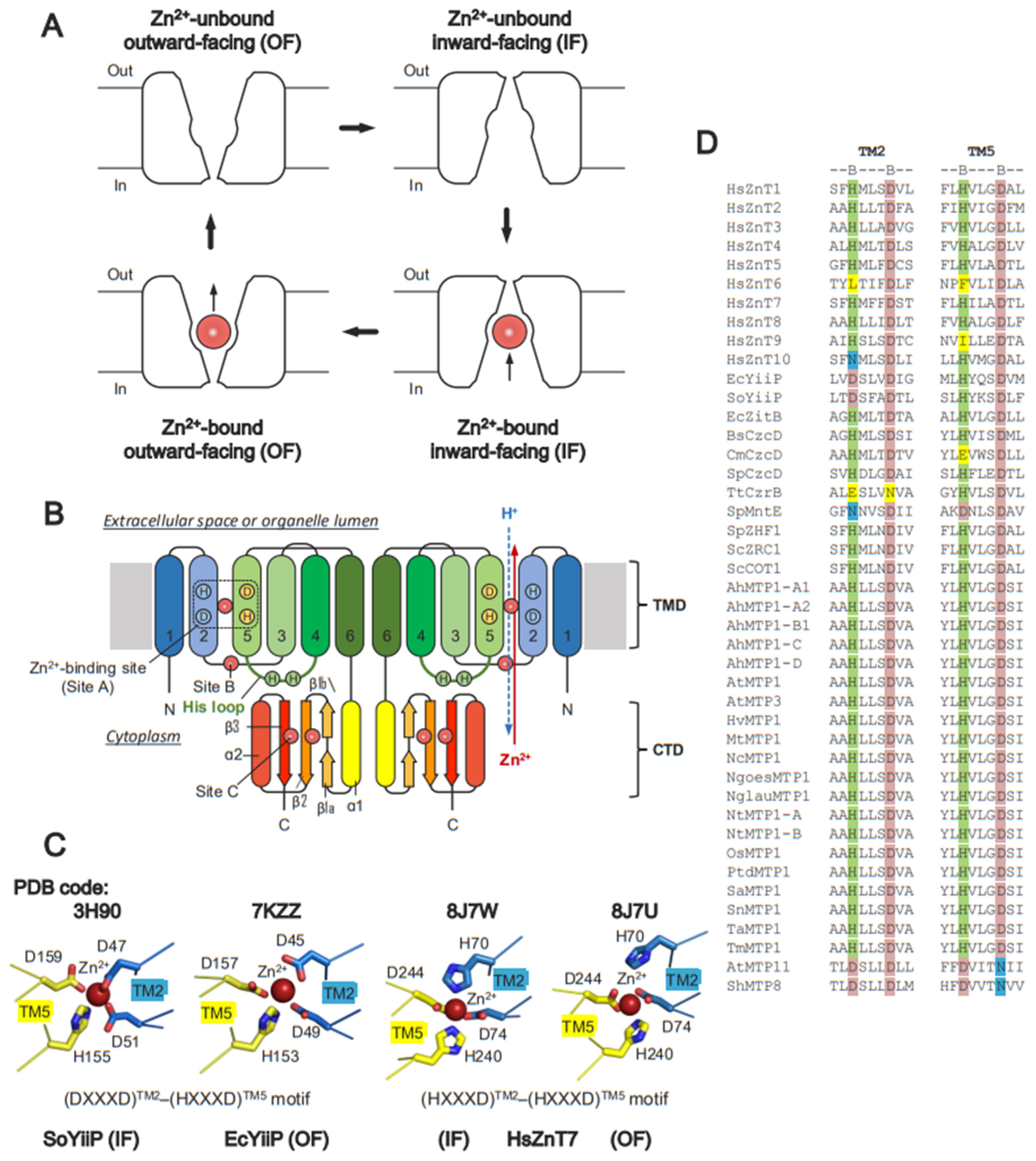
4. Structural Basis of ZnTs

In general, ZnTs adopt inward-facing (IF) or outward-facing (OF) forms and undergo conversion between these two forms during Zn2+ transport (Figure 1A). The IF form creates an open cavity for Zn2+ recruitment from the cytoplasm to the TM metal-binding site. Conversely, in the OF form, an open cavity is formed on the extracellular or organelle luminal side to release Zn2+ (Figure 1A). Unlike ZnTs, ZIPs are supposed to operate by a mechanism named elevator-type transport. Membrane transporters using this mechanism commonly consist of moving and fixed domains. Switching between the outward- and inward-facing forms involves the sliding of the entire moving domain through the bilayer as a rigid body. The substrate-binding site translocates some distance across the bilayer during transport, along with the moving domain [87].
The first X-ray crystal structure of ZnTs was solved for EcYiiP in a Zn2+-bound OF state [41,42]. Subsequently, cryo-EM single-particle analysis identified the structure of a Zn2+-bound IF state for SoYiiP [44,46] (Table 1). More recently, the cryo-EM structures of vertebrate ZnTs human ZnT7 (HsZnT7) [50], human ZnT8 (HsZnT8) [51], and Xenopus ZnT8 (XtZnT8) [52] were reported (Table 1), revealing that these ZnTs can adopt both OF-OF homodimeric and IF-OF heterodimeric conformations [50,51,52]. The presence of the homodimeric and heterodimeric conformations may suggest that the two protomers work independently during Zn2+ transport [50,51,52], although more detailed studies are required to draw a firm conclusion.
All Zn-CDFs share common structural folds. The structural core consists of six TM helices (TM1-TM6) in the TM domain (TMD), and a cytosolic domain (CTD) with a ferredoxin-like fold having an αββαβ secondary structure topology (Figure 1B). The Zn2+-binding site is formed by an HXXXD motif located on TM2 and TM5, individually, near the center of the TMD (Figure 1C). In some Zn-CDFs, the His (H) and Asp (D) residues on the motifs are replaced by Asp (D) and Asn (N), respectively (Figure 1D). Some eukaryotic Zn-CDFs possess a histidine-rich loop (His-loop) flanked by TM4 and TM5. The length of the His loop, and the number and distribution of His residues in the loop, vary among the Zn-CDFs (Figure 2).
As aforementioned, all ZnTs belong to the SLCA30 family, allowing us to compare the conformational details between them. ZnTs show different TM helix arrangements in both the IF and OF forms. In the IF form of bacterial YiiP and HsZnT8, TM4 and TM5 largely swing to the outside on the cytosolic side (relative to their positions in the OF form), using their luminal ends as pivot points (Figure 3A,B). By contrast, TM2 moves slightly toward the center [51]. Notably, in the IF form of HsZnT7, TM5 kinks at the middle, and its cytosolic half is largely bent toward the outside, resulting in the very open cytosolic cavity (Figure 3C) [50]. In this form, part of the His loop is integrated into the cytosolic cavity, and His164 in this loop is directly coordinated to Zn2+ along with His70, Asp74, and Asp244 (Figure 3D) [50]. The detailed mechanistic and functional roles of the His-loop are discussed later.
There are also significant differences in the OF forms of ZnTs with a known structure. The OF form of HsZnT7 has a wider cavity at the luminal side than that of HsZnT8 and EcYiiP due to the more “outside” positions of TM1 and TM2 (Figure 3E,F). An additional conformation has been identified for Zn2+-unbound HsZnT7, in which TM5 packs tightly with TM2, TM3, and TM6 on both the cytoplasmic and luminal sides (Figure 3G) [50]. The TM helix arrangement in this state is highly superimposable to that of a previously reported occluded state of Zn2+-unbound SoYiiP (Figure 3G) [44]. Although the physiological relevance of the Zn2+-unbound occluded state remains unclear, this state may be formed after the release of Zn2+ to the luminal or periplasmic side and before complete conversion to the IF state with a widely open cytosolic cavity. Hereafter, we refer to this state as the “IF resting state” in this review article.

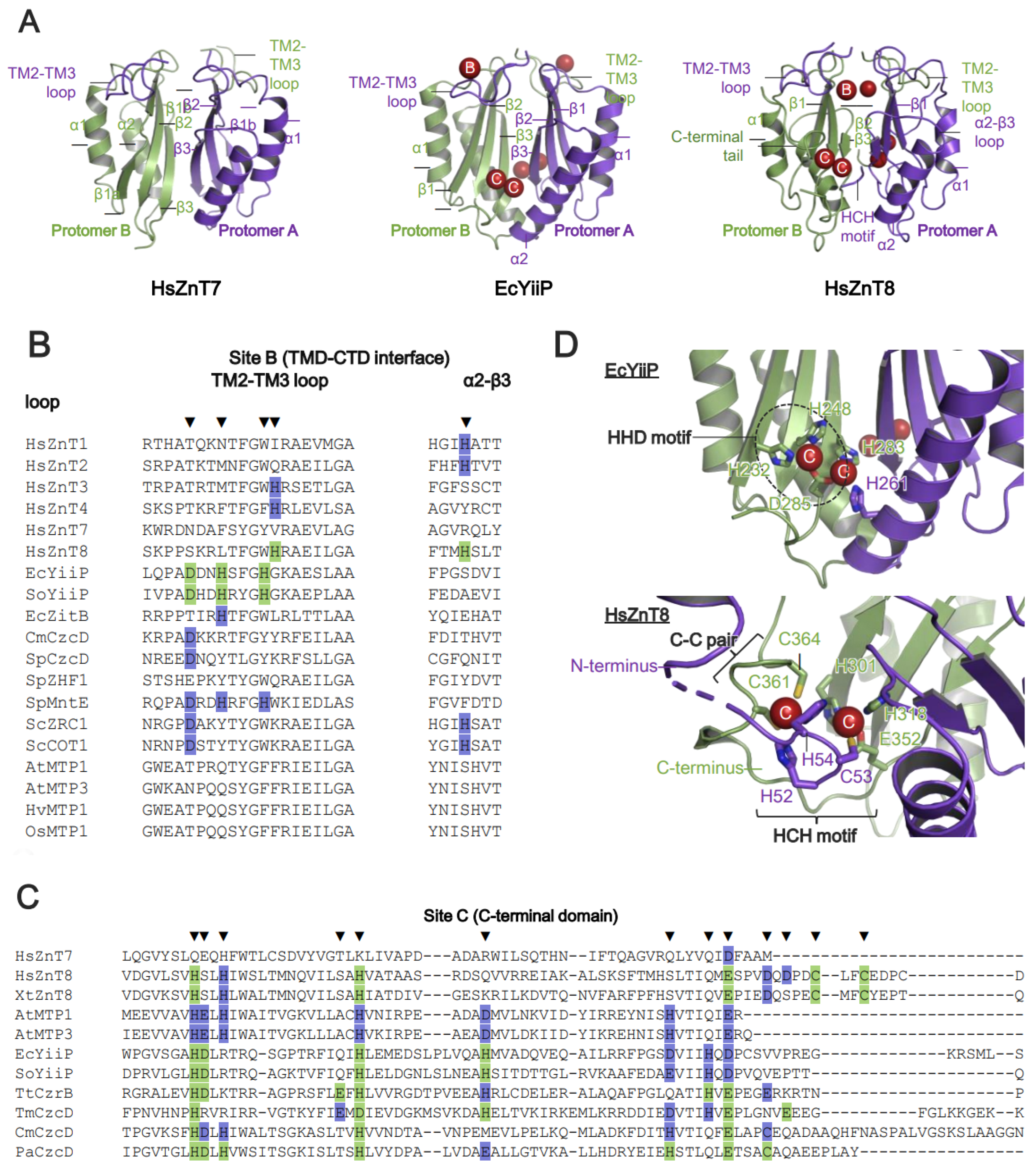
5. Zn2+-Binding Sites on ZnTs

Zn2+-Binding Sites and Metal Ion Selectivity

Metal specificity is an important functional feature of all metal transporters. X-ray crystallographic and cryo-EM analyses revealed one to three Zn2+-binding sites (site A, site B, and site C) in ZnTs. All ZnTs commonly possess a Zn2+-binding site in the TMD (site A). Zn2+ binds transiently to site A before effluxing to the other side, indicating that site A is located on the Zn2+-translocation pathway. Site A contains highly conserved Zn2+-binding motifs formed by the BXXXB motif on TM2 and TM5, in which B is His (H), Asp (D), Asn (N), or Glu (E), and X can be any residues (Figure 1C,D). There are, however, some differences in the sequence of site A (Figure 1D). While human, plant, and yeast ZnTs conserve the (HXXXD)TM2-(HXXXD)TM5 motif, different amino acid sequences are seen in the motifs of bacterial ZnTs; for instance, (DXXXD)TM2-(HXXXD)TM5 in EcYiiP and SoYiiP, and (HXXXD)TM2-(HXXXD)TM5 in EcZitB and CzcD (Figure 1D). Notably, mammalian ZnT6 loses the BXXXB motif in the TMD, and does not therefore have Zn2+ transport activity [62]. Mammalian ZnT10 harboring (NXXXD)TM2-(HXXXD)TM5 transports Mn2+ rather than Zn2+ (Figure 1D) [88,89,90]. AtMTP11 and ShMTP8 harbor (DXXXD)TM2-(DXXXN)TM5 and transport both Mn2+ and Cu2+ (Figure 1D) [66]. Thus, not all ZnTs transport Zn2+ exclusively.
Site B is less conserved at the TMD-CTD interface (Figure 1B and Figure 4A). In bacterial YiiP, this site is located in the TM2-TM3 loop, which contains a DHH motif (Figure 4B) [42,43,44]. In HsZnT8, this site is constituted by His residues from the TM2-TM3 loop in the TMD and from the α2-β3 loop in the CTD, and it has low affinity for Zn2+ [51]. While the functional role of site B is unclear in HsZnT8, bacterial YiiP participates directly in Zn2+ transport [43,44]. Since site B is positioned near the cytosolic Zn2+ entry gate, mutation of two His residues at site B reduces the Zn2+ transport activity of bacterial YiiP [43]; thus, site B is thought to trap Zn2+ in the cytosol, thereby facilitating Zn2+ transport [43,51]. Although the structure of AtMTP1 has not been solved, homology modeling based on the crystal structure of EcYiiP suggests that it lacks site B (Figure 4B). However, mutations of some residues in the TM2-TM3 loop impair the Zn2+ transport activity of AtMTP1, indicating that the TM2-TM3 loop itself is essential for AtMTP1 function [91].
Site C is located at the dimer interface between two CTDs (Figure 1B and Figure 4A). In bacterial YiiP, site C forms a binuclear Zn2+ complex composed of the (HHD)2 motif and stabilizes its dimeric conformation (Figure 4C,D) [42,43]. In HsZnT8, the exact location of site C differs from that in bacterial YiiP. In HsZnT8, two Zn2+ ions are coordinated by an HCH motif from the N-terminal loop, and a Cys-Cys pair from the C-terminal tail, thereby forming a tetrahedral complex (Figure 4D) [51]. The HCH motifs seal off site C and bury the Zn2+ ions inside the protein (Figure 4D). This motif is highly conserved among the ZnT8 orthologues but not among bacterial ZnTs [51]. The N-terminal truncation that accompanies the loss of the HCH motif reduces the Zn2+ uptake activity of HsZnT8 significantly [51]. AtMTP1 also possesses site C within the CTD (Figure 4C), and the lack of this domain results in loss of function [91]. Thus, Zn2+-mediated dimerization via the CTD seems likely to be essential for Zn2+ transport by HsZnT8, bacterial YiiP, and plant MTP1.
By contrast, no Zn2+ ions have been identified at either site B or site C of HsZnT7, although its cryo-EM structures were determined in the presence of high Zn2+ concentrations (10, 200, or 300 μM) of Zn2+ (Figure 4). Consistent with this, residues required for Zn2+ binding are not conserved at site B or site C of HsZnT7 (Figure 4C). Presumably, other elements contribute to the dimerization of the CTD and the Zn2+ transport activity of HsZnT7. Indeed, dimerization of the CTD is mediated by residues within four β-strands and TM2-TM3 loops located at the dimer interface (Figure 4A) [50].

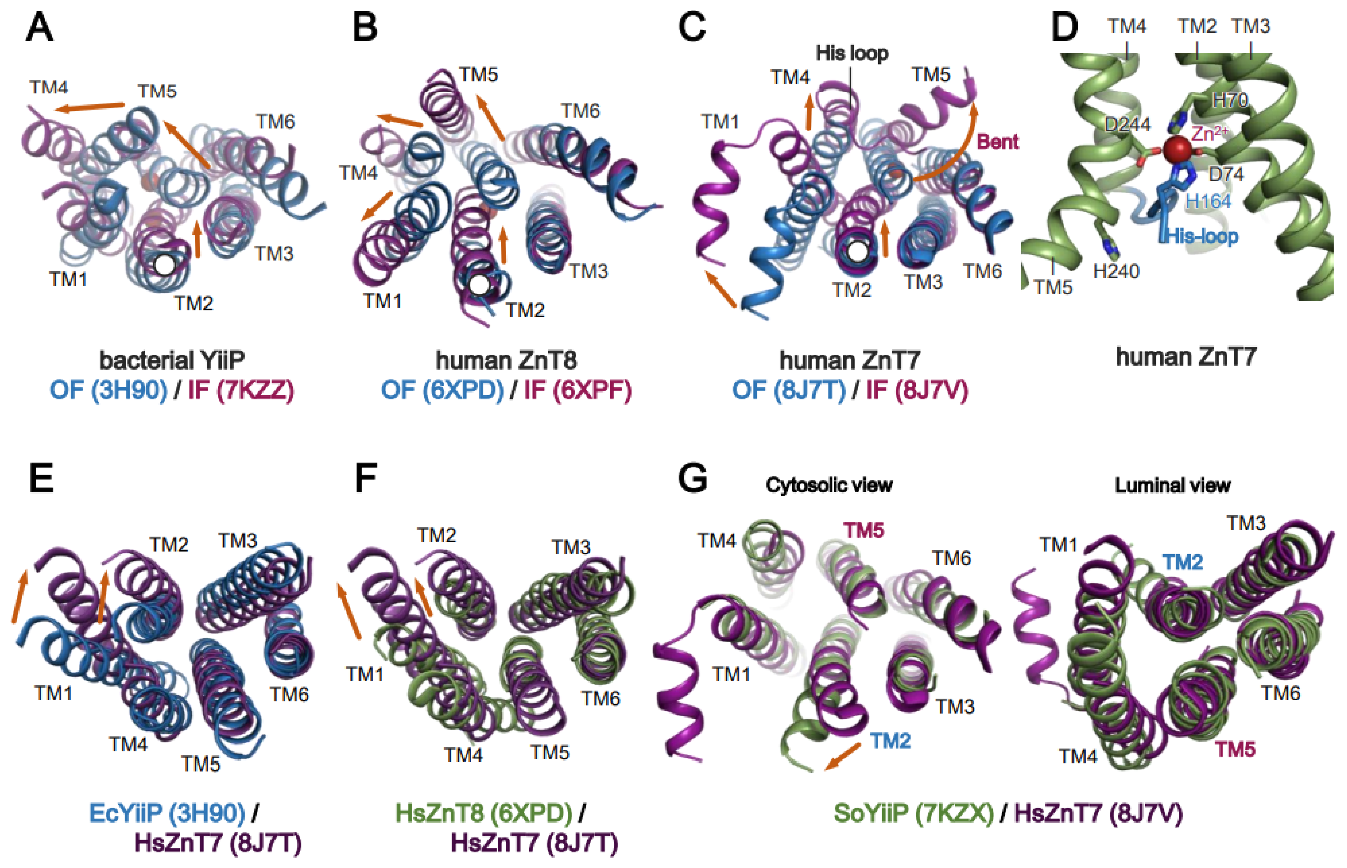
6. Mechanism of Zn2+ Transport by Human ZnT7 and Bacterial YiiP

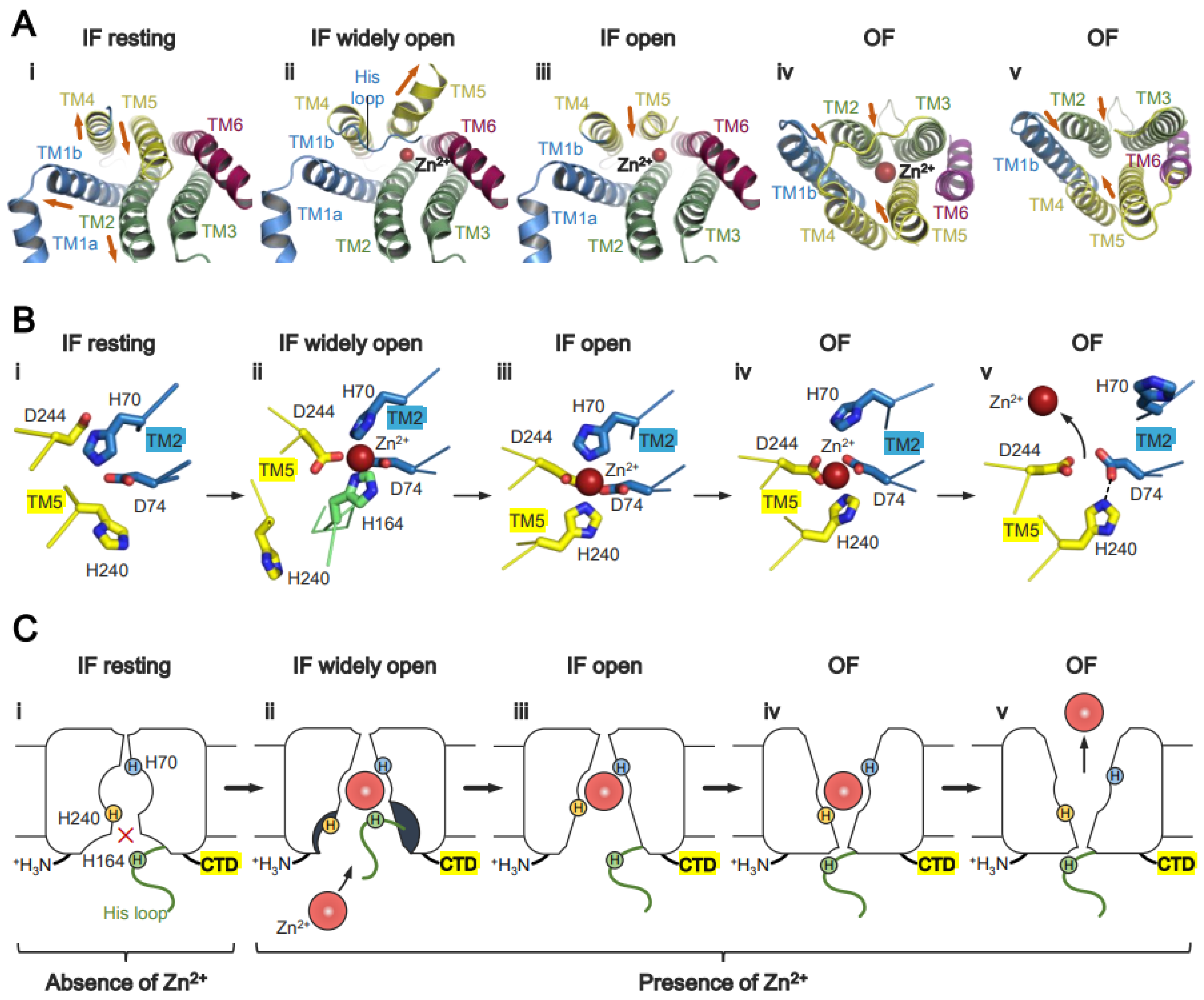
6.1. Zn2+ Transport by HsZnT7

HsZnT7 transports Zn2+ from the cytoplasm to the TM Zn2+-binding site (site A), and then to the Golgi lumen. The cryo-EM structures of HsZnT7 in multiple states have helped to paint a full picture of Zn2+ transport mediated by this transporter (Figure 5) [50]. In the absence of Zn2+, TM5 packs against TM2, TM3, and TM6 at both the cytosolic and luminal sides, forming an “IF resting” state (Figure 3G and Figure 5A). In this state, Zn2+ uptake to site A seems to be blocked due to the closed Zn2+ entry gate (Figure 5A–C(i)). In the presence of Zn2+, however, the cytosolic cavity opens by bending the N-terminal half of TM5, and the His-loop is integrated into the cavity to coordinate with Zn2+ at site A (Figure 5A–C(ii)). In the next step, TM5 returns to a straight conformation concomitant with pulling His164 out of site A. Consequently, His240 on TM5 coordinates with Zn2+ instead of His164 (Figure 5A–C(iii)). Upon conversion to the OF state, the His70 side chain moves away from Zn2+ slightly, while Asp74, His240, and Asp244 remain coordinated with Zn2+ (Figure 5B(iv)). Simultaneously (or shortly thereafter), protons presumably enter the Zn2+ transport channel from the weakly acidic Golgi lumen to protonate His70 and His240. As a result, His70 flips out of site A, and Zn2+ is released to the Golgi lumen (Figure 5A–C(v)). In this context, it is notable that while a His residue is highly conserved among human ZnTs, it is substituted by an Asp residue in bacterial YiiP (Figure 1C,D). Although it is not fully elucidated how many protons are involved in the release of Zn2+, the above mechanistic model suggests that HsZnT7 exchanges one Zn2+ for two protons per cycle.

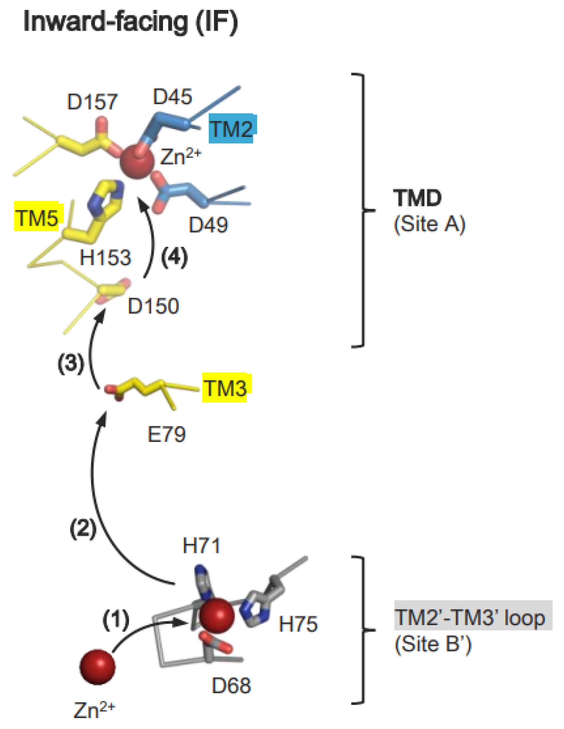
6.2. Zn2+ Transport by Bacterial YiiP

Recent biochemical, structural, and computational studies provide deep insight into the mechanism of Zn2+ transport mediated by bacterial YiiP [43,44]. Initially, Zn2+ is recruited to site B, formed by the TM2-TM3 loop, and then transferred to site A of another protomer (Figure 6) [43,44]. In the Zn2+-free IF state, the TM2-TM3 loop is disordered to allow Zn2+ to approach site B. After binding Zn2+ site B, the TM2-M3 loop forms an ordered conformation [43]. Importantly, the higher affinity of Zn2+ for site A than site B explains sequential Zn2+ transfer from site A to site B [43]. At this step, the negatively charged cytoplasmic cavity seems advantageous for directing Zn2+ to site A [43,92]. In the OF state, the TM2-TM3 loop in a Zn2+-free state interacts with the CTD, forming an extended conformation [43]. Zn2+ is released to the periplasm and is facilitated by the protonation of two residues in site A, or potentially three residues at a lower pH [43].
MD simulations using EcYiiP and SoYiiP as a model metal transporter demonstrate that Zn2+ binds transiently to Asp150 (Asp152 in SoYiiP) on TM5, and to Glu79 (Glu81 in SoYiiP) on TM3, before reaching site A (Figure 6) [92]. In the next step, His153 (His155 in SoYiiP), located at site A, plays a key role in guiding Zn2+ to site A. In support of this, His153 can adopt two conformations to receive Zn2+ from Asp150, after which it forms a tetrahedral Zn2+ complex with Asp45, Asp49, and Asp157 (Figure 6) [49]. Release of Zn2+ can be triggered by protonation of site A residues [43,44]. His153 is likely deprotonated again when it faces the neutral-pH cytoplasm to receive Zn2+ from site B via Asp150 [49,51,54,92,93].

6.3. Role of the Proton Gradient in ZnT-Mediated Zn2+ Transport

Lactose permease (LacY) utilizes a proton motive force for the efficient transport of lactose [94]. Presumably, ZnTs also uses the proton motive force to transport Zn2+ from the cytoplasm to the extracellular space or the organelle lumen [55]. Given that the concentration of labile Zn2+ in the Golgi is much higher than in the cytosol [64,95,96], and that the Golgi is a weakly acidic organelle [97], it makes sense that Golgi-resident ZnTs utilize the energy provided by the Golgi-to-cytosol proton flow to transport Zn2+ to the luminal side. As mentioned above, protonation of His70 and His240 is critical for the IF-to-OF conversion and subsequent release of Zn2+. While HsZnT7 exchanges one Zn2+ for (possibly) two H+ per cycle, EcZitB [47] and EcYiiP [48] exchange one Zn2+ for one H+. SoYiiP conducts the exchange at a ratio of 1:2–3, depending on the external pH [43]. Human ZnT2 is thought to operate at a ratio of 1:2 [54].

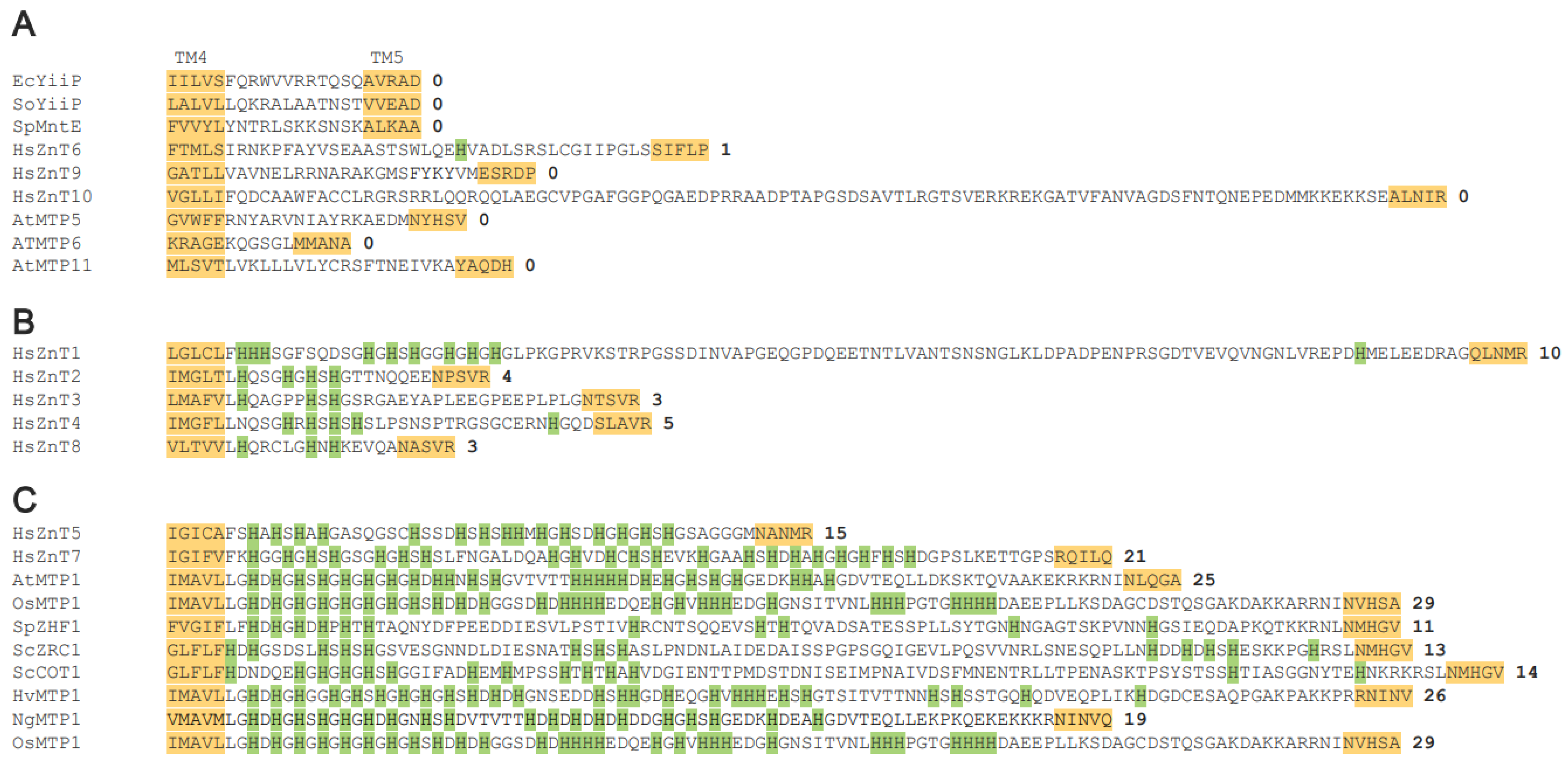
7. Structural and Functional Roles of the Histidine-Rich Loop (His-Loop)

The His-loop is a unique sequence located at the cytoplasmic side between TM4 and TM5 (Figure 1B and Figure 2). The length of this loop, as well as the number and arrangement of His residues, can differ (Figure 2). Most bacterial ZnTs (e.g., YiiP and MntE), several mammalian ZnTs (e.g., ZnTs 6, 9, and 10), and plant MTPs (e.g., AtMTPs 5, 6, and 11) possess a short TM4-TM5 loop without any His residues. Mammalian ZnTs such as ZnTs 2-4 and ZnT8 are also characterized by a short His-loop. Of note, several mammalian ZnTs (e.g., ZnTs 1, 5, and 7), plant MTP1s (e.g., AtMTP1, NgMTP1, OsMTP1, and HvMTP1), and yeast ZnTs (e.g., SpZHF1, ScZRC1, and ScCOT1) have an exceptionally long His-loop (Figure 2). Despite having a relatively long TM4-TM5 loop, ZnT10 has no His residues in the loop (Figure 2). Such variations in the His-loop may underlie the diverse functions and different mechanisms of Zn2+ transport mediated by ZnTs. Indeed, the His-loop also functions as a Zn2+-buffering pocket [98], a Zn2+ selectivity filter [99], a Zn2+ sensor [98,100,101], or a Zn2+-fishing loop [50].
As described above, cryo-EM analysis of HsZnT7 revealed that a short segment of the His-loop inserts into the cytoplasmic cavity, where His164 contained in the His-loop coordinates directly with Zn2+ at site A, while His240 on TM5 is kicked out from site A due to bending of the N-terminal half of TM5 (Figure 5A,B(ii)) [50]. Thus, the His-loop appears to contribute to efficient Zn2+ uptake to site A, but its task seems transient. In other words, HsZnT7 may use the His-loop as a “Zn2+ fishing loop”, and His residues in the loop function as a “fishing hook” that captures Zn2+ and transfers it to site A. Consistent with this, the deletion of the His-loop, or the mutations of all His residues in the loop to Ser, abolishes the Zn2+ transport activity of HsZnT7 [50]. Similarly, deletion of the entire His-loop segment in Noccaea caerulescens MTP1 and AtMTP1 results in substantial loss of Zn2+ transport activity [98,102] although deletion of the first half of the His-loop increases the activity of AtMTP1 significantly [98].
The 63-residue His-loop of HsZnT7 contains 21 His residues and can be divided into two halves: The first half contains seven His residues close to the C-terminus of TM4, and the second half contains 14 His residues close to the N-terminus of TM5 (Figure 2) [50]. The His-loop of HsZnT7 binds Zn2+ with a Kd value of 12 μM; although each half can bind one Zn2+, the first and second halves appear to bind Zn2+ co-operatively [50]. AtMTP1 also contains an exceptionally long His-loop containing 25 His residues and can also be divided into two halves: The first half contains 18 His residues close to the C-terminus of TM4, while the second half contains seven His residues close to the N-terminus of TM5 (Figure 2) [98]. The His-loop of AtMTP1 can bind four Zn2+ ions, with a Kd value of 25 μM [101]. The lack of either half leads to impairment of Zn2+-binding by AtMTP1, suggesting that the two halves of the His-loop work co-operatively to form multiple Zn2+-binding sites [101]. However, stable Zn2+ binding may lead to slower Zn2+ release from AtMTP1, which likely explains the less efficient Zn2+ transport by AtMTP1 WT than by the mutant lacking the first half [98,101]. Consistent with this, the His-loop of AtMTP1 adopts a β-strand conformation to suppress its Zn2+ transport activity at high concentrations of Zn2+, implying a role in sensing excessive Zn2+ levels in the cytosol [101].

8. Zn2+ Transport by Other Zn2+ Transport Systems

8.1. ZIPs

ZIPs play a crucial role in mediating metal import across the plasma membrane as well as export across organelle membranes. ZIPs are classified into four subfamilies based on phylogenetic relationships [102]. In humans, 14 ZIPs have been identified. Despite their relevance to human diseases, the structural and mechanistic details of mammalian ZIPs remain poorly understood. Recent studies on Bordetella bronchiseptica ZIP (BbZIP) have shed light on some structural and mechanistic features [56,103,104]. Crystal structures of BbZIP revealed an IF monomer, with binuclear metal-binding sites potentially occupied by Zn2+ or Cd2+ [104], while cryo-EM studies revealed an IF homodimer [56]. BbZIP is an elevator-type transporter in which a four-TM helix bundle with bound metal ions slides as a rigid body against the dimeric domain composed of the other static TM helices to exert an alternating access mechanism, and its Zn2+ uptake is pH-dependent [56,105,106,107]. However, it is unclear whether ZIPs transfer Zn2+ through a symport or antiport mechanism [56,105]. The presence of a His-rich loop in many mammalian ZIPs, including BbZIP, adds complexity to the mechanisms underlying their regulated Zn2+ transport [108,109]. Plants possess more ZIPs [110], but detailed structural and mechanistic characterization has not been reported for any of them.

8.2. ZntB

ZntB belongs to the prokaryotic CorA family [111,112]. E. coli and P. aeruginosa ZntB transport multiple metal ions, including Zn2+, Co2+, Ni2+, and Cd2+ [53,113]. ZntB forms homogeneous pentagonal structures that span the biological membrane, similar to other members of the CorA family. Each protomer consists of two TM helices and a large cytoplasmic domain [53,113]. ZnTB-mediated Zn2+ transport is stimulated by a pH gradient across the biological membrane. ZntB transports protons and Zn2+ together in the same direction, thereby working as a symporter [53]. In contrast to bacterial YiiP and human ZnTs, ZntB does not seem to use an alternating access mechanism mediated by conformational transitions between IF and OF forms [53]. The cryo-EM structure of PaZntB is similar to that of EcZntB and T. maritima CorA, but with different helical arrangements [53,113,114]. PaZntB is capable of transporting Zn2+, Mg2+, Cd2+, Ni2+, and Co2+. In contrast to other ZntBs, PaZntB is unlikely to use the proton motive force [53,113]. Thus, different mechanisms may operate among ZntBs from various bacterial species [53,111,113,115].

8.3. P-Type ATPases with Zn2+ Transport Activity

Zn2+-transporting P-type ATPases, which belong to class IB (PIB-2-ATPases), are active zinc transporters. While no P-type ATPases with Zn2+ transport activity have been identified in mammals, several have been identified in bacteria and plants. In plants, the PIB-2-ATPases, also known as the heavy metal ATPases (HMAs), are thought to play an important role in the transport of transition metals. In A. thaliana, AtHMA2 and AtHMA4 are zinc transporters that increase Zn2+ levels in roots, stems, and leaves [66,116]. AtHMA1 functions to detoxify Zn2+ in the chloroplast, while AtHMA3 is thought to supply Zn2+ to the ER and vacuole. Little is known about the structural features and Zn2+ transport mechanisms of HMAs [66]. In bacteria, ZntA is an active zinc transporter that is crucial for cellular detoxification and sub-cellular redistribution of Zn2+ [117]. ZntA couples ATP hydrolysis with Zn2+ transport via the “Post-Albers” cycle, during which at least four primary intermediate states (E1, E1P, E2P, and E2) accumulate [118,119,120,121]. Structurally, PIB-2-ATPases possess four domains: A TMD, a nucleotide-binding domain (N domain), an actuator/dephosphorylation domain (A domain), and a phosphorylation domain (P domain) [118,120,122]. In addition, PIB-2-ATPases contain metal-binding domains (MDBs) in the N-terminal region to promote metal recruitment to the TM metal-binding site (114, 116, 118). To date, only the crystal structure of Shigella sonnei ZntA has been reported [117], and the cryo-EM structures of other PIB-2-ATPases are currently unknown.

8.4. Zn2+ Transport by ATP-Binding Cassette Transporters

ATP-binding cassette (ABC) transporters are a ubiquitous superfamily of integrated membrane proteins that transport various substrates across biological membranes by utilizing ATP as an external energy source [123]. In bacteria, ZnuABC is a specialist zinc transporter. It comprises three protein subunits: A periplasmic Zn2+-binding protein (ZnuA), an integrated membrane protein that transports Zn2+ across the cytoplasmic membrane (ZnuB), and an ATPase protein in charge of coupling Zn2+ transport to ATP hydrolysis (ZnuC) [124,125]. In most Gram-negative bacteria, ZnuABC expression is regulated by a Zur (zinc uptake regulatory) protein induced by Zn2+ acquisition [126]. The structure of ZnuABC and its mechanism of Zn2+ transport coupled to ATP hydrolysis are only poorly understood, although crystal structures have been solved for EcZnuA [124,127,128], and Salmonella enterica ZnuA [129].

9. Conclusions

As described above, while some members of the Zn-CDF family share structural and mechanistic features, there are considerable variations among them. It is widely accepted that ZnTs commonly operate as dimers; however, detailed structural analyses conducted so far show that whereas bacterial YiiPs adopt OF-OF and IF-IF homodimers, human ZnTs exist as either OF-OF homodimers or IF-OF heterodimers. No IF-IF homodimers have been observed for mammalian ZnTs, likely due to their conformational instability. Indeed, modes of the TM helix rearrangement between the IF and OF states among ZnTs seem to differ significantly. Of particular note, HsZnT7 undergoes marked bending of TM5 to allow efficient Zn2+ uptake. Concomitantly, a part of the long cytoplasmic His-loop is integrated into the negatively charged cytosolic cavity to facilitate acqusition and efficient transfer of Zn2+ to the TM Zn2+-binding site. In this state, His164 in the His-loop is coordinated directly with Zn2+ in the TMD. Subsequently, His240 (TM5) repaces His164, followed by the conversion from the IF to the OF state. In this context, the essential role of His164 needs to be further explored by conducting additional mutational and structural studies. It will also be interesting to see if another His residue in the His-loop can replace His164 for efficient Zn2+ recruitment when His164 is deleted. Recently, a de novo heterozygous variant of SLC30A7, His164Ser, was found in Joubert syndrome patients [130]. Although no SLC30A7 variants have yet been shown to cause human phenotypes or diseases, SLC30A7 is a candidate gene associated with Joubert syndrome [130].
Undoubtedly, multiple ZnTs work co-operatively to maintain Zn2+ homeostasis in cells. Indeed, disruption of Zn2+ homeostasis leads to disruption of protein homeostasis [64,131] and dysfunction of essential enzymes, transcription factors, and other biomolecules [132,133,134,135], eventually causing many fatal diseases [29,30,31,32,33,34,35,36,37,38,39,40]. In this regard, the mechanisms of Zn2+ homeostasis mediated by various kinds of ZnTs need to be understood more comprehensively. Zinc biology is an important field of research that encompasses biochemical, structural, computational, physiological, and medical approaches.

Author Contributions

Conceptualization: H.B.B. and K.I.; literature collection and analysis: H.B.B.; writing—original draft: H.B.B. and K.I.; writing—review and editing: H.B.B. and K.I. All authors have read and agreed to the published version of the manuscript.

Funding

This work was supported by funding from AMED-CREST (21gm1410006h0001 to K.I.), JSPS KAKENHI (18H03978, 21H04758, and 21H05247 to K.I), Canon Medical Systems Corporation (to K.I.), and the Basis for Supporting Innovative Drug Discovery and Life Science Research (BINDS) from the Japan Agency for Medical Research and Development (AMED) under grant numbers JP19am0101115 (Support No. 1025), JP19am0101078 (Support No. 2293), and JP21am0101079 (Support No. 2343).

Institutional Review Board Statement

Not applicable.

Informed Consent Statement

Not applicable.

Data Availability Statement

Not applicable.

Conflicts of Interest

The authors declare no conflicts of interest with respect to the contents of this article.

References

  1. Skalny, A.V.; Aschner, M.; Tinkov, A.A. Zinc. Adv. Food Nutr. Res. 2021, 96, 251–310. [Google Scholar] [CrossRef]
  2. Zaher, D.M.; El-Gamal, M.I.; Omar, H.A.; Aljareh, S.N.; Al-Shamma, S.A.; Ali, A.J.; Zaib, S.; Iqbal, J. Recent advances with alkaline phosphatase isoenzymes and their inhibitors. Arch. Pharm. 2020, 353, e2000011. [Google Scholar] [CrossRef]
  3. Xiong, L.; Peng, M.; Zhao, M.; Liang, Z. Truncated expression of a Carboxypeptidase A from bovine improves its enzymatic properties and detoxification efficiency of Ochratoxin A. Toxins 2020, 12, 680. [Google Scholar] [CrossRef]
  4. Luan, R.; Ding, D.; Xue, Q.; Li, H.; Wang, Y.; Yang, J. Protective role of zinc in the pathogenesis of respiratory diseases. Eur. J. Clin. Nutr. 2023, 77, 427–435. [Google Scholar] [CrossRef]
  5. Begum, F.; Me, H.M.; Christov, M. The role of zinc in cardiovascular disease. Cardiol. Rev. 2022, 30, 100–108. [Google Scholar] [CrossRef]
  6. Gembillo, G.; Visconti, L.; Giuffrida, A.E.; Labbozzetta, V.; Peritore, L.; Lipari, A.; Calabrese, V.; Piccoli, G.B.; Torreggiani, M.; Siligato, R.; et al. Role of zinc in diabetic kidney disease. Nutrients 2022, 14, 1353. [Google Scholar] [CrossRef]
  7. Li, J.; Cao, D.; Huang, Y.; Chen, B.; Chen, Z.; Wang, R.; Dong, Q.; Wei, Q.; Liu, L. Zinc intakes and health outcomes: An umbrella review. Front. Nutr. 2022, 9, 798078. [Google Scholar] [CrossRef]
  8. Li, Z.; Liu, Y.; Wei, R.; Yong, V.W.; Xue, M. The important role of zinc in neurological diseases. Biomolecules 2022, 13, 28. [Google Scholar] [CrossRef]
  9. Garner, T.B.; Hester, J.M.; Carothers, A.; Diaz, F.J. Role of zinc in female reproduction. Biol. Reprod. 2021, 104, 976–994. [Google Scholar] [CrossRef] [PubMed]
  10. Banupriya, N.; Bhat, B.V.; Sridhar, M.G. Role of zinc in neonatal sepsis. Indian J. Pediatr. 2021, 88, 696–702. [Google Scholar] [CrossRef] [PubMed]
  11. Tamura, Y. The role of zinc homeostasis in the prevention of diabetes mellitus and cardiovascular diseases. J. Atheroscler. Thromb. 2021, 28, 1109–1122. [Google Scholar] [CrossRef] [PubMed]
  12. Allouche-Fitoussi, D.; Breitbart, H. The role of zinc in male fertility. Int. J. Mol. Sci. 2020, 21, 7796. [Google Scholar] [CrossRef]
  13. Michalczyk, K.; Cymbaluk-Płoska, A. The role of zinc and copper in gynecological malignancies. Nutrients 2020, 12, 3732. [Google Scholar] [CrossRef] [PubMed]
  14. Granero, R.; Pardo-Garrido, A.; Carpio-Toro, I.L.; Ramírez-Coronel, A.A.; Martínez-Suárez, P.C.; Reivan-Ortiz, G.G. The role of iron and zinc in the treatment of ADHD among children and adolescents: A systematic review of randomized clinical trials. Nutrients 2021, 13, 4059. [Google Scholar] [CrossRef]
  15. Moshtagh, M.; Amiri, R. Role of zinc supplementation in the improvement of acute respiratory infections among Iranian children: A systematic review. Tanaffos 2020, 19, 1–9. [Google Scholar]
  16. Wang, M.X.; Win, S.S.; Pang, J. Zinc supplementation reduces common cold duration among healthy adults: A systematic review of randomized controlled trials with micronutrients supplementation. Am. J. Trop. Med. Hyg. 2020, 103, 86–99. [Google Scholar] [CrossRef]
  17. Korkmaz-Icöz, S.; Atmanli, A.; Radovits, T.; Li, S.; Hegedüs, P.; Ruppert, M.; Brlecic, P.; Yoshikawa, Y.; Yasui, H.; Karck, M.; et al. Administration of zinc complex of acetylsalicylic acid after the onset of myocardial injury protects the heart by upregulation of antioxidant enzymes. J. Physiol. Sci. 2016, 66, 113–125. [Google Scholar] [CrossRef]
  18. Barnett, J.B.; Dao, M.C.; Hamer, D.H.; Kandel, R.; Brandeis, G.; Wu, D.; Dallal, G.E.; Jacques, P.F.; Schreiber, R.; Kong, E.; et al. Effect of zinc supplementation on serum zinc concentration and T cell proliferation in nursing home elderly: A randomized, double-blind, placebo-controlled trial. Am. J. Clin. Nutr. 2016, 103, 942–951. [Google Scholar] [CrossRef]
  19. Cope, E.C.; Levenson, C.W. Role of zinc in the development and treatment of mood disorders. Curr. Opin. Clin. Nutr. Metab. Care 2010, 13, 685–689. [Google Scholar] [CrossRef]
  20. Plum, L.M.; Rink, L.; Haase, H. The essential toxin: Impact of zinc on human health. Int. J. Environ. Res. Public Health 2010, 7, 1342–1365. [Google Scholar] [CrossRef] [PubMed]
  21. Fox, M.R.S. Zinc Excess. In Zinc in Human Biology. ILSI Human Nutrition Reviews; Mills, C.F., Ed.; Springer: London, UK, 1989; pp. 365–370. [Google Scholar] [CrossRef]
  22. Brown, M.A.; Thom, J.V.; Orth, G.L.; Cova, P.; Juarez, J. Food poisoning involving zinc contamination. Arch. Environ. Health 1964, 8, 657–660. [Google Scholar] [CrossRef]
  23. Skalny, A.V.; Aschner, M.; Lei, X.G.; Gritsenko, V.A.; Santamaria, A.; Alekseenko, S.I.; Prakash, N.T.; Chang, J.S.; Sizova, E.A.; Chao, J.C.J.; et al. Gut Microbiota as a Mediator of Essential and Toxic Effects of Zinc in the Intestines and Other Tissues. Int. J. Mol. Sci. 2021, 22, 13074. [Google Scholar] [CrossRef] [PubMed]
  24. Hamzah-Saleem, M.; Usman, K.; Rizwan, M.; Al-Jabri, H.; Alsafran, M. Functions and strategies for enhancing zinc availability in plants for sustainable agriculture. Front. Plant Sci. 2022, 13, 1033092. [Google Scholar] [CrossRef] [PubMed]
  25. Choudhury, R.; Srivastava, S. Zinc resistance mechanisms in bacteria. Curr. Sci. 2001, 81, 768–775. [Google Scholar]
  26. Pandey, N.; Pathak, G.C.; Sharma, C.P. Zinc is critically required for pollen function and fertilisation in lentil. J. Trace Elem. Med. Biol. 2006, 20, 89–96. [Google Scholar] [CrossRef]
  27. Wissuwa, M.; Ismail, A.M.; Yanagihara, S. Effects of zinc deficiency on rice growth and genetic factors contributing to tolerance. Plant Physiol. 2006, 142, 731–741. [Google Scholar] [CrossRef]
  28. Corbett, D.; Wang, J.; Schuler, S.; Lopez-Castejon, G.; Glenn, S.; Brough, D.; Andrew, P.W.; Cavet, J.S.; Roberts, I.S. Two zinc uptake systems contribute to the full virulence of Listeria monocytogenes during growth in vitro and in vivo. Infect. Immun. 2012, 80, 14–21. [Google Scholar] [CrossRef] [PubMed]
  29. Hara, T.; Yoshigai, E.; Ohashi, T.; Fukada, T. Zinc transporters as potential therapeutic targets: An updated review. J. Pharmacol. Sci. 2022, 148, 221–228. [Google Scholar] [CrossRef]
  30. Zhu, B.; Huo, R.; Zhi, Q.; Zhan, M.; Chen, X.; Hua, Z.C. Increased expression of zinc transporter ZIP4, ZIP11, ZnT1, and ZnT6 predicts poor prognosis in pancreatic cancer. J. Trace Elem. Med. Biol. 2021, 65, 126734. [Google Scholar] [CrossRef]
  31. Lei, P.; Ayton, S.; Bush, A.I. The essential elements of Alzheimer’s disease. J. Biol. Chem. 2021, 296, 100105. [Google Scholar] [CrossRef]
  32. Xu, Y.; Xiao, G.; Liu, L.; Lang, M. Zinc transporters in Alzheimer’s disease. Mol. Brain 2019, 12, 106. [Google Scholar] [CrossRef] [PubMed]
  33. Sikora, J.; Ouagazzal, A.M. Synaptic zinc: An emerging player in Parkinson’s disease. Int. J. Mol. Sci. 2021, 22, 4724. [Google Scholar] [CrossRef] [PubMed]
  34. Davis, D.N.; Strong, M.D.; Chambers, E.; Hart, M.D.; Bettaieb, A.; Clarke, S.L.; Smith, B.J.; Stoecker, B.J.; Lucas, E.A.; Lin, D.; et al. A role for zinc transporter gene SLC39A12 in the nervous system and beyond. Gene 2021, 799, 145824. [Google Scholar] [CrossRef] [PubMed]
  35. Chowanadisai, W.; Lönnerdal, B.; Kelleher, S.L. Identification of a mutation in SLC30A2 (ZnT-2) in women with low milk zinc concentration that results in transient neonatal zinc deficiency. J. Biol. Chem. 2006, 281, 39699–39707. [Google Scholar] [CrossRef] [PubMed]
  36. Lieberwirth, J.K.; Joset, P.; Heinze, A.; Hentschel, J.; Stein, A.; Iannaccone, A.; Steindl, K.; Kuechler, A.; Abou-Jamra, R. Bi-allelic loss of function variants in SLC30A5 as cause of perinatal lethal cardiomyopathy. Eur. J. Hum. Genet. 2021, 29, 808–815. [Google Scholar] [CrossRef] [PubMed]
  37. Hildebrand, M.S.; Phillips, A.M.; Mullen, S.A.; Adlard, P.A.; Hardies, K.; Damiano, J.A.; Wimmer, V.; Bellows, S.T.; McMahon, J.M.; Burgess, R.; et al. Loss of synaptic Zn2+ transporter function increases risk of febrile seizures. Sci. Rep. 2015, 5, 17816. [Google Scholar] [CrossRef] [PubMed]
  38. Pérez, Y.; Shorer, Z.; Liani-Leibson, K.; Chabosseau, P.; Kadir, R.; Volodarsky, M.; Halperin, D.; Barber-Zucker, S.; Shalev, H.; Schreiber, R.; et al. SLC30A9 mutation affecting intracellular zinc homeostasis causes a novel cerebro-renal syndrome. Brain 2017, 140, 928–939. [Google Scholar] [CrossRef] [PubMed]
  39. Tavasoli, A.; Arjmandi-Rafsanjani, K.; Hemmati, S.; Mojbafan, M.; Zarei, E.; Hosseini, S. A case of dystonia with polycythemia and hypermanganesemia caused by SLC30A10 mutation: A treatable inborn error of manganese metabolism. BMC Pediatr. 2019, 19, 229. [Google Scholar] [CrossRef]
  40. Quadri, M.; Federico, A.; Zhao, T.; Breedveld, G.J.; Battisti, C.; Delnooz, C.; Severijnen, L.A.; Di Toro Mammarella, L.; Mignarri, A.; Monti, L.; et al. Mutations in SLC30A10 cause parkinsonism and dystonia with hypermanganesemia, polycythemia, and chronic liver disease. Am. J. Hum. Genet. 2012, 90, 467–477. [Google Scholar] [CrossRef]
  41. Lu, M.; Fu, D. Structure of the zinc transporter YiiP. Science 2007, 317, 1746–1748. [Google Scholar] [CrossRef]
  42. Lu, M.; Chai, J.; Fu, D. Structural basis for autoregulation of the zinc transporter YiiP. Nat. Struct. Mol. Biol. 2009, 16, 1063–1067. [Google Scholar] [CrossRef]
  43. Hussein, A.; Fan, S.; Lopez-Redondo, M.L.; Kenney, I.; Zhang, X.; Beckstein, O.; Stokes, D.L. Energy coupling and stoichiometry of Zn2+/H+ antiport by the cation diffusion facilitator YiiP. eLife 2023, 12, RP87167. [Google Scholar] [CrossRef]
  44. Lopez-Redondo, M.L.; Fan, S.; Koide, A.; Koide, S.; Beckstein, O.; Stokes, D.L. Zinc binding alters the conformational dynamics and drives the transport cycle of the cation diffusion facilitator YiiP. J. Gen. Physiol. 2021, 153, e202112873. [Google Scholar] [CrossRef] [PubMed]
  45. Lopez-Redondo, M.L.; Coudray, N.; Zhang, Z.; Alexopoulos, J.; Stokes, D.L. Structural basis for the alternating access mechanism of the cation diffusion facilitator YiiP. Proc. Natl. Acad. Sci. USA 2018, 115, 3042–3047. [Google Scholar] [CrossRef] [PubMed]
  46. Coudray, N.; Valvo, S.; Hu, M.; Lasala, R.; Kim, C.; Vink, M.; Zhou, M.; Provasi, D.; Filizola, M.; Tao, J.; et al. Inward-facing conformation of the zinc transporter YiiP revealed by cryoelectron microscopy. Proc. Natl. Acad. Sci. USA 2013, 110, 2140–2145. [Google Scholar] [CrossRef] [PubMed]
  47. Chao, Y.; Fu, D. Kinetic study of the antiport mechanism of an Escherichia coli zinc transporter, ZitB. J. Biol. Chem. 2004, 279, 12043–12050. [Google Scholar] [CrossRef] [PubMed]
  48. Chao, Y.; Fu, D. Thermodynamic studies of the mechanism of metal binding to the Escherichia coli zinc transporter YiiP. J. Biol. Chem. 2004, 279, 17173–17180. [Google Scholar] [CrossRef] [PubMed]
  49. Sharma, G.; Merz, K.M. Mechanism of zinc transport through the zinc transporter YiiP. J. Chem. Theory Comput. 2022, 18, 2556–2568. [Google Scholar] [CrossRef] [PubMed]
  50. Bui, H.B.; Watanabe, S.; Nomura, N.; Liu, K.; Uemura, T.; Inoue, M.; Tsutsumi, A.; Fujita, H.; Kinoshita, K.; Kato, Y.; et al. Cryo-EM structures of human zinc transporter ZnT7 reveal the mechanism of Zn2+ uptake into the Golgi apparatus. Nat. Commun. 2023, 14, 4770. [Google Scholar] [CrossRef]
  51. Xue, J.; Xie, T.; Zeng, W.; Jiang, Y.; Bai, X.C. Cryo-EM structures of human ZnT8 in both outward- and inward-facing conformations. eLife 2020, 9, e58823. [Google Scholar] [CrossRef]
  52. Zhang, S.; Fu, C.; Luo, Y.; Xie, Q.; Xu, T.; Sun, Z.; Su, Z.; Zhou, X. Cryo-EM structure of a eukaryotic zinc transporter at a low pH suggests its Zn2+-releasing mechanism. J. Struct. Biol. 2023, 215, 107926. [Google Scholar] [CrossRef] [PubMed]
  53. Gati, C.; Stetsenko, A.; Slotboom, D.J.; Scheres, S.H.W.; Guskov, A. The structural basis of proton driven zinc transport by ZntB. Nat. Commun. 2017, 8, 1313. [Google Scholar] [CrossRef] [PubMed]
  54. Golan, Y.; Alhadeff, R.; Warshel, A.; Assaraf, Y.G. ZnT2 is an electroneutral proton-coupled vesicular antiporter displaying an apparent stoichiometry of two protons per zinc ion. PLoS Comput. Biol. 2019, 15, e1006882. [Google Scholar] [CrossRef] [PubMed]
  55. Cotrim, C.A.; Jarrott, R.J.; Martin, J.L.; Drew, D. A structural overview of the zinc transporters in the cation diffusion facilitator family. Acta Crystallogr. D Struct. Biol. 2019, 75, 357–367. [Google Scholar] [CrossRef] [PubMed]
  56. Pang, C.; Chai, J.; Zhu, P.; Shanklin, J.; Liu, Q. Structural mechanism of intracellular autoregulation of zinc uptake in ZIP transporters. Nat. Commun. 2023, 14, 3404. [Google Scholar] [CrossRef]
  57. Montanini, B.; Blaudez, D.; Jeandroz, S.; Sanders, D.; Chalot, M. Phylogenetic and functional analysis of the Cation Diffusion Facilitator (CDF) family: Improved signature and prediction of substrate specificity. BMC Genom. 2007, 8, 107. [Google Scholar] [CrossRef] [PubMed]
  58. Gaither, L.A.; Eide, D.J. Eukaryotic zinc transporters and their regulation. Biometals 2001, 14, 251–270. [Google Scholar] [CrossRef] [PubMed]
  59. Kambe, T.; Taylor, K.M.; Fu, D. Zinc transporters and their functional integration in mammalian cells. J. Biol. Chem. 2021, 296, 100320. [Google Scholar] [CrossRef]
  60. Kambe, T.; Tsuji, T.; Hashimoto, A.; Itsumura, N. The physiological, biochemical, and molecular roles of zinc transporters in zinc homeostasis and metabolism. Physiol. Rev. 2015, 95, 749–784. [Google Scholar] [CrossRef]
  61. Hara, T.; Takeda, T.A.; Takagishi, T.; Fukue, K.; Kambe, T.; Fukada, T. Physiological roles of zinc transporters: Molecular and genetic importance in zinc homeostasis. J. Physiol. Sci. 2017, 67, 283–301. [Google Scholar] [CrossRef]
  62. Fukunaka, A.; Suzuki, T.; Kurokawa, Y.; Yamazaki, T.; Fujiwara, N.; Ishihara, K.; Migaki, H.; Okumura, K.; Masuda, S.; Yamaguchi-Iwai, Y.; et al. Demonstration and characterization of the heterodimerization of ZnT5 and ZnT6 in the early secretory pathway. J. Biol. Chem. 2009, 284, 30798–30806. [Google Scholar] [CrossRef]
  63. Huang, L.; Tepaamorndech, S. The SLC30 family of zinc transporters: A review of current understanding of their biological and pathophysiological roles. Mol. Asp. Med. 2013, 34, 548–560. [Google Scholar] [CrossRef]
  64. Amagai, Y.; Yamada, M.; Kowada, T.; Watanabe, T.; Du, Y.; Liu, R.; Naramoto, S.; Watanabe, S.; Kyozuka, J.; Anelli, T.; et al. Zinc homeostasis governed by Golgi-resident ZnT family members regulates ERp44-mediated proteostasis at the ER-Golgi interface. Nat. Commun. 2023, 14, 2683. [Google Scholar] [CrossRef]
  65. Kambe, T.; Wagatsuma, T. Metalation and activation of Zn2+ enzymes via early secretory pathway-resident ZNT proteins. Biophys. Rev. 2023, 4, 041302. [Google Scholar] [CrossRef]
  66. Ricachenevsky, F.K.; Menguer, P.K.; Sperotto, R.A.; Williams, L.E.; Fett, J.P. Roles of plant metal tolerance proteins (MTP) in metal storage and potential use in biofortification strategies. Front. Plant Sci. 2013, 4, 144. [Google Scholar] [CrossRef]
  67. Gustin, J.L.; Zanis, M.J.; Salt, D.E. Structure and evolution of the plant cation diffusion facilitator family of ion transporters. BMC Evol. Biol. 2011, 11, 76. [Google Scholar] [CrossRef]
  68. Gao, Y.; Yang, F.; Liu, J.; Xie, W.; Zhang, L.; Chen, Z.; Peng, Z.; Ou, Y.; Yao, Y. Genome-wide identification of metal tolerance protein genes in Populus trichocarpa and their roles in response to various heavy metal stresses. Int. J. Mol. Sci. 2020, 21, 1680. [Google Scholar] [CrossRef]
  69. Arrivault, S.; Senger, T.; Krämer, U. The Arabidopsis metal tolerance protein AtMTP3 maintains metal homeostasis by mediating Zn exclusion from the shoot under Fe deficiency and Zn oversupply. Plant J. 2006, 46, 861–879. [Google Scholar] [CrossRef] [PubMed]
  70. Desbrosses-Fonrouge, A.G.; Voigt, K.; Schröder, A.; Arrivault, S.; Thomine, S.; Krämer, U. Arabidopsis thaliana MTP1 is a Zn transporter in the vacuolar membrane which mediates Zn detoxification and drives leaf Zn accumulation. FEBS Lett. 2005, 579, 4165–4174. [Google Scholar] [CrossRef] [PubMed]
  71. Kobae, Y.; Uemura, T.; Sato, M.H.; Ohnishi, M.; Mimura, T.; Nakagawa, T.; Maeshima, M. Zinc transporter of Arabidopsis thaliana AtMTP1 is localized to vacuolar membranes and implicated in zinc homeostasis. Plant Cell Physiol. 2004, 45, 1749–1758. [Google Scholar] [CrossRef] [PubMed]
  72. Sinclair, S.A.; Krämer, U. The zinc homeostasis network of land plants. Biochim. Biophys. Acta 2012, 1823, 1553–1567. [Google Scholar] [CrossRef] [PubMed]
  73. Fujiwara, T.; Kawachi, M.; Sato, Y.; Mori, H.; Kutsuna, N.; Hasezawa, S.; Maeshima, M. A high molecular mass zinc transporter MTP12 forms a functional heteromeric complex with MTP5 in the Golgi in Arabidopsis thaliana. FEBS J. 2015, 282, 1965–1979. [Google Scholar] [CrossRef]
  74. Miyabe, S.; Izawa, S.; Inoue, Y. The Zrc1 is involved in zinc transport system between vacuole and cytosol in Saccharomyces cerevisiae. Biochem. Biophys. Res. Commun. 2001, 282, 79–83. [Google Scholar] [CrossRef]
  75. MacDiarmid, C.W.; Milanick, M.A.; Eide, D.J. Biochemical properties of vacuolar zinc transport systems of Saccharomyces cerevisiae. J. Biol. Chem. 2002, 277, 39187–39194. [Google Scholar] [CrossRef]
  76. MacDiarmid, C.W.; Milanick, M.A.; Eide, D.J. Induction of the ZRC1 metal tolerance gene in zinc-limited yeast confers resistance to zinc shock. J. Biol. Chem. 2003, 278, 15065–15072. [Google Scholar] [CrossRef] [PubMed]
  77. Lin, H.; Kumánovics, A.; Nelson, J.M.; Warner, D.E.; Ward, D.M.; Kaplan, J. A single amino acid change in the yeast vacuolar metal transporters ZRC1 and COT1 alters their substrate specificity. J. Biol. Chem. 2008, 283, 33865–33873. [Google Scholar] [CrossRef] [PubMed]
  78. Li, L.; Kaplan, J. The yeast gene MSC2, a member of the cation diffusion facilitator family, affects the cellular distribution of zinc. J. Biol. Chem. 2001, 276, 5036–5043. [Google Scholar] [CrossRef]
  79. Wu, Y.H.; Taggart, J.; Song, P.X.; MacDiarmid, C.; Eide, D.J. An MSC2 promoter-lacZ fusion gene reveals zinc-responsive changes in sites of transcription initiation that occur across the yeast genome. PLoS ONE 2016, 11, e0163256. [Google Scholar] [CrossRef]
  80. Ellis, C.D.; Macdiarmid, C.W.; Eide, D.J. Heteromeric protein complexes mediate zinc transport into the secretory pathway of eukaryotic cells. J. Biol. Chem. 2005, 280, 28811–28818. [Google Scholar] [CrossRef]
  81. Ellis, C.D.; Wang, F.; MacDiarmid, C.W.; Clark, S.; Lyons, T.; Eide, D.J. Zinc and the Msc2 zinc transporter protein are required for endoplasmic reticulum function. J. Cell Biol. 2004, 166, 325–335. [Google Scholar] [CrossRef]
  82. Clemens, S.; Bloss, T.; Vess, C.; Neumann, D.; Nies, D.H.; Zur Nieden, U. A transporter in the endoplasmic reticulum of Schizosaccharomyces pombe cells mediates zinc storage and differentially affects transition metal tolerance. J. Biol. Chem. 2002, 277, 18215–18221. [Google Scholar] [CrossRef] [PubMed]
  83. Grass, G.; Otto, M.; Fricke, B.; Haney, C.J.; Rensing, C.; Nies, D.H.; Munkelt, D. FieF (YiiP) from Escherichia coli mediates decreased cellular accumulation of iron and relieves iron stress. Arch. Microbiol. 2005, 183, 9–18. [Google Scholar] [CrossRef] [PubMed]
  84. Wei, Y.; Fu, D. Selective metal binding to a membrane-embedded aspartate in the Escherichia coli metal transporter YiiP (FieF). J. Biol. Chem. 2005, 280, 33716–33724. [Google Scholar] [CrossRef] [PubMed]
  85. Grass, G.; Fan, B.; Rosen, B.P.; Franke, S.; Nies, D.H.; Rensing, C. ZitB (YbgR), a member of the cation diffusion facilitator family, is an additional zinc transporter in Escherichia coli. J. Bacteriol. 2001, 183, 4664–4667. [Google Scholar] [CrossRef] [PubMed]
  86. Guffanti, A.A.; Wei, Y.; Rood, S.V.; Krulwich, T.A. An antiport mechanism for a member of the cation diffusion facilitator family: Divalent cations efflux in exchange for K+ and H+. Mol. Microbiol. 2002, 45, 145–153. [Google Scholar] [CrossRef] [PubMed]
  87. Garaeva, A.A.; Slotboom, D.J. Elevator-type mechanisms of membrane transport. Biochem. Soc. Trans. 2020, 48, 1227–1241. [Google Scholar] [CrossRef] [PubMed]
  88. Nishito, Y.; Tsuji, N.; Fujishiro, H.; Takeda, T.A.; Yamazaki, T.; Teranishi, F.; Okazaki, F.; Matsunaga, A.; Tuschl, K.; Rao, R.; et al. Direct comparison of manganese detoxification/efflux proteins and molecular characterization of ZnT10 protein as a manganese transporter. J. Biol. Chem. 2016, 291, 14773–14787. [Google Scholar] [CrossRef]
  89. Zogzas, C.E.; Aschner, M.; Mukhopadhyay, S. Structural elements in the transmembrane and cytoplasmic domains of the metal transporter SLC30A10 are required for its manganese efflux activity. J. Biol. Chem. 2016, 291, 15940–15957. [Google Scholar] [CrossRef]
  90. Leyva-Illades, D.; Chen, P.; Zogzas, C.E.; Hutchens, S.; Mercado, J.M.; Swaim, C.D.; Morrisett, R.A.; Bowman, A.B.; Aschner, M.; Mukhopadhyay, S. SLC30A10 is a cell surface-localized manganese efflux transporter, and parkinsonism-causing mutations block its intracellular trafficking and efflux activity. J. Neurosci. 2014, 34, 14079–14095. [Google Scholar] [CrossRef]
  91. Kawachi, M.; Kobae, Y.; Kogawa, S.; Mimura, T.; Krämer, U.; Maeshima, M. Amino acid screening based on structural modeling identifies critical residues for the function, ion selectivity and structure of Arabidopsis MTP1. FEBS J. 2012, 279, 2339–2356. [Google Scholar] [CrossRef]
  92. Sala, D.; Giachetti, A.; Rosato, A. An atomistic view of the YiiP structural changes upon zinc(II) binding. Biochim. Biophys. Acta Gen. Subj. 2019, 1863, 1560–1567. [Google Scholar] [CrossRef] [PubMed]
  93. Gupta, S.; Chai, J.; Cheng, J.; D’Mello, R.; Chance, M.R.; Fu, D. Visualizing the kinetic power stroke that drives proton-coupled zinc(II) transport. Nature 2014, 512, 101–104. [Google Scholar] [CrossRef] [PubMed]
  94. Kaback, H.R. A chemiosmotic mechanism of symport. Proc. Natl. Acad. Sci. USA 2015, 112, 1259–1264. [Google Scholar] [CrossRef] [PubMed]
  95. Liu, R.; Kowada, T.; Du, Y.; Amagai, Y.; Matsui, T.; Inaba, K.; Mizukami, S. Organelle-level labile Zn2+ mapping based on targetable fluorescent sensors. ACS Sens. 2022, 7, 748–757. [Google Scholar] [CrossRef] [PubMed]
  96. Kowada, T.; Watanabe, T.; Amagai, Y.; Liu, R.; Yamada, M.; Takahashi, H.; Matsui, T.; Inaba, K.; Mizukami, S. Quantitative imaging of labile Zn2+ in the Golgi apparatus using a localizable small-molecule fluorescent probe. Cell Chem. Biol. 2020, 27, 1521–1531.e8. [Google Scholar] [CrossRef]
  97. Maeda, Y.; Ide, T.; Koike, M.; Uchiyama, Y.; Kinoshita, T. GPHR is a novel anion channel critical for acidification and functions of the Golgi apparatus. Nat. Cell Biol. 2008, 10, 1135–1145. [Google Scholar] [CrossRef]
  98. Kawachi, M.; Kobae, Y.; Mimura, T.; Maeshima, M. Deletion of a histidine-rich loop of AtMTP1, a vacuolar Zn2+/H+ antiporter of Arabidopsis thaliana, stimulates the transport activity. J. Biol. Chem. 2008, 283, 8374–8383. [Google Scholar] [CrossRef]
  99. Podar, D.; Scherer, J.; Noordally, Z.; Herzyk, P.; Nies, D.; Sanders, D. Metal selectivity determinants in a family of transition metal transporters. J. Biol. Chem. 2012, 287, 3185–3196. [Google Scholar] [CrossRef]
  100. Tanaka, N.; Fujiwara, T.; Tomioka, R.; Krämer, U.; Kawachi, M.; Maeshima, M. Characterization of the histidine-rich loop of Arabidopsis vacuolar membrane zinc transporter AtMTP1 as a sensor of zinc level in the cytosol. Plant Cell Physiol. 2015, 56, 510–519. [Google Scholar] [CrossRef]
  101. Tanaka, N.; Kawachi, M.; Fujiwara, T.; Maeshima, M. Zinc-binding and structural properties of the histidine-rich loop of Arabidopsis thaliana vacuolar membrane zinc transporter MTP1. FEBS Open Bio 2013, 3, 218–224. [Google Scholar] [CrossRef] [PubMed]
  102. Jeong, J.; Eide, D.J. The SLC39 family of zinc transporters. Mol. Asp. Med. 2013, 34, 612–619. [Google Scholar] [CrossRef]
  103. Zhang, Y.; Jiang, Y.; Gao, K.; Sui, D.; Yu, P.; Su, M.; Wei, G.W.; Hu, J. Structural insights into the elevator-type transport mechanism of a bacterial ZIP metal transporter. Nat. Commun. 2023, 14, 385. [Google Scholar] [CrossRef]
  104. Zhang, T.; Liu, J.; Fellner, M.; Zhang, C.; Sui, D.; Hu, J. Crystal structures of a ZIP zinc transporter reveal a binuclear metal center in the transport pathway. Sci. Adv. 2017, 3, e1700344. [Google Scholar] [CrossRef]
  105. Hoch, E.; Levy, M.; Hershfinkel, M.; Sekler, I. Elucidating the H+ coupled Zn2+ transport mechanism of ZIP4: Implications in acrodermatitis enteropathica. Int. J. Mol. Sci. 2020, 21, 734. [Google Scholar] [CrossRef] [PubMed]
  106. Franz, M.C.; Pujol-Giménez, J.; Montalbetti, N.; Fernandez-Tenorio, M.; DeGrado, T.R.; Niggli, E.; Romero, M.F.; Hediger, M.A. Reassessment of the transport mechanism of the human zinc transporter SLC39A2. Biochemistry 2018, 57, 3976–3986. [Google Scholar] [CrossRef] [PubMed]
  107. Lin, W.; Chai, J.; Love, J.; Fu, D. Selective electrodiffusion of zinc ions in a Zrt-, Irt-like protein, ZIPB. J. Biol. Chem. 2010, 285, 39013–39020. [Google Scholar] [CrossRef]
  108. Zhang, T.; Sui, D.; Zhang, C.; Cole, L.; Hu, J. Asymmetric functions of a binuclear metal center within the transport pathway of a human zinc transporter ZIP4. FASEB J. 2020, 34, 237–247. [Google Scholar] [CrossRef]
  109. Mao, X.; Kim, B.E.; Wang, F.; Eide, D.J.; Petris, M.J. A histidine-rich cluster mediates the ubiquitination and degradation of the human zinc transporter, hZIP4, and protects against zinc cytotoxicity. J. Biol. Chem. 2007, 282, 6992–7000. [Google Scholar] [CrossRef] [PubMed]
  110. Ajeesh-Krishna, T.P.; Maharajan, T.; Victor-Roch, G.; Ignacimuthu, S.; Antony Ceasar, S. Structure, function, regulation and phylogenetic relationship of ZIP family transporters of plants. Front. Plant Sci. 2020, 11, 662. [Google Scholar] [CrossRef]
  111. Tan, K.; Sather, A.; Robertson, J.L.; Moy, S.; Roux, B.; Joachimiak, A. Structure and electrostatic property of cytoplasmic domain of ZntB transporter. Protein Sci. 2009, 18, 2043–2052. [Google Scholar] [CrossRef]
  112. Worlock, A.J.; Smith, R.L. ZntB is a novel Zn2+ transporter in Salmonella enterica serovar Typhimurium. J. Bacteriol. 2002, 184, 4369–4373. [Google Scholar] [CrossRef]
  113. Stetsenko, A.; Stehantsev, P.; Dranenko, N.O.; Gelfand, M.S.; Guskov, A. Structural and biochemical characterization of a novel ZntB (CmaX) transporter protein from Pseudomonas aeruginosa. Int. J. Biol. Macromol. 2021, 184, 760–767. [Google Scholar] [CrossRef]
  114. Pfoh, R.; Li, A.; Chakrabarti, N.; Payandeh, J.; Pomès, R.; Pai, E.F. Structural asymmetry in the magnesium channel CorA points to sequential allosteric regulation. Proc. Natl. Acad. Sci. USA 2012, 109, 18809–18814. [Google Scholar] [CrossRef]
  115. Wang, D.; Hurst, T.K.; Thompson, R.B.; Fierke, C.A. Genetically encoded ratiometric biosensors to measure intracellular exchangeable zinc in Escherichia coli. J. Biomed. Opt. 2011, 16, 087011. [Google Scholar] [CrossRef]
  116. Mills, R.F.; Francini, A.; Ferreira da Rocha, P.S.; Baccarini, P.J.; Aylett, M.; Krijger, G.C.; Williams, L.E. The plant P1B-type ATPase AtHMA4 transports Zn and Cd and plays a role in detoxification of transition metals supplied at elevated levels. FEBS Lett. 2005, 579, 783–791. [Google Scholar] [CrossRef]
  117. Wang, K.; Sitsel, O.; Meloni, G.; Autzen, H.E.; Andersson, M.; Klymchuk, T.; Nielsen, A.M.; Rees, D.C.; Nissen, P.; Gourdon, P. Structure and mechanism of Zn2+-transporting P-type ATPases. Nature 2014, 514, 518–522. [Google Scholar] [CrossRef] [PubMed]
  118. Dyla, M.; Kjærgaard, M.; Poulsen, H.; Nissen, P. Structure and mechanism of P-type ATPase ion pumps. Annu. Rev. Biochem. 2020, 89, 583–603. [Google Scholar] [CrossRef]
  119. Raimunda, D.; Subramanian, P.; Stemmler, T.; Argüello, J.M. A tetrahedral coordination of zinc during transmembrane transport by P-type Zn2+-ATPases. Biochim. Biophys. Acta 2012, 1818, 1374–1377. [Google Scholar] [CrossRef]
  120. Kühlbrandt, W. Biology, structure and mechanism of P-type ATPases. Nat. Rev. Mol. Cell Biol. 2004, 5, 282–295. [Google Scholar] [CrossRef] [PubMed]
  121. Albers, R.W. Biochemical aspects of active transport. Annu. Rev. Biochem. 1967, 36, 727–756. [Google Scholar] [CrossRef] [PubMed]
  122. Argüello, J.M.; Eren, E.; González-Guerrero, M. The structure and function of heavy metal transport P1B-ATPases. Biometals 2007, 20, 233–248. [Google Scholar] [CrossRef] [PubMed]
  123. Rees, D.C.; Johnson, E.; Lewinson, O. ABC transporters: The power to change. Nat. Rev. Mol. Cell Biol. 2009, 10, 218–227. [Google Scholar] [CrossRef]
  124. Yatsunyk, L.A.; Easton, J.A.; Kim, L.R.; Sugarbaker, S.A.; Bennett, B.; Breece, R.M.; Vorontsov, I.I.; Tierney, D.L.; Crowder, M.W.; Rosenzweig, A.C. Structure and metal binding properties of ZnuA, a periplasmic zinc transporter from Escherichia coli. J. Biol. Inorg. Chem. 2008, 13, 271–288. [Google Scholar] [CrossRef]
  125. Patzer, S.I.; Hantke, K. The ZnuABC high-affinity zinc uptake system and its regulator Zur in Escherichia coli. Mol. Microbiol. 1998, 28, 1199–1210. [Google Scholar] [CrossRef] [PubMed]
  126. Gabbianelli, R.; Scotti, R.; Ammendola, S.; Petrarca, P.; Nicolini, L.; Battistoni, A. Role of ZnuABC and ZinT in Escherichia coli O157:H7 zinc acquisition and interaction with epithelial cells. BMC Microbiol. 2001, 11, 36. [Google Scholar] [CrossRef]
  127. Li, H.; Jogl, G. Crystal structure of the zinc-binding transport protein ZnuA from Escherichia coli reveals an unexpected variation in metal coordination. J. Mol. Biol. 2007, 368, 1358–1366. [Google Scholar] [CrossRef] [PubMed]
  128. Chandra, B.R.; Yogavel, M.; Sharma, A. Structural analysis of ABC-family periplasmic zinc binding protein provides new insights into mechanism of ligand uptake and release. J. Mol. Biol. 2007, 367, 970–982. [Google Scholar] [CrossRef]
  129. Ilari, A.; Alaleona, F.; Petrarca, P.; Battistoni, A.; Chiancone, E. The X-ray structure of the zinc transporter ZnuA from Salmonella enterica discloses a unique triad of zinc-coordinating histidines. J. Mol. Biol. 2011, 409, 630–641. [Google Scholar] [CrossRef]
  130. Penon-Portmann, M.; Eldomery, M.K.; Potocki, L.; Marafi, D.; Posey, J.E.; Coban-Akdemir, Z.; Harel, T.; Grochowski, C.M.; Loucks, H.; Devine, W.P.; et al. De novo heterozygous variants in SLC30A7 are a candidate cause for Joubert syndrome. Am. J. Med. Genet. A 2022, 188, 2360–2366. [Google Scholar] [CrossRef]
  131. Watanabe, S.; Amagai, Y.; Sannino, S.; Tempio, T.; Anelli, T.; Harayama, M.; Masui, S.; Sorrentino, I.; Yamada, M.; Sitia, R.; et al. Zinc regulates ERp44-dependent protein quality control in the early secretory pathway. Nat. Commun. 2009, 10, 603. [Google Scholar] [CrossRef]
  132. Fan, Y.G.; Wu, T.Y.; Zhao, L.X.; Jia, R.J.; Ren, H.; Hou, W.J.; Wang, Z.Y. From zinc homeostasis to disease progression: Unveiling the neurodegenerative puzzle. Pharmacol. Res. 2023, 18, 107039. [Google Scholar] [CrossRef] [PubMed]
  133. Cheng, Y.; Chen, H. Aberrance of zinc metalloenzymes-induced human diseases and its potential mechanisms. Nutrients 2021, 13, 4456. [Google Scholar] [CrossRef] [PubMed]
  134. Kambe, T.; Matsunaga, M.; Takeda, T.A. Understanding the contribution of zinc transporters in the function of the early secretory pathway. Int. J. Mol. Sci. 2017, 18, 2179. [Google Scholar] [CrossRef] [PubMed]
  135. Murakami, M.; Hirano, T. Intracellular zinc homeostasis and zinc signaling. Cancer Sci. 2008, 99, 1515–1522. [Google Scholar] [CrossRef]
Figure 1. Molecular architecture of zinc transporters (ZnTs). (A) Two conformations of ZnTs, inward-facing (IF) and outward-facing (OF), with the metal-binding site open to the cytoplasmic side (In) and extracellular or organelle luminal side (Out), respectively. ZnTs convert between these two forms during the Zn2+ transport cycle. (B) Typical topology model of a ZnT homodimer in which two transmembrane domains (TMDs) and two C-terminal domains (CTDs) tightly contact each other. The Zn2+-binding sites are labeled as site A in the TMD, site B at the interface of the TMD and CTD, and site C in the CTD. Red and blue arrows indicate the directions of Zn2+ and H+ transports, respectively. For clarity, the arrows are shown only in a right protomer. (C) Zn2+ coordination structures of bacterial YiiP (PDB code: 3H90 for SoYiiP in the IF form and 7KZZ for EcYiiP in the OF form) and human ZnT7 (PDB code: 8J7W for the IF form and 8J7U for OF form). Red spheres indicate bound Zn2+. “A” in the red sphere indicates Zn2+ bound to site A. (D) Sequence alignment of Zn2+ coordinating residues at site A. Light green and light pink highlight conserved His and Asp residues, respectively. Yellow highlights loss of the conserved His and Asp residues. Cyan highlights conserved Asn residues required for Mn2+ transport.
Ijms 25 03045 g001
Figure 2. Amino acid sequence of the histidine-rich loop (His-loop) of ZnTs. Some ZnTs have no His-loop (A), while others have a short (B) or exceptionally long His-loops (C). Histidine residues are highlighted in light green. The C-terminal part of TM4 and the N-terminal part of TM5 are shown in orange. Bold numbers on the right denote the number of histidine residues contained in the His-loop. Accession numbers are as follows: Escherichia coli YiiP (EcYiiP, UniProt code: P69380), Shewanella oneidensis YiiP (SoYiiP, UniProt code: Q8E919), Streptococcus pneumoniae MntE (SpMntE, UniProt code: Q8DP19S), Homo sapiens ZnT6 (HsZnT6, UniProt code: Q6NXT4), Homo sapiens ZnT9 (HsZnT9, UniProt code: Q6PML9), Homo sapiens ZnT10 (HsZnT10, UniProt code: Q6XR72), Arabidopsis thaliana MTP5 (AtMTP5, UniProt code: Q6ICY4), Arabidopsis thaliana MTP6 (AtMTP6, UniProt code: Q8L725), Arabidopsis thaliana MTP11 (AtMTP11, UniProt code: O80632), Homo sapiens ZnT1 (HsZnT1, UniProt code: Q9Y6M5), Homo sapiens ZnT2 (HsZnT2, UniProt code: Q9BRI3), Homo sapiens ZnT3 (HsZnT3, UniProt code: Q99726), Homo sapiens ZnT4 (HsZnT4, UniProt code: O14863), Homo sapiens ZnT8 (HsZnT8, UniProt code: Q8IWU4), Homo sapiens ZnT5 (HsZnT5, UniProt code: Q8TAD4), Homo sapiens ZnT7 (HsZnT7, UniProt code: Q8NEW0), Arabidopsis thaliana MTP1 (AtMTP1, UniProt code: Q9ZT63), Oryza sativa MTP1 (OsMTP1, UniProt code: Q688R1), Schizosaccharomyces pombe ZHF1 (SpZHF1, UniProt code: O13918), Saccharomyces cerevisiae ZRC1 (ScZRC1, UniProt code: P20107), Saccharomyces cerevisiae COT1 (ScCOT1, UniProt code: P32798), Hordeum vulgare MTP1 (HvMTP1, UniProt code: A0JJL9), and Noccaea goesingensis MTP1 (NgMTP1, UniProt code: Q6Q4F7).
Ijms 25 03045 g002
Figure 3. Rearrangement of the transmembrane helix of ZnTs during transition between the OF and IF forms. (AC) Superimpositions of the OF (blue) and IF (magenta) forms of bacterial YiiP (PDB codes: 3H90 and 7KZZ, respectively), human ZnT8 (PDB codes: 6XPD and 6XPF, respectively), and human ZnT7 (PDB codes: 8J7T and 8J7V, respectively), viewed from the cytosolic side. (D) Integration of the His-loop of HsZnT7 into the widely open cytosolic cavity, viewed from the side. (E) Superimposition of the OF forms of EcYiiP (blue, PDB code: 3H90) and HsZnT7 (violet, PDB code: 8J7T), viewed from the luminal side. (F) Superimposition of the OF forms of HsZnT8 (green, PDB code: 6XPD) and HsZnT7 (violet, PDB code: 8J7T), viewed from the luminal side. (G) Superimposition of the Zn2+-unbound IF states of HsZnT7 (violet, PDB code: 8J7V) and SoYiiP (green, PDB code: 7KZX), viewed from the cytosolic side. The cytosolic domains and TM helix loops are omitted for clarity. Orange arrows indicate the movement of TM helices during the conversion from the OF to the IF state. TM, transmembrane.
Ijms 25 03045 g003
Figure 4. Zn2+-binding sites in the C-terminal domain of ZnTs. (A) The C-terminal domain (CTD) of dimeric HsZnT7 (left), EcYiiP (middle), and HsZnT8 (right). Red spheres indicate Zn2+ ions. “B” and “C” in the red spheres indicate Zn2+ bound to site B and site C, respectively. Note that HsZnT7 contains no Zn2+ binding sites in the CTD. (B) Sequence alignment of representative ZnT-family members from human, bacteria, and plants around site B, located at the TMD-CTD interface. Zn2+-coordinating residues confirmed by structure analysis are highlighted in light green and marked by black down-pointing triangles. Light violet highlights His and Asp residues that are predicted to be coordinated with Zn2+. (C) Sequence alignment of representative ZnT-family members around site C in the CTD. Zn2+-coordinating residues identified by structure analyses are highlighted in light green and marked by black down-pointing triangles. Light violet highlights residues to be involved in Zn2+ coordination. Note that HsZnT7 lacks site B and site C. TM, transmembrane. (D) Closed-up views of Zn2+-binding sites around the CTD (site C) in EcYiiP (upper) and HsZnT8 (lower). Bound Zn2+ ions are represented by red spheres. Black circle indicates the HHD motif located at site C of EcYiiP. “C” in the red spheres indicate Zn2+ bound to site C. Accession numbers are as follows: Homo sapiens ZnT1 (HsZnT1, UniProt code: Q9Y6M5), Homo sapiens ZnT2 (HsZnT2, UniProt code: Q9BRI3), Homo sapiens ZnT3 (HsZnT3, UniProt code: Q99726), Homo sapiens ZnT4 (HsZnT4, UniProt code: O14863), Homo sapiens ZnT7 (HsZnT7, UniProt code: Q8NEW0), Homo sapiens ZnT8 (HsZnT8, UniProt code: Q8IWU4), Escherichia coli YiiP (EcYiiP, UniProt code: P69380), Shewanella oneidensis YiiP (SoYiiP, UniProt code: Q8E919), Escherichia coli ZitB (EcZitB, UniProt code: P75757), Cupriavidus metallidurans CzcD (CmCzcD, UniProt code: P13512), Streptococcus pneumoniae CzcD (SpCzcD, UniProt code: A0A0B7LW62), Schizosaccharomyces pombe ZHF1 (SpZHF1, UniProt code: O13918), Streptococcus pneumoniae MntE (SpMntE, UniProt code: Q8DP19S), Saccharomyces cerevisiae ZRC1 (ScZRC1, UniProt code: P20107), Saccharomyces cerevisiae COT1 (ScCOT1, UniProt code: P32798), Arabidopsis thaliana MTP1 (AtMTP1, UniProt code: Q9ZT63), Arabidopsis thaliana MTP3 (AtMTP3, UniProt code: Q9LXS1), Hordeum vulgare MTP1 (HvMTP1, UniProt code: A0JJL9), Oryza sativa MTP1 (OsMTP1, UniProt code: Q688R1), Thermus thermophilus CzrB (TtCzrB, UniProt code: Q8VLX7), Thermotoga maritima CzcD (TmCzcD, UniProt code: Q9WZX9), and Pseudomonas aeruginosa CzcD (PaCzcD, UniProt code: Q9I6A3).
Ijms 25 03045 g004
Figure 5. Zn2+ transport mechanism used by human ZnT7. (A) TM helix rearrangement during the transition from the IF state (iiii) to the OF state (iv,v), viewed from the cytosolic side (iiii) and from the luminal side (iv,v). (B) Zn2+-coordination structure at site A. Note that His240 (TM5) and His70 (TM2) undergo striking positional shifts during the Zn2+ transport cycle. His164 (His-loop) is coordinated directly with Zn2+ in the “IF widely open” state and is subsequently replaced by His240 (TM5). His70 moves away from site A to facilitate Zn2+ release to the luminal side. (C) Simplified cartoon showing the Zn2+ transport cycle of human ZnT7. States iv indicate intermediates generated during the transition from the IF state (iiii) to OF states (iv,v).
Ijms 25 03045 g005
Figure 6. Mechanism of Zn2+ recruitment and transfer to site A by bacterial YiiP. Initially, Zn2+ is recruited to site B (step (1)) located at the TMD-CTD interface, and then transferred to site A via steps (24), sequentially.
Ijms 25 03045 g006
Table 1. X-ray and cryo-EM structure of zinc transporters (ZnTs).
ProteinsMain FunctionsOrganismsStatesConformations (PDB Code)LigandsMethodsReferences
YiiPTransport Zn2+ out of the cytoplasm and into the periplasmEscherichia coliHomodimerOutward-facing (2QFI, 3H90)Zn2+X-ray diffraction[41,42]
Shewanella oneidensisHomodimerInward-facing (3J1Z, 5VRF, 7KZZ (1))Zn2+Electron microscopy[44,45,46]
HomodimerInward-facing occluded (7KZX)Zn2+[43]
ZnT7Transport Zn2+ out of the cytoplasm and into the Golgi lumenHomo sapiensHomodimerOutward-facing (8J7T)ApoElectron microscopy[50]
HomodimerOutward-facing (8J7U)Zn2+
HeterodimerInward-facing and outward-facing (8J7V (2))Apo
HeterodimerInward-facing with Zn2+ and outward-facing (8J80 (3))Zn2+, Apo
HeterodimerInward-facing with Zn2+ and outward-facing with Zn2+ (8J7W) (4)Zn2+
ZnT8Transport Zn2+ out of the cytoplasm and into the insulin secretory granuleH. sapiensHomodimerOutward-facing (6XPE)Zn2+Electron microscopy[51]
HeterodimerOutward-facing and inward-facing (6XPF)Apo
Xenopus tropicalisHomodimerOutward-facing (7Y5G)Zn2+[52]
HomodimerOutward-facing (7Y5H (5))Apo
(1) This structure was observed in the presence of 0.5 mM EDTA. (2) This structure was observed in the absence of Zn2+. (3) This structure was observed in the presence of 10 μM Zn2+. (4) This structure was observed with addition of 200 and 300 μM Zn2+. (5) This structure was observed at low pH.
Disclaimer/Publisher’s Note: The statements, opinions and data contained in all publications are solely those of the individual author(s) and contributor(s) and not of MDPI and/or the editor(s). MDPI and/or the editor(s) disclaim responsibility for any injury to people or property resulting from any ideas, methods, instructions or products referred to in the content.

