Article

Cannabidiol Modulates Alterations in PFC microRNAs in a Rat Model of Depression

1
Department of Psychology, School of Psychological Sciences, University of Haifa, Haifa 3498838, Israel
2
The Integrated Brain and Behavior Research Center (IBBRC), University of Haifa, Haifa 3498838, Israel
*
Author to whom correspondence should be addressed.
Int. J. Mol. Sci. 2023, 24(3), 2052; https://doi.org/10.3390/ijms24032052
Submission received: 21 December 2022 / Revised: 11 January 2023 / Accepted: 14 January 2023 / Published: 20 January 2023
(This article belongs to the Special Issue New Insight into Cannabinoid Effects 2.0)

Abstract

:
Cannabidiol (CBD) is a potential antidepressant agent. We examined the association between the antidepressant effects of CBD and alterations in brain microRNAs in the unpredictable chronic mild stress (UCMS) model for depression. UCMS male rats were injected with vehicle or CBD (10 mg/kg) and tested for immobility time in the forced swim test. Alterations in miRNAs (miR16, miR124, miR135a) and genes that encode for the 5HT1a receptor, the serotonergic transporter SERT, β-catenin, and CB1 were examined. UCMS increased immobility time in a forced swim test (i.e., depressive-like behavior) and altered the expression of miRNAs and mRNA in the ventromedial prefrontal cortex (vmPFC), raphe nucleus, and nucleus accumbens. Importantly, CBD restored UCMS-induced upregulation in miR-16 and miR-135 in the vmPFC as well as the increase in immobility time. CBD also restored the UCMS-induced decrease in htr1a, the gene that encodes for the serotonergic 5HT1a receptor; using a pharmacological approach, we found that the 5HT1a receptor antagonist WAY100135 blocked the antidepressant-like effect of CBD on immobility time. Our findings suggest that the antidepressant effects of CBD in a rat model for depression are associated with alterations in miR-16 and miR-135 in the vmPFC and are mediated by the 5HT1a receptor.

1. Introduction

Depression is one of the most common psychiatric disorders worldwide [1]. Antidepressants are the current recommended standard of treatment for depression; however, their effectiveness is only slightly efficacious compared to placebos [2].
There has been growing evidence that cannabidiol (CBD) may have therapeutic effects on depressive symptoms. Given its safety profile, CBD is a promising treatment for mood disorders; however, the exact molecular mechanisms underlying its potential antidepressant effects are largely unknown. Pre-clinical studies demonstrated the antidepressant effects of CBD, expressed as lower immobility time in the forced swimming test (FST) (i.e., less despair) [3,4,5] and higher saccharine consumption in the saccharine preference test (i.e., lower anhedonia) [3,6]. In mice that underwent olfactory bulbectomy (OBX), a model for depression, CBD improved depressive-like symptoms and elevated serotonin and glutamate levels in the prefrontal cortex (PFC) [7]. Human studies suggest that CBD has ameliorating effects in several disorders, which are in high comorbidity with depression, such as insomnia, borderline personality disorder, and social anxiety [8,9].
CBD has a very low affinity for both cannabinoid CB1 and CB2 receptors [10], but it modulates endocannabinoid function through its ability to inhibit the hydrolysis of anandamide and to act as a transient receptor potential vanilloid 1 agonist. Another major mediating pathway for CBD-anti-depressive effects may be through the serotonergic 5-HT1a receptor [4,11,12,13]. CBD administered into the medial PFC (mPFC) was found to induce antidepressant-like effects in the FST through indirect activation of CB1r and 5-HT1a [13]. Repeated administration of CBD was found to prevent long-lasting anxiogenic effects promoted by a single predator exposure; pretreatment with the 5-HT1a antagonist WAY100635 attenuated the CBD effects, suggesting the involvement of 5-HT1a in the mediation of those effects [11].
CBD activates the extracellular signal-regulated kinase (ERK) pathway through the 5-HT1a receptor [14], resulting in β-catenin accumulation in the cytosol and therefore in the cell nucleus [15]. β-catenin is a multi-functional protein that plays an important role in the mature central nervous system; its dysfunction has been implicated in several neuropsychiatric disorders, including depression [16]. We have recently found, that downregulating β-catenin levels in the nucleus accumbens (NAc), blocked the therapeutic-like effects of the fatty acid amide hydrolase (FAAH) inhibitor URB597 on anxiety- and depression-like behaviors in rats exposed to a rat model of post-traumatic stress disorder (PTSD) [17]. It has been shown that β-catenin is a critical regulator in the development of behavioral resilience, activating a network that includes downstream microRNAs (miRNAs, miRs) [18]. miRNAs are small non-coding RNA molecules comprising 19–25 nucleotides. miRNAs are implicated in a range of psychiatric disorders including anxiety and depression [19,20,21,22,23].
Different miRNAs target different mRNAs, leading to various effects, and behavioral phenotypes such as resilience to panic, anxiety, stress, etc. [24,25]. Several miRNAs have been associated with resilience to stress and depression; amongst them are miR-16, miR-124, and miR-135 [18,26,27,28,29,30].
MiR-16 was found to be less abundant in the cerebrospinal fluid of patients with major depression [31] and lower levels of miR-16 in the NAc were associated with susceptibility to stress in mice [18]. Moreover, MiR-16 modulates the expression of the serotonin transporter (SERT), a major target for SSRIs [31,32]. In raphe cells, elevated levels of miR-16 induced a decrease in the expression of SERT [31,32]. We have recently found that in adult males and females, exposed to early life stress (ELS), the FAAH inhibitor URB597 restored an ELS-induced decrease in mPFC miR-135a in females and miR-16 in males and the associated depressive-like phenotype in both sexes [33].
In the PFC, early adolescent stress downregulated miR-135a expression [34], while upregulating in the hippocampus [26,34], suggesting a brain-region-dependent effect. MiR-135a levels were significantly lower in the blood and brain of depressed human patients. MiR-135- knockdown also prompted an increase in 5-HT1a and SERT levels in the raphe nucleus [27]. A similar effect of miR-135a downregulation on 5-HT1a overexpression was seen in the PFC [34].
MiR-124, like miR-135, is a brain-specific miRNA [35]. A decrease in hippocampal miR-124 levels was correlated with depressive-like behaviors in mice that underwent chronic ultra-mild stress (CUMS), while overexpression of miR-124 enhanced behavioral resilience. MiR-124 targets the GSK3β-coding mRNA, as miR-124 overexpression induced GSK3β downregulation [28]. GSK3β is an enzyme that plays a role in β-catenin regulation [15,36,37,38], and its inhibition elevates cytosolic β-catenin, which promotes resilience to depression.
The aim of the present study was to examine in the unpredictable chronic mild stress (UCMS) model for depression, whether the antidepressant properties of CBD are associated with alterations in miRNAs implicated in depression (miR-16, miR-124, and miR-135) and with important target genes of these miRNAs and CBD. We examined the expression of genes that encode for the serotonergic 5HT1a receptor and SERT, and the expression of genes that encode for β-catenin and CB1. This was examined in the ventromedial PFC (vmPFC), NAc, and raphe nucleus, brain regions highly associated with the etiology of depression [39,40,41].

2. Results

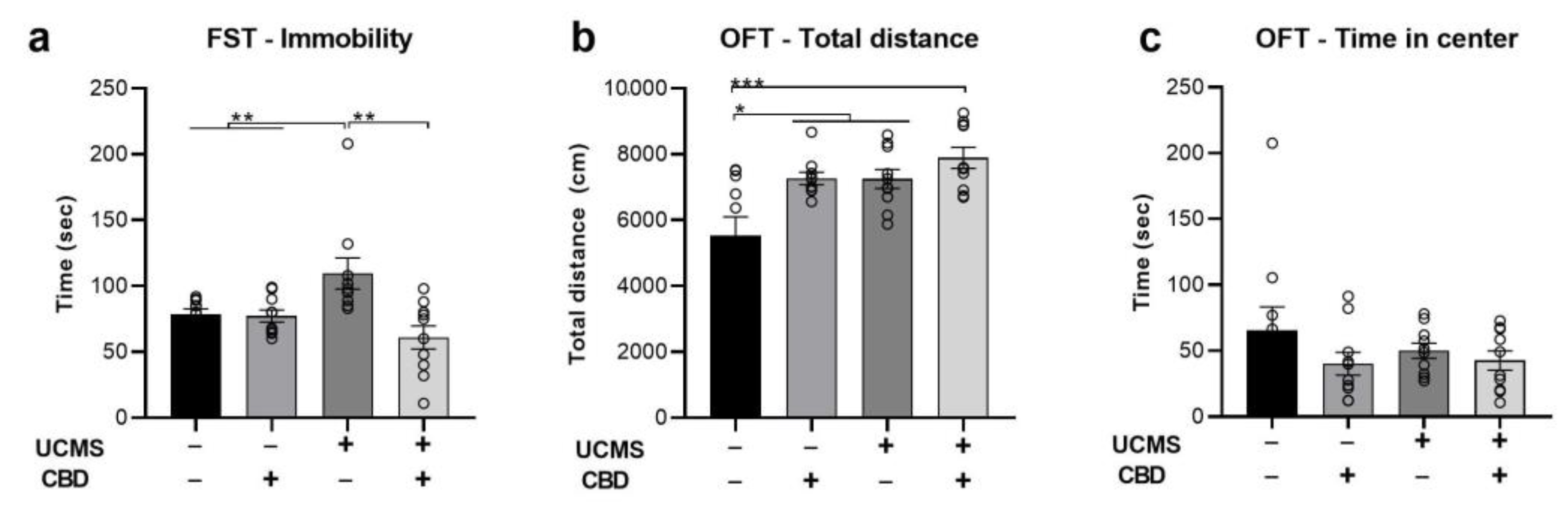
2.1. The Effects of Chronic CBD Administration during UCMS on Behavior

We examined the effects of chronic CBD administration (10 mg/kg, i.p.) during the last 3 weeks of a 6-week UCMS model on immobility in the FST and on motoric and anxiety-like behavior in the open field test (OFT). All analyses were conducted using two-way ANOVA [stress × drug (2 × 2)].
In the FST (Figure 1a), we found a significant effect of drug (F(1,39) = 9.801, p < 0.01) and stress × drug interaction (F(1,39) = 8.732, p < 0.01) with no effect of stress (F(1,39) = 0.874, ns), suggesting that CBD restored UCMS-induced increase in immobility.
In the OFT, we found a significant effect of drug (F(1,39) = 10.586, p < 0.01) and stress (F(1,39) = 10.34, p < 0.01) on locomotion (Figure 1b), with no effect of stress x drug interaction (F(1,39) = 2.236, ns), suggesting that CBD and UCMS increased locomotion behavior compared with the No UCMS vehicle group. Furthermore, we found no effect of stress (F(1,39) = 0.355, ns), drug (F(1,39) = 2.219, ns), or stress × drug interaction (F(1,39) = 0.653, ns) on the time spent in the center of the arena during the first 5 min of the test (Figure 1c).

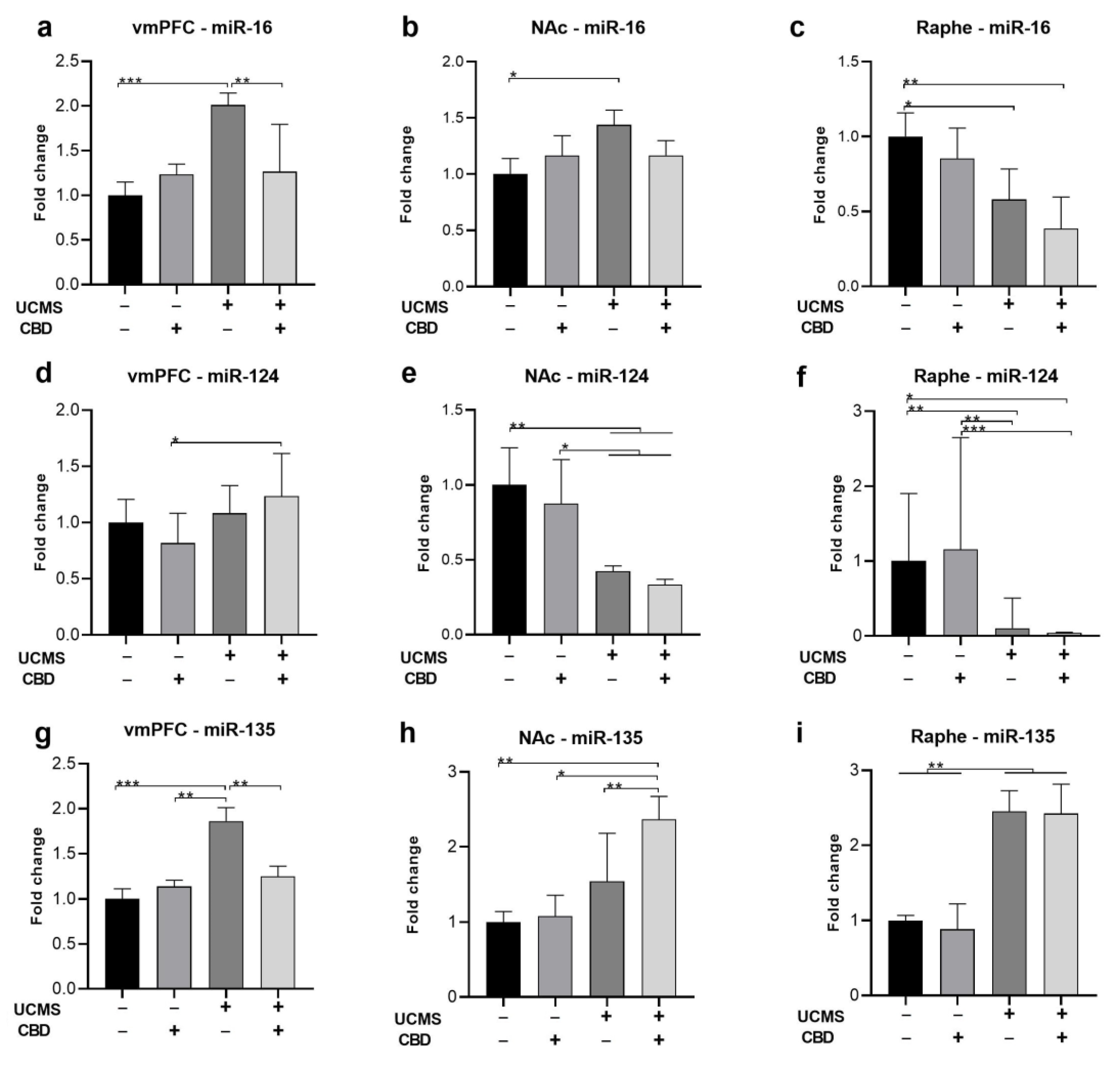
2.2. The Effects of Chronic CBD Administration during UCMS on miRNA Expression

Following the behavioral tests, we assessed the expression of miR-16, miR-124, and miR-135 in the vmPFC, NAc, and raphe nucleus. All analyses were conducted using two-way ANOVA [stress × drug (2 × 2)].

2.2.1. miR-16

In the vmPFC (Figure 2a), we found a significant effect of stress (F(1,35) = 15.648, p < 0.001), drug (F(1,35) = 4.276, p < 0.05), and stress x drug interaction (F(1,35) = 10.682, p < 0.01) on the expression of miR-16, suggesting that CBD restored UCMS-induced upregulation of miR-16.
In the NAc (Figure 2b), a significant effect of stress (F(1,34) = 5.454, p < 0.05), with no effect of drug (F(1,34) = 0.001, ns) or stress x drug interaction (F(1,34) = 2.434, ns) was observed, suggesting that UCMS upregulated miR-16 compared to the controls, an effect that was not observed in CBD-treated rats.
In the raphe (Figure 2c), we found a significant effect of stress (F(1,29) = 15.141, p < 0.001), with no effect of drug (F(1,29) = 2.862, ns) or stress × drug interaction (F(1,29) = 0.18, ns), suggesting that UCMS downregulated miR-16, with no effect for CBD.

2.2.2. miR-124

In the vmPFC (Figure 2d), we found a significant effect of stress (F(1,35) = 7.668, p < 0.01), with no effect of drug (F(1,35) = 0.155, ns), or stress x drug interaction (F(1,35) = 3.507, ns) on the expression of miR-124, suggesting that UCMS upregulated miR-124 in CBD-treated rats.
In the NAc (Figure 2e), and in the raphe (Figure 2f), a significant effect of stress (NAc: F(1,36) = 25.341, p < 0.001; raphe: F(1,37) = 30.532, p < 0.001) was observed, with no effect of drug (NAc: F(1,36) = 1.063, ns; raphe: F(1,37) = 0.527, ns) or stress × drug interaction (NAc: F(1,36) = 0.087, ns; raphe: F(1,37) = 1.03, ns), suggesting that UCMS downregulated miR-124, with no effect for CBD.

2.2.3. miR-135

In the vmPFC (Figure 2g), we found a significant effect of stress (F(1,33) = 16.546, p < 0.001), drug (F(1,33) = 4.745, p < 0.05), and stress × drug interaction (F(1,33) = 8.748, p < 0.01) on the expression of miR-135, suggesting that CBD restored UCMS-induced upregulation of miR-135.
In the NAc (Figure 2h), we found a significant effect of stress (F(1,34) = 6.996, p < 0.05) and drug (F(1,34) = 12.672, p < 0.01) with no effect of stress x drug interaction (F(1,34) = 2.726, ns), suggesting that CBD upregulated miR-135 only in UCMS rats.
In the raphe (Figure 2i), we found a significant effect of stress (F(1,27) = 19.081, p < 0.001), with no effect of drug (F(1,27) = 0.28, ns) or stress × drug interaction (F(1,27) = 0.187, ns), suggesting that UCMS upregulated miR-135, with no effect for CBD.
Pearson bivariate correlations tests (Table 1) were conducted between miRNA expression in the different brain regions and the behavioral measures to explore the association between the depressive-like behavior of the rats and their miR expression. For immobility, the most robust effect was observed with vmPFC miR-135 levels (r = 0.428, p < 0.05), suggesting that increased immobility was associated with vmPFC miR-135 upregulation. A negative correlation was observed with NAc miR-135 levels (r = −0.339, p < 0.05).
For total distance in the OFT, significant correlations were observed with NAc miR-124 (r = 0.455, p < 0.01), raphe miR-135 (r = 0.474, p < 0.05), and raphe miR-16 (r = −0.473, p < 0.01) levels. These suggest that increased locomotion behavior was associated with increased NAc miR-124 and raphe miR-135 and decreased raphe miR-16.

2.3. The Effects of Chronic CBD Administration during UCMS on Possible Target Genes

Previous findings suggested that the effects of CBD on depressive-like behavior may be mediated via serotonergic mechanisms and CB1r activation [4,11-13]. We have recently shown that the stress-preventing effects of FAAH inhibition are mediated by β-catenin [17]. Hence, we next examined alterations in the expression of several target genes in UCMS rats treated with CBD.
We examined the expression of htr1a and slc6a4 genes that encode for the serotonergic 5HT1a receptor and SERT, respectively, and the expression of ctnnb1 and cnr1, the genes that encode for β-catenin and CB1, respectively. All analyses were conducted using two-way ANOVA [stress × drug (2 × 2)].

2.3.1. htr1a

In the vmPFC (Figure 3a), we found a significant effect of stress (F(1,35) = 5.586, p < 0.05) and stress x drug interaction (F(1,35) = 9.861, p < 0.01) but not for drug (F(1,35) = 3.169, ns), suggesting that CBD restored UCMS-induced downregulation of htr1a.
In the NAc (Figure 3b), we found a significant effect of stress (F(1,26) = 6.916, p < 0.05), drug (F(1,26) = 10.584, p < 0.01) and stress x drug interaction (F(1,26) = 4.386, p < 0.05), suggesting that UCMS and CBD downregulated htr1a.
In the raphe (Figure 3c), we found no effect of stress (F(1,29) = 1.158, ns), drug (F(1,29) = 0.684, ns), or stress × drug interaction (F(1,29) = 0.305, ns), suggesting no effect for UCMS or CBD on htr1a.

2.3.2. slc6a4

In the vmPFC (Figure 3d), we found a significant effect of stress (F(1,27) = 14.184, p < 0.01) but not of drug (F(1,27) = 0.49, ns)), or stress x drug interaction (F(1,27) = 0.438, ns), suggesting that UCMS downregulated slc6a4, with no effect for CBD.
In the NAc (Figure 3e), we found no effect of stress (F(1,25) = 0.259, ns), drug (F(1,25) = 0.319, ns), or stress × drug interaction (F(1,25) = 1.253, ns), suggesting no effect for UCMS or CBD on slc6a4.
In the raphe (Figure 3f), we found a significant effect of stress (F(1,27) = 5.272, p < 0.05), drug (F(1,27) = 6.904, p < 0.05) but not for stress x drug interaction (F(1,27) = 0.303, ns), suggesting that UCMS rats treated with vehicle showed upregulation compared to No UCMS-CBD rats.

2.3.3. ctnnb1

In the vmPFC (Figure 3g), we found a significant effect of stress (F(1,35) = 7.864, p < 0.01) and stress × drug interaction (F(1,35) = 5.071, p < 0.05), but not for drug (F(1,35) = 0.453, ns) on the expression of ctnnb1, suggesting that in UCMS-vehicle, but not in UCMS-CBD rats, ctnnb1 was downregulated, an effect that was not observed in CBD-treated rats.
In the NAc (Figure 3h), a significant effect of stress (F(1,34) = 21.045, p < 0.001) was observed, but not of drug (F(1,34) = 0.001, ns) or stress × drug interaction (F(1,34) = 0.051, ns), suggesting that UCMS downregulated ctnnb1, with no effect for CBD.
In the raphe (Figure 3i), we found no effect of stress (F(1,33) = 0.157, ns), drug (F(1,33) = 0.496, ns), or stress × drug interaction (F(1,33) = 0.037, ns), suggesting no effect for UCMS or CBD on ctnnb1.

2.3.4. cnr1

In the vmPFC (Figure 3j), we found a significant effect of stress (F(1,35) = 16.429, p < 0.001) but not for drug (F(1,35) = 0.002, ns) or stress × drug interaction (F(1,35) = 0.314, ns), suggesting that UCMS downregulated cnr1. In the NAc (Figure 3k) and the raphe (Figure 3l), we found no effect of stress (NAc: F(1,32) = 1.736, ns; raphe: F(1,30) = 2.111, ns), drug (NAc: F(1,32) = 0.003, ns; raphe: F(1,30) = 0.32, ns) or stress x drug interaction (NAc: F(1,32) = 0.048, ns; raphe: F(1,30) = 0.116, ns), suggesting no effect for UCMS or CBD on cnr1.
Pearson bivariate correlations tests were conducted between miRNA expression and genes in the various brain regions.
In the vmPFC (Table 2), the most robust correlations were observed between miR-16 and hrt1a (r = 0.591, p < 0.001) and slc6a4 (r = 0.432, p < 0.05), and between miR-135 and hrt1a (r = 0.478, p < 0.01), suggesting that levels of the 5HT1a and SERT genes were associated with these microRNAs.
In the NAc (Table 3), the most robust correlations were observed between miR-16 and ctnnb1 (r = 0.479, p < 0.01) and between miR-124 and ctnnb1 (r = 0.463, p < 0.01), suggesting that levels of these microRNAs were associated with the β-catenin gene.
In the raphe nucleus (Table 4), the most robust correlations were observed between miR-124 and slc6a4 (r = 0.408, p < 0.05) suggesting that levels of this microRNA were associated with SERT gene.

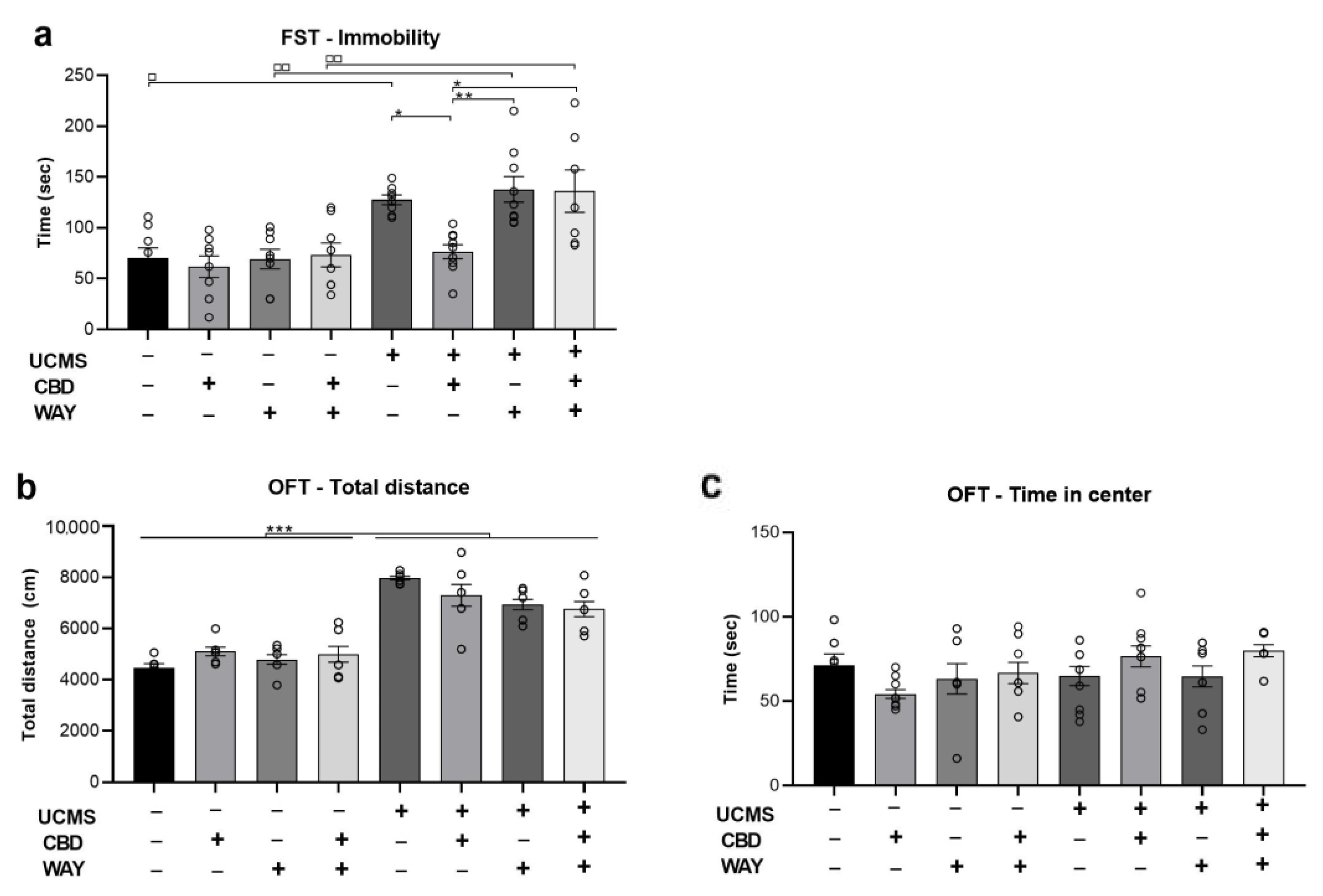
2.4. Does the 5-HT1a Antagonist WAY100635 Block the Effects of Chronic CBD Administration during UCMS on Behavior

As CBD was found to restore UCMS-induced downregulation of the htr1a gene that encodes for the serotonergic 5HT1a receptor, we pharmacologically examined whether the effects of CBD on behavior are mediated by the activation of the 5HT1a receptor. To that end, in a different set of animals, we assessed whether the antidepressant effects of CBD (see Figure 1) are mediated by the serotonergic 5HT1a receptor by administering a 5HT1a receptor antagonist. We administered the 5HT1a-antagonist WAY100635 (0.1 mg/kg) along with CBD every day during the last 3 weeks of UCMS. Other groups were injected with CBD or WAY or vehicle for comparison. All analyses were conducted using two-way ANOVA [stress × drug (2 × 2)].
In the FST (Figure 4a), we found a significant effect of stress (F(1,79) = 40.143, p < 0.001) and drug (F(1,79) = 4.506, p < 0.01) with no effect of stress x drug interaction (F(1,79) = 2.409, ns), suggesting that CBD restored UCMS-induced immobility, an effect that was blocked by the antagonist WAY. UCMS rats injected with vehicle, WAY, or WAY + CBD demonstrated increased immobility compared to No UCMS rats that were injected with vehicle (p < 0.05), WAY (p < 0.01), or WAY + CBD (p < 0.01), respectively.
In the OFT, we found a significant effect of stress (F(1,79) = 185.137, p < 0.001) and stress x drug interaction (F(1,79) = 4.602, p < 0.01) on locomotion (Figure 4b) with no effect of drug (F(1,79) = 1.227, ns), suggesting that UCMS increased locomotion, with no effect for CBD. Furthermore, we found no effect of stress (F(1,79) = 3.197, ns), drug (F(1,79) = 0.928, ns), and stress × drug interaction (F(1,79) = 2.210, ns) on the time spent in the center of the arena in the first 5 min of the test (Figure 4c).

3. Discussion

In this study, we show for the first time that CBD can restore UCMS-induced upregulation of miR-16 and miR-135 in the vmPFC as well as the associated despair-like behavior. UCMS also downregulated the 5-HT1a gene htr1a in the vmPFC; using a pharmacological approach with the 5-HT1a receptor antagonist WAY, we found that the antidepressant-like effects of CBD are mediated by the 5HT1a receptor.
The antidepressant effects of CBD on despair-like behavior in the FST corroborates with previous findings [3,4,5,42,43,44]. In the open field, CBD did not restore the UCMS-induced increase in locomotion activity and UCMS had no significant effect on anxiety-like behavior measured as time spent in the center of the open field.
The expression of miR-16, miR-124, and miR-135 was significantly affected by UCMS, corroborating with previous studies demonstrating that these miRNAs are correlated with anxiety- and depressive-like phenotypes and are significantly downregulated or upregulated following stress exposure, depending upon the brain region studied and the type of stressor [18,23,26,27,28,29,45,46]. Specifically, we found that UCMS decreased the expression of miR-124 in the NAc and raphe and increased miR-16 in the NAc and the raphe and miR-135 in the raphe. However, these effects were not normalized by CBD treatment. Similarly, UCMS affected the target genes, decreasing slc6a4 and cnr1 expression in the vmPFC (genes coding SERT and CB1), and decreasing ctnnb1 (β-catenin) in the PFC and NAc, with no effect for CBD treatment.

3.1. Alterations in miRNAs

3.1.1. miR-16

In general, higher serum levels of miR-16 are associated with a resilient phenotype to stress [47], while patients with major depression exhibit lower CSF expression of the same microRNA [30,48]. Nevertheless, these findings are rarely associated with specific brain regions. In mice, social defeat stress was associated with susceptibility to stress and lower accumbal miR-16 [18], while in rats, ELS-induced stress was associated with lower miR-16 in the vmPFC [32].
It has been shown that maternally deprived rats, but not rats exposed to chronic unpredictable stress, showed higher hippocampal miR-16 expression than control rats [26]. Taken together these findings suggest that different stressors differentially affect the expression of miRs in a brain-region-dependent manner.

3.1.2. miR-124

In the vmPFC, UCMS rats that were treated with CBD had higher levels of miR-124 only compared to control rats who were treated with CBD. UCMS decreased miR-124 in the NAc and in the raphe nucleus with no effect of CBD treatment, suggesting that its antidepressant effects are mediated by a different mechanism.
Previous studies showed that rats that were treated chronically with corticosterone as a model of depression presented higher miR-124 levels in the PFC [49], and vmPFC suppression of miR-124 via lentiviral vector decreased depressive symptoms [50]. Other studies have shown a decrease in miR-124 in the hippocampus following UCMS and that using an agomir to increase miR-124 had an antidepressant-like effect [51]. Similarly, addictive behavior to cocaine was associated with lower NAc levels of this microRNA [52,53,54], thus indicating that miR-124 may play an important role in the reward system. However, in another study, UCMS increased hippocampal miR-124 expression, and downregulation of miR-124 using an antagomir decreased depressive-like behavior [55].

3.1.3. miR-135

Previous findings showed decreased miR-135 in the PFC and raphe of mice exposed to chronic stress [27,33] and decreased PFC miR-135 in rats exposed to early life stress [32,33]. Downregulation of raphe miR-135 was observed following exposure to the chronic social defeat stress model in mice [27].

3.2. Alterations in Serotonergic Targets, β-catenin, and CB1

3.2.1. htr1a (5HT1a Gene)

We found that the UCMS-induced decrease in the 5HT1a gene in the vmPFC was reversed by CBD. This corroborates with the expectation for lower levels of the 5HT1a gene in regions where miR-135 is high, and vice versa [27].
Importantly, we found that the antidepressant-like effects of CBD were mediated by the 5HT1a receptor, as co-administration of CBD and the 5HT1a antagonist, blocked the therapeutic-like effects of CBD in the FST in UCMS rats. This corroborates with a previous study in which CBD was microinjected into the vmPFC in rats exposed to the FST and the OFT [13].
In the NAc, both UCMS and CBD led to a decrease in the 5HT1a gene, as control rats that were treated with vehicle had higher levels of this gene than all the other groups. In the raphe, UCMS or CBD had no effect on 5HT1a gene expression.

3.2.2. slc6a4 (SERT Gene)

SERT modulation is the main mechanism on which SSRIs are based [56]. Total SERT knockout results in depressive and stressed behavior [57], but variations in its expression in different brain regions can lead to a more complex effect. Therefore, lower expression of the SERT gene was expected to be observed in UCMS rats; we found a decrease in slc6a4 in the PFC, with no effect of CBD treatment. In the NAc, no differences were observed between the groups; in the raphe nucleus, UCMS-vehicle rats demonstrated increased levels compared to controls who were treated with CBD.
As a target of miR-16, levels of the SERT gene were expected to be lower in regions where miR-16 is overexpressed, and vice versa [31,58]. We found that UCMS decreased SERT gene expression in the vmPFC while elevating miR-16. However, even though CBD reversed the effect of UCMS on miR-16 expression in the vmPFC, it did not affect the SERT gene, suggesting other mechanisms which are involved in the regulation of this gene (e.g., miR-15a) [58].

3.2.3. ctnnb1 (β-catenin Gene)

We found that UCMS decreased β-catenin gene levels in the vmPFC and NAc, with no effect in the raphe. This is in line with other studies showing decreased expression of β-catenin in the brain and specifically the PFC and NAc [59,60]. CBD did not restore UCMS-induced downregulation of β-catenin, but in the vmPFC, UCMS rats treated with CBD were not different from any of the other groups. Hence, β-catenin is altered following UCMS exposure, and it regulates microRNA expression [18], but our findings did not demonstrate a robust effect of CBD on β-catenin mRNA in UCMS rats.
In general, increased β-catenin expression correlates with resilience to stress and depression [18,61], and CBD was shown to target the Wnt/β-catenin pathway [62].

3.2.4. cnr1 (CB1 Gene)

UCMS decreased the expression of the CB1 gene in the vmPFC, with no effect in the NAc or raphe. In general, CB1 signaling regulates stress responses by modulating the fast feedback inhibition of the hypothalamic–pituitary–adrenal (HPA) axis and its adaptation during exposure to repeated stress [17,63,64,65,66,67,68,69].
CBD functions as a negative allosteric modulator of CB1, and it has been shown to prevent CB1 internalization [70,71]. In our study, CBD did not restore the UCMS-induced decrease in cnr1 in the vmPFC.

4. Materials and Methods

4.1. Subjects

Male Sprague Dawley rats (60 days old) were group-housed at 22 ± 2 °C under 12 h light/dark cycles (lights turned on at 07:00). Rats were allowed water and laboratory rodent chow ad lib, except when the UCMS procedure required deprivation. The experiments were approved by the University of Haifa Ethics and Animal Care Committee, and adequate measures were taken to minimize pain and discomfort (696/20).

4.2. UCMS Protocol

Rats were subjected to a random sequence of mild stressors [72,73] for 6 weeks. These included cage soiling with 300 mL water, group housing, water and/or food deprivation, reversal of light/dark cycle, cage tilting to 45°, and physical restraint (see supplementary information (SI); Table S1). No UCMS rats were handled and were not subjected to the stress protocol.

4.3. Pharmacological Agents

No UCMS and UCMS exposed rats received daily injections (i.p.) of vehicle, CBD (10 mg/kg), or the 5-HT1a-antagonist WAY100635 (WAY; 0.1 mg/kg; Sigma, St. Louis, MO, USA) during the last 3 weeks of the 6-week UCMS model. Drugs were freshly prepared and administered in 1 mL/kg of vehicle. Rats were injected between 15:00 pm and 17:00 pm, irrespective of the stress schedule. Drugs were dissolved in 2% Tween-80 and 98% saline. Doses were based on previous work [74,75].

4.4. Behavioral Tests

All tests occurred in a dim light (15–20 lx) and took place between 1300 and 1600 h.

4.4.1. Locomotor Activity and Anxiety-like Behavior

Locomotion was measured in an open-field test (OFT). The open field arena is an open black box 50 × 50 cm in size. It was thoroughly cleaned between each trial. The movements of the rat were recorded and analyzed using Ethovision (version 11) to measure motor activity (over a period of 30 min.) and anxiety-like behavior measured as time spent in the center (first 5 min).

4.4.2. Forced Swim Test (FST)

Conducted in a cylindrical water container (62 cm diameter, 40 cm height, filled with water at a temperature of 22 °C). The water level was such that the rat could only touch the bottom with the tip of its tail. Rats were exposed to the swim tank for 15 min habituation on the first day and 5 min on the second day. Video films of the second day of each FST session were analyzed for passive coping (immobility). An immobility index was calculated: time spent immobile divided by the total time spent in the arena.

4.5. Quantitative Real-Time PCR (qRT-PCR)

Rats were sacrificed and brain tissues of the vmPFC, NAc, and raphe nucleus were harvested for molecular analysis (see SI, Figure S1). RNA extraction, cDNA preparation, and qRT-PCR were performed as previously described [76] to detect the expression of miRNAs (miR-16, miR-135, miR-124) and mRNAs (htr1a, slc6a4, ctnnb1 and cnr1; genes coding to 5HT1a, SERT, β-catenin, and CB1r, respectively). For miRNA, 500ng of total RNA was reverse transcribed cDNA using qScript microRNA cDNA Synthesis Kit (Quanta Biosciences, Gaithersburg, MD, USA). For mRNA, 1000 ng of total RNA was converted into cDNA using qScript cDNA Synthesis Kit (Quanta Biosciences, Gaithersburg, MD, USA). This was followed by Real-Time SYBR Green qRT-PCR amplification using specific primers (Quanta Biosciences, Gaithersburg, MD, USA) according to the manufacturer’s instructions. RT reactions were carried out by a Step One real-time PCR system (Applied Biosystems, Waltham, MA, USA). Fold-change values were calculated using the ddCt method relative to the housekeeping gene hypoxanthine phosphoribosyl transferase RNU6 (miRNA) or HPRT (mRNA). Primers for both miRNAs (miR-16-5p, miR-124-5p, and miR-135a-5p) and mRNAs (see Table 5) were designed and synthesized by Agentek (Tel Aviv, Israel). Primer suitability was determined using standard curve analysis, melting curve analysis, and linearity and threshold.

4.6. Statistical Analysis

The results are expressed as means ± SEM. For statistical analysis, one-way ANOVA, two-way ANOVA, and Pearson bivariate correlation test were used as indicated. All post hoc comparisons were made using Tukey’s range test. Significance was set at p ≤ 0.05. Data were analyzed using SPSS 27 (IBM, Chicago, IL, USA). Normality assumption was examined using the Kolmogorov–Smirnov and Shapiro–Wilk tests.

5. Conclusions

We show for the first time that CBD can prevent UCMS-induced increases in vmPFC miR-16 and miR-135. The antidepressant effects of CBD in rats exposed to the UCMS model for depression were mediated by the 5HT1a receptor.
CBD seems to have positive effects of diminishing depressive-like behaviors with the advantage of not being addictive or having many side effects [77]. However, the mechanisms underlying its therapeutic effects are still not entirely clear and involve multiple targets.

Supplementary Materials

The following supporting information can be downloaded at: https://www.mdpi.com/article/10.3390/ijms24032052/s1.

Author Contributions

Conceptualization, methodology U.B. and I.A.; formal analysis, investigation, visualization, writing—original draft preparation: U.B.; writing—review and editing, resources, supervision, funding acquisition, I.A. All authors have read and agreed to the published version of the manuscript.

Funding

This research was funded by the Israel Science Foundation (ISF), grant number 993/20 to IA.

Institutional Review Board Statement

The animal study protocol was approved by the University of Haifa Ethics and Animal Care Committee (approval number: 696/20).

Informed Consent Statement

Not applicable.

Data Availability Statement

Not applicable.

Acknowledgments

We thank Symrise for providing CBD.

Conflicts of Interest

The authors declare no conflict of interest. The funders had no role in the design of the study; in the collection, analyses, or interpretation of data; in the writing of the manuscript, or in the decision to publish the results.

References

  1. Maletic, V.; Robinson, M.; Oakes, T.; Iyengar, S.; Ball, S.G.; Russell, J. Neurobiology of depression: An integrated view of key findings. Int. J. Clin. Pract. 2007, 61, 2030–2040. [Google Scholar] [CrossRef] [Green Version]
  2. Pigott, H.E.; Leventhal, A.M.; Alter, G.S.; Boren, J.J. Efficacy and effectiveness of antidepressants: Current status of research. Psychother. Psychosom. 2010, 79, 267–279. [Google Scholar] [CrossRef]
  3. Shbiro, L.; Hen-Shoval, D.; Hazut, N.; Rapps, K.; Dar, S.; Zalsman, G.; Mechoulam, R.; Weller, A.; Shoval, G. Effects of cannabidiol in males and females in two different rat models of depression. Physiol. Behav. 2019, 201, 59–63. [Google Scholar] [CrossRef]
  4. Zanelati, T.V.; Biojone, C.; Moreira, F.A.; Guimarães, F.S.; Joca, S.R.L. Antidepressant-like effects of cannabidiol in mice: Possible involvement of 5-HT1A receptors. Br. J. Pharmacol. 2010, 159, 122–128. [Google Scholar] [CrossRef] [Green Version]
  5. Réus, G.Z.; Stringari, R.B.; Ribeiro, K.F.; Luft, T.; Abelaira, H.M.; Fries, G.R.; Aguiar, B.W.; Kapczinski, F.; Hallak, J.E.; Zuardi, A.W.; et al. Administration of cannabidiol and imipramine induces antidepressant-like effects in the forced swimming test and increases brain-derived neurotrophic factor levels in the rat amygdala. Acta Neuropsychiatr. 2011, 23, 241–248. [Google Scholar] [CrossRef]
  6. Shoval, G.; Shbiro, L.; Hershkovitz, L.; Hazut, N.; Zalsman, G.; Mechoulam, R.; Weller, A. Prohedonic effect of cannabidiol in a rat model of depression. Neuropsychobiology 2016, 73, 123–129. [Google Scholar] [CrossRef]
  7. Linge, R.; Jiménez-Sánchez, L.; Campa, L.; Pilar-Cuéllar, F.; Vidal, R.; Pazos, A.; Adell, A.; Díaz, Á. Cannabidiol induces rapid-acting antidepressant-like effects and enhances cortical 5-HT/glutamate neurotransmission: Role of 5-HT1A receptors. Neuropharmacology 2016, 103, 16–26. [Google Scholar] [CrossRef] [Green Version]
  8. Ferber, S.G.; Hazani, R.; Shoval, G.; Weller, A. Targeting the Endocannabinoid System in Borderline Personality Disorder: Corticolimbic and Hypothalamic Perspectives. Curr. Neuropharmacol. 2021, 19, 360–371. [Google Scholar]
  9. Zhornitsky, S.; Potvin, S. Cannabidiol in humans—The quest for therapeutic targets. Pharmaceuticals 2012, 5, 529–552. [Google Scholar] [CrossRef]
  10. McPartland, J.M.; Glass, M.; Pertwee, R.G. Meta-analysis of cannabinoid ligand binding affinity and receptor distribution: Interspecies differences. Br. J. Pharmacol. 2007, 152, 583–593. [Google Scholar] [CrossRef] [Green Version]
  11. Campos, A.C.; Moreira, F.A.; Gomes, F.V.; Del Bel, E.A.; Guimaraes, F.S. Multiple mechanisms involved in the large-spectrum therapeutic potential of cannabidiol in psychiatric disorders. Philos. Trans. R. Soc. B Biol. Sci. 2012, 367, 3364–3378. [Google Scholar] [CrossRef]
  12. Resstel, L.B.; Tavares, R.F.; Lisboa, S.F.; Joca, S.R.; Corrêa, F.M.; Guimarães, F.S. 5-HT1A receptors are involved in the cannabidiol-induced attenuation of behavioural and cardiovascular responses to acute restraint stress in rats. Br. J. Pharmacol. 2009, 156, 181–188. [Google Scholar] [CrossRef] [PubMed] [Green Version]
  13. Sartim, A.G.; Guimarães, F.S.; Joca, S.R.L. Antidepressant-like effect of cannabidiol injection into the ventral medial prefrontal cortex—Possible involvement of 5-HT1A and CB1 receptors. Behav. Brain Res. 2016, 303, 218–227. [Google Scholar] [CrossRef] [PubMed]
  14. Fogaça, M.V.; Campos, A.C.; Guimarães, F.S. Cannabidiol and 5-HT1A receptors. In Neuropathology of Drug Addictions and Substance Misuse; Academic Press: Cambridge, MA, USA, 2016; pp. 749–759. [Google Scholar]
  15. Aberle, H.; Bauer, A.; Stappert, J.; Kispert, A.; Kemler, R. β-catenin is a target for the ubiquitin–proteasome pathway. EMBO J. 1977, 16, 3797–3804. [Google Scholar] [CrossRef] [PubMed] [Green Version]
  16. Teo, C.H.; Soga, T.; Parhar, I.S. Brain beta-catenin signalling during stress and depression. Neurosignals 2018, 26, 31–42. [Google Scholar] [CrossRef] [PubMed]
  17. Mizrachi Zer-Aviv, T.; Islami, L.; Hamilton, P.J.; Parise, E.M.; Nestler, E.J.; Sbarski, B.; Akirav, I. Enhancing Endocannabinoid Signaling via β-Catenin in the Nucleus Accumbens Attenuates PTSD-and Depression-like Behavior of Male Rats. Biomedicines 2022, 10, 1789. [Google Scholar] [CrossRef]
  18. Dias, C.; Feng, J.; Sun, H.; Mazei-Robison, M.S.; Damez-Werno, D.; Scobie, K.; Bagot, R.; LaBonté, B.; Ribeiro, E.; Liu, X.; et al. β-catenin mediates stress resilience through Dicer1/microRNA regulation. Nature 2014, 516, 51–55. [Google Scholar] [CrossRef] [Green Version]
  19. Hansen, K.F.; Obrietan, K. MicroRNA as therapeutic targets for treatment of depression. Neuropsychiatr. Dis. Treat. 2013, 9, 1011. [Google Scholar]
  20. O’connor, R.M.; Dinan, T.G.; Cryan, J.F. Little things on which happiness depends: microRNAs as novel therapeutic targets for the treatment of anxiety and depression. Mol. Psychiatry 2012, 17, 359–376. [Google Scholar] [CrossRef] [Green Version]
  21. Serafini, G.; Pompili, M.; Hansen, K.F.; Obrietan, K.; Dwivedi, Y.; Shomron, N.; Girardi, P. The involvement of microRNAs in major depression, suicidal behavior, and related disorders: A focus on miR-185 and miR-491-3p. Cell. Mol. Neurobiol. 2014, 34, 17–30. [Google Scholar] [CrossRef]
  22. Ferrúa, C.P.; Giorgi, R.; da Rosa, L.C.; do Amaral, C.C.; Ghisleni, G.C.; Pinheiro, R.T.; Nedel, F. MicroRNAs expressed in depression and their associated pathways: A systematic review and a bioinformatics analysis. J. Chem. Neuroanat. 2019, 100, 101650. [Google Scholar] [CrossRef]
  23. Allen, L.; Dwivedi, Y. MicroRNA mediators of early life stress vulnerability to depression and suicidal behavior. Mol. Psychiatry 2020, 25, 308–320. [Google Scholar] [CrossRef]
  24. Muiños-Gimeno, M.; Espinosa-Parrilla, Y.; Guidi, M.; Kagerbauer, B.; Sipilä, T.; Maron, E.; Pettai, K.; Kananen, L.; Navinés, R.; Martín-Santos, R.; et al. Human microRNAs miR-22, miR-138-2, miR-148a, and miR-488 are associated with panic disorder and regulate several anxiety candidate genes and related pathways. Biol. Psychiatry 2011, 69, 526–533. [Google Scholar] [CrossRef]
  25. Haramati, S.; Navon, I.; Issler, O.; Ezra-Nevo, G.; Gil, S.; Zwang, R.; Hornstein, E.; Chen, A. MicroRNA as repressors of stress-induced anxiety: The case of amygdalar miR-34. J. Neurosci. 2011, 31, 14191–14203. [Google Scholar] [CrossRef] [Green Version]
  26. Bai, M.; Zhu, X.; Zhang, Y.; Zhang, S.; Zhang, L.; Xue, L.; Yi, J.; Yao, S.; Zhang, X. Abnormal hippocampal BDNF and miR-16 expression is associated with depression-like behaviors induced by stress during early life. PLoS ONE 2012, e46921. [Google Scholar] [CrossRef]
  27. Issler, O.; Haramati, S.; Paul, E.D.; Maeno, H.; Navon, I.; Zwang, R.; Gil, S.; Mayberg, H.S.; Dunlop, B.W.; Menke, A.; et al. MicroRNA 135 is essential for chronic stress resiliency, antidepressant efficacy, and intact serotonergic activity. Neuron 2014, 83, 344–360. [Google Scholar] [CrossRef] [Green Version]
  28. Higuchi, F.; Uchida, S.; Yamagata, H.; Abe-Higuchi, N.; Hobara, T.; Hara, K.; Kobayashi, A.; Shintaku, T.; Itoh, Y.; Suzuki, T.; et al. Hippocampal microRNA-124 enhances chronic stress resilience in mice. J. Neurosci. 2016, 36, 7253–7267. [Google Scholar] [CrossRef] [Green Version]
  29. Volk, N.; Pape, J.C.; Engel, M.; Zannas, A.S.; Cattane, N.; Cattaneo, A.; Binder, E.B.; Chen, A. Amygdalar microRNA-15a is essential for coping with chronic stress. Cell Rep. 2016, 17, 1882–1891. [Google Scholar] [CrossRef] [Green Version]
  30. Maurel, O.M.; Torrisi, S.A.; Barbagallo, C.; Purrello, M.; Salomone, S.; Drago, F.; Leggio, G.M. Dysregulation of miR-15a-5p, miR-497a-5p and miR-511-5p is associated with modulation of BDNF and FKBP5 in brain areas of PTSD-related susceptible and resilient mice. Int. J. Mol. Sci. 2021, 22, 5157. [Google Scholar] [CrossRef]
  31. Song, M.F.; Dong, J.Z.; Wang, Y.W.; He, J.; Ju, X.; Zhang, L.; Zhang, Y.H.; Shi, J.F.; Lv, Y.Y. CSF miR-16 is decreased in major depression patients and its neutralization in rats induces depression-like behaviors via a serotonin transmitter system. J. Affect. Disord. 2015, 178, 25–31. [Google Scholar] [CrossRef]
  32. Baudry, A.; Mouillet-Richard, S.; Schneider, B.; Launay, J.M.; Kellermann, O. miR-16 targets the serotonin transporter: A new facet for adaptive responses to antidepressants. Science 2010, 329, 1537–1541. [Google Scholar] [CrossRef] [Green Version]
  33. Portugalov, A.; Zaidan, H.; Gaisler-Salomon, I.; Hillard, C.J.; Akirav, I. FAAH inhibition restores early life stress-induced alterations in PFC microRNAs associated with depressive-like behavior in male and female rats. IJMS 2022, 23, 16101. [Google Scholar] [CrossRef]
  34. Liu, Y.; Liu, D.; Xu, J.; Jiang, H.; Pan, F. Early adolescent stress-induced changes in prefrontal cortex miRNA-135a and hippocampal miRNA-16 in male rats. Dev. Psychobiol. 2017, 59, 958–969. [Google Scholar] [CrossRef] [PubMed]
  35. Adlakha, Y.K.; Saini, N. Brain microRNAs and insights into biological functions and therapeutic potential of brain enriched miRNA-128. Mol. Cancer 2014, 13, 1–18. [Google Scholar] [CrossRef] [PubMed] [Green Version]
  36. Ding, Q.; Xia, W.; Liu, J.C.; Yang, J.Y.; Lee, D.F.; Xia, J.; Bartholomeusz, G.; Li, Y.; Pan, Y.; Li, Z.; et al. Erk associates with and primes GSK-3β for its inactivation resulting in upregulation of β-catenin. Mol. Cell 2005, 19, 159–170. [Google Scholar] [CrossRef]
  37. Mendoza, M.C.; Er, E.E.; Blenis, J. The Ras-ERK and PI3K-mTOR pathways: Cross-talk and compensation. Trends Biochem. Sci. 2011, 36, 320–328. [Google Scholar] [CrossRef] [PubMed] [Green Version]
  38. Ma, C.; Bower, K.A.; Chen, G.; Shi, X.; Ke, Z.J.; Luo, J. Interaction between ERK and GSK3β mediates basic fibroblast growth factor-induced apoptosis in SK-N-MC neuroblastoma cells. J. Biol. Chem. 2008, 283, 9248–9256. [Google Scholar] [CrossRef] [PubMed] [Green Version]
  39. Bewernick, B.H.; Hurlemann, R.; Matusch, A.; Kayser, S.; Grubert, C.; Hadrysiewicz, B.; Axmacher, N.; Lemke, M.; Cooper-Mahkorn, D.; Cohen, M.X.; et al. Nucleus accumbens deep brain stimulation decreases ratings of depression and anxiety in treatment-resistant depression. Biol. Psychiatry 2010, 67, 110–116. [Google Scholar] [CrossRef]
  40. Campbell, S.; MacQueen, G. The role of the hippocampus in the pathophysiology of major depression. J. Psychiatry Neurosci. 2004, 29, 417–426. [Google Scholar]
  41. Koenigs, M.; Huey, E.D.; Calamia, M.; Raymont, V.; Tranel, D.; Grafman, J. Distinct regions of prefrontal cortex mediate resistance and vulnerability to depression. J. Neurosci. 2008, 28, 12341–12348. [Google Scholar] [CrossRef] [Green Version]
  42. Zhu, S.; Shi, R.; Wang, J.; Wang, J.F.; Li, X.M. Unpredictable chronic mild stress not chronic restraint stress induces depressive behaviours in mice. Neuroreport 2014, 25, 1151–1155. [Google Scholar] [CrossRef] [PubMed]
  43. Luo, D.D.; An, S.C.; Zhang, X. Involvement of hippocampal serotonin and neuropeptide Y in depression induced by chronic unpredicted mild stress. Brain Res. Bull. 2008, 77, 8–12. [Google Scholar] [CrossRef] [PubMed]
  44. Bhutani, M.K.; Bishnoi, M.; Kulkarni, S.K. Anti-depressant like effect of curcumin and its combination with piperine in unpredictable chronic stress-induced behavioral, biochemical and neurochemical changes. Pharmacol. Biochem. Behav. 2009, 92, 39–43. [Google Scholar] [CrossRef]
  45. Bangasser, D.A.; Cuarenta, A. Sex differences in anxiety and depression: Circuits and mechanisms. Nat. Rev. Neurosci. 2021, 22, 674–684. [Google Scholar] [CrossRef] [PubMed]
  46. Eid, R.S.; Gobinath, A.R.; Galea, L.A. Sex differences in depression: Insights from clinical and preclinical studies. Prog. Neurobiol. 2019, 176, 86–102. [Google Scholar] [CrossRef] [PubMed]
  47. Zurawek, D.; Kusmider, M.; Faron-Gorecka, A.; Gruca, P.; Pabian, P.; Kolasa, M.; Solich, J.; Szafran-Pilch, K.; Papp, M.; Dziedzicka-Wasylewska, M. Time-dependent miR-16 serum fluctuations together with reciprocal changes in the expression level of miR-16 in mesocortical circuit contribute to stress resilient phenotype in chronic mild stress–an animal model of depression. Eur. Neuropsychopharmacol. 2016, 26, 23–36. [Google Scholar] [CrossRef] [PubMed]
  48. Gheysarzadeh, A.; Sadeghifard, N.; Afraidooni, L.; Pooyan, F.; Mofid, M.R.; Valadbeigi, H.; Bakhtiari, H.; Keikhavani, S. Serum-based microRNA biomarkers for major depression: MiR-16, miR-135a, and miR-1202. J. Res. Med. Sci. Off. J. Isfahan Univ. Med. Sci. 2018, 23, 39. [Google Scholar] [CrossRef]
  49. Dwivedi, Y.; Roy, B.; Lugli, G.; Rizavi, H.; Zhang, H.; Smalheiser, N.R. Chronic corticosterone-mediated dysregulation of microRNA network in prefrontal cortex of rats: Relevance to depression pathophysiology. Transl. Psychiatry 2015, 5, e682. [Google Scholar] [CrossRef] [Green Version]
  50. Gu, Z.; Pan, J.; Chen, L. MiR-124 suppression in the prefrontal cortex reduces depression-like behavior in mice. Biosci. Rep. 2019, 39, BSR20190186. [Google Scholar] [CrossRef] [Green Version]
  51. Lou, D.; Wang, J.; Wang, X. miR-124 ameliorates depressive-like behavior by targeting STAT3 to regulate microglial activation. Mol. Cell. Probes 2019, 48, 101470. [Google Scholar] [CrossRef]
  52. Periyasamy, P.; Liao, K.; Kook, Y.H.; Niu, F.; Callen, S.E.; Guo, M.L.; Buch, S. Cocaine-mediated downregulation of miR-124 activates microglia by targeting KLF4 and TLR4 signaling. Mol. Neurobiol. 2018, 55, 3196–3210. [Google Scholar] [CrossRef]
  53. Cabana-Domínguez, J.; Arenas, C.; Cormand, B.; Fernàndez-Castillo, N. MiR-9, miR-153 and miR-124 are down-regulated by acute exposure to cocaine in a dopaminergic cell model and may contribute to cocaine dependence. Transl. Psychiatry 2018, 8, 1–8. [Google Scholar] [CrossRef] [Green Version]
  54. Dash, S.; Balasubramaniam, M.; Martínez-Rivera, F.J.; Godino, A.; Peck, E.G.; Patnaik, S.; Suar, M.; Calipari, E.S.; Nestler, E.J.; Villalta, F.; et al. Cocaine-regulated microRNA miR-124 controls poly (ADP-ribose) polymerase-1 expression in neuronal cells. Sci. Rep. 2020, 10, 1–15. [Google Scholar] [CrossRef]
  55. Yang, W.; Liu, M.; Zhang, Q.; Zhang, J.; Chen, J.; Chen, Q.; Suo, L. Knockdown of miR-124 reduces depression-like behavior by targeting CREB1 and BDNF. Curr. Neurovascular Res. 2020, 17, 196–203. [Google Scholar] [CrossRef]
  56. Fakhoury, M. Revisiting the serotonin hypothesis: Implications for major depressive disorders. Mol. Neurobiol. 2016, 53, 2778–2786. [Google Scholar] [CrossRef]
  57. Kalueff, A.V.; Olivier, J.D.A.; Nonkes, L.J.P.; Homberg, J.R. Conserved role for the serotonin transporter gene in rat and mouse neurobehavioral endophenotypes. Neurosci. Biobehav. Rev. 2010, 34, 373–386. [Google Scholar] [CrossRef]
  58. Moya, P.R.; Wendland, J.R.; Salemme, J.; Fried, R.L.; Murphy, D.L. miR-15a and miR-16 regulate serotonin transporter expression in human placental and rat brain raphe cells. Int. J. Neuropsychopharmacol. 2013, 16, 621–629. [Google Scholar] [CrossRef] [Green Version]
  59. Dahlhoff, M.; Siegmund, A.; Golub, Y.; Wolf, E.; Holsboer, F.; Wotjak, C.T. AKT/GSK-3β/β-catenin signalling within hippocampus and amygdala reflects genetically determined differences in posttraumatic stress disorder like symptoms. Neuroscience 2010, 169, 1216–1226. [Google Scholar] [CrossRef]
  60. Chen, Y.C.; Tan, Q.R.; Dang, W.; Wang, H.N.; Zhang, R.B.; Li, Z.Y.; Lin, H.; Liu, R. The effect of citalopram on chronic stress-induced depressive-like behavior in rats through GSK3β/β-catenin activation in the medial prefrontal cortex. Brain Res. Bull. 2012, 88, 338–344. [Google Scholar] [CrossRef]
  61. Pilar-Cuéllar, F.; Vidal, R.; Pazos, A. Subchronic treatment with fluoxetine and ketanserin increases hippocampal brain-derived neurotrophic factor, β-catenin and antidepressant-like effects. Br. J. Pharmacol. 2012, 165, 1046–1057. [Google Scholar] [CrossRef] [Green Version]
  62. Vallée, A.; Lecarpentier, Y.; Vallée, J.N. Possible actions of cannabidiol in obsessive-compulsive disorder by targeting the WNT/β-catenin pathway. Mol. Psychiatry 2022, 27, 230–248. [Google Scholar] [CrossRef] [PubMed]
  63. Sbarski, B.; Akirav, I. Cannabinoids as therapeutics for PTSD. Pharmacol. Ther. 2020, 211, 107551. [Google Scholar] [CrossRef] [PubMed]
  64. Hillard, C.J.; Beatka, M.; Sarvaideo, J. Endocannabinoid signaling and the hypothalamic-pituitary-adrenal axis. Compr. Physiol. 2016, 7, 1. [Google Scholar] [PubMed]
  65. Hill, M.N.; McLaughlin, R.J.; Bingham, B.; Shrestha, L.; Lee, T.T.; Gray, J.M.; Hillard, C.J.; Gorzalka, B.B.; Viau, V. Endogenous cannabinoid signaling is essential for stress adaptation. Proc. Natl. Acad. Sci. USA 2010, 107, 9406–9411. [Google Scholar] [CrossRef] [Green Version]
  66. Hill, M.N.; Karatsoreos, I.N.; Hillard, C.J.; McEwen, B.S. Rapid elevations in limbic endocannabinoid content by glucocorticoid hormones in vivo. Psychoneuroendocrinology 2010, 35, 1333–1338. [Google Scholar] [CrossRef] [Green Version]
  67. Alteba, S.; Zer-Aviv, T.M.; Tenenhaus, A.; David, G.B.; Adelman, J.; Hillard, C.J.; Doron, R.; Akirav, I. Antidepressant-like effects of URB597 and JZL184 in male and female rats exposed to early life stress. Eur. Neuropsychopharmacol. 2020, 39, 70–86. [Google Scholar] [CrossRef]
  68. Alteba, S.; Portugalov, A.; Hillard, C.J.; Akirav, I. Inhibition of fatty acid amide hydrolase (FAAH) during adolescence and exposure to early life stress may exacerbate depression-like behaviors in male and female rats. Neuroscience 2021, 455, 89–106. [Google Scholar] [CrossRef]
  69. Burstein, O.; Shoshan, N.; Doron, R.; Akirav, I. Cannabinoids prevent depressive-like symptoms and alterations in BDNF expression in a rat model of PTSD. Prog. Neuro-Psychopharmacol. Biol. Psychiatry 2018, 84, 129–139. [Google Scholar] [CrossRef]
  70. Laprairie, R.B.; Bagher, A.M.; Kelly, M.E.M.; Denovan-Wright, E. Cannabidiol is a negative allosteric modulator of the cannabinoid CB1 receptor. Br. J. Pharmacol. 2015, 172, 4790–4805. [Google Scholar] [CrossRef] [Green Version]
  71. Chung, H.; Fierro, A.; Pessoa-Mahana, C.D. Cannabidiol binding and negative allosteric modulation at the cannabinoid type 1 receptor in the presence of delta-9-tetrahydrocannabinol: An In Silico study. PLoS ONE 2019, 14, e0220025. [Google Scholar] [CrossRef]
  72. Willner, P.; Muscat, R.; Papp, M. Chronic mild stress-induced anhedonia: A realistic animal model of depression. Neurosci. Biobehav. Rev. 1992, 16, 525–534. [Google Scholar] [CrossRef] [PubMed]
  73. Grønli, J.; Murison, R.; Bjorvatn, B.; Sørensen, E.; Portas, C.M.; Ursin, R. Chronic mild stress affects sucrose intake and sleep in rats. Behav. Brain Res. 2004, 150, 139–147. [Google Scholar] [CrossRef] [PubMed]
  74. Sales, A.J.; Fogaça, M.V.; Sartim, A.G.; Pereira, V.S.; Wegener, G.; Guimarães, F.S.; Joca, S.R. Cannabidiol induces rapid and sustained antidepressant-like effects through increased BDNF signaling and synaptogenesis in the prefrontal cortex. Mol. Neurobiol. 2019, 56, 1070–1081. [Google Scholar] [CrossRef] [PubMed]
  75. Rogóż, Z.; Kabziński, M.; Sadaj, W.; Rachwalska, P.; Gądek-Michalska, A. Effect of co-treatment with fluoxetine or mirtazapine and risperidone on the active behaviors and plasma corticosterone concentration in rats subjected to the forced swim test. Pharmacol. Rep. 2012, 64, 1391–1399. [Google Scholar] [CrossRef] [PubMed]
  76. Zaidan, H.; Ramaswami, G.; Barak, M.; Li, J.B.; Gaisler-Salomon, I. Pre-reproductive stress and fluoxetine treatment in rats affect offspring A-to-I RNA editing, gene expression and social behavior. Environ. Epigenetics 2018, 4, 21. [Google Scholar] [CrossRef] [PubMed]
  77. Iffland, K.; Grotenhermen, F. An update on safety and side effects of cannabidiol: A review of clinical data and relevant animal studies. Cannabis Cannabinoid Res. 2017, 2, 139–154. [Google Scholar] [CrossRef]
Figure 1. The effects of CBD treatment on behavior in rats exposed to UCMS. (a) FST: UCMS rats treated with a vehicle spent more time immobile than the No UCMS groups and UCMS rats treated with CBD. (b) OFT total distance: No UCMS rats treated with the vehicle covered less distance compared to all groups. (c) OFT time in the center: no differences between groups were observed. FST—forced swim test; OFT—open field test; UCMS: unpredictable chronic mild stress; CBD: cannabidiol. *, p < 0.05, **, p < 0.01, ***, p < 0.001.
Ijms 24 02052 g001
Figure 2. The effects of CBD treatment on miRNA expression in the vmPFC, NAc, and raphe nucleus in rats exposed to UCMS. miR-16. (a) vmPFC: UCMS rats treated with a vehicle demonstrated miR-16 upregulation compared to all groups. (b) NAc: UCMS rats treated with a vehicle demonstrated miR-16 upregulation compared to No UCMS rats treated with a vehicle. (c) Raphe: UCMS (vehicle, CBD) downregulated miR-16 compared to UCMS rats treated with a vehicle. miR-124: (d) vmPFC: UCMS rats treated with CBD demonstrated miR-124 upregulation compared to No UCMS rats treated with CBD. (e) NAc: UCMS downregulated miR-124 compared to No UCMS. (f) Raphe: UCMS downregulated miR-124 compared to No UCMS. miR-135: (g) vmPFC: UCMS rats treated with a vehicle demonstrated miR-135 upregulation compared to all groups. (h) NAc: UCMS rats treated with CBD demonstrated miR-135 upregulation compared to all groups. (i) Raphe: UCMS upregulated miR-135 compared to No UCMS. vmPFC: ventromedial prefrontal cortex; NAc: nucleus accumbens; miR: microRNA; UCMS: unpredictable chronic mild stress; CBD: cannabidiol. *, p < 0.05, **, p < 0.01, ***, p < 0.001.
Ijms 24 02052 g002
Figure 3. The effects of CBD treatment on serotonergic, β-catenin, and CB1 mRNA expression in the vmPFC, NAc, and raphe nucleus in rats exposed to UCMS. htr1a: (a) vmPFC: UCMS-vehicle downregulated htr1a levels compared to all groups. (b) NAc: UCMS rats treated with a vehicle or CBD and No UCMS rats treated with CBD demonstrated htr1a downregulation compared to No UCMS rats treated with a vehicle. (c) Raphe: no differences were observed between the groups. slc6a4: (d) vmPFC: UCMS rats treated with a vehicle or CBD demonstrated downregulation compared to No UCMS rats treated with a vehicle. (e) NAc: no differences were observed between the groups. (f) Raphe: UCMS-vehicle rats demonstrated upregulation of slc6a4 compared to No UCMS-CBD rats. Ctnnb1: (g) vmPFC: UCMS-vehicle rats demonstrated Ctnnb1 downregulation compared to No UCMS-vehicle rats. (h) NAc: UCMS downregulated Ctnnb1 compared to No UCMS rats treated with a vehicle or CBD. (i) Raphe: no differences were observed between the groups. Cnr1: (j) vmPFC: UCMS rats that were treated with a vehicle or CBD demonstrated Cnr1 downregulation compared to No UCMS rats treated with a vehicle or CBD. (k) NAc: no differences were observed between the groups. (l) Raphe: no differences were observed between the groups. vmPFC: ventromedial prefrontal cortex; NAc: nucleus accumbens; UCMS: unpredictable chronic mild stress; CBD: cannabidiol. *, p < 0.05, **, p < 0.01.
Ijms 24 02052 g003
Figure 4. The effects of the 5-HT1a antagonist WAY100635 on behavior in rats exposed to UCMS and treated with CBD. (a) FST: UCMS rats that were treated with CBD, spent less time immobile than UCMS rats that were treated with vehicle, WAY, or CBD + WAY. Furthermore, UCMS rats treated with vehicle, WAY, or CBD + WAY, showed increased immobility compared to No UCMS rats injected with vehicle, WAY, or CBD + WAY, respectively. (b) OFT: UCMS rats traveled more than No UCMS rats. (c) OFT time in the center: no differences between groups were observed. FST—forced swim test; OFT—open field test; UCMS: unpredictable chronic mild stress; CBD: cannabidiol; WAY: WAY100635, *, p < 0.05; **, p < 0.01; ***, p < 0.001; compared to UCMS-CBD group ◻ *, p < 0.05, ◻◻ **, p < 0.01, ◻◻◻ ***, p < 0.001 white aquares indicate statistical significance in UCMS vs. NO UCMS groups.
Ijms 24 02052 g004
Table 1. Pearson correlation coefficients between miRNA levels and behavioral measures in rats exposed to UCMS and CBD.
miR-16 vmPFCmiR-16 NACmiR-16 RaphemiR-124 vmPFCmiR-124 NACmiR-124 RaphemiR-135 vmPFCmiR-135 NACmiR-135 Raphe
FST—immobilityr = 0.277
p = 0.102
r = 0.250
p = 0.147
r = 0.069
p = 716
r = 0.087
p = 0.613
r = −0.040
p = 0.816
r = 0.054
p = 0.749
r = 0.428 *
p = 0.012
r = −0.339 *
p = 0.046
r = 0.187
p = 0.340
OFT—total distancer = 0.176
p = 0.305
r = 0.322
p = 0.060
r = −0.473 **
p = 0.008
r = −0.159
p = 0.355
r = 0.455 **
p = 0.005
r = 0.060
p = 0.718
r = 0.012
p = 0.944
r = 0.192
p = 0.268
r = 0.474 *
p = 0.011
OFT—time in centerr = 0.0.14
p = 0.933
r = −0.2.33
p = 0.198
r = 0.276
p = 0.140
r = −0.184
p = 0.282
r = −0.142
p = 0.403
r = 0.157
p = 0.347
r = −0.75
p = 0.675
r = −0.229
p = 0.186
r = −0.70
p = 0.723
FST—forced swim test; OFT—open field test; vmPFC: ventromedial prefrontal cortex; NAc: nucleus accumbens; miR: microRNA; UCMS: unpredictable chronic mild stress; CBD: cannabidiol. *, p < 0.05, **, p < 0.01.
Table 2. Pearson correlation coefficients between miRNA levels and genes in the vmPFC in rats exposed to UCMS and CBD.
hrt1aslc6a4ctnnb1cnr1
miR-16r = 0.591 ***
p = 0.000
r = −0.432 *
p = 0.031
r = 0.140
p = 0.436
r = 0.250
p = 0.160
miR-124r = −0.131
p = 0.469
r = 0.281
p = 0.183
r = 0.027
p = 0.880
r = −0.075
p = 0.682
miR-135r = 0.478 **
p = 0.008
r = −0.346
p = 0.091
r = 0.001
p = 0.997
r = 0.258
p = 0.154
*, p < 0.05, **, p < 0.01, ***, p < 0.001.
Table 3. Pearson correlation coefficients between miRNA levels and genes in the NAc in rats exposed to UCMS and CBD.
hrt1aslc6a4ctnnb1cnr1
miR-16r = −0.404
p = 0.051
r = −0.33
p = 0.878
r = 0.479 **
p = 0.006
r = 0.229
p = 0.215
miR-124r = −0.272
p = 0.198
r = −0.56
p = 0.794
r = 0.463 **
p = 0.008
r = 0.224
p = 0.234
miR-135r = −0.324
p = 0.114
r = −0.080
p = 0.724
r = 0.222
p = 0.222
r = 0.226
p = 0.231
**, p < 0.01.
Table 4. Pearson correlation coefficients between miRNA levels and genes in the raphe nucleus in rats exposed to UCMS and CBD.
hrt1aslc6a4ctnnb1cnr1
miR-16r = 0.125
p = 0.579
r = 0.064
p = 0.795
r = −0.205
p = 0.325
r = 0.012
p = 0.956
miR-124r = −0.358
p = 0.056
r = 0.408 *
p = 0.035
r = −0.175
p = 0.330
r = 0.147
p = 0.438
miR-135r = 0.191
p = 0.382
r = 0.232
p = 0.298
r = 0.050
p = 0.815
r = 0.324
p = 0.142
*, p < 0.05.
Table 5. Primers for mRNAs used for real-time PCR.
NameDescriptionGeneBankID (NM)Protein NamePrimer SequenceEfficacyDescription
HprtHousekeeping gene; used as a reference geneNM_012583.2HPRTF: 5′CGCCAGCTTCCTCCTCAG3′NM_012583.2HPRT
Htr1aSerotonergic auto-receptorR: 5′ATAACCTGGTTCATCATCACTAATCAC3′99.83 R: 5′ATAACCTGGTTCATCATCACTAATCAC3′99.83
Slc6a4The serotonergic transporterNM_012585.15HT1aF: 5′CCACGGCTACACCATCTACTC3′NM_012585.15HT1a
F: forward primer; R: reverse primer.
Disclaimer/Publisher’s Note: The statements, opinions and data contained in all publications are solely those of the individual author(s) and contributor(s) and not of MDPI and/or the editor(s). MDPI and/or the editor(s) disclaim responsibility for any injury to people or property resulting from any ideas, methods, instructions or products referred to in the content.

