Article

Long-Term Risk of Hepatic and Extrahepatic-Related Events After Direct Antiviral Therapy for Chronic Hepatitis C: A Prospective Long-Term Study Cohort

by
Luisa Cavalletto
1,*,†,
Eleonora Bertoli
2,†,
Claudia Mescoli
3,
Camillo Aliberti
4,
Maria Giovanna Quaranta
5,
Loreta Kondili
5 and
Liliana Chemello
1,*
1
UOC Clinica Medica 5, Regional Center for Liver Disease Outpatient Unit, Department of Medicine—DIMED, University of Padova, 35128 Padova, Italy
2
Unit of Emergency Medicine, Department of Systems Medicine—DIDAS, University of Padova, 35128 Padova, Italy
3
Unit of Pathology, Department of Medicine—DIMED, University of Padova, 35128 Padova, Italy
4
Unit of Radiology, Pederzoli Hospital Peschiera del Garda, 37019 Verona, Italy
5
Istituto Superiore di Sanità, Global Health Center, 00161 Rome, Italy
*
Authors to whom correspondence should be addressed.
These authors contributed equally to this work.
Cancers 2025, 17(9), 1528; https://doi.org/10.3390/cancers17091528
Submission received: 15 February 2025 / Revised: 2 April 2025 / Accepted: 27 April 2025 / Published: 30 April 2025
(This article belongs to the Special Issue Insights from the Editorial Board Member)

Simple Summary

After availability of oral direct-acting antivirals for treatment of chronic hepatitis C, it is required to define the long-term effectiveness of the extensive use of these novel molecules and, in particular, to analyze the outcome in term of morbidity and mortality, especially in senior cases or with advanced liver disease. The identification of the frail subject, which is not meant to have a significant change in life expectancy, even after HCV eradication, can give the opportunity to produce new algorithms predictive of unfavorable outcomes and can improve management of who maintains a high risk of hepatic- and extrahepatic-related events and, in particular, of hepatocellular carcinoma, liver transplantation, or death.

Abstract

Novel direct antiviral-acting (DAA) molecules significantly improved efficacy and ameliorated outcomes of patients with chronic hepatitis C (CHC). The extensive use of DAA from 2015, due to large access to therapy, maximized rates of viral eradication with a safety profile in the majority of cases. Aims: We evaluated risk factors and the incidence of related clinical events and hepatocellular carcinoma (HCC) in cases with sustained virologic response (SVR) after DAA. We also aimed to apply a score assessment to identify the individual patient with unfavorable outcomes during an average follow-up (FU) of five years. Methods: In total, 470 cases consecutively recruited with CHC have been compared by non-invasive tests (NIT), as APRI, FORNS, FIB-4, LSPS, and transient elastography (TE) liver stiffness measurement (LSM), to identify cutoff related to major event onset. Results: Grouping of cases without or with related events development of both types hepatic (HE) (i.e., HCC or further cirrhosis decompensation or/with hospitalized septic state) or extrahepatic (EHE) (i.e., other tumors, bleeding, or thrombotic episodes and other organs pathologic conditions not liver related)allowed us to select the parameters to propose a novel risk stratification system (RISS) for the identification of the remnant individual patient’s risk for HCC occurrence, orthotopic liver transplant (OLT) need, or death during long-term follow-up (FU). Conclusions: Patients with cirrhosis and portal hypertension (PH) maintained a higher LSM mean value (>25 kPa), showed the lowest reduction of NIT scores, and developed events in 80/108 (74%) cases (67 and 13 of HE and EHE type), even after long-term successful DAA therapy. Furthermore, cases with RISS score ≥ 8 demonstrated a significant incidence of HCC (37/46, 80.4%) and a reduction in survival rate to 65.4% at 5-year FU.

1. Introduction

The benefit of the antiviral therapy for chronic hepatitis C (CHC) with direct-acting antivirals (DAA) is not arguable, as these new molecules showed a wider spectrum of indication (i.e., also in patients with comorbidities or advanced liver disease and older age), sharing a good safety profile and a shorter oral administration (8–12 weeks) [1]. In addition to excellent tolerability, the DAA therapy demonstrated an overtime efficacy, obtaining HCV eradication in up to 90–95% of cases [2], regardless of previous resistance to interferon (IFN) and ribavirin (RBV) or HCV genotype, as pan-genotypic molecules [3].
These assumptions expected for a significant reduction not only of fibrosis (i.e., downstage of liver disease), but especially of rates of all complications related to cirrhosis, such as decompensation with ascites or encephalopathy, bleeding episodes of gastro-esophageal (GE) tract, or development of hepatocellular carcinoma (HCC) [4]. However, in some literature reports, a discrepancy has been highlighted with respect to these significant and expected results with DAA, especially for the most fragile patient categories. Furthermore, many papers focused on unexpected HCC onset [5,6,7] or higher rates of HBV reactivation [8] and, on description of severe drug interaction injury or increased rate of unfavorable outcome during the course or short term after, the DAA therapy [9].
Conversely, in many papers was underlined the beneficial role of DAA, particularly after attainment of sustained virologic response (SVR) [10,11], but in others was shown a remaining and sneaky risk of HCC onset or relapse after effective cure, especially in cases with advanced liver disease, with significant fibrosis or compensated cirrhosis stage [12,13]. Among different studies, the rate of HCC development after SVR ranges from 1.5 to 2.5/100 person-year (p-y) [14,15]. The factors involved in these differences were mostly attributable to the retrospective analysis and probably to the different characteristics of the study population, such as the estimated follow-up period (FU), the patient’s mean age, the liver disease stage, and the presence of hepatic nodules with undefined characteristics [16]. Beyond the aspect relating to liver disease, also the presence of comorbidities (i.e., obesity, diabetes, coinfections, cardiovascular disease, etc.) and risk habits (i.e., alcohol, drugs or tobacco consumption) has been linked to the higher risk of developing HCC [17,18]. Furthermore, interacting conditions during the long-term FU after DAA, which may be related to individual genetics, advanced liver disease without remission, or off-target effect of the used DAA, could likely have a decisive impact on the occurrence or recurrence of HCC. [19,20]. Some studies have investigated the improvement in scores or liver stiffness measurement (LSM) for liver fibrosis and portal hypertension (PH) staging after HCV eradication, documenting different risk categories for HCC incidence and some predictive factors associated with unfavorable outcomes [15,21]. However, limited data focusing on these potential predictors, when evaluated prospectively in the real-life practice of CHC cohorts successfully treated with DAA, are available.
The objective of our study was to define, at long-term FU, after DAA and achievement of SVR, the following goals: (a) the incidence rate of all types of related events, both hepatic (HE) or extrahepatic (EHE), occurred in the cohort; (b) the performance changes of liver fibrosis and PH non-invasive tests (NIT), as APRI, FORNS, FIB-4, LSPS, and of LSM by transient elastography (TE), obtained at basal time (before starting DAA therapy) and at least 36 months or at last FU after end of therapy (EOT); and (c) the identification of at-risk subjects for major clinical events (HCC occurrence, OLT, or death) incidence by a novel risk stratification system (RISS).

2. Methods

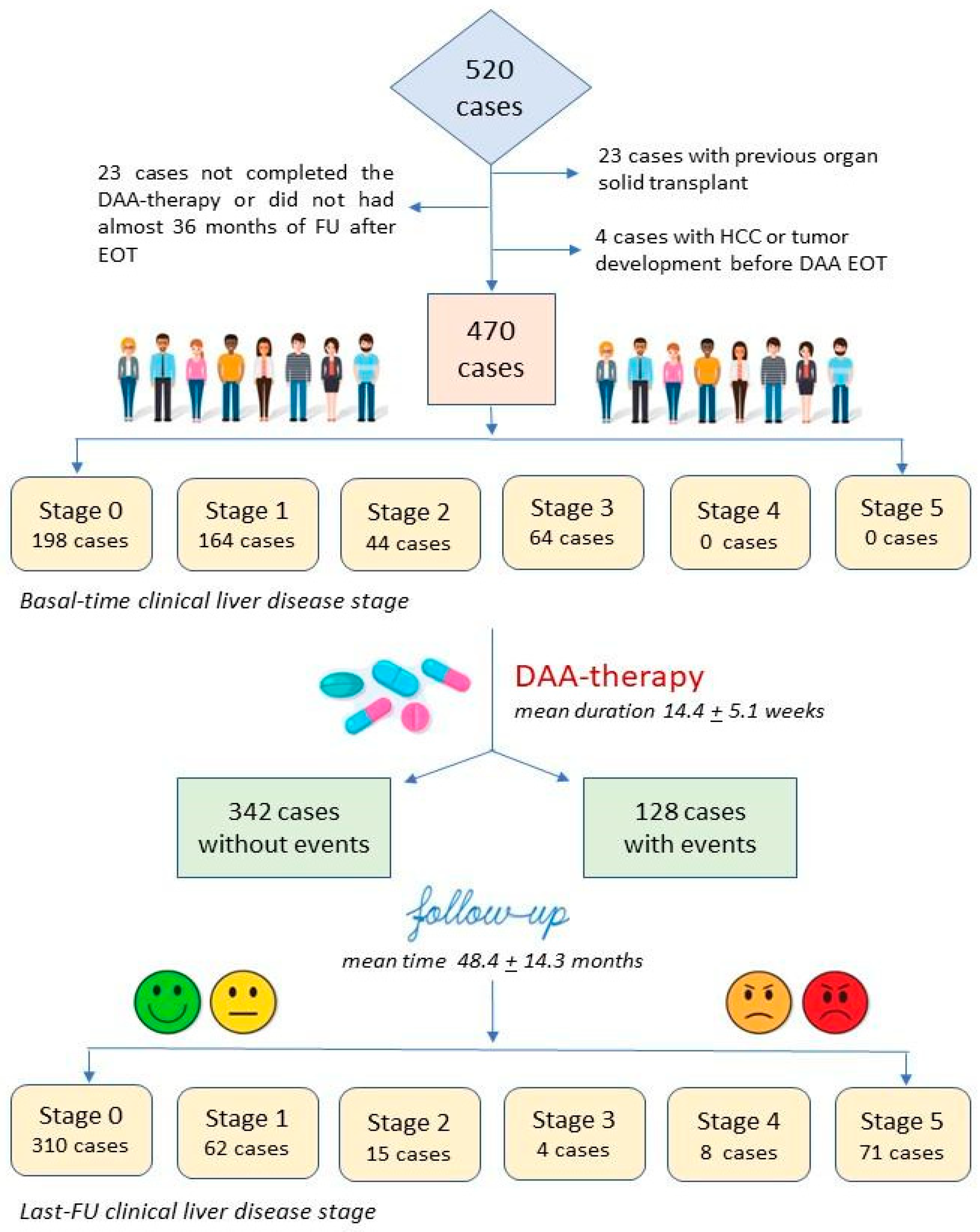
2.1. Study Design and Characteristics of Study Population

This prospective multicenter observational study was carried out in HUB outpatient liver centers at Padua University Hospital. The study protocol was approved by the local ethics committee, as an independent regional project, complies with the Helsinki rules and encloses written consent for all participants. Analyses included data of cases that complete a DAA therapy schedule from 2015 to 2022 and was restricted to participants who fulfilled requirements in terms of eligibility, interventions, and outcome assessment (per-protocol analysis).
The DAA schedule was chosen according to the EASL update guideline [22]. Antiviral treatment was administered to subjects aged between 18 and 75 years, in Italy, with priority for cases with histological features and/or LSM consistent with advanced chronic hepatitis or compensated cirrhosis Child–Pugh–Turcotte (CPT) class A5–6 and B7, after determination of serum viral loads by RT-PCR (Abbott Real-Time HCV, Chicago, IL, USA) and HCV genotyping (INNO-LIPA™ HCV-II, Innogenetics, Gent, Belgium).
All cases with absolute contraindication to DAA therapy, in particular, cirrhosis with recurrent decompensation, gastro-esophageal (GE) re-bleeding varices or active HCC, and patients with a poor motivation to comply and adhere to therapy or with a life expectancy of less than 5 years, were excluded.
In total, 520 cases were recruited, but 50 were excluded for previous solid organ transplantation (23 cases), for incomplete therapy schedules or FU period after EOT (23 cases), and for HCC or solid tumor onset before EOT (4 cases). All cases were fully investigated at basal time (within 1–3 months before DAA therapy start) and during a FU after EOT of at least 36 months, with laboratory and imaging examination to confirm the inclusion criteria to treatment. SVR was defined when HCV-RNA persisted undetectable at 12-week determination from EOT.

2.2. NIT for Estimation of Liver Fibrosis and Portal Hypertension

All cases underwent LSM (with a 50 Hz probe, ranging 1.5–75 kPa) by VCTE (Fibroscan®, Echosens, Paris, France). In particular, we also explored the following parameter to perform the formulas of liver NIT scores [23]: APRI = [(AST/upper limit NV AST) ×100]/number of platelets (109/L), with cirrhosis stage defined at score ≥ 1.5 [24,25]; FORNS = 7.811 − 3.131 × ln [PLT count (109/L)] × 0.781 ln [GGT (U/L)] + 3.467 × ln [age (years)] − 0.014 [cholesterol (mg/dL)], at score ≥ 6.9 [26]; FIB-4 = [age (years)] × AST (U/L)]/[number of platelets (109/L)× RADQ ALT (U/L)], at score > 3.25 [27]; LSPS = LSM x spleen ø (cm)/PLT count (109/L), with clinical significant PH at score > 3.5 [28].

2.3. Assessment of Clinical Liver Disease Stage

The clinical liver disease stage (CLDS), applied in this analysis of cases with CHC receiving DAA therapy, aims to focus on some liver disease clinical stages, which remains not easy to differentiate, especially regarding the presence of cirrhosis [29], and as no measures or values by NIT scores or TE have a real defined range for the classification of cases with significant fibrosis or cirrhosis. It takes into account a six-stage level clinical approach, which refers to one of the following: stage 0 = mild fibrosis (cases without features on abdominal ultrasound or objective clinical examination significant for progressive liver disease and with LSM compatible with fibrosis stage F1–F2 according to METAVIR); stage 1 = significant fibrosis or early cirrhosis without sign of significant PH (i.e., absence of spleen enlargement or GE varices or varicose collateral circles); stage 2 = cirrhosis with presence of GE varices non-at-risk, at gastroscopy; stage 3 = cirrhosis with only one episode of resolved hepatic decompensation (i.e., ascites or GE bleeding); stage 4 = cirrhosis with recurrent episodes of decompensation; stage 5 = development of HCC or end-stage liver disease with further episodes of decompensation related to cirrhosis requiring OLT or resulting in death. This classification also allowed us to follow the change in clinical stage obtained, between clinical examination at basal time and that of the last FU in the cohort (see also Figure 1).

2.4. Risk Stratification Assessment of Events

The risk stratification system (RISS) is a set of conditions and clinical measures counted by a score and based on the probability of presence/absence of an event or clinical value, collected for each individual patient, which allows the identification of cases, which are mainly involved in the development of major events (onset of HCC, OLT, or death), obtained by statistical analysis of the study population and/or validation in different literature reports. The RISS scored 1 point if presenting active smoking, alcohol consumption, comorbidities (consisting of cardiovascular disease or arterial hypertension, obesity or diabetes and MASLD, autoimmunity conditions, or previous or family history of cancers), male sex, age < 50 years, LSM values ≥ 15 kPa; 2 points for aged > 70 years, LSM ≥ 25 kPa or with stage 2 or 3 at CLDS; and 3 points for those with age between 50 and 70 years. This evaluation has a range score of 0–12 for each patient and consists of three categories: at low, moderate, or high risk in cases with score 0–4, 5–7, and ≥8 points, respectively, allowing the identification of susceptible or frail cases due to advanced liver disease and associated conditions, which increase the risk of all events occurring after DAA therapy, especially for HCC, OLT and death incidences.

2.5. Statistical Analysis
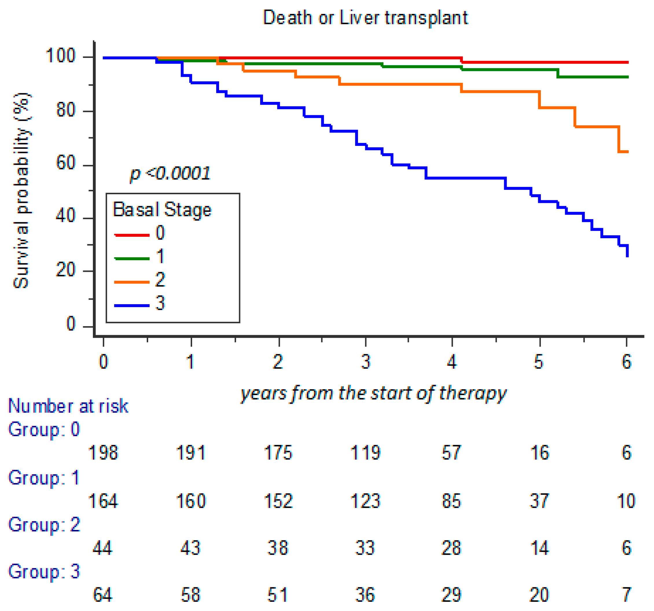
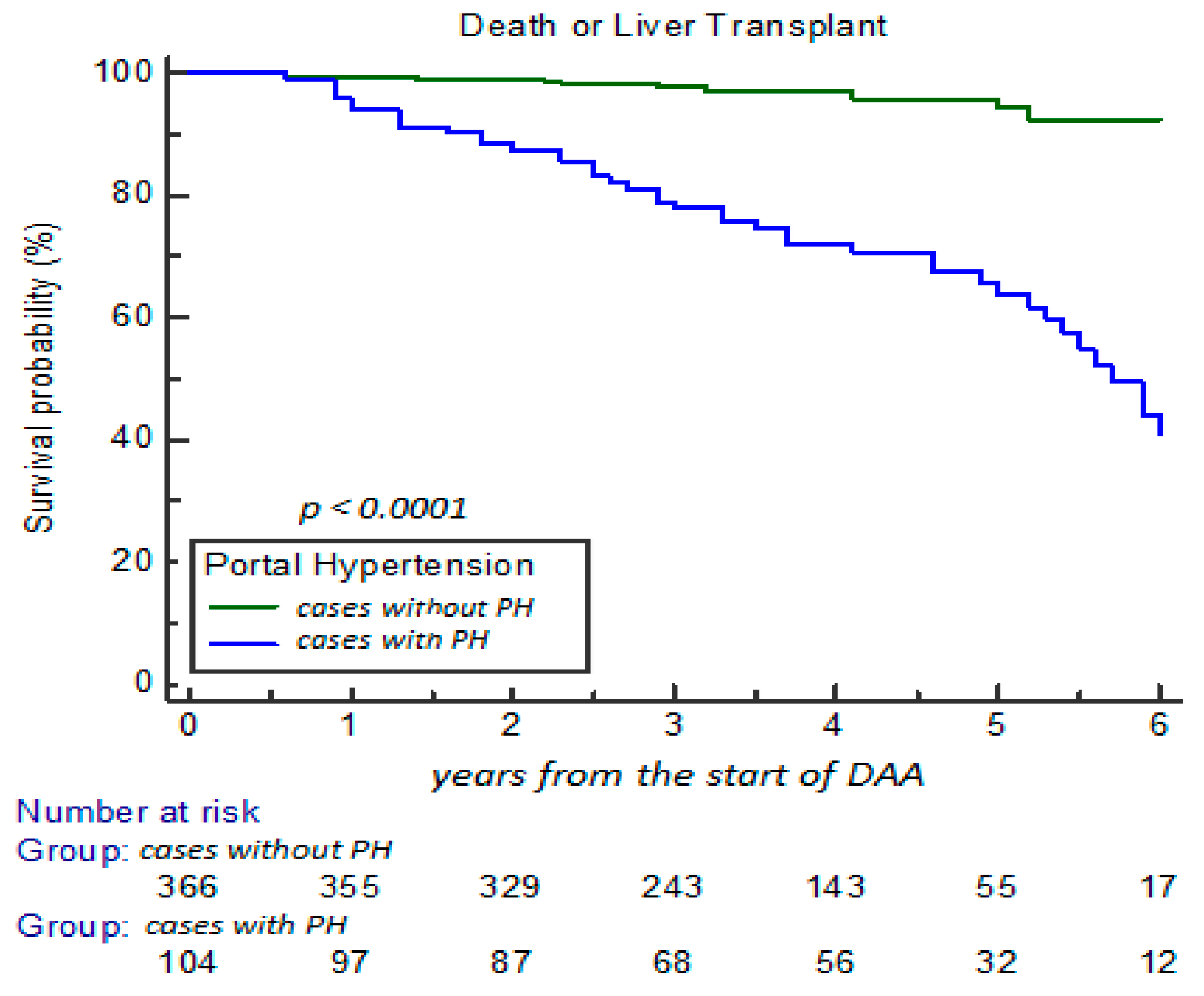
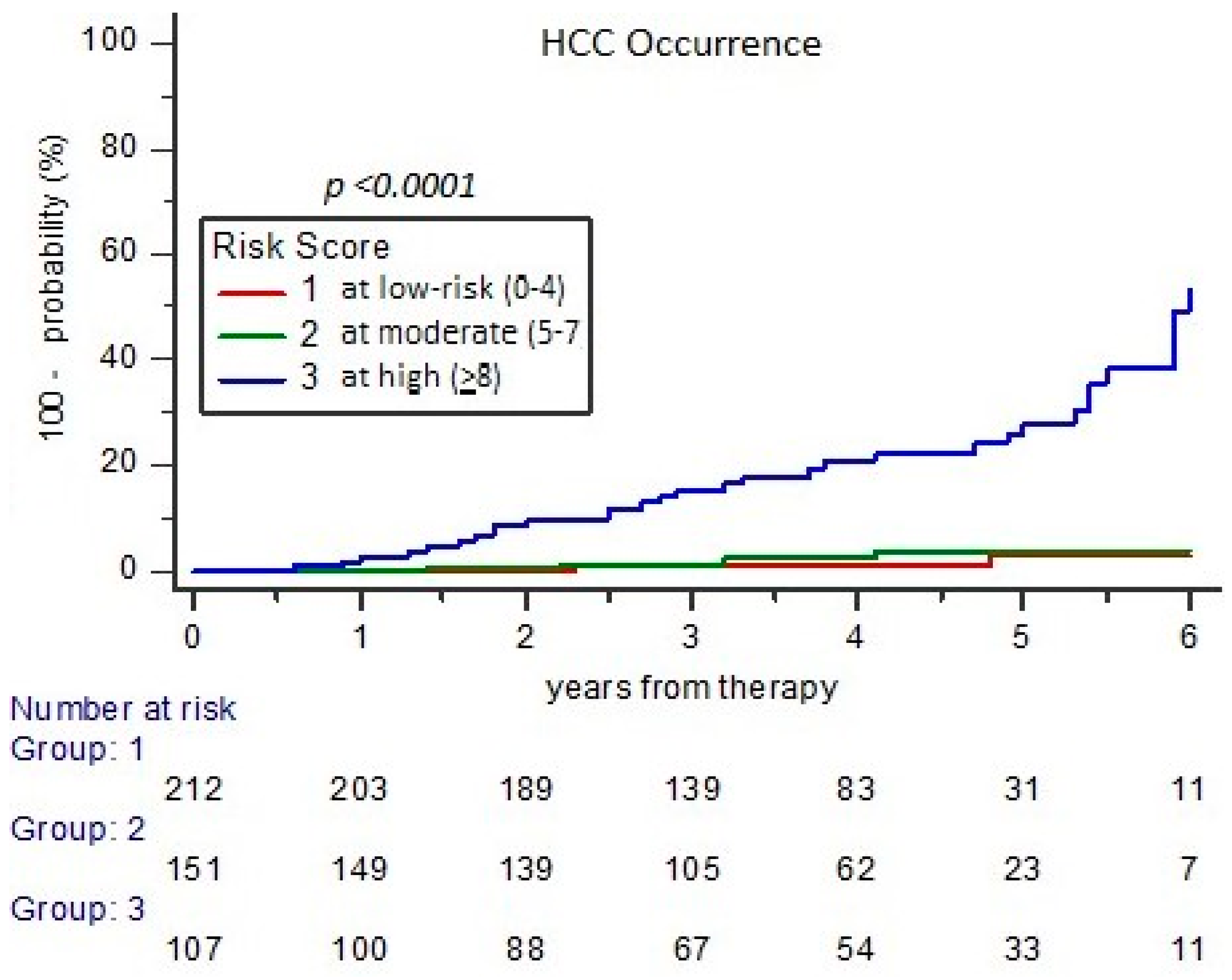
Mean value and standard deviation (SD) were applied to continuous variables showing a normal distribution, and the means were compared using the Student’s t-test for independent samples. The differences between categorical variables were evaluated with Pearson’s chi-square test or Fisher’s test, when appropriate. Differences between data of NIT scores and LSM obtained at different t-time were analyzed using ANOVA analyses of variance. Only significant parameters (p ≤ 0.05) obtained at univariate and multivariate Cox regression analyses were taken into consideration to identify predictors of related events. Analysis with Kaplan–Meier curves were used for estimation of events cumulative incidence or survival probability in cases with major events (HCC, OLT, or deceased) during a 5-year FU of the cohort. Comparison of the curves between/among groups was performed using Log-Rank test. These analyses were performed using the software STATA 9.0 (StataCorp LLC, Lakeway Drive, TX, USA) and MEDCALC software Ltd. (Ostend, Belgium).

3. Results

3.1. Clinical Characteristics and Changes During Long-Term FU

Figure 1 shows the flowchart of the study. Of 520 cases recruited, only 470 patients fully fit the inclusion criteria by a per-protocol analysis. Mean age was 59.4 ± 12.7 years, and 241 (51.3%) were male. Genotype HCV-1 was the most frequent infection type, with 288 (61.3%) cases. Patients received in 268 (57.0%) cases a DAA schedule with sofosbuvir (SOF), in 102 (21.7%) with glecaprevir plus pibrentasvir, in 64 (13.6%) with elbasvir plus grazoprevir, and in 36 (7.7%) with ritonavir-boosted paritaprevir plus ombitasvir and dasabuvir. Treatment mean duration was 14.4 ± 5.1 weeks. Basal time CLDS included 198, 164, 44, and 64 cases at stage 0, 1, 2, and 3, respectively, with 108 (23.0%) cases defined with PH (staged 2 or 3). No cases were included in stages 4 and 5, as these patients had absolute contraindication to be treated with DAA.
During a mean FU of 48.4 ± 14.3 months, we observed 310, 62, 15, 4, 8, and 71 cases with stage 0, 1, 2, 3, 4, and 5, respectively, with 79 (16.8%) cases shifted to stages 4 and 5. Furthermore, 52 (11.1%) cases were deceased, 13 (2.8%) received a liver transplant (OLT), and 40 (8.5%) were dropouts. In total, 128 (27.2%) patients developed events: 76 (59.4%) defined as HE (46 with HCC and 30 with advanced cirrhosis and PH stage, further decompensation episodes and development of abnormal coagulative, or septic state) and 52 of EHE type (22 with other tumor, 30 with bleeding or thrombosis not liver-related and pathological conditions regarding cardiology, neurology, or rheumatology)(see also Table S1, Supplementary Material).
The survival probability analyzed (% cases with OLT or death) at 5-year FU showed a statistical difference among cases in relation to CLDS basal stage 0, 1, 2, and 3, with survival decreased to 98.9%, 95.1%, 79.5%, and 32.8%, respectively (p < 0.0001) (Figure 2). Moreover, the survival probability estimation was significantly reduced from 96.2% to 53.8%, particularly in relation to presence of PH at basal time (48/104 cases; 46.2%; with stage 2 and 3) in comparison to cases with stage 0 and 1 (14/366; 3.8%; p < 0.0001) (Figure 3).

3.2. Development of HE and EHE and the Impact on Clinical NIT Scores

The overall cohort was grouped in cases without incidence of events (342 cases; group 0) and with HE (76 cases; group 1) or EHE (52 cases; group 2) during the long-term FU (Table 1). Patients with HE had the majority of cases with smoking habit (60.5%), alcohol consumption (65,8%), and comorbidities (80.3%), and 69.7% were males. No differences appeared among groups in relation to HCV genotype infection; conversely, cases with events were mostly treated with SOF-based DAA schedules compared to cases without events (105/128 cases, 82.0% vs. 163/342, 47.7%, respectively, p < 0.001).
Considering the CLDS of the cohort at basal time in Table 1, 180/342 (52.6%) cases without events fell into the stage of mild fibrosis (0), and cumulatively, stages 0 and 1 accounted for 91.8% of these (p < 0.001). On the contrary, 67/76 (88.2%) cases with HE, with respect to 28 (8.2%) without events (group 0), were affected by cirrhosis with PH (p < 0.001). Moreover, the number of cases with CLDS stages 0 and 1 with respect to those with 2 and 3 appeared differently distributed with an inverse relationship, between cases with HE (group 1) and EHE (group 2) (all, p < 0.005). Finally, 46 cases developed HCC, and 53 (70%) cases were deceased in group 1, with respect to 9 cases (17.3%) in group 2 (p < 0.001).
Biochemical parameters, particularly those of liver function, confirmed significant differences among groups, especially of group 1 versus group 0 or 2. It should also be noted that between groups 0 and 2, there was a significant difference regarding a more advanced age, the presence of comorbidities, and the greater presence of cases with cirrhosis and PH and toward a decreased trend in the mean of platelet count and albumin level (p = 0.05) in the latter.
Table 2 presents the changes reported according to groups without and with events incidence during long-term FU by the NIT scores for staging of liver fibrosis and PH. Interestingly, all scores have shown a statistical difference between the values obtained at baseline compared to those of the last FU, although higher and significant values for rule-in the stage of cirrhosis are described in NIT scores of the group with HE (group 1). In the latter, the LSM obtained at the last FU remained at very high mean value (28.3 ± 14.4 kPa) compared to those of groups 0 (7.5 ± 4.1 kPa) and 2 (10.0 ± 7.4 kPa), as well as at FORNS and FIB-4 and LSPS scores: These data confirm the residual number of cases with advanced cirrhosis with PH and decompensated stage without signs of remission (staged 4 and 5) after successful DAA therapy.
During long-term FU, 46 cases had HCC occurrence; of these, 9 (19.6%) received OLT, and 28 (60.9%) were deceased, 2 cases after receiving OLT. Cases with HCC had significantly higher LSM in comparison to cases without tumor occurrence (36.1 ± 12.2 vs. 15.6 ± 10.3 kPa; p < 0.001), and also HCC incidence resulted in 31.7% and 68.3%, in cases with LSM > 15 kPa and ≥25 kPa, respectively, at basal time (p < 0.001). During long-term FU, cases that maintained clinical signs of PH and LSM > 25 kPa, showed HR of 7.34 (95%CI 3.77–14.31), with respect to the group with LMS < 25 kPa (HR: 0.14; 0.06–0.26) (p < 0.0001).

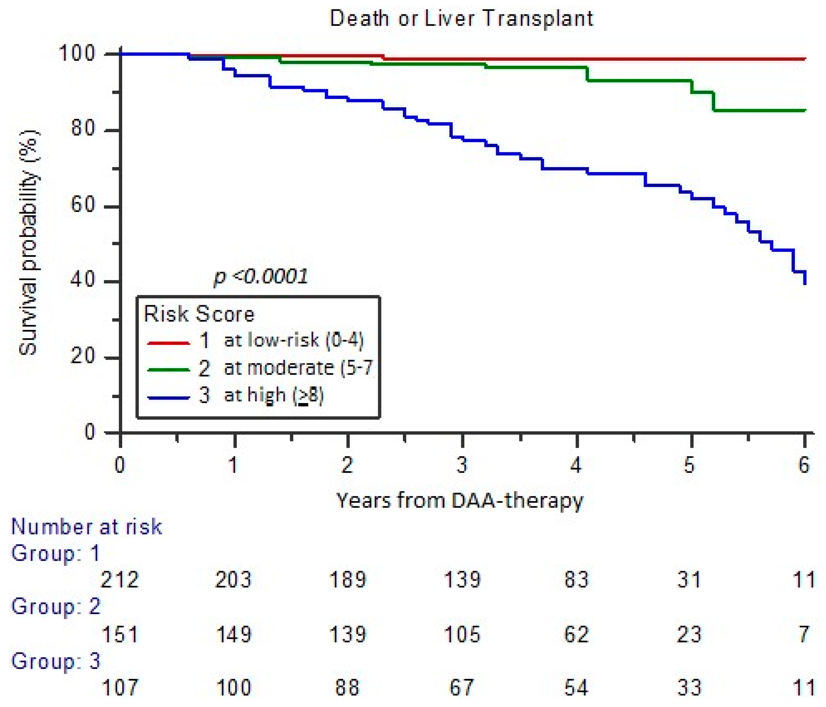
3.3. Assessment of Risk for Major Events Incidence at Long-Term FU (RISS Score)

To identify cases that would not benefit from the efficacy of DAA therapy, even after achieving SVR, we evaluate the risk of incidence of major events (onset of HCC and OLT or death) in the individual patient. The RISS scored into three categories, or score ranges, that defined a patient profile: at low (0–4 points), moderate (5–7), or high (≥8) risk. It was based on variables related to unfavorable outcomes, identified by statistical significance in our cohort, that allowed the plotting of curves for survival analysis.
Among 128 cases with events over 470 cases (27.23%) of the study cohort, those with high RISS score (≥8) had a significantly higher observed versus expected incidence, including 72.0% of all events, and showing, in comparison to moderate and low-risk groups, a HR of 4.2 (95% CI 2.80–6.40) versus 1.72 (1.27–2.64) and 0.57 (0.37–0.88), respectively (p < 0.001) (Figure 4).
Curves of cases with HCC occurrence (total cases 46/470; 9.8%) according to RISS profiles at high, moderate, or low risk had a distribution number of 34.6%, 3.3%, and 1.9%, respectively, showing a HR of 6.16 (95% CI 2.92–12.98), 1.78 (0.87–3.66), and 0.55 (0.27–1.14), respectively (p < 0.0001) (Figure 5). In the high RISS profile were included 37/46 (80.4%) cases with HCC onset during a 5-year FU.
The survival probability of the cohort analyzed, including cases requiring OLT or deceased (total number 62/470 cases; 13.2%) according to RISS score at low versus high risk, appeared to decrease from 98.6% to 55.1% at 5-year FU, with HR of 3.95 (95% CI 2.07–7.54) versus 0.19 (0.10–0.35), respectively (p < 0.0001) (Figure 6). The high RISS profile resulted in 48/62 (77.4%) of cases with OLT or dead at 5-year FU.

4. Discussion

This study aimed to present a series of events, occurring after a mean FU of 4.1 + 1.2 years, in a cohort of 470 patients with CHC achieving viral eradication, evaluated by per-protocol analysis. The extensive use of DAA to treat patients, even those vulnerable due to age, comorbidities, or more advanced liver disease, has led to the assumption that the effectiveness in avoiding liver-related complications could be achievable for all treated subjects. Unfortunately, over time, the use of DAA did not eliminate the incidence of major events, although in real-life practice, the high efficacy of HCV eradication translated into high effectiveness in reducing the HCC incidence risk [10]. Data from the PITER-HCV cohort in Italy [15] showed a HCC weighted cumulative incidence rate of 7.0% in cases with SVR, at 36-month FU, which was independently associated with male sex, increasing age, current alcohol use, HCV genotype 3, platelet count ≤ 120,000/μL, and albumin ≤ 3.5 g/dL.
Our study collected 128 (27.2%) cases with events, and the development of HCC had a 5-year cumulative incidence of 9.8%, but 272 (57.9%) cases staged with significant fibrosis or cirrhosis, including 108 cases (23%) with PH. Cases with HE included 46 cases with HCC and 30 with advanced cirrhosis, of which 88.2% had PH. It is evident that our cohort, being treated between 2015 and 2017, was affected by a priority treatment phase in favor of cases with advanced cirrhosis CPT-A6 and -B7 classes. The greater predisposition of these cases to unfavorable outcomes after DAA has been supported by many studies of literature [30]. In particular, of 350 cases with cirrhosis described in the Asian population, 70 patients (20%) developed HCC, and 9.1% decompensated during therapy, especially in the group with CPT-B or C, which represented 44.3% of the study population [31]. Instead, considering an Italian study with 138 cases aged ≥ 70 years, 70% of cases had compensated cirrhosis, and among these, 8 cases (14.5%) developed HCC [32].
To raise awareness of the careful and responsible use of DAA therapy, in particular in frail-to-treat patients, since exposition can lead to some related events, the objective of having useful tools for predicting its incidence is desirable. In our study, we classified both the clinical aspect of liver disease, proposing a staging classification or CLDS, similar to others in literature [29], which allowed us to identify the subjects most predisposed to major events, but it also contributed to a risk stratification system with three-profile (the RISS score) for the analysis of the morbidity and mortality of the cohort.
With regard to the comparison between cases with EH and EHE, the greater frequency of males in the group with HE was highlighted, but no influence of the HCV genotype infection among groups was seen, and a significant relationship between the use of SOF-based schedules was linked to occurrence of major events (see Table S1, Supplementary Materials). This phenomenon remains difficult to explain, but notice also by other contributions in the literature, where off-target effects related to the DAA use [19] or by drug-to-drug interactions [9] were hypothesized, in particular with regard to a greater incidence/recurrence of HCC [5,6]. Following and larger studies nullify this relation, which was attributed to a cohort bias due to higher number of cases mainly at risk of HCC onset for presence of cACLD [33,34]. In our cohort, we had a cumulative incidence of HCC of 1.6/100 p-y at 5-year FU estimation, which resulted similarly to many other studies of literature [14,15,16,17,18,19,20,21,22,23,24,25,26,27,28,29,30,31,32,33,34,35].
NIT scores varied significantly from basal, among the groups described, at last FU, and it is undoubtedly an expected finding due to HCV eradication. This goal, which drives the effectiveness of treatment, particularly in cases with significant fibrosis or early cirrhosis, is confirmed by a large retrospective cohort in 40,654 cases treated with DAA versus untreated cases for insured patients with CHC, which showed a significant improvement in outcomes, with a higher overall survival among cases with SVR [30]. In contrast, the greatest number of major events occurs in relation to cases with cirrhosis with PH in 88.2%, which also showed a higher mean of LSM (>25 kPa) and the lowest reduction of NIT scores, even after long-term success of DAA therapy.
The Baveno VII “rules of 5” for LSM successfully help to recognize cases with compensated cirrhosis and PH [36], and also in our RISS score, cases with LSM > 15 and ≥25 kPa identified subjects at risk of major events (HCC, OLT, or death), when combined to other parameters (i.e., risk habits, comorbidities, gender and age), that showed a differentiate weight as variables defined strong or weak for HCC correlation.. The novel RISS demonstrated a good prognostic accuracy, with HR 4.24 (95% CI 2.80–6.48) between cases with high versus low (0.57; 0.37–0.88) or moderate-risk (1.72; 1.12–2.64) categories for incidence of all type of events (p < 0.0001). The RISS score ≥ 8 showed a significant correlation with incidence of all events (107/128 cases; 83.6%) and HCC (36/46 cases; 80.4%) and identified the category of cases with a reduction in survival rate to 65.4% at 5-year FU.
These original data add knowledge supporting the relevance of clinical classification of liver disease, which is closely related to the prognosis and survival of cases with CHC successfully treated with DAA, and the proposed RISS could effectively help us with a tailored post DAA FU, for early diagnosis of HCC in individuals who remain at risk.

5. Conclusions

Nowadays, serious complications related to cirrhosis and portal hypertension inevitably maintain the related morbidity and mortality after DAA therapy, despite viral eradication. Our study aimed to focus on the development of adverse outcomes, which may occur after DAA exposure. In particular, hepatic-related events accounted for the largest number of cases staged with advanced liver disease, reaching 88.2%. Only through detailed staging of fibrosis by evaluation of NIT and LSM could we accurately predict the risk of major events onset. All cases with RISS score ≥ 8 had higher LSM (>25 kPa) and lower reduction of NIT scores, even after long-term successful DAA therapy, and had shown a risk of HCC in 80.4% of cases and a reduction in survival rate to 65.4% at 5 years.

Supplementary Materials

The following supporting information can be downloaded at: https://www.mdpi.com/article/10.3390/cancers17091528/s1, Table S1: Type of LR or not-LR events occurred in the cohort during a 5-year FU.

Author Contributions

For this research articles, authors’ contribution were the following: conceptualization, L.C. (Liliana Chemello) and L.C. (Luisa Cavalletto); methodology, L.C. (Luisa Cavalletto), L.C. (Liliana Chemello), E.B., L.K., M.G.Q., C.M., and C.A.; software, L.C. (Luisa Cavalletto); validation, L.C. (Luisa Cavalletto), L.C. (Liliana Chemello), and E.B.; formal analysis, L.C. (Luisa Cavalletto) and L.C. (Liliana Chemello); investigation, E.B. and L.C. (Luisa Cavalletto); resources, L.C. (Liliana Chemello); data curation, E.B., L.C. (Luisa Cavalletto), and L.C. (Liliana Chemello); writing—original draft preparation, L.C. (Liliana Chemello) and L.C. (Luisa Cavalletto); writing—review and editing, L.C. (Liliana Chemello), L.C. (Luisa Cavalletto), E.B., and L.K.; visualization, L.C. (Liliana Chemello); supervision, L.C. (Liliana Chemello), L.C. (Luisa Cavalletto), E.B., L.K., M.G.Q., C.M., and C.A.; funding acquisition, L.C. (Liliana Chemello) and L.C. (Luisa Cavalletto). All authors have read and agreed to the published version of the manuscript.

Funding

This research received no sponsors or external funding. An independent regional project (D.G.R. N°791/2018) formed a network of hepatologic HUB centers and received a grant of Veneto region. We thank University of Padua for supporting scientific research activity through dedicated funds for the development of projects of the Department of Medicine-DIMED (DOR 2021 N°21-47470 and DOR 2024 N°00743).

Institutional Review Board Statement

The Institutional Review Board of Ethics Committee of the AOUP, University Hospital of Padova, gave its approval with protocol number N°4178/AO/17 on the day 18 May 2017. The study was conducted in accordance with the Declaration of Helsinki.

Informed Consent Statement

Informed written consent was obtained from all subjects involved in the study.

Data Availability Statement

All the data used for the study are stored in a database with security measures compliant with privacy legislation and can be consulted in an anonymous form, upon request to the researchers who conducted the study.

Conflicts of Interest

All authors declare no conflicts of interest.

Abbreviations

ALTalanine aminotransferase
APRIaspartate aminotransferase to platelet ratio index
ASTaspartate aminotransferase
BMIbody mass index
cACLDcompensated advanced liver disease
CIconfidential interval
CHCchronic hepatitis C
CLDSclinical liver disease stage
RISSRisk stratification system
DAAdirect-acting antiviral
DILIdrug-induced liver injury
EHEextrahepatic-related events
EOTend of therapy
EVesophageal varices
FIB-4fibrosis-4 index for detection of liver fibrosis or cirrhosis
FORNSstaging of liver fibrosis or cirrhosis according to Forns’ formula
GGTgamma-glutamil transferase
GEgastro-esophageal
HCChepatocellular carcinoma
HCVhuman hepatitis C virus
HEhepatic-related events
HRhazard ratio
LSMliver stiffness measures
LSPSliver stiffness platelet score
MASLDmetabolic dysfunction-associated steatotic liver disease
NITnon-invasive test
OLTorthotopic liver transplantation
PHportal hypertension
SDstandard deviation
SOFsofosbuvir
SVRsustained virologic response
TEtransient elastography

References

  1. Zarębska-Michaluk, D.; Brzdęk, M.; Jaroszewicz, J.; Tudrujek-Zdunek, M.; Lorenc, B.; Klapaczyński, J.; Mazur, W.; Kazek, A.; Sitko, M.; Berak, H.; et al. Best therapy for the easiest to treat hepatitis C virus genotype 1b-infected patients. World J. Gastroenterol. 2022, 28, 6380–6396. [Google Scholar] [CrossRef] [PubMed]
  2. Chen, T.; Terrault, N. Treatment of chronic hepatitis C in patients with cirrhosis. Curr. Opin. Gastroenterol. 2016, 32, 143–151. [Google Scholar] [CrossRef] [PubMed]
  3. Pawlotsky, J.M.; Aghemo, A.; Back, D.; Dusheiko, G.; Forns, X.; Puoti, M.; Sarrazin, C. EASL Recommendations on Treatment of Hepatitis C. J. Hepatol. 2015, 63, 199–236. [Google Scholar]
  4. Huang, C.F.; Yu, M.L. Unmet needs of chronic hepatitis C in the era of direct-acting antiviral therapy. Clin. Mol. Hepatol. 2020, 26, 251–260. [Google Scholar] [CrossRef] [PubMed]
  5. Conti, F.; Buonfiglioli, F.; Scuteri, A.; Crespi, C.; Bolondi, L.; Caraceni, P.; Foschi, F.G.; Lenzi, M.; Mazzella, G.; Verucchi, G.; et al. Early occurrence and recurrence of hepatocellular carcinoma in HCV-related cirrhosis treated with direct-acting antivirals. J. Hepatol. 2016, 65, 727–733. [Google Scholar] [CrossRef]
  6. Reig, M.; Mariño, Z.; Perelló, C.; Iñarrairaegui, M.; Ribeiro, A.; Lens, S.; Díaz, A.; Vilana, R.; Darnell, A.; Varela, M.; et al. Unexpected high rate of early tumor recurrence in patients with HCV-related HCC undergoing interferon-free therapy. J. Hepatol. 2016, 65, 719–726. [Google Scholar] [CrossRef]
  7. Villani, R.; Vendemiale, G.; Serviddio, G. Molecular Mechanisms Involved in HCC Recurrence after Direct-Acting Antiviral Therapy. Int. J. Mol. Sci. 2018, 20, 49. [Google Scholar] [CrossRef]
  8. Bersoff-Matcha, S.J.; Cao, K.; Jason, M.; Ajao, A.; Jones, S.C.; Meyer, T.; Brinker, A. Hepatitis B virus reactivation associated with direct-acting antiviral therapy for chronic hepatitis C virus: A review of cases reported to the U.S. Food and Drug Administration Adverse Event Reporting System. Ann. Intern. Med. 2017, 166, 792–798. [Google Scholar] [CrossRef]
  9. Néant, N.; Solas, C. Drug-drug interactions potential of direct acting antivirals for the treatment of chronic hepatitis C virus infection. Int. J. Antimicrob. Agents 2020, 56, 105–571. [Google Scholar] [CrossRef]
  10. Calvaruso, V.; Craxì, A. Hepatic benefits of HCV cure. J. Hepatol. 2020, 73, 1548–1556. [Google Scholar] [CrossRef]
  11. Laursen, T.L.; Sandahl, T.D.; Kazankov, K.; George, J.; Grønbæk, H. Liver-related effects of chronic hepatitis C antiviral treatment. World J. Gastroenterol. 2020, 26, 2931–2947. [Google Scholar] [CrossRef]
  12. Jakobsen, J.C.; Nielsen, E.E.; Feinberg, J.; Katakam, K.K.; Fobian, K.; Hauser, G.; Poropat, G.; Djurisic, S.; Weiss, K.H.; Bjelakovic, M.; et al. Direct-acting antivirals for chronic hepatitis C. Cochrane Database Syst. Rev. 2017, 9, CD012143. [Google Scholar] [PubMed]
  13. Van der Meer, A.J.; Feld, J.J.; Hofer, H.; Almasio, P.L.; Calvaruso, V.; Fernandez-Rodriguez, C.M.; Aleman, S.; Ganne-Carrié, N.; D’Ambrosio, R.; Pol, S.; et al. Risk of cirrhosis-related complications in patients with advanced fibrosis following hepatitis C virus eradication. J. Hepatol. 2017, 66, 485–493. [Google Scholar] [CrossRef] [PubMed]
  14. Calvaruso, V.; Cabibbo, G.; Cacciola, I.; Petta, S.; Madonia, S.; Bellia, A.; FabioTinè, F.; Distefano, M.; Licata, A.; Giannitrapani, L.; et al. Incidence of hepatocellular carcinoma in patients with HCV-associated cirrhosis treated with direct-acting antiviral agents. Gastroenterology 2018, 155, 411–421. [Google Scholar] [CrossRef]
  15. Quaranta, M.G.; Cavalletto, L.; Russo, F.P.; Calvaruso, V.; Ferrigno, L.; Zanetto, A.; Mattioli, B.; D’Ambrosio, R.; Panetta, V.; Brancaccio, G.; et al. Reduction of the risk of hepatocellular carcinoma over time using direct-acting antivirals: A propensity score analysis of a real-life cohort (PITER-HCV). Viruses 2024, 16, 682. [Google Scholar] [CrossRef] [PubMed]
  16. Sangiovanni, A.; Alimenti, E.; Gattai, R.; Filomia, R.; Parente, E.; Valenti, L.; Marzi, L.; Pellegatta, G.; Borgia, G.; Gambato, M.; et al. Undefined/non-malignant hepatic nodules are associated with early occurrence of HCC in DAA-treated patients with HCV-related cirrhosis. J. Hepatol. 2020, 73, 593–602. [Google Scholar] [CrossRef]
  17. Kanwal, F.; Kramer, J.R.; Asch, S.M.; Cao, Y.; Li, L.; El Serag, H.B. Long-term risk of hepatocellular carcinoma in HCV patients treated with direct acting antiviral agents. Hepatology 2020, 71, 44–55. [Google Scholar] [CrossRef]
  18. Backus, L.I.; Belperio, P.S.; Shahoumian, T.A.; Mole, L.A. Impact of sustained virologic response with direct-acting antiviral treatment on mortality in patients with direct-acting antiviral treatment on mortality in patients with direct-acting antiviral treatment on mortality in patients with advanced liver disease. Hepatology 2019, 69, 487–497. [Google Scholar]
  19. Giovannini, C.; Fornari, F.; Indio, V.; Trerè, D.; Renzulli, M.; Vasuri, F.; Cescon, M.; Ravaioli, M.; Perrucci, A.; Astolfi, A.; et al. Direct antiviral treatments for hepatitis C virus have off-target effects of oncologic relevance in hepatocellular carcinoma. Cancers 2020, 12, 2674. [Google Scholar] [CrossRef]
  20. Faillaci, F.; Marzi, L.; Critelli, R.; Milosa, F.; Schepis, F.; Turola, E.; Andreani, S.; Vandelli, G.; Bernabucci, V.; Lei, B.; et al. Liver angiopoietin-2 is a key predictor of de novo or recurrent HCC after hepatitis C virus direct-acting antivirals. Hepatology 2018, 68, 1010–1024. [Google Scholar] [CrossRef]
  21. Nevola, R.; Tortorella, G.; Rosato, V.; Rinaldi, L.; Imbriani, S.; Perillo, P.; Mastrocinque, D.; La Montagna, M.; Russo, A.; Di Lorenzo, G.; et al. Gender differences in the pathogenesis and risk factors of hepatocellular carcinoma. Biology 2023, 12, 984. [Google Scholar] [CrossRef] [PubMed]
  22. European Association for the Study of the Liver. EASL recommendations on treatment of hepatitis C. J. Hepatol. 2016, 66, 153–194. [Google Scholar]
  23. EASL Clinical practice guideline on non-invasive test for evaluation of liver disease severity and prognosis-2021 update. J. Hepatol. 2021, 75, 659–689. [CrossRef] [PubMed]
  24. Lok, A.S.; Ghany, M.G.; Godman, Z.D.; Wright, E.C.; Everson, G.T.; Sterling, R.K.; Everhart, J.E.; Lindsay, K.L.; Bonkovsky, H.L.; Di Bisceglie, A.M.; et al. Predicting cirrhosis in patients with hepatitis C based on standard laboratory tests: Results of the HALT-C cohort. Hepatology 2005, 42, 282–292. [Google Scholar] [CrossRef]
  25. Lin, Z.H.; Xin, Y.N.; Dong, Q.J.; Dong, Q.J.; Wang, Q.; Jiang, X.J.; Shu-Hui Zhan, S.H.; Sun, Y.; Xuan, S.Y. Performance of the aspartate aminotransferase to platelet ratio index for the staging of hepatitis C-related fibrosis: An updated meta-analysis. Hepatology 2011, 53, 726–736. [Google Scholar] [CrossRef]
  26. Forns, X.; Ampurdanes, S.; Llovet, J.M.; Aponte, J.; Quinto, L.; Martinez-Bauer, E.; Bruguera, M.; Sanchez-Tapias, J.; Rode, J. Identification of chronic hepatitis C patients without hepatic fibrosis by a simple predictive model. Hepatology 2020, 36, 986–992. [Google Scholar] [CrossRef]
  27. Kim, B.K.; Kim, D.Y.; Park, J.Y.; Ahn, S.H.; Chon, C.Y.; Kim, J.K.; Paik, Y.H.; Lee, K.S.; Park, Y.N.; Han, K.H. Validation of FIB-4 and comparison with other simple noninvasive indices for predicting virus-infected patients. Liver Int. 2010, 30, 546–553. [Google Scholar] [CrossRef] [PubMed]
  28. Berzigotti ALSeijo, S.; Arena, U.; Abraldes, J.G.; Vizzutti, F.; Garcia-Pagàn, J.C.; Pinzani, M.; Bosch, J. Elastography, spleen size, and platelet count identify portal hypertension in patients with compensated cirrhosis. Gastroenterology 2013, 144, 102–111. [Google Scholar] [CrossRef]
  29. D’Amico, G.; Morabito, A.; D’Amico, M.; Pasta, L.; Malizia, G.; Rebora, P.; Valsecchi, M.G. Clinical states of cirrhosis and competing risks. J. Hepatol. 2018, 68, 563–576. [Google Scholar] [CrossRef]
  30. Ogawa, E.; Chien, N.; Kam, L.; Yeo, Y.H.; Ji, F.; Huang, D.Q.; Cheung, R.; Nguyen, M.H. Association of direct-acting antiviral therapy with liver and non-liver complications and long-term mortality in patients with chronic hepatitis C. JAMA Intern. Med. 2023, 183, 97–105. [Google Scholar] [CrossRef]
  31. Tahata, Y.; Hikita, H.; Mochida, S.; Enomoto, N.; Kawada, N.; Kurosaki, M.; Ido, A.; Miki, D.; Yoshiji, H.; Takikawa, Y.; et al. Liver-related events after direct-acting antiviral therapy in patients with hepatitis C virus-associated cirrhosis. J. Gastroenterol. 2022, 57, 120–132. [Google Scholar] [CrossRef] [PubMed]
  32. De Santis, A.; Maggi, D.; Lubrano Lobianco, F. Safety and efficacy of directly-acting antiviral therapy for chronic hepatitis C virus in elderly people. Aging Med. 2021, 4, 304–316. [Google Scholar] [CrossRef]
  33. ANRS Collaborative Study Group on Hepatocellular Carcinoma. Lack of evidence of an effect of direct-acting antivirals on the recurrence of hepatocellular carcinoma: Data from three ANRS cohorts. J. Hepatol. 2016, 65, 734–740. [Google Scholar] [CrossRef] [PubMed]
  34. Sapena, V.; Enea, M.; Torres, F.; Celsa, C.; Rios, J.; Rizzo, G.E.M.; Nahon, P.; Mariño, Z.; Tateishi, R.; Minami, T.; et al. Hepatocellular carcinoma recurrence after direct-acting antiviral therapy: An individual patient data meta-analysis. Gut 2022, 71, 593–604. [Google Scholar] [CrossRef] [PubMed]
  35. Kanwal, F.; Kramer, J.R.; Asch, S.M.; Chayanupatkul, M.; Cao, Y.; El-Serag, H.B. Risk of hepatocellular cancer in HCV patients treated with direct acting antiviral agents. Gastroenterology 2017, 153, 996–1005. [Google Scholar] [CrossRef]
  36. de Franchis, R.; Bosch, J.; Garcia-Tsao, G.; Reiberger, T.; Ripoll, C.; on behalf of the Baveno VII Faculty. Baveno VII—Renewing consensus in portal hypertension. J. Hepatol. 2021, 76, 959–974. [Google Scholar] [CrossRef]
Figure 1. Flowchart of patients recruited, grouping by CLDS at basal time and at last FU and by cases without or with incidence of events during a 5-year FU.
Cancers 17 01528 g001
Figure 2. Survival probability according to basal time CLDS at 5-year FU estimation.
Cancers 17 01528 g002
Figure 3. Survival probability according to cases without or with PH at 5-year FU estimation.
Cancers 17 01528 g003
Figure 4. Probability of all events (HE and EHE type) according to categories of RISS.
Cancers 17 01528 g004
Figure 5. Probability of HCC occurrence according to risk categories of RISS.
Cancers 17 01528 g005
Figure 6. Probability of requiring OLT or dead according to risk categories of RISS.
Cancers 17 01528 g006
Table 1. Characteristics of study population according to development of HE or EHE.
Cases
Without Events
n. 342 (72.7%)
p-Level
0 vs. 1
Cases
with HE
n. 76 (16.2%)
p-Level
1 vs. 2
Cases
with EHE
n. 52 (11.0%)
p-Level
0 vs. 2
CharacteristicsGroup 0 Group 1 Group 2
N° males (%)164 (47.9%)<0.00153 (69.7%)0.0124 (31.6%)ns
Age (mean years ± SD)57.6 ± 12.9ns59.6 ± 11.4ns63.6 ± 12.9<0.005
BMI (Kg/m2)25.1 ± 4.0<0.0526.8 ± 4.3ns25.4 ± 4.1ns
Smoking habit155 (45.3%)<0,0546 (60.5%)0.0522 (42.3%)ns
Alcohol consumption175 (51.2%)<0.0550 (65.8%)ns25 (48.1%)ns
Comorbidities196 (57.3%)<0.00161 (80.3%)ns39 (75.0%)<0.05
HCV genotype 1 infection212 (61.9%)ns44 (57.9%)ns32 (61.5%)ns
DAA schedule SOF-based163 (47.7%)<0.00168 (89.5%)0.0137 (71.1%)<0.001
Basal Time Clinical Liver Disease Stage
Mild fibrosis (stage 0)180 (52.6%)<0.0011 (1.3%)<0.00117 (32.7%)<0.001
Significant fibrosis or
cirrhosis without EV (stage 1)
134 (39.2%)<0.0018 (10.5%)<0.00122 (42.3%)ns
Cirrhosis with EV (stage 2)24 (7.0%)0.0113 (17.1%)<0.0057 (13.4%)ns
Cirrhosis with only one known
decompensation (stage 3)
4 (1.2%)<0.00154 (71.1%)<0.0016 (11.5%)0.001
Development of Major Events during FU
Cases with HCC0--46 (60.5%)--0--
Cases with OLT or/and dead0--53 * (70.0%)<0.0019 (17.3%)--
Biochemical Laboratory Parameters
Hemoglobin (g/L)141.8 ± 17.7<0.001129.3 ± 20.90.01137.6 ± 16.6ns
White cell count (mm3)6.1 ± 1.9<0.0014.5 ± 1.9<0.0055.9 ± 1.8ns
PLTS count (mm3)204.8 ± 72.6<0.00196.5 ± 68.4<0.001179.7 ± 87.10.05
INR ratio1.08 ± 0.25<0.0011.22 ± 0.21<0.0051.11 ± 0.26ns
Total bilirubin (μmol/L)11.5 ± 5.7<0.00122.5 ± 9.90.00111.1 ± 5.9ns
ALT (U/L)93.9 ± 89.5<0.00570.5 ± 48.1ns90.7 ± 67.4ns
Gamma-GT (U/L)57.2 ± 60.40.00199.6 ± 86.9ns71.8 ± 63.7ns
Total protein (g/L)74.6 ± 5.0ns74.5 ± 6.7ns73.7 ± 7.9ns
Albumin (g/L)41.5 ± 4.2<0.00135.9 ± 4.80.00539.3 ± 4.80.05
Gamma-globulin (g/L)14.6 ± 4.6<0.00120.7 ± 6.1<0.00515.2 ± 4.8ns
Alpha-fetoprotein (μg/L)8.2 ± 17.6<0.0522.4 ± 50.6ns12.5 ± 19.9ns
* Two cases were transplanted and afterwards deceased. “ns”, not significant.
Table 2. Staging of liver fibrosis and PH by NIT scores. Comparison of values obtained at basal time and at last FU according to development of HE or EHE in the cohort.
Cases Without Eventsp-Level0 vs. 1Cases with HEp-Level1 vs. 2Cases with EHEp-Level0 vs. 2
Liver NITsGroup 0 Group 1 Group 2
N° cases (%) *309 (90%) 60 (79%) 40 (77%)
LSM ** at BT ***11.7 ± 7.7<0.00132.6 ± 12.0<0.0115.2 ± 9.1ns
LSM at last FU7.5 ± 4.1<0.00128.3 ± 14.4<0.00510.1 ± 7.4ns
APRI at BT1.0 ± 0.9<0.0012.5 ± 1.8<0.0011.4 ± 1.2<0.05
APRI at last FU0.4 ± 0.2<0.0011.6 ± 1.2<0.010.6 ± 0.5ns
FORNS at BT8.1 ± 1.5<0.00111.5 ± 1.6<0.019.2 ± 1.8<0.01
FORNS at last FU7.3 ± 1.2<0.00110.9 ± 1.7<0.0018.2 ± 1.7<0.01
FIB-4 at BT2.5 ± 1.4<0.0018.5 ± 4.3<0.0014.4 ± 2.7<0.005
FIB-4 at last FU1.7 ± 0.7<0.0016.9 ± 4.4<0.0013.0 ± 2.1<0.05
LSPS at BT1.0 ± 0.8<0.0018.1 ± 5.0<0.0011.8 ± 1.6<0.05
LSPS at last FU0.6 ± 0.5<0.0016.9 ± 5.4<0.0011.0 ± 0.9ns
* Only cases with complete data for all scores were analyzed. ** TE units (kPa). *** BT, basal time. “ns”, not significant.
Disclaimer/Publisher’s Note: The statements, opinions and data contained in all publications are solely those of the individual author(s) and contributor(s) and not of MDPI and/or the editor(s). MDPI and/or the editor(s) disclaim responsibility for any injury to people or property resulting from any ideas, methods, instructions or products referred to in the content.

