
Plant-Based Biosynthesis of Copper/Copper Oxide Nanoparticles: An Update on Their Applications in Biomedicine, Mechanisms, and Toxicity

 , , , and
Abstract
1. Introduction
2. Copper Nanoparticles
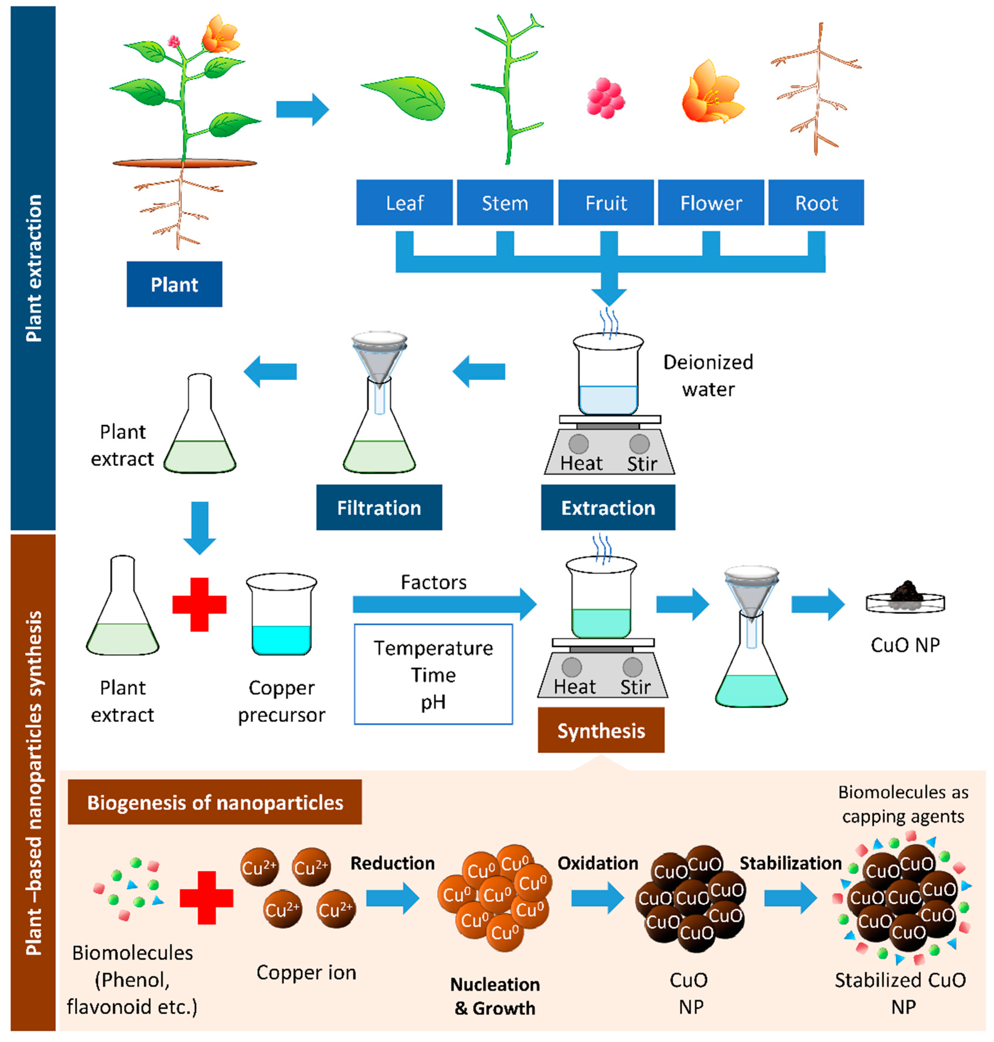
3. Plant-Based Green Synthesis of Copper and Copper Oxide Nanoparticles
3.1. Synthesis Strategy of CuO, Cu2O, and Cu4O3 NPs
3.2. Factors Affecting the Green Synthesis of Cu and CuO NPs
3.2.1. Temperature
3.2.2. Time
3.2.3. Concentration of Plant Extracts
3.2.4. Precursor Used
3.2.5. pH
4. Application of Cu and CuO NPs
4.1. Antibacterial
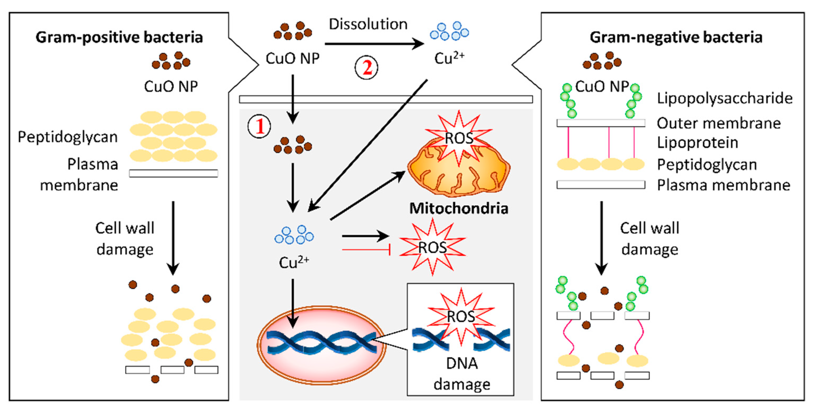
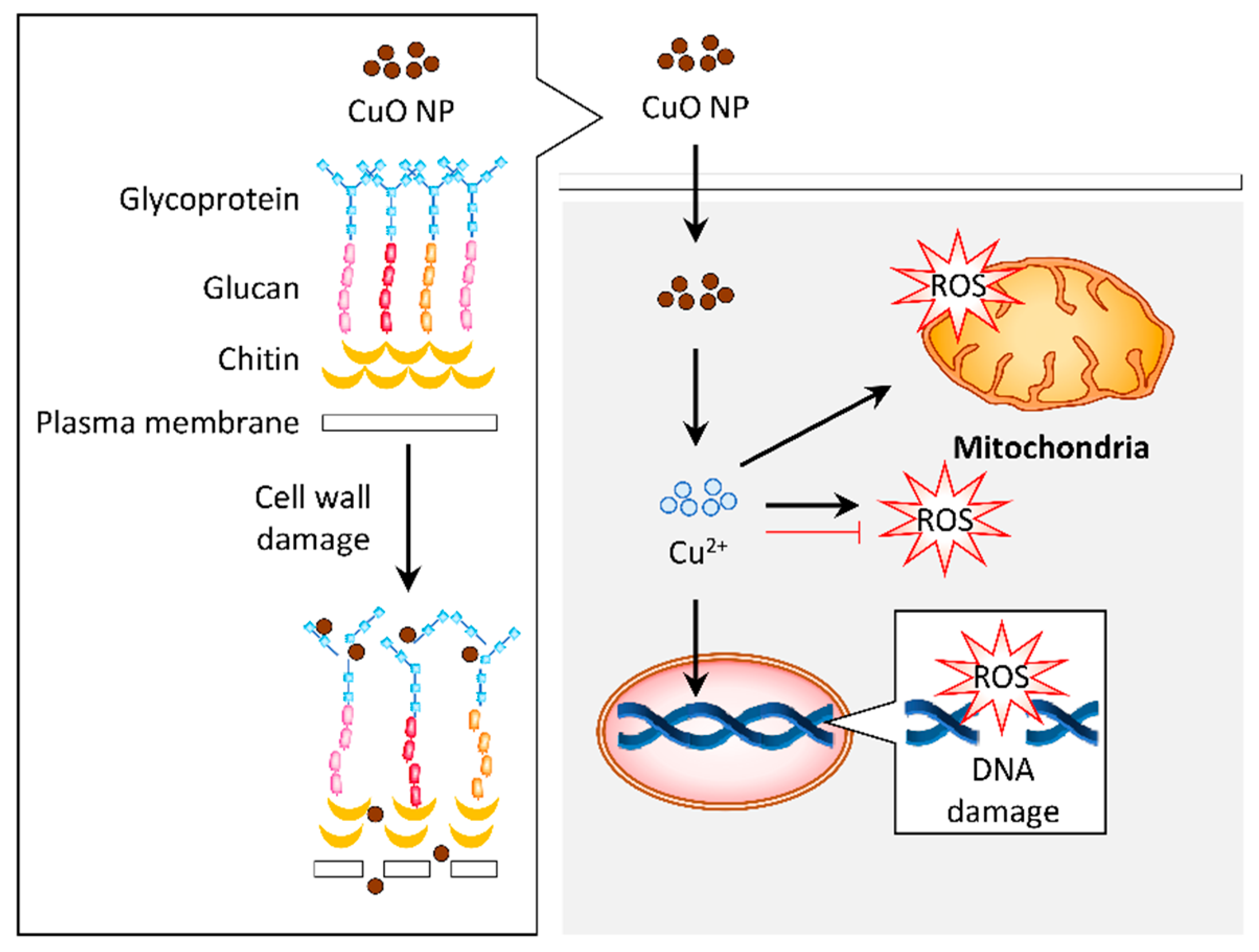
Predicted Mechanism for Antibacterial Activity
4.2. Antifungal
4.3. Anticancer
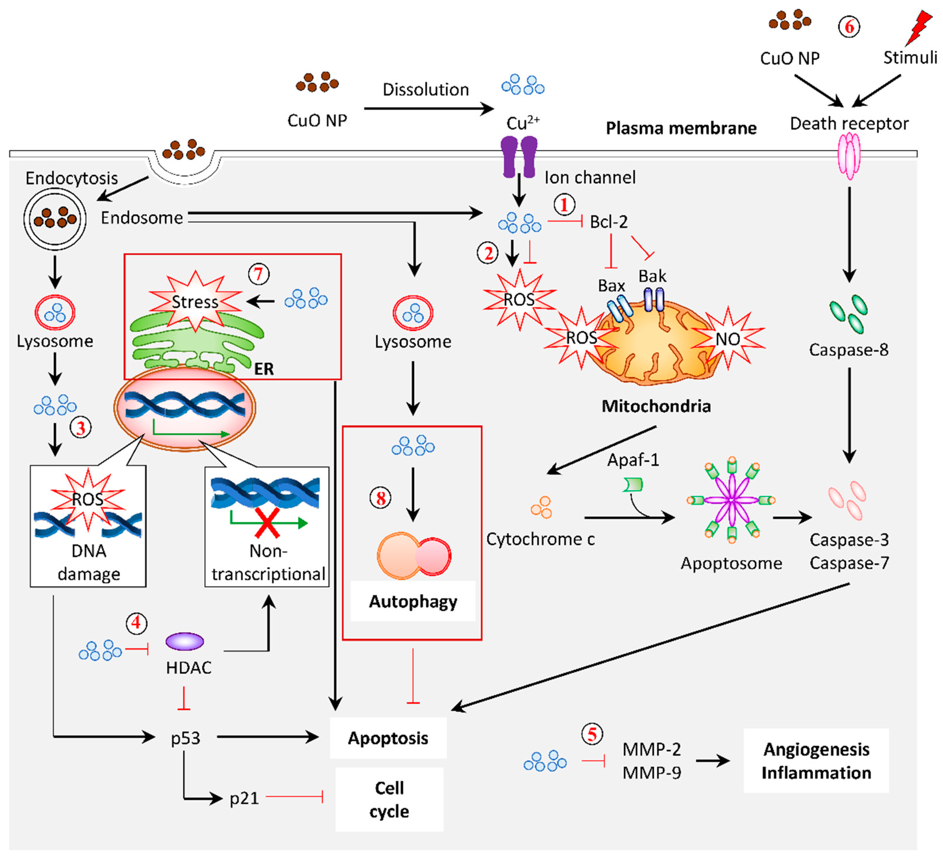
Predicted Mechanism for Anticancer Activity
4.4. Wound Healing and Anti-Inflammatory Activity
5. Toxicity Evaluation
6. Comparison of the Efficacy of Plant-Based-Synthesized and Commercial Cu and CuO NPs
7. Current Status of Cu and CuO NPs and Their Future Perspective in Cancer Therapy
8. Conclusions
Author Contributions
Funding
Institutional Review Board Statement
Informed Consent Statement
Data Availability Statement
Conflicts of Interest
References
- Riggio, C.; Pagni, E.; Raffa, V.; Cuschieri, A. Nano-oncology: Clinical application for cancer therapy and future perspectives. J. Nanomater. 2011, 2011. [Google Scholar] [CrossRef]
- Contado, C. Nanomaterials in consumer products: A challenging analytical problem. Front. Chem. 2015, 3, 48. [Google Scholar] [CrossRef]
- Ibrahim, S.; Charinpanitkul, T.; Kobatake, E.; Sriyudthsak, M. Nanowires nickel oxide and nanospherical manganese oxide synthesized via low temperature hydrothermal technique for hydrogen peroxide sensor. J. Chem. 2016, 2016. [Google Scholar] [CrossRef]
- Yasin, S.M.M.; Ibrahim, S.; Johan, M.R. Effect of zirconium oxide nanofiller and dibutyl phthalate plasticizer on ionic conductivity and optical properties of solid polymer electrolyte. Sci. World J. 2014, 2014. [Google Scholar] [CrossRef]
- Sangeetha, S.; Kalaignan, G.P.; Anthuvan, J.T. Pulse electrodeposition of self-lubricating Ni–W/PTFE nanocomposite coatings on mild steel surface. Appl. Surf. Sci. 2015, 359, 412–419. [Google Scholar] [CrossRef]
- Sahooli, M.; Sabbaghi, S.; Saboori, R. Synthesis and characterization of mono sized CuO nanoparticles. Mater. Lett. 2012, 81, 169–172. [Google Scholar] [CrossRef]
- Safarifard, V.; Morsali, A. Sonochemical syntheses of a nano-sized copper(II) supramolecule as a precursor for the synthesis of copper(II) oxide nanoparticles. Ultrason Sonochem. 2012, 19, 823–829. [Google Scholar] [CrossRef] [PubMed]
- Pandiyarajan, T.; Udayabhaskar, R.; Vignesh, S.; James, R.A.; Karthikeyan, B. Synthesis and concentration dependent antibacterial activities of CuO nanoflakes. Mater Sci Eng. C. 2013, 33, 2020–2024. [Google Scholar] [CrossRef]
- Yahia, I.S.; Farag, A.A.M.; El-Faify, S.; Yakuphanoglu, F.; Al-Ghamdi, A.A. Synthesis, optical constants, optical dispersion parameters of CuO nanorods. Optik 2016, 127, 1429–1433. [Google Scholar] [CrossRef]
- Mohamed, R.M.; Harraz, F.A.; Shawky, A. CuO nanobelts synthesized by a template-free hydrothermal approach with optical and magnetic characteristics. Ceram. Int. 2014, 40, 2127–2133. [Google Scholar] [CrossRef]
- Saif Hasan, S.; Singh, S.; Parikh, R.Y.; Dharne, M.S.; Patole, M.S.; Prasad, B.; Shouche, Y.S. Bacterial synthesis of copper/copper oxide nanoparticles. J. Nanosci. Nanotechnol. 2008, 8, 3191–3196. [Google Scholar] [CrossRef] [PubMed]
- Jiang, T.; Wang, Y.; Meng, D.; Yu, M. Facile synthesis and photocatalytic performance of self-assembly CuO microspheres. Superlattice. Microstruct. 2015, 85, 1–6. [Google Scholar] [CrossRef]
- Karthik, A.; Geetha, K. Synthesis of Copper Precursor, Copper and its oxide Nanoparticles by Green Chemical Reduction Method and its Antimicrobial Activity. J. Appl. Pharm. Sci. 2013, 3, 16–21. [Google Scholar]
- Dang, T.M.D.; Le, T.T.T.; Fribourg-Blanc, E.; Dang, M.C. Synthesis and optical properties of copper nanoparticles prepared by a chemical reduction method. Adv. Nat. Sci. Nanosci. Nanotechnol. 2011, 2, 015009. [Google Scholar] [CrossRef]
- Ealia, S.A.M.; Saravanakumar, M. A review on the classification, characterisation, synthesis of nanoparticles and their application. IOP Conf. Ser. Mater. Sci. Eng. 2017, 263, 032019. [Google Scholar] [CrossRef]
- Cele, T. Preparation of Nanoparticles. In Engineered Nanomaterials-Health and Safety; IntechOpen: London, UK, 2020. [Google Scholar]
- Liu, Q.-M.; Zhou, D.-B.; Yamamoto, Y.; Ichino, R.; Okido, M. Preparation of Cu nanoparticles with NaBH4 by aqueous reduction method. Trans. Nonferr. Metals Soc. China 2012, 22, 117–123. [Google Scholar] [CrossRef]
- Zhu, H.-T.; Lin, Y.-S.; Yin, Y.-S. A novel one-step chemical method for preparation of copper nanofluids. J. Coll. Interface Sci. 2004, 277, 100–103. [Google Scholar] [CrossRef]
- Su, X.; Zhao, J.; Bala, H.; Zhu, Y.; Gao, Y.; Ma, S.; Wang, Z. Fast synthesis of stable cubic copper nanocages in the aqueous phase. J. Phys. Chem. C 2007, 111, 14689–14693. [Google Scholar] [CrossRef]
- Iravani, S. Green synthesis of metal nanoparticles using plants. Green Chem. 2011, 13, 2638–2650. [Google Scholar] [CrossRef]
- Kumar, P.V.; Pammi, S.; Kollu, P.; Satyanarayana, K.; Shameem, U. Green synthesis and characterization of silver nanoparticles using Boerhaavia diffusa plant extract and their anti bacterial activity. Ind. Crops Prod. 2014, 52, 562–566. [Google Scholar] [CrossRef]
- Khan, I.; Saeed, K.; Khan, I. Nanoparticles: Properties, applications and toxicities. Arab. J. Chem. 2019, 12, 908–931. [Google Scholar] [CrossRef]
- Mandal, D.; Bolander, M.E.; Mukhopadhyay, D.; Sarkar, G.; Mukherjee, P. The use of microorganisms for the formation of metal nanoparticles and their application. Appl. Microbiol. Biotechnol. 2006, 69, 485–492. [Google Scholar] [CrossRef] [PubMed]
- Kowshik, M.; Ashtaputre, S.; Kharrazi, S.; Vogel, W.; Urban, J.; Kulkarni, S.K.; Paknikar, K. Extracellular synthesis of silver nanoparticles by a silver-tolerant yeast strain MKY3. Nanotechnology 2002, 14, 95. [Google Scholar] [CrossRef]
- Mukherjee, P.; Ahmad, A.; Mandal, D.; Senapati, S.; Sainkar, S.R.; Khan, M.I.; Parishcha, R.; Ajaykumar, P.; Alam, M.; Kumar, R. Fungus-mediated synthesis of silver nanoparticles and their immobilization in the mycelial matrix: A novel biological approach to nanoparticle synthesis. Nano Lett. 2001, 1, 515–519. [Google Scholar] [CrossRef]
- Chaudhary, R.; Nawaz, K.; Khan, A.K.; Hano, C.; Abbasi, B.H.; Anjum, S. An Overview of the Algae-Mediated Biosynthesis of Nanoparticles and Their Biomedical Applications. Biomolecules 2020, 10, 1498. [Google Scholar] [CrossRef] [PubMed]
- Spadaro, D.; Gullino, M.L. Improving the efficacy of biocontrol agents against soilborne pathogens. Crop Protect. 2005, 24, 601–613. [Google Scholar] [CrossRef]
- Bukhari, S.I.; Hamed, M.M.; Al-Agamy, M.H.; Gazwi, H.S.; Radwan, H.H.; Youssif, A.M. Biosynthesis of Copper Oxide Nanoparticles Using Streptomyces MHM38 and Its Biological Applications. J. Nanomater. 2021, 2021. [Google Scholar] [CrossRef]
- Kumar, V.V.; Nithya, S.; Shyam, A.; Subramanian, N.S.; Anthuvan, J.T.; Anthony, S.P. Natural amino acid based phenolic derivatives for synthesizing silver nanoparticles with tunable morphology and antibacterial studies. Bull. Korean Chem. Soc. 2013, 34, 2702–2706. [Google Scholar] [CrossRef]
- Ibrahim, S.; Jakaria, N.Z.; Rozali, S.; Ghazali, N.N.N.; Ab Karim, M.S.; Sabri, M.F.M. Biosynthesis of Copper Oxide Nanoparticles Using Camellia Sinensis Plant Powder. In Advances in Material Sciences and Engineering; Springer: Berlin, Germany, 2020; pp. 233–238. [Google Scholar]
- Wang, Y.; Yang, Q.W.; Yang, Q.; Zhou, T.; Shi, M.F.; Sun, C.X.; Gao, X.X.; Cheng, Y.Q.; Cui, X.G.; Sun, Y.H. Cuprous oxide nanoparticles inhibit prostate cancer by attenuating the stemness of cancer cells via inhibition of the Wnt signaling pathway. Int. J. Nanomed. 2017, 12, 2569–2579. [Google Scholar] [CrossRef]
- Ammara, S.; Shamaila, S.; Bokhari, A.; Sabah, A. Nonenzymatic glucose sensor with high performance electrodeposited nickel/copper/carbon nanotubes nanocomposite electrode. J. Phys. Chem. Solids 2018, 120, 12–19. [Google Scholar] [CrossRef]
- Verma, N.; Kumar, N. Synthesis and biomedical applications of copper oxide nanoparticles: An expanding horizon. ACS Biomater. Sci. Eng. 2019, 5, 1170–1188. [Google Scholar] [CrossRef]
- Jeffree, A.I.; Karman, S.; Ibrahim, S.; Ab Karim, M.S.; Rozali, S. Biosensors Approach for Lung Cancer Diagnosis—A Review. In RITA 2018; Springer: Berlin, Germany, 2020; pp. 425–435. [Google Scholar]
- Jia, B.; Mei, Y.; Cheng, L.; Zhou, J.; Zhang, L. Preparation of copper nanoparticles coated cellulose films with antibacterial properties through one-step reduction. ACS Appl. Mater. Interfaces 2012, 4, 2897–2902. [Google Scholar] [CrossRef] [PubMed]
- Duan, Z.; Ma, G.; Zhang, W. Preparation of copper nanoparticles and catalytic properties for the reduction of aromatic nitro compounds. Bull. Korean Chem. Soc. 2012, 33, 4003–4006. [Google Scholar] [CrossRef]
- Vijay, M.; Anu, Y. Anticancer activity of camellia Sinensis mediated copper nanoparticles against HT-29, MCF-7, and MOLT-4 human cancer cell lines. Asian J. Pharm. Clin. Res. 2017, 10, 82–88. [Google Scholar]
- Santini, C.; Pellei, M.; Gandin, V.; Porchia, M.; Tisato, F.; Marzano, C. Advances in copper complexes as anticancer agents. Chem. Rev. 2014, 114, 815–862. [Google Scholar] [CrossRef]
- Maqbool, Q.; Iftikhar, S.; Nazar, M.; Abbas, F.; Saleem, A.; Hussain, T.; Kausar, R.; Anwaar, S.; Jabeen, N. Green fabricated CuO nanobullets via Olea europaea leaf extract shows auspicious antimicrobial potential. IET Nanobiotechnol. 2016, 11, 463–468. [Google Scholar] [CrossRef]
- Mohammed, W.M.; Mubark, T.H.; Al-Haddad, R. Effect of CuO nanoparticles on antimicrobial activity prepared by sol-gel method. Int. J. Appl. Eng. Res. 2018, 13, 10559–10562. [Google Scholar]
- Jurj, A.; Braicu, C.; Pop, L.-A.; Tomuleasa, C.; Gherman, C.D.; Berindan-Neagoe, I. The new era of nanotechnology, an alternative to change cancer treatment. Drug Design Dev. Ther. 2017, 11, 2871. [Google Scholar] [CrossRef] [PubMed]
- Vaid, P.; Raizada, P.; Saini, A.K.; Saini, R.V. Biogenic silver, gold and copper nanoparticles-A sustainable green chemistry approach for cancer therapy. Sustain. Chem. Pharm. 2020, 16, 100247. [Google Scholar] [CrossRef]
- Singh, S.; Kumar, N.; Kumar, M.; Agarwal, A.; Mizaikoff, B. Electrochemical sensing and remediation of 4-nitrophenol using bio-synthesized copper oxide nanoparticles. Chem. Eng. J. 2017, 313, 283–292. [Google Scholar] [CrossRef]
- Nasrollahzadeh, M.; Sajadi, S.M.; Rostami-Vartooni, A.; Hussin, S.M. Green synthesis of CuO nanoparticles using aqueous extract of Thymus vulgaris L. leaves and their catalytic performance for N-arylation of indoles and amines. J. Coll. Interface Sci. 2016, 466, 113–119. [Google Scholar] [CrossRef]
- Liu, H.T.; Zheng, S.M.; Xiong, H.F.; Alwahibi, M.S.; Niu, X.L. Biosynthesis of copperoxide nanoparticles using Abies spectabilis plant extract and analyzing its antinociceptive and anti-inflammatory potency in various mice models. Arab. J. Chem. 2020, 13, 6995–7006. [Google Scholar] [CrossRef]
- Ijaz, F.; Shahid, S.; Khan, S.A.; Ahmad, W.; Zaman, S. Green synthesis of copper oxide nanoparticles using Abutilon indicum leaf extract: Antimicrobial, antioxidant and photocatalytic dye degradation activitie. Trop. J. Pharm. Res. 2017, 16, 743–753. [Google Scholar] [CrossRef]
- Awwad, A.; Amer, M. Biosynthesis of copper oxide nanoparticles using Ailanthus altissima leaf extract and antibacterial activity. Chem. Int. 2020, 6, 210–217. [Google Scholar]
- Elemike, E.E.; Onwudiwe, D.C.; Singh, M. Eco-friendly synthesis of copper oxide, zinc oxide and copper oxide–zinc oxide nanocomposites, and their anticancer applications. J. Inorgan. Organomet. Polym. Mater. 2020, 30, 400–409. [Google Scholar] [CrossRef]
- Zhao, H.W.; Su, H.T.; Ahmeda, A.; Sun, Y.Q.; Li, Z.Y.; Zangeneh, M.M.; Nowrozi, M.; Zangeneh, A.; Moradi, R. Biosynthesis of copper nanoparticles using Allium eriophyllum Boiss leaf aqueous extract; characterization and analysis of their antimicrobial and cutaneous wound-healing potentials. Appl. Organomet. Chem. 2020. [Google Scholar] [CrossRef]
- Tahvilian, R.; Zangeneh, M.M.; Falahi, H.; Sadrjavadi, K.; Jalalvand, A.R.; Zangeneh, A. Green synthesis and chemical characterization of copper nanoparticles using Allium saralicum leaves and assessment of their cytotoxicity, antioxidant, antimicrobial, and cutaneous wound healing properties. Appl. Organomet. Chem. 2019, 33, e5234. [Google Scholar] [CrossRef]
- Velsankar, K.; Kumar, R.M.A.; Preethi, R.; Muthulakshmi, V.; Sudhahar, S. Green synthesis of CuO nanoparticles via Allium sativum extract and its characterizations on antimicrobial, antioxidant, antilarvicidal activities. J. Environ. Chem. Eng. 2020, 8. [Google Scholar] [CrossRef]
- Sukumar, K.; Arumugam, S.; Thangaswamy, S.; Balakrishnan, S.; Chinnappan, S.; Kandasamy, S. Eco-friendly cost-effective approach for synthesis of copper oxide nanoparticles for enhanced photocatalytic performance. Optik 2020, 202. [Google Scholar] [CrossRef]
- Dey, A.; Manna, S.; Chattopadhyay, S.; Mondal, D.; Chattopadhyay, D.; Raj, A.; Das, S.; Bag, B.G.; Roy, S. Azadirachta indica leaves mediated green synthesized copper oxide nanoparticles induce apoptosis through activation of TNF-alpha and caspases signaling pathway against cancer cells. J. Saudi Chem. Soc. 2019, 23, 222–238. [Google Scholar] [CrossRef]
- Rehana, D.; Mahendiran, D.; Kumar, R.S.; Rahiman, A.K. Evaluation of antioxidant and anticancer activity of copper oxide nanoparticles synthesized using medicinally important plant extracts. Biomed. Pharmacother. Biomed. Pharmacother. 2017, 89, 1067–1077. [Google Scholar] [CrossRef]
- Sundaram, C.S.; Kumar, J.S.; Kumar, S.S.; Ramesh, P.L.N.; Zin, T.; Rao, U.S.M. Antibacterial and anticancer potential of Brassica oleracea var acephala using biosynthesised copper nanoparticles. Med. J. Malaysia 2020, 75, 677–684. [Google Scholar]
- Chandrasekaran, R.; Yadav, S.A.; Sivaperumal, S. Phytosynthesis and Characterization of Copper Oxide Nanoparticles using the Aqueous Extract of Beta vulgaris L and Evaluation of their Antibacterial and Anticancer Activities. J. Cluster Sci. 2020, 31, 221–230. [Google Scholar] [CrossRef]
- Kumari, P.; Panda, P.K.; Jha, E.; Kumari, K.; Nisha, K.; Mallick, M.A.; Verma, S.K. Mechanistic insight to ROS and apoptosis regulated cytotoxicity inferred by green synthesized CuO nanoparticles from Calotropis gigantea to embryonic zebrafish. Sci. Rep. 2017, 7, 1–17. [Google Scholar] [CrossRef] [PubMed]
- Kumari, P.; Panda, P.K.; Jha, E.; Pramanik, N.; Nisha, K.; Kumari, K.; Soni, N.; Mallick, M.A.; Verma, S.K. Molecular insight to In Vitro biocompatibility of phytofabricated copper oxide nanoparticles with human embryonic kidney cells. Nanomedicine 2018, 13, 2415–2433. [Google Scholar] [CrossRef] [PubMed]
- Fardood, S.T.; Ramazani, A. Black tea extract mediated green synthesis of copper oxide nanoparticles. J. Appl. Chem. Res. 2018, 12, 8–15. [Google Scholar]
- Kiranmai, M.; Kadimcharla, K.; Keesara, N.R.; Fatima, S.N.; Bommena, P.; Batchu, U.R. Green synthesis of stable copper nanoparticles and synergistic activity with antibiotics. Indian J. Pharm. Sci. 2017, 79, 695–700. [Google Scholar]
- Rajeshkumar, S.; Menon, S.; Kumar, S.V.; Tambuwala, M.M.; Bakshi, H.A.; Mehta, M.; Satija, S.; Gupta, G.; Chellappan, D.K.; Thangavelu, L. Antibacterial and antioxidant potential of biosynthesized copper nanoparticles mediated through Cissus arnotiana plant extract. J. Photochem. Photobiol. B Biol. 2019, 197, 111531. [Google Scholar] [CrossRef]
- Wu, S.; Rajeshkumar, S.; Madasamy, M.; Mahendran, V. Green synthesis of copper nanoparticles using Cissus vitiginea and its antioxidant and antibacterial activity against urinary tract infection pathogens. Artif. Cells Nanomed. Biotechnol. 2020, 48, 1153–1158. [Google Scholar] [CrossRef]
- Tshireletso, P.; Ateba, C.N.; Fayemi, O.E. Spectroscopic and Antibacterial Properties of CuONPs from Orange, Lemon and Tangerine Peel Extracts: Potential for Combating Bacterial Resistance. Molecules 2021, 26, 586. [Google Scholar] [CrossRef]
- Prakash, S.; Elavarasan, N.; Venkatesan, A.; Subashini, K.; Sowndharya, M.; Sujatha, V. Green synthesis of copper oxide nanoparticles and its effective applications in Biginelli reaction, BTB photodegradation and antibacterial activity. Adv. Powder Technol. 2018, 29, 3315–3326. [Google Scholar] [CrossRef]
- Chung, I.M.; Abdul Rahuman, A.; Marimuthu, S.; Vishnu Kirthi, A.; Anbarasan, K.; Padmini, P.; Rajakumar, G. Green synthesis of copper nanoparticles using Eclipta prostrata leaves extract and their antioxidant and cytotoxic activities. Exp. Ther. Med. 2017, 14, 18–24. [Google Scholar] [PubMed]
- Ali, K.; Saquib, Q.; Ahmed, B.; Siddiqui, M.A.; Ahmad, J.; Al-Shaeri, M.; Al-Khedhairy, A.A.; Musarrat, J. Bio-functionalized CuO nanoparticles induced apoptotic activities in human breast carcinoma cells and toxicity against Aspergillus flavus: An In Vitro approach. Process. Biochem. 2020, 91, 387–397. [Google Scholar] [CrossRef]
- Sackey, J.; Nwanya, A.; Bashir, A.; Matinise, N.; Ngilirabanga, J.; Ameh, A.; Coetsee, E.; Maaza, M. Electrochemical properties of Euphorbia pulcherrima mediated copper oxide nanoparticles. Mater. Chem. Phys. 2020, 244, 122714. [Google Scholar] [CrossRef]
- Zangeneh, M.M.; Ghaneialvar, H.; Akbaribazm, M.; Ghanimatdan, M.; Abbasi, N.; Goorani, S.; Pirabbasi, E.; Zangeneh, A. Novel synthesis of Falcaria vulgaris leaf extract conjugated copper nanoparticles with potent cytotoxicity, antioxidant, antifungal, antibacterial, and cutaneous wound healing activities under In Vitro and In Vivo condition. J. Photochem. Photobiol. B Biol. 2019, 197, 111556. [Google Scholar] [CrossRef]
- Sankar, R.; Maheswari, R.; Karthik, S.; Shivashangari, K.S.; Ravikumar, V. Anticancer activity of Ficus religiosa engineered copper oxide nanoparticles. Mater. Sci. Eng. C 2014, 44, 234–239. [Google Scholar] [CrossRef]
- Kalaiarasi, A.; Sankar, R.; Anusha, C.; Saravanan, K.; Aarthy, K.; Karthic, S.; Mathuram, T.L.; Ravikumar, V. Copper oxide nanoparticles induce anticancer activity in A549 lung cancer cells by inhibition of histone deacetylase. Biotechnol. Lett. 2018, 40, 249–256. [Google Scholar] [CrossRef]
- Hemmati, S.; Ahmeda, A.; Salehabadi, Y.; Zangeneh, A.; Zangeneh, M.M. Synthesis, characterization, and evaluation of cytotoxicity, antioxidant, antifungal, antibacterial, and cutaneous wound healing effects of copper nanoparticles using the aqueous extract of Strawberry fruit and L-Ascorbic acid. Polyhedron 2020, 180. [Google Scholar] [CrossRef]
- Vishveshvar, K.; Krishnan, M.A.; Haribabu, K.; Vishnuprasad, S. Green synthesis of copper oxide nanoparticles using Ixiro coccinea plant leaves and its characterization. BioNanoScience 2018, 8, 554–558. [Google Scholar] [CrossRef]
- Fardood, S.T.; Ramazani, A.; Asiabi, P.; Joo, S. A novel green synthesis of copper oxide nanoparticles using a henna extract powder. J. Struct. Chem. 2018, 59, 1737–1743. [Google Scholar] [CrossRef]
- Santhosh Kumar, J.; Shanmugam, V. Green synthesis of copper oxide nanoparticles from magnolia champaca floral extract and its antioxidant & toxicity assay using Danio Rerio. Int. J. Recent Technol. Eng. 2020, 8, 5444–5449. [Google Scholar]
- Kiriyanthan, R.M.; Sharmili, S.A.; Balaji, R.; Jayashree, S.; Mahboob, S.; Al-Ghanim, K.A.; Al-Misned, F.; Ahmed, Z.; Govindarajan, M.; Vaseeharan, B. Photocatalytic, antiproliferative and antimicrobial properties of copper nanoparticles synthesized using Manilkara zapota leaf extract: A photodynamic approach. Photodiagn. Photodyn. Ther. 2020, 32, 102058. [Google Scholar] [CrossRef] [PubMed]
- Thiruvengadam, M.; Chung, I.-M.; Gomathi, T.; Ansari, M.A.; Khanna, V.G.; Babu, V.; Rajakumar, G. Synthesis, characterization and pharmacological potential of green synthesized copper nanoparticles. Bioprocess. Biosyst. Eng. 2019, 42, 1769–1777. [Google Scholar] [CrossRef]
- Rajamma, R.; Gopalakrishnan Nair, S.; Abdul Khadar, F.; Baskaran, B. Antibacterial and anticancer activity of biosynthesised CuO nanoparticles. IET Nanobiotechnol. 2020, 14, 833–838. [Google Scholar] [CrossRef] [PubMed]
- Sulaiman, G.M.; Tawfeeq, A.T.; Jaaffer, M.D. Biogenic synthesis of copper oxide nanoparticles using olea europaea leaf extract and evaluation of their toxicity activities: An In Vivo and In Vitro study. Biotechnol. Prog. 2018, 34, 218–230. [Google Scholar] [CrossRef]
- Nagajyothi, P.; Muthuraman, P.; Sreekanth, T.; Kim, D.H.; Shim, J. Green synthesis: In-Vitro anticancer activity of copper oxide nanoparticles against human cervical carcinoma cells. Arab. J. Chem. 2017, 10, 215–225. [Google Scholar] [CrossRef]
- Berra, D.; Laouini, S.; Benhaoua, B.; Ouahrani, M.; Berrani, D.; Rahal, A. Green synthesis of copper oxide nanoparticles by Pheonix dactylifera L leaves extract. Digest J. Nanomater. Biostruct. 2018, 13, 1231–1238. [Google Scholar]
- Nagaraj, E.; Karuppannan, K.; Shanmugam, P.; Venugopal, S. Exploration of Bio-synthesized Copper Oxide Nanoparticles Using Pterolobium hexapetalum Leaf Extract by Photocatalytic Activity and Biological Evaluations. J. Cluster Sci. 2019, 30, 1157–1168. [Google Scholar] [CrossRef]
- Mary, A.A.; Ansari, A.T.; Subramanian, R. Sugarcane juice mediated synthesis of copper oxide nanoparticles, characterization and their antibacterial activity. J. King Saud Univ. Sci. 2019, 31, 1103–1114. [Google Scholar] [CrossRef]
- Khatami, M.; Varma, R.S.; Heydari, M.; Peydayesh, M.; Sedighi, A.; Agha Askari, H.; Rohani, M.; Baniasadi, M.; Arkia, S.; Seyedi, F. Copper oxide nanoparticles greener synthesis using tea and its antifungal efficiency on Fusarium solani. Geomicrobiol. J. 2019, 36, 777–781. [Google Scholar] [CrossRef]
- Yugandhar, P.; Vasavi, T.; Devi, P.U.M.; Savithramma, N. Bioinspired green synthesis of copper oxide nanoparticles from Syzygium alternifolium (Wt.) Walp: Characterization and evaluation of its synergistic antimicrobial and anticancer activity. Appl. Nanosci. 2017, 7, 417–427. [Google Scholar] [CrossRef]
- Akhter, S.M.H.; Mohammad, F.; Ahmad, S. Terminalia belerica mediated green synthesis of nanoparticles of copper, iron and zinc metal oxides as the alternate antibacterial agents against some common pathogens. BioNanoScience 2019, 9, 365–372. [Google Scholar] [CrossRef]
- Gopinath, V.; Priyadarshini, S.; Al-Maleki, A.; Alagiri, M.; Yahya, R.; Saravanan, S.; Vadivelu, J. In Vitro toxicity, apoptosis and antimicrobial effects of phyto-mediated copper oxide nanoparticles. RSC Adv. 2016, 6, 110986–110995. [Google Scholar] [CrossRef]
- Selvan, S.M.; Anand, K.V.; Govindaraju, K.; Tamilselvan, S.; Kumar, V.G.; Subramanian, K.S.; Kannan, M.; Raja, K. Green synthesis of copper oxide nanoparticles and mosquito larvicidal activity against dengue, zika and chikungunya causing vector Aedes aegypti. IET Nanobiotechnol. 2018, 12, 1042–1046. [Google Scholar] [CrossRef]
- Minelli, C.; Shard, A.G. Chemical measurements of polyethylene glycol shells on gold nanoparticles in the presence of aggregation. Biointerphases 2016, 11, 04B306. [Google Scholar] [CrossRef] [PubMed]
- Mourdikoudis, S.; Pallares, R.M.; Thanh, N.T. Characterization techniques for nanoparticles: Comparison and complementarity upon studying nanoparticle properties. Nanoscale 2018, 10, 12871–12934. [Google Scholar] [CrossRef]
- Murthy, H.A.; Abebe, B.; Prakash, C.; Shantaveerayya, K. A review on green synthesis of Cu and CuO nanomaterials for multifunctional applications. Mater. Sci. Res. India 2018, 15, 279–295. [Google Scholar] [CrossRef]
- Singh, J.; Dutta, T.; Kim, K.-H.; Rawat, M.; Samddar, P.; Kumar, P. ‘Green’synthesis of metals and their oxide nanoparticles: Applications for environmental remediation. J. Nanobiotechnol. 2018, 16, 1–24. [Google Scholar] [CrossRef]
- Khanehzaei, H.; Ahmad, M.B.; Shameli, K.; Ajdari, Z. Synthesis and characterization of Cu@Cu2O core shell nanoparticles prepared in seaweed Kappaphycus alvarezii Media. Int. J. Electrochem. Sci. 2014, 9, 8189–8198. [Google Scholar]
- Sampaio, S.; Viana, J.C. Optimisation of the green synthesis of Cu/Cu2O particles for maximum yield production and reduced oxidation for electronic applications. Mater. Sci. Eng. B 2021, 263, 114807. [Google Scholar] [CrossRef]
- Thanuja, J.; Nagaraju, G.; Naika, H.R. Biosynthesis of Cu4O3 nanoparticles using Razma seeds: Application to antibacterial and cytotoxicity activities. SN Appl. Sci. 2019, 1, 1–12. [Google Scholar] [CrossRef]
- Tran, T.H.; Nguyen, V.T. Copper oxide nanomaterials prepared by solution methods, some properties, and potential applications: A brief review. Int. Sch. Res. Not. 2014, 2014. [Google Scholar] [CrossRef]
- Joshi, A.; Sharma, A.; Bachheti, R.K.; Husen, A.; Mishra, V.K. Plant-mediated synthesis of copper oxide nanoparticles and their biological applications. In Nanomaterials and Plant Potential; Springer: Berlin, Germany, 2019; pp. 221–237. [Google Scholar]
- Patra, J.K.; Baek, K.-H. Green nanobiotechnology: Factors affecting synthesis and characterization techniques. J. Nanomater. 2014, 2014. [Google Scholar] [CrossRef]
- Rajendran, K.; Sen, S. Optimization of process parameters for the rapid biosynthesis of hematite nanoparticles. J. Photochem. Photobiol. B Biol. 2016, 159, 82–87. [Google Scholar] [CrossRef]
- Baer, D. Surface Characterization of Nanoparticles: Critical needs and significant challenges. J. Surf. Anal. 2011, 17, 163–169. [Google Scholar] [CrossRef]
- Saif, S.; Tahir, A.; Chen, Y. Green synthesis of iron nanoparticles and their environmental applications and implications. Nanomaterials 2016, 6, 209. [Google Scholar] [CrossRef]
- Mudunkotuwa, I.A.; Pettibone, J.M.; Grassian, V.H. Environmental implications of nanoparticle aging in the processing and fate of copper-based nanomaterials. Environ. Sci. Technol. 2012, 46, 7001–7010. [Google Scholar] [CrossRef]
- Park, Y.; Hong, Y.; Weyers, A.; Kim, Y.; Linhardt, R. Polysaccharides and phytochemicals: A natural reservoir for the green synthesis of gold and silver nanoparticles. IET Nanobiotechnol. 2011, 5, 69–78. [Google Scholar] [CrossRef]
- Fazlzadeh, M.; Rahmani, K.; Zarei, A.; Abdoallahzadeh, H.; Nasiri, F.; Khosravi, R. A novel green synthesis of zero valent iron nanoparticles (NZVI) using three plant extracts and their efficient application for removal of Cr (VI) from aqueous solutions. Adv. Powder Technol. 2017, 28, 122–130. [Google Scholar] [CrossRef]
- Sanjini, N.; Winston, B.; Velmathi, S. Effect of precursors on the synthesis of CuO nanoparticles under microwave for photocatalytic activity towards methylene blue and rhodamine B dyes. J. Nanosci. Nanotechnol. 2017, 17, 495–501. [Google Scholar] [CrossRef] [PubMed]
- Phiwdang, K.; Suphankij, S.; Mekprasart, W.; Pecharapa, W. Synthesis of CuO nanoparticles by precipitation method using different precursors. Energy Proc. 2013, 34, 740–745. [Google Scholar] [CrossRef]
- Asemani, M.; Anarjan, N. Green synthesis of copper oxide nanoparticles using Juglans regia leaf extract and assessment of their physico-chemical and biological properties. Green Process. Synth. 2019, 8, 557–567. [Google Scholar] [CrossRef]
- Akintelu, S.A.; Folorunso, A.S.; Folorunso, F.A.; Oyebamiji, A.K. Green synthesis of copper oxide nanoparticles for biomedical application and environmental remediation. Heliyon 2020, 6, e04508. [Google Scholar] [CrossRef]
- Rajesh, K.; Ajitha, B.; Reddy, Y.A.K.; Suneetha, Y.; Reddy, P.S. Synthesis of copper nanoparticles and role of pH on particle size control. Mater. Today Proc. 2016, 3, 1985–1991. [Google Scholar] [CrossRef]
- Singh, Z.; Singh, I. Copper Oxide Nanoparticles Synthesized at Different pH Pose Varied Genotoxic Effects in Allium cepa. Asian J. Biol. Sci. 2019, 12. [Google Scholar] [CrossRef]
- Madhubala, V.; Kalaivani, T. Phyto and hydrothermal synthesis of Fe3O4@ ZnO core-shell nanoparticles using Azadirachta indica and its cytotoxicity studies. Appl. Surf. Sci. 2018, 449, 584–590. [Google Scholar] [CrossRef]
- Preeth, D.R.; Shairam, M.; Suganya, N.; Hootan, R.; Kartik, R.; Pierre, K.; Suvro, C.; Rajalakshmi, S. Green synthesis of copper oxide nanoparticles using sinapic acid: An underpinning step towards antiangiogenic therapy for breast cancer. J. Biol. Inorgan. Chem. 2019, 24, 633–645. [Google Scholar] [CrossRef] [PubMed]
- Sukumar, S.; Rudrasenan, A.; Padmanabhan Nambiar, D. Green-Synthesized Rice-Shaped Copper Oxide Nanoparticles Using Caesalpinia bonducella Seed Extract and Their Applications. ACS Omega 2020, 5, 1040–1051. [Google Scholar] [CrossRef]
- Sok, S.P.M.; Arshad, N.M.; Azmi, M.N.; Awang, K.; Ozpolat, B.; Hasima Nagoor, N. The apoptotic effect of 1′S-1′-Acetoxychavicol Acetate (ACA) enhanced by inhibition of non-canonical autophagy in human non-small cell lung cancer cells. PLoS ONE 2017, 12, e0171329. [Google Scholar] [CrossRef]
- Zakaria, N.; Mahdzir, M.A.; Yusoff, M.; Mohd Arshad, N.; Awang, K.; Nagoor, N.H. Cytotoxic Effects of Pinnatane a Extracted from Walsura pinnata (Meliaceae) on Human Liver Cancer Cells. Molecules 2018, 23, 2733. [Google Scholar] [CrossRef]
- Riss, T.L.; Moravec, R.A.; Niles, A.L.; Duellman, S.; Benink, H.A.; Worzella, T.J.; Minor, L. Cell viability assays. In Assay Guidance Manual; National Center for Advancing Translational Sciences: Bethesda, MD, USA, 2016. [Google Scholar]
- Perillo, B.; Di Donato, M.; Pezone, A.; Di Zazzo, E.; Giovannelli, P.; Galasso, G.; Castoria, G.; Migliaccio, A. ROS in cancer therapy: The bright side of the moon. Exp. Mol. Med. 2020, 52, 192–203. [Google Scholar] [CrossRef]
- Hayes, J.D.; Dinkova-Kostova, A.T.; Tew, K.D. Oxidative stress in cancer. Cancer Cell 2020. [Google Scholar] [CrossRef]
- Duman, F.; Ocsoy, I.; Kup, F.O. Chamomile flower extract-directed CuO nanoparticle formation for its antioxidant and DNA cleavage properties. Mater. Sci. Eng. C Mater. Biol. Appl. 2016, 60, 333–338. [Google Scholar] [CrossRef]
- Siivola, K.M.; Suhonen, S.; Hartikainen, M.; Catalán, J.; Norppa, H. Genotoxicity and cellular uptake of nanosized and fine copper oxide particles in human bronchial epithelial cells In Vitro. Mutat. Res. 2020, 856–857, 503217. [Google Scholar] [CrossRef]
- He, Z.L.; Yang, X.E.; Stoffella, P.J. Trace elements in agroecosystems and impacts on the environment. J. Trace Elem. Med. Biol. 2005, 19, 125–140. [Google Scholar] [CrossRef]
- Pfeffer, C.M.; Singh, A.T. Apoptosis: A target for anticancer therapy. Int. J. Mol. Sci. 2018, 19, 448. [Google Scholar] [CrossRef]
- Liu, H.; Lai, W.; Liu, X.; Yang, H.; Fang, Y.; Tian, L.; Li, K.; Nie, H.; Zhang, W.; Shi, Y.; et al. Exposure to copper oxide nanoparticles triggers oxidative stress and endoplasmic reticulum (ER)-stress induced toxicology and apoptosis in male rat liver and BRL-3A cell. J. Hazard. Mater. 2021, 401, 123349. [Google Scholar] [CrossRef] [PubMed]
- Soria, N.G.C.; Aga, D.S.; Atilla-Gokcumen, G.E. Lipidomics reveals insights on the biological effects of copper oxide nanoparticles in a human colon carcinoma cell line. Mol. Omics 2019, 15, 30–38. [Google Scholar] [CrossRef]
- Laha, D.; Pramanik, A.; Maity, J.; Mukherjee, A.; Pramanik, P.; Laskar, A.; Karmakar, P. Interplay between autophagy and apoptosis mediated by copper oxide nanoparticles in human breast cancer cells MCF7. Biochim. Biophys. Acta Gen. Subj. 2014, 1840, 1–9. [Google Scholar] [CrossRef]
- Tao, X.; Wan, X.; Wu, D.; Song, E.; Song, Y. A tandem activation of NLRP3 inflammasome induced by copper oxide nanoparticles and dissolved copper ion in J774A.1 macrophage. J. Hazard. Mater. 2021, 411, 125134. [Google Scholar] [CrossRef]
- Jahangirian, H.; Lemraski, E.G.; Webster, T.J.; Rafiee-Moghaddam, R.; Abdollahi, Y. A review of drug delivery systems based on nanotechnology and green chemistry: Green nanomedicine. Int. J. Nanomed. 2017, 12, 2957. [Google Scholar] [CrossRef]
- Lam, P.L.; Wong, W.Y.; Bian, Z.; Chui, C.H.; Gambari, R. Recent advances in green nanoparticulate systems for drug delivery: Efficient delivery and safety concern. Nanomedicine 2017, 12, 357–385. [Google Scholar] [CrossRef] [PubMed]
- Patra, J.K.; Das, G.; Fraceto, L.F.; Campos, E.V.R.; del Pilar Rodriguez-Torres, M.; Acosta-Torres, L.S.; Diaz-Torres, L.A.; Grillo, R.; Swamy, M.K.; Sharma, S. Nano based drug delivery systems: Recent developments and future prospects. J. Nanobiotechnol. 2018, 16, 71. [Google Scholar] [CrossRef] [PubMed]
- Navya, P.; Kaphle, A.; Srinivas, S.; Bhargava, S.K.; Rotello, V.M.; Daima, H.K. Current trends and challenges in cancer management and therapy using designer nanomaterials. Nano Converg. 2019, 6, 23. [Google Scholar] [CrossRef]
- Varghese, R.J.; Zikalala, N.; Oluwafemi, O.S. Green synthesis protocol on metal oxide nanoparticles using plant extracts. In Colloidal Metal Oxide Nanoparticles; Elsevier: Amsterdam, The Netherlands, 2020; pp. 67–82. [Google Scholar]
- Crozier, A.; Clifford, M.N.; Ashihara, H. Plant. Secondary Metabolites: Occurrence, Structure and Role in the Human Diet; John Wiley & Sons: Hoboken, NJ, USA, 2008. [Google Scholar]
- Fierascu, I.; Fierascu, I.C.; Brazdis, R.I.; Baroi, A.M.; Fistos, T.; Fierascu, R.C. Phytosynthesized Metallic Nanoparticles—between Nanomedicine and Toxicology. A Brief Review of 2019’s Findings. Materials 2020, 13, 574. [Google Scholar] [CrossRef] [PubMed]


| Plant Used (Common Name) | Parts of Plant | Plant Metabolites Involved in Bioreduction | Precursor | Tmp. (°C) | pH | Time of Reaction | Cu/CuO NPs | Reference | |
|---|---|---|---|---|---|---|---|---|---|
| Size (nm) | Shapes | ||||||||
| Abies spectabilis (East Himalayan fir) | Leaves | Terpenoids, flavonoids, lignans, steroids, and phenols | CuSO4 | 27 | nil | 2 h | 50 | Spherical | [45] |
| Abutilon indicum (Indian mallow) | Leaves | Phenols and flavonoid | Cu(NO3)2·3H2O | 400 ± 5 (Burned) | nil | 2–5 min | 16.78 | Spherical | [46] |
| Ailanthus altissima (Varnish tree) | Leaves | Proteins, phenols, and alkenes | Cu(OAc)2 | 27 | nil | 4 h | 5–20 | Spherical | [47] |
| Alchornea cordifolia (Christmas Bush) | Leaves | Phenols, steroids, tannins, alkaloids, flavonoids, and xanthones | CuSO4·5H2O | 80–90 °C | nil | 4 h | 16.25 | Spherical | [48] |
| Allium eriophyllum Boiss (Kurdish traditional medicine plant) | Leaves | Neophytadiene and stigmast-5-en-3-ol | CuSO4 | 80 | nil | 16 h | 30–35 | Spherical | [49] |
| Allium saralicum | Leaves | Linolenic acid and methyl ester | CuSO4 | nil | 12 | >1 h | 45–50 | Spherical | [50] |
| Allium sativum (Garlic) | Bulb | Polypenols and saponin | Cu(NO3)2 | 70 | nil | 2–3 h | 20–40 | Spherical and oval-shaped | [51] |
| Annona muricata (Soursop) | Leaves | Flavonoids and phenols | CuSO4·5H2O | 80 | 12 | nil | 30–40 | Spherical and cubical | [52] |
| Azadirachta indica (Neem tree) | Leaves | Phenols, flavonoids, carbohydrate, and saponin | CuSO4 | 27 | nil | nil | 36 ± 8 | Spherical | [53] |
| Azadirachta indica (Neem tree) | Leaves | Phenols and flavonoids | Cu(OAc)2·4H2O | 80 | nil | nil | 12 | Spherical | [54] |
| Brassica oleracea var acephala (Kale) | leaves | Flavanoids, tannins, terpenoids, and phytosterols | CuSO4 | 27 | nil | 15 min | 60–100 | Spherical | [55] |
| Beta vulgaris (Beet) | Leaves | Alcohol and phenol | CuSO4·5H2O | 60 | nil | 30 h | 11.4–63.9 | Spherical and irregular | [56] |
| Calotropis gigantea (Crown flower) | Floral | Flavonol glycosides, cardenolides, saccharides, and lipids | CuCl2 | 37 | nil | 24 h | 25–35 | Spherical | [57] |
| Calotropis gigantean (Crown flower) | Floral | Polysaccharides, proteins, and lipids | CuCl2 | 37 | nil | 24 h | 32 ± 0.9 | Spherical | [58] |
| Camellia sinensis (Black tea) | Leaves | Polyphenols and epigallocatechin gallate | Cu(NO3)2·3H2O | 75 | nil | 12 h | 22–39 | Spherical | [59] |
| Camellia sinensis (Green tea) | Leaves | Polyphenols | CuSO4·5H2O | 95 | nil | nil | 67–99 | Spherical | [60] |
| Camellia sinensis (Green tea) | Leaves | Polyphenols | CuCl2·2H2O | 90 | nil | nil | 10–40 | Spherical | [37] |
| Cissus arnottiana | Leaves | Biomolecules | CuSO4 | 27 | nil | 4 h | 60–90 | Spherical | [61] |
| Cissus vitiginea (South Indian treebine) | Leaves | Polyphenol, anthroquinone, steroids, terpenoids, and tannins | CuSO4 | 27 | nil | nil | 5–20 | Spherical | [62] |
| Citrus (Orange, lemon, tangerine) | Peel of fruit | Phenol | Cu(NO3)2⋅5H2O | 80 | nil | 1 h | 48–76 | Globular | [63] |
| Cordia sebestena (Geiger tree) | Floral | Polyphenols, flavonoids, and tannins | Cu(NO3)2⋅3H2O | 80 | nil | 4 h | 20–35 | Spherical | [64] |
| Eclipta prostrate (False daisy) | Leaves | Steroids, triterpenes, and flavonoids, | Cu(OAc)2 | 50 | 6 | 30 min | 23–57 | Spherical | [65] |
| Eucalyptus globulus (Southern blue gum) | Leaves | Phenol, terpenoids, flavonoids, and tannins | CuSO4 | 30–140 | 8 | 2–6 h | 12–68 | Cuboidal, spherical, and oval-shaped | [66] |
| Euphorbia pulcherrima (Poinsettia) | Floral | Flavonoids and amino acids | Cu(OAc)2·H2O | 27 | 4 | nil | 16.3–153.7 | Cubical | [67] |
| Falcaria vulgaris (Sickleweed; longleaf) | Leaves | Carvacrol and spathulenol | CuSO4 | nil | 12 | >1 h | 20 | Spherical | [68] |
| Ficus religiosa (Sacred fig) | Leaves | Alkaloids, flavonoids, and terpenoids | CuSO4·5H2O | 27 | nil | nil | 577 | Spherical | [69] |
| Ficus religiosa (Sacred fig) | Leaves | Alkaloids, flavonoids, and terpenoids | CuSO4 | 27 | nil | nil | 577 | Spherical | [70] |
| Fragaria ananassa (Strawberry) | Fruit | Flavonol, tannins, and anthocyanins | CuSO4 | 27 | 8 | 1 h | 10–30 | Spherical | [71] |
| Hibiscus rosa-sinensis (Chinese hibiscus) | Leaves | Phenols and flavonoids | Cu(OAc)2·4H2O | 80 | nil | nil | 12 | Spherical | [54] |
| Ixoro coccinea (Jungle geranium) | Leaves | Phenols and alcohols | CuSO4·5H2O | 27 | nil | 24 h | 80–110 | Spherical | [72] |
| Lawsonia inermis (Henna) | Leaves | Hennotannic acid (naphthoquinone), mannitol, and alkaloids | Cu(NO3)2⋅3H2O | 80 | nil | 12 h | 22–38 | Spherical | [73] |
| Magnolia champaca (Champak) | Floral | Starch, flavanol glycosides, and phenol | Cu(OAc)2 | 37 | nil | 24 h | 20–40 | Spherical | [74] |
| Manilkara zapota (Sapodilla) | Leaves | Triterpenoids, flavonoid glycosides, and polyphenol | CuSO4·5H2O | 100 | 12 | Until color change to brownish-black | 18.9–45.2 | Spherical | [75] |
| Millettia pinnata (Seashore Mempari; Pongam) | Flower | Proteins, acids, flavonoids, polyphenols, carboxylic acid, and alkaloids | Cu2(OAc)4(H2O)2 | 25 and 60 | nil | nil | 23 ± 1.10 | Spherical | [76] |
| Moringa oleifera (Drumstick tree) | Leaves | Phenols and flavonoids | Cu(OAc)2·4H2O | 80 | nil | nil | 12 | Spherical | [54] |
| Murraya koenigii (Curry tree) | Leaves | Phenols and flavonoids | Cu(OAc)2·4H2O | 80 | nil | nil | 12 | Spherical | [54] |
| Nilgirianthus ciliates (Sahachara) | Leaves | Phenol, sapanonin, and tannins | CuSO4·5H2O | 100 | nil | 30 min | 20 | Spherical | [77] |
| Olea europaea (Olive) | Leaves | Flavonoids | CuSO4·5H2O | 100 | nil | 24 h | 20–50 | Spherical | [78] |
| Phaseolus vulgaris (Common bean) | Fruits | Phenolic, protease inhibitors, phytic acids, and saponins | CuSO4·5H2O | 120 | nil | 7–8 h | 26.6 | Spherical | [79] |
| Phoenix dactylifera L. (Date palm) | Leaves | Polyphenols, flavonoids, and tannins | CuSO4·5H2O | 70 | nil | 2 h | 20–28 | Spherical | [80] |
| Pterolobium hexapetalum (Indian redwing) | Leaves | Phenols, flavonoid, terpenoids, tannins, alkaloids, carbohydrates, and glycosides | CuSO4·5H2O | 60 | nil | 2 h | 10–50 | Spherical | [81] |
| Saccharum officinarum (Sugarcane) | Stem | Glucose, sucrose, and fructose | Cu(NO3)2 | 80 | 10 | 9 h | 29.5–60.5 | Spherical, square, cube, plate, rectangular | [82] |
| Stachys lavandulifolia (Tea) | Leaves | Biomolecules | CuCl2 | 50 | 10 | nil | <80 | Spherical | [83] |
| Syzygium alternifolium (Mogi) | Fruit | Phenol and primary amines of protein | CuSO4·5H2O | 50 | 8.2– 9 | 2 h | 2–69 | Spherical | [84] |
| Tamarindus indica (Tamarinda; Asam jawa) | Leaves | Phenols and flavonoids | Cu(OAc)2·4H2O | 80 | nil | nil | 12 | Spherical | [54] |
| Terminalia bellirica (Bahera) | Fruits | Tannins | Cu(NO3)2 | 25 | nil | nil | 9–14 | Spherical | [85] |
| Tribulus terrestris (Bindii) | Fruit | Alkaloids, flavonoids, tannins, ascorbic acid, and phenols | CuSO4·5H2O | 90 | nil | 2 h | 5–22 | Spherical | [86] |
| Tridax procumbens (Tridax daisy) | Leaves | Hexadecen, pentadecne, and squalene | CuSO4 | 80 | nil | 4 h | 16 | Spherical | [87] |
| Bacterial Species | Cu/CuO NPs | Diameter of Inhibition Zone (mm)/Inhibition (%) | Reference | ||
|---|---|---|---|---|---|
| Size (nm) | Shapes | Concentration/ Amount | |||
| Gram-negative | |||||
| Campylobacter coli | 48–76 | Globular | 25 μg/mL | 20 (orange peel extract) | [63] |
| 16 (lemon peel extract) | |||||
| 50 μg/mL | 26 (orange peel extract) | ||||
| 25 (lemon peel extract) | |||||
| Escherichia coli | 5–20 | Spherical | 100 μg/mL | 18 | [47] |
| 5–22 | Spherical | MIC: 16 μg/mL | – | [86] | |
| 10–30 | Spherical | 4 mg/mL | 12.4 ± 1.3 | [71] | |
| 10–50 | Spherical | 50 μg/mL | 14 ± 0.22 | [81] | |
| 16.8 | Spherical | 3 mg | 6 ± 0.09 | [46] | |
| 5 mg | 7 ± 0.08 | ||||
| 18.9–45.2 | Spherical | 5 μg/mL | 98% | [75] | |
| 20 | Spherical | 1000 μg/mL | 13 | [77] | |
| 20–40 | Spherical and oval-shaped | 50 μg/mL | 3.90 ± 0.27 | [51] | |
| 100 μg/mL | 8.80 ± 0.54 | ||||
| 150 μg/mL | 11.65 ± 0.67 | ||||
| 29.5–60.5 | Spherical, square, cube, plate, and rectangular | 100 μg | 5 | [82] | |
| 30–35 | Spherical | 4 mg/mL | 14.2 ± 0.83 | [49] | |
| 48–76 | Globular | 25 μg/mL | 18 (orange peel extract) | [63] | |
| 50 μg/mL | 24 (orange peel extract) | ||||
| 60–100 | Spherical | 25 μL | 24 | [55] | |
| 50 μL | 32 | ||||
| 67–99 | Spherical | 10 μL of 170 mL of 1 mM CuSO4·5H2O aqueous solution + 30 mL of 1% green tea extract | 24 ± 1.73 | [60] | |
| Klebsiella | 16.8 | Spherical | 3 mg | 12 ± 0.04 | [46] |
| 5 mg | 14 ± 0.05 | ||||
| Klebsiella pneumonia | 20–40 | Spherical and oval-shaped | 50 μg/mL | 3.50 ± 0.24 | [51] |
| 100 μg/mL | 8.55 ± 0.52 | ||||
| 150 μg/mL | 10.65 ± 0.63 | ||||
| Moraxwlla catarrhalis | 48–76 | Globular | 25 μg/mL | 18 (orange peel extract) | [63] |
| 50 μg/mL | 24 (orange peel extract) | ||||
| Proteus mirabilis | 10–30 | Spherical | 8 mg/mL | 13.2 ± 1.3 | [71] |
| Pseudomonas aeruginosa | 5–22 | Spherical | MIC: 17.5 μg/mL | – | [86] |
| 10–30 | Spherical | 4 mg/mL | 13.8 ± 0.4 | [71] | |
| 20 | Spherical | 1000 μg/mL | 17 | [77] | |
| 20–40 | Spherical and oval-shaped | 50 μg/mL | 3.75±0.26 | [51] | |
| 100 μg/mL | 8.60 ± 0.53 | ||||
| 150 μg/mL | 10.90 ± 0.64 | ||||
| 29.5–60.5 | Spherical, square, cube, plate, and rectangular | 100 μg | 8 | [82] | |
| 30–35 | Spherical | 2 mg/mL | 13.2 ± 0.44 | [49] | |
| 60–100 | Spherical | 25 μL | 16 | [55] | |
| 50 μL | 31 | ||||
| Salmonella typhi | 67–99 | Spherical | 10 μL of 170 mL of 1 mM CuSO4·5H2O aqueous solution + 30 mL of 1% green tea extract | 21 ± 1.00 | [60] |
| Salmonella typhimurium | 10–30 | Spherical | 8 mg/mL | 16.8 ± 1 | [71] |
| 30–35 | Spherical | 4 mg/mL | 12 ± 0 | [49] | |
| Vibrio harveyi | 18.9–45.2 | Spherical | 5 μg/mL | 98% | [75] |
| Vibrio parahaemolyticus | 18.9–45.2 | Spherical | 5 μg/mL | 98% | [75] |
| Gram-positive | |||||
| Bacillus cereus | 5–22 | Spherical | MIC: 21 μg/mL | – | [86] |
| Bacillus subtilis | 10–30 | Spherical | 4 mg/mL | 14.6 ± 0.8 | [71] |
| 10–50 | Spherical | 50 μg/mL | 15 ± 0.29 | [81] | |
| 16.8 | Spherical | 3 mg | 15 ± 0.07 | [46] | |
| 5 mg | 15 ± 0.11 | ||||
| 18.9–45.2 | Spherical | 5 μg/mL | 50% | [75] | |
| 20–40 | Spherical and oval-shaped | 50 μg/mL | 3.35 ± 0.23 | [51] | |
| 100 μg/mL | 8.20 ± 0.50 | ||||
| 150 μg/mL | 10.90 ± 0.62 | ||||
| 29.5–60.5 | Spherical, square, cube, plate, and rectangular | 100 μg | 9 | [82] | |
| 30–35 | Spherical | 2 mg/mL | 13.2 ± 0.83 | [49] | |
| Clostridium perfringens | 48–76 | Globular | 25 μg/mL | 12 (orange peel extract) | [63] |
| 50 μg/mL | 19 (orange peel extract) | ||||
| Listeria monocytogenes | 48–76 | Globular | 25 μg/mL | 9 (lemon peel extract) | [63] |
| 50 μg/mL | 13 (lemon peel extract) | ||||
| Micrococcus luteus | 67–99 | Spherical | 10 μL of 170 mL of 1 mM CuSO4·5H2O aqueous solution + 30 mL of 1% green tea extract | 23.33 ± 2.08 | [60] |
| Staphylococcus aureus | 5–20 | Spherical | 80 μg/mL | 20 | [47] |
| 5–22 | Spherical | MIC: 19.5 μg/mL | – | [86] | |
| 10–30 | Spherical | 4 mg/mL | 12.6 ± 0.8 | [71] | |
| 10–50 | Spherical | 50 μg/mL | 15 ± 0.47 | [81] | |
| 16.8 | Spherical | 3mg | 6 ± 0.09 | [46] | |
| 5 mg | 10 ± 0.11 | ||||
| 18.9–45.2 | Spherical | 5 μg/mL | > 90% | [75] | |
| 20 | Spherical | 1000 μg/mL | 15 | [77] | |
| 20–40 | Spherical and oval-shaped | 50 μg/mL | 2.80 ± 0.19 | [51] | |
| 100 μg/mL | 7.50 ± 0.45 | ||||
| 150 μg/mL | 11.30 ± 0.58 | ||||
| 29.5–60.5 | Spherical, square, cube, plate, and rectangular | 100 μg | 9 | [82] | |
| 30–35 | Spherical | 2 mg/mL | 15.4 ± 1.34 | [49] | |
| 48–76 | Globular | 25 μg/mL | 13 (orange peel extract) | [63] | |
| 17 (lemon peel extract) | |||||
| 50 μg/mL | 25 (orange peel extract) | ||||
| 23 (lemon peel extract) | |||||
| 60–100 | Spherical | 25 μL | 14 | [55] | |
| 50 μL | 24 | ||||
| Staphylococcus saprophyticus | 10–30 | Spherical | 2 mg/mL | 12.4 ± 0.5 | [71] |
| Streptococcus mutans | 20 | Spherical | 1000 μg/mL | 13 | [77] |
| 67–99 | Spherical | 10 μL of 170 mL of 1 mM CuSO4·5H2O aqueous solution + 30 mL of 1% green tea extract | 30 ± 2.00 | [60] | |
| Streptococcus pneumonia | 10–30 | Spherical | 4 mg/mL | 11.8 ± 1 | [71] |
| 30–35 | Spherical | 2 mg/mL | 15.2 ± 0.83 | [49] | |
| 48–76 | Globular | 25 | 8 (tangerine peel extract) | [63] | |
| 50 | 14 (tangerine peel extract) | ||||
| Streptococcus pyrogenes | 20–40 | Spherical and oval-shaped | 50 μg/mL | 3.05 ± 0.21 | [51] |
| 100 μg/mL | 8.15 ± 0.50 | ||||
| 150 μg/mL | 10.65 ± 0.60 | ||||
| Fungal Species | CuO NPs | Diameter of Inhibition Zone (mm)/INHIBITION (%) | Reference | ||
|---|---|---|---|---|---|
| Size (nm) | Shapes | Concentration | |||
| Aspergillus flavus | 5–24 | Cuboidal, spherical, oval-shaped | 10 μg/mL | 9.0 ± 0.13 | [66] |
| 25 μg/mL | 13.16 ± 0.49 | ||||
| 50 μg/mL | 16.9 ± 0.42 | ||||
| 20–40 | Spherical, oval-shaped | 50 μg/mL | 2.35 ± 0.16 | [51] | |
| 100 μg/mL | 6.25 ± 0.36 | ||||
| 150 μg/mL | 9.30 ± 0.58 | ||||
| Aspergillus fumigates | 20–40 | Spherical, oval-shaped | 50 μg/mL | 2.70 ± 0.18 | [51] |
| 100 μg/mL | 7.00 ± 0.42 | ||||
| 150 μg/mL | 9.95 ± 0.65 | ||||
| Aspergillus niger | 20–40 | Spherical, oval-shaped | 50 μg/mL | 2.70 ± 0.18 | [51] |
| 100 μg/mL | 6.60 ± 0.39 | ||||
| 150 μg/mL | 9.96 ± 0.61 | ||||
| Candida albicans | 10–30 | Spherical | 4 mg/mL | 12 ± 1.2 | [71] |
| 20–40 | Spherical, oval-shaped | 50 μg/mL | 2.95 ± 0.20 | [51] | |
| 100 μg/mL | 6.85 ± 0.40 | ||||
| 150 μg/mL | 10.05 ± 0.63 | ||||
| 30–35 | Spherical | 4 mg/mL | 9.0 ± 1.22 | [49] | |
| 60–100 | Spherical | 25 μL | 18 | [55] | |
| 50 μL | 21 | [55] | |||
| Candida glabrata | 10–30 | Spherical | 4 mg/mL | 13 ± 1 | [71] |
| 30–35 | Spherical | 4 mg/mL | 10.2 ± 0.83 | [49] | |
| Candida guilliermondii | 10–30 | Spherical | 2 mg/mL | 13.8 ± 1 | [71] |
| 30–35 | Spherical | 2 mg/mL | 8.6 ± 0.89 | [49] | |
| Candida krusei | 10–30 | Spherical | 2 mg/mL | 12.4 ± 0.5 | [71] |
| 30–35 | Spherical | 2 mg/mL | 10.2 ± 0.83 | [49] | |
| Candida parapsilosis | 10–30 | Spherical | 2 mg/mL | 14.8 ± 1 | [71] |
| Rhizoctonia solani | 18.9–45.2 | Spherical | 50 μg/mL | 24.4% | [75] |
| 100 μg/mL | 56.6% | ||||
| 200 μg/mL | 65.5% | ||||
| Sclerotium oryzae | 18.9–45.2 | Spherical | 50 μg/mL | 61.1% | [75] |
| 100 μg/mL | 88.9% | ||||
| 200 μg/mL | 100% | ||||
| Types of Cells/ Cell Line | Cu/CuO NPs | Toxicity (IC50) (μg/mL) | Biological Function (Targeting) | Reference | |
|---|---|---|---|---|---|
| Size (nm) | Shapes | ||||
| Breast cancer | |||||
| AMJ-13 | 20–50 | Spherical | 1.47 | Antioxidant, loss of membrane potential, and DNA fragmentation | [78] |
| MCF-7 | 5–24 | Cuboidal, spherical, and oval-shaped | >100 | ROS generation, loss of mitochondrial membrane potential, apoptosis, and cell cycle arrest | [66] |
| 10–40 | Spherical | 50.3 | Growth inhibition | [37] | |
| 12 | Spherical | 19.77–27.44 (depends on source of plant extract) | Antioxidant and apoptosis | [54] | |
| 18.9–45.2 | Spherical | 53.89 | Growth inhibition | [75] | |
| 20 | Spherical | 85.58 | Growth inhibition | [77] | |
| 20 | Spherical | 24.5 | ROS generation and antiangiogenic | [111] | |
| 30–40 | Spherical, cubical | 35 | Growth inhibition | [52] | |
| 36 ± 8 | Spherical | 21.56 | ROS and NO generation, apoptosis, DNA fragmentation, induces proinflammatory (TNF-α) cytokines, and inhibits anti-inflammatory cytokine (IL-10) | [53] | |
| >200 | Spherical | 21.5 | ROS generation and antiangiogenic | [111] | |
| MDA-MB-231 | 10–50 | Spherical | 30 | ROS generation | [81] |
| 20 | Spherical | 11 | ROS generation and antiangiogenic | [111] | |
| >200 | Spherical | 7.5 | ROS generation and antiangiogenic | [111] | |
| Cervical cancer | |||||
| HeLa | 12 | Spherical | 26.73–20.32 (depends on the source of plant extract) | Antioxidant and apoptosis | [54] |
| ~26.6 | Spherical | ~0.5 mg/mL | ROS generation, loss of mitochondrial membrane potential, and apoptosis | [79] | |
| 36 ± 8 | Spherical | 24.74 | ROS and NO generation, apoptosis, DNA fragmentation, induces proinflammatory (TNF-α) cytokines, and inhibits anti-inflammatory cytokine (IL-10) | [53] | |
| 60–100 | Spherical | 119.1 μg/mL | Growth inhibition | [55] | |
| Colon cancer | |||||
| HT-29 | 10–40 | Spherical | 33.0 | Growth inhibition | [37] |
| Epithelioma | |||||
| Hep-2 | 12 | Spherical | 21.66–29.58 (depends on source of plant extract) | Antioxidant and apoptosis | [54] |
| Gastric cancer | |||||
| AGS | 5–22 | Spherical | 25–50 | Apoptosis | [86] |
| Leukemia | |||||
| MOLT-4 | 10–40 | Spherical | >80 | Growth inhibition | [37] |
| Liver cancer | |||||
| HepG2 | 23–57 | Spherical, hexagonal, cubical | >500 | Antioxidant | [65] |
| Lung cancer | |||||
| A549 | 12 | Spherical | 18.11–37.19 (depends on the source of plant extract) | Antioxidant and apoptosis | [54] |
| 20 | Spherical | 81.57 | Growth inhibition | [77] | |
| 33.47 | Spherical, irregular | 25 | Apoptosis | [56] | |
| 577 | Spherical | 200 | Loss of mitochondrial membrane potential, ROS generation, and apoptosis | [69] | |
| 577 | Spherical | 200 | Regulates histone deacetylases, downregulates oncogenes and upregulates tumor suppressor genes, intrinsic and extrinsic apoptosis, and downregulates inflammatory genes (TNF-α and COX-2) | [70] | |
| Ovarian cancer | |||||
| SKOV-3 | 20–50 | Spherical | 2.27 | Antioxidant, loss of membrane potential, and DNA fragmentation | [78] |
| Types of Cells/ Cell Line/Animal | Cu/CuO NPs | Toxicity (IC50) | Reference | |
|---|---|---|---|---|
| Size (nm) | Shapes | |||
| Human embryonic kidney cells | ||||
| HEK 293 | 32 ± 0.9 | Spherical | 410 μg/mL | [58] |
| Human umbilical vein endothelial cells | ||||
| HUVEC | 10–30 | Spherical | >1000 μg/mL | [71] |
| Human dermal fibroblast | ||||
| NHDF | 12 | Spherical | >100 μg/mL | [54] |
| HuFb | 20–50 | Spherical | 54.34 μg/mL | [78] |
| L929 | 20 | Spherical | >100 μg/mL | [77] |
| Animal | ||||
| Male Swiss albino mice (BALB/c strain) | 20–50 | Spherical | Lethal at 800 mg/kg | [78] |
| Zebrafish | 20–40 | Spherical | 500 ± 15 mg/L | [74] |
| Cell Line/ Animal Model | Plant-Mediated Cu/CuO NPs | Commercial Cu/CuO NPs | Reference | ||||
|---|---|---|---|---|---|---|---|
| Size (nm) | Shapes | Toxicity/ Area | Size (nm) | Shapes | Toxicity/ Area | ||
| In vitro | |||||||
| MCF-7 | 12 | Spherical | 19.77 ± 0.98 μg/mL | 12 | Spherical | 27.44 ± 2.14 μg/mL | [54] |
| HeLa | 12 | Spherical | 20.32 ± 1.16 μg/mL | 12 | Spherical | 45.31 ± 2.44 μg/mL | [54] |
| A549 | 12 | Spherical | 18.11 ± 0.93 μg/mL | 12 | Spherical | 37.19 ± 2.82 μg/mL | [54] |
| In vivo | |||||||
| Zebrafish | 25–35 | Spherical | 175 ± 10 mg/L | 25–35 | Spherical | 45 ± 10 mg/L | [57] |
| Rat | 10–30 | Spherical | Wound area: 0.9 ± 0.2 cm2 | 10–30 | Spherical | Wound area: 2.1 ± 0.1 cm2 | [71] |
Publisher’s Note: MDPI stays neutral with regard to jurisdictional claims in published maps and institutional affiliations. |
© 2021 by the authors. Licensee MDPI, Basel, Switzerland. This article is an open access article distributed under the terms and conditions of the Creative Commons Attribution (CC BY) license (http://creativecommons.org/licenses/by/4.0/).
Share and Cite
Letchumanan, D.; Sok, S.P.M.; Ibrahim, S.; Nagoor, N.H.; Arshad, N.M. Plant-Based Biosynthesis of Copper/Copper Oxide Nanoparticles: An Update on Their Applications in Biomedicine, Mechanisms, and Toxicity. Biomolecules 2021, 11, 564. https://doi.org/10.3390/biom11040564
Letchumanan D, Sok SPM, Ibrahim S, Nagoor NH, Arshad NM. Plant-Based Biosynthesis of Copper/Copper Oxide Nanoparticles: An Update on Their Applications in Biomedicine, Mechanisms, and Toxicity. Biomolecules. 2021; 11(4):564. https://doi.org/10.3390/biom11040564
Chicago/Turabian StyleLetchumanan, Devanthiran, Sophia P. M. Sok, Suriani Ibrahim, Noor Hasima Nagoor, and Norhafiza Mohd Arshad. 2021. "Plant-Based Biosynthesis of Copper/Copper Oxide Nanoparticles: An Update on Their Applications in Biomedicine, Mechanisms, and Toxicity" Biomolecules 11, no. 4: 564. https://doi.org/10.3390/biom11040564
APA StyleLetchumanan, D., Sok, S. P. M., Ibrahim, S., Nagoor, N. H., & Arshad, N. M. (2021). Plant-Based Biosynthesis of Copper/Copper Oxide Nanoparticles: An Update on Their Applications in Biomedicine, Mechanisms, and Toxicity. Biomolecules, 11(4), 564. https://doi.org/10.3390/biom11040564